non -protein nitrogen compoundsulbld.lf1.cuni.cz/file/2673/non-protein-nitrogen1617-theory.pdf ·...
TRANSCRIPT

ÚSTAV LÉKAŘSKÉ BIOCHEMIE A LABORATORNÍ DIAGNOSTIKY 1. LF UK
Non-protein nitrogen compounds
General Medicine
Lenka Fialová & Martin Vejražka
edited and in part translated by Jan Pláteník
2016/2017

Non-protein nitrogen compounds
1
1 Non-protein compounds of nitrogen
Blood serum contains compounds of nitrogen other than proteins and peptides. Urea,
creatinine, uric acid, ammonia and amino acids are the most important of them and have
implications in clinical biochemistry. These nitrogen compounds remain dissolved even
after precipitating the serum proteins with deproteinizing agents. Metabolism of some
nitrogen compounds is tightly connected.
Major low-molecular weight compounds of nitrogen (acc. to Burtis et al. 1994):
Low-molecular
weight nitrogen compound
Source Clinical and biochemical
significance
Amino acids
Proteins
• Liver disease
• Renal disease
• Inborn errors of amino-acid
metabolism
Ammonia
Amino acids
• Liver disease
• Renal disease
• Inborn errors of enzymes of the
urea cycle
Urea Ammonia • Liver disease
• Renal disease
Creatinine Creatine • Renal disease
Uric acid Purine nucleotides • Purine metabolism disorders
• Excessive cell lysis
Fig. 1: Relationships among some nitrogen compounds
2 Creatinine
Creatinine (inner anhydride of creatine) is formed in muscles by irreversible non-
enzymatic dehydration and cleavage of phosphate from creatine phosphate, which serves in
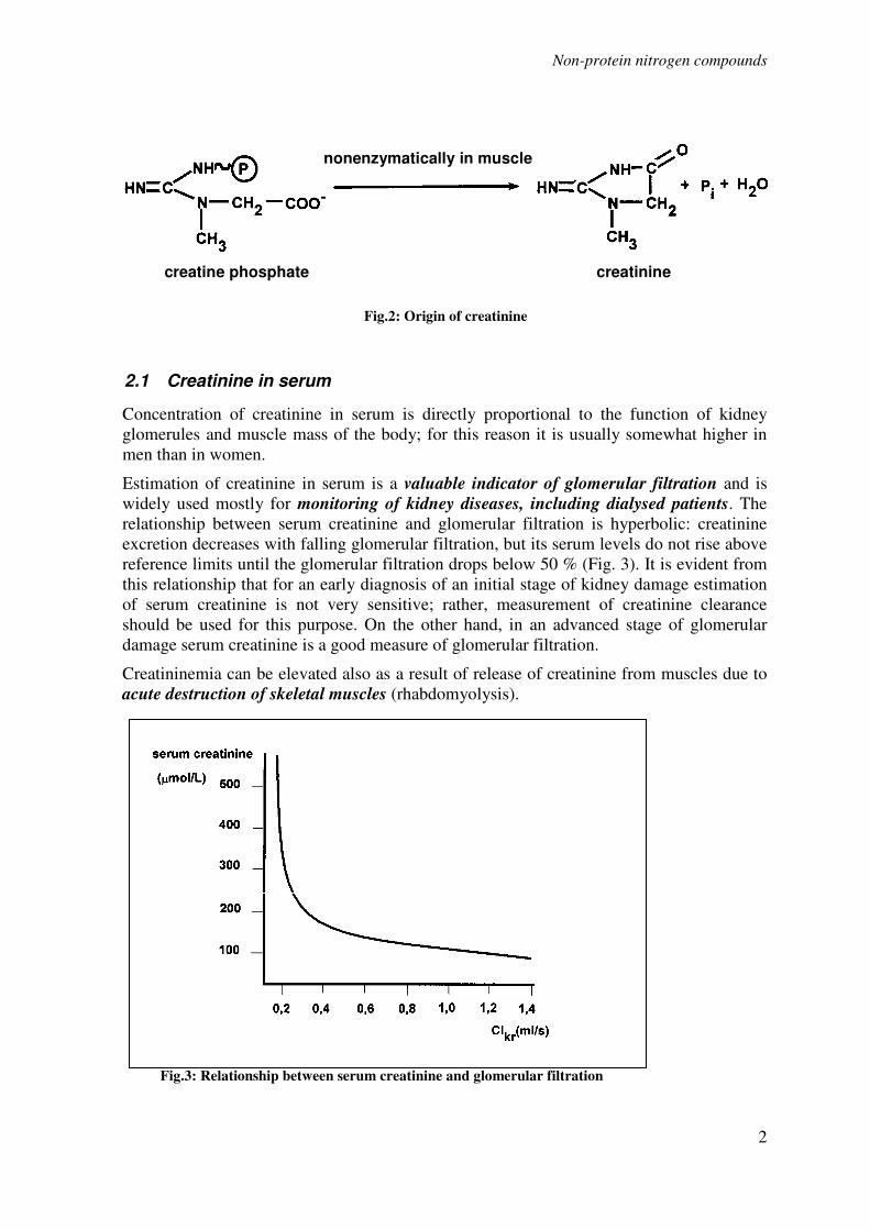
muscle as a source of energy for muscle contraction (Fig. 2).
Rate of creatinine production in the body is relatively constant. It reflects amount of
muscle mass and under condition of physical rest and meat-free diet is stable. Creatinine is
excreted in the kidney by glomerular filtration; an additional secretion by renal tubules
becomes significant only at elevated serum creatinine levels.
Proteins Amino acids Ammonia Urea
proteolysis transamination
oxidative deamination
urea cycle
Non-protein nitrogen compounds
2
Fig.2: Origin of creatinine
2.1 Creatinine in serum
Concentration of creatinine in serum is directly proportional to the function of kidney
glomerules and muscle mass of the body; for this reason it is usually somewhat higher in
men than in women.
Estimation of creatinine in serum is a valuable indicator of glomerular filtration and is
widely used mostly for monitoring of kidney diseases, including dialysed patients. The
relationship between serum creatinine and glomerular filtration is hyperbolic: creatinine
excretion decreases with falling glomerular filtration, but its serum levels do not rise above
reference limits until the glomerular filtration drops below 50 % (Fig. 3). It is evident from
this relationship that for an early diagnosis of an initial stage of kidney damage estimation
of serum creatinine is not very sensitive; rather, measurement of creatinine clearance
should be used for this purpose. On the other hand, in an advanced stage of glomerular
damage serum creatinine is a good measure of glomerular filtration.
Creatininemia can be elevated also as a result of release of creatinine from muscles due to
acute destruction of skeletal muscles (rhabdomyolysis).
Fig.3: Relationship between serum creatinine and glomerular filtration
creatine phosphate creatinine
nonenzymatically in muscle

Non-protein nitrogen compounds
3
Reference values (S-Creatinine):
Women: 44 – 104 µmol/l
Men: 44 – 110 µmol/l
2.2 Creatinine in urine
When compared to other endogenous substances, creatinine excretion into urine is rather
constant during the day; in persons with normal glomerular filtration it reflects amount and
activity of muscles.
Concentration of creatinine in urine can be used as a check of 24-hour urine collection
used for measurements of daily output of some substances into urine. Incorrect or
incomplete urine collection often makes these measurements faulty; and simultaneous
estimation of creatinine output provides a simple way to check it. Creatinine is estimated in
a sample of urine from the 24-hour collection. The result in µmol/kg/day is compared with
the reference values for urinary creatinine output in dependence on age and gender:
Age Creatinine Output (µmol/kg of body weight/day)
Men Women
20 – 29 210± 20 174± 34
30 – 39 194± 13 180± 34
40 – 49 174± 28 156± 34
50 – 59 171± 26 132± 32
60 – 69 149± 26 114± 23
70 – 79 126± 26 104± 19
80 – 89 103± 35 95± 22
90 – 99 83± 28 74± 12
If output of creatinine is 30 % or more below the tabular value, it is almost certain the
collection of urine was incomplete.
Next, urinary creatinine is used for standardisation of urinary output of other substances
if information on the 24-hour volume of urine is entirely missing. Concentration of the
substance of interest is expressed per 1 mmol of creatinine.
Reference values:
Concentration of creatinine in urine (U-Creatinine): 3.0 – 12.0 mmol/l
Daily output of creatinine into urine (dU-Creatinine): 8.8 – 13.3 mmol/24 hours
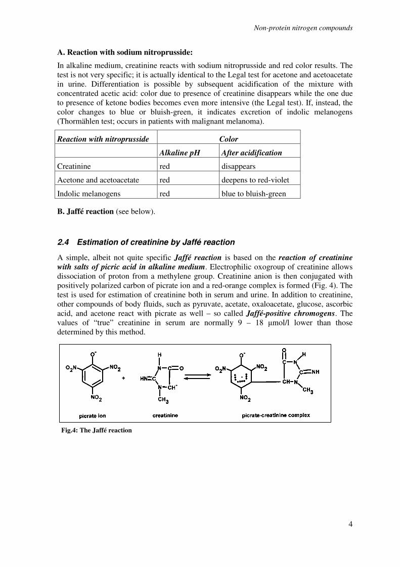
2.3 Properties of creatinine and demonstration of creatinine in urine
Creatinine is well soluble in water. Its aqueous solution is weakly alkaline. Creatinine can
be proved in a sample using the Jaffé reaction and the reaction with sodium
nitroprusside.
Non-protein nitrogen compounds
4
A. Reaction with sodium nitroprusside:
In alkaline medium, creatinine reacts with sodium nitroprusside and red color results. The
test is not very specific; it is actually identical to the Legal test for acetone and acetoacetate
in urine. Differentiation is possible by subsequent acidification of the mixture with
concentrated acetic acid: color due to presence of creatinine disappears while the one due
to presence of ketone bodies becomes even more intensive (the Legal test). If, instead, the
color changes to blue or bluish-green, it indicates excretion of indolic melanogens
(Thormählen test; occurs in patients with malignant melanoma).
Reaction with nitroprusside Color
Alkaline pH After acidification
Creatinine red disappears
Acetone and acetoacetate red deepens to red-violet
Indolic melanogens red blue to bluish-green
B. Jaffé reaction (see below).
2.4 Estimation of creatinine by Jaffé reaction
A simple, albeit not quite specific Jaffé reaction is based on the reaction of creatinine
with salts of picric acid in alkaline medium. Electrophilic oxogroup of creatinine allows
dissociation of proton from a methylene group. Creatinine anion is then conjugated with
positively polarized carbon of picrate ion and a red-orange complex is formed (Fig. 4). The
test is used for estimation of creatinine both in serum and urine. In addition to creatinine,
other compounds of body fluids, such as pyruvate, acetate, oxaloacetate, glucose, ascorbic
acid, and acetone react with picrate as well – so called Jaffé-positive chromogens. The
values of “true” creatinine in serum are normally 9 – 18 µmol/l lower than those
determined by this method.
Fig.4: The Jaffé reaction

Non-protein nitrogen compounds
5
2.5 Clearance of endogenous creatinine
The term clearance in general applies to clearing of the internal environment by all means
of excretion (kidney, liver etc.).
For excretion of low-molecular substances that pass freely the kidney filtration membrane
this relationship applies: U: concentration of substance in urine
V: volume of urine
GF: volume of glomerular filtrate
P: concentration of substance in plasma
If a substance is excreted into urine only by glomerular filtration, amount of the substance
passing glomerular membrane in unit of time must be equal to the amount excreted into
urine in the same time period. And if the amount of substance excreted into urine is U ×
V per second, then there must be a certain (theoretical) volume of blood plasma that has
been “cleared” from the substance. This volume of plasma is called clearance.
Renal clearance of a certain compound is the volume of blood plasma that is completely cleared of the compound in kidneys within unit of time.
Measurement of clearance for various substances can provide information on various renal
functions. If a substance is chosen that is excreted only by glomerular filtration, its
clearance tells us value of the glomerular filtration. If a substance in use is both filtered and
excreted by tubular secretion (e.g. p-aminohippuric acid), its clearance can be used for
estimation of renal blood flow.
Compounds eliminated only by filtration through glomerular membrane can be employed
to measure glomerular filtration. Inulin is an example of substance that passes the
glomerular membrane, and then in tubules is neither secreted nor absorbed; measurement
of clearance of inulin provides the exact value of glomerular filtration. The procedure for
determination of inulin clearance, however, is demanding since a constant plasma level of
inulin must be kept by a continuous intravenous infusion. Therefore, usage of this
examination is limited to research purpose, while in clinical practice clearance of
endogenous creatinine is used for the assessment of glomerular filtration. Creatinine is
excreted largely (about 90 %) by glomerular filtration, and its plasma concentration is
fairly stable. In comparison to clearance of inulin, the value of creatinine clearance is
higher. The examination of creatinine clearance is useful especially in patients with less
severe kidney damage, in which glomerular filtration is lowered to 50 – 80 % of the
normal, and serum creatinine is still below reference limit.
On the other hand, at higher values of serum creatinine (above 180 µmol/l) the fraction of
creatinine excreted by tubular excretion increases and validity of the examination of
creatinine clearance becomes rather limited. In these cases estimation of serum creatinine
is more useful.
2.5.1 Measurement of clearance of endogenous creatinine
In order to measure clearance of creatinine, its concentration in serum and in urine, as well
as the volume of urine per given period of time, must be found. Patient usually collects
urine for 24 hours. Errors in urine collection may be less likely if the collection period is
shortened to 6 or 12 hours. The volume of collected urine is measured in a cylinder with
precision to 10 ml, and after mixing a sample is taken, in which concentration of creatinine
GF × P = U × V
Non-protein nitrogen compounds
6
is estimated. At the end of the collection period a blood sample for estimation of serum
creatinine is taken as well. Information on the patient’s body height and weight
accompanies the samples to the laboratory.
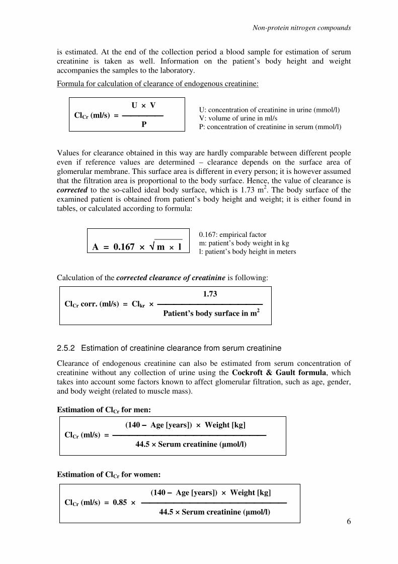
Formula for calculation of clearance of endogenous creatinine:
U: concentration of creatinine in urine (mmol/l)
V: volume of urine in ml/s
P: concentration of creatinine in serum (mmol/l)
Values for clearance obtained in this way are hardly comparable between different people
even if reference values are determined – clearance depends on the surface area of
glomerular membrane. This surface area is different in every person; it is however assumed
that the filtration area is proportional to the body surface. Hence, the value of clearance is
corrected to the so-called ideal body surface, which is 1.73 m2. The body surface of the
examined patient is obtained from patient’s body height and weight; it is either found in
tables, or calculated according to formula:
0.167: empirical factor
m: patient’s body weight in kg
l: patient’s body height in meters
Calculation of the corrected clearance of creatinine is following:
2.5.2 Estimation of creatinine clearance from serum creatinine
Clearance of endogenous creatinine can also be estimated from serum concentration of
creatinine without any collection of urine using the Cockroft & Gault formula, which
takes into account some factors known to affect glomerular filtration, such as age, gender,
and body weight (related to muscle mass).
Estimation of ClCr for men:
Estimation of ClCr for women:
U × V
ClCr (ml/s) = P
_______
A = 0.167 × √√√√ m × l
1.73
ClCr corr. (ml/s) = Clkr ×
Patient’s body surface in m2
(140 −−−− Age [years]) × Weight [kg]
ClCr (ml/s) =
44.5 × Serum creatinine (µmol/l)
(140 −−−− Age [years]) × Weight [kg]
ClCr (ml/s) = 0.85 ×
44.5 × Serum creatinine (µmol/l)

Non-protein nitrogen compounds
7
Recently, this estimation has been replaced by a more reliable calculation, called MDRD
formula. It was suggested by Levey et al. in 1999. MDRD is an empirical formula that
was derived from data of a large, multicentric study on the effect of diet in renal disease
(Modification of Diet in Renal Disease – MDRD).
General formula is
GF = 2.83 × (0.0113 × serum creatinine)
-0.999 × age
-0.176 × (2.8 × serum urea)
-0.17 × (0.1 × serum albumin)
0.318
For women, the result is to be multiplied by 0.762.
Results of this estimation are in good agreement with measured values especially in
patients with decreased glomerular filtration. None of these calculations is suitable for
people with normal or just moderately decreased renal function.
Reference values for ClCr (ml/s):
The glomerular filtration decreases with age:
Age (years): 13 – 49 50 – 59 60 – 69 70 and more
Women: 1.58 – 2.67 1.0 – 2.1 0.90 – 1.80 0.8 – 1.3
Men: 1.63 – 2.60 1.2 – 2.4 1.05 – 1.95 0.7 – 1.0
The ideal clearance of creatinine with respect to the age can be also found using the
equation:
Cl = − 0.00946 × age [years] + 2.118. The patient’s value should be within ± 30 %.
2.6 Estimation of glomerular filtration using serum concentration of cystatin C
Cystatin C is a protein composed of 120 amino acids. Many tissues produce cystatin C at
various rates. This protein is one of the most important inhibitors of extracellular cystein
proteases. Rate of its synthesis is almost constant and unaffected by inflammation,
catabolism or diet. Cystatin C is due to its relatively low molecular weight (approx.
13,000) freely filtered through glomerular membrane. Then, it is fully reabsorbed and
degraded in proximal tubules. Thus, plasma concentration of cystatin C is a good
measure of glomerular filtration, while its urinary concentration correlates with
impairment of tubular function. Concentration of cystatin C can be measured by
immunochemical techniques. Reference values differ according to the analytical method
used; however, introduction of international standardized calibration is expected.
Estimation of cystatin C provides several considerable advantages: it is sensitive for early
stage glomerular defect; there is no need for 24-hour urine collection that is frequently
faulty; and its measurement is not influenced by non-specific reactions as for creatinine.
Although this estimation is rather expensive and still reserved for research it is believed
that cystatin C will broaden the repertoire of commonly used examinations of renal
functions in the near future.
Non-protein nitrogen compounds
8
2.7 Fractional excretion (FE)
The amount of a compound excreted to definitive urine depends on glomerular filtration
(i.e. on the amount of the compound in primitive urine) and on tubular secretion and
reabsorption. To make the explanation simpler, we will assume only compounds that are
not eliminated by tubular secretion (or with negligible tubular secretion) in the following
text.
Portion of the compound from primitive urine that is finally excreted to definitive urine is
called fractional excretion (FE). FE of certain compound can be between 0 and 1
(between 0 and 100 %). FE value equal to zero means that the compound is fully
reabsorbed in tubuli; on the other hand, FE 100% means that all substance filtered across
glomerular membrane passes to definitive urine. Tubular reabsorption (TR) is a “mirror”
value to FE – it is the portion of substance that is reabsorbed by tubular cells. Assuming
that tubular secretion is negligible it goes that
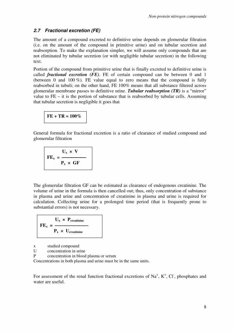
General formula for fractional excretion is a ratio of clearance of studied compound and
glomerular filtration
The glomerular filtration GF can be estimated as clearance of endogenous creatinine. The
volume of urine in the formula is then cancelled out; thus, only concentration of substance
in plasma and urine and concentration of creatinine in plasma and urine is required for
calculation. Collecting urine for a prolonged time period (that is frequently prone to
substantial errors) is not necessary.
x studied compound
U concentration in urine
P concentration in blood plasma or serum
Concentrations in both plasma and urine must be in the same units.
For assessment of the renal function fractional excretions of Na+, K
+, Cl
-, phosphates and
water are useful.
FE + TR = 100%
Ux × V
FEx =
Px × GF
Ux × Pcreatinine
FEx =
Px × Ucreatinine
Non-protein nitrogen compounds
9
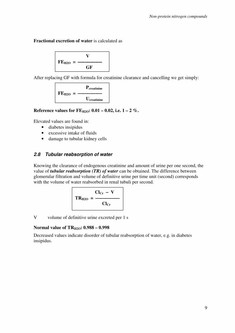
Fractional excretion of water is calculated as
After replacing GF with formula for creatinine clearance and cancelling we get simply:
Reference values for FEH2O: 0.01 – 0.02, i.e. 1 – 2 %.
Elevated values are found in:
• diabetes insipidus
• excessive intake of fluids
• damage to tubular kidney cells
2.8 Tubular reabsorption of water
Knowing the clearance of endogenous creatinine and amount of urine per one second, the
value of tubular reabsorption (TR) of water can be obtained. The difference between
glomerular filtration and volume of definitive urine per time unit (second) corresponds
with the volume of water reabsorbed in renal tubuli per second.
V volume of definitive urine excreted per 1 s
Normal value of TRH2O: 0.988 – 0.998
Decreased values indicate disorder of tubular reabsorption of water, e.g. in diabetes
insipidus.
V
FEH2O =
GF
Pcreatinine
FEH2O =
Ucreatinine
ClCr −−−− V
TRH2O =
ClCr

Non-protein nitrogen compounds
10
3 Urea
In quantitative terms, urea is the most significant degradation product of protein and amino
acids in the body. It is produced in the liver from ammonia released by deamination
reactions from amino acids. Urea diffuses freely through cell membranes and so its
concentrations in plasma and intracellular fluid are equal.
Urea is excreted from the body mainly in the kidney by a combination of glomerular
filtration and tubular reabsorption. The latter is variable: lower at higher diuresis while
increasing when diuresis is low.
Concentration of urea in the blood depends on amount of protein in the diet, excretion
by the kidney, and metabolic function of the liver.
For instance, serum urea can increase due to high protein intake in the food. One gram of
protein (dietary or endogenous) can give rise to 5.74 mmol (0.34 g) of urea. Increased
concentration of serum urea without changes in other non-protein nitrogen compounds
(esp. creatinine) is a hallmark of an intense protein catabolism, which occurs e.g. in
starvation, febrile state or malignancy. Children have lower catabolism of protein, and
show demonstrably lower serum urea levels.
Serum urea increases in diseases of the kidney that lead to marked restriction of
glomerular filtration (below 30 %); simultaneously, high levels of creatinine are found.
Unlike creatinine, estimation of urea is not suitable for an early detection of decrease in
glomerular filtration. Sensitivity of the urea estimation for renal insufficience is low: it
starts to exceed the reference values when more than 75 % of glomerular filtration is lost.
On the other hand, urea is a sensitive parameter of renal hypoperfusion – in this case not
only glomerular filtration is lowered but also tubular reabsorption of urea is increased.
Serum concentration of urea therefore rises much more quickly than that of creatinine. In
renal failure of the pre-renal type (e.g. in hypoperfusion, most frequently in dehydration)
the ratio of serum concentrations of urea and creatinine (in µmol/l) is above 160.
In liver function failure, synthesis of urea falls down, and so its concentration in the
serum.
Concentration of urea in serum and urine can also be used for calculation of nitrogen
balance.
Some causes of changes in serum urea concentration:
Serum urea increased: Serum urea decreased:
Impaired kidney function Low-protein diet
High-protein diet Impaired liver function
High catabolism of protein Late pregnancy (growth of foetus demands protein)
Dehydration

Non-protein nitrogen compounds
11
Reference values (fS-Urea): Women: 2.0 – 6.7 mmol/l
Men: 2.8 – 8.0 mmol/l
Output (loss) of urea in urine (dU-urea):
An adult excretes daily 167-583 mmol/ 24 hours, depending on protein uptake in food and
protein catabolism. Urinary urea output is calculated according to the formula:
dU-urea(mmol/24 hours) = U-urea (mmol/l) x volume of urine per 24 hours (l)
Principle of urea estimation in serum and urine:
In general, urea in biological fluids can be estimated either directly, or indirectly as
ammonia. In the indirect assay, first an enzyme urease cleaves urea to carbon dioxide and
ammonia that in aqueous medium exists as ammonium ion. Next, the amount of
ammonium is estimated by the Berthelot’s reaction: ammonium ion with sodium
hypochlorite and phenol or salicylate form a colored product. The reaction is catalyzed by
sodium nitroprusside.
Nowadays, the recommended routine method for measurement of the ammonium ions
generated by the urease reaction utilizes conversion of α-ketoglutarate to glutamate. The
reaction is catalyzed by glutamate dehydrogenase, and coupled to oxidation of NADH to
NAD+ (Warburg’s optical test).
Urease Urea + H2O + 2H+ 2NH4
+ + CO2 Glutamate dehydrogenase NH4
+ + 2-oxoglutarate + NADH L-glutamate + NAD+ + H2O

Non-protein nitrogen compounds
12
4 Uric acid
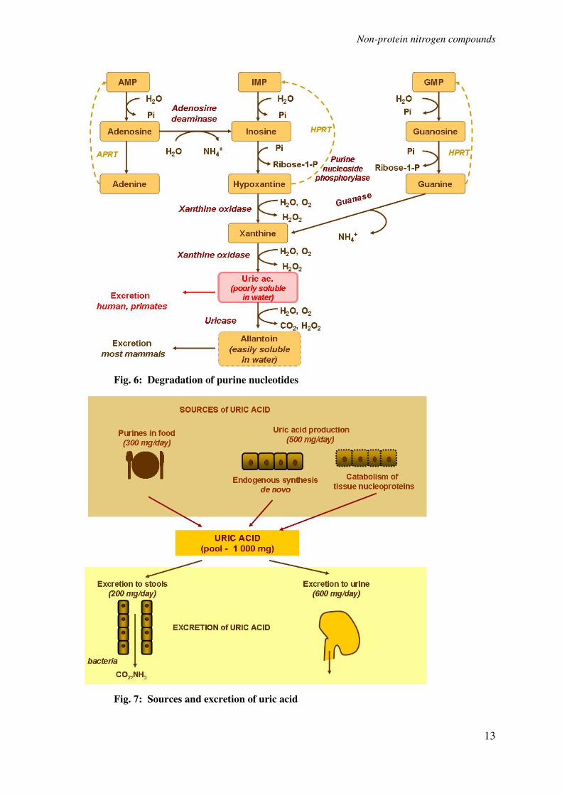
Uric acid is the end product of purine metabolism in humans (Fig. 5). Purine nucleotides
are building blocks of the nucleic acids and other compounds with important roles in
metabolism (e.g. ATP, NAD+). Most cells are capable to synthesize purine nucleotides de
novo. An important intermediate in purine synthesis is 5-phosphoribosyl-1-diphosphate
(PRPP) that is formed in reaction catalyzed by PRPP-synthetase. In the course of the
subsequent reactions, the purine core is formed and principal purine nucleotide, inosine
monophosphate (IMP) is produced. IMP is then converted to other purine nucleotides,
adenosine monophosphate (AMP) and guanosine monophosphate (GMP).
Nucleic acids from own cell nuclei as well as from nutrition are catabolized to nucleotides,
nucleosides and bases. Purine bases are finally metabolized by xanthine oxidase to uric
acid. Degradation of purines is terminated at this point in humans and primates. Other
mammals can further convert uric acid by uricase to allantoin which is better soluble in
water. A part of purines is used for re-synthesis of nucleotides by hypoxanthine-guanine
phosphoribosyltransferase (HPRT) and adenine phosphoribosyltransferase (APRT)
(“salvage pathway”, Fig. 6).
Fig. 5: Uric acid
Total amount of uric acid in the body is approximately 1 g. Uric acid originates from three
sources: food nucleotides, degradation of tissue nucleotides and biosynthesis. Uric acid is
not only a waste product but it also protects cells from reactive oxygen species.
75 – 80 % of uric acid is eliminated by kidneys (see later). The rest (20 – 25 %) is
eliminated by gastrointestinal tract where it can be further degraded to NH3 and CO2 by
bacterial flora (Fig. 7).
Lactim form (Enol-form)
Lactam form (Oxo-form)
Non-protein nitrogen compounds
13
Fig. 6: Degradation of purine nucleotides
Fig. 7: Sources and excretion of uric acid

Non-protein nitrogen compounds
14
4.1 Properties of uric acid
Uric acid is poorly soluble in water. At pH values below 5.5, as typical for urine, most
molecules of uric acid exist in the non-dissociated and thus less soluble form. Uric acid can
then form crystals or concrements. Coolness contributes to insolubility of uric acid. The
solubility of uric acid rises with increasing pH value. At physiological blood pH 7.4 the
ionized form prevails and more soluble sodium or potassium urate is formed.
Oxidative cleavage with concentrated nitric acid can be used for the uric acid detection.
Imidazole ring of purine is opened and two molecules join, forming purpuric acid. Salts of
purpuric acid are colored. Addition of ammonia yields murexide (ammonium salt of
purpuric acid, Fig. 8). Murexide reaction is employed in urine stone analysis.
Fig. 8: Murexide reaction
4.2 Estimation of uric acid concentration
Most state-of-art techniques for measuring uric acid concentration employ uricase, an
enzyme converting uric acid into allantoin, hydrogen peroxide and carbon dioxide (Fig. 9).
Decrease in uric acid concentration in the reaction mixture may be determined by direct
photometry at 290 – 293 nm, because uric acid and allantoin differ in their absorption
spectra. Uric acid has an absorption peak at this wavelength while allantoin does not
absorb light in this range.
Another choice is indirect measurement in which hydrogen peroxide formed in the uricase
reaction is used in a coupled reaction catalyzed by peroxidase. A quinonimine dye is
formed by oxidative coupling of (usually) 4-aminoantipyrine and a suitable derivative of
phenol (e.g. N-ethyl-N-(2-hydroxy-2-sulphopropyl)-m-toluidine)). Intensity of the
resulting color is proportional to the concentration of uric acid in solution. Ascorbic acid
interferes with this method; therefore ascorbate oxidase is added to eliminate such an
influence.
Uric acid Purpuric acid Murexide

Non-protein nitrogen compounds
15
1. Uricase reaction
2. Oxidative coupling of 4-aminoantipyrine and a phenolic compound
peroxidase
2 H2O2 + 4-AAP + phenolic comp. 4 H2O + colored product
Fig. 9: Estimation of uric acid
4.3 Uric acid in blood serum
Concentration of uric acid in blood depends on intake of purines in food, intensity of its
production and on its excretion. Especially increased levels of uric acid are of clinical
significance.
Hyperuricemia results from overproduction or decreased excretion of uric acid.
Concentration of urates can exceed their solubility in hyperuricemia.
Overproduction of uric acid
• Excessive de novo synthesis of purines associated with hyperuricemia is found in
some genetically determined impairments of purine metabolism. Partial or
complete defect of hypoxanthine-guanine phosphoribosyltransferase (Lesch-
Nyhan’s syndrome) may serve as an example. Re-utilization of purine nucleotides
is impaired and purines are therefore degraded to uric acid. Another genetic
disorder leading to increased production of uric acid is increased activity of
phosphoribosylphosphate synthetase.
• Anticancer therapy (cytostatic chemotherapy, radiotherapy) induces extensive cell
lysis and leads to increased formation of uric acid. Purine nucleotides are released
from degraded nucleic acids and metabolized to uric acid. Some hematological
diseases associated with excessive production (polycythemia vera) or degradation
of cells (leukemia, hemolytic anemia) are, in a similar way, joined with
hyperuricemia.
• Increased intake of foodstuffs rich on purines (offal, meat, legume, in smaller
extent also chocolate, cocoa and coffee) also leads to overproduction of uric acid.
Kidneys may fail to compensate the overload with uric acid and uricemia grows up.
• Alcohol increases the level of uric acid. Increased production of lactate inhibits
renal secretion of uric acid. The decreased excretion of uric acid is later replaced
with high uricosuria.
uric acid allantoin
uricase

Non-protein nitrogen compounds
16
Decreased excretion of uric acid
Decreased excretion of uric acid is one of the most frequent causes of hyperuricemia.
• Decreased tubular secretion of uric acid is common in patients with
hyperuricemia. The cause of this disorder is unknown.
• Excretion of uric acid is lowered in states associated with decreased glomerular
filtration and impaired tubular function.
Reference values (fS-UA): Women 140 – 340 µmol/l
Men 220 – 420 µmol/l
4.4 Uric acid in urine
Majority of uric acid (75 – 80 %) is eliminated by kidneys. It is freely filtered in glomeruli
(uric acid is only minimally bound to proteins) and then most of it is reabsorbed in the
proximal tubuli. Next, uric acid is again secreted in the distal part of proximal tubuli and
once again reabsorbed. Approximately 0.6 g of uric acid per day (3.6 mmol/day) is
excreted under purine-free diet. Normally, with a common diet, these values are higher –
about 0.8 g/day (5.0 mmol/day, Fig. 10). Tubular secretion of uric acid is inhibited if other
organic ions (e.g. acetoacetic acid, β-hydroxybutyric acid, lactate or some drugs) are also
excreted to a high extent.
Fig. 10: Excretion of uric acid
Non-protein nitrogen compounds
17
Uric acid is a considerable risk factor for both urinary tract and renal parenchyma.
If the excretion of uric acid is increased there is a higher risk of urate urolithiasis.
Especially important is this risk in individuals with permanently acidic and concentrated
urine. Urate concrements are usually made of pure uric acid, sometimes of sodium urate.
Ammonium urate may precipitate in weakly alkaline urine, typically in case of urinary
infection.
Crystals of sodium urate can precipitate even in renal interstitium and cause inflammatory
response (chronic interstitial nephritis).
Acute renal failure is a relatively rare disorder that results from sudden increase of uric
acid in blood (e.g. cytostatic therapy in patients with leukemia) in case that urine is
concentrated and acidic at the same time (dehydration). Crystals of uric acid are formed in
distal tubuli and collecting ducts under these conditions and drainage of urine is blocked
(acute urate nephropathy).
Examination of uric acid in urine is especially important in patients with increased blood
concentration of uric acid and in patients with urolithiasis.
The amount of uric acid in urine can be expressed in several ways:
a) Concentration in the morning sample of urine. According to results of this test,
the amount and type of beverages is adapted in order to decrease the urate
concentration.
b) The amount of uric acid excreted in 24 hours. Urine is collected for 24 hours and
a mixed sample is analyzed. This method can distinguish between hyperuricemia
from increased production and decreased excretion.
c) Uric acid/creatinine ratio in a random sample of urine. Results give information
similar to the previous technique but it is not necessary to collect urine for 24
hours.
d) Clearance of uric acid may be calculated according to formula
UUA: concentration of uric acid in urine (mmol/l)
PUA: concentration of uric acid in serum or plasma (mmol/l)
V: volume of urine in ml/s
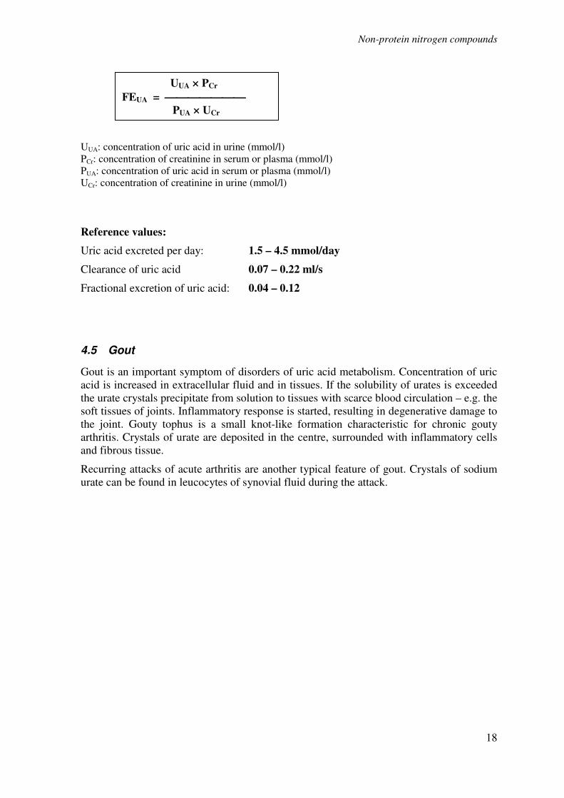
e) Fractional excretion of uric acid. This parameter gives summary information on
transport of uric acid in renal tubuli. The role of dysfunction of renal tubular cells
on hyperuricemia may be assessed using this value. It can be calculated for
randomly taken sample of urine and blood in which the concentration of uric acid
and creatinine is examined. Fractional excretion of uric acid is calculated using the
formula:
UUA × V
ClUA (ml/s) =
PUA
Non-protein nitrogen compounds
18
UUA: concentration of uric acid in urine (mmol/l)
PCr: concentration of creatinine in serum or plasma (mmol/l)
PUA: concentration of uric acid in serum or plasma (mmol/l)
UCr: concentration of creatinine in urine (mmol/l)
Reference values:
Uric acid excreted per day: 1.5 – 4.5 mmol/day
Clearance of uric acid 0.07 – 0.22 ml/s
Fractional excretion of uric acid: 0.04 – 0.12
4.5 Gout
Gout is an important symptom of disorders of uric acid metabolism. Concentration of uric
acid is increased in extracellular fluid and in tissues. If the solubility of urates is exceeded
the urate crystals precipitate from solution to tissues with scarce blood circulation – e.g. the
soft tissues of joints. Inflammatory response is started, resulting in degenerative damage to
the joint. Gouty tophus is a small knot-like formation characteristic for chronic gouty
arthritis. Crystals of urate are deposited in the centre, surrounded with inflammatory cells
and fibrous tissue.
Recurring attacks of acute arthritis are another typical feature of gout. Crystals of sodium
urate can be found in leucocytes of synovial fluid during the attack.
UUA × PCr
FEUA =
PUA × UCr