non-enzymatic glycation of aminotransferases and the possibilities
TRANSCRIPT

4
Non-Enzymatic Glycation of Aminotransferases and the Possibilities of Its Modulation
Iva Boušová, Lenka Srbová and Jaroslav Dršata Department of Biochemical Sciences, Charles University in Prague,
Faculty of Pharmacy in Hradec Králové Czech Republic
1. Introduction
Enzymes are catalytic protein molecules performing specific functions in their native form. Native structure is one of basic conditions for normal function of proteins including enzymes. Catalytic activity of enzyme may be decreased by both non-covalent modulation by true inhibitors and covalent modifications by metabolites in the body or natural products. One example of such modulation is non-enzymatic glycation of proteins by sugars.
Enzymes are very good models for studies of protein interactions with other molecules, including sugars (e.g., Arai et al., 1987; Dolhofer & Wieland, 1978; Okada et al., 1994; Okada et al., 1997; Sakurai et al., 1987). Advantage of these studies is the fact that enzyme interactions may be investigated not only by common methods of studies of protein properties like changes in spectral characteristics, molecular weight, charge, solubility etc. but also by measurement of their catalytic activity as the most characteristic property of enzyme molecules. For such studies, it is important to use the enzyme with known structure and reaction mechanism, available in sufficient purity, and measurable by simple assay method. Aminotransferases belong to such enzymes. That is the reason why research activities of our laboratory devote to inhibition of aminotransferases by low-molecular compounds for many years (e.g. Dršata & Veselá, 1984; Netopilová et al., 1991; Netopilová et al., 2001). Our studies of aminotransferase glycation belong to the efforts made also by several other research groups (see e.g. Okada & Ayabe, 1997; Fitzgerald et al., 2000; Seidler & Kowalewski 2003; Hobart et al., 2004).
1.1 Aminotransferases
Aspartate aminotransferase (AST, EC 2.6.1.1) and alanine aminotransferase (ALT, EC 2.6.1.2), enzymes frequently assessed in clinical laboratories, catalyse reversible transfer of α-amino group from amino acids aspartate and alanine to the acceptor 2-oxoglutarate, respectively. The resulting products are oxaloacetate, pyruvate and glutamate. In metabolism, AST activity mediates the connection between the metabolism of amino acids
www.intechopen.com

Enzyme Inhibition and Bioapplications
86
and saccharides and participates in the transportation of reduced equivalents across the membrane of mitochondria as a part of the malate–aspartate shuttle. The catalytic activities of AST in cells are implemented by two isoenzymes—the cytosolic and the mitochondrial one, which differ in primary structure and in some properties (Yagi et al., 1985; Metzler et al., 1979).
Porcine heart cytosolic AST, which was used in further described experiments of our research group, is a homodimer with molecular mass of about 93,150 Da. Both subunits are composed of 412 amino acid residues. This dimer contains 38 lysine and 52 arginine residues with six Lys–Arg and four Arg–Lys sequence pairs (Seidler & Kowalewski, 2003). Presence of these amino acid residues makes glycation of aminotransferases in vitro as well as in vivo possible. The Lys258 binding coenzyme PLP could be one of the possible targets of glycating agents as can be seen from the loss of enzymatic activity due to the effect of methylglyoxal as well as other glycating agents (e.g. Boušová et al., 2009; Seidler & Kowalewski, 2003).
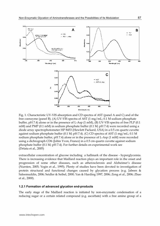
Aminotransferases are characterised by the presence of coenzyme pyridoxal-5’-phosphate (PLP) and its direct participation in catalysis. The protein part of aminotransferase molecules consists of two identical, non-covalently bound subunits, which are composed of one small and one large domain. PLP coenzyme forms a Schiff base with ε-amino group of lysine residue (Lys313 in ALT and Lys258 in AST) located in an active site of the larger domain of each subunit (Kirsch et al., 1984). The PLP form of AST shows, depending on pH, a major absorption peak at 360 nm (an active, unprotonized form of the coenzyme, prevailing at lower pH values), and/or a peak at 430 nm (an inactive, protonized form, increasing at lower pH values) (Kirsch et al., 1984). After a reaction with L-aspartate during the first part of a ping-pong transaminating reaction, the pyridoxamine-5’-phosphate (PMP) form of the coenzyme appears and the original absorption maximum shifts to 325–330 nm (Fig. 1A). While free PLP or PMP are not optically active substances, the coenzyme bound in the active site of AST shows circular dichroism (CD) spectra in the range of 300–500 nm, which are similar to absorption spectra (Fig. 1C). The CD effect is caused by the change in the electronic configuration of the molecule (Kelly & Price, 2000). Circular dichroism clears away absorption characteristics of optically inactive components, which facilitates identification of the specific coenzyme signal and its changes (Dršata et al., 2005). This method is also a powerful tool for characterizing secondary (190-240 nm, chromophore is peptide bond) and tertiary (250-320 nm, chromophores are aromatic amino acids and disulfide bonds) structures of studied proteins as well as determining whether the protein is folded (Kelly & Price, 2000).
1.2 Non-enzymatic glycation of proteins
Non-enzymatic glycation, also called Maillard reaction, was first described in 1912 by Louis Camille Maillard (Monnier, 1989). This non-enzymatic browning process had been first extensively studied by food chemists and later has become the center of attention of geological or agricultural sciences. Much later it was recognized that the process is important for medical science (Monnier, 1989; Singh et al., 2001). A very common but serious human disease is diabetes mellitus, which in its untreated or unsuccessfully treated form is accompanied with development of chronic complications due to increased intra- and
www.intechopen.com
Non-Enzymatic Glycation of Aminotransferases and the Possibilities of Its Modulation
87
Fig. 1. Characteristic UV-VIS absorption and CD spectra of AST (panel A and C) and of the free coenzyme (panel B). (A) UV-VIS spectra of AST (1 mg/mL; 0.1 M sodium phosphate buffer, pH 7.4) alone or in the presence of L-Asp (1 mM), (B) UV-VIS spectra of free PLP (0.1 mM) and PMP (0.1 mM) in sodium phosphate buffer (0.1 M; pH 7.4) were recorded using a diode array spectrophotometer HP 8453 (Hewlett Packard, USA) in a 0.5 cm quartz cuvette against sodium phosphate buffer (0.1 M; pH 7.4). (C) CD spectra of AST (1 mg/mL; 0.1 M sodium phosphate buffer, pH 7.4) alone or in the presence of L-Asp (1 mM) were recorded using a dichrograph CD6 (Jobin Yvon, France) in a 0.5 cm quartz cuvette against sodium phosphate buffer (0.1 M; pH 7.4). For further details on experimental work see (Dršata et al., 2005)
extracellular concentration of glucose including a hallmark of the disease - hyperglycemia. There is increasing evidence that Maillard reaction plays an important role in the onset and progression of some other diseases, such as atherosclerosis and Alzheimer’s disease (Nursten, 2005; Yegin et al., 1995). Plenty of studies have been devoted to investigation of protein structural and functional changes caused by glycation process (e.g. Jabeen & Saleemuddin, 2006; Seidler & Seibel, 2000; Yan & Harding 1997, 2006; Zeng et al., 2006; Zhao et al., 2000).
1.2.1 Formation of advanced glycation end-products
The early stage of the Maillard reaction is initiated by non-enzymatic condensation of a reducing sugar or a certain related compound (e.g. ascorbate) with a free amino group of a
www.intechopen.com

Enzyme Inhibition and Bioapplications
88
protein, a lipid or a nucleic acid. In the case of glucose, the reaction first leads to the formation of acid-labile Schiff base, which undergoes a rearrangement to a relatively stable Amadori product, e.g. fructosamine. Only a small portion of these Amadori-adducts experiences further rearrangements leading to an irreversible formation of advanced glycation end-products (AGEs) (Monnier, 1989). The reaction with fructose proceeds in a similar way, but it is called Heyns rearrangement and two separate Heyns products are generated (Suarez et al., 1989). The formation of Schiff base proceeds in the range of hours and it is fully reversible, while Amadori rearrangement takes days and is reversible only to a certain extent.
In the intermediate stage, Amadori product subsequently degrades and various reactive intermediates are formed. These products are known as α-dicarbonyls or α-oxoaldehydes and are represented by products like methylglyoxal (MGO), 3-deoxyglucosone (3-DG), and glyoxal (GO). Also a Schiff base is a potential source of reactive α-dicarbonyls, because it can be fragmented to MGO and GO. These dicarbonyls possess higher reactivity towards proteins than the parent monosaccharide. They are capable of forming various cross-links as well as chromo/fluorophoric adducts called AGEs, upon reaction with proteins (Schalkwijk et al., 2004; Wolff et al., 1991). Both MGO and 3-DG form adducts with proteins and nucleic acids up to 10,000 times more readily than glucose (Beisswenger et al., 2003). The accumulation of α-dicarbonyl compounds is termed carbonyl stress (Miyata et al., 1999). The other process proceeding during the intermediate stage of glycation is metal catalyzed autoxidation of glucose, in which the carbonyl compounds (arabinose and glyoxal), H2O2 and free radicals are formed (Hunt et al., 1988; Wolff & Dean, 1987). The generated free radicals initiate further oxidative steps. The glycation process accompanied by oxidation steps is called glycoxidation (Baynes, 1991). Various pathways incorporated in the formation of AGEs are shown in Fig. 2.
The advanced glycation end-products are formed during the late stage of glycation over a period of weeks, thereby affecting predominantly long-lived proteins, such as collagen and lens crystallins. They represent a heterogeneous group of compounds rising from different precursors. The chemical structures of AGEs have not been fully described yet. These compounds are formed either by oxidative pathway (pentosidine and CML) or by non-oxidative pathway (pyrraline, DOLD, GOLD, MOLD, and CEL) as can be seen in Fig. 2. Proteins modified by advanced glycation are characterized by a much higher molecular weight than the original protein, a yellow-brown pigmentation, a typical fluorescent spectra (λex/λem: 370/440 nm), an ability to form various cross-links, and by their biological half-life, which is comparable to the half-lives of parent proteins (Lapolla et al., 2005; Singh et al., 2001).
Glucose is the least reactive of the common sugars and that is probably the reason for its evolutionary selection as the principal sugar in vivo (Bunn et al., 1978). Because of its low reactivity towards proteins, AGEs have been thought to form only at long-lived extracellular proteins, such as collagen, crystallines, and myelin. Recently also rapid intracellular AGE formation by various intracellular sugars (e.g. fructose, ribose, glyceraldehyde, dihydroxyacetone phosphate, glyceraldehyde-3-phosphate, glyoxal, methylglyoxal, and 3-deoxyglucosone) in vivo has been described. The rate of glycation is directly proportional to the percentage of sugar in the open-chain form and the rate for fructose (0.7% open-chain) is 7.5-fold faster than that of glucose (0.002% open-chain). More strikingly, the glycolytic intermediate glyceraldehyde-3-phosphate (100% open-chain) forms 200-fold more glycated proteins than do equimolar amounts of glucose (Schalkwijk et al., 2004).
www.intechopen.com

Non-Enzymatic Glycation of Aminotransferases and the Possibilities of Its Modulation
89
Fig. 2. Three stages of non-enzymatic glycation reaction. CML, N-ε-(carboxymethyl)lysine; GOLD, glyoxal-lysine dimer; CEL, N-ε-(carboxyethyl)lysine; MOLD, methylglyoxal-lysine dimer; 3-DG, 3-deoxyglucosone; DOLD, deoxyglucosone-lysine dimer
1.2.2 Structure of AGEs
The advanced glycation end-products found under physiological conditions can be classified according to their fluorescent properties and their ability to form cross-links. The first group is represented by fluorescent AGE cross-links, which are thought to be responsible for a major share of the deleterious effects of AGEs in diabetes and aging. Fluorescence is a good qualitative indicator used to estimate AGEs formation. Pentosidine, crossline, and various vesperlysines are members of this group. However, also non-fluorescent AGE cross-links are found in vivo. Their isolation and identification is more complicated than in the case of fluorescent AGE cross-links. It is thought that they account just for 1% of all cross-links rising under physiological conditions. Various imidazolium dilysine cross-links (GOLD, MOLD), arginine-lysine cross-links, and glucosepan belong to this group. Last but not least, a group of non-cross-linking protein bound AGE structures have been identified in vivo. These structures may exert deleterious effects as precursors of cross-links or as biological receptor ligands inducing a variety of adverse cellular and tissue changes. The well-known members of this group are pyrraline, carboxyalkyllysines (CML, CEL), imidazolones, and argpyrimidine (Ulrich & Cerami, 2001). Classification and examples of each above mentioned group are shown in Fig. 3.
www.intechopen.com

Enzyme Inhibition and Bioapplications
90
Fig. 3. Classification of AGEs formed under physiological conditions including several examples to each group. [Lys] represents a desamino-lysine residue; [Arg] stands for a desguanidino-arginine residue; R represents either hydrogen atom (GOLD), methyl group (MOLD), 1,2,3-trihydroxypropyl (DOLD) or 2,3,4-trihydroxybutyl group (imidazolone A and B).
1.2.3 Therapeutic strategies targeting the AGEs
The therapeutic intervention to the glycation process has followed three main approaches. A first approach follows inhibition of RAGE by neutralizing antibodies or suppression of post-receptor signaling using antioxidants. A second one is inhibition of AGE formation process by carbonyl-blocking agents (aminoguanidine) or by antioxidants. The last approach is reducing AGE deposition by using cross-link breakers or by enhancing cellular uptake and degradation.
Interactions of AGEs with the receptor for AGEs (RAGE) have been implicated in the development of diabetic vascular complications, which cause various disabilities and shortened life expectancy, and reduced quality of life in patients with diabetes. These undesirable effects can be suppressed by the use of specific antibodies to RAGE, soluble
www.intechopen.com

Non-Enzymatic Glycation of Aminotransferases and the Possibilities of Its Modulation
91
RAGE or by suppression of post-receptor signaling using antioxidants (Hudson et al., 2003; Stuchbury & Münch, 2005). The secreted RAGE form, named soluble RAGE (sRAGE), acts as a decoy to trap ligands and prevent interaction with cell surface receptors (Bucciarelli et al., 2002). Soluble RAGE was shown to have important inhibitory effects in several cell culture and transgenic mouse models, in which it prevented or reversed full-length RAGE signaling. The administration of sRAGE has been shown to suppress accelerated diabetic atherosclerosis (Park et al., 1998).
Aminoguanidine, also known by its trade name Pimagedine (Alteon Inc.), is a prototype therapeutic agent for prevention of the AGEs formation. It is a low-molecular, highly nucleophilic hydrazine compound that rapidly reacts with α-dicarbonyl compounds such as MGO, GO, and 3-DG to prevent formation of AGE cross-links. The products of the scavenging reaction are substituted 3-amino-1,2,4-triazines. Aminoguanidine does not affect the formation of the Schiff base and Amadori products (Thornalley et al., 2000). Clinical trials of aminoguanidine in overt diabetic nephropathy (ACTION) were performed, but they were early terminated due to safety concerns. Reported side effects of aminoguanidine in clinical therapy were gastrointestinal disturbance, abnormalities in liver function tests, flu-like symptoms, and a rare vasculitis (Bolton et al., 2004; Thornalley, 2003). Other nucleophilic compounds, which are designed to trap reactive carbonyl intermediates in AGE formation, are for example OPB-9195, diaminophenazine, tenilsetam, and pyridoxamine (Baynes & Thorpe, 2000; De La Cruz et al., 2004). With regard to the presence of free radicals and oxidative steps in the course of glycoxidation, compounds with antioxidant effect such as α-lipoic acid, α-tocopherol, ascorbic acid, and ß-carotene were tested. Dipeptide carnosine, pyridoindole derivative stobadine, hypolipidemic drug probucol, and mucolytic remedy N-acetylcysteine are just a few more examples of the compounds with described antioxidant properties, which were tested in order to estimate their potential protective effect in the process of glycation. Also some antioxidant enzymes such as superoxide dismutase, catalase, and selenium-dependent glutathione peroxidase may protect proteins against impairment caused by non-enzymatic glycation (De La Cruz et al., 2004; Kyselova et al., 2004).
Aminoguanidine and other compounds mentioned before can inhibit the formation of new AGE cross-links, but they are not able to cleave those already formed. Vasan et al. (1996) reported the first of cross-link breakers, phenyl thiazolium bromide (PTB). This anti-AGE agent chemically breaks α-dicarbonyl compounds by cleaving the carbon-carbon bond between the carbonyls. Under physiological conditions, PTB is not stable and therefore its analogs were tested and alagebrium chloride (ALT-711), a highly potent cross-link breaker with higher stability, has been discovered. This compound successfully completed preclinical studies and Phase II clinical study on healthy volunteers (Yamagishi et al., 2008). Unfortunately, the specific types of AGEs affected by alagebrium are more important in rats than humans; hence the promising results in animals were never repeated in human studies. Randomized Phase II clinical trial (BENEFICIAL) in patients with chronic heart failure (Willemsen et al., 2010) has been terminated early due to financial constraints.
The objective of current study was to evaluate potential antiglycation activity of two mitochondrial antioxidants, α-phenyl N-tert-butyl nitrone (PBN) and N-tert-butyl hydroxylamine (NtBHA). PBN is a nitrone that traps free radicals and protects against
www.intechopen.com

Enzyme Inhibition and Bioapplications
92
damage in different models such as inflammation, ischemia reperfusion, and aging. Its decomposition product NtBHA mimics PBN and is much more effective in delaying senescence of human lung fibroblasts IMR90 (Atamna et al., 2000). NtBHA appears to act on mitochondria to delay alterations in function (Atamna et al., 2001). Supplementation with NtBHA improved the respiratory control ratio of mitochondria from liver of old rats (Atamna et al., 2001).
1.3 Summary of existing results
Our laboratory deals with research of the protein glycation in vitro for many years. Aminotransferases were chosen as suitable model proteins, because they are commercially available in a highly purified and stable form, their structures and mechanism of catalysis are well known, and at least two simple methods for their enzyme activity assessment are in use. This subchapter summarizes results, which have been obtained by our research group up to now.
In our laboratory, we found decrease in alanine aminotransferase (ALT, EC 2.6.1.2) activities in the presence of several reducing monosaccharides in vitro. The decrease in the catalytic activity of ALT from porcine heart after 20 days of incubation with D-glucose, D-fructose, D-ribose or D,L-glyceraldehyde varied and was dependent on the nature of glycating agent (the percentage of sugar present in open-chain form). The dependence of enzyme inactivation on the presence of these sugars and time of incubation is presented in Fig. 4 (Beránek et al., 2001). As was described earlier, the rate of glycation is directly proportional to the percentage of sugar in the open chain form and the rate for fructose (0.7% open-chain) is 7.5-fold faster than that of glucose (0.002% open-chain). More strikingly, the glycolytic intermediate glyceraldehyde 3-phosphate (100% open-chain) forms 200-fold more glycated proteins than do equimolar amounts of glucose (Schalkwijk et al., 2004).
Fig. 4. An effect of glycation on ALT activity. ALT was incubated with 50 mM sugars in sodium phosphate buffer (0.1 M, pH 7.4) at 25 °C. Aliquots of samples were taken at days 0, 2, 6, 9, 13, 20 and remaining enzyme activities in samples were determined. Activity was expressed as a percentage of the activity of the control sample (without sugars). ● ALT with D-glucose; ALT with D-ribose; ALT with D-fructose; ■ ALT with D,L-glyceraldehyde
www.intechopen.com
Non-Enzymatic Glycation of Aminotransferases and the Possibilities of Its Modulation
93
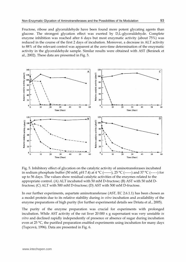
Fructose, ribose and glyceraldehyde have been found more potent glycating agents than glucose. The strongest glycation effect was exerted by D,L-glyceraldehyde. Complete enzyme inhibition was reached after 6 days but most enzymatic activity (about 75%) was reduced in the course of the first 2 days of incubation. Moreover, a decrease in ALT activity to 88% of the relevant control was apparent at the zero-time determination of the enzymatic activity in the glyceraldehyde sample. Similar results were obtained with AST (Beránek et al., 2002). These data are presented in Fig. 5.
Fig. 5. Inhibitory effect of glycation on the catalytic activity of aminotransferases incubated in sodium phosphate buffer (50 mM, pH 7.4) at 4 °C (―――), 25 °C (––––) and 37 °C (–·–·–) for up to 56 days. The values show residual catalytic activities of the enzymes related to the appropriate control. (A) ALT incubated with 50 mM D-fructose; (B) AST with 50 mM D-fructose; (C) ALT with 500 mM D-fructose; (D) AST with 500 mM D-fructose.
In our further experiments, aspartate aminotransferase (AST, EC 2.6.1.1) has been chosen as a model protein due to its relative stability during in vitro incubation and availability of the enzyme preparations of high purity (for further experimental details see Dršata et al., 2005).
The purity of the enzyme preparation was crucial for experiments with prolonged incubation. While AST activity of the rat liver 20 000 x g supernatant was very unstable in
vitro and declined rapidly independently of presence or absence of sugar during incubation even at 25 °C, the purified preparation enabled experiments using incubation for many days (Tupcová, 1996). Data are presented in Fig. 6.
www.intechopen.com

Enzyme Inhibition and Bioapplications
94
Fig. 6. Comparison of stability of AST in rat liver supernatant and in purified preparation from pig heart during in vitro incubation at 25 °C. The incubation mixture was diluted before the assay in order to obtain activities within the analytical range of the method. Values are mean ± S.D. of three independent samples. Each sample was measured three-times and the mean was used to calculate the value presented.
Moreover, glycation of AST was accompanied by a decrease in its catalytic activity in dependence on the concentration and activity of the glycating agent, while the concentration effect was not clearly demonstrated in case of ALT (Dršata et al., 2002). The effect of substrates (2-oxoglutarate and L-aspartate) on AST stability and on glycation process was assessed as well. There was no effect of 2-oxoglutarate on the control AST activity throughout the experiment, which demonstrated a high stability of the pyridoxal form both in the presence and absence of this substrate. On the other hand, the presence of 25 mM L-aspartate (inducing the pyridoxamine form of the enzyme) caused a rapid decrease in AST activity even in the control reaction (Dršata et al., 2002). The results of AST incubation with D-ribose and D-fructose in presence of 0.5 mM 2-oxoglutarate suggested that AST glycation could be partly prevented by this substrate. This finding supports the idea that Lys258 in the active center of AST is involved in glycation of the free enzyme (Fig. 7, taken from Dršata et al., 2002).
The in vitro model of protein glycation (AST) by D-fructose has been then established and experimentally used. (Boušová et al., 2005a). As mentioned above, attempts have been made by researchers to investigate various chemical compounds as potential antiglycating agents. With this model, influence of potential antiglycating compounds with antioxidant activities was investigated. In an attempt to reduce glycoxidation process and formation of AGEs, influence of endogenous antioxidant uric acid (0.2-1.2 mM) on glycoxidation process of AST by 50 mM and 500 mM D-fructose in vitro was studied. Uric acid at 1.2 mM concentration reduced AST activity decrease caused by incubation of the enzyme with 50 mM sugar up to 25 days at 37 °C (Fig. 8), as well as formation of total fluorescent AGE products (Fig. 9). The results obtained supported the hypothesis that uric acid has beneficial effects in controlling protein glycoxidation (Fig. 8 and 9).
www.intechopen.com

Non-Enzymatic Glycation of Aminotransferases and the Possibilities of Its Modulation
95
Fig. 7. Influence of substrates on AST activity during glycation by D-ribose in vitro. Concentration of the enzyme (purified Serva preparation) in the incubation mixture: 0.413 mg/ml, final catalytic concentration in the mixture 7.83 µkat/mg. Values are mean ± S.D. of 6 (control) or 3 (with D-ribose) independent samples. Each sample was measured three-times and the mean was used to calculate the value presented. L-Asp = L-aspartate; 2-OG = 2-oxoglutarate
Fig. 8. Effect of glycation on AST activity and intervention of uric acid (UA) in the process. AST (1.33 mg/ml) was incubated with and without fructose (Frc) in 0.1 mM phosphate buffer, pH 7.4 at 37 °C in the presence or absence of 1.2 mM uric acid. AST activity was expressed as percentage of that of control sample (without sugar), which was considered as 100% ± S.D. (%) at every interval. Each point represents an average of three experiments in interval 0-12 days, and an average of two experiments on the 15th and 21st day. Each experiment was performed in triplicate (*data with P<0.05, Student’s t-test).
www.intechopen.com

Enzyme Inhibition and Bioapplications
96
Fig. 9. Formation of fluorescent products of glycation under the conditions of long lasting incubation. AST (1.33 mg/ml) was incubated with or without D-fructose in sodium phosphate buffer (0.1 M, pH 7.4) at 37 °C in the presence or absence of 1.2 mM uric acid up to 25 days. Aliquots of samples were taken on days 0, 1, 3, 5, 25, and arising fluorescent AGE products in samples were determined at specific wavelengths of excitation and emission (λex/λem) corresponding to total AGEs (370/440 nm). Data of relative fluorescence were expressed in arbitrary units per mg of protein ± S.D., with 1 AU corresponding to the fluorescence of BSA 1.0 mg/ml. Every point in days 0 and 5 represents an average of four experiments (10 samples), in days 1 and 3 an average of three experiments for mixtures with 50 mM fructose (7 samples) and of two experiments for mixtures with 500 mM fructose (4 samples), and in day 25 an average of three experiments (7 samples), (*data with P<0.05, Student’s t-test).
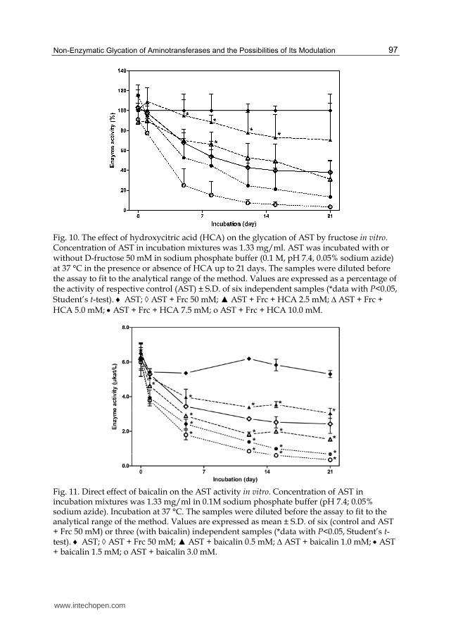
Also further studies of our research group have been pointed at possible protective activity of selected natural compounds (e.g. hydroxycinnamic acids, flavonoids, arbutin, hydroxycitric acid) on AST glycation by fructose. The results have shown that these compounds can exert also negative effects on enzyme activity but some of them have been able to slow down the course of AST modification by glycating agent. The compound with overall positive activity has been hydroxycitric acid (Fig. 10), which is major active component in the fruit rinds of certain species of the plant Garcinia. The effect of this compound was concentration-depended and its positive activity was most pronounced at 2.5 mM concentration. On the other hand, flavonoid baicalin (Fig. 11) and its aglycone baicalein rapidly decreased the in vitro activity of the enzyme in all concentrations used (0.5–3 mM), and no beneficial effects of these compounds on glycation of the enzyme by fructose were found (Boušová et al., 2005b).
www.intechopen.com
Non-Enzymatic Glycation of Aminotransferases and the Possibilities of Its Modulation
97
Fig. 10. The effect of hydroxycitric acid (HCA) on the glycation of AST by fructose in vitro. Concentration of AST in incubation mixtures was 1.33 mg/ml. AST was incubated with or without D-fructose 50 mM in sodium phosphate buffer (0.1 M, pH 7.4, 0.05% sodium azide) at 37 °C in the presence or absence of HCA up to 21 days. The samples were diluted before the assay to fit to the analytical range of the method. Values are expressed as a percentage of the activity of respective control (AST) ± S.D. of six independent samples (*data with P<0.05, Student’s t-test). AST; AST + Frc 50 mM; ▲ AST + Frc + HCA 2.5 mM; ∆ AST + Frc + HCA 5.0 mM; AST + Frc + HCA 7.5 mM; o AST + Frc + HCA 10.0 mM.
Fig. 11. Direct effect of baicalin on the AST activity in vitro. Concentration of AST in incubation mixtures was 1.33 mg/ml in 0.1M sodium phosphate buffer (pH 7.4; 0.05% sodium azide). Incubation at 37 °C. The samples were diluted before the assay to fit to the analytical range of the method. Values are expressed as mean ± S.D. of six (control and AST + Frc 50 mM) or three (with baicalin) independent samples (*data with P<0.05, Student’s t-test). AST; AST + Frc 50 mM; ▲ AST + baicalin 0.5 mM; ∆ AST + baicalin 1.0 mM; AST + baicalin 1.5 mM; o AST + baicalin 3.0 mM.
www.intechopen.com

Enzyme Inhibition and Bioapplications
98
Following experiments were conducted using methylglyoxal (MGO) as a glycating agent, because this compound has higher glycating potential than reducing monosaccharides and the incubation period has been shortened to one week only. Changes in the catalytic activity of AST caused by MGO were observable even after 120 min of incubation at 37 °C. Antiglycating activity of hydroxycitric (Fig. 12) and uric acid has been studied in this modified model (Boušová et al., 2009).
Fig. 12. Effect of glycation on AST activity and its intervention by hydroxycitric acid. AST (5 µg/ml) was incubated with or without methylglyoxal (0.5 mM) in 0.1 M sodium phosphate buffer (pH 7.4) at 37 °C in the presence or absence of hydroxycitric acid (1.0 and 2.5 mM). Catalytic activity of AST was expressed as percentage of each sample activity at the time 0, which was 100% ± S.D. (%). Every point represents an average of two independent experiments, in which assays were performed in triplicates († data with P<0.05 and *data with P<0.01, Student’s t-test).
2. Methods
Following parameters have been assessed: enzyme activity, fluorescence (total AGEs and argpyrimidine), amount of primary amino groups, molecular charge of AST (using native polyacrylamide gel electrophoresis), and protein cross-linking and aggregation (using polyacrylamide gel electrophoresis under reducing conditions with subsequent western blotting). Structures of all tested compounds are shown in Fig. 13.
www.intechopen.com

Non-Enzymatic Glycation of Aminotransferases and the Possibilities of Its Modulation
99
Fig. 13. Structures of tested compounds
2.1 Sample preparation and incubation
The enzyme suspension was centrifuged at 5000 rpm at 4 °C for 20 minutes, the supernatant was removed, and protein pellet was reconstituted in 0.1 M sodium phosphate buffer (pH 7.4, 0.05% sodium azide) and the stock solution of 1.0 mg/ml was prepared. This stock solution was used for the preparation of four different types of incubation mixtures: (a) control samples (with buffer only), (b) methylglyoxal-modified samples (with MGO in a final concentration of 0.5 mM), (c) direct protein-antioxidant interaction samples (with individual antioxidants in a final concentration 0.5-10 mM), (d) antiglycation samples (with individual antioxidants in a final concentration of 0.5-10 mM and MGO in a final concentration 0.5 mM). The inhibitory effect of α-phenyl N-tert-butyl nitrone and N-tert-butyl hydroxylamine on protein glycation was compared to the effect of aminoguanidine (AG) in a concentration of 1.0 mM and Trolox in a concentration of 2.5 mM. The final concentrations of the enzyme were 5 µg/ml for catalytic activity assessment and 0.5 mg/ml for electrophoresis, amine content and fluorescence measurements. All incubation mixtures were incubated in the dark at 37 °C for up to 14 days. The low-molecular compounds were removed using Amicon centrifugal filtration device with 0.1 M sodium phosphate buffer (pH 7.4), protein content was measured using Bradford assay, and adjusted to the concentration 0.5 mg/ml. Aliquots were stored frozen at -20 °C until analysis. All samples were assessed in triplicates and experiments were repeated twice if not stated otherwise.
2.2 Enzyme assay
Catalytic activity of AST was assessed spectrophotometrically using kinetic UV method with addition of PLP (Bergmeyer et al., 1986). Sample aliquots were diluted by 0.1 M sodium phosphate buffer (pH 7.4) to obtain enzyme activities within the analytical range of the method used. Sampling and measuring was carried out at 37 °C in the intervals 0, 120, and 240 minutes using Helios ß spectrophotometer. Absorbance changes at 340 nm were
www.intechopen.com

Enzyme Inhibition and Bioapplications
100
used to calculate enzyme activities. All results of enzyme assays were expressed in µkat/l and usually recalculated as activities relative to those of the value of individual sample at time 0.
2.3 Fluorescence measurements
Formation of fluorescent AGEs and argpyrimidine were measured using the method of Wu & Yen (2005) with some modifications. Briefly, samples were incubated for 7 days at 37 °C. The aliquots were taken away at time 0, 3 and 7 days and stored frozen at -20 °C. Aliquots of time 0 were used as unincubated blanks. Fluorescence of samples was measured at excitation and emission wavelengths of 330 nm/410 nm (fluorescent AGEs) and 320 nm/380 nm (argpyrimidine) against corresponding blanks in 96-well-plate by microplate reader (Tecan Infinity M200) using 0.1 mg of protein per well. The percentage inhibition of AGEs and argpyrimidine formation were calculated according to following formula: % inhibition = [1 - (fluorescence of test group/fluorescence of glycated control)] x 100%.
2.4 Determination of primary amino groups
Amine content, which is a measure of protein glycation, was estimated spectrophotometrically with trinitrobenzenesulfonic acid (Steinbrecher, 1987) using ß-alanine as the standard. Sample containing 50 µg of AST was incubated with 0.1% trinitrobenzenesulfonic acid in alkaline conditions for 2 h at 37 °C. The reaction was stopped by acidification (1 M HCl) and addition of 10% sodium dodecyl sulfate. The absorbance of trinitrophenyl-amino acid complex was measured at 340 nm. The standard curve was linear in the range 5–100 nmol of NH2.
2.5 Effect of glycation on molecular charge of AST
Native polyacrylamide gel electrophoresis (PAGE) was used to investigate the changes in the molecular charge of AST due to glycation. Electrophoresis was performed in discontinuous system with 4% stacking gel and 7.5% separating non-denaturating gel (Ornstein, 1964; Davies, 1964). All lanes were loaded with 9 µg of protein. Electrophoresis was performed at 30 mA for 2 hours using Mini ProteanIII apparatus. The gel was then stained by colloidal Coomassie Blue G250, scanned, and relative migration distances were calculated from Rf using Quantity One software. Electrophoretic mobilities were expressed as a rise in percentage mobility compared to the native enzyme (control).
2.6 SDS-PAGE and western blotting
Protein cross-linking and aggregation were assessed using a sodium dodecyl sulfate polyacrylamide gel electrophoresis (SDS–PAGE) on Mini ProteanIII apparatus (BioRad). SDS-PAGE was performed using discontinuous system with 4% stacking gel and 10% separating gel (Laemmli, 1970). Lanes were loaded with 4 µg of protein. Proteins after electrophoretic separation were transferred to PVDF membrane (0.2 µm, Bio-Rad) at a constant voltage 100 V for 90 minutes (Mini Trans-Blot Electrophoretic Transfer Cell, Bio-Rad). After blotting, membranes were blocked with 8% non-fat dry milk in Tris buffered saline-Tween-20 buffer (TBST) overnight at 4°C, then washed in TBST and reacted with primary antibody (dilution 1:1000) for 45 minutes at room temperature. Subsequently,
www.intechopen.com
Non-Enzymatic Glycation of Aminotransferases and the Possibilities of Its Modulation
101
membranes were washed six times with TBST and incubated with secondary antibody for 45 minutes (dilution 1:1000). The blots were extensively washed in 0.1 M TRIS buffer containing 5 mM MgCl2.6H2O (pH 9.5), covered with chemiluminescent substrate DuoLux (Vector Laboratories) and incubated for 5 minutes. The membranes were then exposed to X-ray film (CL-XPosure film, Thermo Fisher Scientific), developed by standard developing process, and images were recorded with a GelDoc XR system. The blots were densitometrically quantified using Quantity One software.
2.7 Statistical analysis
Values of catalytic activity are given as means ± S.D. and mostly expressed in % of the time 0 of individual samples ± relative S.D. Values of fluorescence (AU) are given as means ± S.D. Statistical significance was determined using Student’s t-test and differences were regarded as significant when P<0.05 and P<0.01, respectively.
3. Results and discussion
The activity of tested compounds was compared to the effect of known carbonyl-blocking agent aminoguanidine and to the effect of Trolox (water-soluble derivative of vitamin E), which is often used in various methods for assessing antioxidant/antiradical properties of potential antioxidants as reference compound.
3.1 Enzyme assay
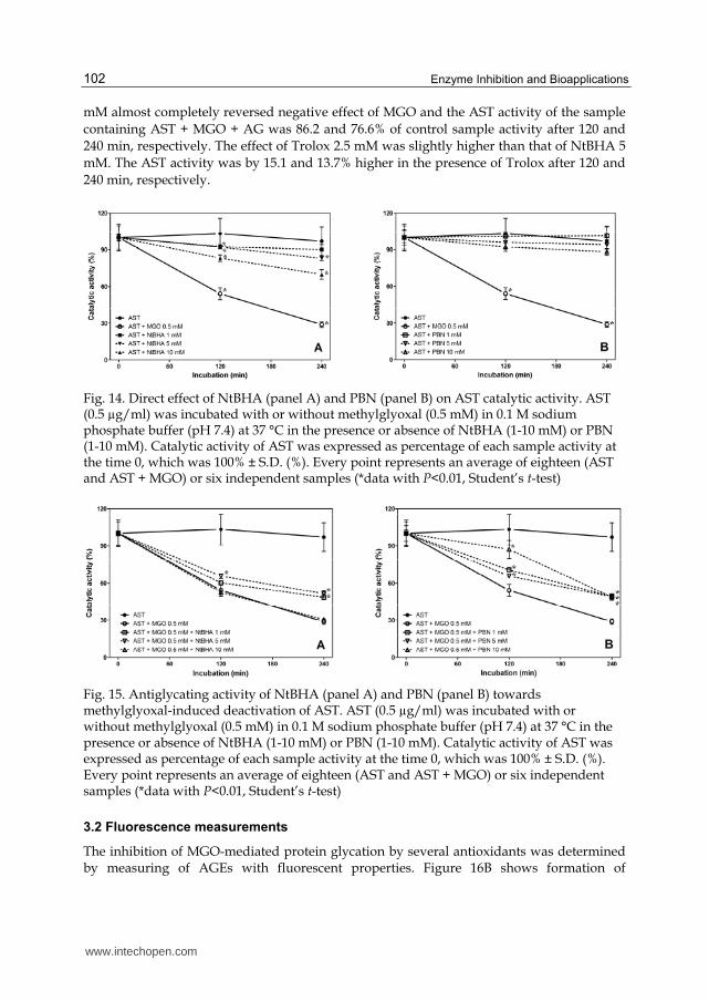
Activity of control sample (AST alone) was stable throughout the experiment. Some of tested compounds had a more or less pronounced negative direct effect on enzyme activity, which was probably due to a direct interaction of their molecules with the molecule of the enzyme. PBN itself had no harmful influence on stability and catalytic activity of AST in the concentrations used (Fig. 14B), while NtBHA caused concentration-dependent decrease in AST catalytic activity, which was statistically significant at concentrations of NtBHA 1 mM and higher (Fig. 14A). Aminoguanidine 1.0 mM caused significant decrease of enzyme activity by 21.7% after 240 min of incubation. This inhibitory effect of AG may be explained by its binding to PLP coenzyme forming a Schiff base, which disturbs tissue distribution of PLP in vivo and decreases its concentration in liver (Okada & Ayabe, 1995; Taguchi et al., 1998). Trolox 2.5 mM did not influence AST activity.
Following incubation of enzyme with MGO 0.5 mM, a rapid decline of AST activity was observed. The enzymatic activity decreased to 53.1 and 30.1% of control sample after 120 and 240 min, respectively. In addition, positive antiglycation effects were observed with some compounds. NtBHA exerted antiglycation influence only at 5 mM concentration after 120 and 240 min and at 1 mM concentration after 240 min of incubation (Fig. 15A). Negative direct effect of this compound observed at 10 mM concentration probably outweighed its positive antiglycation activity. The catalytic activity of AST was by 33.1% and 16.5% higher in the presence of PBN 10 mM and 1 mM after 120 min of incubation with MGO compared to the activity of sample containing AST + MGO only (Fig. 15B), respectively. PBN 1-10 mM protected AST against MGO-induced glycation also after 240 min of incubation, when all three concentrations increased activity of AST by 20%. In comparison, aminoguanidine 1.0
www.intechopen.com
Enzyme Inhibition and Bioapplications
102
mM almost completely reversed negative effect of MGO and the AST activity of the sample containing AST + MGO + AG was 86.2 and 76.6% of control sample activity after 120 and 240 min, respectively. The effect of Trolox 2.5 mM was slightly higher than that of NtBHA 5 mM. The AST activity was by 15.1 and 13.7% higher in the presence of Trolox after 120 and 240 min, respectively.
Fig. 14. Direct effect of NtBHA (panel A) and PBN (panel B) on AST catalytic activity. AST (0.5 µg/ml) was incubated with or without methylglyoxal (0.5 mM) in 0.1 M sodium phosphate buffer (pH 7.4) at 37 °C in the presence or absence of NtBHA (1-10 mM) or PBN (1-10 mM). Catalytic activity of AST was expressed as percentage of each sample activity at the time 0, which was 100% ± S.D. (%). Every point represents an average of eighteen (AST and AST + MGO) or six independent samples (*data with P<0.01, Student’s t-test)
Fig. 15. Antiglycating activity of NtBHA (panel A) and PBN (panel B) towards methylglyoxal-induced deactivation of AST. AST (0.5 µg/ml) was incubated with or without methylglyoxal (0.5 mM) in 0.1 M sodium phosphate buffer (pH 7.4) at 37 °C in the presence or absence of NtBHA (1-10 mM) or PBN (1-10 mM). Catalytic activity of AST was expressed as percentage of each sample activity at the time 0, which was 100% ± S.D. (%). Every point represents an average of eighteen (AST and AST + MGO) or six independent samples (*data with P<0.01, Student’s t-test)
3.2 Fluorescence measurements
The inhibition of MGO-mediated protein glycation by several antioxidants was determined by measuring of AGEs with fluorescent properties. Figure 16B shows formation of
www.intechopen.com

Non-Enzymatic Glycation of Aminotransferases and the Possibilities of Its Modulation
103
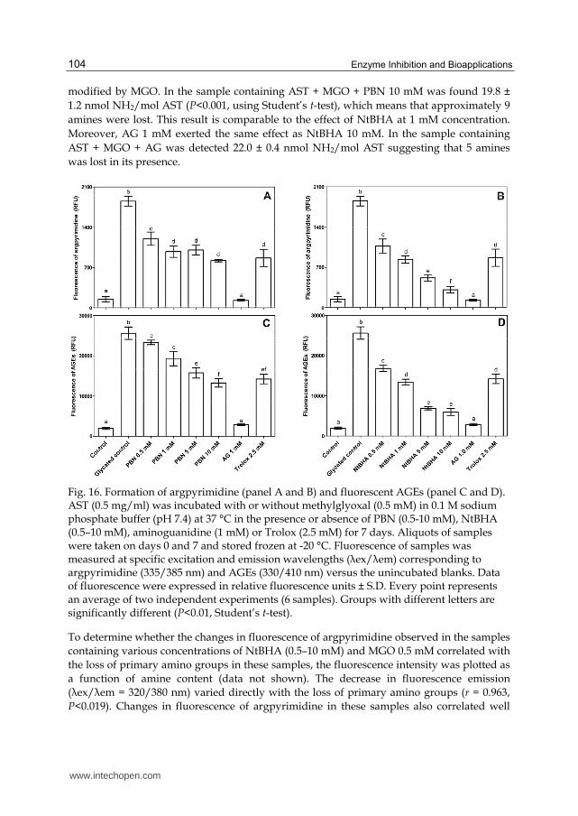
argpyrimidine and the effect of NtBHA on this process. Sample containing AST + MGO exerted 12.5 times higher fluorescence intensity than the control sample without MGO after 7 days of incubation. NtBHA caused statistically significant decrease in the formation of argpyrimidine during incubation (inhibition by 42.3–83.1%). The most remarkable decline in argpyrimidine formation was observed at 10 mM concentration of NtBHA, which exhibited inhibition by 83.1%. Effect of PBN on the argpyrimidine formation was less remarkable (inhibition by 35.2–55.8%) but still highly significant (Fig. 16A). The influence of AG 1.0 mM was well-pronounced (93.2%), while the effect of Trolox 2.5 mM was much weaker (53.2%) and comparable to the activity of NtBHA 1 mM and PBN 1-10 mM.
The effect of NtBHA on the formation of ‘‘non-specific’’ AGE products is presented in Fig. 16D. Methylglyoxal caused almost 13 fold increase in concentration of AGEs with fluorescent properties compared to the control sample (AST alone) after 7 days of incubation. Positive effect of NtBHA reached almost the same extent as in the case of argpyrimidine formation with statistically significant inhibition of glycation (34.3–76.6%). Little bit lower rate of inhibition (8.9–48.2%) was obtained also with PBN (Fig. 16C). Aminoguanidine and Trolox showed 88.7 and 44.4% suppressing effect on AGEs generation, respectively.
As for fluorescence measurement, control sample showed stable but not negligible fluorescence, since the start of the experiment. Most of this fluorescence is probably constituted by general fluorescence properties of this protein. Presence of pyridoxal-5’-phosphate coenzyme in the molecule of AST also contributes to basal fluorescence of the enzyme. Results of fluorescence measurements clearly show an inhibiting effect of NtBHA and PBN on the formation of AGE products. NtBHA was also quite effective in the inhibition of argpyrimidine generation. Apart from these findings, the use of fluorescence method for evaluation of protein glycation is limited by its imprecision. The measurement of some well-identified AGEs (e.g., pentosidine and carboxymethyllysine) by techniques as HPLC or ELISA could give more precise information on this matter (Boušová et al., 2009).
3.3 Determination of primary amino groups
Following incubation of AST with methylglyoxal, there was a decrease in amine content compared to control (Table 1). Unmodified AST exhibited 25.2 ± 0.4 nmol NH2/mol AST versus 13.9 ± 1.1 nmol NH2/mol AST for sample containing AST + MGO (P<0.001, using Student’s t-test). This difference represents a 45% decrease in amine content due to chemical modification of the primary amines (α-amino group of N-terminal amino acids and ε-amino group of Lys residues). Native AST in the dimer form contains 40 primary amines (38 Lys residues and 2 N-terminal amino acids), suggesting that approximately 18 amines were modified by methylglyoxal. Modification of primary amino group of Lys258 in AST molecule by MGO may be also responsible for the loss of its catalytic activity, because this Lys residue binds coenzyme PLP in the active centre of AST and thus directly participates in the enzymatic catalysis.
PBN as well as NtBHA significantly protected AST against the loss of primary amino groups induced by MGO. Their effect was concentration-dependent and more pronounced in the case of NtBHA. Sample containing AST + MGO + NtBHA 10 mM exhibited 21.8 ± 0.7 nmol NH2/mol AST (P<0.001, using Student’s t-test) suggesting that about 5 amines were
www.intechopen.com
Enzyme Inhibition and Bioapplications
104
modified by MGO. In the sample containing AST + MGO + PBN 10 mM was found 19.8 ± 1.2 nmol NH2/mol AST (P<0.001, using Student’s t-test), which means that approximately 9 amines were lost. This result is comparable to the effect of NtBHA at 1 mM concentration. Moreover, AG 1 mM exerted the same effect as NtBHA 10 mM. In the sample containing AST + MGO + AG was detected 22.0 ± 0.4 nmol NH2/mol AST suggesting that 5 amines was lost in its presence.
Fig. 16. Formation of argpyrimidine (panel A and B) and fluorescent AGEs (panel C and D). AST (0.5 mg/ml) was incubated with or without methylglyoxal (0.5 mM) in 0.1 M sodium phosphate buffer (pH 7.4) at 37 °C in the presence or absence of PBN (0.5-10 mM), NtBHA (0.5–10 mM), aminoguanidine (1 mM) or Trolox (2.5 mM) for 7 days. Aliquots of samples were taken on days 0 and 7 and stored frozen at -20 °C. Fluorescence of samples was measured at specific excitation and emission wavelengths (λex/λem) corresponding to argpyrimidine (335/385 nm) and AGEs (330/410 nm) versus the unincubated blanks. Data of fluorescence were expressed in relative fluorescence units ± S.D. Every point represents an average of two independent experiments (6 samples). Groups with different letters are significantly different (P<0.01, Student’s t-test).
To determine whether the changes in fluorescence of argpyrimidine observed in the samples containing various concentrations of NtBHA (0.5–10 mM) and MGO 0.5 mM correlated with the loss of primary amino groups in these samples, the fluorescence intensity was plotted as a function of amine content (data not shown). The decrease in fluorescence emission (λex/λem = 320/380 nm) varied directly with the loss of primary amino groups (r = 0.963, P<0.019). Changes in fluorescence of argpyrimidine in these samples also correlated well
www.intechopen.com

Non-Enzymatic Glycation of Aminotransferases and the Possibilities of Its Modulation
105
with the formation of fluorescent AGEs (r = 0.991, P<0.005). In addition, the amine content measured in samples containing AST + MGO + NtBHA (0.5-10 mM) was plotted as a function of AGEs fluorescence, indicating that MGO-induced formation of AGEs was directly proportional to an irreversible loss of primary amino groups in AST molecule (r = 0.946, P<0.027). Similar results were obtained also in samples containing AST + MGO + PBN (0.5-10 mM).
Sample Amine content Number of primary NH2
(nmol NH2/mol AST) remaining modified
AST 25.2 ± 0.4a 40 0
AST + MGO 0.5 mM 13.9 ± 1.1b 22 18
AST + MGO + PBN 0.5 mM 17.9 ± 0.9c 28 12
AST + MGO + PBN 1 mM 18.2 ± 1.1cd 29 11
AST + MGO + PBN 5 mM 18.6 ± 1.7cd 30 10
AST + MGO + PBN 10 mM 19.8 ± 1.2d 31 9
AST + MGO + NtBHA 0.5 mM 17.3 ± 0.4c 27 13
AST + MGO + NtBHA 1 mM 19.5 ± 0.4d 31 9
AST + MGO + NtBHA 5 mM 20.8 ± 0.8de 32 8
AST + MGO + NtBHA 10 mM 21.8 ± 0.7ef 35 5
AST + MGO + AG 1 mM 22.0 ± 0.4f 35 5
a,b,c,d,e,f Groups with different letters vary significantly (P<0.05, Student’s t-test)
Table 1. Effect of PBN, NtBHA and AG on the changes in AST primary amine content induced by MGO 0.5 mM
3.4 Effect of glycation on molecular charge of AST
Native PAGE was run several times, and the representative native PAGE gel is presented in Fig. 17. Mobility of MGO-modified protein to the positive pole significantly increased (by 41%) after 7 days of incubation compared to the mobility of control sample (AST alone). This result indicates the progressive loss of the positive charge in the MGO-modified AST during the glycation reaction. NtBHA showed concentration-dependent protective effect against changes in AST molecular change induced by MGO, when the relative mobility of sample containing AST + MGO + NtBHA 10 mM was increased only by 9.2% compared to the mobility of control. The enzyme incubated in the presence of both MGO and PBN showed a smaller rise in mobility, up to 21.6% in the case of PBN 10 mM. The effect of this compound on the protein electrophoretic mobility ranged from 21.6 to 26.3%. Aminoguanidine 1 mM completely reversed effect of MGO and the relative mobility of sample containing AST + MGO + AG was only slightly increased (by 0.35%) against the mobility of control, whereas Trolox 2.5 mM showed similar effect on molecular charge of AST as PBN, i.e., the mobility was increased by 27% compared to control sample (data not shown). These data indicated that the molecule of enzyme became more anionic due to glycation and that PBN as well as NtBHA had significant inhibitory effect on the middle stage of glycation process.
www.intechopen.com

Enzyme Inhibition and Bioapplications
106
Fig. 17. Protective effect of NtBHA and PBN on changes in molecular change of AST caused by MGO-induced glycation. AST (0.5 mg/ml) was incubated with or without MGO (0.5 mM) and NtBHA (0.5-10 mM) or PBN (0.5-10 mM) in sodium phosphate buffer (0.1 M, pH 7.4) at 37 °C for 7 days and then subjected to native PAGE. Proteins were visualized by Coomassie Blue G250. Gels were scanned and Rf was obtained using Quantity One software
Methylglyoxal-induced chemical modifications led to a change in molecular charge of AST, which became more anionic as revealed by native PAGE. These results indicate the progressive loss of the positive charge in the glycation-modified AST molecule, which is caused by the irreversible modification of Arg and Lys residues (Kang, 2006; Nagai et al., 2000) as was confirmed by determination of amine content. Both PBN and NtBHA partially protected native AST against glycation by MGO. The antiglycation activity was more pronounced in the case of NtBHA mainly at higher concentrations tested. The antiglycation activity of NtBHA 10 mM was a little bit lower than that of AG 1 mM.
3.5 SDS-PAGE and western blotting
The ability of aggregation and cross-link formation of tested antioxidants was determined by SDS-PAGE under denaturing conditions (Fig. 18). MGO readily reacts with lysine and arginine residues to produce high molecular weight protein products. Incubation of AST with MGO 0.5 mM at 37 °C for 7 days resulted in the formation of protein aggregates with molecular weight about 85, 107, and 145 kDa corresponding to protein dimer, trimer, and tetramer, respectively. No presence of protein dimer and tetramer, and lower concentration of protein trimer were observed in samples containing AST alone (lane 2), AST + NtBHA 10 mM (lane 7), and AST + AG 1 mM (data not shown). Also lower concentrations of NtBHA were able to partially protect formation of protein tetramer, although they had no effect on formation of protein dimer and trimer. On the other hand, PBN as well as Trolox were not able to prevent formation of protein cross-links and high molecular weight aggregates. Additional bands with molecular weight 20–35, 57 and 63 kDa were constituted of several contaminating proteins present in commercial preparation (Fig. 18).
www.intechopen.com

Non-Enzymatic Glycation of Aminotransferases and the Possibilities of Its Modulation
107
Western blotting with specific antibody against advanced glycation end products derived from MGO (anti-MGO [3C]) was used to confirm formation of protein aggregates as a result of MGO activity. The presence of high molecular weight protein cross-links in samples containing AST + MGO, AST + MGO + NtBHA, and AST + MGO + PBN was observed (data not shown). These protein aggregates had molecular weight about 85, 107, and 145 kDa corresponding to AST dimer, trimer, and tetramer, respectively. Quantitative differences between bands of samples with and without PBN or Trolox were not observed. These compounds are not able to prevent formation of protein cross-links. On the other hand, some reduction in the amount of AST tetramer was observed in samples containing NtBHA. These results suggest that NtBHA possesses, at least in part, antiglycation properties. Nevertheless, aminoguanidine 1 mM completely inhibited formation of protein aggregates, since no bands of AST dimer, trimer or tetramer were present.
The electrophoretic techniques confirmed the results obtained by other methods; i.e., changes in protein molecule caused by the presence of methylglyoxal and positive antiglycating effect of NtBHA. Methylglyoxal-induced chemical modifications led to a change in molecular charge of AST, which became more anionic as revealed by native PAGE. The SDS-PAGE and subsequent western blotting clearly showed formation of protein cross-links with higher molecular weight than native enzyme. NtBHA partially protected native AST from glycation by MGO and also exhibited mild anti-cross-linking activity.
Fig. 18. Formation of protein cross-links on reaction of AST with methylglyoxal. AST (0.5 mg/ml) was incubated with or without methylglyoxal (0.5 mM) in sodium phosphate buffer (0.1 M, pH 7.4) at 37 °C in the presence or absence of N-tert-butyl hydroxylamine (0.5–10 mM) for 7 days and then subjected to SDS-PAGE. Electrophoretic separation was performed on 4% stacking and 10% resolving polyacrylamide gels under reducing conditions. Bands were visualized with silver staining. Each lane was loaded with 4 µg of protein. MM = Mw marker; AST = aspartate aminotransferase; MGO = methylglyoxal; NtBHA = N-tert-butyl hydroxylamine
www.intechopen.com
Enzyme Inhibition and Bioapplications
108
Modification of proteins caused by methylglyoxal can be accompanied by formation of free radicals. Lee et al. (1998) identified three types of free radical species in samples containing methylglyoxal and bovine serum albumin by electron spin resonance spectroscopy. These radicals (methylglyoxal dialkylimine radical cation, methylglyoxal radical anion, and superoxide anion radical) were formed by direct 1-electron transfer process. Scavenging ability of NtBHA and PBN were already described (Lee et al., 2004; Atamna et al., 2001). It can be assumed that the positive antiglycation activity of these compounds may be at least partly attributed to their scavenging ability.
4. Conclusion
- Catalytic activity, which is biologically the most important property of all enzymes, is fully dependent on the native structure of the enzyme. Changes in the structure of enzymes usually lead to the progressive loss of their catalytic activities. These changes may be caused by reversible binding of various low-molecular inhibitors or by irreversible modification. Such example of the irreversible change is non-enzymatic glycation of proteins by various reducing monosaccharides or reactive α-dicarbonyl compounds (e.g., MGO).
- In our studies, aspartate aminotransferase (AST) was used in glycation studies as model protein, which possesses catalytic properties.
- Catalytic activity of aminotransferases in vitro has been found to be impaired by glycating agents. The extent of this effect depends on the activity and concentration of the agent, susceptibility of given enzyme to such modification as well as on the duration of action (see Fig. 4, Fig. 5 and Fig. 8).
- Among several glycating agents, effect of fructose is strong enough for investigation of changing AST properties during long time of incubation. Nevertheless, methylglyoxal, an intermediate of glycation process, is more reactive and permits to investigate the process of AST glycation in vitro in shorter course of time.
- Catalytic activity of AST as the protein function may serve as the most important criterion of glycation effect. Beside this, molecular properties of purified AST and character of glycoxidation permit using other methods of investigation of molecular changes during the process, like fluorescence of advanced glycation end-products, decrease in primary amino groups in the protein molecule, and protein cross-linking and aggregation.
- Several approaches of therapeutic intervention to the glycation process have been used (e.g., reduction in deposition of already formed AGEs, inhibition of new AGEs formation, and inhibition of the receptor for AGE).
- In our own experiments, compounds with described antioxidant and potential antiglycating activities have been studied. Among the compounds studied, hydroxycitric acid, uric acid, and two mitochondrial antioxidants α-phenyl N-tert-butyl nitrone and N-tert-butyl hydroxylamine had pronounced antiglycating activity against protein glycation by methylglyoxal (hydroxycitric acid, PBN and NtBHA) and by fructose (hydroxycitric and uric acids). On the other hand, flavonoids baicalin and baicalein exerted overall negative influence on the catalytic activity of AST alone and in the combination with fructose. Other studied compounds (i.e., ferulic, isoferulic, o-coumaric, and p-coumaric acids, arbutin and methylarbutin) showed no positive antiglycation activity.
www.intechopen.com

Non-Enzymatic Glycation of Aminotransferases and the Possibilities of Its Modulation
109
- The goal of our research group is to participate in the search for compounds with potential antiglycating activity with a perspective of their use as remedies against diabetic complications.
5. Acknowledgment
This study was supported by the Charles University in Prague (Project SVV 263 004).
6. References
Arai, K.; Iizuka, S.; Tada, Y.; Oikawa, K. & Taniguchi, N. (1987). Increase in the glucosylated form of erythrocyte Cu-Zn-superoxide dismutase in diabetes and close association of the nonenzymatic glucosylation with the enzyme activity. Biochimica et Biophysica
Acta - General Subjects, Vol.924, No.2, pp. 292-296, ISSN 0304-4165 Atamna, H.; Paler-Martínez, A. & Ames, B.N. (2000). N-t-butyl hydroxylamine, a hydrolysis
product of alpha-phenyl-N-t-butyl nitrone, is more potent in delaying senescence in human lung fibroblasts. Journal of Biological Chemistry, Vol.275, No.9, pp. 6741-6748, ISSN 0021-9258
Atamna, H.; Robinson, C.; Ingersoll, R.; Elliott, H. & Ames, B.N. (2001). N-t-butyl hydroxylamine is an antioxidant that reverses age-related changes in mitochondria in vivo and in vitro. The FASEB Journal, Vol.15, No.12, pp. 2196-2204, ISSN 0892-6638
Baynes, J.W. (1991). Role of oxidative stress in development of complications in diabetes. Diabetes, Vol.40, No.4, pp. 405-412, ISSN 0012-1797
Baynes, J.W. & Thorpe, S.R. (2000). Glycoxidation and lipoxidation in atherogenesis. Free
Radical Biology & Medicine, Vol.28, No.12, pp. 1708-1716, ISSN 0891-5849 Beisswenger, P.J.; Howell, S.K.; Nelson, R.G.; Mauer, M. & Szwergold, B.S. (2003). Alpha-
oxoaldehyde metabolism and diabetic complications. Biochemical Society
Transactions, Vol.31, No.Pt 6, pp. 1358-1363, ISSN 0300-5127 Beránek, M.; Dršata, J. & Palička, V. (2001). Inhibitory effect of glycation on catalytic activity
of alanine aminotransferase. Molecular and Cellular Biochemistry, Vol.218, No.1-2, pp. 35-39, ISSN 0300-8177
Beránek, M.; Dršata, J. & Palička, V. (2002). In vitro glycation of aminotransferases: A process closely depending on the employed experimental conditions. Acta Medica, Vol.45, No.3, pp. 89-92, ISSN 1211-4286
Bergmeyer, H.U.; Horder, M. & Rej, R. (1986). International Federation of Clinical Chemistry (IFCC) Scientific Committee, Analytical Section: approved recommendation (1985) on IFCC methods for the measurement of catalytic concentration of enzymes. Part 2. IFCC method for aspartate aminotransferase (L-aspartate: 2-oxoglutarate aminotransferase, EC 2.6.1.1). Journal of Clinical Chemistry and Clinical Biochemistry, Vol.24, No.7, pp 497–510, ISSN 0340-076X
Bolton, W.K.; Cattran, D.C.; Williams, M.E.; Adler, S.G.; Appel, G.B.; Cartwright, K.; Foiles, P.G.; Freedman, B.I.; Raskin, P.; Ratner, R.E.; Spinowitz, B.S.; Whittier, F.C.; Wuerth, J.P. & ACTION I Investigator Group (2004). Randomized trial of an inhibitor of formation of advanced glycation end products in diabetic nephropathy. American Journal of Nephrology, Vol.24, No.1, pp. 32-40, ISSN 0250-8095
www.intechopen.com

Enzyme Inhibition and Bioapplications
110
Boušová, I.; Vukasović, D.; Juretić, D.; Palička, V. & Dršata, J. (2005a). Enzyme activity and AGE formation in a model of glycoxidation of AST by D fructose in vitro. Acta
Pharmaceutica, Vol.55, No.1, pp. 107-114, ISSN 1330-0075 Boušová, I.; Martin, J.; Jahodář, L.; Dušek, J.; Palička, V. & Dršata, J. (2005b). Evaluation of in
vitro effects of natural substances of plant origin using a model of protein glycoxidation. Journal of Pharmaceutical and Biomedical Analysis, Vol.37, No.5., pp. 957-962, ISSN 0731-7085
Boušová, I.; Bakala, H.; Chudáček, R.; Palička, V. & Dršata, J. (2005c). Glycation-induced inactivation of aspartate aminotransferase, effect of uric acid. Molecular and Cellular
Biochemistry, Vol.278, No.1-2, pp. 85-92, ISSN 0300-8177 Boušová, I.; Bacílková, E.; Dobrijević, S. & Dršata J. (2009). Glycation of aspartate
aminotransferase by methylglyoxal, effect of hydroxycitric and uric acid. Molecular
and Cellular Biochemistry, Vol.331, No.1-2, pp. 215-223, ISSN 0300-8177 Bucciarelli, L.G.; Wendt, T.; Rong, L.; Lalla, E.; Hofmann, M.A.; Goova, M.T.; Taguchi, A.;
Yan, S.F.; Yan, S.D.; Stern, D.M. & Schmidt, A.M. (2002). RAGE is a multiligand receptor of the immunoglobulin superfamily: implications for homeostasis and chronic disease. Cellular and Molecular Life Sciences, Vol.59, No.7, pp. 1117-1128, ISSN 1420-682X
Bunn, H.F.; Gabbay, K.H. & Gallop, P.M. (1978). The glycosylation of hemoglobin: relevance to diabetes mellitus. Science, Vol.200, No.4337, pp. 21-27, ISSN 0036-8075
Davies, B.J. (1964). Disc electrophoresis. II. Method and application to human serum proteins. Annals of the New York Academy of Sciences, Vol.121, pp. 404–427, ISSN 0077-8923
De La Cruz, J.; González-Correa, J.; Guerrero, A. & De la Cuesta, F. (2004). Pharmacological approach to diabetic retinopathy. Diabetes/Metabolism Research and Reviews, Vol.20, No.2, pp. 91-113, ISSN 1520-7552
Dolhofer, R. & Wieland, O.H. (1978). In vitro glycosylation of hemoglobins by different sugars and sugar phosphates. FEBS Letters, Vol.85, No.1, pp. 86-90, ISSN 0014-5793
Dršata, J.; Beránek, M. & Palička, V. (2002). Inhibition of aspartate aminotransferase by glycation in vitro under various conditions. Journal of Enzyme Inhibition and
Medicinal Chemistry, Vol.17, No.1, pp. 31-36, ISSN 1475-6366 Dršata, J.; Boušová, I. & Maloň, P. (2005). Determination of quality of pyridoxal-5´-
phosphate enzyme preparations by spectroscopic methods. Journal of Pharmaceutical
and Biomedical Analysis, Vol.37, No.5., pp. 1173-1177, ISSN 0731-7085 Dršata, J. & Veselá, J. (1984). Inhibition of liver aminotransferases with some potential
cytostatic agents. Cesko-Slovenska Farmacie, Vol.33, No.9, pp. 372-375, ISSN 0009-0530
Fitzgerald, C.; Swearengin, T.A.; Yeargans, G.; McWhorter, D.; Cucchetti, B. & Seidler, N.W. (2000). Non-enzymatic glycosylation (or glycation) and inhibition of the pig heart cytosolic aspartate aminotransferase by glyceraldehyde 3-phosphate. Journal of
Enzyme Inhibition, Vol.15, No.1, pp. 79-89, ISSN 8755-5093 Hudson, B.I.; Bucciarelli, L.G.; Wendt, T.; Sakaguchi, T.; Lalla, E.; Qu, W.; Lu, Y.; Lee, L.;
Stern, D.M. & Naka, Y. (2003). Blockade of receptor for advanced glycation endproducts: a new target for therapeutic intervention in diabetic complications
www.intechopen.com

Non-Enzymatic Glycation of Aminotransferases and the Possibilities of Its Modulation
111
and inflammatory disorders. Archives of Biochemistry and Biophysics, Vol.419, No.1, pp. 80-88, ISSN 0003-9861
Hunt, J.V.; Dean, R.T. & Wolff, SP. (1988). Hydroxyl radical production and autoxidative glycosylation. Glucose autoxidation as the cause of protein damage in the experimental glycation model of diabetes mellitus and ageing. The Biochemical
Journal, Vol.256, No.1, pp. 205-212, ISSN 0264-6021 Jabeen, R. & Saleemuddin, M. (2006). Polyclonal antibodies inhibit the glycation-induced
inactivation of bovine Cu,Zn-superoxide dismutase. Biotechnology and Applied
Biochemistry, Vol.43, No. Pt 1, pp. 49-53, ISSN 0885-4513 Kang, J.H. (2006). Oxidative modification of human ceruloplasmin by methylglyoxal: an in
vitro study. Journal of Biochemistry and Molecular Biology, Vol.39, No.3, pp. 335–338, ISSN 1225-8687
Kelly, S.M. & Price, N.C. (2000). The Use of Circular Dichroism in the Investigation of Protein Structure and Function. Current Protein and Peptide Science, Vol.1, No.4, pp. 349-384, ISSN 1389-2037
Kirsch, J.F.; Eichele, G.; Ford, G.C.; Vincent, M.G.; Jansonius, J.N.; Gehring, H. & Christen, P. (1984). Mechanism of action of aspartate aminotransferase proposed on the basis of its spatial structure. Journal of Molecular Biology, Vol.174, No.3, pp. 497-525, ISSN 0022-2836
Kyselova, Z.; Stefek, M. & Bauer, V. (2004). Pharmacological prevention of diabetic cataract. Journal of Diabetes and its Complications, Vol.18, No.2, pp. 129-140, ISSN 1056-8727
Lapolla, A.; Traldi, P. & Fedele, D. (2005). Importance of measuring products of non-enzymatic glycation of proteins. Clinical Biochemistry, Vol.38, No.2, pp. 103-115, ISSN 0009-9120
Lee, J.H.; Kim, I.S. & Park, J.W. (2004). The use of N-t-butyl hydroxylamine for radioprotection in cultured cells and mice. Carcinogenesis, Vol.25, No.8, pp. 1435-1442, ISSN 0143-3334
Lee, C.; Yim, M.B.; Chock, P.B.; Yim, H.S. & Kang, S.O. (1998). Oxidation-reduction properties of methylglyoxal-modified protein in relation to free radical generation. Journal of Biological Chemistry, Vol.273, No.39, pp. 25272–25278, ISSN 0021-9258
Metzler, C.M.; Rogers, P.H.; Arnone, A.; Martin, D.S. & Metzler, D.E. (1979). Investigation of crystalline enzyme-substrate complexes of pyridoxal phosphate-dependent enzymes. Methods in Enzymology, Vol.62, pp. 551-558, ISSN 0076-6879
Monnier, V.M. (1989). Toward a Maillard reaction theory of aging. Progress in Clinical and
Biological Research, Vol.304, pp. 1-22, ISSN 0361-7742 Nagai, R.; Matsumoto, K.; Ling, X.; Suzuki, H.; Araki, T. & Horiuchi, S. (2000).
Glycolaldehyde, a reactive intermediate for advanced glycation end products, plays an important role in the generation of an active ligand for the macrophage scavenger receptor. Diabetes, Vol.49, No.10, pp. 1714–1723, ISSN 0012-1797
Netopilová, M.; Haugvicová, R.; Kubová, H.; Dršata, J. & Mareš, P. (2001). Influence of convulsants on rat brain activities of alanine aminotransferase and aspartate aminotransferase. Neurochemical Research, Vol.26, No.12, pp. 1285-1291, ISSN 0364-3190
www.intechopen.com

Enzyme Inhibition and Bioapplications
112
Netopilová, M.; Veselá, J. & Dršata, J. (1991). Influence of 5-[2-N,N-dimethylamino)ethoxy]-7-oxo-7H-benzo(c)fluorene hydrochloride (benflurone) on the activity of rat liver aspartate and alanine aminotransferases. Drug Metabolism and Drug Interactions, Vol.9, No.3-4, pp. 301-309, ISSN 0792-5077
Nursten, H. (2005). The Maillard Reaction : Chemistry, Biochemistry and Implications (1st edition), The Royal Society of Chemistry, ISBN 0-85404-964-9, Cambridge
Okada, M.; Sogo, A. & Ohnishi, N. (1994). Glycation reaction of aspartate aminotransferase by various carbohydrates in an in vitro system. Journal of Nutritional Biochemistry, Vol.5, No.10, pp. 485-489, ISSN 0955-2863
Okada, M. & Ayabe, Y. (1995). Effects of aminoguanidine and pyridoxal phosphate on glycation reaction of aspartate aminotransferase and serum albumin. Journal of
Nutritional Science and Vitaminology, Vol.41, No.4, pp. 43-50, ISSN 0301-4800 Okada, M.; Murakami, Y. & Miyamoto, E. (1997). Glycation and inactivation of aspartate
aminotransferase in diabetic rat tissues. Journal of Nutritional Science and
Vitaminology, Vol.43, No.4, pp. 463-469, ISSN 0301-4800 Ornstein, L. (1964). Disc electrophoresis. I. Background and theory. Annals of the New York
Academy of Sciences, Vol.121, pp. 321–349, ISSN 0077-8923 Park, L.; Raman, K.G.; Lee, K.J.; Lu, Y.; Ferran, L.J.; Chow, W.S.; Stern, D. & Schmidt, A.M.
(1998). Suppression of accelerated diabetic atherosclerosis by the soluble receptor for advanced glycation endproducts. Nature Medicine, Vol.4, No.9, pp. 1025-1031, ISSN 1078-8956
Sakurai, T.; Matsuyama, M. & Tsuchiya, S. (1987). Glycation of erythrocyte superoxide dismutase reduces its activity. Chemical and Pharmaceutical Bulletin, Vol.35, No.1, pp. 302-307, ISSN 0009-2363
Schalkwijk, C.G.; Stehouwer, C.D. & van Hinsbergh, V.W. (2004). Fructose-mediated non-enzymatic glycation: sweet coupling or bad modification. Diabetes/Metabolism
Research and Reviews, Vol.20, No.5, pp. 369-82, ISSN 1520-7552 Seidler, N.W. & Kowalewski, C. (2003). Methylglyoxal-induced glycation affects protein
topography. Archives of Biochemistry and Biophysics, Vol.410, No.1, pp. 149–154, ISSN 0003-9861
Seidler, N.W. & Seibel, I. (2000). Glycation of Aspartate Aminotransferase and Conformational Flexibility. Biochemical and Biophysical Research Communications, Vol.277, No.1, pp. 47-50, ISSN 1090-2104
Singh, R.; Barden, A.; Mori, T. & Beilin, L. (2001). Advanced glycation end-products: a review. Diabetologia, Vol.44, No.2, pp. 29-146, ISSN 1432-0428
Steinbrecher, U.P. (1987). Oxidation of human low density lipoprotein results in derivatization of lysine residues of apolipoprotein B by lipid peroxide decomposition products. Journal of Biological Chemistry, Vol.262, No.8, pp. 3603–3608, ISSN 0021-9258
Stuchbury, G. & Münch, G. (2005). Alzheimer's associated inflammation, potential drug targets and future therapies. Journal of Neural Transmission, Vol.112, No.3, pp. 429-453, ISSN 0300-9564
Suarez, G.; Rajaram, R.; Oronsky, A.L. & Gawinowicz, M.A. (1989). Nonenzymatic glycation of bovine serum albumin by fructose (fructation). Comparison with the Maillard
www.intechopen.com

Non-Enzymatic Glycation of Aminotransferases and the Possibilities of Its Modulation
113
reaction initiated by glucose. Journal of Biological Chemistry, Vol.264, No.7, pp. 3674-3679, ISSN 0021-9258
Taguchi, T.; Sugiura, M.; Hamada, Y. & Miwa, I. (1998). In vivo formation of a Schiff base of aminoguanidine with pyridoxal phosphate. Biochemical Pharmacology, Vol.55, No.10, pp. 1667-1671, ISSN 0006-2952
Thornalley, P.J. (2003). Use of aminoguanidine (Pimagedine) to prevent the formation of advanced glycation endproducts. Archives of Biochemistry and Biophysics, Vol.419, No.1, pp. 31-40, ISSN 0003-9861
Thornalley, P.J.; Yurek-George, A. & Argirov, O.K. (2000). Kinetics and mechanism of the reaction of aminoguanidine with the [alpha]-oxoaldehydes glyoxal, methylglyoxal, and 3-deoxyglucosone under physiological conditions. Biochemical Pharmacology, Vol.60, No.1, pp. 55-65, ISSN 0006-2952
Tupcová, P. (1996). Influence of sugars on activity of aminotransferases in vitro, Diploma thesis, Charles University in Prague, Faculty of Pharmacy, Hradec Králové
Ulrich, P. & Cerami, A. (2001). Protein glycation, diabetes, and aging. Recent Progress in
Hormone Research, Vol.56, pp. 1-22, ISSN 0079-9963 Vasan, S.; Zhang, X.; Zhang, X.; Kapurniotu, A.; Bernhagen, J.; Teichberg, S.; Basgen, J.;
Wagle, D.; Shih, D.; Terlecky, I.; Bucala, R.; Cerami, A.; Egan, J. & Ulrich, P. (1996). An agent cleaving glucose-derived protein crosslinks in vitro and in vivo. Nature, Vol.382, No.6588, pp.275-278, ISSN 0028-0836
Willemsen, S.; Hartog, J.W.; Hummel, Y.M.; Posma, J.L.; van Wijk, L.M.; van Veldhuisen, D.J. & Voors, A.A. (2010). Effects of alagebrium, an advanced glycation end-product breaker, in patients with chronic heart failure: study design and baseline characteristics of the BENEFICIAL trial. European Journal of Heart Failure. Vol.12, No.3, pp. 294-300, ISSN 1388-9842
Wolff, S.P. & Dean, R.T. (1987). Glucose autoxidation and protein modification. The potential role of 'autoxidative glycosylation' in diabetes. The Biochemical Journal, Vol.245, No.1, pp. 243-250, ISSN 0264-6021
Wolff, S.P.; Jiang, Z.Y. & Hunt, J.V. (1991). Protein glycation and oxidative stress in diabetes mellitus and ageing. Free Radical Biology & Medicine, Vol.10, No.5, pp. 339-352, ISSN 0891-5849
Wu, C.H. & Yen, G.C. (2005). Inhibitory effect of naturally occurring flavonoids on the formation of advanced glycation end products. Journal of Agricultural and Food
Chemistry, Vol.53, No.8, pp. 3167–3173, ISSN 0021-8561 Yagi, T.; Kagamiyama, H.; Nozaki, M. & Soda, K. (1985). Glutamate-aspartate transaminase
from microorganisms. Methods in Enzymology, Vol.113, pp. 83-89, ISSN 0076-6879 Yamagishi, S.; Nakamura, K.; Matsui, T.; Ueda, S.; Fukami, K. & Okuda, S. (2008). Agents
that block advanced glycation end product (AGE)-RAGE (receptor for AGEs)-oxidative stress system: a novel therapeutic strategy for diabetic vascular complications. Expert Opinion on Investigational Drugs, Vol.17, No.7, pp. 983-996, ISSN 1354-3784
Yan, H. & Harding, J.J. (1997). Glycation-induced inactivation and loss of antigenicity of catalase and superoxide dismutase. The Biochemical Journal, Vol.328, No.Pt 2, pp. 599-605, ISSN 0264-6021
www.intechopen.com
Enzyme Inhibition and Bioapplications
114
Yan, H. & Harding, J.J. (2006). Carnosine inhibits modifications and decreased molecular chaperone activity of lens alpha-crystallin induced by ribose and fructose 6-phosphate. Molecular Vision, Vol.12, pp. 205-14, ISSN 1090-0535
Yegin, A., Özben, T. & Yegin, H. (1995). Glycation of lipoproteins and accelerated atherosclerosis in non-insulin-dependent diabetes mellitus. International Journal of
Clinical & Laboratory Research, Vol.25, No.3, pp. 157-161, ISSN 0940-5437 Zeng, J.; Dunlop, R.A.; Rodgers, K.J. & Davies, M.J. (2006). Evidence for inactivation of
cysteine proteases by reactive carbonyls via glycation of active site thiols. The
Biochemical Journal, Vol.398, No.2, pp. 197-206, ISSN 0264-6021 Zhao, W.; Devamanoharan, P.S. & Varma, S.D. (2000). Fructose induced deactivation of
antioxidant enzymes: preventive effect of pyruvate. Free Radical Research, Vol.33, No.1, pp. 23-30, ISSN 1071-5762
www.intechopen.com

Enzyme Inhibition and BioapplicationsEdited by Prof. Rakesh Sharma
ISBN 978-953-51-0585-5Hard cover, 314 pagesPublisher InTechPublished online 09, May, 2012Published in print edition May, 2012
InTech EuropeUniversity Campus STeP Ri Slavka Krautzeka 83/A 51000 Rijeka, Croatia Phone: +385 (51) 770 447 Fax: +385 (51) 686 166www.intechopen.com
InTech ChinaUnit 405, Office Block, Hotel Equatorial Shanghai No.65, Yan An Road (West), Shanghai, 200040, China
Phone: +86-21-62489820 Fax: +86-21-62489821
Enzyme Inhibition and Bioapplications is a concise book on applied methods of enzymes used in drug testing.The present volume will serve the purpose of applied drug evaluation methods in research projects, as well asrelatively experienced enzyme scientists who might wish to develop their experiments further. Chapters arearranged in the order of basic concepts of enzyme inhibition and physiological basis of cytochromes followedby new concepts of applied drug therapy; reliability analysis; and new enzyme applications from mechanisticpoint of view.
How to referenceIn order to correctly reference this scholarly work, feel free to copy and paste the following:
Iva Boušová, Lenka Srbová and Jaroslav Dršata (2012). Non-Enzymatic Glycation of Aminotransferases andthe Possibilities of Its Modulation, Enzyme Inhibition and Bioapplications, Prof. Rakesh Sharma (Ed.), ISBN:978-953-51-0585-5, InTech, Available from: http://www.intechopen.com/books/enzyme-inhibition-and-bioapplications/non-enzymatic-glycation-of-aminotransferases-and-the-possibilities-of-its-modulation

© 2012 The Author(s). Licensee IntechOpen. This is an open access articledistributed under the terms of the Creative Commons Attribution 3.0License, which permits unrestricted use, distribution, and reproduction inany medium, provided the original work is properly cited.