nomenclature, symbols, units and their usage in...
TRANSCRIPT

Pure & App!. C/tern., Vol. 45, pp. 105—123. Pergamon Press, 1976. Printed in Greal Britain.
INTERNATIONAL UNION OFPURE AND APPLIED CHEMISTRY
ANALYTICAL CHEMISTRY DIVISION
COMMISSION ON SPECTROCHEMICAL AND OTHEROPTICAL PROCEDURES FOR ANALYSIS
NOMENCLATURE, SYMBOLS, UNITS AND THEIRUSAGE IN SPECTROCHEMICAL ANALYSIS—Ill.
ANALYTICAL FLAME SPECTROSCOPY ANDASSOCIATED NON-FLAME PROCEDURES
(RULES APPROVED 1975)
PERGAMON PRESSOXFORD NEW YORK PARIS FRANKFURT

ANALYTICAL CHEMISTRY DIVISIONCOMMISSION ON SPECTROCHEMICAL AND OTHER
OPTICAL PROCEDURES FOR ANALYSISt
NOMENCLATURE, SYMBOLS, UNITS AND THEIRUSAGE IN SPECTROCHEMICAL ANALYSIS—Ill.
ANALYTICAL FLAME SPECTROSCOPY ANDASSOCIATED NON-FLAME PROCEDURES
(RULES APPROVED 1975)
CONTENTS
1. Introduction2. Terms and symbols for general quantities and constants3. Terms, symbols, and units for the description of the analytical apparatus
3.1. Transformation of sample into vapour3.1.1. Descriptive terms concerning nebulizer-flame systems
3.1.1.1. Nebulization, desolvation, volatilization, and atomization3.1.1.2. Nebulizers3.1.1.3. Burners and flames
3.1.2. Terms, symbols, and units for measurable quantities relating to nebulizer-flame systems3.1.3. Terms concerning special sampling, atomizing, and exciting devices
3.1.3.1. Special sampling devices for flames3.1.3.2. Electrical flame-like plasmas for atomization and excitation3.1.3.3. Non-flame atomizing devices
3.1.3.3.1. Resistance-heated devices3.1.3.3.2. Hollow-cathode devices3.1.3.3.3. Radiation-heated devices
3.2. Light sources in atomic absorption and atomic fluorescence spectroscopy3.3. Optical systems3.4. Photodetectors3.5. The electrical measuring system3.6. Survey of terms, symbols, and units for measurable quantities in the optical and measuring systems
4. Terms and symbols relating to the analytical procedure and the performance of an analysis4.1. General analytical terminology in flame spectroscopy4.2. Analytical calibration4.3. Assessment of an analytical procedure
4.3.1. Measurement scatter4.3.2. Limit of detection, precision, and accuracy
4.4. Interferences by concomitants4.4.1. General4.4.2. Classification of interferences
4.4.2.1. Spectral interferences4.4.2.2. Non-spectral interferences
4.4.3. Reduction of errors due to interferences for given instrumental conditions5. Terms, symbols, and units relating to radiant energy and its interaction with matter
5.1. Descriptive terms relating to the emission, absorption, and fluorescence of radiation5.1.1. Emission5.1.2. Absorption and self-absorption5.1.3. Fluorescence
5.2. Terms, symbols, and units for measurable quantities6. Terms, symbols, and units relating to the gaseous state of matter
6.1. Descriptive terms concerning the gaseous state of matter6.2. Terms, symbols, and units for measurable quantities
7. Index of terms
1. INTRODUCTION
tChairman: V. A. Fassel (USA); Secretary: B. F. Scribner Part III is a sequel to Parts I [Pure App!. Chem. 30 653(USA); Members: C. Th. J. Alkemade (Netherlands), L. S. Birks (1972)1 and II [Pure App! Chem 45 (2) 99—103 (1976)] of
lovakia)JP(deceased)(UK),E.Pliko(Czechos- the Nomenclature for Spectrochemical Analysis. Whereas
Associate Members: R. Jenkins (USA), H. Kaiser (Germany) A. Parts I and II are mainly concerned with some generalKvalheim (Norway), R. Muller (Switzerland), I. Rubeika recommendations, Part III deals specifically with analyti-(Czechoslovakia), A. Strasheim (South Africa), V. Vukanoviè cal flame spectroscopy and associated procedures.(Yugoslavia), J. Walters (USA). The purpose of this nomenclature, as well as its
107

108 COMMISSION ON SPECTROCHEMICAL AND OTHER OPTICAL PROCEDURES FOR ANALYSIS
adaptation to the documents developed by IUPAC andIUPAP in more general fields of chemistry and physics,has already been explained in the Foreword of Part I. Inthe development of Part III, the recommendations ofIUPAC, IUPAP and the International Commission onIllumination (CIE), and of the foregoing Parts I and IIwere taken as a starting point. Deviations from theserecommendations occur only in some exceptional casesand mainly concern the choice of a consistent set ofsymbols within the restricted field of analytical flamespectroscopy. Deviations may also be found in a fewcases where previous international documents are not inagreement among themselves. Such deviations are exp-licitly identified in the Notes added to this document.
In those instances of specific usage in flame spectros-copy which are not already established by internationalconventions, compromises often had to be made betweenthe historically developed terminology and nomenclatureand those based on more logical considerations. Someexpressions which have found their way into the languageof practical analysts, but which could be misleading, havebeen abandoned. The use of alternative terms for thesame item was considered undesirable and, as a rule, onlyone term has been recommended for each item. Somedifferences in terminology that developed historicallyamong the practitioners of emission and atomic absorptionmethods have been reconciled.
During the period in which this document was inpreparation, several national organizations developedtheir own nomenclature for atomic absorption spectros-copy. Through mutual deliberations, serious conflictsbetween national documents of restricted scope and theIUPAC nomenclature were significantly reduced. Thesimultaneous emergence of these national initiativesclearly proves the general need for a well defined
terminology in this field and calls for internationalcooperation.
Part III is concerned with the analytical application offlame spectroscopy by emission, absorption, and fluores-cence methods. These three branches of analysis havemany terms in common and a uniform terminology seemsmandatory. In the context of this document, an "ordi-nary" flame may be defined as a continuously flowing gasmixture at atmospheric pressure, which emerges from aburner and is heated by combustion. Although thisdocument is primarily concerned with systems thatcomprise such ordinary flames, burners, and nebulizers,attention is also paid to similar procedures comprisingother sampling, atomizing, and/or exciting devices (seeSection 3.1.3). These procedures bear some resemblanceto ordinary flame spectroscopy with regard to themethods of measuring emission, absorption, or fluores-cence signals, as well as to the rather simple nature of thedevices involved and the simplicity of the spectraobtained. Arc and spark spectroscopy involving moreelaborate and expensive instrumentation are not discus-sed in this document.
As can be seen from the Table of Contents, thisdocument is subdivided into several sections correspond-ing to the different aspects of analytical flame spectros-copy. A general classification and consistent terminologyof the different branches of flame spectroscopy arepresented in Table 1.1. For some terms, abbreviations aresuggested for practical usage. To comply with theinternationally agreed restricted meaning of "photome-ter" (see Section 4.5 in Part I), the term "flamephotometry" had to be abandoned, although it wasrealized that many of the older practitioners of flameemission spectroscopy may have difficulty in giving upthis long-cherished term. On the other hand, the
Absorption Emission Fluorescence
Methods:General classification Absorption spectroscopyt Emission spectroscopyt Fluorescence spectroscopyt
Instruments:General classification Absorption spectrometer Emission spectrometert Fluorescence spectrometert
When atomic lines areobserved
Atomic absorptionspectroscopy (AAS)Atomic absorptionspectrometer
Atomic emissionspectroscopy (AES)Atomic emissionspectrometer
Atomic fluorescencespectroscopy (AFS)Atomic fluorescencespectrometer
When a flame is used asa means for vaporization,atomization and/orexcitationT
Flame absorptionspectroscopy (FAS)Flame absorptionspectrometer
Flame emissionspectroscopy (FES)Flame emissionspectrometer
Flame fluorescencespectroscopy (FFS)Flame fluorescencespectrometer
When both a flame isused and atomic linesare observed
Flame atomic absorptionspectroscopy (FAAS)Flame atomic absorptionspectrometer
Flame atomic emissionspectroscopy (FAES)Flame atomic emissionspectrometer
Flame atomic fluorescencespectroscopy (FAFS)Flame atomic fluorescencespectrometer
Note: The term flame photometry (flame photometer) has been abandoned (see also Part I, Section 4.5).tSpectroscopy may be replaced by the more restrictive term spectrometry when quantitative measurements of
intensities at one or more wavelengths are performed with a spectrometer (see below).tThe term spectrometer as it is used here implies that quantitative measurements of intensities at one or more
wavelengths are performed with a photoelectric detector. Wavelength selection may be accomplished, e.g. with amonochromator or optical filter.
§When molecular species are observed, "molecular" is substituted for "atomic".¶Alternative, but presently less common means of vaporization, atomization and/or excitation are, for example,
furnaces, flame-like (electrical) plasmas, and cathodic sputtering tubes (see Section 3.1.3). The appropriate adjectiveshould replace the term flame.
Table 1.1. Classification of methods and instruments

Nomenclature, symbols, units and their usage in spectrochemical analysis—Ill 109
abandonment of this term removes the illogical juxtaposi-tion of terms such as "emission flame photometry" and"atomic absorption spectroscopy," which are often foundin the literature. The more uniform terminology presentedin Table 1.1 should help to bridge the gap betweenemission and absorption methods.
Descriptive terms denoting processes or instrumentalcomponents and terms for measurable quantities arepresented separately. The descriptive terms and some ofthe most important quantitative terms are explained in anarrative form. To facilitate reference, all quantitativeterms are presented in tables, together with their symbolsand practical units. Notes have been added to the text aswell as to the tables in order to provide additionalexplanation or justification, or to warn against improperusage of terms. These notes may readily be passed overon first reading, as all specifically recommended terms areincorporated in the text or the tables themselves. Suchterms, when first met and defined in the text, are printed initalics for easy recognition.
This nomenclature is not intended to provide acomprehensive set of terms. Definitions and explanationsof specific terms are only given when this is required tomake their meaning unambiguous. General physical orchemical concepts and quantities are presented in mostcases without further elucidation. To facilitate the use ofthis document, terms and symbols employed in flamespectroscopy which were presented in the foregoing PartsI and II have been recapitulated. This applies in particularto the general quantitative terms listed in Tables 2.1 and5.1. Important terms such as "concentration," "analyticalcurve," "sensitivity," etc. are briefly explained in Section4 with explicit reference to Parts I and II for a fullerexplanation.
In view of the limited number of letters in the alphabet,symbols have been selected carefully, while makingallowance for existing international recommendations aswell as for long usage. In some cases, alternative symbolshave been added, which allows the user to select a set ofsymbols that is most consistent in a particular context.Alternative symbols that are recommended withoutpreference are listed after each other and separated bycommas. When a particular symbol is preferred, it is listedfirst while the alternative symbols are separated from it bya row of dots. The printing of symbols and indices shouldconform to the general international rules recapitulated inSection 2.3 of Part I.
2. TERMS AND SYMBOLS FOR GENERAL
QUANTITIES AND CONSTANTS
Table 2.1 lists terms and symbols for some generalphysical and chemical quantities which are commonlyused in analytical spectroscopy. This Table is partly anabstract from, and partly forms a supplement to, Sections3.1 and 7.2 of Part I of the Nomenclature on Spec-trochemical Analysis.
3. TERMS, SYMBOLS, AND UNITS FOR THEDESCRIPTION OF THE ANALYTICAL APPARATUS
The functions of an analytical flame spectrometer ingeneral are:
(a) Transformation of the solution to be analyzed into avapour containing free atoms or molecular compounds ofthe analyte (see Section 4) in the flame;
(b) Selection and detection of the optical signal (arisingfrom the analyte vapour) which carries information on thekind and concentration of the analyte;
Terms Symbol Note
MassAtomic weight
(A, = 12 for carbon'2C) of species X
Atomic mass ofspecies X
VolumeSolid angleTimeFrequency (in optical
spectroscopy)Frequency (in
electrotechnics)WavelengthWavenumber (1/A)Gas pressureTotal pressure of
gas mixturePartial pressure of
species XNumber of particles(Number) density of
particles (number perunit volume)
(Number) density ofspecies X
Thermodynamic (orabsolute) temperature
Velocity of light(in vacuo)
Gas constantAvogadro constantBoltzmann constantPlanck constantElementary charge The symbol for
"electron" is eand should notbe printed initalics.
(c) Amplification and read-out of the electrical signal.Terms for the description of component parts of a flamespectrometer (and similar systems) and of processesoccurring therein are discussed below. Terms for thedescription of processes and properties related inparticular to the gaseous state of matter in flames arediscussed in Section 5.
3.1. Transformation of sample into vapour3.1.1. Descriptive terms concerning nebulizer-flame sys-tems
3.1.1.1. Nebulization, desolvation, volatilization, andatomization. With a pneumatic nebulizer driven under theaction of a compressed gas stream, the solution isaspirated from the sample container and nebulized into amist or aerosol of fine droplets. The term sprayer denotesthat particular part of a nebulizer where the aspiratedliquid is disrupted by the gas-jet into a spray.
By desolvation, i.e. evaporation of the solvent from thedroplets, this mist is converted into a dry aerosolconsisting of a suspension of solid or molten particles ofthe solute. In the high-temperature environment of theflame, volatilization of these particles follows.
In atomic flame spectroscopy, the atomization, i.e. theconversion of volatilized analyte into free atoms, should
Table 2.1. Terms and symbols for general quantities and con-stants
m(Ar), Ar(X)
mx, m(X)
V11,0)
ii
fA
cT, V
pPt
Px, p(X)
Nn
flx, n(X), [XI
T
c
RNAkhe
The unit for T isthe kelvin (K)

110 COMMISSION ON SPECTROCHEMICAL AND OTHER OPTICAL PROCEDURES FOR ANALYSIS
be as complete as possible in order to obtain a maximumsignal. Any system which is capable of converting theanalyte into an atomic vapour is called an atomizer (seealso Section 3.1.3).
3.1.1.2. Nebulizers. In the chamber-type nebulizer, thenebulizing gas-jet stream emerges from the sprayer into aspray chamber. In such a chamber, the gas-jet stream ishomogeneously mixed with the mist droplets. Some ofthese droplets may evaporate, coalesce, or deposit on thechamber walls and subsequently drain as waste.
Nebulizers can be described as follows:According to the source of energy used for nebulization,as, for example, pneumatic or ultrasonic nebulizer.According to the way the liquid is taken up, e.g. suction,gravity -fed, controlled-flow, and reflux-nebulizers.According to the relative position of the capillaries forthe nebulizing gas and the aspirated liquid, e.g. angularand concentric nebulizers.
Special devices are the nebulizer with heated spraychamber, the twin nebulizer, and the drop generator.
Note: Nebulizers by themselves should not be called atomizers.
3.1.1.3. Burners and flames. Flames are produced bymeans of a burner to which fuel and oxidant are supplied,usually in the form of gases. With the premix burner, fueland oxidant are thoroughly mixed inside the burnerhousing before they leave the burner ports and enter theprimary-combustion or inner zone of the flame. This typeof burner usually produces an approximately laminarflame, and is commonly combined with a separate unit fornebulizing the sample.
In contrast, a direct-injection burner combines thefunctions of nebulizer and burner. Here oxidant and fuelemerge from separate ports and are mixed above theburner orifices through their turbulent motion. The flameproduced by such a burner is turbulent. Most commonly,the oxidant is also used for aspirating and nebulizing thesample. However, when the fuel is used for this purpose,the term reversed direct-injection burner is applied. Ineach case, the mist droplets enter the flame directly,without passing through a spray chamber.
Note: The term total-consumption burner, which is often used, isnot recommended.
Premix burners are distinguished as Bunsen-, Méker-,or slot-burners according to whether they have one largehole, a number of small holes, or a slot as outlet port(s) forthe gas mixture, respectively. When several parallel slotsare present, they are identified as multislot burners (e.g. athree-slot burner). The small diameter of the holes in theMéker burner or the narrowness of the slot in theslot-burner usually prevents the unwanted flash -back ofthe flame into the burner housing.
At the edge of the flame where the hot gas comes intocontact with the surrounding air, secondary combustionoccurs and the secondary combustion or outer zone isformed. The region of the flame confined by the inner andouter zones, where in many instances the conditions forflame analysis are optimum, is called the interzonal region,or, when the combustion zones have the form of a cone,the interconal zone.
Sometimes provision is made to screen the observedportion of the flame gases from direct contact with thesurrounding air. This may be done either mechanically byplacing a tube on the top of the burner around the flame,which produces a zonal separation (separated flame), or
aerodynamically by surrounding the flame with a sheathof inert gas that emerges from openings at the rim of theburner top (shielded flame). Observations can thus bemade without disturbances from the secondary-combustion zone.
To promote the atomization of elements that readilyform oxides in the vapour phase in the flame, a fuel-richflame is often chosen, where reducing conditions favourthe dissociation of the metal oxides.
3.1.2. Terms, symbols and units for measurable quantitiesrelating to nebulizer-flame systems
An easily measurable quantity is the rate of liquidconsumption by the nebulizing system, defined as thevolume of liquid sample that is consumed per unit of time(symbol: F1: see Table 3.1). In the particular, but commoncase of a pneumatic nebulizer, the term rate of liquidaspiration is more specific.
Often only a fraction of the analyte solution that isaspirated passes through the flame cross-section at theobservation height (see Table 3.2) in a form that isaccessible for spectroscopic observation. There are lossesof different kinds that limit this fraction and consequentlythe sensitivity of the method. Examples of such losseswith premix burners are the waste of solution in the spraychamber, the burner, and the tubes between them,because of deposition of mist droplets on the walls. Withdirect-injection burners, there may be losses of dropletsthat are ejected from the flame because of the turbulentmotion of the gases leaving the burner. Moreover, theresidence time of the larger droplets in the flame may beinsufficient for complete desolvation (see Section 3.1.1.1).Similarly, the particles formed after desolvation may notbe completely volatilized (see Section 3.1.1.1) at theobservation height. Finally, it should be recognized thatonly part of the vapour produced by the analyte mayconsist of free atoms.
To describe these losses quantitatively, the followingterms are recommended. These terms relate to the amountof analyte aspirated per second and entering the flame persecond or passing through the total horizontal flamecross-section per second at the observation height indifferent states.
The efficiency of nebulization, En, is the ratio of theamount of analyte entering the flame to the amount ofanalyte aspirated.
Notes: The quantity n isnot related to the amount of solvent butto the amount of analyte. Its value cannot be determinedunambiguously by simply comparing the volume of solutiondrained per second from the spray chamber with the aspirationrate. Correction must usually be made for the difference in analyteconcentration in the drained and aspirated solution, respectively,due to the partial evaporation of solvent from the mist dropletsdeposited on the walls.
The quantity €. is not merely characteristic of the operation ofthe nebulizer, but of the nebulizer-burner system as a whole.
The (local) fraction desolvated, f3s, is the ratio of theamount of analyte passing in the desolvated state (i.e.either as adry aerosol or as a vapour) to the total amountof analyte passing. When this fraction varies with height,due to progressive evaporation of the aerosol droplets inthe flame, it is appropriate to speak of the local fractiondesolvated.
Notes: Losses due to incomplete volatilization of the dry aerosol(which depend largely on the nature and concentration of thesolute) are not covered by the definition of Ps but by the definition

Nomenclature, symbols, units and their usage in spectrochemical analysis—Ill 111
of fL. The quantity j3, will usually depend on the solute whereas f3will depend on the solvent.
When this ratio varies markedly with height over the volume offlame observed, the amount of analyte found in this volume in theconsidered state is related to the average value of this ratio overthis volume.
The (local) fraction volatilized, IL, is the ratio of theamount of analyte passing in the gaseous state to the totalamount of analyte passing in the desolvated state. Thegaseous state includes free atoms as well as molecules.
The (local) fraction atomized, a, is the ratio of theamount of analyte passing as free neutral (or ionized)atoms to the total amount of analyte passing in thegaseous state.
Note: This fraction is determined by chemical reactions in thegaseous state. The bond strengths of the molecular compoundswhich the analyte may form in the flame play an important part aswell as the composition and the temperature of the flame.
The overall (local) efficiency of atomization, Ca, isdefined as the ratio of the amount of analyte that passesthrough the flame cross-section at the observation height,as free neutral (or ionized) atoms, to the amount ofanalyte aspirated. Therefore, Ca = Enf3s/3v13a. The atomicsignal strength obtained for a given solution concentrationis proportional to the product PiCa. It is noted that a maydepend on F1.
In Table 3.1, the above quantitative terms and somefurther terms for measurable quantities belonging to thissection are listed, together with their symbols and units.
3.1.3. Terms concerning special sampling, atomizing andexciting devices
3.1.3.1. Special sampling devices for flames. Samplesmay be introduced into flames by means other thannebulizers. The samples may be deposited on a samplingioop, sampling boat, or sampling cup made fromplatinum, tungsten, or other high melting point materials,and subsequently thermally vaporized.
Note: These devices are heated only by the flame and not by anadditional source of energy.
In atomic absorption spectroscopy a long tube device issometimes used with a nebulizer-flame system to increasethe sensitivity. The increase in sensitivity is achieved by"retaining" the combustion gases with the atomizedanalyte over an extended path length by means of a tubecoaxial with the optical axis. The tube is made frommaterial capable of withstanding the flame temperature.The flame gases enter the tube at one end and leave at theother, or when a T-shaped tube arrangement is used, thegases enter at the centre and flow toward the two openends. T-shaped tubes are called T-tubes.
Table 3.1. Transformation of sample into vapour. Terms, symbols, and units for measurablequantities
Terms
Travel time (time needed forsubstance to be carriedfrom base of flame to theobservation volume)
Transit time (time needed forsubstance to pass throughthe observation volume)
(Vertical) rise velocity offlame gas
Burning velocity (of flamefront)
Flow rate of unburnt gasmixture
Flow rate of X, e.g.air, 02, etc.
Practicalunit
cm3 s'
Note
For definition, see the text ofSection 3.1.2. In the usualcase of a pneumaticnebulizer F is called therate of liquid aspiration.
1 For definition, see thetext of Section 3.1.2.
IL 1 For definition, see thetext of Section 3.1.2.
/L 1 For definition, see thetext of Section 3.1.2.
I For definition, see thetext of Section 3.1.2.
Ca 1 For definition, see thetext of Section 3.1.2.
Tf K When the temperature varieslocally in the flame, it ismore appropriate to speakof the local flametemperature.
tts
yr cms'
vb cm 5'
cm3 s' Measured at atmosphericpressure and at roomtemperature.
Fx cm3 s' Measured at atmosphericpressure and at roomtemperature.
Symbol
F,Rate of liquid consumption
Efficiency of nebulization
Fraction desolvated
Fraction volatilized
Fraction atomized
Efficiency of atomization
Flame temperature
ttv
112 COMMISSION ON SPECTROCHEMICAL AND OTHER OPTICAL PROCEDURES FOR ANALYSIS
Note : The long tube has often been referred to as an absorptioncell. However, the term absorption cell should be reserved fordevices closed by optical windows.
3.1.3.2. Electrical flame-like plasmas for atomizationand excitation. Electrical flame-like plasmas may becurrent-carrying plasmas or current-free plasmas. Plas-mas may be formed by an arc discharge in a chamber andtransferred through an appropriate opening to form aplasma jet. Flame-like plasmas can also be generated byhigh-frequency fields with or without the use of elec-trodes. With the electrodeless plasma, the electromagneticfield is usually inductively coupled to the plasma(inductively-coupled plasma). The single-electrodeplasma is formed on a metallic tip that is connected to ahigh-frequency generator.
Notes : Both chamber-type nebulizer systems and nebulizerswhich spray the aerosol directly into the plasma are used tointroduce the sample.
The term "plasma flame," often used for flame-like plasmas, isnot recommended. The term "flame" should be reserved for hotgases which are produced by combustion. Similarly, the term"plasma burner" is discouraged. As a general descriptive term,"plasma torch" is often used.
3.1.3.3. Non-flame atomizing devices3.1.3.3.1. Resistance-heated devices. When small
amounts of liquid are to be analysed or if the sample is tobe atomized directly from the solid state, different typesof atomizing devices with electrical resistance heating canbe used. The sample can be introduced on, or into, anelectrically conductive support made of a material withhigh melting point and heated by an electrical current. Thedevice may be specified according to the material andshape, such as a carbon or metal filament, loop, ribbon orbraid atomizer. Atomizing devices using heated carbon orgraphite tubes are called carbon- or graphite-tubefurnaces. If a rod is used with a hole drilled perpendicu-larly to its axis, such furnaces are referred to as carbon-or graphite-rod furnaces. Cup shaped atomizers usedmost often in AFS are called carbon- or graphite-cupatomizers.
Normally an electrical current flowing through the wallsof the support causes the temperature to rise by resistanceheating. Other less often used types have a separateresistance wire wound around the walls of the tubes orrods. Another type of resistance-heated graphite-tubefurnace is used with a d.c. arc discharge to accelerate theatomizing process. In this case, the sample is not placeddirectly into the tube but on the tip of an anode electrodeand evaporated by the arc into the heated tube.
Note: The terms "graphite cell" or "graphite cuvette" should bereserved for devices with closed ends.
3.1.3.3.2. Hollow-cathode devices. In AAS, a hollow-cathode discharge can also be used as an atomizer whenthe sample is used either as the cathode or placed in thehollow cathode in a low-pressure discharge chamber. In acooled hollow cathode the cathode cylinder is cooled bywater, liquid nitrogen, or other means. Under suchconditions samples are atomized by cathodic sputteringeven at high current densities. In a hot hollow cathodesamples are atomized primarily by thermal evaporation.
3.1.3.3.3. Radiation-heated devices. Solid samples canbe evaporated and atomized by radiation sources, such aspulsed-discharge lamps and lasers. Because a laser beamcan be brought to a sharp focal point on the samplesurface, a local analysis can be made with this device.
3.2. Light sources in atomic absorption and atomicfluorescence spectroscopy
In AAS and AFS an auxiliary light source is required toproduce the radiation which is to be absorbed (and partlyre-emitted as fluorescence) by the analyte in the atomizer(see Section 3.1.1.1).
Notes : The traditional term "light" here also includes other thanvisible radiation, although according to the International Commis-sion on Illumination (C.I.E. Publication No. 17, E-l.1., 1970), thisterm should be restricted to visible radiation (see footnote inSection 1 of this Part and Section 4.5 of Part I).
The general term "source" includes not only lamps but also theuse of an auxiliary flame into which a constant amount of analytevapour is introduced to produce the (primary) radiation.
Light sources are conveniently distinguished as(spectral) continuum sources or (spectral) line sources.Examples of spectral continuum sources used in AAS orAFS are the tungsten-filament lamp and the high -pressurexenon lamp. These lamps radiate light as a resultof the high temperature of the filament or as a result of agas discharge. They belong to the class of thermalradiators (see also the definition of thermal radiation inSection 5.1.1).
Notes: Usually a spectral line source is employed containingatoms of the same element as the analyte. The line spectrum maybe often superimposed upon a background that may be bothdiscrete and/or a continuum.
The International Commission on Illumination (C.I.E. Publica-tion No. 17, E-l.l., 1970) calls a spectral line source a"spectroscopic lamp."
Planck's law describes the spectral radiance BAb (see Table 5.1)of a so-called full radiator (or black body) as a function ofwavelength A and temperature T. A black body is a thermalradiator having an absorption factor a(A) (see Table 5.1) equal tounity at all wavelengths. In thermal equilibrium, according toKirchhoff's law, the spectral radiance BA of any radiator witha(A)1 is given by
BA =
where BAb is to be taken at the temperature of the radiator.
Spectral line sources can be realized by an electricaldischarge through a gas, a metal vapour, or a mixture ofboth at a low total pressure (the so-called low-pressuredischarge lamps). The following types have foundapplication in AAS and AFS as light sources.
In the hollow -cathode lamp accelerated electronsgenerate positive ions upon collisions with atoms of thecarrier gas (usually a noble gas). These ions gain energy inthe electric field and collide with the cathode whichusually has a hollow cylindrical form. Atoms of thecathode material are released by these collisions (cathodicsputtering). These atoms are excited in the discharge andradiate their spectral lines. These lamps can be madeeither with the cathode sealed within the same enclosure(sealed lamp) or the cathode element can be changed bydismantling the lamp (demountable lamp). The cathodematerial may be composed of pnly one element (single-element lamp) or of several elements (multi-elementlamp).
Note: Carrier gas has also been called fill-gas, but this term isusually limited to sealed lamps.
Another common type of spectral line source is themetal-vapour lamp operated at low vapour pressures.The lamp is filled with a noble gas while the metal vapouris produced from the volatile element by the thermaleffect of the discharge.

Nomenclature, symbols, units and their usage in spectrochemical analysis—Ill 113
The (high -frequency excited) electrodeless-dischargelamp contains a noble gas at low pressure and somevolatile metal or metal salt (such as an iodide or chloride).A discharge produced in the noble gas by high-frequencyfields generates electrons which by collisions excite theanalyte atoms.
3.3. Optical systemsThe functions of the optical system are to transfer, to
select (spectrally, spatially, and temporally), and, possi-bly, to encode (by modulation) the radiation flux to bereceived by the photodetector. In addition, in AAS andAFS, the optical system should provide for the efficientconduction of the light beam through the analyte vapourcontained in the atomizer (e.g. flame). In this Section,optical components, special systems, and their propertieswhich are of interest in analytical flame spectroscopy willbe considered. In Section 5 of Part I of this Nomencla-ture, general terms applicable to optical systems andspectroscopic instruments were considered. Those defini-tions will not be repeated here, but some of these termsare included in Table 3.2 for convenience, together withthe recommended symbols.
The general term spectrometer, as it is used in thisdocument (see Table 1.1), implies that quantitativemeasurements of intensities at one or more wavelengthsare performed with a photoelectric detector. Spectralisolation of the desired radiation may be performed bymeans of (optical) filters (absorption filter, interferencefilter, etc.) or by a dispersing system. When thespectrometer is able to isolate only one narrowwavelength region, the instrument is called a mono -chromator. A polychromator is a multichannel spectrome-ter that allows spectra! isolation of a number of narrowwavelength regions.
A resonance spectrometer consists of a "reservoir" ofanalyte atoms, which is irradiated by an external radiationbeam containing, among others, the resonance frequen-cies of the analyte atoms. The vapour in the reservoir isexcited specifically by this resonance radiation. Theresulting fluorescence radiation which is related to theintensity of the analyte resonance radiation in the originalradiation beam, is measured.
In AAS, the sensitivity (see Section 4.2) may beimproved by means of a multipass system, which allowsthe radiation beam from the light source to pass severaltimes through the analyte vapour before it reaches thedetector.
Most optical systems in use in flame spectroscopy aresingle-beam systems. For double-beam systems used inAAS, the radiation from the light source is split into thesample beam and the reference beam.
The optical signal can be modulated or pulsed by theperiodic interruption or intensity variation of the lightbeam of interest. Light modulation when combined withan a.c. measuring system provides some advantages. Forexample, in FES, the electrical signal arising from theflame emission may thus be distinguished from the d.c.dark current (see Section 3.4) of the photodetector. InAAS and AFS, modulation of the primary radiation beam(prior to its entrance into the flame), provides for thediscrimination between the absorption or fluorescencesignal and the thermal emission of the flame. Lightmodulation can, be achieved by means of a mechanicalchopper, or by modulating the electrjcal current throughthe lamp.
Table 3.2 contains some further terms and theirsymbols for measurable quantities connected with theoptical system.
Table 3.2. Selection, detection, and readout of the analyticalsignal. Terms, symbols, and units for measurable quantities
PracticalTerms Symbol unit Note
mm5 See Part I of thisnomenclature series.
h mm See Part I of thisnomenclature series.
Am nm See Part I, 2.8 and5.2.2 respectively,for the use of A andchoice of symbol.See Part I, 2.8 and5.2.2 respectively,
nm for the use of A andchoice of symbol.
nm See Part I, 2.8 and5.2.2 respectively,for the use of A andchoice of symbol.
cm2sr For definition, seePart I, 5.3.2.
nm See Part I, 2.8 and5.2.2 respectively,for the use of A andchoice of symbol. SeeTable 5.1, Part IIIfor definition ofhalf-intensity width.
Aa nm
T, To.
hOb5
fmod
The official symbolI for current maygive confusion withI for intensity.
Id A For definition seeSection 3.4. Theofficial symbol Ifor current may giveconfusion with I forintensity.
A For definition seeSection 3.4. Theofficial symbol Ifor current may giveconfusion with I forintensity.
or
Am
G
6A5
5
Entrance-slit widthof monochromatorEntrance-slit heightof monochromatorSpectral bandwidthof monochromator
10%- or 1%-width ofoptical filter (measuredbetween points of 10%or 1% of maximumtransmission factor)Wavelength formaximum transmissionof optical filteror monochromatorOptical conductance
Half-intensity widthof source line
Half-intensity widthof absorption lineResponse time of asystem (time neededto reach the readingthat is a specifiedfraction, e.g. 99%,of the final value)Observation height(above top of burner)Frequency oflight-modulationElectrical current foroperating light source
Dark current(of photodetector)
Photocurrent
Solid angle overwhich emission ismeasuredSolid angle overwhich fluorescenceis measuredSolid angle overwhich radiation isabsorbed by flamefrom light source
cm
Hz
A
sr
QF sr
sr
114 COMMISSION ON SPECTROCHEMICAL AND OTHER OPTICAL PROCEDURES FOR ANALYSIS
3.4. PhotodetectorsThe most commonly applied photodetectors are the
vacuum phototube (without internal amplification), thephotomultiplier tube (with internal amplification bysecondary emission of electrons) and the photovoltaic cell(a semiconductor device producing an electromotiveforce upon irradiation).
Note : The photovoltaic cell is often called a barrier-layer cell orphotodiode.
There are two components in the output current of aphotodetector. One component, the photocurrent, i, isthat portion induced by the radiation. The detector is saidto be linear if the photocurrent is proportional to theradiant flux. The other component is called the darkcurrent, Id, because it continues to exist when theradiation flux is blocked.
The responsivity of a photodetector (or in more generalterms, the sensitivity) is the chartge in output current perunit change in radiant flux. The responsivity is wavelengthdependent; the curve describing this dependence is calledthe spectral response curve.
Table 3.2 lists the quantitative terms and symbolsrelated to photodetectors.
3.5. The electrical measuring systemIn the electrical measuring system, the electrical signal
delivered by the photodetector (see Section 3.4) isprocessed and converted to a reading on an appropriatereadout device. The reading provides a measure (seeSection 4.1) of the radiant flux emitted, absorbed orfluoresced in the flame. In the absence of analyte, thesignal trace recorded as a function of time or ofwavelength is called the baseline. In order to speed up theevaluation of an analysis, the readout is sometimesdirectly calibrated in terms of concentration or amount ofanalyte. A non-linear analytical curve (see Section 4.2)may be linearized by means of a curve corrector. Thebackground (see Section 4.1) can be compensatedelectronically by means of a background corrector.
Readings of small differences in two relatively largesignals may often be made more readily by scaleexpansion of a segment of the reading range. When zerosuppression is used, one of the signals is suppressed bydisplacing the zero of the meter, usually by electronicmeans, and the difference signal is increased by increasingthe amplifier gain.
The response time (see also Table 3.2) is the timeneeded for the readout to reach a specified fraction (whichmust be stated) of its final value if the photodetector issuddenly exposed to the radiation flux. If this fraction ischosen to be (1 — l/e) =063, then the response time iscalled the time constant.
Scatter and drift will be discussed in Section 4.3.1.
3.6. Survey of terms, symbols, and units for measurablequantities in the optical and measuring systems
(See Table 3.2).
4. TERMS AND SYMBOLS RELATING TO THE
ANALYTICAL PROCEDURE AND THE
PERFORMANCE OF AN ANALYSIS
4.1. General anal ytical terminology inflame spectroscopyIn analytical flame spectroscopy, the sample may be a
solution or is brought into solution. We distinguishbetween the solvent (e.g. an alcohol—water mixture), the
analyte, i.e. the element sought, and the concomitants, i.e.any species other than the analyte and the solvent. Someconcomitants may be present in known and constantconcentrations through having been added during chemi-cal pretreatment of the sample, and are called additives.Others, which were in the original sample, may havevariable and/or unknown concentrations.
The border line between the solvent and the concomit-ant cannot always be sharply drawn. For example, whenalcohol is added to the solution in constant knownproportions (in order to improve nebulization or toproduce chemiluminescence), it is regarded as a part ofthe solvent; when present in the original sample, inunknown or varying concentration, it may be regarded asa concomitant.
A reference solution is a solution with the same solventas the sample, and contains the analyte, and possiblysome concomitants, in known concentrations.
A blank solution is a solution that does not intentionallycontain the analyte, but in other respects has, as far aspossible, the same composition as the sample solution. Asolvent blank consists only of the solvent.
The analytical result is the final value of concentration,c, or quantity, q, of the element sought, after allsubprocedures and evaluations have been performed. Forexample, the analytical result may be obtained from ameter reading which provides a measure of some physicalquantity, such as analyte emission intensity, absorbance,or fluorescence intensity measured at the wavelength ofan analytical line. The physical quantity carrying theinformation on the analyte concentration, is called the(analyte) signal.
Note: If the position of the sensitivity knob of the instrument ischanged, the reading, but not the measure, is changed.
The measure obtained when a blank solution isnebulized into the flame. is called the blank measure, XbI.The measure, x, obtained when the (reference) solution isnebulized, can be corrected for the blank measure, Xbl,either instrumentally or numerically. The difference,x — Xbl, is called the net measure. The signal that isobserved when no solution or solvent is nebulized into theflame, is called the flame background (emission orabsorption). The background observed when a blanksolution is nebulized in FES, FAS or FFS, is called theblank background (emission, absorption, or scattering,respectively).
Note: See also Part I, Section 6, for a further explanation of somegeneral terms.
4.2. Analytical calibrationThe relationship between the measure, x, of the signal
and the solution concentration, c, of the analyte is givenby the analytical curve. This curve is generally establishedby making measurements on a series of referencesolutions.
The derivative of the function x = g(c), dx/dc, is thesensitivity of the analytical procedure. When the analyti-cal curve is non-linear, the sensitivity is a function ofconcentration.
In AAS, it is often desirable to compare slopes ofanalytical curves at low analyte concentration values fordifferent lines and/or elements. This is often done byreporting concentration values of the analyte correspond-ing to 1% net absorption or 00044 absorbance. The termcharacteristic concentration is recommended for thisparticular concentration value.

Nomenclature, symbols, units and their usage in spectrochemical analysis—Ill 115
Note : In the past the term 'sensitivity' has been incorrectly usedfor this concentration value.
Different techniques may be used for obtaining theanalytical result. When the analytical-curve technique isused, the analytical result is read from an analytical curvecovering the concentration range of interest.
When the bracketing technique is applied, the analyticalresult is found by graphical or numerical (usually linear)interpolation between measures of two reference solu-tions, one having a slightly lower, and the other a slightlyhigher analyte concentration than the unknown samplesolution.
When the (analyte) addition technique is applied,successive known quantities of the analyte are added toaliquots of the sample solution. The net measures of thesolutions thus obtained are plotted against the addedconcentrations. This plot is extrapolated to intercept thenegative concentration axis. The analytical result is foundfrom the corresponding concentration value.
Direct methods, i.e. those where the analyte producesthe measured signal, are generally used in flame spec-trometry. For some elements, indirect methods have beenapplied. A given amount of another element, whosemeasured signal depends on the analyte concentrationpresent, is added to the sample solution. If the change inthe signal of the latter element is measured, a measure ofthe analyte concentration is obtained (e.g. the determina-tion of phosphate by the addition and measurement ofstrontium). In some cases, the added element reacts withthe analyte in the solution before nebulization, theresulting compound being separated. The remainder of theadded element, or the amount of added element that isseparated because of the compound formation, is thenmeasured (e.g. the chloride content may be determined bythe addition and measurement of silver).
Note: See also Part II for a further explanation of some generalterms.
4.3. Assessment of an analytical procedure4.3.1. Measurement scatter
The measure observed when a sample solution isanalyzed is, in general, composed of two portions: theblank measure and the net measure (see Section 4.1). Thenet measure refers to the (analyte) signal (see Section 4.1)and is thus the useful, informative portion. The blankmeasure can be isolated and observed when a blanksolution is treated in exactly the same way as the samplesolution. Because the analyte should not be present in theblank solution, the blank measure contains no informationabout the analyte.
When replicate determinations are made on a givensample or blank, the measured values will not be constant,but will show scatter. If the causes of the scatter are notknown (or considered), they cannot be controlled andtherefore appear accidental. Blank scatter and signalscatter both contribute to the uncertainty of the analyticalresult.
The numerical value for the scatter may be taken as thestandard deviation, which is determined from a suffi-ciently large series of repeated determinations. For a setof n measured values x the standard deviation, s, is, ingeneral, defined and calculated by the formula:
n 1/2
s (x — i)2/(n —
1)]
x =
is the average.The scatter in general is a composite quantity produced
by random fluctuations from different sources. If theserandom fluctuations in the measure x are independent, theformula for the propagation of variance (square of thestandard deviation) can be applied to this case in thefollowing symbolic form:
5l Si2+5j+ 5j+ ...
Typical sources of scatter in the blank and net measureare the following: (not all independent)
Preparation of solution (accidental differences in weigh-ing, adsorption, etc.);Accidental external contaminations of solution;Variation of impurities in reagents;Random fluctuations of nebulization and transport;Random fluctuations of desolvation, atomization, andexcitation;Random fluctuation in the background;Random fluctuations from the light source;Uncontrolled variations in the optical system;Random fluctuations in the photodetector, such as shotnoise in the photocurrent and dark current, and low-frequency flicker noise arising from instabilities in aphotocathode;Electronic noise in the electrical measuring device, suchas shot noise and thermal or Johnson noise which is dueto the thermal agitation of charge carriers in resistors;Drift in amplifiers;Reading errors, accumulated rounding errors in calcula-tions, calibration errors, etc.
In a complicated measuring system the contribution ofthese different sources can rarely be predicted theoreti-cally. Blank scatter, Sbl, can only be determined by makingblank analyses and then treating the measures, Xbl,statistically. It is therefore highly important that such aseries of blank analyses be planned carefully. All causesfor random fluctuations involved in the original analyticalprocedure must play their full part, except those whichare directly bound to the presence of the analyte.
4.3.2. Limit of detection, precision, and accuracyThe merits of an analytical procedure may be character-
ized by its limit of detection, precision, and accuracy.The limit of detection expressed as concentration, cL,
or quantity, qL, is derived from the smallest measure, XL,that can be accepted with confidence as genuine and is notsuspected to be only an accidentally high value of theblank measure. The valueof XL is givenbythe equation:
XL = Xbl + ksb,
where lb is the mean and Sb the standard deviation of theblank measures, and k a numerical factor chosenaccording to the specified confidence level. The limit ofdetection is obtained directly from the analytical curve. Avalue of k = 3 is strongly recommended for the reasonsgiven in Part II, Section 4.1. Although a value of 2 hasoften been used, this value is not recommended. To avoidambiguity, the k-value should be indicated as follows:XL(k=3). The time constant of the measuring device shouldbe stated specifically, so that meaningful comparison
where

116 COMMISSION ON SPECTROCHEMICAL AND OTHER OPTICAL PROCEDURES FOR ANALYSIS
between the limits of detection for different instrumentsmay be made.
The precision of an analytical procedure can beconveniently expressed by the standard deviation, s, or bythe relative standard deviation Sr 5/C in the analyticalresult (see Part II, Section 2.4).
The accuracy relates to the difference between theanalytical result obtained by a given analytical procedureand the (known or assumed) true analyte concentration inthe sample.
The bias characterizes the systematic error in a givenanalytical procedure and is the (positive or negative)deviation of the mean analytical result from the (known orassumed) true value.
If materials with certified analyte concentration values(i.e. the best estimates of the true values) are notavailable, and the results cannot be compared with thoseobtained by other reliable methods, several test experi-ments may be used to check the accuracy of theprocedure. The recovery test is based on the addition of aknown amount of the analyte to the sample at an earlystage of the analytical procedure. This amount should befound when the analytical result for the sample solutionwithout addition is subtracted from the result for thesample solution with addition. In the dilution test,different known dilutions of the sample are made and theresults obtained are compared.
The correct outcome of these tests is a necessary butnot sufficient proof of the absence of systematic errors ina given analytical procedure.
Note: For a further explanation, see also Part II and Pure App!.Chem. 18 (3), 439 (1969).
4.4. Interferences by concomitants4.4.1. General
The presence of concomitants in the sample can causeinterferences, i.e. systematic errors in the measure of thesignal. An interference may be due to a particularconcomitant or to the combined effect of severalconcomitants. A concomitant causing an interference iscalled an interferent. When C is an interferent and A theanalyte, there is an interference of C on A. When anelement X acts as an interferent on an analysis element Y,and Y acts as an interferent on the analysis element X, amutual interference is said to exist.
Note: The influence of the solvent or of the flame background(without nebulization) on the measure is not called an interfer-ence. This is logical, because the sample and the referencesolutions presumably contain the same solvent (see Section 4.1).
An interference will cause an error in the analyticalresult only if the interference is not adequately accountedfor in the evaluation procedure. The analyte concentra-tion determined when the interference is not accountedfor is called the apparent concentration. If the apparentconcentration is larger than the true concentration, anenhancement occurs or, if smaller, a depression. Theinterference curve relates the measure, or apparentconcentration of the analyte, to the concentration ofinterferent, at fixed analyte concentration.
4.4.2. Classification of interferences4.4.2.1. Spectral interferences. Spectral interferences
are due to incomplete isolation of the radiation emitted orabsorbed by the analyte from other radiation detected bythe instrument. Their occurrence may be established by
comparing the measures of the analyte-free blank solutionand the solvent blank (1).
Spectral interferences are usually strongly dependenton the spectral bandwidth of the monochromator.Spectral interference may arise:
In FES—from radiation (spectral continuum, molecularbands, or atomic lines, called interfering lines) emitted bythe concomitants. Spectral interference may also arisefrom stray or scattered light or spectral ghosts that reachthe detector;—by the indirect effect of the concomitants on flamebackground (which is sometimes difficult to distinguishfrom their direct contribution to the background).
In FAS and/or FFS—by absorption or fluorescence ofradiation by overlapping molecular or atomic lines ofconcomitants;—by thermal emission of concomitants transmitted by themonochromator, or received by the photodetector asstray light, when the light source is not modulated;—by scattering of source radiation by nonvolatilizedparticles formed by the concomitants;—by the indirect effect of the concomitants on the blankbackground absorption or scattering in the flame;—by foreign line absorption and/or fluorescence if thecorresponding radiation happens to be emitted by the lightsource, in addition to the analysis line, within the spectralbandwidth of the monochromator, particularly when acontinuum source is used.
Note 1: The spectral interference found when a blank solution isnebulized need not be identical with the true spectral interference,since the presence of analyte in the sample might, in turn,influence the emission or absorption of the concomitants throughone of the effects to be discussed in this Section.
4.4.2.2. Non -spectral interferences. For interferencesother than spectral, the analyte signal itself is directlyaffected. The non -spectral interferences may be classifiedaccording to the following viewpoints:
(a) To the place or stage at which the particularinterference occurs, i.e. transport, solute-volatilization,vapour-phase and spatial-distribution interferences;
(b) To the effects on different elements, i.e. specificand non -specific interferences;
(c) To the properties which are decisive for themechanism of the interference, i.e. physical and chemicalinterferences.
Note: The latter classification is discouraged since it can easilylead to confusion. Some physical processes (e.g. volatilization)determined by physical properties of the particles formed by theanalyte in the presence of an interferent depend on the chemicalproperties of the analyte and interferent.
These different classifications do not exclude oneanother. If the interference cannot be specified, the termeffect may be used. Thus the matrix effect is a compositeinterference due to all of the concomitants, except for theadditives (see Part I, Section 6.1); the anion, cation ororganic effect includes all interferences caused by thepresence of different anions, cations or organic con-stituents of the sample. When a particular solvent otherthan water is used, its effect on the signal (as comparedwith an aqueous solution) is not to be considered as aninterference (see Note in Section 4.4.1).
Transport interferences affect the amount of desolvatedsample passing through the horizontal flame cross-sectionper unit time at the observation height. They includefactors affecting the rate of liquid consumption, F1, the

Nomenclature, symbols, units and their usage in spectrochemical analysis—Ill 117
efficiency of nebulization, €,,, and the fraction desolvated,PS. Theymay be classified as non-specific (and physical).
Solute-volatilization interferences are due to changes inthe volatilization rate of the dry aerosol particles in thecase when volatilization of the analyte is incomplete in thepresence and/or absence of the concomitant. Theseinterferences can be either specific, if the analyte andinterferent form a new phase of different thermostability,as when Mg and Al form MgA12O4 in an air-acetyleneflame, or non-specific, if the analyte is simply dispersed ina large excess of the interferent, as when Ag is dispersedin Th02. If the interferent has a high boiling point, thislatter case is sometimes referred to as blocking interfer-ence. It is often difficult •to make sharp distinctionsbetween the specific and non-specific solute-volatilizationinterferences.
Note: Solute-volatilization interferences do not necessarilydepress the signal. Effects due to compounds causing explosivedisintegration of the solid aerosol particles and consequentenhancement also belong to this group.
Vapour-phase interferences are caused by a change inthe fraction of analyte dissociated,t ionized, or excited inthe gaseous phase. These interferences may be calleddissociation, ionization, and excitation interferences re-spectively. An excitation interference may occur whenthe concomitant alters the flame temperature. Experimen-tally these interferences may be easily recognizedbecause they take place even when twin nebulizers areused for aspirating the analyte and the interferentseparately. All interferences of this type are specific.
Spatial-distribution interference may occur whenchanges in concentration of concomitants affect the massflow rates or mass flow patterns of the analyte species inthe flame. If they are caused by changes in the volume andrise velocity of the gases formed by combustion, inextreme cases manifesting themselves by changes in thesize and/or shape of the flame, they are non-specific andare called flame-geometry interferences. However ifcaused by changes in diffusion processes they may bespecific. Thus the lateral diffusion interferences arisewhen the presence of concomitants delays the vaporiza-tion of spray droplets or solid particles thereby shorteningthe time available for lateral diffusion of the analytegaseous species before they reach the viewing field of thespectrometer.
4.4.3. Reduction of errors due to interferences for giveninstrumental conditions
Several techniques may be used to reduce or eliminateanalytical errors resulting from various types of interfer-ences. Apart from changing the instrumental conditions,the following techniques represent some of those incurrent use:
In the baseline technique (see Section 3.5), backgroundspectral interference is corrected. The background isinterpolated from background readings on both sides ofthe analytical line and the value thus found is subtractedfrom the reading at the peak of the line.
In the reference-element technique, the measure of theanalyte is compared with the measure of a referenceelement (see Part I, 6.2.2). This technique is used mainlyfor minimizing non-specific interferences.
In the (analyte) addition technique (see Section 4.2),errors arising from both specific and non-specific interfer-ences but not from spectral interferences are minimized.
In the simulation technique, reference solutions suffi-ciently similar in quantitative composition to the samplesolutions to be analyzed are used so that the interferencesin the reference and sample solution are equivalent.
In the buffer-addition technique, an additive (called aspectrochemical buffer) is added to both the sample andreference solutions for the purpose of making themeasure of the analyte less sensitive to variations ininterferent concentration. Additives that may serve asspectrochemical buffers are:
Suppressors reduce emission, absorption or light scatter-ing by an interferent, thus removing or lowering spectralinterference.Releasers reduce solute-volatilization interferences byforming a compound preferentially with the interferent,thus preventing the analyte from entering a thermallystable compound.Protective agents combine chemically with the analyte orthe interferent in such a way as to reduce the type ofinterferences discussed under 4.4.2.2.Ionization buffers are added to increase the free-electronconcentration in the flame gases thus repressing andstabilizing the degree of ionization (see Section 6.2).Volatilizers increase the fraction volatilized (see Section3.1.2) either by forming more volatile compounds or byincreasing the total surface area of all analyte particles(e.g. by explosive disintegration or by dispersal of theanalyte in a highly volatile matrix).Saturators are interferents added in sufficiently highconcentration to the sample solution to reach thesaturation (plateau) of the interference curve (seeSection 4.4.1).
5. TERMS, SYMBOLS, AND UNITS RELATING TO
RADIANT ENERGY AND ITS INTERACTION WITH MATTER
Relevant descriptive terms relating to the emission,absorption, and fluorescence of optical radiation arediscussed in this section. Terms for measurable quantitiesare listed separately in a table, together with theirrecommended symbols and practical units.
5.1. Descriptive terms relating to the emission, absorption,and fluorescence of radiation5.1.1. Emission
The emission spectrum of a light source may consist ofseparate spectral lines (see Section 7.6.1 in Part I) and/ora spectral continuum with a continuous distribution offrequencies or wavelengths. Spectral lines emitted by freeneutral atoms and ions are called atomic lines and ioniclines, respectively (see Section 7.6.1 in Part I). Thespectral lines emitted by free molecules are groupedtogether in spectral bands. When the resolution of themonochromator (see Section 5.2.2 in Part I) is insufficient,some bands may appear as a (quasi-)continuum in therecorded spectrum.
The emission of an atomic line is the result of atransition of the atom from a state of higher excitation to astate of lower excitation. When the lower state of thetransition is the ground state, the line is called a resonanceline. When the ground state is a multiplet, only transitionsto the lowest multiplet component should be called aresonance line.
tHere "dissociation" means the formation of free neutral atomsfrom free molecules in the gaseous phase (see also Section 6.1).The term atomization is here not appropriate because the latteralso covers the formation of free atomic ions (see Section 3.1.1.1).

118 COMMISSION ON SPECTROCHEMICAL AND OTHER OPTICAL PROCEDURES FOR ANALYSIS
Note : Some handbooks define a resonance line as the line thatoriginates from the lowest excitation state for which an opticaltransition to the ground state is allowed. Other handbooks adoptthe wider definition presented in the text (compare, e.g. Lexiconder Physik, edited by H. Franke, Stuttgart, with the Handbook ofthe American Institute of Physics). The broader definitionconforms to common usage in analytical flame spectroscopy. Ifdesired, the resonance line (or doublet) originating from the lowestexcited level(s) may be specifically called the first resonance line(or doublet).
The radiation originating from a source where allparticles are in a state of thermal equilibrium (see Section6.1), is called thermal radiation. This term applies for theradiation of a spectral continuum as well as of isolatedspectral lines or bands.
When the excited state from which the transitionoriginates is mainly populated as a direct result of achemical reaction, the radiation process is calledchemiluminescence.
Note : The adjective "thermal" as such does not indicate the kindof process (collisional, chemical or radiative) that is responsiblefor the excitation of the radiating substance. Note, however, thatthe term chemiluminescence specifies the kind of excitationprocess. These two concepts therefore do not necessarily excludeeach other. In flames, "thermal chemiluminescence" can exist, ifthe chemical species involved in the chemiluminescent reactionare in chemical equilibrium. Suprathermal chemiluminescenceresults if the concentrations of the chemical species taking part inthe excitation reaction are above the equilibrium value.
5.1.2. Absorption and self-absorptionWhen a light beam traverses a flame or other hot gases
into which a sample is nebulized, its intensity (in the beamdirection) may be attenuated by several processes (1).First, radiation may be lost due to (real) absorption,whereby photon energy is indirectly converted into heat(2). Furthermore, scattering by particles in the condensed(1) or gaseous phase (3), may change the direction (but notthe energy or frequency) of the incident photons.Resonance fluorescence (see Section 5.1.3) is a specialcase of scattering by free atoms or molecules. Finally,photons may be removed from the original beam whenthey are transformed into photons of different frequencyand direction as a result of non-resonance fluorescence(see Section 5.1.3).
Note (1): In atomizers (see Section 3.1.1) which incorporateoptical windows, trivial reflection losses may also occur. Randomreflections of the light beam by unevaporated droplets in the flameare for practical reasons not distinguished here from scatteringeffects.Note (2): Conversion of radiation into heat occurs when an atomthat has been excited by photon absorption subsequently loses itsexcitation energy through collisional processes. The energy lost isconverted to kinetic energy (see also Section 5.1.3). Note that theexpression: "absorption of a radiation beam in a medium" meansthe indirect conversion of radiant energy into heat, whereas theexpression: "absorption of a photon by an atom" only means thetransition of the atom to a higher excited level. When the excitedatom returns to the lower level by re-emission of a photon of thesame energy, no conversion of photon energy into heat results.Note (3): Scattering by free atomic or molecular species is notimportant in flames when the frequency of the photon does notcorrespond to any of the optically allowed transitions in the atomor molecule.
The absorption of a photon, by which the atom is raisedfrom a lower level to a higher one, is the reverse processof photon emission. Each atomic line appearing as anemission line, can thus in principle also occur as anabsorption line. However, since the overwhelming major-
ity of the atoms in a flame are normally in the groundstate, absorption of photons (whether followed byconversion into heat or by fluorescence), is usuallydetectable only with resonance lines (see Section 5.1.1) orwith lines absorbed by atoms or ions in low-lying excitedstates.
For a similar reason, self-absorption (see Part I,Section 7.6.2) is usually found with the resonance linesemitted by the flame or spectral line source. There is acertain probability that the photons of the resonance linewhich are generated inside the light source, e.g. a flame ora hollow-cathode lamp, may be absorbed on their way outby ground-state atoms and of being partly converted intoheat. This results in aloss of intensity of the total outgoingradiation. The loss will be larger, the thicker the cloud ofatoms and/or the higher their concentration (4).
Self-reversal of emission lines is a special case ofself-absorption which occurs when the radiating coreinside the light source (flame, hollow-cathode lamp) issurrounded by a mantle of atomic vapour in which little orno excitation takes place (see also Part I, Section 7.6.3).With a flame, this situation occurs when the temperaturein the mantle is appreciably lower than at the centre. Theabsorption, in the mantle, of the line radiation from thecore is then no longer (fully) compensated by the emissionof the mantle itself. Since the absorption factor (see Table5.1) has a peak value at the centre of the line, theuncompensated loss of radiation will here be morepronounced than in the line wings. This may result in theappearance of a minimum or reversal dip in the centre ofthe line profile. In the extreme case, when practically onlythe line wings remain, the line may appear as two diffuselines.
Note (4): At high concentrations, where self-absorption becomesnoticeable, the intensity of a resonance emission line willtherefore increase less than proportionately with increasing atomconcentration in the flame. If the emitting species occurs in a zoneof the flame with a homogeneous radial temperature distribution,the relation between line intensity and atom concentration isdescribed by the curve-of-growth. This curve has a linear branchin the range of low concentrations where self-absorption is stillnegligible. At high concentrations, the intensity increases as thesquare-root of the concentration (square-root-branch).
5.1.3. FluorescenceThe absorption of photons from a primary beam to
raise an atom to a higher excited state may be followeddirectly or indirectly by (secondary) photon emission; thisprocess is called atomic fluorescence. When thewavelengths of the absorbed radiation in the excitingbeam and of the re-emitted radiation are identical,resonance fluorescence is said to occur. Resonancefluorescence may be considered to be a special case ofscattering (see Section 5.1.2). When the wavelengths ofthe two radiations are different, several cases can bedistinguished. Direct line fluorescence exists when thetransitions in the absorption and fluorescence processhave a common upper level. When the upper levels aredifferent, stepwise line fluorescence occurs. Stokes andanti-Stokes fluorescence apply when the wavelength ofthe fluorescence radiation is longer or shorter, respec-tively, than that of the absorbed radiation.
Notes: In resonance fluorescence the line need not be a resonanceline (see Section 5.1.1), although this will most often be the case.
In stepwise line fluorescence, atoms excited to an upper level bythe primary beam are transferred, usually by collisions, to anotherexcited level from which the fluorescent line is emitted.
Nomenclature, symbols, units and their usage in spectrochemical analysis—Ill 119
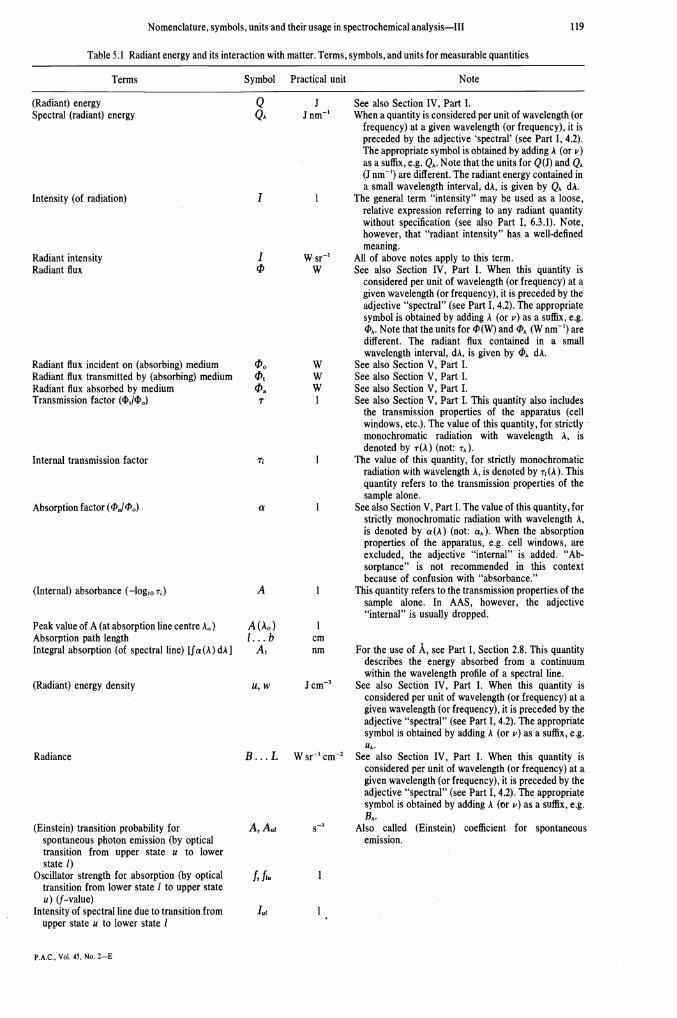
Table 5.1 Radiant energy and its interaction with matter. Terms, symbols, and units for measurable quantities
Terms Symbol Practical unit Note
Q J See also Section IV, Part I.Q A Jnm' When a quantity is considered per unit of wavelength (or
frequency) at a given wavelength (or frequency), it ispreceded by the adjective 'spectral' (see Part I, 4.2).The appropriate symbol is obtained by adding A (or v)as a suffix, e.g. QA. Note that the units for Q(J) and QA(J nm') are different. The radiant energy contained ina small wavelength interval, dA, is given by QA dA.I 1 The general term "intensity" may be used as a loose,relative expression referring to any radiant quantitywithout specification (see also Part I, 6.3.1). Note,however, that "radiant intensity" has a well-definedmeaning.
I W sr1 All of above notes apply to this term.'I, W See also Section IV, Part I. When this quantity is
considered per unit of wavelength (or frequency) at agiven wavelength (or frequency), it is preceded by theadjective "spectral" (see Part I, 4.2). The appropriatesymbol is obtained by adding A (or v) as a suffix, e.g.&. Note that the units for '1(W) and A (Wnm) aredifferent. The radiant flux contained in a smallwavelength interval, dA, is given by dA.
W See also Section V, Part I.cP W See also Section V, Part I.cPa W See also Section V, Part I.T 1 See also Section V, Part I. This quantity also includes
the transmission properties of the apparatus (cellwindows, etc.). The value of this quantity, for strictlymonochromatic radiation with wavelength A, isdenoted by T(A) (not: TA).
1 The value of this quantity, for strictly monochromaticradiation with wavelength A, is denoted by r(A). Thisquantity refers to the transmission properties of thesample alone.
a 1 See also Section V, Part I. The value of this quantity, forstrictly monochromatic radiation with wavelength A,is denoted by a(A) (not: aA). When the absorptionproperties of the apparatus, e.g. cell windows, areexcluded, the adjective "internal" is added. "Ab-sorptance" is not recommended in this contextbecause of confusion with "absorbance."
A 1 This quantity refers to the transmission properties of thesample alone. In AAS, however, the adjective"internal" is usually dropped.
A (A0)1... b
A1 For the use of A, see Part I, Section 2.8. This quantitydescribes the energy absorbed from a continuumwithin the wavelength profile of a spectral line.
u, w J cm3 See also Section IV, Part I. When this quantity isconsidered per unit of wavelength (or frequency) at agiven wavelength (or frequency), it is preceded by theadjective "spectral" (see Part I, 4.2). The appropriatesymbol is obtained by adding A (or v) as a suffix, e.g.UA.
B.. . L W sr1 cm2 See also Section IV, Part I. When this quantity isconsidered per unit of wavelength (or frequency) at agiven wavelength (or frequency), it is preceded by theadjective "spectral" (see Part I, 4.2). The appropriatesymbol is obtained by adding A (or v) as a suffix, e.g.BA.
A,A1 s' Also called (Einstein) coefficient for spontaneousemission.
(Radiant) energySpectral (radiant) energy
Intensity (of radiation)
Radiant intensityRadiant flux
Radiant flux incident on (absorbing) mediumRadiant flux transmitted by (absorbing) mediumRadiant flux absorbed by mediumTransmission factor (4?,I4?)
Internal transmission factor
Absorption factor (ckJI'0)
(Internal) absorbance (—log10 r)
Peak value of A (at absorption line centre A0)Absorption path lengthIntegral absorption (of spectral line) [fa(A) dA]
(Radiant) energy density
Radiance
(Einstein) transition probability forspontaneous photon emission (by opticaltransition from upper state u to lowerstate 1)
Oscillator strength for absorption (by opticaltransition from lower state 1 to upper stateu) (f—value)
Intensity of spectral line due to transition fromupper state u to lower state 1
P.A.C., Vol. 45, No. 2—E
cmnm
f,flu
Ii

120 COMMISSION ON SPECTROCHEMICAL AND OTHER OPTICAL PROCEDURES FOR ANALYSIS
Table 5.1 (Contd)
Terms Symbol Practical unit Note
Wavelength at (atomic) line centre A0 nm For the use of A, see Part I, Section 2.8.Quantum efficiency of fluorescence (number Y, Yq I
of photons re-emitted per second/number ofprimary photons absorbed per second)
Power efficiency of fluorescence (radiant flux Y, 1
re-emitted/primary radiant flux absorbed)Total quantum efficiency of fluorescence [= Y Yt 1
for the case when the upper level of thefluorescence transition is populated directlyor indirectly (by 2-step process) by absorp-tion of more than I spectral line].
Half-intensity width (full width at half peak ÔÀ nm The term half-width is sometimes used instead ofheight of a spectral line profile) half-intensity width, but may readily be misunder-
stood as half of the full width. For the use of A, seePart I, Section 2.8.
Doppler half-intensity width (of spectral 5AD nm The term half-width is sometimes used instead ofline due to Doppler broadening) half-intensity width, but may readily be misunder-
stood as half of the full width. For the use of A, seePart I, Section 2.8.
Collisional half-intensity width (of spectral 5A nm The term half-width is sometimes used instead ofline due to collisional broadening) half-intensity width, but may readily be misunder-
stood as half of the full width. For the use of A, see
a-parameter [\/i(&tc/ôAD)] a IPart I, Section 2.8.
Also called: line-broadening parameter or dampingconstant. In this definition of a, the natural line-broadening is disregarded. When natural line-broadening is important, it should be included in thenumerator.
Collisions of fluorescing atoms with other atoms ormolecules are said to quench the fluorescence when theydestroy the state of excitation brought about by absorptionof the primary photons. The number of secondary photonswill then be smaller than the number of primary photonsabsorbed. The extent of quenching is determined by thecompetition between the rates of radiative and collisionalde-excitation of the excited atoms, and quantitativelyexpressed by the efficiency of fluorescence (for definitionsee Table 5.1).
5.2. Terms, symbols, and units for measurable quantitiesTable 5.1 presents terms with their symbols and
practical units for some measurable quantities belongingto this Section. Section IV of Part I lists additional terms.Although practical units generally conform, alternativesymbols that differ from those in Part I are occasionallyrecommended.
6. TERMS, SYMBOLS, AND UNITS RELATING TO
THE GASEOUS STATE OF MA11ER
Analytical flame spectroscopy and similar techniquesare based on the interaction of radiation with the analyte.The strength of this interaction depends on the propertiesand state of the analyte in the vapour phase. In thefollowing, we restrict ourselves to this phase (thetransformation of the analyte from the condensed phaseinto the vapour "phase has already been discussed inSection 3.1). A few descriptive terms will be mentioned,followed by a list of terms for measurable quantities withtheir symbols and practical units.
6.1. Descriptive terms concerning the gaseous state ofmatter
In (thermodynamic) equilibrium, the state of a system isgenerally described by the thermodynamic or absolutetemperature (Table 2.1) which occurs as a univeralparameter in the distribution laws. These laws determine
the state of excitation, ionization, and dissociation inwhich the analyte is found in the gaseous phase.Note: The Maxwell—Boltzmann law describes the distributionover the translational and internal energies of the particles.Planck's law describes the distribution of the radiant energy overthe spectrum. Saha's law describes the distribution of the particlesover their different states of ionization, while the mass action lawdetermines the fraction of element that is bound in molecular form.
In atomic spectroscopy, the term "dissociation" usuallyrefers to the splitting of a free molecule into fragments,one or more of which is a free neutral analyte atom, and isthus important in connection with the atomization (seeSection 3.1.1.1).
Strictly speaking, thermodynamic equilibrium impliesthat the spectral energy density obeys Planck's law (seeNote in Section 3.2). In flames of analytical interest, thiscondition is not met except at the centre of stronglyself-absorbed resonance lines. However, radiative proces-ses usually play a minor part in the population of theexcited states, at least if the flame is not irradiated by anexternal light beam. The distribution of the particles overtheir various energy states and over the various forms inwhich they may occur (as neutral or ionized atoms, or asmolecules) is then hardly affected by deviation fromPlanck's law. The system is then said to be in thermalequilibrium. This implies that there is chemical equilib-rium between all chemical species (including ions andelectrons) in the system, as well as physical equilibriumfor the translational and internal energies of the particles.However, there need then be no radiative equilibrium.
Note: In the absence of radiative equilibrium, it is still meaningfulto define thermal radiation as it has been done in Section 5.1.1.
6.2. Terms, symbols, and units for measurable quantitiesMany of the terms listed in Table 6.1 are also found in
Part I, Section 7.3, where the terms are discussed in moredetail.

Nomenclature, symbols, units and their usage in spectrochemical analysis—Ill 121
Table 6.1. Properties and state of matte r. Terms, symbols, and units for measurable quantities
Terms Symbol Practical unit Note
Excitation energy J. . . eV
(Number) density of free ions (M)
(Number) density of free electrons(Number) denisty of molecules MXTotal (number) density of element
M(nM+nM++nMx)Ionization constant (n1njn in
equilibrium at T)Dissociationconstant (flMflX/flMX in
equilibrium at T)
See Part 1,7.4. The electron volt (eV), as a practical unitfor energy on an atomic scale, is still in common use.When several excited states are to be distinguished,the excitation energy of state q may be written as Eq.When several kinds of particles are also considered,the excitation energy of state q of a particle X may bewritten as Eq(X).
See Part I, 7.4.See Part I, 7.4.See Part I, 7.4.This is the minimum energy required to dissociate one
molecule of XY at zero K in perfect gas state. Thefragments must be specified when the moleculecontains more than 2 atoms. The molar dissociationenergy refers to one mole of substance XY. Inchemistry, the symbol D, is customarily used.
VM denotes an atom; [M] denotes number density of M in
the flame (see also Table 2.1)./3d . . a 1 M denotes an atom; [M] denotes number density of M in
the flame (see also Table 2.1).gq 1 When several kinds of particles are considered, the
symbol may be extended by adding the chemicalsymbol X as in: gq(X).
g0 1 When several kinds of particles are considered, thesymbol may be extended by adding the chemicalsymbol X as in: g0(X).
Z, Q 1 When several kinds of particles are considered, thesymbol may be extended by adding the chemicalsymbol X as in: Z(X).
flq cm3 When several kinds of particles are considered, thesymbol may be extended by adding the chemicalsymbol X as in: flq(X).
n0 cm3 When several kinds of particles are considered, thesymbol may be extended by adding the chemicalsymbol X as in: nq(X).
7. INDEX OF TERMSt
absolute temperature (Table 2.1, Section 6.1)absorption (Section 5.1.2)accuracy (Section 4.3.2)additive (Section 4.1)aerosol (Section 3.1.1.1)analyte (Section 4.1)(analyte) addition technique (Sections 4.2, 4.4.3)(analyte) signal (Section 4.1)analytical curve (Section 4.1)analytical-curve technique (Section 4.2)analytical result (Section 4.1)angular nebulizer (Section 3.1.1.2)anion effect (Section 4.4.2.2)
anti-Stokes fluorescence (Section 5.1.3)apparent concentration (Section 4.4.1)aspiration (Section 3.1.1.1)atomic fluorescence (Section 5.1.3)atomic line (Section 5.1.3)atomization (Section 3.1.1.1)atomizer (Section 3.1.1.1)
background corrector (Section 3.5)baseline technique (Section 4.4.3)bias (Section 4.3.2)blank background (Section 4.1)blank measure (Section 4.1)blank scatter (Section 4.3.1)blank solution (Section 5.1)
— bracketing technique (Section 4.2)buffer-addition technique (Section 4.4.3)Bunsen-burner (Section 3.1.1.3)burner (Section 3.1.1.3)
VJ...eV
VJ...eV
VexcE0, Evion, vi
Ed1, D0, D,
Vd
Excitation potentialIonization energyIonization potentialDissociation energy
Dissociation potentialDegree of ionization [Mi)]
Degree of dissociation I + [MX])]
Statistical weight of particle in state q
Statistical weight of particle in ground state
Partition function
(Number) density of particles in state q(see also Table 2.1)
(Number) density of particles in ground state
(Number) density of free atoms (M) nat, fla, nMn (M), [M]n10, n, nMn(M), [M]
ne, [e]flMX, [MXIflt, (nt)M,
[MItK1, K(T)
Kd, Kd(T)
cm3
cm3
cm3cm3cm3
cm3
cm3
tQuantitative terms and some qualitative terms that are definedonly in the tables are not included in this index. They are easilyfound in the tables under the appropriate headings.
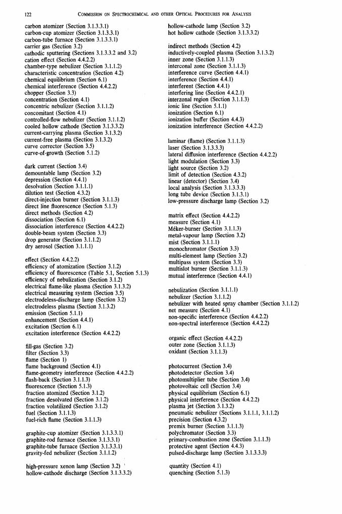
122 COMMISSION ON SPECTROCHEMICAL AND OTHER OPTICAL PROCEDURES FOR ANALYSIS
carbon atomizer (Section 3.1.3.3.1)carbon-cup atomizer (Section 3.1.3.3. 1)carbon-tube furnace (Section 3.1.3.3.1)carrier gas (Section 3.2)cathodic sputtering (Sections 3.1.3.3.2 and 3.2)cation effect (Section 4.4.2.2)chamber-type nebulizer (Section 3.1.1.2)characteristic concentration (Section 4.2)chemical equilibrium (Section 6.1)chemical interference (Section 4.4.2.2)chopper (Section 3.3)concentration (Section 4.1)concentric nebulizer (Section 3.1.1.2)concomitant (Section 4.1)controlled-flow nebulizer (Section 3.1.1.2)cooled hollow cathode (Section 3.1.3.3.2)current-carrying plasma (Section 3.1.3.2)current-free plasma (Section 3.1.3.2)curve corrector (Section 3.5)curve-of-growth (Section 5.1.2)
dark current (Section 3.4)demountable lamp (Section 3.2)depression (Section 4.4.1)desolvation (Section 3.1.1.1)dilution test (Section 4.3.2)direct-injection burner (Section 3.1.1.3)direct line fluorescence (Section 5.1.3)direct methods (Section 4.2)dissociation (Section 6.1)dissociation interference (Section 4.4.2.2)double-beam system (Section 3.3)drop generator (Section 3.1.1.2)dry aerosol (Section 3.1.1.1)
effect (Section 4.4.2.2)efficiency of atomization (Section 3.1.2)efficiency of fluorescence (Table 5.1, Section 5.1.3)efficiency of nebulization (Section 3.1.2)electrical flame-like plasma (Section 3.1.3.2)electrical measuring system (Section 3.5)electrodeless-discharge lamp (Section 3.2)electrodeless plasma (Section 3.1.3.2)emission (Section 5.1.1)enhancement (Section 4.4.1)excitation (Section 6.1)excitation interference (Section 4.4.2.2)
fill-gas (Section 3.2)filter (Section 3.3)flame (Section 1)flame background (Section 4.1)flame-geometry interference (Section 4.4.2.2)flash-back (Section 3.1.1.3)fluorescence (Section 5.1.3)fraction atomized (Section 3.1.2)fraction desolvated (Section 3.1.2)fraction volatilized (Section 3.1.2)fuel (Section 3.1.1.3)fuel-rich flame (Section 3.1.1.3)
graphite-cup atomizer (Section 3.1.3.3.1)graphite-rod furnace (Section 3.1.3.3.1)graphite-tube furnace (Section 3.1.3.3.1)gravity-fed nebulizer (Section 3.1.1.2)
high-pressure xenon lamp (Section 3.2)hollow-cathode discharge (Section 3.1.3.3.2)
hollow-cathode lamp (Section 3.2)hot hollow cathode (Section 3.1.3.3.2)
indirect methods (Section 4.2)inductively-coupled plasma (Section 3.1.3.2)inner zone (Section 3.1.1.3)interconal zone (Section 3.1.1.3)interference curve (Section 4.4.1)interference (Section 4.4.1)interferent (Section 4.4.1)interfering line (Section 4.4.2.1)interzonal region (Section 3.1.1.3)ionic line (Section 5.1.1)ionization (Section 6.1)ionization buffer (Section 4.4.3)ionization interference (Section 4.4.2.2)
laminar (flame) (Section 3.1.1.3)laser (Section 3.1.3.3.3)lateral diffusion interference (Section 4.4.2.2)light modulation (Section 3.3)light source (Section 3.2)limit of detection (Section 4.3.2)linear (detector) (Section 3.4)local analysis (Section 3.1.3.3.3)long tube device (Section 3.1.3.1)low-pressure discharge lamp (Section 3.2)
matrix effect (Section 4.4.2.2)measure (Section 4.1)Méker-burner (Section 3.1.1.3)metal-vapour lamp (Section 3.2)mist (Section 3.1.1.1)monochromator (Section 3.3)multi-element lamp (Section 3.2)multipass system (Section 3.3)multislot burner (Section 3.1.1.3)mutual interference (Section 4.4.1)
nebulization (Section 3.1.1.1)nebulizer (Section 3.1.1.2)nebulizer with heated spray chamber (Section 3.1.1.2)net measure (Section 4.1)non-specific interference (Section 4.4.2.2)non-spectral interference (Section 4.4.2.2)
organic effect (Section 4.4.2.2)outer zone (Section 3.1.1.3)oxidant (Section 3.1.1.3)
photocurrent (Section 3.4)photodetector (Section 3.4)photomultiplier tube (Section 3.4)photovoltaic cell (Section 3.4)physical equilibrium (Section 6.1)physical interference (Section 4.4.2.2)plasma jet (Section 3.1.3.2)pneumatic nebulizer (Sections 3.1.1.1, 3.1.1.2)precision (Section 4.3.2)premix burner (Section 3.1.1.3)polychromator (Section 3.3)primary-combustion zone (Section 3.1.1.3)protective agent (Section 4.4.3)pulsed-discharge lamp (Section 3.1.3.3.3)
quantity (Section 4.1)quenching (Section 5.1.3)
Nomenclature, symbols, units and their usage in spectrochemical analysis—Ill 123
radiative equilibrium (Section 6.1)rate of liquid aspiration (Section 3.1.2)rate of liquid consumption (Section 3.1.2)reading (Section 3.5)recovery test (Section 4.3.2)reference beam (Section 3.3)reference element (Section 4.4.3)reference-element technique (Section 4.4.3)reference solution (Section 5.1)reflux nebulizer (Section 3.1.1.2)relative standard deviation (Section 4.3.2)releaser (Section 4.4.3)resistance-heated device (Section 3.1.3.3.1)resonance fluorescence (Section 5.1.3)resonance line (Section 5.1.1)resonance spectrometer (Section 3.3)response time (Section 3.5)responsivity (Section 3.4)reversal dip (Section 5.1.2)reversed direct-injection burner (Section 3.1.1.3)
sample (Section 4.1)sample beam (Section 3.3)sampling boat (Section 3.1.3.1)sampling cup (Section 3.1.3.1)sampling ioop (Section 3.1.3.1)saturation (plateau) (Section 4.4.3)saturator (Section 4.4.3)scale expansion (Section 3.5)scatter (Section 4.3.1)scattering (Section 5.1.2)sealed lamp (Section 3.2)secondary-combustion zone (Section 3.1.1.3)self-absorption (Section 5.1.2)self-reversal (Section 5.1.2)sensitivity (Section 4.2)separated flame (Section 3.1.1.3)shielded flame (Section 3.1.1.3)simulation technique (Section 4.4.3)single-beam system (Section 3.3)single-electrode plasma (Section 3.1.3.2)single-element lamp (Section 3.2)slot burner (Section 3.1.1.3)solute-volatilization interference (Section 4.4.2.2)
solvent blank (Section 3.5)spatial-distribution interference (Section 4.4.2.2)specific interference (Section 4.4.2.2)spectral band (Section 5.1.1)spectral continuum (Section 5.1.1)(spectral) continuum source (Section 3.2)spectral interference (Section 4.4.2.1)(spectral) line source (Section 3.2)spectral line (Section 5.1.1)spectral response curve (Section 3.4)spectrochemical buffer (Section 4.4.3)spectrometer (Table 1.1, Section 3.3)spectrometry (Table 1.1)spray chamber (Section 3.1.1.2)sprayer (Section 3.1.1.1)standard deviation (Section 4.3.1)stepwise line fluorescence (Section 5.1.3)Stokes fluorescence (Section 5.1.3)suction nebulizer (Section 3.1.1.2)suppressor (Section 4.4.3)suprathermal chemiluminescence (Section 5.1.1)
T-tubes (Section 3.1.3.1)thermal equilibrium (Section 6.1)thermal radiation (Section 5.1.1)(thermodynamic) equilibrium (Section 6.1)thermodynamic temperature (Table 2.1, Section 6.1)three-slot burner (Section 3.1.1.3)time constant (Section 3.5)transport interference (Section 4.4.2.2)tungsten-filament lamp (Section 3.2)turbulent (flame) (Section 3.1.1.3)twin nebulizer (Section 3.1.1.2)
ultrasonic nebulizer (Section 3.1.1.2)
vacuum phototube (Section 3.4)yapour-phase interference (Section 4.4.2.2)variance (Section 4.3.1)volatilization (Section 3.1.1.1)volatilizer (Section 4.4.3)
zero suppression (Section 3.5)