no slide titleca01001129.schoolwires.net/cms/lib7/ca01001129/centr… · ·...
TRANSCRIPT
Chapter 7
Lecture Outline
Copyright (c) The McGraw-Hill Companies, Inc. Permission required for reproduction or display.
7-1
7-2
Bone Tissue
• tissues and organs of the skeletal system
• histology of osseous tissue
• bone development
• physiology of osseous tissue
• bone disorders
7-3
Bone as a Tissue
• osteology – the study of bone
• skeletal system - composed of bones, cartilages, and ligaments– form strong flexible framework of the body
– cartilage – forerunner of most bones• covers many joint surfaces of mature bone
• ligaments – hold bones together at the joints
• tendons – attach muscle to bone
7-4
Functions of the Skeleton
• support – hold the body up, supports muscles, mandible and maxilla support teeth
• protection – brain, spinal cord, heart, lungs
• movement – limb movements, breathing, action of muscle on bone
• electrolyte balance – calcium and phosphate ions
• acid-base balance – buffers blood against excessive pH changes
• blood formation – red bone marrow is the chief producer of blood cells
7-5
Bones and Osseous Tissue
• bone (osseous tissue) - connective tissue with the matrix hardened by calcium phosphate and other minerals
• mineralization or calcification – the hardening process of bone
• individual bones consist of bone tissue, bone marrow, cartilage, adipose tissue, nervous tissue, and fibrous connective tissue
• continually remodels itself and interacts physiologically with all of the other organ systems of the body
• permeated with nerves and blood vessels, which attests to its sensitivity and metabolic activity
7-6
Shapes of Bones
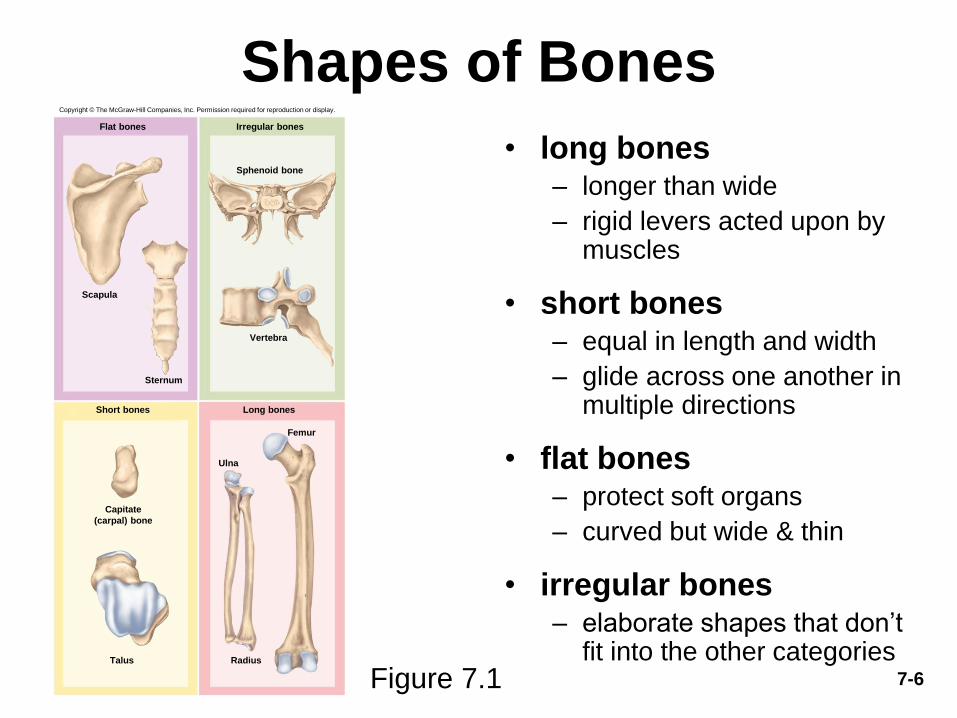
• long bones– longer than wide
– rigid levers acted upon by muscles
• short bones – equal in length and width
– glide across one another in multiple directions
• flat bones– protect soft organs
– curved but wide & thin
• irregular bones– elaborate shapes that don’t
fit into the other categoriesFigure 7.1
Copyright © The McGraw-Hill Companies, Inc. Permission required for reproduction or display.
Femur
Scapula
Sternum
Sphenoid bone
Radius
Ulna
Irregular bonesFlat bones
Short bones Long bones
Vertebra
Capitate
(carpal) bone
Talus
7-7
General Features of Bones
• compact (dense) bone – outer shell of long bone
• diaphysis (shaft) - cylinder of compact bone to provide leverage
• medullary cavity (marrow cavity) - space in the diaphysis of a long bone that contains bone marrow
• epiphyses - enlarged ends of a long bone
– enlarged to strengthen joint and attach ligaments and tendons
• spongy (cancellous) bone covered by more durable compact bone
– skeleton about three-fourths compact and one-fourth spongy bone by weight
– spongy bone found in ends of long bones, and the middle of nearly all others
• articular cartilage – a layer of hyaline cartilage that covers the joint surface where one bone meets another
– allows joint to move more freely and relatively friction free
• nutrient foramina – minute holes in the bone surface that allows blood vessels to penetrate
7-8
General Features of Bones• periosteum – external sheath that covers bone except where there is
articular cartilage
– outer fibrous layer of collagen
• some outer fibers continuous with the tendons that attach muscle to bone
• perforating (Sharpey’s) fibers – other outer fibers that penetrate into the bone matrix
• strong attachment and continuity from muscle to tendon to bone
– inner osteogenic layer of bone forming cells
• important to growth of bone and healing of fractures
• endosteum – thin layer of reticular connective tissue lining marrow cavity
– has cells that dissolve osseous tissue and others that deposit it
• epiphyseal plate (growth plate) – area of hyaline cartilage that separates the marrow spaces of the epiphysis and diaphysis
– enables growth in length
– epiphyseal line – in adults, a bony scar that marks where growth plate used to be
7-9
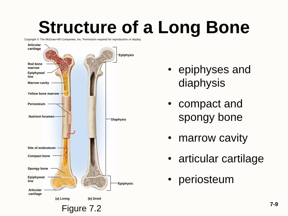
Structure of a Long Bone
• epiphyses and
diaphysis
• compact and
spongy bone
• marrow cavity
• articular cartilage
• periosteum
Figure 7.2
Copyright © The McGraw-Hill Companies, Inc. Permission required for reproduction or display.
(a) Living (b) Dried
Marrow cavity
Periosteum
Nutrient foramen
Site of endosteum
Compact bone
Spongy bone
Epiphysis
Epiphysis
Diaphysis
Articular
cartilage
Epiphyseal
line
Red bone
marrow
Yellow bone marrow
Epiphyseal
line
Articular
cartilage

7-10
Structure of a Flat Bone• sandwich-like
construction
• two layers of compact
bone enclosing a
middle layer of spongy
bone
– both surfaces of flat
bone covered with
periosteum
• diploe – spongy layer
in the cranium
– absorbs shock
– marrow spaces lined
with endosteum
Copyright © The McGraw-Hill Companies, Inc. Permission required for reproduction or display.
Suture
Outer compact
bone
Spongy bone
(diploe)
Inner compact
bone
Trabeculae
Figure 7.3
7-11
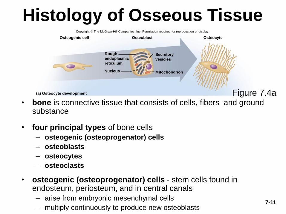
Histology of Osseous Tissue
• bone is connective tissue that consists of cells, fibers and ground substance
• four principal types of bone cells
– osteogenic (osteoprogenator) cells
– osteoblasts
– osteocytes
– osteoclasts
• osteogenic (osteoprogenator) cells - stem cells found in endosteum, periosteum, and in central canals
– arise from embryonic mesenchymal cells
– multiply continuously to produce new osteoblasts
Osteogenic cell Osteoblast Osteocyte
(a) Osteocyte development
Nucleus Mitochondrion
Rough
endoplasmic
reticulum
Secretory
vesicles
Copyright © The McGraw-Hill Companies, Inc. Permission required for reproduction or display.
Figure 7.4a
7-12
Histology of Osseous Tissue• osteoblasts – bone forming cells
– line up as single layer of cells under endosteum and periosteum
– are nonmitotic
– synthesize soft organic matter of matrix which then hardens by mineral deposition
– stress and fractures stimulate osteogenic cells to multiply more rapidly and increase number of osteocytes to reinforce or rebuild bone
– secrete osteocalcin – thought to be the structural protein of bone
• stimulates insulin secretion of pancreas
• increases insulin sensitivity in adipocytes which limit the growth of adipose tissue
• osteocytes – former osteoblasts that have become trapped in the matrix they have deposited– lacunae – tiny cavities where osteocytes reside
– canaliculi – little channels that connect lacunae
– cytoplasmic processes reach into canaliculi
– some osteocytes reabsorb bone matrix while others deposit it
– contribute to homeostatic mechanism of bone density and calcium and phosphate ions
– when stressed, produce biochemical signals that regulate bone remodeling
7-13
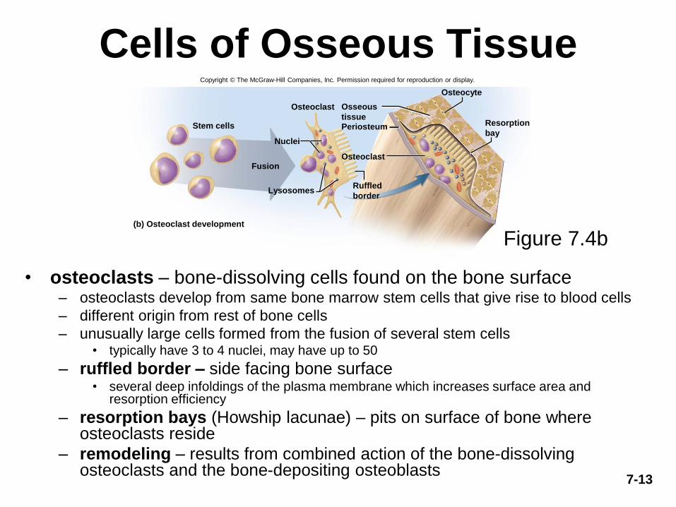
Cells of Osseous Tissue
• osteoclasts – bone-dissolving cells found on the bone surface– osteoclasts develop from same bone marrow stem cells that give rise to blood cells
– different origin from rest of bone cells
– unusually large cells formed from the fusion of several stem cells• typically have 3 to 4 nuclei, may have up to 50
– ruffled border – side facing bone surface• several deep infoldings of the plasma membrane which increases surface area and
resorption efficiency
– resorption bays (Howship lacunae) – pits on surface of bone where osteoclasts reside
– remodeling – results from combined action of the bone-dissolving osteoclasts and the bone-depositing osteoblasts
Copyright © The McGraw-Hill Companies, Inc. Permission required for reproduction or display.
Osteocyte
Stem cells
Osteoclast
Fusion
Periosteum
(b) Osteoclast development
Osteoclast
Nuclei
Lysosomes
Osseous
tissueResorption
bay
Ruffled
border
Figure 7.4b
7-14
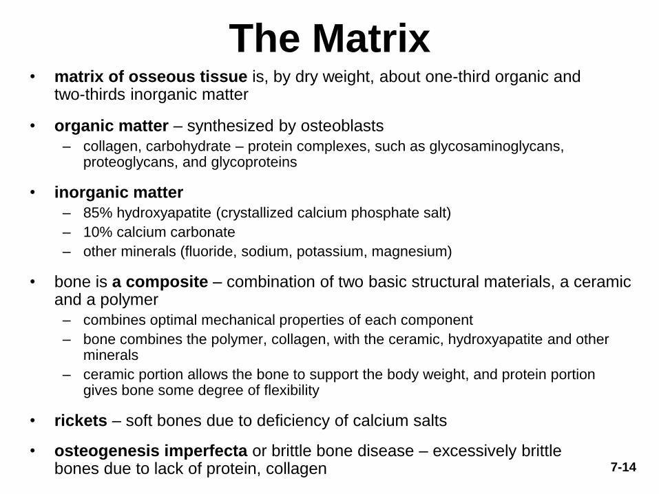
The Matrix• matrix of osseous tissue is, by dry weight, about one-third organic and
two-thirds inorganic matter
• organic matter – synthesized by osteoblasts
– collagen, carbohydrate – protein complexes, such as glycosaminoglycans, proteoglycans, and glycoproteins
• inorganic matter
– 85% hydroxyapatite (crystallized calcium phosphate salt)
– 10% calcium carbonate
– other minerals (fluoride, sodium, potassium, magnesium)
• bone is a composite – combination of two basic structural materials, a ceramic and a polymer
– combines optimal mechanical properties of each component
– bone combines the polymer, collagen, with the ceramic, hydroxyapatite and other minerals
– ceramic portion allows the bone to support the body weight, and protein portion gives bone some degree of flexibility
• rickets – soft bones due to deficiency of calcium salts
• osteogenesis imperfecta or brittle bone disease – excessively brittle bones due to lack of protein, collagen
7-15
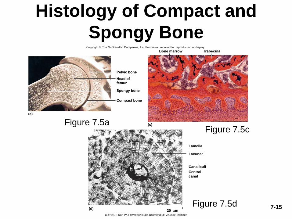
Histology of Compact and
Spongy Bone
Figure 7.5aFigure 7.5c
Figure 7.5d
Spongy bone
Compact bone
Head offemur
Pelvic bone
(a)
Bone marrow Trabecula
(c)
Lamella
Lacunae
(d)
Canaliculi
Central
canal
20 m
Copyright © The McGraw-Hill Companies, Inc. Permission required for reproduction or display.
a,c: © Dr. Don W. Fawcett/Visuals Unlimited; d: Visuals Unlimited
7-16
Compact Bone• osteon (haversian system) – the basic structural unit of compact bone
– formed by a central canal and its concentric lamella connected to each other by canaliculi
– a cylinder of tissue around a central canal
– perforating (Volkmann) canals are transverse or diagonal passages along the length of the osteon
– collagen fibers “corkscrew” down the matrix of the lamella giving it a helical arrangement
– helices coil in one direction in one lamella and in the opposite direction in the next lamella for added strength
– blood flow - skeleton receives about half a liter of blood per minute
– nutrient foramina – on the surface of bone tissue that allow blood vessels and nerves to enter the bone
• open into the perforating canals that cross the matrix and feed into the central canals
• innermost osteocytes near central canal receive nutrients and pass them along through their gap junction to neighboring osteocytes
• they also receive wastes from their neighbors and transfer them to the central canal maintaining a two-way flow of nutrients and waste
– not all of the matrix is organized into osteons
– circumferential lamellae - inner and outer boundaries of dense bone
• – run parallel to bone surface
– interstitial lamellae – remains of old osteons that broke down as bone grew and remodeled itself
7-17
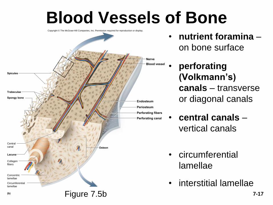
Blood Vessels of Bone• nutrient foramina –
on bone surface
• perforating
(Volkmann’s)
canals – transverse
or diagonal canals
• central canals –
vertical canals
• circumferential
lamellae
• interstitial lamellaeFigure 7.5b
Periosteum
Endosteum
Perforating fibers
Perforating canal
Osteon
Lacuna
Nerve
Blood vessel
(b)
Spongy bone
Spicules
Central
canal
Collagen
fibers
Concentric
lamellae
Circumferential
lamellae
Trabeculae
Copyright © The McGraw-Hill Companies, Inc. Permission required for reproduction or display.
7-18
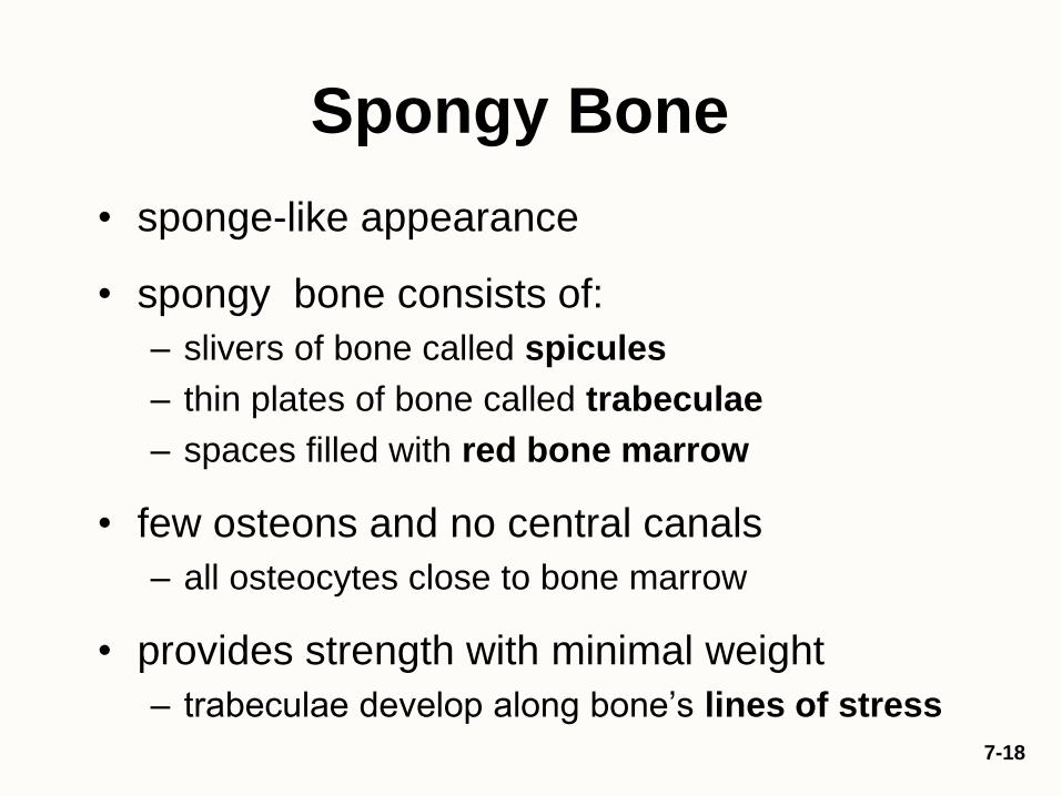
Spongy Bone
• sponge-like appearance
• spongy bone consists of:
– slivers of bone called spicules
– thin plates of bone called trabeculae
– spaces filled with red bone marrow
• few osteons and no central canals
– all osteocytes close to bone marrow
• provides strength with minimal weight
– trabeculae develop along bone’s lines of stress

7-19
Design of Spongy Bone
Figure 7.6
Copyright © The McGraw-Hill Companies, Inc. Permission required for reproduction or display.
Greater trochanter
Compact bone
Head
Lines of stress
Shaft (diaphysis)
Trabeculae of
spongy bone
© Robert Calentine/Visuals Unlimited
7-20
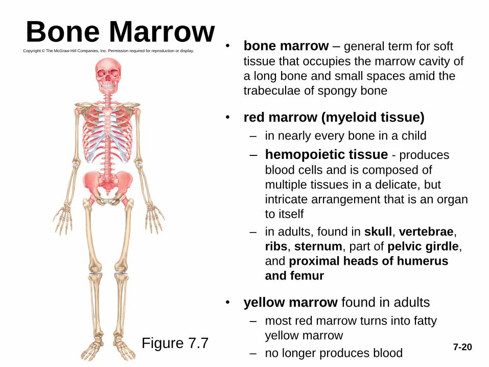
Bone Marrow• bone marrow – general term for soft
tissue that occupies the marrow cavity of
a long bone and small spaces amid the
trabeculae of spongy bone
• red marrow (myeloid tissue)
– in nearly every bone in a child
– hemopoietic tissue - produces
blood cells and is composed of
multiple tissues in a delicate, but
intricate arrangement that is an organ
to itself
– in adults, found in skull, vertebrae,
ribs, sternum, part of pelvic girdle,
and proximal heads of humerus
and femur
• yellow marrow found in adults
– most red marrow turns into fatty
yellow marrow
– no longer produces blood
Copyright © The McGraw-Hill Companies, Inc. Permission required for reproduction or display.
Figure 7.7

7-21
Bone Development
• ossification or osteogenesis – the formation of bone
• in the human fetus and infant, bone develops by two methods:
– intramembranous ossification
– endochondral ossification
7-22
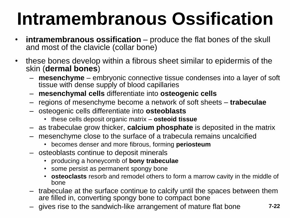
Intramembranous Ossification• intramembranous ossification – produce the flat bones of the skull
and most of the clavicle (collar bone)
• these bones develop within a fibrous sheet similar to epidermis of the skin (dermal bones)– mesenchyme – embryonic connective tissue condenses into a layer of soft
tissue with dense supply of blood capillaries
– mesenchymal cells differentiate into osteogenic cells
– regions of mesenchyme become a network of soft sheets – trabeculae
– osteogenic cells differentiate into osteoblasts• these cells deposit organic matrix – osteoid tissue
– as trabeculae grow thicker, calcium phosphate is deposited in the matrix
– mesenchyme close to the surface of a trabecula remains uncalcified• becomes denser and more fibrous, forming periosteum
– osteoblasts continue to deposit minerals• producing a honeycomb of bony trabeculae
• some persist as permanent spongy bone
• osteoclasts resorb and remodel others to form a marrow cavity in the middle of bone
– trabeculae at the surface continue to calcify until the spaces between them are filled in, converting spongy bone to compact bone
– gives rise to the sandwich-like arrangement of mature flat bone
7-23
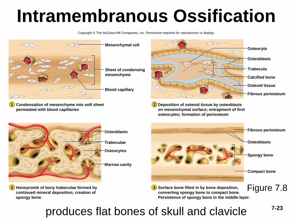
Intramembranous Ossification
produces flat bones of skull and clavicle
Copyright © The McGraw-Hill Companies, Inc. Permission required for reproduction or display.
Mesenchymal cell
Blood capillaryOsteoid tissue
Osteocyte
Calcified bone
Osteoblasts
Fibrous periosteum
Osteocytes
Trabeculae
Osteoblasts
Osteoblasts
Spongy bone
Compact bone
Marrow cavity
Fibrous periosteum
2
3 4
1
Sheet of condensing
mesenchyme
Condensation of mesenchyme into soft sheet
permeated with blood capillaries
Honeycomb of bony trabeculae formed by
continued mineral deposition; creation of
spongy bone
Trabecula
Deposition of osteoid tissue by osteoblasts
on mesenchymal surface; entrapment of first
osteocytes; formation of periosteum
Surface bone filled in by bone deposition,
converting spongy bone to compact bone.
Persistence of spongy bone in the middle layer.
Figure 7.8
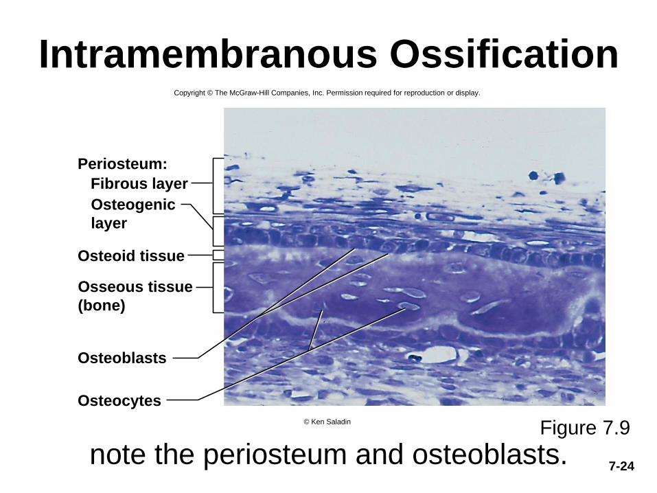
7-24
Intramembranous Ossification
note the periosteum and osteoblasts.Figure 7.9
Copyright © The McGraw-Hill Companies, Inc. Permission required for reproduction or display.
Periosteum:
Fibrous layer
Osteoid tissue
Osteoblasts
Osteocytes
Osteogenic
layer
Osseous tissue
(bone)
© Ken Saladin
7-25
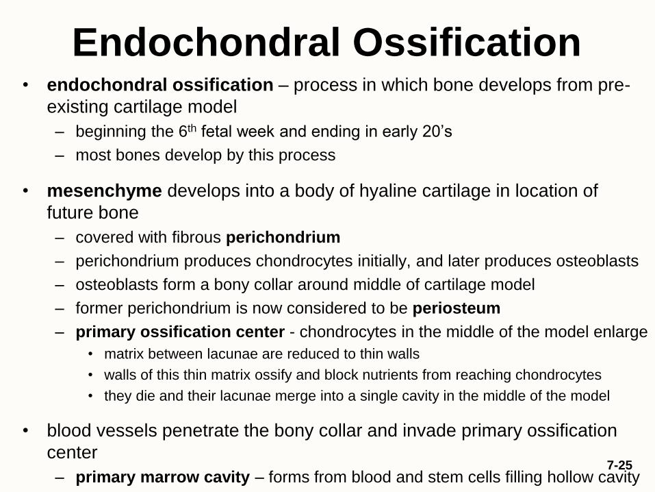
Endochondral Ossification• endochondral ossification – process in which bone develops from pre-
existing cartilage model
– beginning the 6th fetal week and ending in early 20’s
– most bones develop by this process
• mesenchyme develops into a body of hyaline cartilage in location of
future bone
– covered with fibrous perichondrium
– perichondrium produces chondrocytes initially, and later produces osteoblasts
– osteoblasts form a bony collar around middle of cartilage model
– former perichondrium is now considered to be periosteum
– primary ossification center - chondrocytes in the middle of the model enlarge
• matrix between lacunae are reduced to thin walls
• walls of this thin matrix ossify and block nutrients from reaching chondrocytes
• they die and their lacunae merge into a single cavity in the middle of the model
• blood vessels penetrate the bony collar and invade primary ossification
center
– primary marrow cavity – forms from blood and stem cells filling hollow cavity

7-26
Primary Ossification Center and
Primary Marrow Cavity
Figure 7.10 (2-3)
Bony collar
Periosteum
2
Enlarging
chondrocytes
Primary
ossification
center
Formation of
primary
ossification center,
bony collar, and
periosteum
Copyright © The McGraw-Hill Companies, Inc. Permission required for reproduction or display.
Vascular invasion,
formation of primary
marrow cavity, and
appearance of
secondary
ossification center
3
Secondary
ossification
center
Primary
marrow
cavity
7-27
Endochondral Ossification
• blood vessels penetrate the bony collar and invade primary
ossification center
– primary marrow cavity – forms from blood and stem cells filling hollow
cavity
– stem cells give rise to osteoblasts and osteoclasts
– osteoblasts line cavity and deposit osteoid tissue and calcify it
• forming temporary network of trabeculae
– wave of cartilage death progresses toward the ends
• osteoclasts follow the wave dissolving the cartilage remnants enlarging the
marrow cavity
– metaphysis – region of transition from cartilage to bone at each end of
primary marrow cavity
• secondary ossification center – created by chondrocyte enlargement
and death in the epiphyses
– become hollowed out by the same process generating a secondary
marrow cavity in epiphyses
• cavity expands outward from the center in all directions
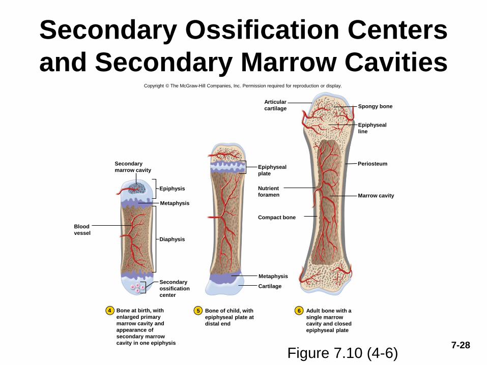
7-28
Secondary Ossification Centers
and Secondary Marrow Cavities
Figure 7.10 (4-6)
Copyright © The McGraw-Hill Companies, Inc. Permission required for reproduction or display.
Metaphysis
Diaphysis
Epiphysis
Cartilage
Metaphysis
Spongy bone
Marrow cavity
Compact bone
Periosteum
4 5 6
Secondary
marrow cavity
Blood
vessel
Secondary
ossification
center
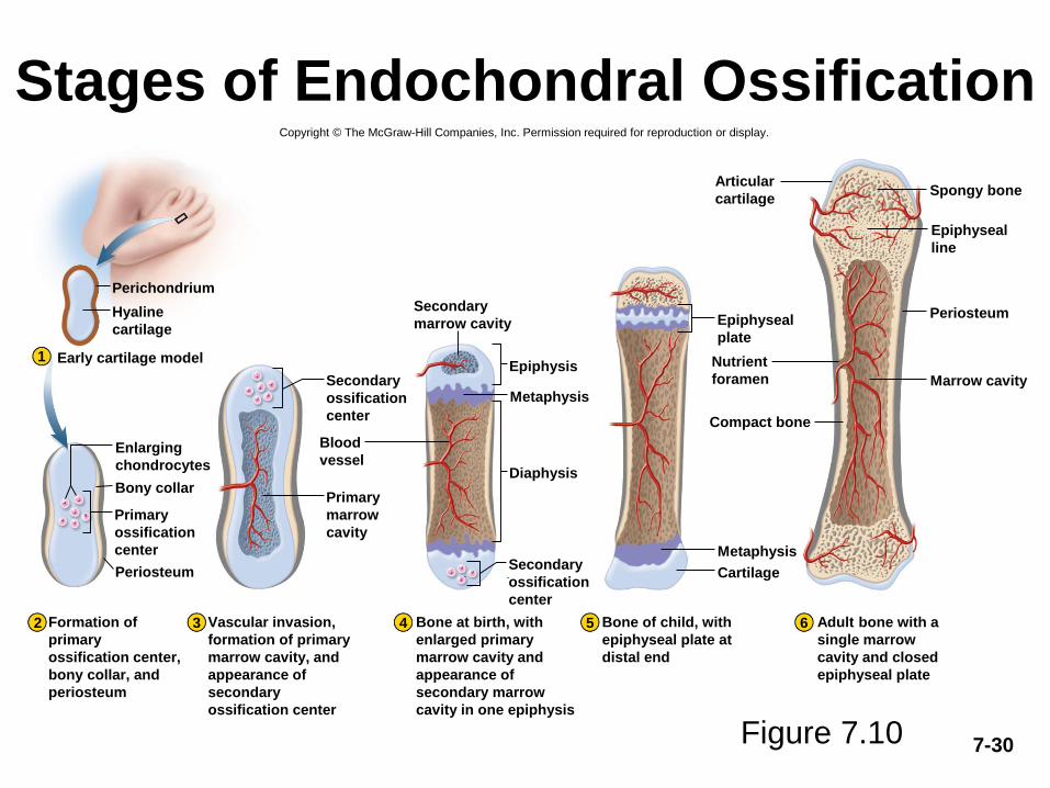
Bone at birth, with
enlarged primary
marrow cavity and
appearance of
secondary marrow
cavity in one epiphysis
Bone of child, with
epiphyseal plate at
distal end
Adult bone with a
single marrow
cavity and closed
epiphyseal plate
Epiphyseal
plate
Articular
cartilage
Epiphyseal
line
Nutrient
foramen
7-29
Endochondral Ossification
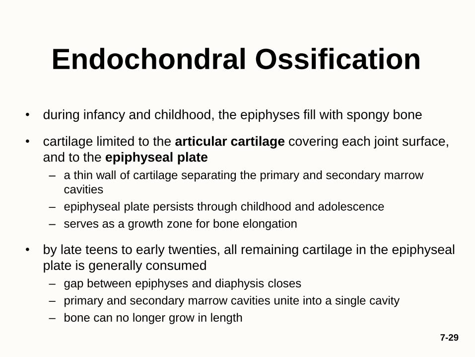
• during infancy and childhood, the epiphyses fill with spongy bone
• cartilage limited to the articular cartilage covering each joint surface,
and to the epiphyseal plate
– a thin wall of cartilage separating the primary and secondary marrow
cavities
– epiphyseal plate persists through childhood and adolescence
– serves as a growth zone for bone elongation
• by late teens to early twenties, all remaining cartilage in the epiphyseal
plate is generally consumed
– gap between epiphyses and diaphysis closes
– primary and secondary marrow cavities unite into a single cavity
– bone can no longer grow in length
7-30
Stages of Endochondral Ossification
Figure 7.10
Copyright © The McGraw-Hill Companies, Inc. Permission required for reproduction or display.
Perichondrium
Bony collar
Periosteum
Metaphysis
Diaphysis
Epiphysis
Cartilage
Metaphysis
Spongy bone
Marrow cavity
Compact bone
Periosteum
2
Early cartilage model1
3 4 5 6
Hyaline
cartilage
Enlarging
chondrocytes
Primary
ossification
center
Secondary
ossification
center
Blood
vessel
Primary
marrow
cavity
Secondary
marrow cavity
Secondary
ossification
center
Epiphyseal
plate
Nutrient
foramen
Articular
cartilage
Epiphyseal
line
Adult bone with a
single marrow
cavity and closed
epiphyseal plate
Bone of child, with
epiphyseal plate at
distal end
Bone at birth, with
enlarged primary
marrow cavity and
appearance of
secondary marrow
cavity in one epiphysis
Vascular invasion,
formation of primary
marrow cavity, and
appearance of
secondary
ossification center
Formation of
primary
ossification center,
bony collar, and
periosteum
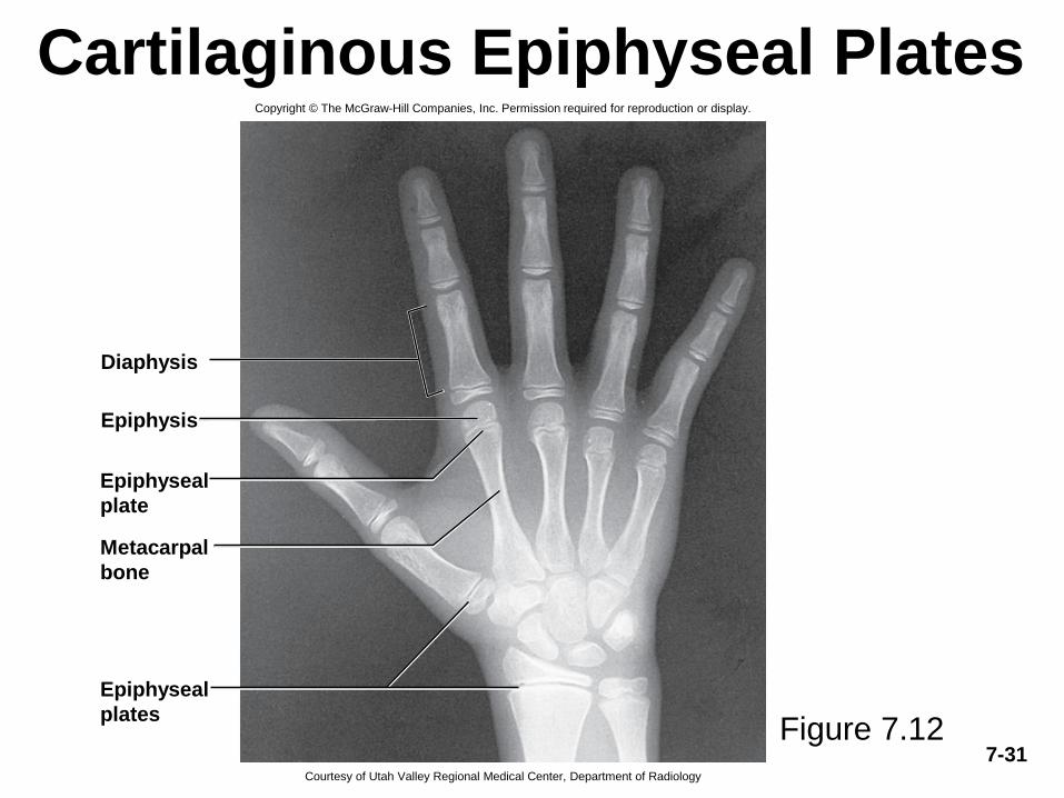
7-31
Cartilaginous Epiphyseal PlatesCopyright © The McGraw-Hill Companies, Inc. Permission required for reproduction or display.
Diaphysis
Epiphysis
Epiphyseal
plate
Metacarpal
bone
Epiphyseal
plates
Courtesy of Utah Valley Regional Medical Center, Department of Radiology
Figure 7.12
7-32
Bone Growth and Remodeling
• ossification continues throughout life with the
growth and remodeling of bones
• bones grow in two directions: length and width
• bone elongation
– epiphyseal plate – a region of transition from
cartilage to bone
• functions as growth zone where the bones elongate
• consists of typical hyaline cartilage in the middle
• with a transition zone on each side where cartilage is being
replaced by bone
• metaphysis is the zone of transition facing the marrow cavity
7-33
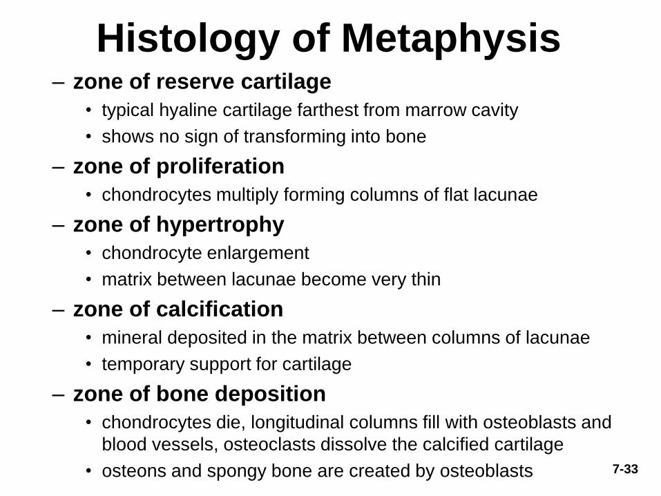
Histology of Metaphysis– zone of reserve cartilage
• typical hyaline cartilage farthest from marrow cavity
• shows no sign of transforming into bone
– zone of proliferation
• chondrocytes multiply forming columns of flat lacunae
– zone of hypertrophy
• chondrocyte enlargement
• matrix between lacunae become very thin
– zone of calcification
• mineral deposited in the matrix between columns of lacunae
• temporary support for cartilage
– zone of bone deposition
• chondrocytes die, longitudinal columns fill with osteoblasts and
blood vessels, osteoclasts dissolve the calcified cartilage
• osteons and spongy bone are created by osteoblasts
7-34
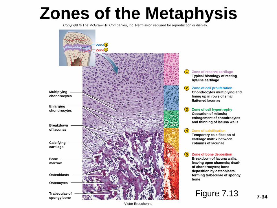
Zones of the Metaphysis
Figure 7.13
Osteoblasts
Osteocytes
1
2
3
4
5
1
5
Zone
Zone
Multiplying
chondrocytes
Enlarging
chondrocytes
Breakdown
of lacunae
Calcifying
cartilage
Bone
marrow
Trabeculae of
spongy bone
Zone of reserve cartilage
Typical histology of resting
hyaline cartilage
Zone of cell proliferation
Chondrocytes multiplying and
lining up in rows of small
flattened lacunae
Zone of cell hypertrophy
Cessation of mitosis;
enlargement of chondrocytes
and thinning of lacuna walls
Zone of calcification
Temporary calcification of
cartilage matrix between
columns of lacunae
Zone of bone deposition
Breakdown of lacuna walls,
leaving open channels; death
of chondrocytes; bone
deposition by osteoblasts,
forming trabeculae of spongy
bone
Victor Eroschenko
Copyright © The McGraw-Hill Companies, Inc. Permission required for reproduction or display.

7-35
Fetal Skeleton at 12 Weeks
Figure 7.11
Copyright © The McGraw-Hill Companies, Inc. Permission required for reproduction or display.
Humerus
Ulna
Femur
Radius
Mandible
Scapula
Ribs
Pelvis© Biophoto Associates/Photo Researchers, Inc.
Cranial
bones
Vertebrae
7-36
Bone Growth and Remodeling• interstitial growth - bones increase in length
– bone elongation is really a result of cartilage growth within epiphyseal plate
– epiphyses close when cartilage is gone – epiphyseal line
– length-wise growth is finished
• occurs at different ages in different bones
• appositional growth - bones increase in width throughout life
– the deposition of new bone at the surface
– osteoblasts on deep side of periosteum deposit osteoid tissue
• Become trapped as tissue calcifies
– lay down matrix in layers parallel to surface
• forms circumferential lamellae over surface
– osteoclasts of endosteum enlarge marrow cavity
• bone remodeling occurs throughout life - 10% per year
– repairs microfractures, releases minerals into blood, reshapes bones in response to use and disuse
– Wolff’s law of bone - architecture of bone determined by mechanical stresses placed on it and bones adapt to withstand those stresses
• remodeling is a collaborative and precise action of osteoblasts and osteoclasts
• bony processes grow larger in response to mechanical stress
7-37
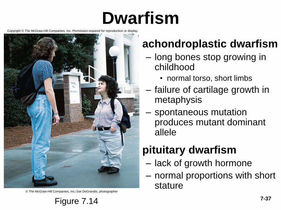
Dwarfism
• achondroplastic dwarfism– long bones stop growing in
childhood• normal torso, short limbs
– failure of cartilage growth in metaphysis
– spontaneous mutation produces mutant dominant allele
• pituitary dwarfism– lack of growth hormone
– normal proportions with short stature
Figure 7.14
Copyright © The McGraw-Hill Companies, Inc. Permission required for reproduction or display.
© The McGraw-Hill Companies, Inc./Joe DeGrandis, photographer
Physiology of Osseous Tissue
• a mature bone remains a metabolically active organ
– involved in its own maintenance of growth and
remodeling
– exerts a profound influence over the rest of the body
by exchanging minerals with tissue fluid• disturbance of calcium homeostasis in skeleton disrupts function
of other organ systems
– especially nervous and muscular
7-38
7-39
Mineral Deposition
• mineral deposition (mineralization) - a crystallization process in which
calcium phosphate, and other ions are taken from the blood plasma and
deposited in bone tissue
– osteoblasts produce collagen fibers that spiral the length of the osteon
– fibers become encrusted with minerals that harden the matrix
• calcium and phosphate (hydroxyapatite) from blood plasma are deposited along the
fibers
• the calcium and phosphate ion concentration must reach a critical value called the
solubility product for crystal formation to occur
• most tissues have inhibitors to prevent this so they do not become calcified
• osteoblasts neutralize these inhibitors and allow salts to precipitate in the bone
matrix
• first few crystals (seed crystals) attract more calcium and phosphate from solution
• abnormal calcification (ectopic ossification)
– may occur in lungs, brain, eyes, muscles, tendons or arteries
(arteriosclerosis)
– calculus – calcified mass in an otherwise soft organ such as the lung
7-40
Mineral Resorption
• mineral resorption – the process of dissolving bone and releasing minerals into the blood– performed by osteoclasts at the “ruffled border”
– hydrogen pumps in membrane secrete hydrogen into space between the osteoclast and bone surface
– chloride ions follow by electrical attraction
– hydrochloric acid (pH 4) dissolves bone minerals
– acid phosphatase enzyme digests the collagen
• orthodontic appliances (braces) reposition teeth– tooth moves because osteoclasts dissolve bone ahead of the
tooth, where the pressure on the bone is the greatest
– osteoblasts deposit bone more slowly in the low-pressure zone behind the tooth
7-41
Calcium Homeostasis• calcium and phosphate are used for much more than bone structure
• phosphate is a component of DNA, RNA, ATP, phospholipids, and pH buffers
• calcium needed in neuron communication, muscle contraction, blood clotting, and exocytosis
• minerals are deposited in the skeleton and withdrawn when they are needed for other purposes
• about 1100g of calcium in adult body
– 99% in the skeleton
• as easily exchangeable calcium ions and more stable hydroxyapatite reserve
• 18% of adult skeleton exchanged with blood each year
• normal calcium concentration in blood plasma is normally 9.2 to 10.4 mg/dl – 45% as Ca2+ can diffuse across capillary walls and affect other tissues – rest in reserve, bound to plasma proteins
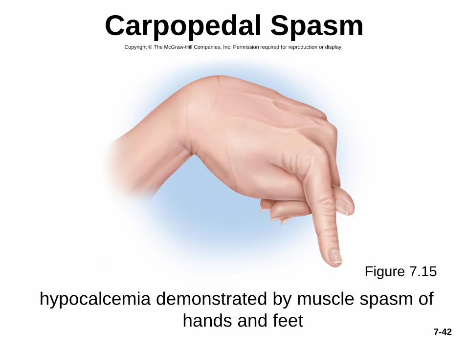
– hypocalcemia - blood calcium deficiency
• causes excess excitability of muscle, tremors, spasms or tetany (inability to relax)
– Na+ enters cells too easily and excites nerves and muscles
– hypercalcemia - blood calcium excess
• sodium channels less responsive and nerve and muscle less excitable than normal (sluggish reflexes, depression)
7-42
Carpopedal Spasm
hypocalcemia demonstrated by muscle spasm of
hands and feet
Copyright © The McGraw-Hill Companies, Inc. Permission required for reproduction or display.
Figure 7.15
7-43
Ion Imbalances
• hypercalcemia is rare
• hypocalcemia has a wide variety of causes
– vitamin D deficiency
– diarrhea
– thyroid tumors
– underactive parathyroids
– pregnancy and lactation
– accidental removal of parathyroid glands during thyroid surgery
• calcium homeostasis depends on a balance between
dietary intake, urinary and fecal loses, and exchanges
between osseous tissue
• calcium homeostasis is regulated by three hormones:
– calcitriol, calcitonin, and parathyroid hormone
7-44
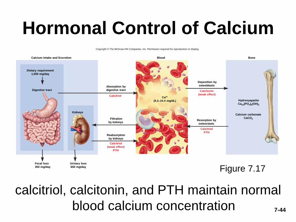
Hormonal Control of Calcium
calcitriol, calcitonin, and PTH maintain normal
blood calcium concentration
Figure 7.17
Copyright © The McGraw-Hill Companies, Inc. Permission required for reproduction or display.
Calcium Intake and Excretion
Digestive tract
Kidneys
Fecal loss
350 mg/day
Urinary loss
650 mg/day
Calcitriol
Calcitriol
(weak effect)
PTH
Blood
Calcitonin
(weak effect)
Calcitriol
PTH
Bone
Absorption by
digestive tract
Filtration
by kidneys
Reabsorption
by kidneys
Deposition by
osteoblasts
Resorption by
osteoclasts
Dietary requirement
1,000 mg/day
Ca2+
(9.2–10.4 mg/dL) Hydroxyapatite
Ca10(PO4)6(OH)2
Calcium carbonate
CaCO3
7-45
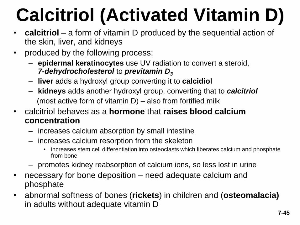
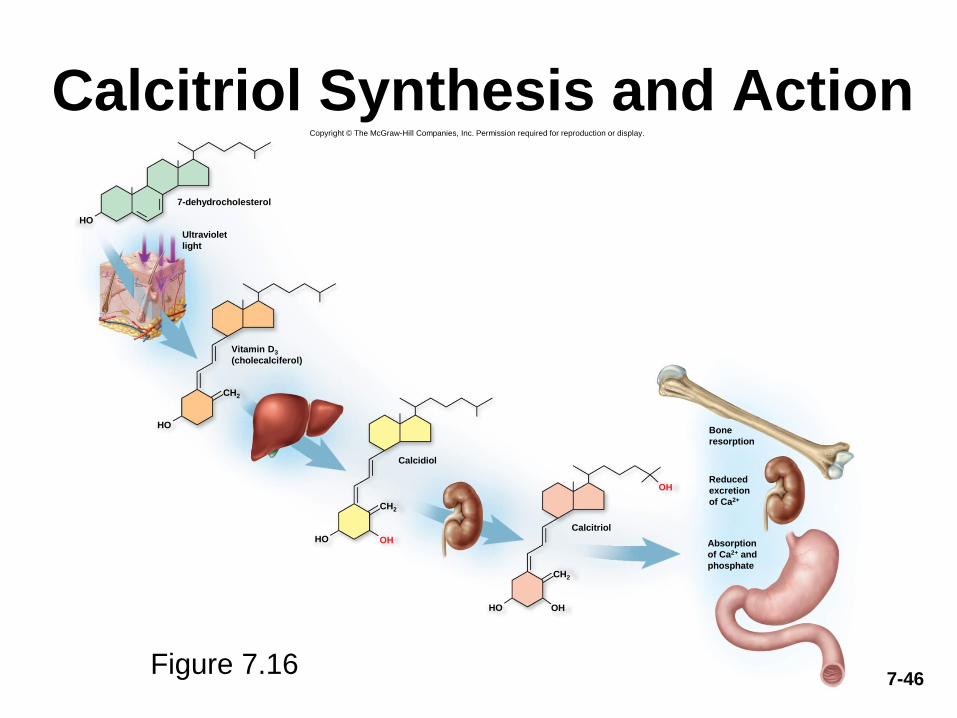
Calcitriol (Activated Vitamin D)• calcitriol – a form of vitamin D produced by the sequential action of
the skin, liver, and kidneys
• produced by the following process:
– epidermal keratinocytes use UV radiation to convert a steroid, 7-dehydrocholesterol to previtamin D3
– liver adds a hydroxyl group converting it to calcidiol
– kidneys adds another hydroxyl group, converting that to calcitriol
(most active form of vitamin D) – also from fortified milk
• calcitriol behaves as a hormone that raises blood calcium concentration
– increases calcium absorption by small intestine
– increases calcium resorption from the skeleton• increases stem cell differentiation into osteoclasts which liberates calcium and phosphate
from bone
– promotes kidney reabsorption of calcium ions, so less lost in urine
• necessary for bone deposition – need adequate calcium and phosphate
• abnormal softness of bones (rickets) in children and (osteomalacia) in adults without adequate vitamin D
Calcitriol Synthesis and ActionCopyright © The McGraw-Hill Companies, Inc. Permission required for reproduction or display.
HO
HO
CH2
HO OH
CH2
HO OH
OH
CH2
7-dehydrocholesterol
Calcidiol
Calcitriol
Ultraviolet
light
Vitamin D3
(cholecalciferol)
Bone
resorption
Reduced
excretion
of Ca2+
Absorption
of Ca2+ and
phosphate
Figure 7.167-46
7-47
Calcitonin
• calcitonin - secreted by C cells (clear cells) of the thyroid gland when calcium concentration rises too high
• lowers blood calcium concentration in two ways:– osteoclast inhibition
• reduces osteoclast activity as much as 70%
• less calcium liberated from bones
– osteoblast stimulation
• increases the number and activity of osteoblasts
• deposits calcium into the skeleton
• important in children, weak effect in adults– osteoclasts more active in children due to faster remodeling
– deficiency does not cause disease in adults
• reduces bone loss in women during pregnancy & lactation
7-48
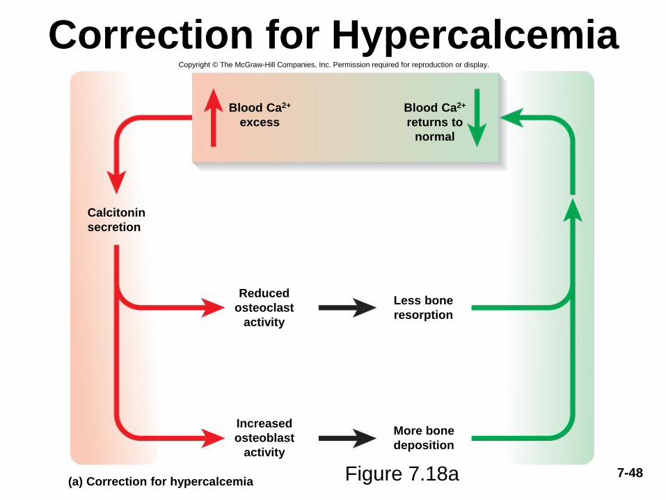
Correction for HypercalcemiaCopyright © The McGraw-Hill Companies, Inc. Permission required for reproduction or display.
(a) Correction for hypercalcemia
Calcitonin
secretion
Blood Ca2+
excess
Blood Ca2+
returns to
normal
Reduced
osteoclast
activity
Less bone
resorption
Increased
osteoblast
activity
More bone
deposition
Figure 7.18a
7-49
Parathyroid Hormone
• parathyroid hormone (PTH) – secreted by the parathyroid glands which adhere to the posterior surface of thyroid gland
• PTH released with low calcium blood levels
• PTH raises calcium blood level by four mechanisms– binds to receptors on osteoblasts
• stimulating them to secrete RANKL which raises the osteoclast population
– promotes calcium reabsorption by the kidneys, less lost in urine
– promotes the final step of calcitriol synthesis in the kidneys, enhancing calcium raising effect of calcitriol
– inhibits collagen synthesis by osteoblasts, inhibiting bone deposition
• sporadic injection or secretion of low levels of PTH causes bone deposition, and can increase bone mass
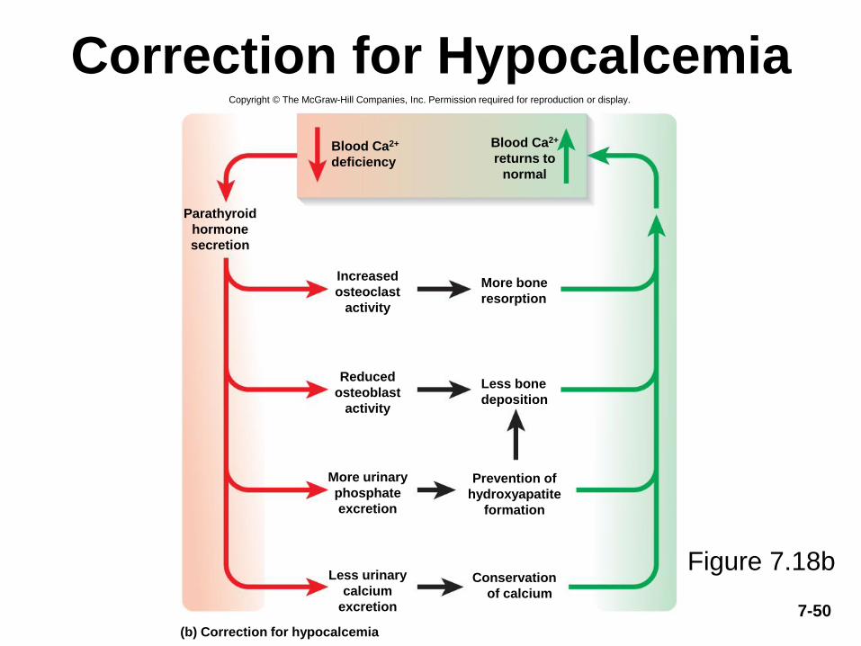
7-50
Correction for HypocalcemiaCopyright © The McGraw-Hill Companies, Inc. Permission required for reproduction or display.
(b) Correction for hypocalcemia
Blood Ca2+
deficiency
Parathyroid
hormone
secretion
Increased
osteoclast
activity
Blood Ca2+
returns to
normal
More bone
resorption
Less bone
deposition
Prevention of
hydroxyapatite
formation
More urinary
phosphate
excretion
Reduced
osteoblast
activity
Conservation
of calcium
Less urinary
calcium
excretion
Figure 7.18b
7-51
Phosphate Homeostasis• average adult has 500 – 800 g of phosphorus
• 85-90% of phosphate is in the bones
• normal plasma concentration is 3.5 – 4.0 mg/dl
• occurs in two principal forms:– HPO4
2- and H2PO4- (monohydrogen & dihydrogen phosphate
ions)
• phosphate levels are not regulated as tightly as calcium levels– no immediate functional disorders
• calcitriol promotes its absorption by small intestine & promotes bone deposition
• PTH lowers blood phosphate level by promoting its urinary excretion
7-52
Other Factors Affecting Bone• at least 20 or more hormones, vitamins, and growth
factors affect osseous tissue
• bone growth especially rapid in puberty & adolescence– surges of growth hormone, estrogen, and testosterone occur and
promote ossification
– these hormones stimulate multiplication of osteogenic cells, matrix deposition by osteoblasts, and chondrocyte multiplication and hypertrophy in metaphyses
– girls grow faster than boys and reach full height earlier
• estrogen stronger effect than testosterone on bone growth
– males grow for a longer time and taller
• anabolic steroids cause growth to stop – epiphyseal plate “closes” prematurely
– results in abnormally short adult stature

7-53
Fractures and Their Repair
• stress fracture – break caused by abnormal
trauma to a bone
– falls, athletics, and military combat
• pathological fracture – break in a bone weakened
by some other disease
– bone cancer or osteoporosis
– usually caused by stress that would not break a healthy
bone
• fractures classified by structural characteristics
– direction of fracture line
– break in the skin
– multiple pieces
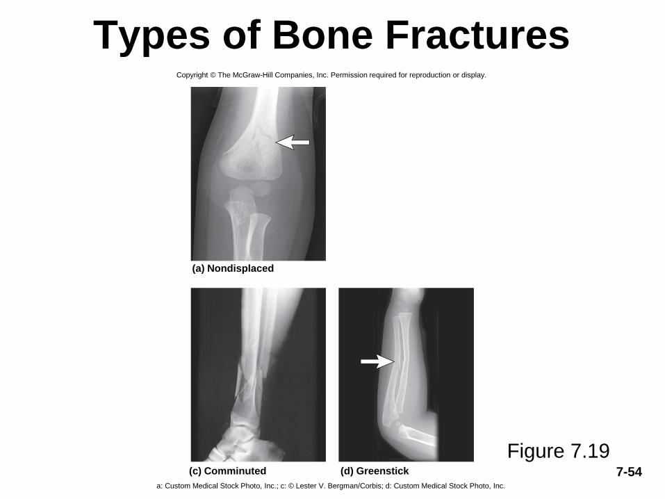
7-54
Types of Bone Fractures
Figure 7.19
Copyright © The McGraw-Hill Companies, Inc. Permission required for reproduction or display.
(a) Nondisplaced
(c) Comminuted (d) Greenstick
a: Custom Medical Stock Photo, Inc.; c: © Lester V. Bergman/Corbis; d: Custom Medical Stock Photo, Inc.
7-55
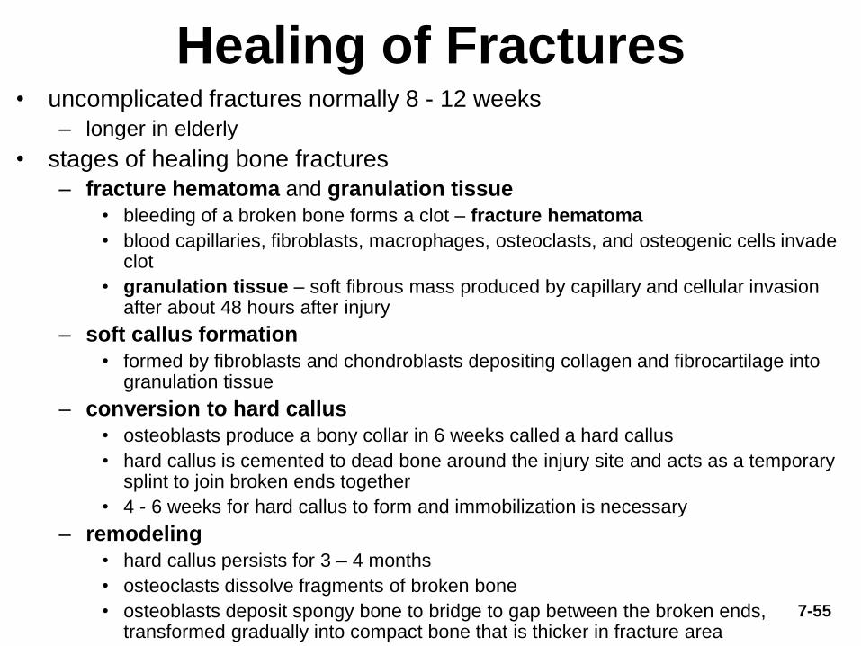
Healing of Fractures • uncomplicated fractures normally 8 - 12 weeks
– longer in elderly
• stages of healing bone fractures
– fracture hematoma and granulation tissue
• bleeding of a broken bone forms a clot – fracture hematoma
• blood capillaries, fibroblasts, macrophages, osteoclasts, and osteogenic cells invade clot
• granulation tissue – soft fibrous mass produced by capillary and cellular invasion after about 48 hours after injury
– soft callus formation
• formed by fibroblasts and chondroblasts depositing collagen and fibrocartilage into granulation tissue
– conversion to hard callus
• osteoblasts produce a bony collar in 6 weeks called a hard callus
• hard callus is cemented to dead bone around the injury site and acts as a temporary splint to join broken ends together
• 4 - 6 weeks for hard callus to form and immobilization is necessary
– remodeling
• hard callus persists for 3 – 4 months
• osteoclasts dissolve fragments of broken bone
• osteoblasts deposit spongy bone to bridge to gap between the broken ends, transformed gradually into compact bone that is thicker in fracture area
7-56
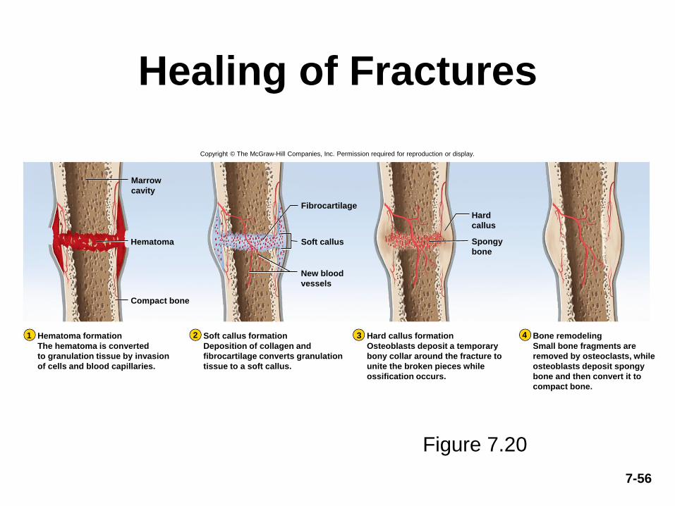
Healing of Fractures
Figure 7.20
Copyright © The McGraw-Hill Companies, Inc. Permission required for reproduction or display.
Fibrocartilage
Soft callusHematoma
Compact bone
1 2 3 4
Marrow
cavity
Hematoma formation
The hematoma is converted
to granulation tissue by invasion
of cells and blood capillaries.
Hard callus formation
Osteoblasts deposit a temporary
bony collar around the fracture to
unite the broken pieces while
ossification occurs.
Bone remodeling
Small bone fragments are
removed by osteoclasts, while
osteoblasts deposit spongy
bone and then convert it to
compact bone.
Soft callus formation
Deposition of collagen and
fibrocartilage converts granulation
tissue to a soft callus.
Hard
callus
Spongy
bone
New blood
vessels
7-57
Treatment of Fractures• closed reduction – procedure in which the bone fragments are
manipulated into their normal positions without surgery
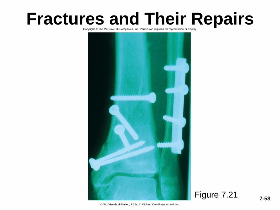
• open reduction – involves surgical exposure of the bone and the use of plates, screws, or pins to realign the fragments
• cast – normally used to stabilize and immobilize healing bone
• traction used to treat fractures of the femur in children
– aligns bone fragments by overriding force of the strong thigh muscles
– risks long-term confinement to bed
– rarely used for the elderly
– hip fractures are usually pinned in elderly and early ambulation (walking) is encouraged to promote blood circulation and healing
• electrical stimulation accelerates repair
– suppresses the effects of parathyroid hormone
• orthopedics – the branch of medicine that deals with prevention and correction of injuries and disorders of the bones, joints, and muscles
7-58
Fractures and Their Repairs
Figure 7.21
Copyright © The McGraw-Hill Companies, Inc. Permission required for reproduction or display.
© SIU/Visuals Unlimited; 7.22a: © Michael Klein/Peter Arnold, Inc.
7-59
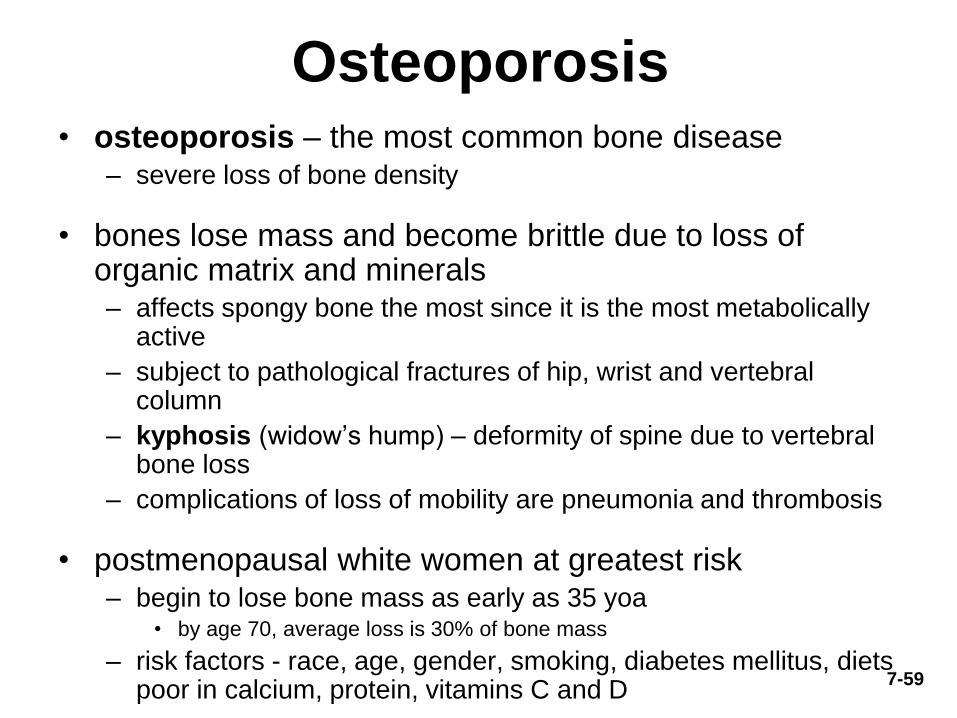
Osteoporosis • osteoporosis – the most common bone disease
– severe loss of bone density
• bones lose mass and become brittle due to loss of organic matrix and minerals– affects spongy bone the most since it is the most metabolically
active
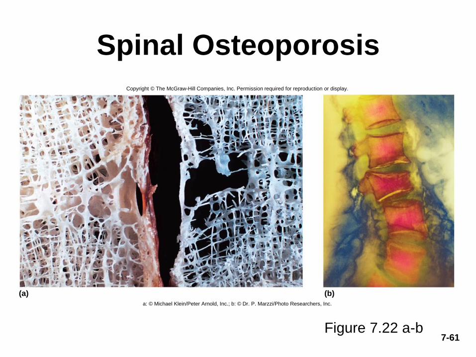
– subject to pathological fractures of hip, wrist and vertebral column
– kyphosis (widow’s hump) – deformity of spine due to vertebral bone loss
– complications of loss of mobility are pneumonia and thrombosis
• postmenopausal white women at greatest risk– begin to lose bone mass as early as 35 yoa
• by age 70, average loss is 30% of bone mass
– risk factors - race, age, gender, smoking, diabetes mellitus, diets poor in calcium, protein, vitamins C and D

7-60
Osteoporosis• estrogen maintains density in both sexes inhibits resorption
by osteoclasts– testes and adrenals produce estrogen in men
– in women, rapid bone loss after menopause since estrogen blood level drops below 30 ng/mL
• osteoporosis is common in young female athletes with low body fat causing them to stop ovulating and ovarian estrogen secretion is low
• treatments– estrogen replacement therapy (ERT) slows bone resorption, but
increases risk of breast cancer, stroke and heart disease
– drugs Fosamax/Actonel destroys osteoclasts
– PTH slows bone loss if given as daily injection• Forteo (PTH derivative) increases density by 10% in 1 year
– may promote bone cancer so use is limited to 2 years
– best treatment is prevention - exercise and calcium intake (1000 mg/day) between ages 25 and 40
7-61
Spinal Osteoporosis
Figure 7.22 a-b
Copyright © The McGraw-Hill Companies, Inc. Permission required for reproduction or display.
(a) (b)
a: © Michael Klein/Peter Arnold, Inc.; b: © Dr. P. Marzzi/Photo Researchers, Inc.