nmr structure and dynamics of the rna-binding site for the histone
TRANSCRIPT
NMR structure and dynamics of the RNA-bindingsite for the histone mRNA stem-loopbinding protein
ERIC S. DEJONG,1 WILLIAM F. MARZLUFF, 2 and EDWARD P. NIKONOWICZ 1
1Department of Biochemistry and Cell Biology, Rice University, Houston, Texas 77251-1892, USA2Program in Molecular Biology and Biotechnology, Department of Biochemistry and Biophysics,University of North Carolina, Chapel Hill, North Carolina 27599, USA
ABSTRACT
The 39 end of replication-dependent histone mRNAs terminate in a conserved sequence containing a stem-loop. This26-nt sequence is the binding site for a protein, stem-loop binding protein (SLBP), that is involved in multiple aspectsof histone mRNA metabolism and regulation. We have determined the structure of the 26-nt sequence by multidimen-sional NMR spectroscopy. There is a 16-nt stem-loop motif, with a conserved 6-bp stem and a 4-nt loop. The loop isclosed by a conserved U•A base pair that terminates the canonical A-form stem. The pyrimidine-rich 4-nt loop, UUUC,is well organized with the three uridines stacking on the helix, and the fourth base extending across the major grooveinto the solvent. The flanking nucleotides at the base of the hairpin stem do not assume a unique conformation,despite the fact that the 5 9 flanking nucleotides are a critical component of the SLBP binding site.
Keywords: 3 9 processing; hairpin; heteronuclear; multidimensional; pre-mRNA; tetraloop
INTRODUCTION
Maturation of mRNA in metazoans involves multipleprocessing steps of the pre-mRNA transcript, includingexcision of introns and the addition of a poly(A) tail tothe 39 end+ However, the replication-dependent histonemRNAs are unique, as the histone genes lack intronsand the mature histone mRNAs terminate with a stem-loop (hairpin) secondary structure rather than poly(A)tails (Birnstiel et al+, 1985; Marzluff, 1992; Dominski &Marzluff, 1999)+ Processing of the histone pre-mRNAtranscript involves a single endonucleolytic cleavageevent 39 to the stem-loop (Gick et al+, 1986)+ Two de-fined trans-acting components participate in the pro-cessing reaction—a protein that specifically binds thestem-loop (Wang et al+, 1996; Martin et al+, 1997) andthe U7 snRNP that interacts with a purine-rich element
located downstream of the cleavage site (Mowry &Steitz, 1987; Soldati & Schümperli, 1988)+ There arelikely to be additional factors required for processing,including a heat-labile factor that has not been wellcharacterized (Gick et al+, 1987; Lüscher & Schümperli,1987)+ Following 39-end processing, the histone mRNAis rapidly transported to the cytoplasm+ The 39 terminalhairpin is necessary for efficient pre-mRNA processing(Pandey et al+, 1994) and mRNA export (Eckner et al+,1991; Williams et al+, 1994) and is essential for regu-lation of the histone mRNA half-life (Pandey & Marzluff,1987)+
The primary structure of the 39 end of histone mRNAsis highly conserved and serves as the principle recog-nition element of the stem-loop binding protein (SLBP)+The minimal recognition site for the SLBP is 26 nt (Wil-liams & Marzluff, 1995) and it is predicted to form a6-bp stem capped by a 4-nt pyrimidine-rich loop+ The 59end of the stem is flanked by A/C-rich sequences es-sential for SLBP binding (Williams & Marzluff, 1995)and the 39 end of the stem is followed by a consensuselement ACCCA or ACCA, which is the cleavage targetand plays a minor role in SLBP binding (Furger et al+,1998)+ High affinity binding of the SLBP to the hairpindepends on nucleotide identity within the loop, the stem,and the immediate 59 and 39 flanking sequences+ Thefirst and third residues of the loop are uridine nucleo-
Reprint requests to: Edward P+ Nikonowicz, Department of Bio-chemistry and Cell Biology, Rice University, Houston, Texas 77251-1892, USA; e-mail: edn@bioc+rice+edu+
Abbreviations: DQF-COSY: double quantum filtered correlatedspectroscopy; HetCor: heteronuclear correlation; HMQC: heteronu-clear multiple quantum coherence;HSQC: heteronuclear single quan-tum coherence; NH: imino; NH2: amino; NMR: nuclear magneticresonance; NOE: nuclear Overhauser effect; NOESY: NOE spec-troscopy; NTP: nucleoside triphosphate; rMD: restrained moleculardynamics; rmsd: root mean square deviation; SLBP: stem-loop bind-ing protein+
RNA (2002), 8:83–96+ Cambridge University Press+ Printed in the USA+Copyright © 2002 RNA Society+DOI: 10+1017+S1355838201013863
83

tides in all metazoans except for Caenorhabditis ele-gans, which has a C in the first position (Roberts et al+,1989; Marzluff, 1992)+ The second position is predom-inantly occupied by uridine+ U, C, and A are found in thefourth position but never G+
The sequence of the stem is critical for RNA binding(Battle & Doudna, 2001; Williams & Marzluff, 1995)+The consensus stem sequence consists of two G•Cbase pairs, followed by three Y-R base pairs that gen-erally contain at least two C•G base pairs, and an in-variant U•A base pair at the top of the stem+Mutagenesisstudies showed that the second G•C base pair is par-ticularly critical for SLBP binding (Battle & Doudna,2001)+Although the proposed loop is composed of 4 nt,it is not a member of the unusually stable family of 4-ntloops known as tetraloops+ Indeed, the most similartetraloop family, UNCG, is excluded by the invariantabsence of guanine at position four+ The 59-U and 39-Anucleotides that close the loop are invariant+ Similarsequence motifs in which apposing U and A nucleo-tides flank a loop but do not base pair, such as stem-loop IIa of yeast U2 snRNA, suggest that the histone 39mRNA hairpin might alternatively have a stem of 5 bpand a loop of 6 nt+ The reduced length of the stem andthe conserved adenine nucleotides that flank the 59side of the hairpin that could pair with nucleotides in theloop suggest the possibility that the SLBP recognitionelement contains a pseudoknot+
Although the SLBP is believed to recognize a uniquetertiary structure adopted by the 39 element, cleavageof the phosphate backbone within the loop does notimpair the ability of the RNA to bind the SLBP (Williams& Marzluff, 1995)+ This result coupled with the findingthat critical determinants for binding reside in the stemsequence and the flanking sequence 59 of the stem,suggest that multiple contacts exist between the SLBPand the histone mRNA 39 end+
We have used heteronuclear nuclear magnetic res-onance (NMR) spectroscopy to determine the solutionstructure and dynamics of the 39 end of histone mRNAin a 28-nt RNA molecule that contains the 26 nt nec-essary for SLBP binding+ Our results confirm the pre-dicted secondary structure of a 6-bp stem capped by a4-nt loop+ The nucleotides flanking the 59 and 39 endsof the stem are largely disordered and presumably onlybecome ordered after forming specific contacts withSLBP+ The implications of the stem-loop structure forSLBP binding are discussed+
RESULTS
Two RNA molecules (Fig+ 1) were used in our structuralstudy of the histone mRNA 39 binding site for the SLBP+SL28 comprises the wild-type binding site for mamma-lian SLBP+ The proposed secondary structure is a stem-loop flanked on either side by short single-strandedRNA sequences+ Several conditions were tested to as-
sess the effects of counterions and pH+ K1 is the phys-iologically relevant counterion within the cell and yieldedthe best quality spectrum+ Na1 and Mg21 cause slightbroadening and small chemical shift changes of theimino (NH) resonances, but do not lead to the appear-ance of new peaks+ The reduction of pH from 6+8 to 5+7also results in slight broadening and weakening of theNH resonances+
Chemical shift assignments
The sequence-specific assignment of SL28 is hinderedby resonance overlap of 19 and pyrimidine base reso-nances+ Approximately 65% of the 1H and 13C reso-nances can be unambiguously assigned using standardheteronuclear methodologies+ Nonetheless, the NHspectrum with 5 G NH resonances characteristic ofG•C base pairs and 1 U NH resonance characteristic ofan A•U base pair clearly indicate that SL28 forms ahairpin (Fig+ 2)+ A pseudoknot conformation with theflanking As paired with the loop Us should produceadditional downfield shifted U NH resonances+Althoughthe NH spectrum largely establishes the global fold ofSL28, poor resolution of several nonexchangeable res-onances from the loop nucleotides impair a high reso-lution structure determination of the RNA+ To facilitateassignment of SL28 and improve the accuracy of thethree-dimensional structure, SL16, the RNA hairpin with-out the flanking sequences, was studied (Fig+ 1)+
The exchangeable NH and amino (NH2) resonanceswere assigned using two-dimensional nuclear Over-hauser effect (NOE) spectroscopy (NOESY) and two-dimensional 15N-edited NOESY experiments+ Briefly,the NH resonance of the U•A base pair was first iden-tified by the characteristic 15N chemical shift of its NHresonance+ The remaining NH resonances were as-signed using the weaker NOE connectivities betweenNH proton resonances of neighboring base pairs+ Theseconnectivities are continuous in the helix from G2 toG11+ The cytidine NH2 resonances were assigned usingthe strong intra-base-pair C NH2 to G NH NOE cross-peaks+ Independent confirmation of the resonance as-
FIGURE 1. Sequence and proposed secondary structures of (A)the conserved RNA binding site for the histone SLBP, (B) the 28-ntRNA molecule, SL28, used in this study, and (C) the 16-nt RNA mol-ecule, SL16, also used in this study+ Residues in SL28 are numberedrelative to the 59 terminal G of the proposed stem-loop+
84 E.S. DeJong et al.

signments was provided by strong cytidine intrabaseNH2 to H5 NOE cross-peaks in G•C base pairs+ TheNH proton resonances of U7, U8, and U9 are resolvedbut broad with chemical shifts 10+5–11+0 ppm, a regionof the NH proton spectrum characteristic of unpaireduridine nucleotides+ The NH2 resonances of G1, G2,and A11 were not observed and could not be assigned+All other exchangeable proton and protonated nitrogenresonances were assigned+Although divalent metal ionsare not required for SLBP binding or 39 end processing,a two-dimensional 15N-1H heteronuclear multiple quan-tum coherence (HMQC) spectrum was collected forSL16 after addition of 10 mM Mg21+ No chemical shiftchanges are observed and only the U6 NH resonanceis slightly weakened+ This suggests that Mg21 may as-sociate with the RNA loop and accelerate solvent ex-change of the U6 NH proton, but does not inducesignificant structural changes+ This is consistent withthe finding that binding of SLBP and histone pre-mRNAprocessing occur efficiently in 20 mM EDTA+
The nonexchangeable 1H and 13C resonances of SL16
(Fig+ 1) were assigned using standard heteronucleartechniques (Pardi, 1995; Dieckmann & Feigon, 1996)+
Most of the base and ribose 1H-13C correlations areresolved and none of the resonances have spectralcharacteristics indicative of intermediate exchange+ All16 ribose spin systems were identified using three-dimensional HCCH-COSY and three-dimensionalHCCH-TOCSY experiments+ Intraresidue base-to-sugarcorrelations were identified using two-dimensional 15N-1H HSQC experiments optimized to yield the multiplebond correlations H5-N1,H8-N9, and H19-N1/N9 (Dieck-mann & Feigon, 1996)+ All residues except C3 and C16
yielded the desired base-ribose correlations+Sequential assignments for the nonexchangeable res-
onances were made using three-dimensional 13C-editedNOESY experiments to identify sequential H6/8-H19NOE connectivities (Pardi, 1995)+ The intraresidue 15N-1H base-to-sugar correlations permitted intraresiduecross-peaks to be distinguished from interresidue cross-peaks+ The sequential H6/8-H19 NOE connectivities arecontinuous through all 16 nt in the 180 ms NOESYspectrum+ The H6/8-H29 interresidue connectivities alsoare continuous through the hairpin except between C10
and A11+ This is consistent with disruption of the back-bone introduced by the C29-endo ribose ring confor-mation of C10+ Interestingly, i to i 1 2 NOE cross-peaksfrom U9 H19 to A11 H8 and U9 H6 to A11 H19 were ob-served, suggesting exclusion of the C10 base from theloop+
All internucleotide 31P resonances are dispersed be-tween 23+4 and 24+6 ppm+All 15 31P resonances wereable to be assigned using the H39-P correlations fromtwo-dimensional 31P-1H heteronuclear correlation(HetCor) spectra+ Several P-H49 and P-H59/H50 corre-lations also were present in the HetCor spectra+ Theseassignments were later confirmed from a sequentialwalk in a two-dimensional 31P-1H hetero-TOCSY-NOESY experiment+
The secondary structure shared by SL28 and SL16 isevident through comparison of the NH 1H spectra(Fig+ 2)+ Except for the 59 terminal residue of the hairpinG1, the peaks in each spectrum have nearly identicalchemical shifts, indicating that SL16 and SL28 have acommon secondary structure+ Importantly, the match-ing chemical shifts of the corresponding unpaired U NHresonances demonstrate that the loop uridines do notinteract with nucleotides of the flanking sequences+
The nonexchangeable resonances of the SL28 RNAmolecule were assigned using the same methods em-ployed to assign SL16, except that a pyrimidine-C5-deuterated molecule was used to help reduce overlapin NOESY spectra (Nikonowicz et al+, 1998)+ All sixadenine C2-H2 correlations are resolved and only A11
of SL28 has 1H and 13C chemical shifts that match theC2-H2 pair of A11 in SL16 (Fig+ 4)+ Similarly, the A11
H2-N1/N3 cross-peaks of SL28 align with the A11 H2-N1/N3 correlations of SL16, confirming the A11 chemi-cal environments are equivalent in the two molecules(Fig+ 3A)+ The chemical shifts of the base 1H-13C cor-
FIGURE 2. One-dimensional NH 1H spectra of SL28 (A) and SL16(B) RNA molecules+ The flanking regions of SL28 cause upfield anddownfield shifts of the G1 and G2 resonances, respectively, relative tothose of SL16+ The chemical shifts of the remaining NH resonances,including those corresponding to the loop uridine nucleotides, are thesame and demonstrate that SL16 and SL28 have equivalent second-ary structures+ The identical peak pattern of the loop uridine reso-nances also confirms that the adenine bases of the 59 flankingsequence do not interact with the loop to form a pseudoknot+
Structure of the RNA-binding site for histone SLBP 85
relations of residues within the 16-nt hairpin of SL28 areidentical with those of SL16 (Fig+ 4)+ The terminal G1
residue is an exception due to the presence of theflanking sequences+ Using the resonance assignmentsof the SL16 hairpin, it was possible to identify the cor-responding ribose spin systems of SL28 from the three-dimensional 13C-1H HCCH-TOCSY spectrum+ The 15N-1H multiple-bond correlated spectrum (Fig+ 3B) yieldedimproved resolution of hairpin and flanking region baseand ribose 19 1H resonances and facilitated sequentialresonance assignment of the SL28 RNA using the H6/8-H19 region of the NOESY spectrum+ The 1H, 13C, and15N chemical shifts are listed in the Appendix+
Structures of the SL 16 and SL 28 molecules
The structures of SL16 and SL28 were calculated usinga restrained molecular dynamics routine starting from40 structures with completely random backbone di-hedral angles+ The calculations for SL16 used a total of296 conformationally restrictive distance constraints and50 dihedral angle constraints (Table 1) to produce 12converged structures (Fig+ 5)+ For SL28, 333 NOE-derived distance constraints and 66 dihedral angle con-straints were used to produce 11 converged structures+The converged structures had an average of 11 dis-tance constraint violations between 0+1 and 0+3 Å, ran-domly distributed throughout the hairpins+All convergedstructures violated no NOE constraints by more than
0+3 Å+ The heavy atoms of the SL16 final convergedstructures superpose on the average structure with anaverage root mean square deviation (rmsd) of 1+25 Å+The heavy atoms of the 16 nt composing the hairpin ofSL28 superpose with an average rmsd of 1+34 Å+ Thelocal precision of the hairpins is better, though, with theloop (U6-A11) and stem (G2-U6 and A11-C15) regions ofSL28 having rmsds of 0+51 Å and 0+85 Å, respectively+
The conformations of SL16 and the central hairpin ofSL28 differ only slightly near the helix terminus and aredescribed together+ The pyrimidine C5-deuterated SL16
RNA hairpin was important for extraction of loop regionbase–base and base–H19 constraints due to reso-nance crowding in both 1H and 13C dimensions+ Fig-ure 6 summarizes the experimental distance constraintsfor the loop and upper stem+ The abundance of dis-tance constraints in the loop region (residues U6-A11)defines the conformation of these nucleotides very pre-cisely (Fig+ 5A; Table 1)+ Sequential NOEs between H5and H6 protons from U6 through U8 indicate that U7 andU8 form a 59 base stack+ NOEs from U9 H6 to A11 H2and H8 and the near absence of interresidue NOEsinvolving the C10 base indicate the proximity of U9 andA11 and the relative distal position of the C10 base+
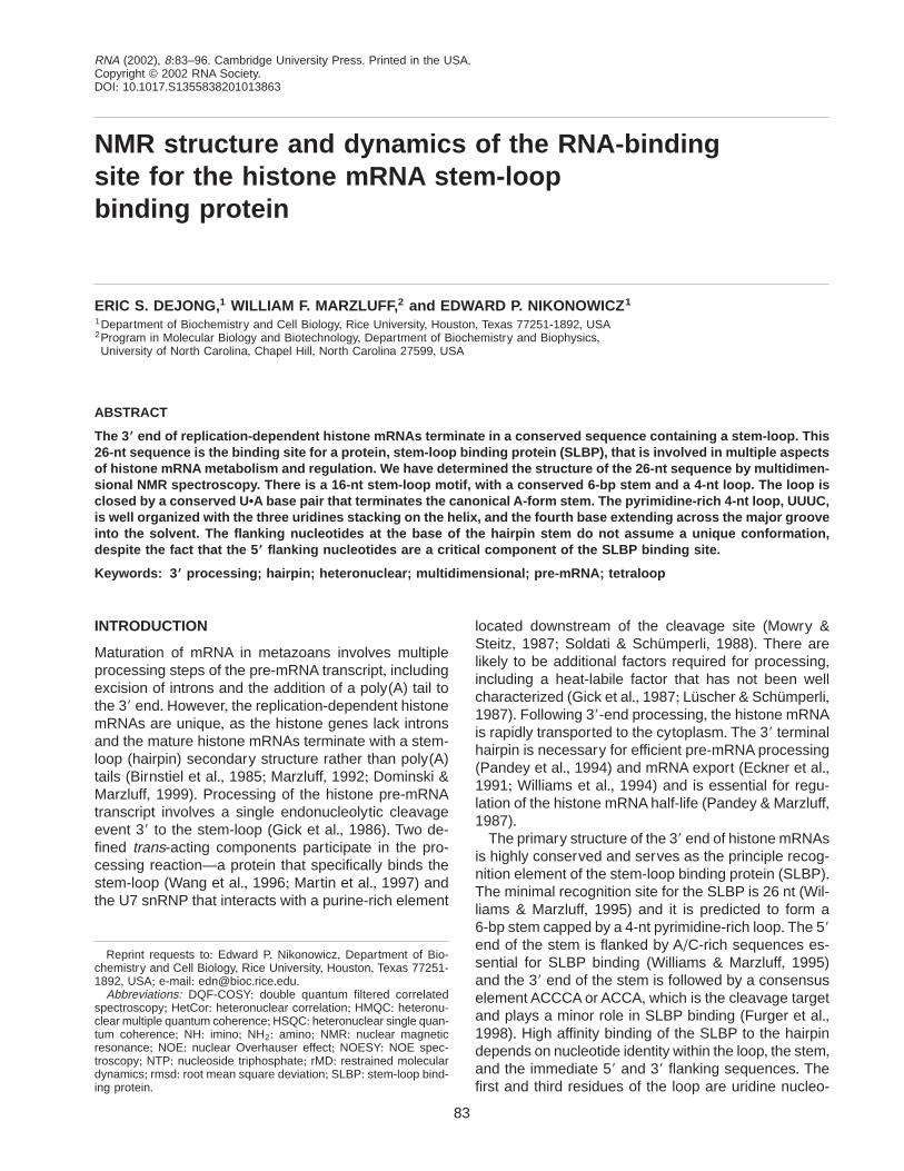
FIGURE 3. Base proton region of the HNN-COSY spectrum (A) andsugar proton region of the multiple-bond 15N-1H HSQC spectrum (B)of SL28+ The U6 NH proton exchanges with solvent too rapidly to giverise to cross-peaks in the NOESY spectrum, preventing confirmationof the U6-A11 base pair by conventional means+ However, the U6H3-to-A11 N1 hydrogen-bond-mediated coupling of U6 N3 to A11 N1yields the A11 H2-U6 N3 correlation (A)+ The six adenine H2-N1, N3correlation pairs are connected in A and the four uridine H5-N1,N3 correlation pairs are connected in B+
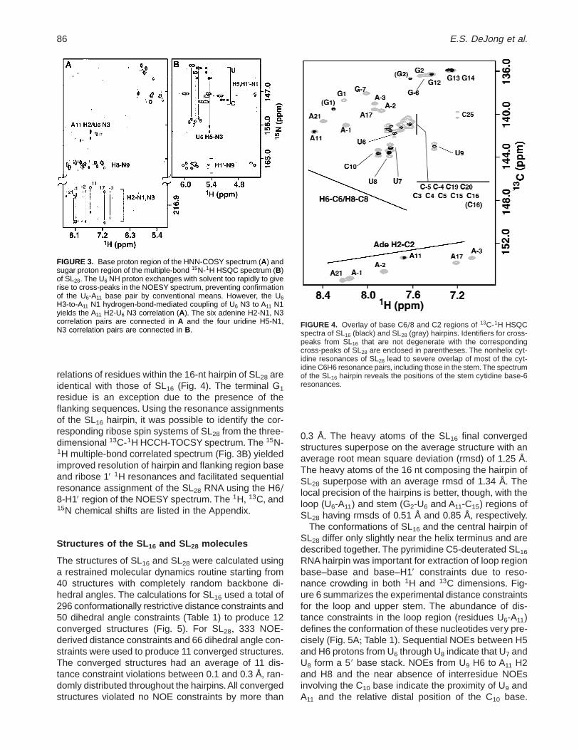
FIGURE 4. Overlay of base C6/8 and C2 regions of 13C-1H HSQCspectra of SL16 (black) and SL28 (gray) hairpins+ Identifiers for cross-peaks from SL16 that are not degenerate with the correspondingcross-peaks of SL28 are enclosed in parentheses+ The nonhelix cyt-idine resonances of SL28 lead to severe overlap of most of the cyt-idine C6H6 resonance pairs, including those in the stem+The spectrumof the SL16 hairpin reveals the positions of the stem cytidine base-6resonances+
86 E.S. DeJong et al.

Unusual interresidue sugar-to-sugar and sugar-to-baseNOEs also play a key role in defining the structure ofthe loop (Fig+ 6)+ These include NOEs from U6 H29 toU7 H19, U8 H19 to U9 H19, C10 H59/50 to A11 H8, and U9
H19, H39 to A11 H8+ These NOEs indicate that the sugarrings and 3 of the 4 bases of the loop pack togethertightly in an arrangement distinctly different from stan-dard helices+
Several nucleotides in the loop have unusual sugar–phosphate backbone conformations+ The large H19–H29 couplings of residues U8, U9, and C10 indicate thattheir ribose sugar rings have the C29-endo conforma-tion and the .5 Hz P–H9/H50 coupling of residues U8,U9, and C10 exclude b from the standard trans confor-mation+ H49–H59/H50 couplings .5 Hz in the DQF-COSY spectrum indicate that residues U7 to U9 havenonstandard g backbone angles+ These angles wereleft unconstrained and occupy the trans and gauche2
conformations, respectively, in all converged structures(Fig+ 7)+ The e torsion angles of U9 and C10 adopt thegauche2 conformation and lie outside the trans regiontypical of A- or B-form geometries+ These angles ap-pear to facilitate the turn of the phosphate backbonesin the loop+ A nonstandard trans conformation for the aand z backbone angles is predicted to cause a down-
field shift of the corresponding 31P resonance (Goren-stein, 1984)+ Because the 31P resonances of SL28 allcluster at 24+0 ppm, it is unlikely any of these anglesadopt the trans conformation+ However, no a and zangles were constrained+ Except for residues U6 andA11, which have gauche1 a torsion angles, U7 to C10
adopt the more common gauche2 conformation+ Sim-ilarly, the z torsion angles tend to have the gauche2
conformation except for those of U8 and U9, which liebetween gauche1 and trans+ However, none of the a orz torsion angles in the loops of the converged struc-tures have a true trans conformation (Fig+ 7)+
A superposition of the loop regions from the 11 con-verged structures is shown in Figure 5A and the mini-mized average structure is shown in Figure 7A+ Thehelical base stack continues up the 59 side of the loop,with U8 stacking on top of U7, which in turn stacks ontop of the U6•A11 base pair+ Residue U9 stacks aboveA11 and is nearly coplanar with U7+ However, there is noevidence to support hydrogen-bond interactions be-tween U7 and U9+ Residue C10 does not participate ineither the 59 or 39 base stack but instead its base pointsout into solution on the major groove side of the helix+The stacking of the loop nucleotides has the net effectof extending the helix by an additional base pair step+
The pattern of intra- and interresidue H6/8-H19 andH6/8-H29 NOEs in the stem region does not signifi-cantly differ from that expected of standard A-form ge-ometry+ Although the U6 NH resonance has no NOEsthat would permit the direct identification of a base pairpartner, the U6 NH nitrogen and hydrogen nuclei res-onate in the downfield region of the spectrum char-acteristic of Watson–Crick A•U base pairs+ The onlypossible cross-strand partner for U6 is A11+ The J(N, N)-HNN-COSY spectrum (Hennig & Williamson, 2000)yields a U6 N3 to A11 H2 cross-peak, confirming thepresence of the U6 NH3 to A11 N1 hydrogen bond andthus the integrity of the U6•A11 base pair (Fig+ 3)+ Thebases of the terminal G1•C16 and U6•A11 base pairshave interresidue NOEs consistent with base–basestacking+ The sugar and phosphate backbone torsionangles in the stem also lie within the limits of A-formRNA helices+
The 59 and 39 flanking regions are poorly constrainedby the NOE and J-coupling data+ The sparseness ofconstraints for these residues leads to an array of con-formations+ The spectral data indicate that the A-rich 59sequence does not interact with the U-rich loop+ Fur-ther, there is no evidence to suggest the presence ofcross-strand A1•C interactions between pH 5+7 and 6+8+Although the nucleotides in these regions exhibit intra-and interresidue NOEs sufficient to provide sequentialresonance assignments and to confirm that the glyco-sidic bonds of all flanking region nucleotides have theanti conformation, no interresidue base–base NOEscould be identified that would indicate the flanking res-idues adopt a base-stacked helical conformation+
TABLE 1 + Summary of experimental distance and dihedral angleconstraints and refinement statistics for SL16 and SL28+
Constraint SL16 SL28
NOE distance constraintsIntraresiduea 87 110Interresidue 135 189Mean number per residue 16 11
NOE constraints by categoryVery strong (0+0–3+0 Å) 6 7Strong (0+0–4+0 Å) 36 36Medium (0+0–5+0 Å) 91 113Weak (0+0–6+0 Å) 85 105Very weak (0+0–7+0 Å) 4 4
Base pair constraintsTotal 34 34
Dihedral angle constraintsRibose ringb 24 24Backbone 55 55Mean number per residue 4+9 2+8
ViolationsAverage distance constraints . 0+3 Åc 0 0Average dihedral constraints . 0+58d 19 19
Rmsd from ideal geometrye
Heavy atoms (Å) 1+45 7+63Backbone atoms (Å) 1+52 7+70
aOnly conformationally restrictive constraints are included+bThree torsion angles within each ribose ring were used to con-
strain the ring to either the C29-endo or C39-endo conformation+ Thering pucker of residues G27 to A21, U7, and A17 to A21 were notconstrained+
cA distance violation of 0+3 Å corresponds to 5+0 kcal energy penalty+dA dihedral angle violation of 0+58 corresponds to 0+05 kcal energy
penalty+eCalculated against the minimized average structure+
Structure of the RNA-binding site for histone SLBP 87

13C Relaxation measurements
The reorientation of a 13C-1H bond vector on the pico-second time scale can be assessed through its carbonT1r relaxation: The longer the relaxation time, the moremobile the 13C-1H pair (Yamazaki et al+, 1994)+ The T1r
relaxation times for the base C6 and C8 and ribose C19positions of SL16 are listed in Table 2+ Cross-peak over-
lap in the SL28 spectrum permitted accurate measure-ment of the adenine C2 nuclei and only a few C8 nucleiand could not be used to assess the relative mobilitiesof stem and loop nucleotides+ The C10 C6 nucleus hasa relaxation time of 80 ms, whereas the U6, U9 andstem cytidine C6 nuclei have relaxation times of 46–55 ms+ Loop nucleotides U7 and U8 have relaxationtimes intermediate between the stem residues and C10+
FIGURE 5. Stereoview of the local superposition of all 11 converged structures of SL28 for the loop (A), stem (B), and stemplus flanking regions (C)+ All views are into the major groove+ The rmsds between the individual structures and the averagestructure are listed in Table 1+ The loop and stem regions are generally well defined+ The disorder of the flanking regions andof C10 are consistent with the dynamic character of these residues reflected in the relatively long T1r relaxation times+ Thecalculations for the SL16 RNA hairpin yielded a similar distribution of converged structures+
88 E.S. DeJong et al.

A similar pattern is repeated for the C19 nuclei+ Theflanking sequence C2 T1r values range from 24 to 80 mswith an average of 42 ms whereas the T1r value of A11
C2 is 26 ms+ The increased mobilities of residue C10
and of the flanking residues indicated by their long re-laxation times are consistent with a looped-out confor-mation of the cytidine base and little conformationalrigidity among flanking nucleotides+
DISCUSSION
SLBP binds specifically to a 26-nt sequence that ispresent at the 39 end of the replication-dependent his-tone mRNAs of all metazoans+ It also binds to the his-tone pre-mRNA and this binding event is probably theinitial step in histone pre-mRNA processing (Dominski& Marzluff, 1999; Dominski et al+, 1999)+ Extensive mu-tagenesis of the 26-nt sequence has defined the pri-
FIGURE 6. Schematic diagram summarizing several NOEs identi-fied in the bulge and upper stem regions of the RNA hairpins+ Ribosesugar conformations are indicated as C39-endo (open), C29-endo(filled), or mixed C39/C29-endo (gray)+
FIGURE 7. Stereoviews of the minimized average structure of the hairpin SL28 towards the major groove (A) and the loopand closing base pairs towards the minor groove (B)+ Nucleotides that are specifically required for SLBP binding are pink,conserved R and Y nucleotides are gray, and the variable loop nucleotide (C10) is yellow+ In the loop, functional groups ofthe conserved nucleotides available for hydrogen bond interactions with the SLBP are red: phosphoryl oxygen atoms;green: base nitrogen and oxygen atoms; and black: 29 oxygen atoms+ The sugar-phosphate backbone is not distorted in thestem region and the G2•C15 base pair is not unusually positioned within the helix+ In the loop, U7 stacks on the U6•A11 basepair and U8 stacks on top of U7+ U9 is positioned above A11 approximately coplanar with U7 and points into the helix, but thereis no evidence of hydrogen bonding between U9 and U7+
Structure of the RNA-binding site for histone SLBP 89

mary sequence requirements (Williams & Marzluff,1995; Battle & Doudna, 2001) for binding+ In addition,the large number of histone 39 ends in the databaseallows one to deduce the functional consensus se-quence from sequences in the database+
Structure of the RNA-binding sitefor histone SLBP
Despite many unusual non-A-form structural charac-teristics, the UUUC hairpin tetraloop has a very definedtertiary structure (Fig+ 7)+ The 59 base stack of the stemcontinues into the loop, with the U7 base stacking onthe U6• A11 base pair in a normal A-form conformation,whereas the U8 base stacks on U7+ The U9 base stacksabove A11 and the planes of the U7 and U9 bases arerotated ;258 to each other+ The C10 base does notstack at all, but instead points out into solution parallelto the helix axis+ Although the N3 of U7 points towardthe O4 of U9 on the minor groove side of the helix(Fig+ 7), there is no spectral evidence of hydrogen bondsbetween these bases+ Thus, unless a network of water-mediated hydrogen bonds is organized within the loop,the preciseness of the structure calculations suggeststhat the loop may be effectively stabilized by stackingand hydrophobic interactions alone+ The long T1r re-laxation times of the C10 C6 and C19 atoms and inter-mediate T1r relaxation times of the U8 and U9 C6 atoms(Table 2) indicate that these residues are mobile andmay occupy a greater region of space than indicatedby the structure calculations (Fig+ 5A)+ However, theT1r relaxation times do not provide direct informationon the amplitudes of the nucleotide motions, which maybe small+ Nevertheless, the conformational heteroge-neity of C10 is consistent with the limited number ofNOEs to its base+
The structure of the hairpin presented here is similarto that presented in the accompanying manuscript (Za-nier et al+, 2002)+ The stems are 6 bp in length, the thirduridine of the loop stacks on the adenine of the loop-
closing A•U base pair, and the loop cytidine extendsout away from the helix+ The conformational differ-ences are localized to the stacking arrangement of theloop uridine residues (Fig+ 7; Zanier et al+, 2002, Fig+ 5)and appear to be the result of the different buffer con-ditions+ The greater ionic strength, more alkaline pH,and the use of K1 as the counterion in this study ap-pear to stabilize the 59-stack of U7 and U8+ The obser-vation of the loop NH resonances in this study suggeststhat the conformation of the loop affords a greater de-gree of protection of the NH protons from solvent ex-change+ These conformational differences are likely dueto small differences in the energy of these two possiblestates of the loop, and the actual structure of the loopin the cell is not known+
The conformations of the loops of both molecules aredistinct from the structures of the unusually thermosta-ble classes’ tetraloops+ The nucleotides at positions 1and 4 of the pyrimidine-rich tetraloop motifs, UNCGand CUUG, form base pairs that enhance the thermo-dynamic stability of the RNA hairpin (Cheong et al+,1990; Jucker & Pardi, 1995)+ That guanine has beenselected against in the fourth position of the loop ofhistone mRNA stem-loop sequences is consistent withthe importance of a unique structure of the loop tohistone mRNA metabolism+
The stem of the RNA hairpin contains six Watson–Crick base pairs and exhibits no evidence for unusualconformational deviations from canonical A-form ge-ometry+ Importantly, the experimental NOE and torsionangle constraints do not suggest backbone or intraheli-cal structure features unique to the G2•C15/C3•G14 basepair junction that could serve as a specific recognitionelement for the histone SLBP (Fig+ 7A)+ The apparentlack of helical deformation at this junction and the pre-sentation of minor groove functional groups unique toG•C base pairs indicate these residues may form base-specific contacts to the histone SLBP+
The sequences flanking the stem do not adopt aunique secondary structure or tertiary fold+ Indeed, thisregion does not even exhibit the propensity to form astable secondary structure as revealed by the lack ofprotonation of A23, A22, A21, and A21 at an unusuallyhigh pH+ The protonated form of adenine participates inthe AH1•C base pair (Legault & Pardi, 1994; Cai &Tinoco, 1996) and is made possible by an increase ofthe adenine pKa+ The apparently normal pKa values ofthe flanking sequence adenine bases combined withthe long relaxation times of their C2 atoms support thevery dynamic behavior of these residues+
Phylogenetic conservation of residueswithin the 3 9 stem-loop structure
A previous prediction of the structure of the 39 end ofhistone mRNA (Gabb et al+, 1992) shows some simi-
TABLE 2 + C6, C8, and C19 T1r relaxation times for the SL16 RNAmolecule+
T1r (ms) T1r (ms)
Residue C6/8 C19 Residue C6/8 C19
G1 58 45 U9 55 71G2 57 56 C10 80 84C3 47 n+m+a A11 67 54C4 46 n+m+ G12 54 68C5 48 n+m+ G13 55 77U6 49 80 G14 55 75U7 63 73 C15 47 74U8 67 78 C16 49 n+m+
The uncertainty in the measured relaxation times is 65%+an+m+: not measured+
90 E.S. DeJong et al.

larity to the structure we determined, with a 59 stack onthe stem and the fourth base of the loop flipped out+The stem is an A-form helix with no dramatic alter-ations from the canonical A-form geometry caused bythe sequence+ In the free RNA, there is no evidence forextensive ordering of the 5 nt 59 to the stem, althoughthese nucleotides make a large contribution to the over-all binding affinity for SLBP (Williams & Marzluff, 1995)+Thus it is likely that these nucleotides adopt a definedstructure only after binding to the SLBP+
The mammalian histone mRNAs (over 100 differenthistone genes sequenced) have a very strong con-sensus in the stem-loop+ The two G•C base pairs atthe base of the stem are invariant and they are fol-lowed by Y•R base pairs, at least two of which areC•G base pairs, and the top base pair is U•A, whichis also invariant+ The first and third bases of the loopare always uridines, with the second base a pyrimi-dine in more than 95% of the genes+ Detailed bindingstudies of the base pairs in the stem indicate that thesecond G•C base pair makes the largest single con-tribution to binding (Battle & Doudna, 2001), suggest-ing that there may be specific contacts between theRNA-binding domain (RBD) of SLBP and functionalgroups of the bases+ Introduction of an additional basepair into the stem greatly reduced binding of SLBP tothe stem-loop, suggesting that there may be preciseorientation of the single-stranded 59 flanking sequencewith the base pairs near the base of the stem (Wil-liams & Marzluff, 1995)+ Thus it is likely that the SLBPrecognizes the stem-loop by recognizing the sequenceof the stem, particularly the bottom base pairs andthe pattern of pyrimidine–purine base pairs in the last4 bp of the stem, and the 59 stack of the loop+ Thevariable fourth base, which is flipped out into the sol-vent, is unlikely to be an important contact for theSLBP+ Because the variable fourth base of the loopis on the major groove face of the A-helix, we spec-ulate that the SLBP binding site is on the other face,with the SLBP making specific contacts in the minorgroove of the stem, as is the case for several otherRNA-binding proteins (Draper, 1999)+ However, thestructural data for the RNA alone does not allow usto exclude other binding mechanisms, such as in-duced fit, that lead to structural rearrangement of theloop nucleotides+ The sequence and structure of the39 flanking region is not likely to be critical for high-affinity binding, because the SLBP binds both to thepre-mRNA and to the mature histone mRNA+ The con-servation of the sequence of the 39 flanking region isnecessary to specify the cleavage site and not asimportant for binding SLBP (Furger et al+, 1998)+ Theloop may also provide specific contacts for the SLBP;alternatively the substitutions of purines in positions 1and 3 of the loop for the uridines may result in dis-ruption of the overall geometry of the 59 stack on thestem-loop, and thus interfere with RNA binding+
It is curious that there is a conserved U•A base pairat the top of stem+ Most short stems are closed with aG•C base pair to impart extra stability to the structure+Thus we would not have been overly surprised if theactual structure of the stem-loop was a 5-bp stem anda 6-base loop+We have previously shown that convert-ing the U•A base pair to a C•G base pair reduces bind-ing affinity of SLBP and converting it to a U•G base pairwas more deleterious, consistent with the base pairbeing present in the functional stem-loop structure (Wil-liams & Marzluff, 1995)+ A functional correlation ofchanges in binding activity and histone mRNA expres-sion showed an excellent agreement with these obser-vations: there was 5–10-fold less mRNA expressed fromthe C•G stem-loop, less from the U•G stem loop and nodetectable mRNA when the U•A base pair was changedto U•C (Pandey et al+, 1994)+
Role of the SLBP–stem-loop RNA complex
The stem-loop at the 39 end of histone mRNA is in-volved in many of the functions of the histone mRNA+High-affinity binding of SLBP to the stem-loop is nec-essary for efficient 39 processing of the histone mRNA(Dominski et al+, 2001), so alterations in the stem-loopresult in decreased synthesis of histone mRNA (Pan-dey et al+, 1994)+ Other functions of the stem-loop in-clude regulation of histone mRNA half-life (Pandey &Marzluff, 1987), and this may not be solely due to bind-ing of SLBP+ It is possible that the U•A base pair isinvolved in other functions of the stem-loop and it maybe necessary, for example, to disrupt that base pair aspart of the mechanism of histone mRNA degradation+This could account for the strong selection of the U•Abase pair in the stem-loop structure+
There are few RNA-binding proteins that specificallyrecognize stem sequences of a stem-loop structure thatis binding motif+ More commonly the major specificitylies in the single-stranded region (Oubridge et al+, 1994;Draper, 1999) or in a defect in the stem (Puglisi et al+,1992; Valegård et al+, 1994)+ The SLBP has a novelRNA-binding domain, and there are no other proteinswith a similar sequence to the SLBP RBD in the hu-man, Drosophila (Sullivan et al+, 2001) or C. elegansgenomes (Martin et al+, 2000)+ Thus it may be a uniqueRNA-binding protein that evolved as part of the regu-latory mechanism for coordinately and precisely regu-lating the histone mRNA levels during the metazoancell cycle+No similar proteins are present either in yeastor Arabidopsis, and the fungal and plant histone mRNAsall end in poly(A) tails+ Because SLBP is the majortrans-acting factor in regulation of histone mRNA in themammalian cell cycle (Whitfield et al+, 2000), under-standing how this protein recognizes its RNA target willbe very important to understanding how SLBP carriesout the posttranscriptional regulation of histone mRNAfunction and metabolism+
Structure of the RNA-binding site for histone SLBP 91

MATERIALS AND METHODS
All enzymes were purchased from Sigma Chemical (St+ Louis,Missouri) except for T7 RNA polymerase, which was pre-pared as described (Davanloo et al+, 1984)+ Deoxyribonucle-ase I type II, pyruvate kinase, adenylate kinase, and nucleotidemonophosphate kinase were obtained as powders, dissolvedin 15% glycerol, 1 mM dithiothreitol, and 10 mM Tris-HCl,pH 7+4, and stored at 220 8C+ Guanylate kinase and nucle-ase P1 were obtained as solutions and stored at 220 8C+Unlabeled 59 nucleoside triphosphates (59-NTPs) were pur-chased from Sigma, phosphoenolpyruvate (potassium salt)was purchased from Bachem, and 99% [15N]-ammonium sul-fate and 99% [13C]-glucose were purchased from CambridgeIsotope Labs (Andover, Massachusetts)+
Preparation of RNA samples
The RNA sequences depicted in Figure 1 were prepared by invitro transcription with T7 RNA polymerase using a syntheticDNA template (Milligan et al+, 1987) and either unlabeled or15N- and 13C-labeled 59-NTPs (Nikonowicz et al+, 1992)+To as-sist in the NMR assignment process, uniformly 13C-enrichedsample of the RNA molecules were prepared in which the py-rimidine C5 positions were deuterated (Nikonowicz et al+,1998)+The RNA molecules were purified using 20% (w/v) pre-parative polyacrylamide gels, electroeluted (Schleicher &Schuell), and precipitated with ethanol+ The purified RNA mol-ecules were resuspended in 1+0 M NaCl, 20 mM KPi, pH 6+8,and 2+0 mM EDTA, and extensively dialyzed against 20 mMKCl, 20 mM KPi, pH 6+8, and 0+02 mM EDTA using aCentricon-3 concentrator (Millipore,Bedford,Massachusetts)+All RNA samples were concentrated to a volume of 240 mL,lyophilized to a powder, and either resuspended in 90% H2O/10% D2O or exchanged twice with 99+9% D2O and resus-pended in 99+96% D2O+The samples were then heated to 90 8Cfor 60 s and snap cooled on ice+ The sample concentrationsvaried between 100 and 150 A260 O+D+ in 240 mL (;2–3 mM)+
NMR spectroscopy
All spectra were acquired on a Bruker, AMX-500 spectrom-eter equipped with 1H-{13C, 15N} and 1H-{13C, 31P} reso-nance probes+ Solvent suppression for spectra collected in90% H2O was achieved using spin-lock pulses or binomial1read pulses with maximum excitation at 12+5 ppm+ Typically,the data points were extended by 25% using linear predictionfor the indirectly detected dimensions and the data were apo-dized using 1 Hz line broadening and 65 degree shifted sine-bell functions+ NMR spectra were processed and analyzedusing Felix 95+0 (Molecular Simulations,San Diego,California)+
Two-dimensional 13C-1H HMQC and HSQC spectra werecollected to identify 13C-1H chemical shift correlations forSL16 and SL28 RNAs+ Sugar spin systems were assignedusing three-dimensional HCCH-TOCSY (24 ms DIPSI-3 spinlock) experiments collected in D2O+A two-dimensional HCCH-TOCSY (52 ms DIPSI-3 spin lock) was collected to establishthe intrabase H2-C2-C8-H8 correlations in adenine residuesin SL28 RNA+ Two-dimensional 15N-1H HSQC experimentswere acquired in D2O and optimized for two- and three-bondcouplings to identify intraresidue base-sugar correlations(Pardi, 1995; Dieckmann & Feigon, 1997)+
Sequential assignments and distance constraints for thenonexchangeable resonances were derived at 25 8C fromtwo-dimensional 1H-1H NOESY spectra (160, 250, and 350 msmixing times) and three-dimensional 13C-edited NOESY spec-tra (180, 280, and 400 ms mixing time) optimized for theribose resonances in v2 and v3+ For the exchangeable res-onances, two-dimensional 15N-1H HSQC spectra werecollected to identify 15N-1H chemical shift correlations+ Two-dimensional 1H-1H NOESY spectra optimized for imino (NH)proton resonances in v2 were acquired at 250 and 400 msmixing times in H2O and at 7 8C to obtain distance restraintsinvolving the exchangeable protons+ A J(N, N)-HNN COSYexperiment was acquired to confirm the presence of the U6•A11
base pair (Hennig & Williamson, 2000)+3JH-H coupling constants were determined from DQF-COSY
experiments acquired in D2O with 31P decoupling+ 3JC-P cou-pling constants were determined using the spin-echo differ-ence method (Legault et al+, 1995)+ 3JP-H couplings weremeasured using 31P-1H HetCor experiments+
13C T1r relaxation times were measured using two-dimensional 13C-1H ctHSQC-based experiments (Yamazakiet al+, 1994) optimized for C2, C6, C8, and C19 resonances+A2+3-kHz 13C spin lock field was used with delays of 4, 8, 12,20, 28, 36, 48, 68, and 96 ms+ The 28-ms experiment wascollected twice to provide an estimate of the error of themeasured intensities+ The 13C-1H cross peak volumes werefit to a single exponential decay+
Distance and torsion angle constraints
Interproton distance estimates were obtained from cross-peak intensities in two-dimensional NOESY and three-dimensional 13C-edited NOESY spectra+ Cross-peakintensities were calibrated using the pyrimidine H5-H6 fixeddistance of 2+54 Å+ NOE cross-peak intensities were classi-fied as very strong, strong, medium, or weak and assignedupper distance bounds of 3+0, 4+0, 5+0, or 6+0 Å, respectively+Cross-peaks only observed in the longest mixing time spec-tra were classified as very weak and given an upper bound of7+0 Å to accommodate the possibility of spin diffusion+
Base pairs were identified by downfield shifted NH or NH2
proton resonances and by observation of strong G•C NH–NH2 or A•U H2–NH NOEs+ Hydrogen bonds were introducedas distance restraints of 2+9 6 0+3 Å between donor andacceptor heavy atoms and 2+0 6 0+2 Å between acceptor andhydrogen atoms+ Constraints identified in this way were in-cluded in the calculations for the five G•C and one U•A basepairs of the stem+
Ribose ring pucker and backbone dihedral constraints werederived from 3JH-H, 3JH-P, and 3JC-P couplings (Varani et al+,1996)+ Ribose rings with 3JH19-H29 . 7 Hz and 3JH39-H49 , 5 Hzand with C39 and C49 resonances between 76 and 80 and 85and 86 ppm, respectively, were constrained to the C29-endoconformation+ Residues with 3JH19-H29 , 5 Hz and large3JH39-H49 . 5 Hz couplings were constrained to the C39-endoconformation (Varani et al+, 1996)+ Residues with intermedi-ate 3JH19-H29 couplings were left unconstrained+ For residuesin which H49-H59 and H49-H50 peaks in the DQF-COSY spec-tra were clearly absent, representing couplings ,5 Hz, gwas constrained to the gauche1 conformation (60 6 208)+ gwas left unconstrained for residues with clear 3JH49-H59
or 3JH49-H50 . 5 Hz (indicating either the trans or gauche2
92 E.S. DeJong et al.
conformation) or residues with weak 3JH49-H59 or 3JH49-H50, toreflect the possibility of conformational averaging (Varani et al+,1996)+ b was constrained to the trans conformation (180 6408) for residues in which P-H59 and P-H50 peaks in theHetCor spectra were clearly absent, representing couplings,5 Hz+ For residues in which P-H59 and P-H50 peaks couldbe observed, b was constrained to exclude the trans confor-mation+ e was constrained to exclude the gauche1 confor-mation (2125 6 1058) for residues with 3JP-H39 . 5 Hz and3JP-C29 , 5 or 3JP-C29 . 5 Hz (Varani et al+, 1996)+ Dihedralangle restraints were not imposed for a and z+
Structure refinement
The dihedral angles of SL16 and SL28 model structures wererandomized to generate 40 sets of coordinates for a simu-lated annealing/restrained molecular dynamics (rMD) routineusing X-PLOR 3+851 (Brünger, 1992)+ The calculation proto-col was divided into three stages: global fold, refinement, andfinal minimization+ The first stage consisted of 10 ps of rMD at1200 K using only ribose pucker, base pair, and NOE con-straints, 15 ps of rMD at 1200 K during which repulsive vander Waals forces were introduced, 9 ps of rMD while coolingto 300 K, and minimized+ Torsion angles b, g, e were intro-duced during an additional 5 ps of rMD at 1200 K while ap-plying all other constraints and cooling to 300 K over 9 ps+ Thestructures were then refined with 500 cycles of constrainedminimization, 10 ps of rMD at 300 K using all constraints, and1,000 cycles of constrained minimization+ The final stage con-sisted of conjugate gradient energy minimization using all con-straints and repulsive van der Waals potentials+The structureswere analyzed using X-PLOR 3+851 and Insight II+
Note: Atomic coordinates for the refined structures havebeen deposited with the Protein Data Bank under accessioncode 1ju7 and 1jwc+
ACKNOWLEDGMENTS
We thank Malgorzata Michnicka for preparation of the T7RNA polymerase and synthesis of the labeled 59-nucleotidetriphosphates, Dr+ S+Moran for acquisition of the HNN-COSYexperiment, and Dr+ F+ Haberle of Bruker Institute for provid-ing access to the 1H-{13C, 31P} triple resonance probe+ Thiswork was supported by Welch Foundation grant C1277 toE+P+N+ and by National Institutes of Health Grant GM29832 toW+F+M+ E+S+D+ was supported by National Institutes of HealthPredoctoral Training Grant GM08280+
Received August 17, 2001; returned for revisionSeptember 27, 2001; revised manuscript receivedOctober 17, 2001
REFERENCES
Battle DJ, Doudna JA+ 2001+ The stem-loop binding protein forms ahighly stable and specific complex with the 39 stem-loop of his-tone mRNAs+ RNA 7:123–132+
Birnstiel ML, Busslinger M, Strub K+ 1985+ Transcription terminationand 39 processing: The end is in site! Cell 41:349–359+
Brünger AT+ 1992+ X-PLOR Version 3.1 Manual, New Haven, Con-necticut: Yale University+
Cai Z, Tinoco IJ+ 1996+ Solution structure of loop A from the hairpin
ribozyme from tobacco ringspot virus satellite+ Biochemistry 35:6026–6036+
Cheong C, Varani G, Tinoco I Jr+ 1990+ Solution structure of an un-usually stable RNA hairpin, 59GGAC(UUCG)GUCC+ Nature 346:613–614+
Davanloo P, Rosenburg AH, Dunn JJ, Studier FW+ 1984+ Cloning andexpression of the gene for bacteriophage T7 RNA polymerase+Proc Natl Acad Sci USA 81:2035–2039+
Dieckmann T, Feigon J+ 1996+ Assignment methodology for largerRNA oligonucleotides: Application to an ATP-binding RNA apta-mer. J Biomol NMR 9:259–272+
Dominski Z, Erkmann JA, Greenland JA, Marzluff WF+ 2001+ Muta-tions in the RNA binding domain of stem-loop binding proteindefine separable requirements for RNA binding and histone pre-mRNA processing+ Mol Cell Biol 21:2008–2017+
Dominski Z, Marzluff WF+ 1999+ Formation of the 39 end of histonemRNA+ Gene 239:1–14+
Dominski Z, Zheng L-X, Sanchez R, Marzluff WF+ 1999+ The stem-loop binding protein facilitates 39 end formation by stabilizing U7snRNP binding to the histone pre-mRNA+ Mol Cell Biol 19:3561–3570+
Draper DE+ 1999+ Themes in RNA-protein recognition. J Mol Biol293:255–270+
Eckner R, Ellmeier W, Birnstiel ML+ 1991+ Mature mRNA 39 endformation stimulates RNA export from the nucleus+ EMBO J 10:3513–3522+
Furger A, Schaller A, Schümperli D+ 1998+ Functional importance ofconserved nucleotides at the histone RNA 39 processing site+RNA 4:246–256+
Gabb HA, Harris ME, Pandey NB, Marzluff WF, Harvey SC+ 1992+Molecular modeling to predict the structural and biological effectsof mutations in a highly conserved histone mRNA loop sequence+J Biomol Struc Dynamics 9:1119–1131+
Gick O, Krämer A, Keller W, Birnstiel ML+ 1986+Generation of histonemRNA 39 ends by endonucleolytic cleavage of the pre-mRNA ina snRNP-dependent in vitro reaction+ EMBO J 5:1319–1326+
Gick O, Krämer A, Vasserot A, Birnstiel ML+ 1987+ Heat-labile regu-latory factor is required for 39 processing of histone precursormRNAs+ Proc Natl Acad Sci USA 84:8937–8940+
Gorenstein DG+ 1984+ Phosphorus-31 NMR: Principles and Applica-tions, New York: Academic Press+
Hennig M,Williamson JR+ 2000+Detection of N-H•••N hydrogen bond-ing in RNA via scalar couplings in the absence of observableimino proton resonances+ Nucleic Acids Res 28:1585–1593+
Jucker FM, Pardi A+ 1995+ Solution structure of the CUUG hairpinloop:A novel RNA tetraloop motif+ Biochemistry 34:14416–14427+
Legault P, Jucker FM, Pardi A+ 1995+ Improved measurement of 13C,31P J coupling constants in isotopically labeled RNA+ FEBS Lett362:156–160+
Legault P, Pardi A+ 1994+ In situ probing of adenine protonation inRNA by 13C NMR+ J Am Chem Soc 116:8390–8391+
Lüscher B, Schümperli D+ 1987+ RNA 39 processing regulates his-tone mRNA levels in a mammalian cell mutant+ A processingfactor becomes limiting in G1-arrested cells+EMBO J 6:1721–1726+
Martin F, Michel F, Zenklusen D, Müller B, Schümperli D+ 2000+ Pos-itive and negative mutant selection in the human histone hairpin-binding protein using the yeast three-hybrid system+Nucleic AcidsRes 28:1594–1603+
Martin F, Schaller A, Eglite S, Schümperli D, Müller B+ 1997+ Thegene for histone RNA hairpin binding protein is located on humanchromosome 4 and encodes a novel type of RNA binding protein+EMBO J 16:769–778+
Marzluff WF+ 1992+ Histone 39 ends: Essential and regulatory func-tions+ Gene Expr 2:93–97+
Milligan JF, Groebe DR, Witherell GW, Uhlenbeck OC+ 1987+ Oligo-ribonucleotide synthesis using T7 RNA polymerase and syntheticDNA templates. Nucleic Acids Res 15:8783–8789+
Mowry KL, Steitz JA+ 1987+ Identification of the human U7 snRNP asone of several factors involved in the 39 end maturation of histonepremessenger RNA’s+ Science 238:1682–1687+
Nikonowicz EP, Michnicka M, DeJong E+ 1998+ Improved NOE-based sequential correlation of base-19 proton resonances in la-beled nucleic acids. J Am Chem Soc 120:3813–3814+
Nikonowicz EP, Sirr A, Legault P, Jucker FM, Baer LM, Pardi A+ 1992+Preparation of 13C and 15N labelled RNAs for heteronuclear multi-dimensional NMR studies+ Nucleic Acids Res 20:4507–4513+
Structure of the RNA-binding site for histone SLBP 93

Oubridge C, Ito N, Evans PR, Teo C-H, Nagai K+ 1994+ Crystal struc-ture at 1+92 Å resolution of the RNA binding domain of the U1Aspliceosomal protein complexed with an RNA hairpin. Nature372:432–438+
Pandey NB,Marzluff WF+ 1987+ The stem-loop structure at the 39 endof histone mRNA is necessary and sufficient for regulation ofhistone mRNA stability+ Mol Cell Biol 7:4557–4559+
Pandey NB, Williams AS, Sun J-H, Brown VD, Bond U, Marzluff WF+1994+ Point mutations in the stem-loop at the 39 end of mousehistone mRNA reduce expression by reducing the efficiency of 39end formation+ Mol Cell Biol 14:1709–1720+
Pardi A+ 1995+ Multidimensional heteronuclear NMR experiments forstructure determination of isotopically labeled RNA+ Methods En-zymol 261:350–380+
Puglisi JD, Tan R, Calnan BJ, Frankel AD, Williamson JR+ 1992+Conformation of the TAR RNA-arginine complex by NMR spec-troscopy+ Science 257:76–80+
Roberts SB, Emmons SW, Childs G+ 1989+ Nucleotide sequences ofCaenorhabditis elegans core histone genes+ Genes for differenthistone classes share common flanking sequence elements+ JMol Biol 206:567–577+
Soldati D, Schümperli D+ 1988+ Structural and functional character-ization of mouse U7 small nuclear RNA active in 39 processing ofhistone pre-mRNA+ Mol Cell Biol 8:1518–1524+
Sullivan E, Santiago C, Parker ED, Dominski Z, Yang X, Lanzotti DJ,Ingledue TC, Marzluff WF, Duronio RJ+ 2001+ Drosophila stemloop binding protein coordinates accumulation of mature histonemRNA with cell cycle progression+ Genes & Dev 15:173–187+
Valegård K, Murray JB, Stockley PG, Stonehouse NJ, Liljas L+ 1994+Crystal structure of an RNA bacteriophage coat protein-operatorcomplex. Nature 371:623–626+
Varani G, Aboul-ela F, Allain FH-T+ 1996+ NMR investigation of RNAstructure. Prog Nucleic Magn Reson Spectrosc 29:51–127+
Wang Z-F,Whitfield ML, Ingledue TI, Dominski Z, Marzluff WF+ 1996+The protein which binds the 39 end of histone mRNA: A novelRNA-binding protein required for histone pre-mRNA processing+Genes & Dev 10:3028–3040+
Whitfield ML, Zheng L-X, Baldwin A, Ohta T, Hurt MM, Marzluff WF+2000+ Stem-loop binding protein, the protein that binds the 39end of histone mRNA, is cell cycle regulated by both translationaland posttranslational mechanisms+ Mol Cell Biol 20:4188–4198+
Williams AS, Ingledue TC, Kay BK, Marzluff WF+ 1994+ Changes inthe stem-loop at the 39 terminus of histone mRNA affects itsnucleocytoplasmic transport and cytoplasmic regulation+ NucleicAcids Res 22:4660–4666+
Williams AS, Marzluff WF+ 1995+ The sequence of the stem andflanking sequences at the 39 end of histone mRNA are criticaldeterminants for the binding of the stem-loop binding protein+Nucleic Acids Res 23:654–662+
Yamazaki T,Muhandiram R, Kay LE+ 1994+ NMR experiments for themeasurement of carbon relaxation properties in highly enriched,uniformly 13C, 15N-labeled proteins: Application to 13Ca carbons+J Am Chem Soc 116:8266–8278+
Zanier K, Luten I, Crombie C, Müller B, Schümperli D, Linge JP,Nilges M, Sattler M+ 2002+ Structure of the histone mRNA hairpinrequired for cell cycle regulation of histone gene expression+ RNA8:29–46+
APPENDIX
Tables A1–A4 list the chemical shifts of the 1H, 13C, and 15Nresonances+
TABLE A1 + Chemical shifts (in parts per million) of the proton resonances of the SL28 RNA+
Res+ H19 H20 H39 H49 H59/H50 H6/H8 H5 H2 NH2 NH
G27 5+88 na na na na 7+99 na naG26 5+83 na na na na 7+43 na naC25 5+56 na na na na 7+64 5+27 naC24 na na na na na na na naA23 5+82 na na na na 7+92 7+06 naA22 5+51 na na na na 7+85 7+83 naA21 6+01 na na na na 8+17 8+14 naG1 5+92 4+99 4+78 4+54 4+43/4+22 8+01 na 11+90G2 5+94 4+61 4+63 4+59 4+55/4+33 7+55 na 13+11C3 5+62 4+47 4+50 4+48 4+59/4+14 7+73 5+33 8+82/7+02C4 5+55 4+46 4+48 4+45 4+57/4+11 7+81 5+60 8+69/7+01C5 5+54 4+43 4+47 4+43 4+60/4+09 7+81 5+54 8+57/6+97U6 5+76 4+46 4+53 4+41 4+50/4+08 7+72 5+48 13+68U7 5+57 4+25 4+44 4+35 4+23/4+06 7+77 5+78 naU8 5+90 4+39 4+48 4+37 4+09/3+99 7+76 5+88 naU9 5+55 4+18 4+52 3+98 3+96/3+93 7+36 5+72 10+48C10 6+06 4+47 4+61 4+59 4+29/4+14 7+88 6+00 7+16/6+70A11 6+00 4+82 4+68 4+61 4+45/4+28 8+37 7+63 naG12 5+75 4+61 4+53 4+50 4+52/4+17 7+42 8+50/7+17 12+57G13 5+83 4+62 4+57 4+53 4+50/4+09 7+25 8+70/7+03 12+57G14 5+82 4+03 4+22 4+17 4+53/4+06 7+26 8+58/7+01 13+03C15 5+80 4+56 4+51 4+53 4+51/4+08 7+67 5+29 8+75/7+10C16 5+51 4+39 4+50 4+41 4+50/4+11 7+65 5+51 8+60/7+23A17 5+88 na na na na 7+92 7+19 naC18 5+07 na na na na 7+19 5+19 naC19 5+52 na na na na 7+60 5+51 naC20 5+72 na na na na 7+67 5+61 naA21 6+07 na na na na 8+45 8+20 na
The nonexchangeable 1H chemical shifts were measured at 25 8C and pH 6+8 and are referenced to the residual 2HOHresonance at 4+76 ppm+ Exchangeable proton chemical shifts were measured at 128 and referenced to the H2O resonanceat 4+89 ppm+ The uncertainties in the chemical shift values are ;0+02 ppm+ The 59 and 50 protons are not stereospecificallyassigned+
na: not available+
94 E.S. DeJong et al.

TABLE A2 + Chemical shifts (in parts per million) of the carbon and nitrogen resonances of the SL28 RNA+
Atom C19 C29 C39 C49 C59 C6/C8 C5 C2 N1/N9 N1/N3 N1/N3 N2/N4/N6
G27 93+13 na na na na 138+34 na na naG26 92+94 na na na na 136+66 na na naC25 92+56 na na na na na na na naC24 na na na na na na na na naA23 93+22 na na na na 139+88 153+71 na na naA22 92+14 na na na na 139+97 154+99 na na naA21 90+22 na na na na 141+32 155+61 na na naG1 93+13 75+29 75+3 83+9 67+4 139+03 165+21 145+37 naG2 92+84 75+55 73+2 82+8 66+5 137+24 166+62 145+70 naC3 94+12 75+80 72+4 82+2 64+9 141+14 97+30 187+90 96+34C4 94+49 75+53 72+5 82+2 65+0 141+48 98+58 187+77 95+52C5 94+48 75+66 72+4 82+2 64+9 141+36 98+13 187+90 95+52U6 92+63 75+87 73+9 83+4 65+4 141+95 104+15 183+20 158+95U7 93+12 75+51 75+5 84+5 67+0 143+10 105+14 181+50 155+01U8 89+52 75+35 78+1 86+2 68+0 143+62 105+80 179+55 154+96U9 90+14 76+28 77+5 85+4 68+1 142+97 105+60 180+27 154+79C10 91+26 76+22 76+8 84+6 67+9 143+53 99+52 187+09 91+76A11 93+12 75+74 74+5 83+2 67+2 140+86 153+63 167+73 174+45/166+79 naG12 92+75 75+77 73+2 82+3 66+2 136+76 167+24 144+41 84+70G13 93+01 75+73 73+1 82+9 65+7 136+49 166+67 145+01 84+63G14 93+12 77+82 69+8 83+5 65+3 136+45 166+24 146+00 84+58C15 93+95 75+70 72+2 82+9 64+8 141+07 98+561 187+81 96+44C16 93+37 75+39 73+0 82+6 65+6 141+62 99+45 189+32 naA17 93+03 na na na na 139+32 154+18 na na naC18 93+50 na na na na 140+35 na na naC19 94+15 na na na na na na na naC20 92+75 na na na na 143+43 na na naA21 90+94 na na na na 142+15 155+87 na na na
The 15N chemical shifts are reported relative to an external standard of NH4OH and were recorded at 12 8C and pH 6+8+The 13C chemical shifts are reported relative to an external standard of TSP and were recorded at 25 8C+ The chemical shiftshave uncertainties of ;0+05 and ;0+03 ppm for 13C and 15N, respectively+ The 39, 49, and 59 13C resonance chemical shiftshave been rounded to the nearest 0+1 ppm to reflect the added uncertainty in identifying the cross-peak centers due tospectral congestion+
na: not available+
TABLE A3 + Chemical shifts (in parts per million) of the proton resonances of the SL16 RNA+
Atom H19 H20 H39 H49 H59/H50 H6/H8 H5 H2 NH2 NH
G1 5+85 4+97 4+79 4+59 4+41/4+30 8+17 na 12+85G2 5+96 4+60 4+62 4+59 4+55/4+35 7+65 na 13+44C3 5+62 4+47 4+50 4+49 4+59/4+14 7+76 5+33 8+80/7+02C4 5+55 4+46 4+48 4+45 4+57/4+11 7+80 5+59 8+69/7+01C5 5+54 4+40 4+49 4+43 4+58/4+10 7+81 5+54 8+57/6+99U6 5+76 4+48 4+53 4+40 4+50/4+09 7+71 5+48 13+68U7 5+55 4+25 4+46 4+34 4+24/4+03 7+77 5+79 naU8 5+90 4+39 4+48 4+38 4+08/3+99 7+78 5+88 naU9 5+55 4+18 4+52 3+99 3+97/3+92 7+36 5+73 10+48C10 6+08 4+47 4+59 4+60 4+27/4+15 7+88 6+00 7+16/6+70A11 6+00 4+81 4+66 4+61 4+44/4+29 8+37 7+63 naG12 5+75 4+63 4+52 4+50 4+52/4+18 7+42 8+50/7+17 12+57G13 5+84 4+66 4+53 4+52 4+53/4+11 7+25 8+70/7+03 12+57G14 5+82 4+05 4+20 4+19 4+53/4+06 7+26 8+58/7+01 13+08C15 5+82 4+57 4+50 4+51 4+52/4+08 7+67 5+26 8+77/7+07C16 5+59 4+36 4+46 4+44 4+58/4+07 7+73 5+61 8+50/7+15
The nonexchangeable 1H chemical shifts were measured at 25 8C and pH 6+8 and are referenced to the residual 2HOHresonance at 4+76 ppm+ Exchangeable proton chemical shifts were measured at 128 and referenced to the H2O resonanceat 4+89 ppm+ The uncertainties in the chemical shift values are ;0+02 ppm+ The 59 and 50 protons are not stereospecificallyassigned+
na: not available+
Structure of the RNA-binding site for histone SLBP 95
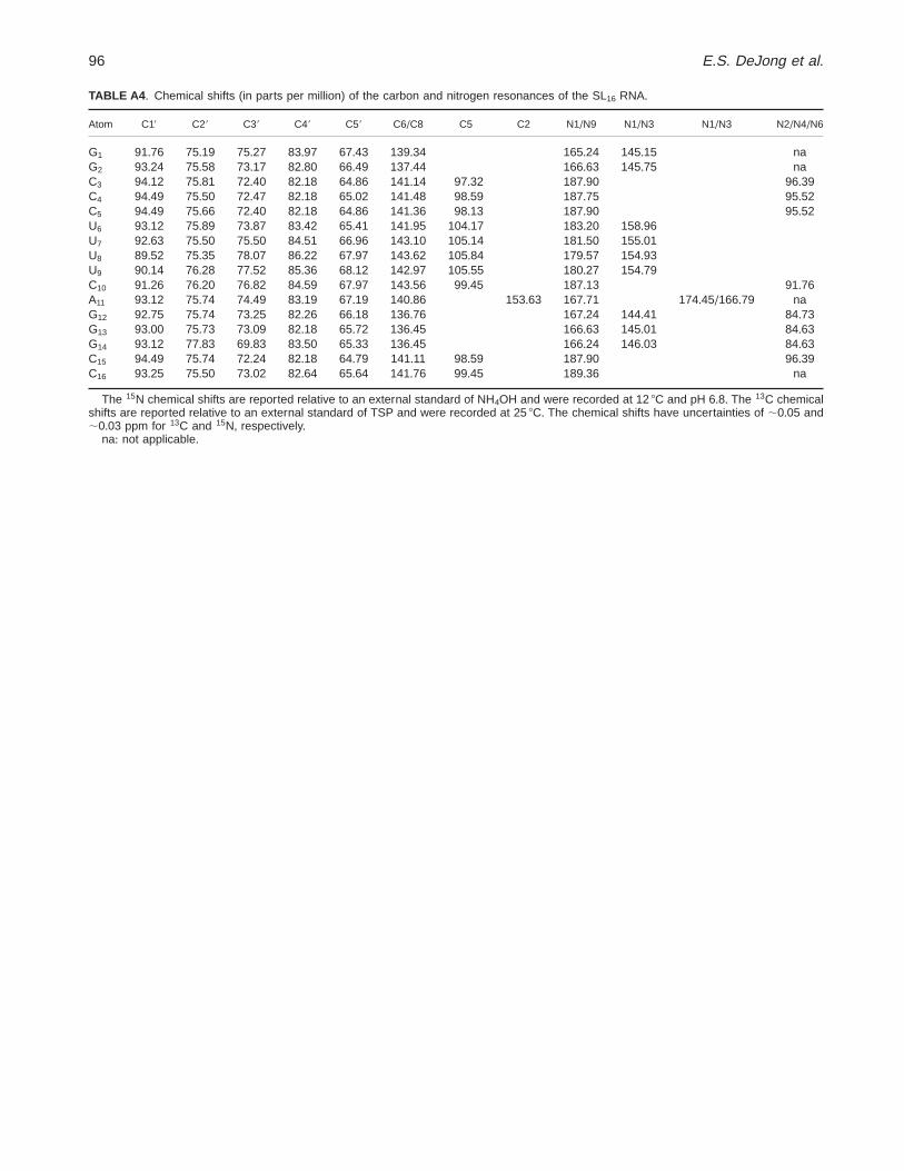
TABLE A4 + Chemical shifts (in parts per million) of the carbon and nitrogen resonances of the SL16 RNA+
Atom C19 C29 C39 C49 C59 C6/C8 C5 C2 N1/N9 N1/N3 N1/N3 N2/N4/N6
G1 91+76 75+19 75+27 83+97 67+43 139+34 165+24 145+15 naG2 93+24 75+58 73+17 82+80 66+49 137+44 166+63 145+75 naC3 94+12 75+81 72+40 82+18 64+86 141+14 97+32 187+90 96+39C4 94+49 75+50 72+47 82+18 65+02 141+48 98+59 187+75 95+52C5 94+49 75+66 72+40 82+18 64+86 141+36 98+13 187+90 95+52U6 93+12 75+89 73+87 83+42 65+41 141+95 104+17 183+20 158+96U7 92+63 75+50 75+50 84+51 66+96 143+10 105+14 181+50 155+01U8 89+52 75+35 78+07 86+22 67+97 143+62 105+84 179+57 154+93U9 90+14 76+28 77+52 85+36 68+12 142+97 105+55 180+27 154+79C10 91+26 76+20 76+82 84+59 67+97 143+56 99+45 187+13 91+76A11 93+12 75+74 74+49 83+19 67+19 140+86 153+63 167+71 174+45/166+79 naG12 92+75 75+74 73+25 82+26 66+18 136+76 167+24 144+41 84+73G13 93+00 75+73 73+09 82+18 65+72 136+45 166+63 145+01 84+63G14 93+12 77+83 69+83 83+50 65+33 136+45 166+24 146+03 84+63C15 94+49 75+74 72+24 82+18 64+79 141+11 98+59 187+90 96+39C16 93+25 75+50 73+02 82+64 65+64 141+76 99+45 189+36 na
The 15N chemical shifts are reported relative to an external standard of NH4OH and were recorded at 12 8C and pH 6+8+ The 13C chemicalshifts are reported relative to an external standard of TSP and were recorded at 25 8C+ The chemical shifts have uncertainties of ;0+05 and;0+03 ppm for 13C and 15N, respectively+
na: not applicable+
96 E.S. DeJong et al.