nitric oxide synthase in pancreatic islets during trauma...
TRANSCRIPT

LUND UNIVERSITY
PO Box 117221 00 Lund+46 46-222 00 00
Nitric Oxide Synthase in Pancreatic Islets During Trauma and Parenteral Feeding
Qader, Saleem
2004
Link to publication
Citation for published version (APA):Qader, S. (2004). Nitric Oxide Synthase in Pancreatic Islets During Trauma and Parenteral Feeding. SaleemSa'aed Qader, MD, MSc, MPH, Department of Surgery, Lund University Hospital, SE-221 85 Lund, Sweden,.
General rightsCopyright and moral rights for the publications made accessible in the public portal are retained by the authorsand/or other copyright owners and it is a condition of accessing publications that users recognise and abide by thelegal requirements associated with these rights.
• Users may download and print one copy of any publication from the public portal for the purpose of private studyor research. • You may not further distribute the material or use it for any profit-making activity or commercial gain • You may freely distribute the URL identifying the publication in the public portalTake down policyIf you believe that this document breaches copyright please contact us providing details, and we will removeaccess to the work immediately and investigate your claim.
Download date: 27. Dec. 2019

From the Department of Surgery and Gastroenterology Lund University Hospital
Lund University, Lund, Sweden Bulletin No, 124
NITRIC OXIDE SYNTHASE IN PANCREATIC ISLETS DURING
TRAUMA AND PARENTERAL FEEDING
AKADEMISK AVHANDLING
Som för avläggande av doktorsexamen I medicinsk vetenskap I ämnet kirurgi vid Medicinska fakulteten vid Lunds Universitet
kommer att offentligen försvaras i Föreläsningssal 3, Centralblocket, Universitetssjukhuset, Lund Torsdagen den 23 september 2004, kl 09:00
Av
Saleem Sa’aed Qader MD, MSc, MPH
Handledare: Fakultetsopponent: Docent: Mats Ekelund Docent: Folke Hammarqvist Docent: Albert Salehi Huddinge Universitetssjukhus Lund Stockholm

2

An Academic Dissertation
Regarding The
IN PTRAUM
NITRIC OXIDE SYNTHASE ANCREATIC ISLETS DURING A AND PARENTERAL FEEDING
Saleem Sa’aed Qader MD, MSc, MPH
Department of Surgery and Gastroenterology, Lund University Hospital
Faculty of Medicine Lund University Lund, Sweden
Lund University
Lund, September 23, 2004
3
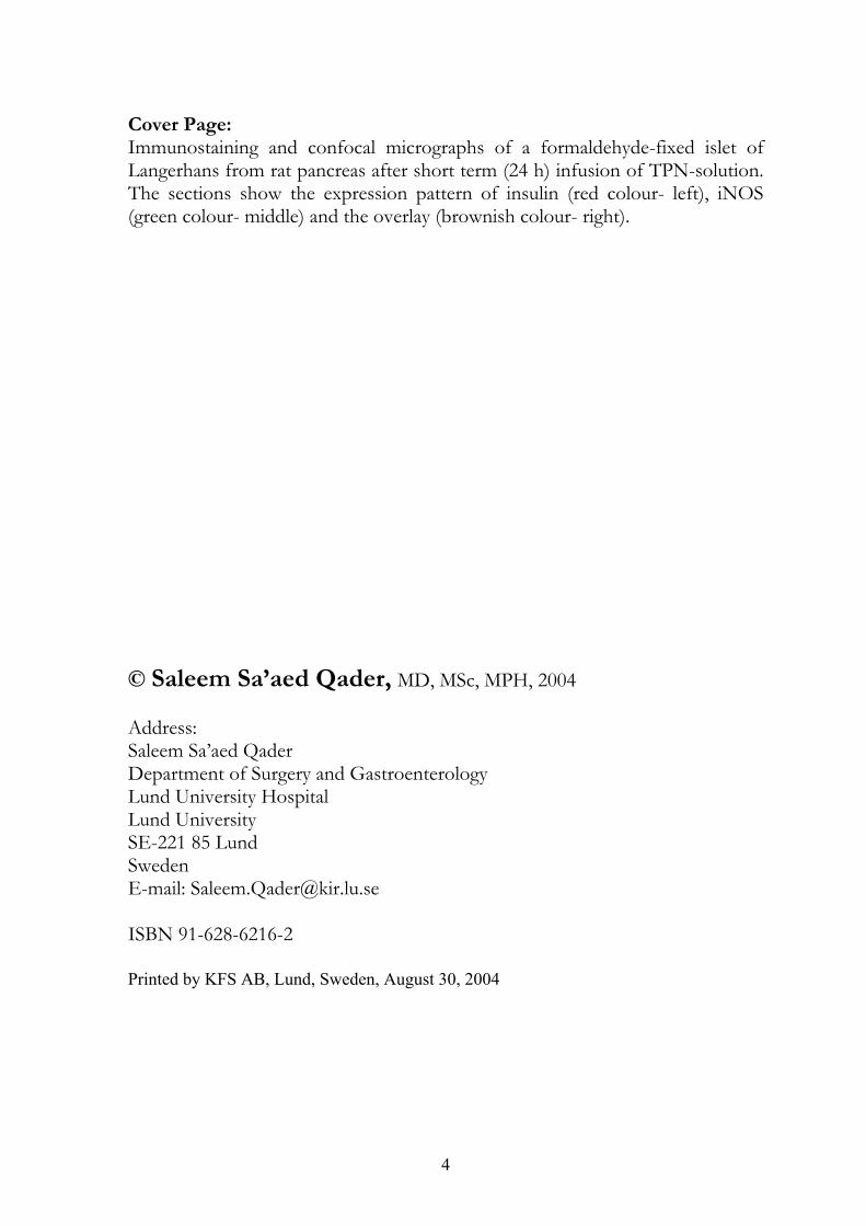
Cover Page: Immunostaining and confocal micrographs of a formaldehyde-fixed islet of Langerhans from rat pancreas after short term (24 h) infusion of TPN-solution. The sections show the expression pattern of insulin (red colour- left), iNOS (green colour- middle) and the overlay (brownish colour- right). © Saleem Sa’aed Qader, MD, MSc, MPH, 2004 Address: Saleem Sa’aed Qader Department of Surgery and Gastroenterology Lund University Hospital Lund University SE-221 85 Lund Sweden E-mail: [email protected] ISBN 91-628-6216-2 Printed by KFS AB, Lund, Sweden, August 30, 2004
4

5

6

7

8

CONTENTS LIST OF ORIGINAL PAPERS 11 ABBREVIATIONS 13 1. INTRODUCTION 15 1.1. BACKGROUND 15
1.1.1. Types of Diabetes Mellitus 15 1.1.2. Insulin Sensitivity 17 1.1.3. Obesity and NIDDM 17 1.1.4. Impact of Diabetes Mellitus on Health and Society 18
1.2. ISLETS OF LANGERHANS 18 1.2.1. Historical Aspects 18 1.2.2. Anatomy of Islets of Langerhans 19
1.2.2.1. Morphology 19 1.2.2.2. Histology of Islets of Langerhans 19
1.2.2.3. Nerve Supply 21 1.3. INSULIN SECRETION 22
1.3.1. The proximal event 23 1.3.2. The distal event 25
1.4. PITUITARY ADENYLATE CYCALSE-ACTIVATING POLYPEPTIDE 25 1.4.1. PACAP Receptors 26 1.4.2. The Biological activity of PACAP 27 1.4.3. Effects of PACAP on Endocrine pancreas 27
1.5. GHRELIN 28 1.5.1. Physiological Effects of Ghrelin 29 1.5.2. Regulation of Ghrelin Secretion 30
1.6. NITRIC OXIDE 30 1.6.1. Nitric Oxide Synthase System 31 1.6.2. Mechanisms of Biological Activity of NO 33 1.6.3. Nitric Oxide and Endocrine Pancreas 34 1.6.4. Mechanism for the Toxic Effects of NO on β-cells 34
1.7. TOTAL PARENTERAL NUTRITION 35 1.7.1. History 35 1.7.2. TPN in Clinical Use 35 1.7.3. Effects of TPN on Endocrine Pancreas 36
1.8. ACUTE PANCREATITIS 37 1.8.1. Pathophysiology 37 1.8.2. Acute Pancreatitis and Insulin Secretion 38
2. AIMS 39 2.1. GENERAL AIM 39 2.2. SPECIFIC AIMS 39 3. MATERIALS AND METHODS 41 3.1. ETHICS 41
9

3.2. ANIMALS 41 3.2.1. Exclusion Criteria 41
3.3. DRUGS AND CHEMICALS 41 3.4. COMPOSITION OF TPN-SOLUTION 42 3.5. SURGICAL PROCEDURES 42
3.5.1. Procedure for TPN Infusion 42 3.5.2. Induction of Acute Pancreatitis 43 3.5.3. Perfusion of Pancreas 43
3.6. OXYNTIC MUCOSAL BIOPSY 44 3.7. IN VIVO EXPERIMENTS 44 3.8. ISOLATION OF ISLETS OF LANGERHANS 44 3.9. IN VITRO EXPERIMENTS 45 3.10. BIOCHEMICAL AND RADIOIMMUNOLOGICAL ANALYSIS 45
3.10.1. Determination of Insulin and Glucagon 45 3.10.2. Determination of Plasma Glucose 45 3.10.3. Determination of Islets cAMP and cGMP 45 3.10.4. Determination of Protein 46 3.10.5. HPLC Analysis 46 3.10.6. Western Blot Analysis 46
3.11. HISTOCHEMISTRY AND IMMUNOHISTOCHEMISTRY 47 3.11.1. Confocal Microscopy 47 3.11.2. Immunohistochemistry 47
3.12. STATISTICAL ANALYSIS 48 4. RESULTS AND DISCUSSION 49 5. CONCLUSIONS 59 6. SUMMARY AND FUTURE ASPECTS 61 7. SUMMARY IN SWEDISH 63 8. ACKNOWLEDGEMENTS 65 9. GRANTS 67 10. REFERENCES 69 11. APPENDIX (PAPERS I-VI) 85
10

LIST OF ORIGINAL PAPERS
All works in this PhD thesis were carried out at the Department of Surgery, Faculty of Medicine, Lund University, Sweden in collaboration with the Institute of Physiological Sciences, Division of Pharmacology, Lund University, Sweden (Paper I-VI). All papers will be referred to in the text by their Roman numerals:
I. Saleem S. Qader, Mats Ekelund, Roland Andersson, Stefanie Obermuller and Albert Salehi. Acute pancreatitis, expression of inducible nitric oxide synthase and defective insulin secretion. Cell Tissue Res (2003) 313:271–279.
II. Albert Salehi, Saleem S. Qader, Eva Ekblad and Mats Ekelund.
Defective insulin secretion during total parenteral nutrition in rat and its normalization by pituitary adenylate cyclase-activating polypeptide 27. Regul Pept. (2004) 119: 83-91.
III. Saleem S. Qader, Javier Jimenez-Feltström, Mats Ekelund, Ingmar
Lundquist and Albert Salehi. Expression of islet inducible nitric oxide synthase and inhibition of glucose stimulated insulin release after long-term lipid infusion in the rat is counteracted by PACAP27: Submitted to AJP- Cell Physiology.
IV. Mats Ekelund, Saleem S. Qader, Javier Jimenez-Feldström and
Albert Salehi. Selective induction of inducible nitric oxide synthase in pancreatic islet of rat after an intravenous glucose challenge: Manuscript.
V. Saleem S. Qader, Albert Salehi, Rolf Håkanson, Ingmar Lundquist
and Mats Ekelund. Long-term infusion of nutrients (total parenteral nutrition) suppresses circulating ghrelin in food-deprived rat: Submitted to Regulatory Peptides.
VI. Saleem S. Qader, Ingmar Lundquist, Mats Ekelund, Rolf Håkanson
and Albert Salehi. Ghrelin activates neuronal constitutive nitric oxide synthase in pancreatic islet cells while inhibiting insulin release and stimulating glucagon release: Submitted to Regulatory Peptides.
The published papers are reprinted by permission of the copyright owners.
11

12

ABBREVIATIONS AA Arachidonic acid AC Adenylate cyclase ADP Adenosine 5’-diphosphate ATP Adenosine 5’-triphosphate AP Acute pancreatitis cAMP Adenosine 3,5-cyclic monophosphate CCK Cholecystokinin CGRP Calcitonin gene-related peptide cNOS Constitutive Nitric Oxide Synthase CPT 1 Carnithine palmitoyl- transferase I CaM Calmodulin DAG Diacyl glycerol DM Diabetes mellitus 2-DG 2-Deoxy-d-glucose eNOS/ ecNOS Endothelial Nitric Oxide Synthase ER Endoplasmic reticulum FFA Free fatty acid GDM Gestational Diabetes Mellitus GHS-R GH-secretagogue receptor GIP Glucose dependant insulinotropic polypeptide GLP-1 Glucagon-like peptide-1 GLP-2 Glucagon-like peptide-2 GRF GH-releasing factor GRP Gastrin-releasing peptide GSIS Glucose-stimulated insulin secretion GIT Gastrointestinal tract HPLC High-Performance Liquid Chromatography IBMX 3-isobutyl-1-methylxanthine i.c.v. Intracerebroventricular IDDM Insulin-dependant diabetes mellitus IL-1 Interleukin-1 IL-6 Interleukin-6 iNOS Inducible Nitric Oxide Synthase IP3 Inositol-1, 4, 5-triphosphate i.v. Intravenous
13

MODY Maturity Onset Diabetes of the Young MODS Multiple Organ Dysfunction Syndrome NADPH-d Nicotinamide adenine dinucleotide hydrogenphosphate-
diaphorase NIDDM Non-insulin-dependant diabetes mellitus NO Nitric oxide NOS Nitric oxide synthase NPY Neuropeptide Y nNOS/ ncNOS Neuronal Nitric Oxide Synthase PAC1-R PACAP receptor type 1 PACAP Pituitary adenylate cyclase-activating polypeptide PACAP-LI PACAP-like immunoreactivity PHM Peptide histidine-methionine PI3 kinase Phosphotidylinositol 3-kinase PIP2 Phosphotidyl-inositol-biphosphate PKA Protein kinase A PKC Protein kinase C PLA2 Phospholipase A2PLC Phospholipase C PLD Phospholipase D PP Pancreatic polypeptide RIA Radioimmunoassay SU Sulphonylureas SUR Sulphonylurea receptor SP Substance P T1D Type 1 diabetes mellitus T2D Type 2 diabetes mellitus TNF-α Tumour necrosis factor- α VDCC Voltage dependant L-type Ca2+ channels VIP Vasoactive intestinal peptide VPAC1-R VIP/PACAP receptor type 1 VPAC2-R VIP/PACAP receptor type 2
14

1. INTRODUCTION The first description of diabetes dates back to 1500 years before Christ when a pharaoh’s doctor noticed the accumulation of ants around the urine of some people rather than others. Hess Raa described it as a curable disease. It was then spoken of by Gallinious in Roman books. But the most accurate description of the disease and its complications appeared in a book, The Law in Medicine, by the president Ibn Sina (Avicenna) in the 10th century. Treating diabetes by changing the diet is certainly the oldest form of therapy and has been practiced in Egypt since 1500 B.C. This was confirmed by Professor George Ebers discovery of a large ancient Egyptian Papyrus in Upper Egypt in Luxor (Egyptian Diabetes Center, March 26th - March 29th, 2004). The name “diabetes” comes from the Greek word for a siphon (movement of fluid through a tube). The sweet taste of the diabetic urine was first coined by Araetus of Cappodocia (81-133 A.D.). Later, the word “mellitus” (honey sweet) was added by Thomas Willis (Britain) in 1675. In 1776 Dobson (Britain) for the first time confirmed the presence of excess sugar in urine and blood of diabetic patients as a cause of their sweetness (Ahmed AM, 2002).
1.1. BACKGROUND Diabetes mellitus is a metabolic disease caused by inherited or acquired deficiency in production of insulin by pancreas or by ineffectiveness of the insulin or by both, characterized by a high level of blood glucose. There are many risk factors for diabetes mellitus and the disease by itself acts as a risk factor for many other diseases. The classical complications notably are; diabetic ketoacidosis, hypoglycaemia, infections, renal failure, neuropathy, retinopathy and cardiovascular diseases. The life expectancy of the diabetic patients is about one-third less than that of the general population (Laing SP, 1999). 1.1.1. TYPES OF DIABETES MELLITUS (DM) 1. Insulin Dependant Diabetes Mellitus In insulin dependant diabetes mellitus (IDDM), also called “Type-1 diabetes mellitus” or “Juvenile diabetes mellitus”, the pancreas cannot produce insulin due to destruction of the insulin producing β-cells. It affects mainly the children and younger age group with a peak incidence between 10 and 14 years of age. The average annual increase in incidence in European children under 15 years of age is 3.4%. The treatment is daily insulin injections throughout the patient’s life. Pancreatic transplantation is currently the only known therapy for IDDM that establishes a long-term insulin-independent euglycaemic state. Islet transplantation may be another treatment.
15

2. Non-insulin Dependant Diabetes Mellitus Non-insulin dependant diabetes mellitus (NIDDM) also known as “Type-2 diabetes mellitus” or “adult onset diabetes mellitus”, is more common than IDDM and it constitutes about 85-95% of all diabetes in developed countries (WHO, 1994). In NIDDM there is a deficiency in the level of insulin secretion or reduced insulin sensitivity. Insulin resistance or reduced insulin sensitivity is an important risk factor for the development of NIDDM (DeFronzo RA, 1991). In these individuals, circulating insulin level is often increased initially in order to overcome the decreased peripheral insulin sensitivity. Eventually the clinical picture of diabetes develops indicating that an appreciable reduction in the β-cell function has occurred (Ferrannini E, 1997). As the β-cell function continues to decrease, the patient progresses from normal glucose tolerance to an abnormal metabolic state known as impaired glucose tolerance (IGT), which then culminate to diabetes with primarily postprandial hyperglycaemia to diabetes with fasting hyperglycaemia, a process that usually takes about 5 years (Lebovitz HE, 2001a). Such progression is however not inevitable; approximately 70% of individuals with IGT are expected to develop the disease (Diabetes Atlas IDF 18th, 2003). The increase in the incidence of diabetes mellitus differs between different populations. Studies in British (Keen H, 1982), Danish (Agner E, 1982) and Finish (Stengard JH, 1993) populations show that the increase in the incidence rate is 1.5-2% per year, while in the Dutch population (Heine RJ, 1996) and in South African Indians (Motala AA, 1993) the increase in incidence rate is about 13-14% per year.
Risk Factors Obesity, high fat diet, and low physical activity are the most important risk factors in developing NIDDM (Froguel P, 2003). Furthermore, genetic susceptibility is clearly needed for the development of NIDDM, but in most cases, it is not sufficient to induce the disease. Therefore, DM is regarded as a heterogenous disease that is caused by both genetic and environmental factors. First-degree relatives of NIDDM patients have a 40% lifetime risk of developing the disease, and the prevalence of NIDDM differs among different ethnic groups living in the same country (Barbetti F, 1996). Finally, diseases of the pancreas e.g. acute and chronic pancreatitis, hemochromatosis, pancreatic surgery, cystic fibrosis and pancreatic cancer are among other risk factors for the development of NIDDM. 3. Maturity Onset Diabetes of the Young Maturity onset diabetes mellitus (MODY) is a subtype of NIDDM and accounts for 2-5% of the cases of NIDDM (Froguel P, 1999). It is caused by a mutation in a single gene and is characterized by an autosomal dominant inheritance over three generations; onset is usually at less than 25years of age and constitutes a primary defect in insulin secretion (Fajans SS, 1990). Genetic studies have
16

shown that MODY can be caused by mutations in the genes encoding the glycolytic enzyme glucokinase (Froguel P, 1992). 4. Gestational Diabetes Mellitus Gestational Diabetes Mellitus (GDM) is defined as glucose intolerance that begins or is first recognized during pregnancy and affects approximately 7% of all pregnant women. A markedly obese woman with glycosuria, strong family history of diabetes mellitus and personal history of GDM is considered at high risk for gestational diabetes (Farrell M, 2003).
1.1.2. INSULIN SENSITIVITY
Reduced insulin sensitivity or insulin resistance is the essential metabolic abnormality in the development of NIDDM. Due to differences in tissue sensitivitie to insulin, the development of insulin resistance initially results in decreased disposal of glucose into the muscle and fat cells leading to postprandial hyperglycaemia, followed later by a more pronounced deficiency of insulin action, resulting in increased hepatic glucose output and overt fasting and all-day hyperglycaemia (DeFronzo RA, 1998). Predicting insulin resistance in normoglycaemic individuals is important, as diabetes intervention programs are more likely to be successful at this stage rather than after the development of IGT. Family history of diabetes, blood pressure (BP), fasting triglycerides, HDL, glucose, insulin and hepatic enzymes are known to correlate with insulin resistance (Laakso M, 1993; Matthews D, 1985). Insulin resistance is associated with high coronary and cerebrovascular mortality. 1.1.3. OBESITY AND NON-INSULIN DEPENDANT DIABETES
MELLITUS The risk of NIDDM is clearly linked to obesity, which forms the principle risk factor of the disease. An excess of body fat especially central obesity (within the abdomen) has potentially harmful consequences. There are several plausible explanations to the increased incidence of NIDDM in obese people. One reason is probably factors secreted from the adipose tissue with adverse effects on the β-cells e.g. free fatty acids and cytokines such as tumour necrosis factor-α (TNF- α). These substances promote resistance to insulin and may adversely affect the ß-cell function. Hepatic fat accumulation decreases insulin activation of glycogen synthase and increases gluconeogenesis and consequently hepatic insulin resistance (Samuel VT, 2004). Other factors behind the close relations between obesity and NIDDM are related to local accumulation of visceral fat and increased tissue acyl-CoA derivatives, which has specific effect in the insulin signal transduction and toxic effects on the ß-cell function. On the other hand there are some factors secreted from adipose tissue e.g. adiponectin with salutary effects e.g. it enhances insulin sensitivity in skeletal muscle and liver and it has protective effects on the vascular functions.
17

1.1.4. IMPACT OF DIABETES MELLITUS ON HEALTH AND SOCIETY
The incidence of diabetes mellitus is increasing worldwide. Currently some 194 million people worldwide or 5.1% in the adult population have diabetes with a female predominance and the incidence is expected to increase to 333 million, or 6.3% by 2025, during which the greatest number of persons with diabetes is expected to be in the South-East Asian Region with about 82 million NIDDM patients. It is the fourth or the fifth leading cause of death in most developed countries. In 2003, diabetes was one of the most common, non-communicable diseases globally. By 1995, diabetes was the number one cause of amputation, blindness, and end-stage renal disease and the 7th leading cause of mortality listed on death certificates. The lowest rate of NIDDM are generally found in rural communities where people are living lifestyles incorporating high levels of physical activity and low fat diet. It is rare or even absent less than 3% in some traditional communities in developing countries, like in Tanzanian Bantus (Ahrén B and Corrigan CB, 1984). On the other hand, the incidence of NIDDM is extremely high in other communities, e.g. more than 50% of the Pima Indians in Arizona, USA have diabetes (Knowler WC, 1978). Type 2 diabetes in children, some as young as 8 years of age, is an emerging problem with potentially serious outcome (Pihoker C, 1998). The estimated number of people with IGT, currently 314 million or 8.2% in the adult population have IGT, exacerbates the diabetes situation. The annual direct health care costs of diabetes worldwide, for people 20-79 years of age, is estimated to be at least 153 billion US dollars, and increasing continuously worldwide (International Diabetes Federation, 2003).
1.2. ISLETS OF LANGERHANS 1.2.1. HISTORICAL ASPECTS
The endocrine pancreas represented by the islets of Langerhans, were first described by Paul Langerhans in 1869 in his doctoral thesis on “Microscopic Anatomy of the Pancreas”. Langerhans observed that the islets were richly innervated but did not mention anything about the function of the islets. Five years after Langerhans death Laguesse named them “the islets of Langerhans” and he stated that these islets produce an anti-diabetic internal secretion (Morrison H, 1937). In 1921-1922, the Canadians “Banting, Best, Macleod and Collip” discovered insulin (Banting FG and Best CH, 1922). This made a revolution in the history of diabetes and led to the treatment of the first patient with diabetes in 1922. Isolation of insulin known as acomatol or pancreatin was first carried out before the Canadian research workers by Paulesco, Reuter and Zuelzer. Successful treatment of diabetes with pancreatic extracts was actually performed long before the Canadian isolation of insulin.
18

In 1923, Murlin et al described a hyperglycaemic factor in cat pancreas which was designated as glucagon (Murlin FC, 1923). 1.2.2. ANATOMY OF ISLETS OF LANGERHANS
1.2.2.1. Morphology The islets of Langerhans are groups of endocrine cells varying from a few hundreds to a few thousands; it forms 1-2% of the adult pancreatic mass, dispersed diffusely and embedded throughout the exocrine parenchyma of the gland with a tendency toward a higher islet concentration in the pancreatic tail region. A fine capsule consisting of fibroblasts and collagen surrounds them. The islets are ovoid clusters of cells measuring 0.1-0.24 millimetres in diameter and 500,000-1 million islets are found in the adult human pancreas. Islets of Langerhans have a very rich blood supply constituting about 20% of the blood supply of the gland during resting conditions and increasing after meals (Jansson L and Hellerstrom C, 1983; Lifson N, 1980), reflecting the very important role the islets play in the regulation of the metabolism. The arterioles supply the core of the islets first and reach the β-cells by passing through the discontinuity in the mantle zone (Figure 1) and form a fine capillary network among the β-cells which then supply the mantle cells and distally form efferent venules. The islets vasculature differs from that of exocrine pancreas in that they are wider and thinner walled and have more fenestrations (Henderson JR and Moss MC, 1985), enabling an extensive exchange of molecules. Islet blood flow is regulated by several factors (Jansson L, 1994), e.g. high blood glucose has been shown to increase the blood flow level in relation to the total pancreatic blood flow. There is, however, no relationship between the extent of the islets blood flow and insulin secretion (Jansson L, 1985). 1.2.2.2. Histology of islets of Langerhans The islets are composed of at least 4 different types of cells in both human and rat pancreas (Figure 1):
1. Insulin secreting β-cells (B-cells): 65-80% of the total cell population, localised mainly in the centre forming the core of the islets. Insulin has hypoglycaemic property by stimulating glucose uptake by peripheral tissue and increase glycogen storage in the liver. In addition it also inhibits glucagon secretion.
2. Glucagon secreting α-cells (A-cells): 10-15% of the total cell population, localised with other non-insulin secreting cells forming a mantle around the core of the islets. Glucagon has hyperglycaemic effect and stimulates glycogenolysis and gluconeogenesis in the liver, stimulates proteolysis to promote gluconeogenesis.
19

3. Somatostatin secreting δ-cells (D-cells): 5% of the total cell population, localised to the mantle zone (Luft R, 1974). Somatostatin inhibits both insulin and glucagon secretion (Alberti KG, 1973).
4. Pancreatic polypeptide PP-cells (F-cells): 10-15% of the total cell population, localised in the mantle zone. It produces pancreatic polypeptide (PP) which belongs to the neuropeptide Y (NPY) family. The pancreatic polypeptide (PP) is localised almost entirely within the pancreas, although detectable levels have been reported throughout the GI tract (Eva Ekblad and Frank Sundler, 2002).
5. Recently, ghrelin-cells have been isolated as a separate islet cell population in human fetal, neonatal, and adult pancreas. Ghrelin is not co-expressed with any known islet hormone and the ghrelin cells may therefore constitute a new cell type (Wierup N, 2002).
Fα δ
β β
β β
β β
β
β
ββ
β
Arteriole
Venule
Venule
Venule Ach, GRP, VIP, PACAP
NA, NPY, Galanin
Nutrients, GIP, GLP-1, GLP-2 Adrenaline Other
nerves
Sympathetic nerves
Parasympathetic nerves
β β
β
β β β β
β
β
β β
β β
β
β β β β
β
β
β β
β
β
β
β β β β
β β
β
β
δ
δ
δ δ
δ
δ
δ
δ
δ
δ δ
δ
δ
δ
FF
F
F
F
F
F
F
FF
F
α
α
α
α
α
α
α
α α
α
α
α
α α
α
α
α
α
α
CCK NO
β
β
β β
β
β β
β
β
β
β β
β
β β β
β
β
β
β
β β
β
β
β
β
β
β
β
β
Sensory nerves CGRP, SP
Figure 1: Illustration of the anatomy of pancreatic islets, showing the β-cell mass in the core of the islet and the surrounding mantle zone formed by α-cells, δ-cells, and F-cells. Afferent arteriols penetrate into the centre of the islet where, permeable, fenestrated, efferent venules are formed. Finally main branches of the autonomic nerves with their respective neurotransmitters are also shown (adopted from Ahrén B, 2000).
20

There are regional differences in the composition of the mantel cells within the pancreas. This difference is based on the embryological derivation of the pancreas. In the tail and the body of the pancreas, the mantel zone is rich in glucagon cells and poor in PP-cells, whereas in the head of the pancreas only few glucagon cells but many PP-cells are found. In recent years several polypeptides have been discovered and shown to be co-localised in the pancreatic islets with other important islet hormones. The physiological role of these peptides is still uncertain but most of them affect the insulin and glucagon secretion e.g. β-cells produce islet amyloid polypeptide (IAPP) and pancreastatin, while α -cells produce peptide YY (PYY), δ-cells produce diazepam binding inhibitor (DBI) and calcitonin gene-related polypeptide (CGRP). More recently adrenomedullin, a novel peptide has been demonstrated in PP-cells of the adult pancreas. Furthermore, during the last 2 decades several neuropeptides has been found to be localised to islet nerve terminals (Martinez A, 1998). 1.2.2.3. Nerve Supply
Islets of Langerhans have a rich nerve supply from parasympathetic, sympathetic and sensory nerves. The innervation of endocrine pancreas is much denser than that of the exocrine part of pancreas. In general, the nerve fibres enter the islets along the blood vessels and form either a peri-insular network in the mantle zone or pass directly to an endocrine cell. Parasympathetic nerves The cholinergic nerve fibres innervating the islets are of postganglionic origin and emanate from the intra-pancreatic ganglia. These ganglia penetrate the islets to terminate close to the endocrine cells and are controlled by preganglionic fibres originating in the dorsal motor nucleus of the vagus (Ahrén B, 1986; Brunicardi FC, 1995). There are 4 different neurotransmitters localised to islet parasympathetic nerves (acetylcholine, VIP, PACAP and GRP). All these neurotransmitters are released by activation of the vagal nerve and stimulate insulin and glucagon secretion. Sympathetic nerves The adrenergic nerves innervating the islets are postganglionic with most nerve cell bodies located in the celiac ganglion or in the paravertebral sympathetic ganglia. The preganglionic nerve fibres originate from nerve cell bodies in the hypothalamus and leave the spinal cord at the level of C8 to L3 to reach the paravertebral or celiac ganglia (Brunicardi FC, 1995). There are 3 different neurotansmitters localised to the islet sympathetic nerve fibres (noradrenaline, galanin and NPY). Activation of the sympathetic nerves inhibit basal and glucose-stimulated insulin secretion (Ahrén B, 2000).
21

Sensory nerves In addition to the cholinergic and adrenergic nerve supply of the islets, each individual islet is also extensively innervated by a network of sensory nerves harbouring the sensory neuropeptides calcitonin gene-related peptide (CGRP) and substance P (SP). These sensory nerves have been shown to innervate mainly the peripheral portion of the islets (Karlsson S, 1992; Rosenfeld MG, 1983). During recent years, it has been shown that the islet sensory nerves are involved in the regulation of islet hormone secretion. Other nerves Recently, in addition to these nerves other nerve fibres have also been found in the endocrine pancreas. Nerves containing nitric oxide synthase participate in the regulation of islet function, which is supported by the finding that inhibition of nitric oxide synthase inhibits insulin secretion induced by 2-DG (2-Deoxy-d-glucose) in mice (Ahrén B, 1995). CCK is also localised to the islet nerves (Rehfeld JF and Goltermann NR, 1980). CCK is a potent stimulator of insulin secretion through activation of CCK-A receptors, which are known to be present on islet β-cells (Verspohl EJ, 1986). Finally, nerves originating in ganglia in the duodenum might pass directly to the pancreas and innervate pancreatic ganglia, suggesting a direct entero-pancreatic neural mechanism (Kirchgessner AL, 1990).
1.3. INSULIN SECRETION Insulin is stored in approximately 13000 secretory vesicles or “granules” in the β-cells waiting to be released to the blood stream. The signals modulating insulin secretion are integrated at the level of the β-cells, enabling an optimal discharge of insulin from each individual cell. Insulin secretion is regulated by different mechanisms including both glucose and non-glucose factors of endocrine, neurocrine, paracrine, and autocrine nature (Barg S, 2002). The mechanism of insulin secretion may be divided into proximal and distal events. The proximal part is represented by the initial response to stimulus and activation of a second messenger, which leads to transduction of the signal to exocytosis process. The distal part includes movement of insulin containing granules, fusion with the plasma membrane and release of insulin. Five major intracellular signal transduction pathways are found (Figure 2). Activation of any one of these pathways depends on the property of the agent stimulating insulin secretion from the β-cells. All these pathways lead to increased mobilization of insulin containing granules from reserve pool to rapidly releasable pool near the plasma membrane of the β-cell (Ahrén B, 2000; Gopel S, 2004, Rorsman P, 2003).
22

1.3.1. THE PROXIMAL EVENT “SIGNALLING PATHWAYS” It includes the following pathways:
1. Glucose Regulating Metabolic Pathway Glucose, in a direct proportion to the extra-cellular glucose level, enters the cell through a specific glucose transporter in the plasma membrane (GLUT-2 in the rat), which is present in the β-cells and is insulin insensitive. After phosphorylation of the glucose molecule by means of glucokinase, the intracellular ATP/ADP ratio is increased and the ATP dependant K+ channels are closed. Closure of KATP channels causes depolarisation of the cell which in turn activates the voltage dependant L-type Ca2+ channels (VDCC) in the plasma cell membrane. This Ca2+ influx increases the intracellular Ca2+ [Ca2+]i concentration by about 10-fold which results in exocytosis of insulin containing granules (Kanno T, 2002). The mechanism by which elevation of [Ca2+]i concentration leads to exocytosis of insulin containing granules is not fully understood but it seems that activation of a Ca2+/ calmodulin-dependant protein kinase by acting at some late stage in the secretory process is involved. After insulin release the cells are re-polarized by the action of intracellular Ca2+ to activate Ca2+ dependant K+ channels to inhibit the L-type Ca2+ channels (Holz GG and Habener JF, 1992). The intracellular Ca2+ concentration returns to basal level also by action of a Na+- Ca2+ counter transport channel (Yoshihashi K, 1996) and by sequestration of Ca2+ into the endoplasmic reticulum (ER). Furthermore, glucose has been shown to stimulate insulin secretion by a Ca2+ independent mechanism, although this might not contribute significantly in the regulation of insulin secretion by glucose (Komatsu M, 1997; Rorsman P, 2003; Sato Y, 1998). 2. cAMP/Protein Kinase A pathway The second mechanism is receptor-mediated activation of the G protein, which activates adenylate cyclase (AC) by generating cAMP from ATP and subsequently activating protein kinase A (PKA). PKA stimulates exocytosis in several ways by phosphorylating different intracellular proteins and increasing the uptake of extracellular Ca2+ thus raising the cytosolic concentration of Ca2+and induce exocytosis. PKA also exerts direct effect on the distal events of exocytosis, which causes mobilization of insulin containing granules from the reserve pool to the readily releasable pool. Furthermore, cAMP/PKA induces an inward current in beta cells, which is mainly caused by an influx of Na+ into the cell, causing depolarisation followed by opening of the L-type Ca2+ channels and raising the cytosolic concentration of Ca2+and potentiation of insulin secretion. Besides, activation of PKA has several other effects on the β-cell function such as inhibition of cell apoptosis and inhibition of iNOS expression.
23

3. Phospholipase C Pathway The third mechanism is activation of phospholipase C (PLC), which hydrolyses phosphoinositides and produce inositol-1, 4, 5-triphosphate (IP3) and diacyl glycerol (DAG). IP3 diffuses in to the cytoplasm and promotes liberation of Ca2+ from Ca2+ storage sites, which increases the cytosolic Ca2+ concentration. DAG activates protein kinase C (PKC) that simulates the distal event in exocytosis. 4. Phosphlipase D Pathway The fourth mechanism is stimulation of phospholipase D (PLD). When PLD is activated it hydrolyses phosphatidyl choline to produce phosphatidic acid, choline and DAG (Exton JH, 1997). DAG is synthesised by further processing of phosphatidic acid.
Ach CCK GRP
GRP
Na+Ca2
INSULIN
G
Glucose
ATP
cAMP
PKA
AC
AA
closure
ATP/ADP ratio
Glucose metabolism
K+
depol. depol.
[Ca2+]cyt
ER Ca2+
G
G
G
PLD
PLC
VIP GLP-1 Glucagon
PLA2
PKC
DAG
IP3
PIP2
CCK
Path 4
Path 2
GLUT-2
SUR
Path 1
Path 3
Sulphonyl ureas
Path 5
Figure 2: Illustration of mechanisms of exocytosis showing the 5 pathways of intra-cellular signalling systems involved in the regulation of insulin secretion in the pancreatic β-cell. For further details, see the text. G =G-protein, depol. =Depolarisation of the cell, (adopted from Ahrén B, 2000)
24

5. Phospholipase A2 Pathway The last mechanism is activation of phospholipase A2 (PLA2). When PLA2 is activated as for example by Ach and CCK-8 (Koshimura K, 1997) it causes formation of arachidonic acid (AA) and subsequent production of arachidonic acid metabolites through the cyclooxygenase and lipooxygenase pathways, mainly prostaglandins and leukotrienes. AA and its metabolites stimulate insulin secretion by enhancing Ca2+ influx into the cell, and release of Ca2+ from intracellular stores, and also by activation of the distal part of the exocytotic machinery (Jones PM and Persaud SJ, 1993). 1.3.2. THE DISTAL EVENT
The proximal part of insulin secretion as described above leads to increase in cytosolic concentration of Ca2+, stimulate the formation of cAMP and arachidonic acid and its metabolites, as well as activates PKA and PKC, which all are stimulators of the distal event in exocytosis of insulin from the β-cells. The distal part of exocytosis includes the intracellular movement of insulin-containing vesicles, and fusion of these with the plasma membrane. The mechanism of the distal event of exocytosis is not studied as well as the proximal event of insulin secretion. The insulin containing granules are called a reserve pool and they need further modification before they can be released into the extracellular space (Renström E, 1997). Fusion of the insulin containing granules with the cell membrane occurs after priming of the vesicle containing insulin. The group of the primed vesicles is known as readily releasable pool. Circulating insulin level depends on the rate of exocytosis rather than on the speed at which insulin is synthesised in the β-cells (Kanno T, 2002). 1.4. PITUITARY ADENYLATE CYCALSE-
ACTIVATING POLYPEPTIDE (PACAP) PACAP belongs to the PACAP/glucagon superfamily; it is the most recent discovered neuropeptide in the family and was originally discovered as an amidated peptide of 38 amino acids. It is the most ancient and tightly conserved neuropeptide (96% over 700 million years) in terms of length and sequence identity of the nucleotides and amino acids. Therefore, it is the most likely ancestral molecule of the superfamily. The tight conservation of PACAP suggests that its function may be essential for survival. PACAP was originally isolated from an extract of ovine hypothalamus on the basis of its ability to stimulate cAMP formation in rat pituitary cells (Miyata A, 1989). Like other hypophysiotropic neurohormones, PACAP is contained in extra-hypothalamic neurons as well as in numerous peripheral tissues. It has been identified in both the brain and the gonads. Furthermore, PACAP has been shown to be localised in nerves in islets of Langerhans (Fridolf T, 1992). PACAP exists also in a C-terminally truncated 27 amino acid long form (Miyata A, 1990). Both peptides
25
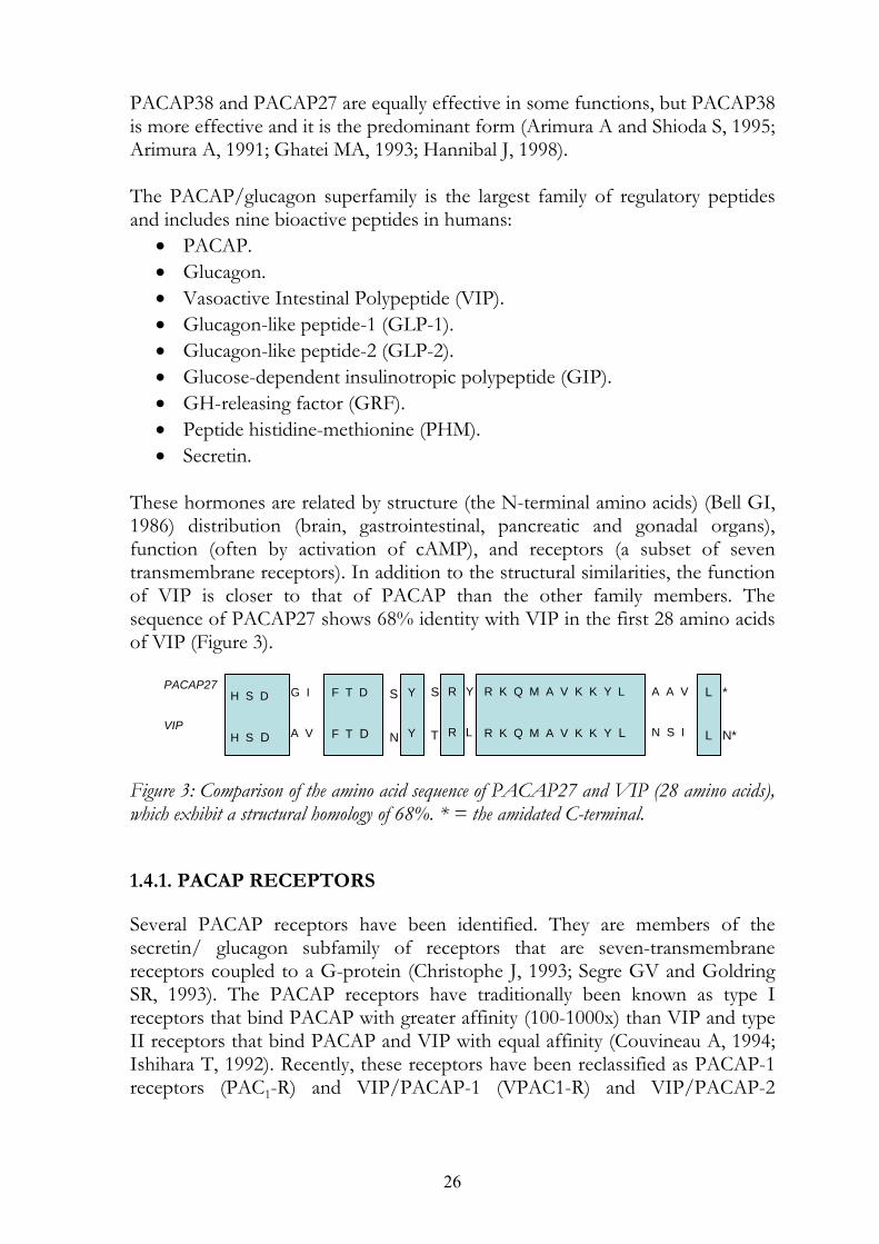
PACAP38 and PACAP27 are equally effective in some functions, but PACAP38 is more effective and it is the predominant form (Arimura A and Shioda S, 1995; Arimura A, 1991; Ghatei MA, 1993; Hannibal J, 1998). The PACAP/glucagon superfamily is the largest family of regulatory peptides and includes nine bioactive peptides in humans:
• PACAP. • Glucagon. • Vasoactive Intestinal Polypeptide (VIP). • Glucagon-like peptide-1 (GLP-1). • Glucagon-like peptide-2 (GLP-2). • Glucose-dependent insulinotropic polypeptide (GIP). • GH-releasing factor (GRF). • Peptide histidine-methionine (PHM). • Secretin.
These hormones are related by structure (the N-terminal amino acids) (Bell GI, 1986) distribution (brain, gastrointestinal, pancreatic and gonadal organs), function (often by activation of cAMP), and receptors (a subset of seven transmembrane receptors). In addition to the structural similarities, the function of VIP is closer to that of PACAP than the other family members. The sequence of PACAP27 shows 68% identity with VIP in the first 28 amino acids of VIP (Figure 3).
PACAP27 S L * R A A V Y R K Q M A V K K Y L G I Y F T D S H S D VIP R K Q M A V K K Y L R N S I L F T D A V Y T L N* H S D N
Figure 3: Comparison of the amino acid sequence of PACAP27 and VIP (28 amino acids), which exhibit a structural homology of 68%. * = the amidated C-terminal. 1.4.1. PACAP RECEPTORS Several PACAP receptors have been identified. They are members of the secretin/ glucagon subfamily of receptors that are seven-transmembrane receptors coupled to a G-protein (Christophe J, 1993; Segre GV and Goldring SR, 1993). The PACAP receptors have traditionally been known as type I receptors that bind PACAP with greater affinity (100-1000x) than VIP and type II receptors that bind PACAP and VIP with equal affinity (Couvineau A, 1994; Ishihara T, 1992). Recently, these receptors have been reclassified as PACAP-1 receptors (PAC1-R) and VIP/PACAP-1 (VPAC1-R) and VIP/PACAP-2
26

(VPAC2-R) receptors (Harmar AJ, 1998). PACAP receptors and their affinity to PACAP and VIP are listed in Table 1. Name Relative affinity PAC1-R PACAP38 ~ PACAP27 >>VIP VPAC1-R PACAP38 ~ PACAP27 ~ VIP VPAC2-R PACAP38 ~ PACAP27 ~ VIP Table 1: PACAP receptor sub-types and their affinities (adopted from Rawlings SR, 1996). 1.4.2. THE BIOLOGICAL ACTIVITY OF PACAP
The wide distribution of PACAP receptors and receptors shared with VIP provide clear evidence that PACAP has many target sites and functions. PACAP has been found to exert pleiotropic effects (Vaudry D, 2000; Gonzalez BJ, 1997) including:
1. Regulation of the cell cycle and development: • Proliferation • Differentiation • Apoptosis: both protecting cells from apoptosis and in triggering
apoptosis depending on the circumstances. 2. Regulation of smooth muscles (vascular, bronchial, intestinal and the
cardiac muscle). 3. Regulates the immune system: PACAP receptors are associated with
many immune cells. 4. Endocrine / paracrine regulator. 5. Regulation of bone metabolism. 6. Exocrine regulator: gastrointestinal secretion and pancreatic secretion. 7. Regulator in the nerve system and modulation of neurotransmitter
release. In pancreas the PACAP-immunoreactive fibers innervate exocrine acini and the islets of Langerhans as well as the small arteries of the connective tissue (Köves K, 1993; Tornoe K, 1997). 1.4.3. EFFECTS OF PACAP ON ENDOCRINE PANCREAS In the endocrine pancreas, PACAP appears to be much more potent than VIP or other regulatory peptides in the PACAP/glucagon superfamily in stimulating pancreatic hormone secretion. In vivo administration of PACAP causes a significant increase in plasma insulin levels in mice (Filipsson K, 1998a; Fridolf T, 1992), calf (Edwards AV, 1997) dog (Kawai K, 1992) and humans (Filipsson K, 1997). In vitro study has shown that PACAP acts as a potent stimulator to insulin secretion from islets isolated from rat pancreas (Yada T, 1994). In
27

addition PACAP is a strong stimulator of glucagon secretion. Intravenous injection of PACAP increases plasma glucagon concentrations in mice (Fridolf T, 1992) and humans (Filipsson K, 1997). The effect of PACAP is mediated through PAC1-R and involves activation of the adenylyl cyclase pathway (Borboni P, 1999) (Figure 4).
PACAP
Figure 4: Mechanism of action of PACAP in the β-cell, showing that when PACAP binds to its specific receptor on β -cells it activates AC (Adenylate cyclase) through activated G-protein (Trimeric GTP-binding regulatory protein) coupled to the receptor. Thus the receptor activated G-protein activates AC, which stimulates the conversion of ATP to cAMP. The subsequent increase in cellular cAMP content activates PKA. cAMP/PKA acts as an intracellular signaling pathway.
1.5. GHRELIN
Ghrelin a novel 28-amino acid orexigenic and adipogenic hormone was discovered in 1999 by Kojima and co-workers, as a natural endogenous bioactive ligand for the growth hormone secretagogue receptor (GHS-R) (Bowers CY, 2001; Hosoda H, 2000b; Kojima M, 1999; Kojima M, 2001). It was called ghrelin, a term that contains “ghre-” as the etymological root for “growth” in many languages and “relin”, a suffix for releasing substances i.e. “growth hormone release”, which is a characteristic effect of ghrelin (Hosoda H, 2000b; Kojima M, 1999). The third amino acid, serine is modified by n-
G-prot AC
ATP cAMP
Activates PKA
Insulin secretion Apoptosis
Insulin secretion
? iNOS expression/activity cNOS activity
?
?
28

octanoic acid, a modification essential for binding to the GHS-R and for release of growth hormone (Figure 5). By using antibodies against the octanoyl-modified serine and the C-terminal portion, two major molecular forms were demonstrated in various tissues: ghrelin itself and the nonmodified des-n-octanoyl form, designated as des-Gln14 ghrelin, which is a second endogenous ligand for GHS-R (Hosoda H, 2000a). Ghrelin, is predominantly produced by A-like cells in the oxyntic mucosa in the gastric fundus (Date Y, 2000; Dornonville de la Cour C, 2001; Kagotani Y, 2001; Kojima M, 1999), whereas substantially lower amounts are derived from the bowel (Date Y, 2000; Kagotani Y, 2001), the pituitary (Korbonits M, 2001), the kidney (Mori K, 2000), the placenta (Gualillo O, 2001), and the hypothalamus. Pancreatic ghrelin cells are numerous from midgestation to early postnatal period (10% of all endocrine cells) and clearly outnumbers those in the stomach. The cells are few but regularly seen in adults in the islet periphery, in exocrine tissue, in ducts, and in pancreatic ganglia. (Kagotani Y, 2001; Kojima M, 1999). (CH2)6-CH3
C=O
O
NH2 – G
–COOH
S L S P E H Q
QL KAP K P
K
Q
V
QR KE S
P R
R F S G
Figure 5: The structure of human ghrelin. The third amino acid serine is octanoylated. Residue number 14, glutamine is missing in des-Gln14 ghrelin.
1.5.1. PHYSIOLOGICAL EFFECTS OF GHRELIN Ghrelin stimulates growth hormone (GH) release by interacting with GH-secretagogue receptors (GHS-R) in the anterior pituitary. This receptor had been identified several years earlier than ghrelin, but only synthetic agents, referred to as GH-secretagogues (GHS), were known to bind to the GHS-R and release GH until the discovery of ghrelin (Howard AD, 1996). Ghrelin is involved in energy homeostasis and peripheral daily administration of ghrelin causes weight gain by reducing fat utilization in mice and rats, whereas intracerebroventricular (i.c.v.) administration generate a dose-dependant increase
29

in food intake and subsequently body weight (Tschop M, 2000). In man, intravenous (i.v.) ghrelin has been shown to stimulate food intake. Ghrelin is released from the stomach in response to fasting and increases feeding behaviour by acting on the arcuate nucleus of the hypothalamus (Akio Inui, 2001; Wren AM, 2001). The orexigenic actions of ghrelin are mediated by hypothalamic neuropeptide Y and agouti-related protein (Asakawa A, 2001; Kamergai J, 2001; Nakazato M, 2001). It has been demonstrated that i.c.v. or i.v. administration of ghrelin stimulates the secretion of GH (Kojima M, 1999), insulin, gastrin (Lee H-M, 2002), gastric acid (Date Y, 2001) and gastric motility (Masuda Y, 2000). Furthermore, ghrelin may have direct cardiovascular effects. 1.5.2. REGULATION OF GHRELIN SECRETION
The stomach has been identified as the major source of circulating ghrelin. Plasma ghrelin-like immunoreactivity levels in totally gastrectomised patients are reduced to 35% of those in controls (Ariyasu H, 2001). Ghrelin is markedly increased in patients with anorexia nervosa; weight gain decreases ghrelin concentration in these subjects (Otto B, 2001). In contrast, the ghrelin levels are decreased in obese Caucasians compared with lean (Tschop M, 2001), whereas weight loss increase circulating levels of ghrelin in obesity (Hansen TK, 2002). Plasma ghrelin levels may also reflect acute feeding states. Plasma ghrelin-like immunoreactivity in humans is increased during fasting and reduced immediately after feeding (Ariyasu H, 2001). Secretion of ghrelin is not affected by the stomach expansion per se. In rats, stomach filling with water does not change ghrelin levels whereas filling with dextrose significantly reduce serum ghrelin levels (Tschop M, 2000). In man oral administration of glucose but not the same volume of water reduces the mean plasma ghrelin concentration (Shiiya T, 2002). However, plasma ghrelin levels also decrease rapidly after intravenous glucose administration. These results indicate that ghrelin is an appetite-stimulatory peptide, signalling to the hypothalamus when an increase in energy demand is encountered (Inui A, 2001). 1.6. NITRIC OXIDE
Alfred Nobel did a pioneering discovery on nitroglycerin as an explosive, culminating in the invention of dynamite. In addition to the explosive property, nitroglycerin is a vasoodilator widely used since over 100 years for the treatment of angina pectoris. Just over a hundred years after Alfred Nobel’s death in 1998, the Nobel Prize in Physiology/Medicine was presented to Drs. Robert Furchgott, Ferid Murad and Louis Ignarro, in part for the discovery that NO acts as a biological mediator produced by mammalian cells (John L Wallace & Mark JS Miller, 2000).30

1.6.1. NITRIC OXIDE SYNTHASE SYSTEM NO is produced by a family of enzymes, Nitric Oxide Synthase (NOS), which was first identified and described in 1989. Three major isoforms were cloned and purified between 1991 and 1994. Isomeric forms of NOS representing at least three distinct gene products have been cloned in bovine, rat, mice and human tissue (Figure 6) (Christopherson KS & Bredt DS, 1997; Nathan C, 1997). NO is synthesised from a guanidino group of L-argenine and can be produced by almost all mammalian cells including endothelium lining the vasculature, neurones of the central and enteric nervous system and cells of the immune system (Moncada S, 1992; Nathan C and Xie Q-w, 1994). NOS isoforms differ in their dependence on calcium as well as in their expression and activity. NOS isoforms can be devided into 2 functional classes called:
• Constitutive Nitric Oxide Synthase (cNOS); ncNOS, ecNOS. • Inducible Nitric Oxide Synthase (iNOS).
The NOS isoforms and the common nomenclatures: 1. Neuronal NOS (ncNOS, Type I, NOS-I, and NOS-1); a neurally associated constitutive nitric oxide synthase found in neurons of the brain and the enteric nervous system. 2. Inducible NOS (iNOS, Type-II, NOS-II, and NOS-2); expressed in endothelium, epithelium, hepatocytes, chondrocytes and inflammatory cells. 3. Endothelial NOS (ecNOS, Type III, NOS-III and NOS-3); a constitutive enzyme normally present primarily in endothelium lining the vasculature.
ecNOS (constitutive)
↑ NO 1. Maintains blood
pressure. 2. Inhibits platelet
aggregation. 3. Inhibits leukocyte
adhesion.
ncNOS (constitutive)
↑ NO 1. Promotes GI Motility. 2. Neurotransmitter. 3. Inhibits insulin secretion.
iNOS (inducible)
↑↑↑ NO 1. Host defence. 2. Inflammation and pain. 3. Tissue destruction
(Cartilage, epithelium. 4. Inhibition of insulin
secretion. 5. Induces apoptosis.
Figure 6: Nitric oxide synthase isoforms and their main functions (adopted from Abramson SB, 2000).
31

cNOS (ecNOS and ncNOS) is constitutively present and shows Ca2+/CaM dependence i.e. their activities are regulated by the intracellular Ca2+ concentrations. In contrast, iNOS has CaM tightly bound to it as a prosthetic group holding the enzyme in active state independent of intracellular Ca2+ concentrations. iNOS is transcriptionally regulated by factors e.g. cytokines (TNF-α, IL1-beta) and other inflammatory mediators (Kubes P, 2000). NADPH
NADP
Reductase domain Oxygenase domain
NH2
+
L-arginine + O2
L-citrulline + NO
COOH
COOH
NH2
ZnS4
Intracellular targets or organisms
Remote target RS-NO
Adjacent target cell or organisms
NO
+RS
heme
heme
e-
CaM
FMNe- e-
N N
Fe3+NN
BH4
FAD
Figure 7: Illustration of the nitric oxide synthase system. The reductase and oxygenase domains are shown with their binding sites for NADPH, FAD, FMN and heme, L-argenine, and tetrahydrobiopterin (BH4). Between these regions lies the binding site for calmodulin (CaM). NOS functions as a dimer consisting of two identical monomers, which can be functionally and structurally divided into two major domains, the C-terminal reductase domain and the N- terminal oxygenase domain. The reductase and oxygenase domains together provide the complete machinery required for NO production. Different cofactors and substrates are required for the production of NO. Dimerization starts with the binding of heme. The binding of the heme and formation of a dimer makes it possible for tetrahydrobiopterin (BH4) to bind to the NOS dimmer, which leads to the formation of a stable dimmer (List BM, 1997; Venema RC, 1997). The pteridine tetrahydrobiopterin (BH4) is a key feature of NOS, affecting dimerization and electron transfer, although its full role in catalysis remains to be determined. ZnS4 is involved in this process. Electron (e-) is donated by NADPH to the reductase domain of the enzyme and proceeds via FAD and FMN redox carriers to the oxygenase domain, and then they interact with the heme and BH4 at the active site to catalyse the reaction of oxygen with L-argenine, generating an equimolar concentration of L-citrulline and NO as product (Albrecht EW, 2003). Electron flow through the reductase domain requires the presence of bound Ca2+/CaM. In some circumstances NO– may be produced instead of NO. Once NO is formed it may diffuse to targets. NO which is normally short-lived, may form stable adducts by interacting with thiol groups on carrier or storage protein (RS). This stable RS-NO may have local or remote actions.
32

1.6.2. MECHANISMS OF BIOLOGICAL ACTIVITY OF NO NO is the smallest synthetic product of mammalian cells and it is soluble in both water and lipids, thereby enhancing a free diffusion in the environment of the cell. It is a free radical molecule that has unpaired electrons which makes it extremely reactive (Nathan CF, 1992). Target molecules include oxygen, other radicals, thiol groups and metals such as iron. NO has a short half life, about 10 seconds and the interaction of NO with oxygen results in oxidation of NO to nitrite and nitrate (NO 2- and NO 3-) inactivating the molecule. Furthermore, NO can combine with other reactive molecules for example superoxid anion O2
- and form other radicals, including peroxynitrite (ONOO-), nitrogen dioxide, or hydroxyl radicals with the capacity to injure the target cells. Conversely, NO may provide a mechanism to “detoxify” other radicals. The interaction of NO with other targets e.g. other molecules containing heme-groups (hemoproteins) and/or iron-sulphur clusters and thiols form the basis for the mechanism by which NO exerts many of its effects, and forms complexes that activate or inactivate the target enzymes. Under conditions of high NO production a number of enzymes can be inhibited by NO-enzyme interaction (Feldman PL, 1993; Nathan CF, 1992; Drapier JC, 1986; Hibbs JB Jr, 1987; Stuehr DJ, 1989) (Table 2). The inhibition of these and other enzymes is believed to be the mechanism by which cytokine-generated NO can inhibit the growth of target cells, which may be in the form of invading microorganisms, tumour cells, or lymphocytes. Although the action of NO are mostly local within the cell, it can also diffuse to targets in the extracellular space or to adjacent cells or organisms, for example, for leukocyte derived NO to kill engulfed organism it must traverse the cell membranes.
Table 2: Enzyme targets of nitric oxide (Billiar TR, 1995). Enzyme Function Activation Soluble guanylate cyclase cGMP formation *Cyclooxygenase Eicosanoid synthesis Inactivation Aconitase TCA cycle NADH: ubiquinone oxireductase Electron transfer Succinate: ubiquinone oxireductase Electron transfer Riboneucleotide reductase DNA synthesis Glyceraldehyde-3-phosphate- Glycolysis Dehydrogenase Gluconeogenesis Cytochrome P450 Biotransformation NADPH oxidase O2 radical generation *Cyclooxygenase Eicosanoid synthesis
*Inducible type cyclooxygenase appears to be activated under stimulation with low concentration of nitric oxide, whereas higher concentrations may inhibit the cyclooxygenases.
33

1.6.3. NITRIC OXIDE AND ENDOCRINE PANCREAS Both cNOS and iNOS have been detected in the islets of Langerhans and in the vessels supplying them. When cNOS is activated it produces pulsatile low amounts of NO (picomolar- nanomolar) for a short period of time in response to receptor stimulation which acts as an intracellular signalling for insulin secretion (Panagiotidis G, 1992a; Salehi A, 1996; Salehi A, 2001a; Schmidt HH, 1992). iNOS is not a normal cellular constituent and can only be expressed in response to pathophysiological stimuli. When iNOS is expressed following exposure to diverse stimuli, such as inflammatory cytokines e.g. IL-1-β, TNF-α and lipopolysaccharide (LPS) it produces large amounts of NO in a sustained and mostly uncontrolled fashion in the β-cells (Flodstrom M and Eizirik DL, 1997; Henningsson R, 2002; McDaniel ML, 1997; Salehi A, 2001a). Thus, iNOS generates significantly greater and more sustained amounts of NO when compared to the constitutive isoforms (Nathan C and Xie Q-w, 1994; Nathan CF, 1997) and these levels of NO are regarded to be toxic to the β-cells. 1.6.4. MECHANISM FOR THE TOXIC EFFECTS OF NO ON β-
CELLS The large sustained amount of iNOS derived NO is toxic to the cell and involved in the β-cell damage and dysfunction and development of type 1 diabetes mellitus (Inada C, 1995). The toxic effects of NOS-derived NO on β-cell function could be through the following possible mechanisms (Figure 8):
1. The huge amount of NO has been reported to impair several vital sites in the β-cells (Eizirik DL and Pavlovic D, 1997; Henningsson R, 2002; McDaniel ML, 1996; McDaniel ML, 1997; Mosen H, 2000; Salehi A, 2001a) (Table 2) e.g. the Krebs cycle enzyme aconitase, the mitochondrial electron transfer chain and the nuclear DNA and ion channels which subsequently results in β-cell dysfunction and apoptosis (McDaniel ML, 1996; Rabinovitch A, 1996).
2. Activation of poly (ADP-ribose) synthase that results in the decrease of NAD content, leading to eventual cell death (Inada C, 1995; Radons J, 1994).
3. Mediation of blood flow dysfunction in hyperglycemia-induced deficiency in microcirculation (Moldovan S, 1996).
4. Influencing the activity of ionic channels (Krippeit-Drews P, 1995). For example it has been shown that NO opens K+ channels through suppression of phosphofructokinase activity and this in turn inhibits glucose induced insulin release in pancreatic β-cells (Tsumura Y, 1994).
34

5. Induction of the cleavage of DNA into nucleosomal fragments of 180-200 bp, nuclear shrinkage, chromatin condensation and apoptotic body formation (Kaneto H, 1995).
6. Recently, it has been found that NO-induced apoptosis in β-cells is mediated by the endoplasmic reticular (ER)-stress pathway. NO causes ER stress and leads to apoptosis through induction of ER stress-associated apoptosis factor CHOP (Araki E, 2003).
7. NO disrupts mitochondrial respiration (Brorson JR, 1999), which will derive the cells of energy source and eventually lead to cell death.
8. S-nitrosylation of glutathione system and/or important regulatory proteins at the distal site in the secretory process is possible targets (Akesson B, 1999; Henningsson R, 2002; Panagiotidis G, 1995; Salehi A, 1998).
1.7. TOTAL PARENTERAL NUTRITION (TPN) 1.7.1. HISTORY
Between 1261 and 1288 AD Ibn El-Nefis (the discoverer of the pulmonary circulation) wrote a chapter on the best mode for dissecting bones, peripheral vessels and internal organs of the chest (heart, lung, big vessels and the diaphragm) in his book Sharh Tashrih Al-Qanun (Rabie EA, 2003). Then, in 1628 William Harvey described the blood circulation, which formed the basis for intravenous infusion, and Wren and Elsholtz gave the first intravenous injection in the 17th century. Many investigations were performed during the following centuries showing that solutions containing electrolytes and glucose could be given intravenously in humans. The observation in the late 1930s when Robert Elman for the first time showed that amino acids in the form of protein hydrolysate could be administered safely to humans was the first step for TPN (Wretlind A, 1992). During the following years, major efforts were done to prepare fat in the form of an emulsion. The first safe fat emulsion, Intralipid®, was introduced in 1961 (Schubert O and Wretlind A, 1961). 1.7.2. TPN IN CLINICAL USE Enteral nutrition (EN) is the normal physiological pathway in maintaining nutrition and life of the patients. EN preserves the gut integrity, immune functions and reduces infectious complications (Kudsk KA, 1992). However, in certain conditions it is not possible to maintain this nutritional pathway. In these circumstances, the nutrition may be provided parenterally. By the use of glucose, fat and protein (TPN), it has been possible to obtain a good nutritional condition when the oral rout is not possible. TPN has clinical applications in preventing and treating starvation and malnutrition (Wretlind A and Szczygiel B, 1998) and it is indicated in various diseases and conditions such as the short
35

bowel syndrome and hypermetabolic states seen in e.g. sepsis, trauma and burns. It is also used to obtain bowel rest in some conditions e.g. inflammatory bowel disease (Fabio Guilherme Campos, 2002). Generally, the complications associated with TPN are associated with greater morbidity than those with enteral nutrition. Most adverse effects seem to be the result of gastrointestinal atrophy induced by food deprivation. In general, complications associated with TPN can be summarized as the following; adverse effects on gastrointestinal tract (GIT) such as atrophy of pancreas, intestinal mucosa and increase in the mucosal permeability with subsequent bacterial translocation (Mok KT and Meng HC, 1993; Pederson RA, 1985). Thus, presence of the intra-luminal nutrients and passage of food through GIT probably plays an important role in maintaining the integrity of GIT and regulation of the function of the glands. Furthermore, metabolic complications (fluid overload, hypertriglyceridemia, hypocalcaemia, hyperglycaemia and specific nutrient deficiencies) and immune suppression are other side effects of TPN (Monson JRT, 1986). 1.7.3. EFFECTS OF TPN ON ENDOCRINE PANCREAS (INSULIN SECRETION) TPN causes hyperlipidemia in the form of increased levels of free fatty acids (FFA), triglycerides, phospholipids and cholesterol (Ekelund M, 1994). Hyperlipidemia with an elevation of the FFA level is associated with impaired glucose tolerance and increased insulin resistance (Felber JP, 1988; Randle PJ, 1986). Hyperlipidemia in rats induced by intravenous infusion of intralipid has been shown to inhibit the β-cell functions selective for the glucose-stimulated insulin secretion (Sako Y and Grill VE, 1990), and produce a condition similar to that of NIDDM but with normal blood glucose level. Possible mechanisms involved in this process are: 1. Hyperlipidemia will result in disturbance of the metabolic process in the β-cell. Oxidation of FFA will inhibit the glucose metabolism in the β-cell and this will in turn inhibit glucose stimulated insulin secretion. 2. Absence of the stimulatory effects of incretin hormones e.g. the gastro-intestinal peptide CCK (cholecystokinine) and GIP (glucose-dependant insulinotropic polypeptide) in the enteroinsular axis. There is no insulin release from islets incubated at low glucose concentration in rat islets subjected to TPN for 6 days (Pederson RA and Brown JC, 1979). The insulin response to GIP in an isolated pancreas from TPN treated rats is greatly exaggerated and it has been suggested that the GIP receptors on the β-cells are up-regulated due to low levels of serum GIP throughout TPN (Pederson RA, 1985). During TPN treatment, plasma CCK concentration remains at fasting level (Fan BG, 1997) which may be another reason behind impaired insulin secretion during TPN.
36

3. Lundquist et al have shown that the islet lysosomal acid alpha-glucoside-hydrolases are involved in the process of nutrient-induced insulin secretion (Lundquist I, 1996). In addition Salehi et al have shown that TPN induces generalized suppression of the islet lysosomal/vacuolar system and impairment of the islet lysosome- acid glucan-1, 4-alpha-glucosidase activity which is associated with an impairment of glucose-stimulated insulin secretion (Salehi A, 2001b). 4. It has also been shown that under certain conditions i.e. in the presence of inflammatory agents or during a period of elevated plasma lipids or glucose the iNOS activity and expression is strongly induced in pancreatic β-cells which causes suppression of cNOS isoenzyme (Eizirik DL and Darville MI, 2001; Eizirik DL and Pavlovic D, 1997; Henningsson R, 2002; McDaniel ML, 1997; Salehi A, 2001a; Salehi A, 2001b).
1.8. ACUTE PANCREATITIS Acute pancreatitis (AP) is a common emergency condition. The incidence of acute pancreatitis varies considerably in different studies, countries and during different time periods. A low incidence has been reported in England (10/100,000) (Corfield A, 1985; Giggs J, 1988) and in Germany (15/100,000) (Assmus C, 1996), while the incidence is higher in USA (40-80/100,000) and in Finland (70/100,000) (Jaakkola M and Nordback I, 1993). Approximately 80% of all cases can be attributed to either gall stones or alcohol (Karne S and Gorelick F, 1999). The severity of acute pancreatitis varies from a mild self-limiting to a sever fulminating fatal condition. Fortunately, most of the cases are mild and conservative treatment results in rapid recovery. However, severe AP constitutes 15–20% of all cases (Barie PS, 1996; Steinberg W and Tenner S, 1994). In severe AP the inflammatory process in the pancreas is often aggressive with frequent involvement of regional tissues and remote organ systems (Banerjee A, 1995; Grönroos J, 1999; Mann D, 1994). 1.8.1. PATHOPHYSIOLOGY
The pathogenesis of acute pancreatitis is only partially known. However, acute pancreatitis is characterised by acinar cell injury (local inflammation). The major function of pancreatic acinar cells is synthesis, storage and secretion of powerful digestive enzymes and their inactive proenzymes, zymogens. In acute pancreatitis, the secretion of digestive enzymes from the acinar cells is blocked and the separation of digestive enzymes from the proteins is disturbed. AP starts with local inflammation due to infiteration of pancreas by activated macrophages and mast cells and a variety of inflammatory mediators of different chemical and functional classes are elaborated in the inflammatory process, such as arachidonic acid metabolites, nitric acid, cytokines (IL-1, IL-6, IL-8, TNF-α,
37

MIP1-α, MIP1-β, histamine, serotonine, platelet activating factor, leukotrines) and reactive oxygen species. This will result in acute inflammation which leads to increased vascular permeability, modulation of leukocyte trafficking, localised tissue destruction and gastrointestinal tract failure which results in increased permeability and enteric bacterial translocation and generalised inflammation which eventually will end in multiple organ dysfunction syndrome (MODS). 1.8.2. ACUTE PANCREATITIS AND INSULIN SECRETION
Endocrine pancreatic dysfunction often accompanies exocrine pancreatic impairment and vice versa because of their close functional and anatomical relations (Diaz-Rubio JL, 2002). Alcoholic pancreatitis is more often complicated by impaired glucose tolerance and diabetes mellitus than the other causes of pancreatitis. Pancreatic endocrine function impairment following acute pancreatitis is associated with decreased plasma insulin level. Furthermore, endocrine pancreatic function impairment is significantly more common after severe than after mild acute pancreatitis (Malecka-Panas E, 2002). Hyperglycemia during acute pancreatitis can be due to abnormalities in insulin secretion, increase in counter regulatory hormones release, or decrease in glucose utilization by peripheral tissues. High blood glucose levels are associated with severe acute pancreatitis and it is regarded as one of the prognostic factors in acute pancreatitis. Some patients are discharged with diabetes after an AP episode, while others develop diabetes during the first year of follow-up (Diaz-Rubio JL, 2002). Abe et al (Abe N, 2002) showed that glucose stimulated insulin secretion was impaired in islets isolated from rats with acute pancreatitis although the islets remained histologically intact and they concluded that the decrease in insulin secretion is possibly caused by impairment of some pancreatic β-cell functions.
38

2. AIMS 2.1. GENERAL AIM
To investigate the activity and expression of nitric oxide synthase isoenzymes in pancreatic islets during trauma and total parenteral nutrition. 2.2. SPECIFIC AIMS 1. Does acute pancreatitis influence glucose-stimulated insulin secretion and NOS isoenzymes expression and activity in pancreatic islets? (Paper I). 2. Is TPN induced impairment of glucose-stimulated insulin secretion related to cAMP production in pancreatic islets? Could cAMP stimulating agents such as PACAP27, PACAP38 and VIP restore normal insulin secretory capacity of islets in the TPN treated rats? (Paper II, III) 3. Does short-term (24 h) nutrient (glucose or intralipid) therapy affect the β-cell function in the rats? (Paper IV). 4. Does TPN affect the serum and oxyntic mucosal ghrelin? Does ghrelin affect insulin secretion? (Paper V, VI).
39

40

3. MATERIALS AND METHODS
3.1. ETHICS The studies were approved by the local animal welfare committee, Lund University, Lund, Sweden. 3.2. ANIMALS Male Sprague-Dawley rats (B&K, Sollentuna, Sweden) (175-220 g) were used in all studies. Before the experiments, the animals were fed a standard pellet diet (B&K, Sollentuna, Sweden) and tap water ad libitum. They were housed for 5 days prior to use in cages under conditions of constant temperature (22 °c) and humidity and subjected to a 12-hours light/dark cycle. All freely fed control rats were provided free access to standard pellet food and tap water throughout the experiments while rats treated with TPN were not allowed any oral intake of either water or food. All the animals in both the TPN and the control groups were kept individually in metabolic cages. 3.2.1. EXCLUSION CRITERIA
Rats having problems with the infusion system e.g. blockage, displacement of the catheter or leakage from the wound or the catheter were excluded from the studies. Signs of infection during or at the end of the experiment were also basis for exclusion. Rats with significant weight loss compared to the controls at the end of the experiment were also excluded from the study. 3.3. DRUGS AND CHEMICALS Collagenase (CLS IV) was obtained from Sigma Chemicals; St. Louis, MO. Bovine serum albumin was purchased from ICN Biochemicals, High Wycombe, UK. VIP, PACAP27 and PACAP38 were from Peninsula Europe (Merseyside, St. Helens, UK). The radioimmunoassay kit for cyclic AMP measurement was purchased from Amersham Pharmacia Biotech (Uppsala, Sweden). The radioimmunoassay kit for insulin determination was obtained from Diagnostika (Falkenberg, Sweden). The human ghrelin antiserum was obtained from Phoenix Pharmaceuticals, Belmont, CA, USA. The tracer was radioiodinated (I125 – labelled ghrelin -28) and used as standard from Yanaihara Institute, Shizuoka, Japan. The gastrin antiserum (2604) was a kind gift from Professor J. F. Rehfeld, Rigshospitalet, Copenhagen, Danmark. The different constituents
41

(Table 3) in the TPN solution were kindly provided by Fresenius-Kabi (Uppsala, Sweden). All other drugs were obtained from Sigma Chemicals; St. Louis, MO. 3.4. COMPOSITION OF TPN-SOLUTION The TPN solution was prepared under sterile conditions at the laboratory (Table 3) and given in an amount corresponding to approximately 270Kcal/kg/day.
Table 3: components of TPN solution (per 1222 ml)
1Vamin 14 g N/L 250 ml 2Glucose 50% 400 ml 3Glucose 5% 300 ml 4Addex-Natriumklorid 20 ml 5Addex-Magnessium 2.5 ml 6Addex-Kalium 20 ml 7Trace elements 10 ml 8Soluvit 10 ml 9Lipid soluble vitamin adult 10 ml 10Intralipid 200ml
*Contents; 1/ Vamin; 85 g amino acids (350 Kcl/L), 2/ Glucose 500 g/L ( 2000 Kcl/L), 3/ Glucose 50 g/L (200 Kcl/L), 4/ Addex NaCl; Na+ (4 mmol/ml), Cl- (4 mmol/ml), 5/ Addex Magnessium; Mg 2+ (1 mmol/ml), SO4
2- (1mmol/ml), 6/ Addex KCl; K+ (2 mmol/ml), Cl- (2 mmol/ml), 7/ Trace elements; Cr3+ (0.2 µmol/10 ml), Cu2+ (20 µmol/10 ml), Fe3+ (20 µmol/10 ml), Mn2+ (5 µmol/10 ml), Zn2+ (100 µmol/10 ml), F- (50 µmol/10 ml), I- (1.0 µmol/10 ml), MoO4
2- (0.2 µmol/10 ml), SeO32- (0.4 µmol/10 ml), 8/ Solvit; Vit B1 (2.5
mg/10 ml), Vit B2 (3.6 mg/10 ml), Nicotinamide (40 mg/10 ml), Vit B6 (4 mg/10 ml), Pontotenic acid (15 mg/10 ml), Vit C (100 mg/10 ml), Biotin (60 µg/10 ml), Folinic acid (0.4 mg/10 ml), B12 (5.0 µg/10 ml), 9/ Lipid soluble vitamine adult; Vit A (0.99 mg/ml), Vit D2 (5 µg/ml), Vit E (9.1 mg/ml), Vit K1 (150 µg/ml), 10/ Intralipid 2000 Kcl/l; Soya bean oil (200g/l), Lecithine (12 g/l), Glycerol (22 g/l). The caloric intake has been measured earlier and corresponds to approximately 270kcal/kg/d in both control and TPN treated rats. 3.5. SURGICAL PROCEDURES 3.5.1. PROCEDURE FOR TPN INFUSION The rats intended for TPN were anaesthesised by an intraperitoneal injection of 5 % chloral hydrate (1 ml/100g body weight) or by an intramuscular mixture of ketamine (Ketalar®) (70 mg/kg) and xylazine (Rompun®) (25 mg/kg) before the operation. The neck of the rat was shaved and the operative field washed with iodine solution. The operation was performed under sterile conditions. A
42
silicon-rubber catheter (Medical Grade Silicone Tubing), 0.635 mm in inner diameter and 1.1938 mm in outer diameter was inserted into the right external jugular vein according to the method of Steiger (Steiger E, 1972). After operations the rats were individually housed in metabolic cages and infused continuously with 5% glucose solution at a rate of 1 ml/h over night. Thereafter, infusion of TPN solution was started and continued for 7-10 days. The dose of the TPN solution was given according to the body weight of the rats. The rats serving as freely fed controls underwent the same operative procedure including insertion of the catheter. No TPN solution was infused and they resumed free oral feeding directly after recovery from anaesthesia. The catheter in both groups were flushed with 100 U/kg /day of low molecular weight heparin (Fragmin®; Pharmacia, Uppsala, Sweden) every second day. There was no significant difference between TPN rats and controls with respect to the body weight at the end of the experiments. 3.5.2. INDUCTION OF ACUTE PANCREATITIS The rats were anaesthetised with 5% chloral hydrate (1ml/100 g body weight) administered intraperitoneally and operated under aseptic conditions. The proximal and the distal end of the common bile duct were clamped and a thin polyethylene catheter (0.66 mm OD, Protex LTD, Hythe, Kent, England) was introduced into the biliary-pancreatic duct. Acute pancreatitis was induced by intraductal infusion of 0.2 ml glycylglycine-NaOH (0.025mol/l) buffer, pH 8.0, containing 5% sodium taurodeoxycholate (0.04 ml/min) sterilised at 100 ºC for 20 minutes. Sham operation (control) included laparotomy and isolation of the common bile duct, though without bile salt injection. A detailed description of the methodology has been reported previously (Andersson and Wang 1999). 3.5.3. PERFUSION OF PANCREAS
Three hours after induction of acute pancreatitis, the pancreatic vasculature was perfused. The pancreatic perfusion technique included ligation of the celiac trunk and subsequent cannulation of the superior mesenteric artery and the portal vein respectively. The tube in the superior mesenteric artery was connected to a pumping devise and the one in the portal vein was connected to a syringe aspirating the blood at 1 minute interval. The perfusion was achieved by using Krebs Ringer bicarbonate buffer (1.0 mmol/l or 20.0 mmol/l glucose) containing 0.20% bovine serum albumin (BSA). The medium was gassed with 95% O2–5% CO2 to obtain constant pH (7.40) and oxygenation. The flow rate was maintained at 0.4 ml/min. After 15 min of equilibration, the venous effluent was collected at 1 minute intervals by a Teflon cannula. After 10 min of
43

perfusion with low (1.0mmol/l) glucose in the perfusate high glucose (20.0mmol/l) was introduced and lasted for 20 minutes. Time 0 was defined as the start of perfusion. The blood samples were centrifuged and the plasma was collected, immediately frozen and stored at -20 ºC until analysis. The rats were sacrificed at the end of the experiments. 3.6. OXYNTIC MUCOSAL BIOPSY The stomach was opened along the major curvature and rinsed in saline. Thereafter the acid producing (oxyntic) mucosa was scraped off the muscular wall of the stomach. Oxyntic mucosa were frozen and stored at -20 °C until analysis. The mucosa was weighed, frozen and extracted in boiling 0.5 M acetic acid for 10 minutes (1ml/100 mg tissue). After centrifugation at 5000X g for 20 minutes, the supernatant lyophilised and reconstituted in assay buffer (0.04 M Na2 HPO4. 2H2O, 0.01 M NaH2PO4. H2O, 4 mM NaN3, 7 mM EDTA, 5% Trasylol, 0.25% BSA), giving a concentration of 1-5 mg tissue per millilitre buffer.
3.7. IN VIVO EXPERIMENTS
A blood sample was taken from the jugular catheter for measuring the basal levels of plasma insulin at time 0. Then glucose (800 mg/kg body weight) or glucose + PACAP27 (5.0 nmol/kg body weight) was injected as a bolus via the jugular catheter directly after stopping the TPN infusion. Blood samples were then taken from the jugular catheter at 3 min after the injection. Plasma was collected, immediately frozen, and stored at -20 °c until analysis for insulin and glucagon.
3.8. ISOLATION OF ISLETS OF LANGERHANS
Preparation of pancreas The distal end of the pancreatic duct was clamped and injected with approximately 5 ml of ice-cold collagenase solution via cannulation of the biliary pancreatic duct (Salehi AA and Lundquist I, 1993). Thereafter, the pancreas was dissected and carefully separated from the surrounding tissue and then placed in a glass scint-tube (20 ml) and in a water bath (30 cycles/ minute) at 37 ºC for 11 minutes. Isolation of islets The pancreatic islets were separated from the acinar tissue by vigorous shaking in ice cold Hank’s solution for several minutes. After sedimentation for about 20 minutes the islets were collected under a stereomicroscope at the room temperature.
44

3.9. IN VITRO EXPERIMENTS The freshly isolated islets were pre-incubated for 30 minutes in an incubation box (30 cycles/minute) at 37 ºC in Krebs Ringer bicarbonate (KRB) buffer, pH 7.4, supplemented with 10 mmol/l HEPES, 0.1% bovine serum albumin, and 1.0 mmol/l glucose as previously described (Salehi AA and Lundquist I, 1993). Each incubation vial contained 12 islets in 1.0 ml buffer solution and was gassed with 95% O2-5% CO2 to obtain constant pH and oxygenation. After pre-incubation the buffer was changed to a medium supplemented with test agents, and the islets were incubated for 60 minutes. All incubations were performed at 37 ºC in an incubation box (30cycles/minute). Immediately after incubation, aliquots of the medium were removed for assay of insulin.
3.10. BIOCHEMICAL AND RADIOIMMUNO-LOGICAL ANALYSIS
3.10.1. DETERMINATION OF INSULIN AND GLUCAGON The concentration of insulin and glucagon were determined by RIA (Ahrén B and Lundquist I, 1982; Heding L, 1966; Panagiotidis G, 1992b). 3.10.2. DETERMINATION OF PLASMA GLUCOSE
Plasma glucose concentration was determined enzymatically (Bruss ML and Black AL, 1978). 3.10.3. DETERMINATION OF ISLET cAMP AND cGMP
After incubation, the islets were thoroughly washed in glucose-free KRB buffer and collected and stored in 500 µl of ice-cold 10 % trichloroacetic acid (TCA) containing the phosphodiesterase inhibitor IBMX (3-isobutyl-1-methylxanthine) (0.2 mmol/l), followed by immediate freezing in a -70 ºC ethanol bath. Before assay, 500 µl of H2O was added, and the samples were sonicated three times for 5 seconds followed by centrifugation at 1100 g for 15 minutes. The supernatants were then collected and extracted with water-saturated diethyl ether (4x2 ml). The aqueous phase was removed and freeze dried, using a Lyovac GT 2 freeze dryer. The residue was then dissolved in 450 µl of Na-acetate buffer (50 mmol/l, pH 6.2). The amounts of cyclic AMP and cyclic GMP were quantified with [125I]-cyclic AMP and [125I]-cyclic GMP radioimmunoassay kits (RIANEN; Du Pont Company, Boston, MA). [3H] –cyclic GMP was added to the TCA islet homogenate in order to determine the recovery of cyclic AMP and cyclic GMP during the ether extraction. The mean recovery was 90 %.
45

3.10.4. DETERMINATION OF PROTEIN Protein was determined according to Bradford (Bradford MM, 1976). 3.10.5. HIGH-PERFORMANCE LIQUID CHROMATOGRAPHY
(HPLC) ANALYSIS OF NOS ACTIVITY The freshly isolated islets were thoroughly washed and collected in ice-cold buffer (200 µl) containing HEPES (20.0 mmol/l), EDTA (0.50 mmol/l) and d, l-dithiothreitol (DTT) (1.0 mmol/l), pH 7.2, and stored at -20 ºC for subsequent NOS analysis. In brief, after sonication on ice, the buffer solution containing the islet homogenate was supplemented to contain also CaCl2 (0.45 mmol/l), calmodulin (25 U/ml), NADPH (2.0 mmol/l) and L-arginine (0.2 mmol/l) in a total volume of 450 µl. For the assay of iNOS, both calmodulin and CaCl2 were omitted from the buffer as previously described (Henningsson R, 2002; Salehi A, 1996). The homogenate was then incubated at 37 ºC under constant air bubbling (1.0 ml/min) for 180 min. Aliquots of the incubated medium (200 µl) were mixed with an equal volume of O-phthaldialdehyde reagent solution in a glass vial and then passed through a 1-ml Amprep CBA cation-exchange column for high-performance liquid chromatography (HPLC) analysis. The amount of L-citrulline formed (NO and L-citrulline are produced in equimolar concentrations) was then measured in a Hitachi F1000 fluorescence spectrophotometer (Merck, Darmstadt, Germany) as previously described (Salehi A, 1996). 3.10.6. WESTERN BLOTT ANALYSIS Approximately 250 islets were collected in Hanks’ buffer (100 µl) and sonicated on ice (3x10 s). Homogenate samples representing 10 µg of total protein from islet tissue were then run on 10% SDS-polyacrylamide gel. After electrophoresis, proteins were transferred to nitrocellulose membranes by electrotransfer (10–15 V, 60 min) (semidry transfer cell, B10-RAD, Richmond, CA). The membranes were blocked in 9 mmol/l TRISHCl (pH 7.4) containing 5% non-fat milk powder for 40 min at 37 ºC. Immunoblotting with rabbit anti-mouse iNOS (N-7782) or ncNOS (N-7155) (1:2000) (Sigma, St. Louis, MO) was performed for 16 h at room temperature. The membrane was washed twice and then incubated with alkaline-phosphatase conjugated goat antirabbit IgG (1:10,000) (Sigma) for 90 min. Antibody binding to ncNOS and iNOS was detected using 0.25 mmol/l CDP-Star (Tropix, Bedford, MA) for 5 min at room temperature. The chemiluminescence signal was visualized by exposing the membranes to Dupont Cronex X-ray films for 1–5 min. An appropriate standard, i.e. molecular mass markers, was run in all analyses. The intensities of the bands were quantified by densitometry (Bio-Rad GS-710 Densitometer).
46
3.11. HISTOCHEMISTRY AND IMMUNO- HISTOCHEMISTRY
3.11.1. CONFOCAL MICROSCOPY The freshly isolated islets were fixed with 4% formaldehyde, permeabilized with 5% Triton X-100, and unspecific sites blocked with 5% normal donkey serum (Jackson Immunoresearch Laboratories, Inc.). cNOS and iNOS were detected with the corresponding rabbit-raised primary antibodies (BD Transduction Lab., USA) in combination with Cy2-conjugated anti-rabbit IgG (Jackson Immunoresearch Laboratories, Inc.). For staining of insulin, islets were incubated with a guinea pig-raised anti-insulin antibody (Eurodiagnostica, Malmö, Sweden) followed by a Cy5-conjugated anti-guinea pig IgG antibody (Jackson Immunoresearch Laboratories, Inc.). Fluorescence was visualized with a Zeiss LSM510 confocal microscope by sequentially scanning at (excitation/emission) 488/505–530 nm (Cy2) and 633/>650 nm (Cy5). 3.11.2. IMMUNOHISTOCHEMISTRY The mid-portion of the pancreas from both controls and TPN- treated rats were dissected out and placed over night in a mixture of 2% formaldehyde and 0.2% picric acid in phosphate buffer (pH 7.2) followed by thorough rinsing in Tyrode’s solution containing 10% sucrose. Specimens were then frozen on dry ice and cut in a cryostat to a thickness of 10 µm. For the detection of VIP-containing nerve fibres, a polyclonal VIP antiserum raised in rabbit (code no 7852, dilution 1:1280; Eurodiagnostica, Malmö, Sweden) was used. This antiserum does not cross-react with PACAP38 or PACAP27. For the detection of PACAP containing nerve fibres, a polyclonal antiserum raised against pure ovine PACAP27 (code no. 88121-3, dilution 1:1280; kind gift from Prof. A. Arimura, Louisiana, USA) was used. This antiserum cross-reacts with both PACAP38 and PACAP27 but not with VIP. After incubation with primary antibodies over a night at 4 °C in a moist chamber, the sections were exposed (60 min) to fluorescein isothiocyanate-conjugated swine anti-rabbit IgG antiserum (DAKO, Glostrup, DK), diluted 1:400; after which the sections were mounted and analysed using a fluorescence microscope with appropriate filter settings. For controls, the antisera were inactivated by the dilution of an excess amount of antigen (10-100 µg of synthetic peptide/ml diluted antiserum).
47

3.12. STATISTICAL ANALYSIS
Probability levels of random differences were determined by analysis of variance followed by Tukey-Kramers’ multiple comparisons test. Student unpaired t-test were also used to detect the level of the significance for the difference between sets of data. Results are expressed as means ± SEM. P<0.05 was considered statistically significant.
48

4. RESULTS AND DISCUSSION Acute pancreatitis causes hyperglycaemia due to a defective insulin secretion and a marked induction of iNOS in β-cells (Paper I) 1. Acute pancreatitis (AP) impairs glucose-stimulated insulin secretion (GSIS): In vivo study by using pancreatic perfusion 3 hours after induction of acute pancreatitis and in vitro incubation of islets isolated from rats 3 hours after induction of acute pancreatitis showed impaired glucose-stimulated insulin secretion both at basal (1 mmol/l) and high (20 mmol/l) glucose concentration compared to controls. Glucose-stimulated insulin secretion showed a typical biphasic pattern when the glucose concentration in the perfusate was raised from 1.0 mmol/l to 20.0 mmol/ l in the control rats. In acute pancreatitis, however, this biphasic pattern was less obvious as a sign of a marked impairment of glucose-stimulated insulin release. 2. Acute pancreatitis causes marked iNOS expression and activity in islets β-cells: Marked induction of iNOS activity and expression with concomitant suppression of cNOS in AP was demonstrated by different methods (Immunohistochemical, HPLC, and Western blot analysis). Double labelling for iNOS and insulin showed that almost all insulin-positive cells were positive for iNOS. But iNOS was also expressed in some non-insulin secreting cells. iNOS was not only present in the cytoplasm but also exhibited a vesicle-like distribution in the β-cells. However, there was only a minor colocalization between insulin and iNOS, indicating that iNOS-positive vesicles were different from insulin-containing dense-core granules. In acute pancreatits the pancreas is infiltrated by activated inflammatory cells (macrophages and lymphocytes) which results in marked production of cytokines like IL-1, IL-6 and TNF-α which causes subsequent tissue destruction (Andersson R and Wang XD, 1999; Chen CC, 1999; Sweiry JH and Mann GE, 1996). Abnormalities of glucose metabolism in human have been reported early in the course of acute pancreatitis (Buscher HC, 1999). By both in vitro incubation of islets from rats with AP and in vivo study during pancreatic perfusion (a model regarded to be more physiological) we demonstrated that both basal and glucose-stimulated insulin secretion were markedly impaired in AP rats. Both the first and the second phase of insulin secretion were markedly defective in AP rats compared to controls. A decreased GSIS especially loss of the first phase and severely impaired second phase of insulin secretion is a common feature in NIDDM. In the present study we demonstrated a strong expression and marked increase in the activity of iNOS and a marked reduction in cNOS expression and activity
49

in islets isolated from AP rats. The decreased cNOS activity may be explained by the fact that iNOS derived NO might exert a negative feedback on cNOS expression and activity (Henningsson R, 2002; Salehi A, 2001a), suggesting an interactive mechanism between islet iNOS and cNOS (Salehi A, 2001a). Minc-Golomb et al’s (Minc-Golomb D, 1994) findings are in concordance with our finding; they showed that exposure of neural cells to cytokines up-regulates the activity of iNOS and down-regulates the activity of cNOS. Interestingly, in vitro incubation of islets or a single beta cell with cytokines or NO donors has been shown to result in impaired glucose-stimulated insulin secretion (Darville MI and Eizirik DL, 1998; Eizirik DL and Pavlovic D, 1997; McDaniel ML, 1996). Furthermore, in vitro studies have shown that pancreatic islets exposed to different cytokines express iNOS mRNA, increased iNOS activity and an elevated NO production that subsequently damages several vital sites in the β-cells (McDaniel ML, 1996; Sandler S, 1994; Yamada K, 1993) (Figure 8). The effect may be exerted mainly through impairment in mitochondrial oxidative metabolism and formation of nitrosyl-iron complexes (Akesson B, 1999; Salehi A, 1996). The NO induced S-nitrosylation may then disturb the balance of glutathione system, impairing the function of critical thiol groups which is known to be essential for insulin secretion (Ammon HP, 1997; Ammon HP and Mark M, 1985). Apart from an excessive production of NO, AP also results in the synthesis of reactive oxygen species such as superoxide (O2
-) (Chen CC, 1999; Kwon G, 1998). Interestingly, both NO and O2
- have been suggested as free radical mediators of cytokine-induced β-cell destruction (Eizirik DL and Darville MI, 2001; Eizirik DL and Pavlovic D, 1997; McDaniel ML, 1996). NO and O2
- are known to cause DNA damage accompanied by poly (ADP-ribose) synthase activation, which subsequently results in NAD depletion in β-cells (Delaney CA and Eizirik DL, 1996). The fall in the cellular content of NAD thus markedly reduces the cellular activities and, therefore, the β-cell may be more susceptible to damage caused by depletion in NAD content. Moreover, combination of these two highly reactive molecules (NO, O2
-) results in the formation of peroxynitrite, which is a powerful oxidant and cytotoxic agent. TPN induces impairment of glucose-stimulated insulin secretion through cAMP suppression, which may be reversed by PACAP and VIP (Paper II, III).
1. Impaired GSIS in TPN- treated rats was improved by PACAP and VIP: In vitro incubation of TPN-treated rat islets exhibited marked suppression of basal and high glucose-stimulated insulin secretion and incubation with VIP, PACAP27 and PACAP38 showed dose dependant potentiation of glucose-stimulated insulin secretion in both control and TPN-treated rats. The effect
50

was more marked in TPN rats and the response was even greater to PACAP than to VIP. Furthermore, glucose dose-dependently increased cAMP accumulation in the islets of controls, which was not the case in TPN-treated rats at high glucose concentration. On the other hand, islets from TPN treated rats showed more marked production of cAMP than the controls in response to VIP, PACAP27 and PACAP38. In vivo administration of PACAP27 in combination with glucose to TPN-treated rats showed increased insulin response to the same level as of glucose alone in the control group. 2. TPN induction of iNOS expression in β-cells is abolished by PACAP27: Isolated islets from TPN treated rats showed marked iNOS expression in the β-cells. In vitro incubation of islets isolated form TPN treated rats together with PACA27 showed marked suppression of both iNOS and cNOS isoenzymes compared to controls. PACAP27 induced a decrease in cNOS in control rats as well. The effect of PACAP27 on iNOS and cNOS activity in β-cells from TPN treated animals was abolished by incubation of islets with PACA27 and a PKA inhibitor (H-89). 3. PACAP27 and cAMP, cGMP production: In vitro incubation of TPN-treated rat islets with PACAP27 at 8.3mmol/l showed a marked increase in cAMP production and a marked suppression of cGMP compared to control rats. Concomitantly, islets ability to secrete insulin was greatly increased in TPN-treated rat islets compared to freely fed rats. 4. Increased PACAP and VIP-containing nerve fibres in islets of Langerhans from TPN-treated rats: PACAP and VIP-containing nerve fibres were demonstrated in both the exocrine and the endocrine pancreas, as well as around the blood vessels in both control and TPN rats. VIP-containing nerve fibres were more frequent than PACAP immunoflourescent nerve fibres and markedly increased within the endocrine pancreas in TPN-treated rats. The present data suggests that the insulin secretory capacity of TPN-treated rat islets is impaired. We have previously shown that TPN-treated rats are hyperlipidemic (FFA, triglyceride and cholesterol) and euglycaemic (Salehi A, 2001b). Chronic exposure of pancreatic islets to high concentration of FFA has been reported to cause defective β-cell function (Salehi A, 2001a; Vigili de Kreutzenberg S, 1988). It alters the coupling of glucose metabolism to insulin secretion by influencing different enzymes or signals involved in the insulin secretory machinery such as carnithine palmitoyl- transferase-1 (CPT 1), uncoupling protein 2 and it increases the NO production (Rubi B, 2002; Salehi A, 2001a; Zhang CY, 2001; Zhou YP, 1996).
51

Therefore, we suggest that increased NO production, exerted by a marked iNOS expression and activity, and the associated reduction of insulin secretory capacity of β-cells is a consequence of increased plasma lipid and not glucose or a combination of both (Poitout V and Robertson RP, 2002). Furthermore, it has been reported that cultured islets isolated from Zucker diabetic (fa/fa) rats in the presence of FFA is associated with an enhanced NO production and apoptosis (Shimabukuro M, 1997). The mechanism by which NO restrains GSIS are still unclear, one hypothesis is that S-nitrosylation of the glutathione system and/or important regulatory proteins at the distal site in the secretory process are possible target (Akesson B, 1999; Henningsson R, 2002; Panagiotidis G, 1995; Salehi A, 1998). In addition, it has been reported that NO impairs several vital sites in the β-cells (Figure 8). Apart from the negative effect of NO on GSIS it seems that NO has no appreciable effect on insulin secretory mechanism elicited by insulin secretagogues directly activating the cyclic AMP system (Akesson B and Lundquist I, 1999; Ammon HP and Mark M, 1985; Salehi A, 1996; Salehi A, 2001a; Salehi A, 2003), since such secretagogues apparently act independently of regulating thiol-groups (Ammon HP and Mark M, 1985). Regarding the role of ncNOS in the regulation of insulin secretion some controversies exist whether it acts as a physiological signalling in insulin secretion or it acts as a negative modulator and suppresses insulin secretion (Henningsson R, 2002; Jaffrey SR, 2001). A clear indication that ncNOS-derived NO could act as a negative modulator of GSIS is coming from the finding that inhibition of ncNOS by selective NOS inhibitors such as L-NAME or L-NMMA positively affects the insulin response to glucose (Akesson B, 1999; Henningsson R, 2000; Henningsson R, 2002; Panagiotidis G, 1995; Salehi A, 1998; Tsuura Y, 1998). Moreover, stimulation of ncNOS-derived NO is inhibitory to the first phase insulin release in perifused rat islets (Henningsson R, 2002). In addition, the ability of PACAP27 to potentiate GSIS by increasing cAMP production in incubated islets isolated from free fed controls was accompanied by marked suppression of ncNOS activity. PACAP27 dramatically reduced the activities of iNOS and ncNOS isoenzymes as well as the cyclic GMP production. Double immunostaining for insulin and iNOS confirmed the reduction of iNOS fluorescence intensity in the islets isolated from PACAP27 treated group. TPN-treated rat islets have been shown to exhibit an increased level of cyclic nucleotides (Salehi A, 2001a). Markedly increased cyclic GMP was associated with impaired glucose-stimulated insulin secretion. The slight increase in cyclic AMP content of TPN-treated islet was probably an effect of the cyclic GMP-inhibited cyclic AMP phosphodiesterase (Trovati M and Anfossi G, 1998). Cyclic AMP enhances insulin secretion by both PKA-dependant and PKA-independent pathways (Eliasson L, 2003; Salehi A, 2001a; Salehi A, 2003).
52

Agents stimulating cAMP production greatly restore the insulin secretory capacity of TPN-treated rat islets (Salehi A, 2001a; Salehi A, 2001b). TPN caused impairment of glucose-stimulated cAMP production reflecting the relation between the cyclic AMP level and GSIS. The neuropeptides VIP, PACAP27 and 38 caused dose dependant potentiation of insulin secretion in TPN-treated rat islets, but the effect was less in the case of VIP. Islets from freely fed rats showed almost equipotent responses to PACAP and VIP, a difference that may be explained by the difference in the nutritional conditions (Bertrand G, 1996; Filipsson K, 1998; Inagaki N, 1996; Tsutsumi M, 2002). PACAP27 enhances dramatically GSIS in TPN-treated rat islets even greater than that of PACP27-treated control islets (Salehi A, 2001b). The cyclic AMP system is supposed to be the main messenger for PACAP to mediate insulin secretion from the β-cells (Jamen F, 2002). Therefore, we suggest that these stimulating effects during TPN treatment are by suppressing the expression and activiy of iNOS through cyclic AMP/PKA pathway. This is based on the fact that the suppression of iNOS activity in the islets isolated from TPN-treated rats was totally counteracted by a PKA selective inhibitor, H-89. Moreover, the beneficial effect of PACAP27 in normalising the defective GSIS in TPN-treated rats was further emphasised by the present finding that PACAP27 increased the insulin releasing capacity of islets from TPN-treated rats to a value exceeding those of controls. Although a dysfunction in incretin hormone secretion and other nutrient-stimulated transduction system may be the primary defect in TPN-treated rat islets (Salehi A, 2001a; Salehi A, 2001b), it is also possible that there may be alterations in expression of PACAP receptors and/or signal transduction mechanism mediating the action of these neuropeptides. It has been reported that there is relation between altered G-protein function and the increased plasma FFA and triglyceride levels in Zucker diabetic (fa/fa) rats (Guerre-Millo M, 1997). PACAP receptors are G-protein coupled; therefore, it is conceivable to assume that there is an alteration in the PACAP signalling pathway in the pancreatic islets of TPN-treated animals. Our finding is in concordance with a recent report demonstrating that PAC1-dificient mice display a defective insulin secretory response to PACAP27 and PACAP38, whereas the response to VIP was preserved (Jamen F, 2000). An alternative explanation could be the increased number of VIP containing nerve fibres in pancreatic islets evoked by TPN treatment. An enhanced intra-islet VIP level may in turn increase the sensitivity of β-cells to agents mainly acting through cAMP system (Persson-Sjogren S, 2001).
53

Short term (24 h) nutrient (glucose or intralipid) therapy impairs β- cell function through induction of marked iNOS expression and activity (Paper IV). 1. Intravenous short term nutrient therapy affects plasma insulin, glucagon and glucose: No difference in plasma glucose, insulin and glucagon could be found in freely fed or orally fed glucose rats. Moderate reduction of plasma insulin was found after short-term nutrient infusion (glucose and intralipid) therapy, while plasma glucagon was markedly increased after glucose infusion and decreased in intralipid infused rats. Apart from high plasma glucose in the i.v. infused glucose rats the plasma glucose was within normal range in the other groups. 2. Intravenous short term nutrient therapy induces iNOS expression and activity: No iNOS immunoreactivity could be detected in freely fed rats or animals fed glucose orally. Marked iNOS expression and activity was demonstrated in the β-cells by different parameters (immunohistochemistry, HPLC and Western blotting) in both groups of rats infused with glucose or intralipid. Double immunolabelling of islets for iNOS and insulin showed that most of the iNOS-immunoreactive cells were insulin immunoreactive. Total NOS activity was enhanced due to a marked expression of iNOS, whereas cNOS activity in the islets of the rats infused with glucose and intralipid treated group were slightly reduced but not apparently different from the control groups (freely fed and orally fed glucose). Impaired glucose tolerance (both i.v. and oral) has been reported in hyperlipidemic and hyperglycaemic individuals (Robertson RP, 2004; Shimabukuro M, 1997; Unger RH, 1995) and glucagon has been blamed for the defect (Ljungqvist O and Soreide E, 2003; Ljungqvist O, 1994). NO acts as a negative modulator to the β-cell function whether it has been produced by iNOS or cNOS (Figure 8). Hyperglycaemia and hyperlipidemia (FFA), which underlies the metabolic syndrome and diabetes, has been blamed for the impaired β-cell function in the form of suppressed GSIS. However, it has also been reported that hyperlipidemia evoked lipotoxicity alone is insufficient to cause any disturbances in the β-cell function without signs of glucotoxicity (Robertson RP, 2004). In contrast, our results from short term i.v. infusion of glucose (hyperglycaemic) or intralipid (normoglycaemic and hyperlipidemic) therapy caused marked stimulation of iNOS expression and activity. iNOS induced NO may underlie the toxic effect of both glucotoxicity and lipotoxicity on the β-cell function.
54

TPN treated rats exhibit reduced serum ghrelin, gastric ghrelin content and oxyntic mucosal weight. Ghrelin causes suppression of insulin secretion (Paper V, VI). 1. TPN suppressed serum ghrelin and gastric ghrelin content: Long-term TPN treatment suppressed serum ghrelin as well as gastrin and pancreastatin concentrations in rats. The serum ghrelin concentration was reduced by 31%, while gastrin was reduced by 83% and pancreastatin by 36% compared to freely fed controls. On the other hand short term fasting for 48 hours increased the serum ghrelin concentration by 81%, while the serum gastrin was reduced by 86% and the serum pancreastatin by 64% compared to freely fed controls. The mucosal ghrelin concentration was reduced by 40% in the TPN treated rats, while the content was reduced by 51%. The weight of the oxyntic mucosa in the TPN treated rats was reduced by 26% 2. TPN treated rats are almost euglycaemic, normoinsulinemic and hyperlipidemic at basal conditions: The plasma insulin and glucose concentrations in the TPN rats were within normal range, while, the serum concentrations of free fatty acids, triglycerides and cholesterol were increased in the TPN treated rats (76%, 72% and 48%, respectively). 3. Ghrelin affects both insulin and glucagon secretion: High doses of ghrelin inhibited insulin secretion but sub-physiological concentration showed no effect on insulin secretion while it enhanced glucagon secretion at a wide range of concentration (0.1-100 nmol l-1). 4. Ghrelin enhances ncNOS expression Ghrelin induced ncNOS expression and suppressed insulin secretion but stimulated glucagon secretion in islets incubated at high concentrations. To further prove this effect, incubation of islets with NOS inhibitor L-NAME and NO scavenger cPT10 at 8.3mmol l-1 glucose together with ghrelin were done. Both abolished the inhibitory effect of ghrelin on insulin secretion and the stimulatory effect on glucagon secretion. The serum ghrelin is known to fluctuate pre- and postprandially, it rises before meals and decreases after feeding during daytime (Dornonville de la Cour C, 2001; Toshinai K, 2001). The prevalent view is that ghrelin acts as a hunger hormone (Dornonville de la Cour C, 2004). TPN-treated rats had low ghrelin concentrations and fasted rats had high concentrations of serum ghrelin compared to freely fed rats. Since TPN treated rats are well nourished, it seems that the nutritional state rather than the presence of food in the upper GI tract determines the level of ghrelin. Both plasma insulin and glucose levels were within normal ranges in TPN treated rats. Hence, it seems less likely that insulin
55

or glucose was the reason to the impaired ghrelin secretion. Gomez et al (Gomez G, 2004) suggested that all nutrients (protein, fat and carbohydrates) inhibit ghrelin secretion whether given orally or systemically. We have earlier shown that long-term TPN in rats causes hyperlipidemia (increased serum cholesterol, triglycerides and phospholipids) and altered fat metabolism (Ekelund M, 1994). Beck et al (Beck B, 2002) showed that a high fat diet lowered plasma ghrelin level while a carbohydrate-rich diet raised plasma ghrelin level. Therefore, we suggest that the hyperlipidemia accompanying TPN treatment in rats accounts for the suppressed serum ghrelin concentration although down regulation of plasma ghrelin has been reported after insulin infusion in humans (Mohlig M, 2002; Saad MF, 2002). However, in these animals serum insulin levels were within normal range. ncNOS derived NO is a powerful inhibitor of GSIS and an important stimulator of glucagon secretion. Ghrelin has a similar effect on both insulin and glucagon secretion. Since β-cell and α-cells harbour the ncNOS protein it is possible that NO acts as a second messenger in the mechanism of action of ghrelin on the insulin and glucagon secretion. It has earlier been shown that the inhibitory effect of ghrelin on glucose-stimulated insulin secretion is at least partly exerted through the NOS-NO pathway since it inhibits the insulin response to both glucose and carbachol (Salehi A, 2001a; Salehi A, 2003). Both stimulate NOS activities while it enhances insulin response to IBMX (the cyclic AMP pathway) providing cAMP exerts a counteracting effect. The inhibitory effect of ghrelin on the insulin secretion could be counteracted by addition of either NOS inhibitor L-NAME or the NO scavenger cPT10 to the incubation media, a finding which further supports the possibility of NO acting as a second messenger of the ghrelin action on the insulin secreting cells which is in concordance with previous report that ncNOS-derived NO is a powerful inhibitor of GSIS and an important stimulator of glucagon release (Reimer MK, 2003; Salehi A, 2004). Stimulated glucagon release by L-arginine or carbachol can be suppressed by different types of NOS inhibitors both in vivo and in vitro (Salehi A, 1996). Thus, it is conceivable to suggest that NO acts as a positive modulator of glucagon secretion. An effect, which has been proven by addition of NO gas to incubated islets showed a marked induction of glucagon secretion (Salehi A, 1998a; Salehi A, 1998b). Using a cultured α-cell line has also showed this. Hence, ghrelin acts as a glucagon secretagogue.
56

FFA, Glucose
↑↑ iNOS
metabolism
β -cell
Figure 8: Illustraion of possible mechanisms for the toxic effect of NO on the β-cell function in pancreatic islet. N=nucleus, M= mitochondria. The star represents inhibiton of the path, red big arrows indicate suppression, black arrows indicate normal physiological process and doted arrows indicate effect of NO on the enzymes (activation or inhibition).
Depolarisation
Poly (ADP-ribose) Synthase
K ATP
Apoptosis & cell death
N
[Ca 2+]i
↑↑↑ NO
ATP
Other signals
cNOS
↑ NO
Suppression of insulin secretion
NIDDM IDDM
Ca 2+
K+
S-nitrosylation of glutathione group
M Aconitase /NADH
Peroxynitrite
?
57

58

5. CONCLUSIONS
1. Acute pancreatitis impairs glucose-stimulated insulin secretion in rats. Furthermore, AP induces marked expression and enhanced activity of iNOS with marked NO production in pancreatic β-cells. Hypothetically the induced iNOS activity, which is an important negative modulator of nutrient-stimulated insulin secretion, might be one reason behind the defective insulin secretion. 2. The defect in glucose-stimulated insulin secretion seen after TPN-treatment is associated with a reduced cAMP production by pancreatic islets. Direct activation of the cAMP system normalises the defective β-cell function in TPN-treated rats. PACAP27 reverses the impaired glucose-stimulated insulin secretion seen after long-term TPN treatment in rats by suppressing the expression of iNOS through cyclic AMP/PKA-dependant pathway via a receptor-mediated elevation of cAMP. 3. Infusion of glucose or intralipid for 24 hours in rats causes a marked iNOS induction and an impaired insulin secretion. The impairment in the β-cell function may be the result of exclusive production of NO, which in high amounts is deleterious for the β-cell function. 4a. The serum ghrelin concentration depends on the nutritional state of the individual rather than the presence of food in the GI tract. Hyperlipidemia accompanying TPN treatment may account for the suppressed activity of ghrelin cells and reduced ghrelin secretion. 4b. Ghrelin inhibits insulin secretion and stimulates glucagon secretion through an increased NO production by activating cNOS in pancreatic islet cells.
59

60

6. SUMMARY AND FUTURE ASPECTS Trauma (acute pancreatitis) and nutrients induced increased iNOS- expression in pancreatic islets. The nutrient stimulated expression of iNOS was found to be due to the route of administration. Thus, glucose given orally did not induce any iNOS activity, while the same concentration of glucose provided intravenously did. A mixture of nutrients (amino acids, fat and glucose) given for 8 days (total parenteral nutrition (TPN)) induced the same changes in the iNOS-expression. The induction of iNOS was, however, seen already after 24 hours of continuous intravenous administration of nutrients and as early as 3 hours after induction of pancreatitis. The increased iNOS expression was accompanied by an impaired glucose stimulated insulin secretion (GSIS), This impairment could be counteracted by PACAP 27 and 38. Further studies showed that PACAP27 and 38 suppressed iNOS expression via a cAMP/PKA dependent system. Ghrelin, a hormone mainly produced in gastric endocrine cells, has been shown to suppress GSIS. In present study it is clearly shown that the suppression of insulin secretion is associated with an increased ncNOS activity and elevated NO production. Furthermore, it is shown that ghrelin is decreased during long-term food deprivation (TPN treatment). It is suggested that the plasma ghrelin level reflects the nutritional state of the individual and that lipids may act as a signalling molecule to suppress ghrelin secretion by A-like cells in the stomach. Our finding may form a base for further exploration of new treatments for the metabolic syndrome and NIDDM. One possible way of treatment is to hinder the deterioration of the insulin secretory capacity of the β-cell. A possible pharmacological target may be the NOS activity in the β-cell. Further studies are, however, needed to confirm that the activity of the NOS enzymes actually is disturbed in diabetic patients. In TPN and after trauma (acute pancreatitis) it seems probable that the NOS enzymes are involved very early in the impairment of the insulin secretion. Although the plasma glucose concentration is fairly normal during TPN, the β-cells seem unable to respond adequately to a glucose challenge. It seems reasonable to assume that an improvement of the insulin secretory capacity may positively affect the outcome of patients treated with TPN as well as trauma patients. One target that should be investigated in the future is agents stimulating cyclic AMP production and PKA activity such as PACAP/VIP. Treatment with an agonist for these receptors may inhibit the expression and activity of iNOS and thereby restoring the insulin secretory capacity of the β-cells. Further, the time course and reversibility of iNOS activity after a period of TPN treatment also be investigated.
61

Since the β-cells seem to respond with a marked iNOS expression during intravenous glucose infusion within 24 hours the impairment may also affect the postoperative recovery after elective surgery preceded or followed by a shorter or longer fasting period. Thus, being able to suppress the iNOS enzyme activity and restore insulin secretion may also improve the ordinary postoperative recovery period. It has been suggested that ghrelin may be a hormone to be used in treatment of obesity. The present results imply that the content in the gastrointestinal tract does not affect the ghrelin secretion. If so, possibly an antagonist to ghrelin could mimic a situation with low ghrelin concentrations and therefore hinder signals that stimulate food intake when the concentration of nutrients in the blood stream are decreasing.
62

7. SUMMARY IN SWEDISH SAMMANFATTNING PÅ SVENSKA Den första beskrivningen av diabetes mellitus gjordes redan 1500 före Kristus när en faraos läkare noterade att myror samlade sig omkring urin från vissa människor men inte andras. På 1700-talet beskrevs för första gången närvaron av större mängder socker i såväl blod som urin hos diabetiska patienter. På 1920-talet upptäcktes insulin i bukspottkörteln (pancreas) och samtidigt behandlades den första diabetes patienten med extrakt från grispancreas. Insulin produceras i små cellöar (Langerhanska cellöarna) spridda och inbäddade i pancreas körtelvävnad. Dessa cellöar, som påvisades i slutet av 1800-talet, innehåller ett flertal olika celltyper. β-cellerna producerar insulin och utgör den största andelen av cellerna, de ligger i de centrala delarna av cellöarna. I periferin finner man α-cellerna (glukagon), δ-cellerna (somatostatin), F-cellerna (pancreatisk polypeptid) samt övriga celler varav en del tycks innehålla hormonet ghrelin. Ghrelin är ett relativt nyligen påvisat hormon som framförallt produceras i magsäcken. Det har föreslagits att ghrelin styr vårt födointag. Bland annat är blodhalterna av ghrelin låga vid fasta för att strax efter en måltid stiga. Huruvida ghrelin kan påverka insulinutsöndringen är oklart. Dessutom finns det nerver från olika delar av nervsystemet i öarna; parasympatiska, sympatiska, sensoriska och övriga nerver. Dessa nerver innehåller ett flertal olika signalsubstanser av betydelse bland annat för regleringen av insulinutsöndringen. I de parasympatiska nerverna finner man bland annat PACAP (Pituitary adenylate cyclase-activating polypeptide) och VIP (Vasoactive intestinal eptide). Tidigare studier har visat att PACAP/VIP reglerar β-cellernas aktivitet, dessutom stimuleras utsöndringen av glukagon. PACAP/VIP verkar via receptorer (mottagare på cellytan). Insulinutsöndringen styrs på ett komplicerat sätt. I β-cellen är styrningen av energiutnyttjandet via omvandling av ATP till ADP och cAMP en central mekanism. Likaså är Ca2+ av vikt för att reglera frisättningsmekanismen när små säckar av insulin lämnar sitt innehåll vid cellytan. NO (kväveoxid) är den minsta produkt från humana celler. NO lever mycket kort tid innan det omvandlas till nitrit och nitrat. NO produceras av enzym (NO-syntetas) som det finns olika former av, beroende på var man finner dem. I de Langerhanska cellöarna påträffas två former av enzymet, ett konstitutivt och ett inducerbart. Det har tidigare ansetts att den konstitutiva formen är av betydelse för reglering av insulinfrisättningen och att den inducerbara formen är skadlig för β-cellen.
63
Total parenteral nutrition (TPN) användes kliniskt för att ge näring till patienter som av olika anledningar inte kan äta. TPN innebär att alla näringsämnen ges i.v. (intra venöst – direkt i blodåder). Genom att ge alla näringsämnen direkt i blodet kommer mag-tarm-kanalens normala styrsystem att kopplas ur. I den kliniska vardagen är det ännu vanligare att ge sockerlösningar i.v. under kortare tider, till exempel inför en operation. Bukspottkörtelinflammation är ett livshotande tillstånd vanligen orsakat av alkohol eller gallstenar. Det är väl känt att akut bukspottkörtelinflammation i det akuta skedet påverkar sockerbalansen och att många patienter i efterförloppet utvecklar diabetes. I avhandlingen visas att TPN orsakar en försämrad sockertolerans i så motto att β-cellerna inte förmår producera insulin i samma mängd som normala β-celler. Det visas att ett av problemen är att omvandlingen av ATP till cAMP är störd. I vidare undersökningar kan det visas att det inducerbara NOS uppregleras vid TPN. Förändringarna kommer mycket snabbt och i princip räcker det att ge sockerlösning för att förändra aktiviteten av NOS enzymet. Om man ger PACAP/VIP till dessa djur (in vivo) eller fripreparerade öceller (in vitro) kan man förhindra denna uppreglering och få en insulinfrisättning som liknar den som ses hos normala djur. Samma förändringar i iNOS- aktiviteten ses vid akut pancreatit och sannolikt i samband med andra trauma. Det visas också att de TPN-behandlade djuren uppvisar mycket låga halter av ghrelin, ett oväntat resultat eftersom dessa djur inte haft något innehåll i sin magsäck under totalt 8 dygn. Detta talar för att hormonkoncentrationen styrs av näringsinnehållet i blodet och inte vad som finns inne i magtarmkanalen. Det kan dessutom visas att ghrelin minskar insulinfrisättningen doch i fysiolgiska koncentrationer.
64

8. ACKNOWLEDGEMENTS I would like to express my sincere gratitude and thanks to all colleagues and other people who, in one way or another, encouraged, helped and supported me throughout my PhD study. I would like to thank especially: Both my principle tutors and supervisors:
Associate Professor Mats Ekelund (Department of Surgery), Associate Professor Albert Salehi (Institution of Physiological Science), for their endless help and kindness, for my introduction into the world of research. They have been enthusiastic and very supportive mentors and they have always taken time to discuss experiments, results and manuscripts and have done a tremendous work in improving and organising the papers and works. I am grateful that I have shared your vast knowledge in this research field, for excellent guidance, constructive criticism and rapid reply to all my wondering even during holidays and evening times.
Professor Ingmar Lundquist, Professor Rolf Håkanson and Professor Roland
Anderson co-authors for their cooperation and revision of the manuscripts. My co-author Javier Jiemenz- Feltström for his cooperation, discussion and long
time work together. Dr. Juris Galvanovskis for his help regarding confocal microscopy. Ms. Britt-Marie Nilson and Ms. Brit Carlson for their technical assistance and
cooperation I found all the time. Ms. Monica Randell for her great professional experience in animal experiments,
Jeppe Stridh and Michael Kindler at BMC/ In vivo Department, for their cooperation and help.
Ms. Monica Kiedser for being an excellent efficient secretary and always
willing to help. Dr. Ebdulemir Ali for his invitation in the start, sponsorship, encouragement
and cooperation during my stay in Sweden, really I am proud of your kindness and help.
Associate Professor Sven-Åke Olsson for his invitation to Sweden in the start,
Associate Professor Per-Anders Larsson, Docent Christer Staël von Holstein and Docent Else Ribbe for their help and cooperation.
65

Excellency of Mr. Nechirvan Barzani, The Prime Minister of the Southern Kurdistan Government in Erbil, for his great cooperation and support.
Mr. Håkan Damm, the Counsellor at the Swedish Embassy in Jordan for his
help and cooperation. Mr. Ahmed Ismail and Mr. Yousif Mirza for their help and support regarding
administrative issues in Jordan and in my country. Kurdish Medical Association in Sweden especially Dr. Kareem Tawfiq Arif for
their invitation to their Annual Meeting in Stockholm. My previous tutors Dr. Fryad Majeed Gafouri, Dr. Husham Yusif Mansoor, Dr.
Dashti Dzayi and Dr. Hamanejm Jaf for their support and encouragement during my postgraduate training.
Mr. Aziz Mansoorbeg, Mr. Jawdat Al-Bahaaddin and their families for their
great encouragement, support and cooperation during my study. My friends Dr. Marwan Dib, Dr. Omran Bakoush, Dr. Asad Zidan, Dr.
Kamaran Daham, Wafa Sultan, Dara Maghdid and Sardar Othman and all other friends who supported and encouraged me.
My dear parents Sa’aed Qader and Haibat Mawlud for their faithful love, infinite
supports, care and encouragement throughout my life and especially during the period of my study, really they are responsible for everything I have and they could bring me to this level, without their supports it was impossible to complete the work…I love you.
My dear brothers (Hakim, Salim and Meqdad) and my dear sisters (Suhaila,
Bagesee, Saria, Nihayat and Nishan) for their endless love, support and encouragement during my life and for their self-sacrifices, really they offered me a big part of their life, especially Hakim during my study in Sweden. I am grateful for everything you did for me and I am proud of that…I love you.
My dear wife (Yasameen A. Shakir) and my dear children (Arez, Ahmed, Karez
and Parez) for their love, patience, support and endless cooperation during my work with amazing cheerful temperaments despite I left them for long times during days and nights for my work, I am proud of you…I love you.
Finally to all those who love me, and really they are too many; thank you and I
love you all.
66

9. GRANTS
The work was supported by grants from the medical faculty of Lund University, the Swedish Research Council (grants 4286 and 04x-1007), the Albert- Påhlsson foundation, the Crafoord foundation, the Magnus Bergwalls foundation and Fresenius-Kabi (Uppsala, Sweden), the Golje foundation.
67

68

10. REFERENCES Abe N, WT, Ozawa S, Masaki T, Mori T, Sugiyama M, Ishida H, Nagamatsu S,
Atomi Y. 2002. Pancreatic endocrine function and glucose transporter (GLUT-2) expression in rat acute pancreatitis. Pancreas: 149-53.
Abramson SB, AA, Clancy RM, Attur M. 2000. The role of nitric oxide in tissue destruction. Best Pract Res Clin Rheumatol. 15:831-45.
Agner E, Thorsteinsson B, and Eriksen M. 1982. Impaired glucose tolerance and diabetes mellitus in elderly subjects. Diabetes Care. 5:600-4.
Ahmed AM. 2002. History of diabetes mellitus. Saudi Med J. 23:373-8. Ahrén B and Lundquist I. 1982. Glucagon immunoreactivity in plasma from
normal and dystrophic mice. Diabetologia. 22:258-6. Ahrén B, and Corrigan CB. 1984. Prevalence of diabetes mellitus in north-
western Tanzania. Diabetologia. 26:333-6. Ahrén B, T GJ, Porte D Jr. 1986. Neuropeptidergic versus cholinergic and
adrenergic regulation of islet hormone secretion. Diabetologia. 29:827-36. Ahrén B, K, Scheurink AJ, Steffens AB. 1995. Involvement of nitric oxide in
neuroglycopenia-induced insulin and glucagon secretion in the mouse. Eur J Pharmacol. 280:27-35.
Ahrén B. 2000. Autonomic regulation of islet hormone secretion-Implications for health and disease. Diabetologia. 43:393-410.
Akesson B and Lundquist I. 1999. Nitric oxide and hydroperoxide affect islet hormone release and Ca 2+ efflux. Endocrine. 11: 99-107.
Akesson B, HR, Salehi A, Lundquist I. 1999. Islet constitutive nitric oxide synthase and glucose regulation of insulin release in mice. J Endocrinol. 163: 39-48.
Akio Inui. 2001. Ghrelin: an orexigenic and somatotrophic signal from the stomach. Nature Reviews Neuroscience. 2:551-560.
Alberti KG, CN, Christensen SE, Hansen AP, Iversen J, Lundbaek K, Seyer- Hansen K, Ørskov H. 1973. Inhibition of insulin secretion by somatostatin. Lancet. 2: 1299-1301.
Albrecht EW, SC, Heeringa P, Henning RH, van Goor H. 2003. Protective role of endothelial nitric oxide synthase. J Pathol. 199: 8-17.
Ammon HP and Mark M. 1985. Thiols and pancreatic beta-cell function: a review. Cell Biochem Funct. 3: 157-171.
Ammon HP. 1997. Hyper- and hypoinsulinemia in type-2 diabetes: what may be wrong in the secretory mechanism of the B-cell. Exp Clin Endocrinol Diabetes. 105:43-47.
Andersson R and Wang XD. 1999. Gut barrier dysfunction in experimental acute pancreatitis. Ann Acad Med Singapore. 28:141-146.
Araki E, OS, Mori M. 2003. Impact of endoplasmic reticulum stress pathway on pancreatic beta-cells and diabetes mellitus. Exp Biol Med. 228:1213-7.
69

Arimura A, S-VA, Miyata A, Mizuno K, Coy DH and Kitada C. 1991. Tissue distribution of PACAP as determined by RIA: Highly abundant in the rat brain and testes. Endocrinology. 129:2787-2789.
Arimura A and Shioda S. 1995. Pituitary adenylate cyclase-activating polypeptide (PACAP) and its receptors: Neuroendocrine and endocrine interaction. Front Neuroendocrinol. 16:53-88.
Ariyasu H, T.K, Tagami T et al. 2001. Stomach is a major source of circulating ghrelin, and feeding state determines plasma ghrelin-like immunoreactivity levels in humans. J Clin Endocrinol Metab. 86:4753-4758.
Asakawa A, IA, Kaga T, Yuzuriha H, Nagata T, Ueno N, Makino S, Fujimiya M, Niijima A, Fujino MA, Kasuga M. 2001. Ghrelin is an appetite-stimulatory signal from stomach with structural resemblance to motilin. Gastroenterology. 120:337-45.
Assmus C, PM, Gottesleben F, Drüke M, Lankisch P. 1996. Epidemiology of acute pancreatitis in a defined German population. Digestion. 57:A217.
Banerjee A, KA, Bache E, Parberry A, Doran J, Nicholson M. 1995. An audit of fatal acute pancreatitis. Postgrad Med J. 71:472-5.
Banting FG and Best CH. 1922. The internal secretion of pancreas. J. Lab. Clin. Med. 7:251-266.
Barbetti F. 1996. Pathophysiology of non-insulin-dependent diabetes and the search for candidate genes: dangerous liaisons? Acta Diabetol. 33:257-62.
Barg S, Eliasson L, Renstrom E, Rorsman P. 2002. A subset of 50 secretory granules in close contact with L-type Ca2+ channels accounts for first-phase insulin secretion in mouse beta-cells. Diabetes. 51 Suppl 1:S74-82.
Barie PS. 1996. A critical review of antibiotic prophylaxis in severe acute pancreatitis. Am J Surg. 172:38-43.
Beck B, MN, Stricker-Krongrad A. 2002. Ghrelin, macronutrient intake and dietary preferences in Long-Evans rats. Biochem Biophys Res Commun. 292:1031-5.
Bell GI. 1986. The glucagon superfamily: precursor structure and gene organization. Peptides. 7:27-36.
Bertrand G, PR, Maisonnasse Y, Bockaert J, Loubatieres- Mariani MM. 1996. Comparative effects of PACAP and VIP on pancreatic endocrine secretions and vascular resistance in rat. Br J Pharmacol. 117:764–70.
Billiar TR. 1995. Nitric oxide. Novel biology with clinical relevance. Ann Surg. 221:339-49.
Borboni P, PO, Pierucci D, Cicconi S, Magnaterra R, Federici M, Sesti G, Lauro D, D'Agata V, Cavallaro S and Marlier LN. 1999. Molecular and functional characterization of pituitary adenylate cyclase-activating polypeptide (PACAP- 38)/vasoactive intestinal polypeptide receptors in pancreatic beta-cells and effects of PACAP-38 on components of the insulin secretory system. Endocrinology. 140:5530-5537.
Bowers CY. 2001. Unnatural growth hormone-releasing peptide begets natural ghrelin. J Clin Endocrinol Metab. 86:1464-1469.
70

Bradford MM. 1976. A rapid and sensitive method for the quantitation of microgram quantities of protein utilizing the principle of protein-dye binding. Anal Biochem. 72:248-254.
Brorson JR, SP, Zhang H. 1999. Nitric oxide acutely inhibits neuronal energy production. The Committees on Neurobiology and Cell Physiology. J. Neurosci. 19:147-158.
Brunicardi FC, SD, Andersen DK. 1995. Neural regulation of the endocrine pancreas. Int J Pancreatol. 18:177-95.
Bruss ML and Black AL. 1978. Enzymatic microdetermination of glycogen. Anal Biochem. 84:309-12.
Buscher HC, JM, Ong GL, van Goor H, Weber RF, Bruining HA. 1999. Beta-cell function of the pancreas after necrotizing pancreatitis. Dig Surg. 16:496-500.
Chen CC, WS, Lee FY, Chang FY, Lee SD. 1999. Proinflammatory cytokines in early assessment of the prognosis of acute pancreatitis. Am J Gastroenterol. 94:213-218.
Christophe J. 1993. Type I receptors for PACAP (a neuropeptide even more important than VIP). Biochim Biophys Acta. 1154:183-199.
Christopherson KS & Bredt DS. 1997. Perspective series: nitric oxide synthase. Nitric oxide in excitable tissues. physiological roles and disease. Journal of Clinical investigation. 100:2424-2429.
Corfield A, CM, Williamson R. 1985. Acute pancreatitis: a lethal disease of increasing incidence. Gut. 26:724-9.
Couvineau A, R-FC, Darmoul D, Maoret J-J, Carrero I, Ogier-Denis E, LaburtheM. 1994. Human intestinal VIP receptor: cloning and functional expression of two cDNA encoding proteins with different N-terminal domains. Biochem Biophys Res Commun. 200:769-776.
Darville MI and Eizirik DL. 1998. Regulation by cytokines of the inducible nitric oxide synthase promoter in insulin-producing cells. Diabetologia. 41:1101-1108.
Date Y, KM, Hosoda H et al. 2000. Ghrelin, a novel growth hormonereleasing acylated peptide, is synthesized in a distinct endocrine cell type in the gastrointestinal tracts of rats and humans. Endocrinology. 141:4255-4261.
Date Y, NM, Murakami N, Kojima M, Kangawa K, Matsukura S. 2001. Ghrelin acts in the central nervous system to stimulate gastric acid secretion. Biochem Biophys Res Commun. 280:904-7.
DeFronzo RA, F E. 1991. Insulin resistance: a multifaceted syndrome responsible for NIDDM, obesity, hypertension, dyslipidemia, and atherosclerotic cardiovascular disease. Diabetes Care. 14:173-194.
DeFronzo RA. 1998. Pathogenesis of type 2 diabetes: metabolic and molecular implications for identifying diabetes genes. Diabetes Reviews. 5:177-269.
Delaney CA and Eizirik DL. 1996. Intracellular targets for nitric oxide toxicity to pancreatic beta-cells. Braz J Med Biol Res. 29:569- 579.
Diabetes Atlas IDF 18th. 2003.
71

Diaz-Rubio JL, T-DA, Robles-Diaz G. 2002. Diabetes mellitus in acute pancreatitis. Rev Gastroenterol Mex. 67:278-84.
Dornonville de la Cour C, NP, Håkansson R. 2004. Section of ghrelin from rat stomach A-like cells in response to submucosal microinfusion of regulatory peptides and candidate neurotransmitters, Lund, (manuscript).
Dornonville de la Cour C, BM, Sandvik AK et al. 2001. A-like cells in the rat stomach contain ghrelin and do not operate under gastrin control. Regul Pept. 99:141-150.
Drapier JC, H. JJ. 1986. Murine cytotoxic activated macrophages inhibit aconitase in tumor cells. Inhibition involves the iron-sulfur prosthetic group and is reversible. J Clin Invest. 78:790-7.
Edwards AV, BS, Ghatei MA. 1997. Pancreatic endocrine responses to the peptides VIP and PACAP in the conscious calf. Exp Physiol. 82:717-727.
Egyptian Diabetes Center. March 26th - March 29th , 2004. Workshop on : Type 2 Diabetes,.
Eizirik DL and Pavlovic D. 1997. Is there a role for nitric oxide in beta-cell dysfunction and damage in IDDM? Diabetes Metab Rev. 13:293-307.
Eizirik DL, and DMI. 2001. Beta-cell apoptosis and defense mechanisms: lessons from type 1 diabetes. Diabetes mellitus G Ital Nefrol. 50 Suppl 1:S64-S69.
Ekelund M, Roth B, Trelde H, Ekstrom U, and Nilsson-Ehle P. 1994. Effects of total parenteral nutrition on lipid metabolism in rats. JPEN J Parenter Enteral Nutr. 18:503-9.
Eliasson L, MX, Renstrom E, Barg S, Berggren PO, Galvanovskis J, et al. 2003. SUR1 regulates PKA-independent cAMP-induced granule priming in mouse pancreatic B-cells. J Gen Physiol . 121:181– 97.
Ekblad E and Sundler F. 2002. Distribution of pancreatic polypeptide and peptide YY. Peptides. 23:251-261.
Exton JH. 1997. Phospholipase D: enzymology, mechanisms of regulation, and function. Physiol Rev. 77:303-20.
Fabio Guilherme Campos, Dan L. Waitzberg, Magaly Gemio Teixeira, Donato Roberto Mucerino, and A.H.-G.a.D.R. Kiss. 2002. INFLAMMATORY BOWEL DISEASES. PRINCIPLES OF NUTRITIONAL THERAPY. REV. HOSP. CLIN. FAC. MED. S. PAULO 57(4):187-198.
Fajans SS. 1990. Scope and heterogeneous nature of MODY. Diabetes Care. 13:49-64.
Fan BG, Axelson J, Sternby B, Rehfeld J, Ihse I, and Ekelund M. 1997. Total parenteral nutrition affects the trophic effect of cholecystokinin on the exocrine pancreas. Scand J Gastroenterol. 32:380-6.
Farrell M. 2003. Improving the care of women with gestational diabetes. MCN Am J Matern Child Nurs. 28(5):301-5).
Felber JP, Golay A., Felley C., and Jequier E. 1988. Regulation of glucose storage in obesity and diabetes: metabolic aspects. Diabetes Metab Rev. 4:691-700.
72

Feldman PL, GO, Stuehr DJ. 1993. The surprising life of nitric oxide.. Chem Eng News. 71:26-39.
Ferrannini E. 1997. Insulin resistance is central to the burden of diabetes. Diabetes Metab Rev. 13:81- 86.
Filipsson K, TK, Holst J, Ahre´n B. 1997. Pituitary adenylate cyclase-activating polypeptide stimulates insulin and glucagon secretion in humans. J Clin Endocrinol Metab. 82:3093-3098.
Filipsson K, PG, Scheurink AJ and Ahrén B. 1998a. PACAP stimulates insulin secretion but inhibits insulin sensitivity in mice. Am J Physiol. 274:E834-E842.
Filipsson K, SF, Hannibal J, Ahrén B. 1998b. PACAP and PACAP receptors in insulin producing tissues: localization and effects. Regul Pept. 74:167– 75.
Flodstrom M and Eizirik DL. 1997. Interferon-gamma-induced interferon regulatory factor-1 (IRF-1) expression in rodent and human islet cells precedes nitric oxide production. Endocrinology. 138:2747-2753.
Fridolf T, SF, Ahrén B. 1992. Pituitary adenylate cyclase-activating polypeptide (PACAP): Occurrence in rodent pancreas and effects on insulin and glucagon secretion in the mouse. Cell Tissue Res. 269:275-279.
Froguel P, VM, Sun F, et al. 1992. Close linkage of glucokinase locus on chromosome 7p to early-onset non-insulindependent diabetes mellitus. Nature. 356:162-4.
Froguel P, VG. 1999. Molecular genetics of maturity-onset diabetes of the young. Trends Endocrinol Metab. 10:142-6.
Froguel P. 2003. Maturity-onset diabetes of the young (MODY): the history of its dismemberment. Ann Endocrinol (Paris). 64:S12-6.
Ghatei MA, TK, Suzuki Y, Gardiner J, Jones PM and Bloom SR. 1993. Distribution, molecular characterization of pituitary adenylate cyclase-activating polypeptide and its precursor encoding messenger RNA in human and rat tissue. J Endocrinol. 136:159-166.
Giggs J, BJ, Katschinski B. 1988. The epidemiology of primary acute pancreatitis in Greater Nottingham: 1969-1983. Soc Sci Med. 26:79-89.
Gomez G, EE, Greeley GH Jr. 2004. Nutrient inhibition of ghrelin secretion in the fasted rat. Regul Pept. 117:33-6.
Gonzalez BJ, BM, Vaudry D, Fournier A, Vaudry H. 1997. Pituitary adenylate cyclase-activating polypeptide promotes cell survival and neurite outgrowth in rat cerebellar neuroblasts. Neuroscience. 78:419-430.
Gopel S, Zhang Q, Eliasson L, Ma XS, Galvanovskis J, Kanno T, Salehi A, Rorsman P. 2004 Capacitance measurements of exocytosis in mouse pancreatic alpha-, beta-and delta-cells within intact islets of Langerhans. J Physiol. 556(Pt 3):711-26.
Grönroos J, N. E. 1999. Mortality in acute pancreatitis in Turky University Central Hospital 1971-1995. Hepatogastroenterology. 46:2572-4.
Gualillo O, CJ, Blanco M, et al. 2001. Ghrelin, a novel placental derived hormone. Endocrinology. 142:788-794.
73

Guerre-Millo M, BG, Lodish HF, Lavau M, Cushman SW. 1997. Rab 3D in rat adipose cells and its overexpression in genetic obesity (Zucker fatty rat). Biochem J. 321:89– 93.
Hannibal J, E.E., Mulder H, Sundler F and Fahrenkrug J. 1998. Pituitary adenylate cyclase-activating polypeptide (PACAP) in the gastrointestinal tract of the rat: Distribution and effects of capsaicin or denervation. Cell Tissue Res. 291:65-79.
Hansen TK, D R, Hosoda H, et al. 2002. Weight loss increases circulating levels of ghrelin in human obesity. Clin Endocrinol (Oxf). 56:203-206.
Harmar AJ, A A, Gozes I, Journot L, Laburthe M, Pisegna JR, Rawlings SR, Robberecht P, Said SI, Sreedharan SP, Wank SA, Waschek JA. 1998. Nomenclature of receptors for vasoactive intestinal peptide and pituitary adenylate cyclase-activating polypeptide. J Pharmacol Exp Ther. 50:265-270.
Heding L. 1966. A simplified insulin radioimmunoassay method. In: Donato L, Milhaud G, Sircis J, editors. Labelled Proteins in Tracer Studies. Euratom, Brussels. 345-50.
Heine RJ, NG, Mooy JM. 1996. New data on the rate of progression of impaired glucose tolerance to NIDDM and predicting factors. Diabet Med. 13:S12-4.
Henderson JR and Moss MC. 1985. A morphometric study of the endocrine and exocrine capillaries of the pancreas. Q J Exp Physiol. 70:347-56.
Henningsson R, A P, Lindstrom E, and Lundquist I. 2000. Chronic blockade of NO synthase paradoxically increases islet NO production and modulates islet hormone release. Am J Physiol Endocrinol Metab. 279:95-107.
Henningsson R, S A, Lundquist I. 2002. Role of nitric oxide synthase isoforms in glucose-stimulated insulin release. Am J Physiol Cell Physiol. 283:C296-C304.
Hibbs JB Jr, VZ, Taintor RR. 1987. L-arginine is required for expression of the activated macrophage effector mechanism causing selective metabolic inhibition in target cells. J Immunol. 138:550-65.
Holz GG and Habener JF. 1992. Signal transduction crosstalk in the endocrine system: pancreatic beta-cells and the glucose competence concept. Trends Biochem Sci. 17:388-93.
Hosoda H, KM, Matsuo H, Kangawa K. 2000a. Ghrelin and des-acyl ghrelin: Two major forms of rat ghrelin peptide in gastrointestinal tissue. Biochem Biophys Res Commun. 279:909-913.
Hosoda H, K M, Matsuo H, Kangawa K. 2000b. Purification and characterization of rat des-Gln14-Ghrelin, a second endogenous ligand for the growth hormone secretagogue receptor. J Biol Chem. 275:2.
Howard AD, F S, Cully DF, Arena JP, Liberator PA, Rosenblum CI, Hamelin M, Hreniuk DL, Palyha OC, Anderson J, Paress PS, Diaz C, Chou M, Liu KK, McKee KK, Pong SS, Chaung LY, Elbrecht A, Dashkevicz M, Heavens R, Rigby M, Sirinathsinghji DJ, Dean DC, Melillo DG, Van der Ploeg LH, et al. 1996. A receptor in pituitary and hypothalamus that functions in growth hormone release. Science. 273:974-7.
74

Inada C, Y K, Takane N, Nonaka K,. 1995. Poly (ADP-ribose) synthesis induced by nitric oxide in a mouse beta-cell line. Life Sci. 56:1467-1474.
Inagaki N, K H, Seino S. 1996. PACAP/VIP receptors in pancreatic beta-cells: their roles in insulin secretion. Ann NY Acad Sci. 805:44– 51.
International Diabetes Federation. 2003. Cost-effective approaches to diabetes care and prevention. IDF Task Force on Diabetes Health Economics. International Diabetes Federation, Brussels.
Inui A. 2001. Ghrelin: An orexigenic and somatotrophic signal from the stomach. Nat Rev Neurosci. 2:551-560.
Ishihara T, S R, Mori K, Takahashi K, Nagata S. 1992. Functional expression and tissue distribution of a novel receptor for vasoactive intestinal polypeptide. Neuron. 8:811-819.
Jaakkola M and Nordback I. 1993. Pancreatitis in Finland between 1970 and 1989. Gut. 34:1255-60.
Jaffrey SR, E-BH, Ferris CD, Tempst P, and Snyder SH. 2001. Protein S-nitrosylation: a physiological signal for neuronal nitric oxide. Nat Cell Biol. 3: 193-197.
Jamen F, P K, Bertrand G, Rodriguez-Henche N, Puech R, Bockaert J, et al. 2000. PAC1 receptor-deficient mice display impaired insulinotropic response to glucose and reduced glucose tolerance. J Clin Invest. 105: 1307–15.
Jamen F, P R, Bockaert J, Brabet P, and Bertrand G. 2002. Pituitary adenylate cyclaseactivating polypeptide receptors mediating insulin secretion in rodent pancreatic islets are coupled to adenylate cyclase but not to PLC. Endocrinology. 143: 1253-1259.
Jansson L and Hellerstrom C. 1983. Stimulation by glucose of the blood flow to the pancreatic islets of the rat. Diabetologia. 25:45-50.
Jansson L. 1985. Dissociation between pancreatic islet blood flow and insulin release in the rat. Acta Physiol Scand. 124:223-8.
Jansson L. 1994. The regulation of pancreatic islet blood flow. Diabetes Metab Rev. 10:407-16.
John L Wallace & Mark JS. Miller. 2000. Nitric oxide in Mucosal defense: A little Goes a long way. Gastroenterology. 119:512-520.
Jones PM and Persaud SJ. 1993. Arachidonic acid as a second messenger in glucose-induced insulin secretion from pancreatic beta-cells. J Endocrinol. 137:7-14.
Kagotani Y, S I, Yamazaki M, Nakamura K, Hayashi Y, Kangawa K. 2001. Localization of ghrelin-immunopositive cells in the rat hypothalamus and intestinal tract. In Proceedings of the 83rd Annual Meeting of The Endocrine Society, Denver, CO. 337.
Kamergai J, T H, Shimizu T, Ishii S, Sugihara H & Wakabayashi I. 2001. Chronic central infusion of ghrelin increases hypothalamic neuropeptide Y and Agouti-related protein mRNA levels and body weight in rats. Diabetes. 50:2438-2443.
75

Kaneto H., F J, Seo H G, Suzuki K, Matsuoka T, Nakamura M, Tastumi H, Yamasaki Y, Kamada T, Taniguchi N. 1995. Apoptotic cell death triggered by nitric oxide in pancreatic beta cells. Diabetes. 44:733-738.
Kanno T, Rorsman P, Gopel SO. 2002. Glucose-dependent regulation of rhythmic action potential firing in pancreatic beta-cells by K (ATP)-channel modulation. J Physiol. 545(Pt 2):501-7.
Karlsson S, S F, Ahrén B. 1992. Neonatal capsaicin-treatment in mice: effects on pancreatic peptidergic nerves and 2-deoxy-D-glucose-induced insulin and glucagon secretion. J Auton Nerv Syst. 39:51-9.
Karne S and Gorelick F. 1999. Etiopathogenesis of acute pancreatitis. Surg Clin North Am. 79:699-710.
Kawai K, OC, Watanabe Y, Suzuki S, Yamashita K and Ohashi S. 1992. Pituitary adenylate cyclase-activating polypeptide stimulates insulin release from the isolated perfused rat pancreas. Life Sci. 50:257-261.
Keen H, J R J, McCartney P. 1982. The ten-year follow-up of the Bedford survey (1962-1972): glucose tolerance and diabetes. Diabetologia. 22:73-8.
Kirchgessner AL, G M. 1990. Innervation of the pancreas by neurons in the gut. J Neurosci. 10:1626-42.
Knowler WC, B PH, Hamman RF, Miller M. 1978. Diabetes incidence and prevalence in Pima Indians: a 19-fold greater incidence than in Rochester, Minnesota. Am J Epidemiol. 108:497-505.
Kojima M, H H, Date Y, Nakazato M, Matsuo H, Kangawa K. 1999. Ghrelin is a growth-hormone-releasing acylated peptide from stomach. Nature. 402:656-660.
Kojima M, H H, Matsuo H, Kangawa K. 2001. Ghrelin: discovery of the natural endogenous ligand for the growth hormone secretagogue receptor. Trends Endocrinol Metab. 12:118-122.
Komatsu M, S T, Noda M. Straub S G, Aizawa T, Sharp G W. 1997. Augmentation of insulin release by glucose in the absence of extracellular Ca2+: new insights into stimulus-secretion coupling. Diabetes. 46:1928-38.
Korbonits M, K M, Kangawa K, Grossman AB. 2001. Presence of ghrelin in normal and adenomatous human pituitary. Endocrine. 14:101-104.
Koshimura K, M Y, Mitsushima M, Hori T, Kato Y. 1997. Activation of Na+ channels in GH3 cells and human pituitary adenoma cells by PACAP. Peptides. 18:877-83.
Krippeit-Drews P, K KD, Welker S, Zempel G, Roenfeldt M, Ammon H P, Lang F, Drews G. 1995. The effect of nitric oxide on the membrane potential and ionic currents of mouse pancreatic beta cells. Endocrinology. 36:5363-5369.
Kubes P. 2000. Inducible nitric oxide synthase: a little bit of good in all of us. Gut. 47:6-9.
Kudsk KA, C M, Fabian TC et al. 1992. Enteral versus parenteral feeding. Ann. Surg. 215: 503-513.
Kwon G, C J, Hauser S, Hill JR, Turk J, McDaniel ML.1998. Evidence for involvement of the proteasome complex (26S) and NFkappaB in IL-
76

1beta-induced nitric oxide and prostaglandin production by rat islets and RINm5F cells. Diabetes. 47:583-591.
Köves K, A A, Vigh S, Somogyvari-Vigh A and Miller J. 1993. Immunohistochemical localization of PACAP in the ovine digestive system. Peptides. 14:449-455.
Laakso M. 1993. How good a marker is insulin level for insulin resistance? Am J Epidemiol. 137:959-965.
Laing SP, SA, Slater SD et al. 1999. All-cause mortality in patients with insulin treated diabetes mellitus. The British Diabetic Association Cohort Study. Diabetic Med. 16:459-65.
Lebovitz HE. 2001a. Diagnosis, classification, and pathogenesis of diabetes mellitus. J Clin Psychiatry. 62:5-9; discussion 40-1.
Lee H-M, W G, Englander EW, Kojima M & Greeley GH. 2002. Ghrelin, a new gastrointestinal endocrine peptide that stimulates insulin secretion, and dietary manipulations. Endocrinology. 143:185-190.
Lifson N, K KG, Mayrand R R, Lender EJ. 1980. Blood flow to the rabbit pancreas with special reference to the islets of Langerhans. Gastroenterology. 79:466-73.
List BM, K B, Volker C, et al. 1997. Characterization of bovine endothelial nitric oxide synthase as a homodimer with down-regulated uncoupled NADPH oxidase activity: tetrahydrobiopterin binding kinetics and role of haem in dimerization. Biochem J. 323:159-165.
Ljungqvist O, T A, Gutniak M, Haggmark T, and Efendic S. 1994. Glucose infusion instead of preoperative fasting reduces postoperative insulin resistance. J Am Coll Surg. 178:329-336.
Ljungqvist O and Soreide E. 2003. Preoperative fasting. Br J Surg. 90:400-406. Luft R, ES, Hokfelt T, Johansson O, Arimura A. 1974. Immunohistochemical
evidence for the localization of somatostatin--like immunoreactivity in a cell population of the pancreatic islets. Med Biol. 52:428-30.
Lundquist I, P G, Salehi A. 1996. Islet acid glucan-1,4-alpha-glucosidase: a putative key enzyme in nutrient-stimulated insulin secretion. Endocrinology. 137:1219-25.
Malecka-Panas E, G A, Kropiwnicka A, Zlobinska A, Drzewoski J. 2002. Endocrine pancreatic function in patients after acute pancreatitis. Hepatogastroenterology. 49:1707-12.
Mann D, H M, Hittinger R, Glazer G. 1994. Multicentre audit of death from acute pancreatitis. Br J Surg. 81:890-3.
Martinez A, C F, Teitelman G. 1998. Expression pattern for adrenomedullin during pancreatic development in the rat reveals a common precursor with other endocrine cell types. Cell Tissue Res. 293:95-100.
Masuda Y, T T, Inomata N, et al. 2000. Ghrelin stimulates gastric acid secretion and motility in rats. Biochem Biophys Res Commun. 276:905-908.
Matthews D, H J, Rudenski A, Naylor B, Treacher D, Turner R. 1985. Homeostasis model assessment: insulin resistance and B-cell function
77

from fasting plasma glucose and insulin concentrations in man. Diabetologia. 28:412- 419.
McDaniel ML, Kwon G, Hill JR, Marshall CA, and C. JA. 1996. Cytokines and nitric oxide in islet inflammation and diabetes. Proc Soc Exp Biol Med. 211:24-32.
McDaniel ML, C J, Kwon G, Hill JR. 1997. A role for nitric oxide and other inflammatory mediators in cytokine-induced pancreatic beta-cell dysfunction and destruction. Adv Exp Med Biol. 426:313-3.
Minc-Golomb D, TI, Schwartz JP. 1994. Expression of inducible nitric oxide synthase by neurones following exposure to endotoxin and cytokine. Br J Pharmacol. 112:720-722.
Miyata A, A A, Dahl RR, Minamino N, Uehara A, Jiang L, Culler MD, Coy DH. 1989. Isolation of a novel 38 residue-hypothalamic polypeptide which stimulates adenylate cyclase in pituitary cells. Biochem Biophys Res Commun. 164:567-74.
Miyata A, J L, Dahl RR, Kitada C, Kubo K, Fujino M, Minamino N and Arimura A. 1990. Isolation of a neuropeptide corresponding to the N-terminal 27 residues of the pituitary adenylate cyclase-activating polypeptide with 38 residues (PACAP38). Biochem Biophys Res Commun. 170:643-648.
Mohlig M, S J, Otto B, Ristow M, Tschop M, Pfeiffer AF. 2002. Euglycemic hyperinsulinemia, but not lipid infusion, decreases circulating ghrelin levels in humans. J Endocrinol Invest. 25:36-8.
Mok KT and Meng HC. 1993. Intestinal, pancreatic, and hepatic effects of gastrointestinal hormones in a total parenteral nutrition rat model. JPEN J Parenter Enteral Nutr. 17:364-9.
Moldovan S, L E, Zhang RS, Kleinman R, Guth P, Brunicardi FC. 1996. Glucose-induced islet hyperemia is mediated by nitric oxide. Am. J. Surg. 171:16-20.
Moncada S. 1992. The L-arginine: nitric oxide pathway. Acta Physiol Scand. 145:201-27.
Monson JRT, R C, MacFie J et al. 1986. Immunorestrictive effects of lipid emulsion during total parenteral nutrition. Br J Surg. 73:843-846.
Mori K, YA, Takaya K et al. 2000. Kidney produces a novel acylated peptide, ghrelin. FEBS Lett. 486:213-216.
Morrison H. 1937. Contribution to the microscopic anatomy of pancreas. Reprint of the German original with an English translation and an introductory essay. Bulletin of the Institute of the History of Medcine, The John Hopkins Press, Baltimore. V.
Mosen H, S A, Lundquist I. 2000. Nitric oxide, islet acid glucan-1, 4-alpha-glucosidase activity and nutrient-stimulated insulin secretion. J Endocrinol. 165:293-300.
Motala AA, O MA, Gouws E. 1993. High risk of progression to NIDDM in South-African Indians with impaired glucose tolerance. Diabetes. 42:556-63.
78
Murlin FC, CH, Gibbs C BF, Stokes AM. 1923. Aqueous extracts of pancreas: Influence of the carbohydrate metabolism of depancreatized animals. J. Biol. Chem. 56:253-296.
Nakazato M, M N, Date Y, Kojima M, Matsuo H, Kangawa K, Matsukura S. 2001. A role for ghrelin in the central regulation of feeding. Nature. 409:194-8.
Nathan CF. 1992. Nitric oxide as a secretory product of mammalian cells. FASEB J. 6:3051-3064.
Nathan CF and Xie Q-w. 1994. Nitric oxide synthase: Roles, tolls and controls. Cell. 78:915-18.
Nathan CF. 1997. Perspective series: nitric oxide and nitric oxide synthases. Inducible nitric oxide synthase: what difference does it make? Journal of Clinical investigation. 100:2417-2423.
Otto B, CU, Fruehauf E et al. 2001. Weight gain decreases elevated plasma ghrelin concentrations of patients with anorexia nervosa. Eur J Endocrinol. 145:669-673.
Panagiotidis G, A P, Lundquist I. 1992a. Inhibition of islet nitric oxide synthase increases arginine-induced insulin release. Eur J Pharmacol. 229:277-278.
Panagiotidis G, S A, Westermark P, Lundquist I. 1992b. Homologous islet amyloid polypeptide: effects on plasma levels of glucagon, insulin and glucose in the mouse. Diabetes Res Clin Pract. 18:167-71.
Panagiotidis G, A B, Rydell EL and Lundquist I. 1995. Influence of nitric oxide synthase inhibition, nitric oxide and hydroperoxide on insulin release induced by various secretagogues. Br J Pharmacol. 114:289-96.
Pederson RA and Brown JC. 1979. Effect of cholecystokinin, secretin, and gastric inhibitory polypeptide on insulin release from the isolated perfused rat pancreas. Can J Physiol Pharmacol. 57:1233-7.
Pederson RA, I S, Buchan AM, Chan CB, Brown JC. 1985. The effect of total parenteral nutrition (TPN) on the enteroinsular axis in the rat. Regul Pept. 10:199-206.
Persson-Sjogren S, F S, Shi CL, Taljedal IB. 2001. Mouse islets cultured with vasoactive intestinal polypeptide: effects on insulin release and immunoreactivity for tyrosine hydroxylase. Pancreas. 22: 84– 90.
Pihoker C, S C, Lensing SY, Cradock MM, Smith J. 1998. Non-insulin dependant diabetes mellitus in African-American youths of Arkansa. Clin Pediatr (phila). 37.
Poitout V and Robertson RP. 2002. Minireview: Secondary beta-cell failure in type 2 diabetes--a convergence of glucotoxicity and lipotoxicity. Endocrinology. 143:339-342.
Rabie E. Abdel-Halim, T EA-M. 2003. The functional anatomy of uretero-vesical junction. Ahistorical review. Saudi Med J. 24:815-819.
Rabinovitch A, S-PW, Sorensen O, Bleackley RC. 1996. Inducible nitric oxide synthase (iNOS) in pancreatic islets of non-obese diabetic mice: identification of iNOS expressing cells and relationships to cytokines expressed in the islets. Endocrinology. 137:2093-2099.
79

Radons J, H B, Burkle A., Hartmann B, Rodriguez M L, Kroncke K D, Burkart V, Kolb H. 1994. Nitric oxide toxicity in pancreatic beta cells involves poly (ADPribose) polymerase activation and concomitant NAD+ depletion. Biochem. Biophy. Res. Comm. 199:1270-1277.
Randle PJ, K A, Espinal J. 1986. Mechanisma decreasing glucose oxidation in diabetes and starvation : role of lipid fuels and hormones. Diabetes Metab Rev. 4: 623-38.
Rawlings SR, H M. 1996. Pituitary adenylate cyclase-activating polypeptide (PACAP) and PACAP/vasoactive intestinal polypeptide receptors: action on the anterior pituitary gland. Endocr. Rev. 17: 4-29.
Rehfeld JF, L L, Goltermann NR, Schwartz TW, Holst JJ, Jensen SL, Morley, and J S. 1980. Neural regulation of pancreatic hormone secretion by the C-terminal tetrapeptide of CCK. Nature. 284:33-8.
Reimer MK, P G, and Ahrén B. 2003. Dose-dependent inhibition by ghrelin of insulin secretion in the mouse. Endocrinology. 144:916-921.
Renström E. 1997. Properties of calcium dependant pancreatic hormone secretion: evidence for distinct functional pools of secretary granules. Thesis. In Dept. of Medical Biophysics. Sweden, Göteborg University.
Robertson RP, H J, Tran PO, and Poitout V. 2004. Beta-cell glucose toxicity, lipotoxicity, and chronic oxidative stress in type 2 diabetes. Diabetes. 53:S119-124.
Rorsman P, Renstrom E. 2003. Insulin granule dynamics in pancreatic beta cells. Diabetologia. 46(8):1029-45. Rosenfeld MG, M J, Amara SG, Swanson LW, Sawchenko PE, Rivier J, Vale
WW, Evans RM. 1983. Production of a novel neuropeptide encoded by the calcitonin gene via tissue-specific RNA processing. Nature. 304:129-35.
Rubi B, A P, Herrero L, Ishihara H, Asins G, Serra D, et al. 2002. Adenovirus-mediated overexpression of liver carnitine palmitoyltransferase I in INS1E cells: effects on cell metabolism and insulin secretion. Biochem J. 364:219–26.
Saad MF, B B, Hwa CM, Jinaguoda S, Fahmi S, Kogosov E, Boyadjian R. 2002. Insulin regulates plasma ghrelin concentration. J Clin Endocrinol Metab. 87:3997-4000.
Sako Y. and Grill VE. 1990. A 48-hour lipid infusion in the rat time-dependently inhibits glucose-induced insulin secretion and B cell oxidation through a process likely coupled to fatty acid oxidation. Endocrinology. 127:1580-9.
Salehi A and Lundquist I. 1993. Islet lysosomal enzyme activities and glucose-induced insulin secretion: effects of mannoheptulose, 2-deoxyglucose and clonidine. Pharmacology. 46:155-63.
Salehi A, C M, Henningson R, Lundquist I. 1996. Islet constitutive nitric oxide synthase: biochemical determination and regulatory function. Am J Physiol Cell Physiol. 270:C1634-1641.
80

Salehi A, P F, and Lundquist I. 1998a. The nitric oxide synthase inhibitor NG-nitro-L-arginine methyl ester potentiates insulin secretion stimulated by glucose and L-arginine independently of its action on ATP-sensitive K+ channels. Biosci Rep. 18:19-28.
Salehi A, P F, Lundquist I. 1998b. Signal transduction in islet hormone release: interaction of nitric oxide with basal and nutrient-induced hormone responses. Cell Signal. 10:645-51.
Salehi A, E M, Henningsson R, Lundquist I. 2001a. Total parenteral nutrition modulates hormone release by stimulating expression and activity of inducible nitric oxide synthase in rat pancreatic islets. Endocrine. 16:97-1.
Salehi A, F B, Ekelund M, Nordin G, Lundquist I. 2001b. TPN-evoked dysfunction of islet lysosomal activity mediates impairment of glucose-stimulated insulin release. Am J Physiol Endocrinol Metab. 281:E171-179.
Salehi A, E M, Lundquist I. 2003. Total parenteral nutrition-stimulated activity of inducible nitric oxide synthase in rat pancreatic islets is suppressed by glucagon-like peptide-1. Horm Metab Res. 35:48–54.
Salehi A, Dornonville de la Cour C, Hakanson R, and Lundquist I. 2004. Effects of ghrelin on insulin and glucagon secretion: a study of isolated pancreatic islets and intact mice. Regul Pept. 118:143-150.
Samuel VT, L Z, Qu X, Elder BD, Bilz S, Befroy D, Romanelli AJ, Shulman GI. 2004. Mechanism of Hepatic Insulin Resistance in Non-alcoholic Fatty Liver Disease. J Biol Chem. 279:32345-53.
Sandler S, E D, Sternesjo J, Welsh N. 1994. Role of cytokines in regulation of pancreatic B-cell function. Biochem Soc Trans. 22:26-30.
Sato Y, N M, Henquin JC. 1998. Relative contribution of Ca2+-dependent and Ca2+-independent mechanisms to the regulation of insulin secretion by glucose. FEBS Lett. 421:115-9.
Schmidt HH, W T, Ishii K, Sheng H, Murad F. 1992. Insulin secretion from pancreatic B cells caused by l-arginine-derived nitrogen oxides. Science. 255:721-723.
Schubert O and Wretlind A. 1961. Intravenous infusion of fat emulsions, phosphatides and emulsifying agents. Acta Chir Scand (suool). 278:1-21.
Segre GV and Goldring SR. 1993. Receptors for secretin, calcitonin, parathyroid hormone (PTH)/PTH-related peptide, vasoactive intestinal peptide, glucagonlike peptide 1, growth hormone-releasing hormone, and glucagon belong to a newly discovered G-protein- linked receptor family. Trends Endocrinol Metab. 4:309-314.
Shiiya T, N M, Mizuta M et al. 2002. Plasma ghrelin levels in lean and obese humans and the effect of glucose on ghrelin secretion. J Clin Endocrinol Metab. 87:240-244.
Shimabukuro M, O M, Lee Y, and Unger RH. 1997. Role of nitric oxide in obesity-induced beta cell disease. J Clin Invest. 100: 290-295.
Steiger E, V H, Dudrick SJ. 1972. A technique for long-term intravenous feeding in unrestrained rats. Arch Surg. 104:330-2.
81

Steinberg W and Tenner S. 1994. Acute pancreatitis. N Engl J Med. 330:1198-210.
Stengard J H, P J, Tuomilehto J, Kivinen P, Kaarsalo E, Tamminen M, Nissinen A, Karvonen M J. 1993. Changes in glucose tolerance among elderly Finnish men during a five-year follow-up: the Finnish cohorts of the Seven Countries Study. Diabete Metab. 19:121-9.
Stuehr DJ, N C. 1989. Nitric oxide. A macrophage product responsible for cytostasis and respiratory inhibition in tumor target cells. J Exp Med. 169:1543-55.
Sweiry JH and Mann GE. 1996. Role of oxidative stress in the pathogenesis of acute pancreatitis. Scand J Gastroenterol Suppl. 219:10-15.
Tornoe K, H J, Fahrenkrug J and Holst JJ. 1997. PACAP-(1-38) as neurotransmitter in pig pancreas: Receptor activation revealed by the antagonist PACAP-(6-38). Am J Physiol. 273:G436-G446.
Toshinai K, M M, Nakazato M, Date Y, Murakami N, Kojima M Kangawa K, Matsukura S. 2001. Upregulation of ghrelin expression in the stomach upon fasting, insulin-induced hypoglycemia, and leptin adminstration. Biochem Biophys Res Commun. 281:1220-1225.
Trovati M and Anfossi G. 1998. Insulin, insulin resistance and platelet function: similarities with insulin effects on cultured vascular smooth muscle cells. Diabetologia. 41: 609-622.
Tschop M, S D, Heiman ML. 2000. Ghrelin induces adiposity in rodents. Nature. 407:908-913.
Tschop M, W C, Tataranni PA, Devanarayan V, Ravussin E, Heiman ML. 2001. Circulating ghrelin levels are decreased in human obesity. Diabetes. 50:707-709.
Tsumura Y, I H, Hayashi S, Sakamoto K, Horie M, Seino Y. 1994. Nitric oxide opens ATP-sensitive channels through suppression of phosphofructokinase activity and it inhibits glucose-induced insulin release in pancreatic beta cells. J. Gen. Physiol. 104:1079-1098.
Tsutsumi M, C T, Liang Y, Li Y, Yang L, Zhu J, et al. 2002. A potent and highly selective VPAC2 agonist enhances glucose-induced insulin release and glucose disposal: a potential therapy for type 2 diabetes. Diabetes. 51: 1453– 60.
Tsuura Y, I H, Shinomura T, Nishimura M, and Seino Y. 1998. Endogenous nitric oxide inhibits glucose-induced insulin secretion by suppression of phosphofructokinase activity in pancreatic islets. Biochem Biophys Res Commun. 252: 34-38.
Unger RH. 1995. Lipotoxicity in the pathogenesis of obesity-dependent NIDDM. Genetic and clinical implications. Diabetes. 44: 863-870.
Vaudry D, Gonzalez BJ, Basille M, Yon L, Fournier A, Vaudry H. 2000. Pituitary adenylate cyclase-activating polypeptide and its receptors: from structure to functions. Pharmacol Rev. 52(2):269-324.
82

Venema RC, J H, Zou R, Ryan JW, Venema VJ. 1997. Subunit interactions of endothelial nitric-oxide synthase. Comparisons to the neuronal and inducible nitric-oxide synthase isoforms. J Biol Chem. 272: 1276-1282.
Verspohl EJ, A HP, Williams JA, Goldfine ID. 1986. Evidence that cholecystokinin interacts with specific receptors and regulates insulin release in isolated rat islets of Langerhans. Diabetes. 35:38-43.
WHO. 1994. Prevention of diabetes mellitus. Technical Report Series no. 844. WHO, Geneva.
Wierup N, S H, Mulder H, Sundler F. 2002. The ghrelin cell: a novel developmentally regulated islet cell in the human pancreas. Regul Pept. 107:63-9.
Vigili de Kreutzenberg S, L G, Riccio A, Giunta F, Bonato R, Petolillo M, Tiengo A, Del Prato S. 1988. Metabolic control during total parenteral nutrition: use of an artificial endocrine pancreas. Metabolism. 37:510-3.
Wren AM, S L, Cohen MA, et al. 2001. Ghrelin enhances appetite and increases food intake in humans. J Clin Endocrinol Metab. 86:59-92.
Wretlind A. 1992. Recollections of pioneers in nutrition: landmarks in the development of parenteral nutrition. J Am Coll Nutr. 11:366-73.
Wretlind A and Szczygiel B. 1998. Total parenteral nutrition. History. Present time. Future. Pol Merkuriusz Lek. 4:181-5.
Yada T, S M, Ihida K, Nakata M, Murata F, Arimura A, Kikuchi M. 1994. Pituitary adenylate cyclase activating polypeptide is an extraordinarily potent intra-pancreatic regulator of insulin secretion from islet beta-cells. J Biol Chem. 269:1290-3.
Yamada K, O S, Inada C, Takane N, Nonaka K. 1993. Nitric oxide and nitric oxide synthase mRNA induction in mouse islet cells by interferon-gamma plus tumor necrosis factor-alpha. Biochem Biophys Res Commun. 197:22-27.
Yoshihashi K, S I, Kanno T. 1996. Contribution of Na+/Ca2+ exchanger in maintaining [Ca2+]c at a stable state in rat pancreatic islets. Jpn J Physiol. 46:473-80.
Zhang CY, B G, Perret P, Krauss S, Peroni O, Grujic D, et al. 2001. Uncoupling protein-2 negatively regulates insulin secretion and is a major link between obesity, beta cell dysfunction, and type 2 diabetes. Cell. 105: 745– 55.
Zhou YP, L Z, Grill VE. 1996. Inhibitory effects of fatty acids on glucose-regulated B-cell function: association with increased islet triglyceride stores and altered effect of fatty acid oxidation on glucose metabolism. Metabolism. 45:981- 6.
83

84

11. APPENDIX (PAPERS I-VI)
85

86