nih public access 1,# 1,2,# melissa r. sarantos1...
TRANSCRIPT

Glucocorticoids Suppress Selected Components of theSenescence-Associated Secretory Phenotype
Remi-Martin Laberge1,#, Lili Zhou1,2,#, Melissa R. Sarantos1, Francis Rodier3, AdamFreund1, Peter L.J. de Keizer1, Su Liu1, Marco Demaria1, Yu-Sheng Cong2, Pankaj Kapahi1,Pierre-Yves Desprez1,4, Robert E. Hughes1, and Judith Campisi1,5,*
1Buck Institute for Research on Aging, 8001 Redwood Boulevard, Novato, CA 94945, USA2Institute of Cell Biology, College of Life Sciences, Beijing Normal University, Beijing, China1008753Centre de Recherche du Centre Hospitalier de l’Université de Montréal (CRCHUM)/Institut duCancer de Montréal, Department of Radiology, Radio-Oncology and Nuclear Medicine, Universitéde Montréal, Montréal, QC H2L 4M1, Canada4California Pacific Medical Center, Research Institute, 475 Brannan Street, San Francisco, CA94107, USA5Lawrence Berkeley National Laboratory, Life Sciences Division, 1 Cyclotron Road, Berkeley, CA94720, USA
SUMMARYCellular senescence suppresses cancer by arresting the proliferation of cells at risk for malignanttransformation. Recently, senescent cells were shown to secrete numerous cytokines, growthfactors and proteases that can alter the tissue microenvironment and may promote age-relatedpathology. To identify small molecules that suppress the senescence-associated secretoryphenotype (SASP), we developed a screening protocol using normal human fibroblasts and alibrary of compounds that are approved for human use. Among the promising library constituentswas the glucocorticoid corticosterone. Both corticosterone and the related glucocorticoid cortisoldecreased the production and secretion of selected SASP components, including several pro-inflammatory cytokines. Importantly, the glucocorticoids suppressed the SASP without revertingthe tumor suppressive growth arrest, and were efficacious whether cells were induced to senesceby ionizing radiation or strong mitogenic signals delivered by oncogenic RAS or MAP kinasekinase 6 overexpression. Suppression of the prototypical SASP component IL-6 required theglucocorticoid receptor, which, in the presence of ligand, inhibited IL-1α signaling and NF-κBtransactivation activity. Accordingly, co-treatments combining glucocorticoids with theglucocorticoid antagonist RU-486 or recombinant IL-1α efficiently reestablished NF-κBtranscriptional activity and IL-6 secretion. Our findings demonstrate feasibility of screening forcompounds that inhibit the effects of senescent cells. They further show that glucocorticoidsinhibit selected components of the SASP, and suggest that corticosterone and cortisol, two FDA-
*Correspondence: Judith Campisi, Buck Institute for Research on Aging, 8001 Redwood Boulevard, Novato, CA 94945, USA;Telephone: 1-415-209-2066; Fax: 1-415-493-3640; [email protected].#These authors contributed equally
The authors report no financial or other conflict of interest relevant to the subject of this article.
AUTHOR CONTRIBUTIONSRML, LZ, MRS, FR and AF designed and performed the experiments, analyzed the data and wrote the manuscript; PLJK, SL and MDperformed the experiments and analyzed the data; YSC, PK and PYD analyzed the data and wrote the manuscript; REH and JCdesigned the experiments, analyzed and interpreted the data and wrote the manuscript.
NIH Public AccessAuthor ManuscriptAging Cell. Author manuscript; available in PMC 2013 August 01.
Published in final edited form as:Aging Cell. 2012 August ; 11(4): 569–578. doi:10.1111/j.1474-9726.2012.00818.x.
NIH
-PA Author Manuscript
NIH
-PA Author Manuscript
NIH
-PA Author Manuscript
approved drugs, might exert their effects in part by suppressing senescence-associatedinflammation.
Keywordsaging; cancer; inflammation; IL-6; IL-8; MMP-3
INTRODUCTIONCellular senescence is a potent tumor suppressive mechanism that arrests the proliferation,essentially irreversibly, of cells at risk for malignant transformation (Campisi 2001; Collado& Serrano 2010). There is increasing evidence that the senescence response may be adouble-edged sword, having both beneficial and deleterious effects (Campisi 2003; Adams2009; Bartholomew et al. 2009; Coppé et al. 2010) owing to the complexity of the senescentphenotype (Campisi 2011; Rodier & Campisi 2011). This duality is consistent with theconcept of evolutionary antagonistic pleiotropy (Williams 1957), which posits the existenceof processes that are beneficial to young organisms but detrimental in old organisms. Thus,cellular senescence may protect organisms from cancer, especially early in life, but later inlife it may promote pathologies associated with aging. This duality, and the complexity ofthe senescence response, suggests it may be challenging to develop drugs that selectivelysuppress the deleterious effects of cellular senescence, while preserving its beneficialeffects.
Why might cellular senescence be antagonistically pleiotropic? The senescence growtharrest, which confers substantial protection against cancer, is clearly beneficial. However, anaccumulation of growth-arrested cells can also limit tissue regeneration (Beausejour &Campisi 2006). Further, senescent cells secrete numerous cytokines, growth factors andproteases, which we term the senescence-associated secretory phenotype (SASP) (Coppe etal. 2008; Coppe et al. 2010). Depending on the physiological context, SASP components canbe beneficial or deleterious. For example, SASP matrix metalloproteinases (MMPs) canlimit fibrosis during tissue repair (Krizhanovsky et al. 2008; Jun & Lau 2010), but, incontrast, can disrupt normal tissue structure and function (Parrinello et al. 2005). SASPMMPs and other SASP components can also stimulate tumor growth in vivo (Krtolica et al.2001; Liu & Hornsby 2007). Similarly, the SASP components interleukin (IL)-6 and IL-8can reinforce the growth arrest of cells that senesce in response to activated oncogenes(Acosta et al. 2008; Kuilman et al. 2008), but these cytokines can also stimulate malignantphenotypes: epithelial-mesenchyme transitions, cell migration and invasiveness insusceptible premalignant or minimally malignant epithelial cells (Coppé et al. 2010; Labergeet al. in press).
Among the prominent SASP components are numerous proteins with pro-inflammatoryactivities (Davalos et al. 2010; Freund et al. 2010). Low-level, chronic inflammation is ahallmark of aging tissues, and inflammation is a major cause of, or contributor to, virtuallyevery major age-related pathology, including cancer (Ferrucci et al. 2004; Franceschi et al.2007; Chung et al. 2009; Davalos et al. 2010; Freund et al. 2010). Thus, senescent cells,which increase with age and at sites of age-related pathology, might stimulate local chronicinflammation and tissue remodeling, thereby fueling both the degenerative diseases of agingas well as age-related cancer. The recent demonstration that elimination of senescent cells ina progeroid mouse model prevented or significantly delayed the development of several age-related pathologies (Baker et al. 2011) strongly support the idea that cellular senescence isindeed causally implicated in generating aging phenotypes and limiting health span.
Laberge et al. Page 2
Aging Cell. Author manuscript; available in PMC 2013 August 01.
NIH
-PA Author Manuscript
NIH
-PA Author Manuscript
NIH
-PA Author Manuscript

Given the potentially deleterious effects of the SASP, it might be clinically advantageous toidentify means to modulate or selectively impair the SASP without affecting its beneficialeffects, particularly the tumor suppressive growth arrest. Towards this end, we developed amethod to screen small molecular weight compounds for abilities to selectively suppress theSASP, and identified two glucocorticoids that have this ability.
Glucocorticoids are a class of steroid hormones that includes cortisol, corticosterone,dexamethasone and related analogs, all of which have wide-ranging tissue-specific effectson metabolism and immune function (Gross & Cidlowski 2008; Zanchi et al. 2010).Accordingly, glucocorticoids are used to treat diverse medical conditions, including asthma,allergies, autoimmune diseases and certain cancers (Schlossmacher et al. 2011).Glucocorticoids have potent anti-inflammatory activities. They suppress inflammationmainly by either inducing immune cell apoptosis, or by activating or repressing genesencoding anti-inflammatory or pro-inflammatory cytokines, respectively. The latter activityis mediated by the ubiquitously expressed glucocorticoid receptor (GR), which exists inmultiple isoforms and posttranslationally modified states (Zanchi et al. 2010; Oakley &Cidlowski 2011).
Here, we demonstrate that two glucocorticoids produced by the adrenal gland – cortisol, theprimary glucocorticoid used by humans, and corticosterone, used primarily by rodents butproduced as a steroidogenic intermediate in humans, can decrease the production andsecretion of selected components of the SASP, including the pro-inflammatory prototypicalSASP component IL-6. Repression of IL-6 production was due in large measure to theability of the glucocorticoids to downregulate two important pathways that regulate theSASP: IL-1α signaling and NF-κB transcriptional activity. Our findings validate thefeasibility of screening for novel and selective SASP modulators, and identify the GR as anew regulator of the phenotype.
RESULTSCorticosterone and cortisol identified as compounds that suppress IL-6 secretion
To identify small molecules that are potential SASP modulators, we devised a screeningstrategy that entailed administering compounds to parallel 96-well plates containing humanfibroblasts (strain HCA2) that were either quiescent or senescent (see ExperimentalProcedures). The compounds we tested comprised the Prestwick Chemical Library, acollection of approximately 1120 Federal Drug Administration-approved drugs. Thecompounds were added to duplicate wells at a single concentration (2.5 μM). After 48 h, weremoved the medium from each well, and lysed the cells. We used ELISAs to assay themedium for IL-6, a major SASP factor, as an indication of whether a compound suppressedor enhanced the SASP. We assayed the cell lysates for ATP as a surrogate for cell number.The ATP assay allowed us to eliminate highly toxic compounds, or compounds that grosslyaltered cell number.
Of the 1120 drugs we tested, several suppressed IL-6 secretion without altering ATP levels.These drugs, then, were candidates for having the ability to suppress the SASP withoutcausing cell toxicity or reversing the senescence growth arrest. Among these candidateswere hormones of the glucocorticoid family, of which corticosterone was the most potent.
To confirm the ability of corticosterone to suppress senescence-associated IL-6 secretion,we prepared fresh HCA2 fibroblast cultures and induced senescence by X-irradiation (10Gy). Under these conditions, cells arrest growth within 24–48 h, but require 4–5 d beforeSASP components are detected in the medium (Coppe et al. 2008; Rodier et al. 2009; Coppeet al. 2010; Freund et al. 2011). We added varying concentrations of corticosterone
Laberge et al. Page 3
Aging Cell. Author manuscript; available in PMC 2013 August 01.
NIH
-PA Author Manuscript
NIH
-PA Author Manuscript
NIH
-PA Author Manuscript

immediately after irradiation, and maintained the cells in the drug for 6 d. On the 6th day, weincubated the cells in serum-free medium with or without corticosterone, collected theconditioned medium 24 h later, and assayed the medium for IL-6 by ELISA. Corticosteronedecreased IL-6 secretion in a dose-dependent manner (Fig. 1A). At 20 nM, corticosteronereduced IL-6 secretion by approximately 50%; maximal suppression (>90%) was achievedat 500 nM. The ability of corticosterone to suppress IL-6 secretion by senescent cells wasnot peculiar to HCA2 cells. A similar reduction was observed using another humanfibroblast strain (IMR-90 from fetal lung) (Fig. S1A).
Corticosterone is the main GR Iigand in rodents and other species; however, in humans, themain GR ligand is the closely related glucocorticoid cortisol (Gross & Cidlowski 2008;Zanchi et al. 2010). We therefore tested cortisol for ability to suppress IL-6 secretion byhuman fibroblasts induced to senesce by X-irradiation. Cortisol decreased IL-6 secretion in adose-dependent manner, and was more potent than corticosterone (Fig. 1B). Cortisolreduced senescence-associated IL-6 secretion by 50% at sub-nM concentrations (160–800pM) and >90% at 100 nM.
To determine whether or to what extent corticosterone or cortisol suppressed the entireSASP, we used antibody arrays to interrogate the relative secretion of 120 cytokines andgrowth factors. We incubated presenescent and senescent cells with 500 nM corticosteroneor 100 nM cortisol (Fig. 1C). Both glucocorticoids strongly suppressed the secretion ofseveral pro-inflammatory cytokines and chemokines, including IL-6, IL-8, GM-CSF andMCP-2. In addition, they suppressed the secretion of several growth and angiogenic factorssuch as VEGF. Neither glucocorticoid suppressed all components of the SASP (Fig. 1C),and thus were selective SASP modulators.
The ability of corticosterone and cortisol to suppress senescence-associated IL-6 secretionwas not limited to cells induced to senesce by X-irradiation. Both glucocorticoids wereeffective in cells induced to senesce by overexpression of oncogenic RAS or MKK6(mitogen-activated protein kinase kinase 6) (Fig. 1D), which induce a growth arrest, cellenlargement, senescence-associated β-galactosidase (SA-Bgal) expression and a robustSASP (Coppe et al. 2008; Freund et al. 2011).
The suppression of IL-6 secretion by glucocorticoids required that the steroids be present foran extended period during which the SASP is being established. In irradiated cells, whichinduces senescence synchronously, the SASP takes 3–4 d, beginning 1–2 d after irradiation,to become established (Coppe et al. 2008; Rodier et al. 2009). Pretreating cells withcorticosterone prior to inducing senescence by X-irradiation, or treating for only 24 himmediately following irradiation or after establishment of the SASP (7 d after irradiation),had no effect on IL-6 secretion (Fig. 1E). However, continuous exposure to corticosteronefor 7 d after irradiation strongly suppressed IL-6 secretion (Fig. 1E).
In contrast to their effects on the SASP, corticosterone and cortisol had no effect on thefraction of cells that expressed SA-Bgal (Fig. S1B). In addition, neither glucocorticoidreversed the enlarged senescent morphology at any time during treatment, nor did eitherreverse the senescence growth arrest. Thus, cells made senescent by X-irradiation andtreated with corticosterone or cortisol for 7 d maintained their low 24 h BrdU labeling index(Fig. S1C). Further, although the SASP depends on constitutive low level DNA damageresponse (DDR) signaling (Rodier et al. 2009) emanating from persistent DNA damage foci(Rodier et al. 2011), corticosterone and cortisol had no effect on the number of persistentDNA damage foci in the nuclei of cells induced to senescent by X-irradiation (Fig. S1D;S1E). Taken together, our data show that corticosterone and cortisol decrease the secretion
Laberge et al. Page 4
Aging Cell. Author manuscript; available in PMC 2013 August 01.
NIH
-PA Author Manuscript
NIH
-PA Author Manuscript
NIH
-PA Author Manuscript

of prominent SASP factors without affecting other prominent senescent phenotypes,including the growth arrest.
Glucocorticoids require the glucocorticoid receptor for ability to suppress the SASPBecause most SASP factors are upregulated at the level of mRNA abundance (Coppe et al.2008; Coppe et al. 2010), we determined the effects of the glucocorticoids on the mRNAlevels of several important SASP factors (IL-6, IL-8, MMP-3, IL-1α, MCP-2, MCP-3, GM-CSF). Whereas mRNA levels of the non-SASP factor IL-5 remained unchanged, all mRNAsencoding SASP factors were strongly reduced by corticosterone and cortisol (Fig. 2A),suggesting that the glucocorticoids act at the level of transcription.
Glucocorticoids are ligands for GR isoforms, which, upon ligand binding, translocate to thenucleus where they alter the transcription of numerous genes; most of the physiologicaleffects of glucocorticoids depend on the GR (Gross & Cidlowski 2008; Zanchi et al. 2010;Oakley & Cidlowski 2011). We first asked whether GR expression changed as aconsequence of senescence or addition of corticosterone or cortisol (Fig. 2B). GR mRNAlevels appeared to slightly increase in senescent cells relative to presenescent cells (althoughthese changes were not statistically significant), and were unaffected by glucocorticoidaddition. The GR was largely cytoplasmic in presenescent cells, and remained cytoplasmicup to 7 d after the cells were induced to senesce by X-irradiation (Fig. 2C). However, theGR translocated into the nucleus in response to either corticosterone or cortisol (Fig. 2C),indicating that both these glucocorticoids can activate the GR. In contrast, the relatedmineralocorticoid receptor, which also binds cortisol and can physically interact with theGR, remained cytoplasmic after corticosterone or cortisol addition (Fig. S2A). Thus,corticosterone and cortisol each specifically induce GR nuclear localization in senescentHCA2 cells.
To test the idea that the ability of corticosterone and cortisol to suppress the expression ofselected SASP components was mediated by the GR, we used RNA interference (RNAi) andlentiviruses that express short hairpin (sh) RNAs designed to deplete cells of the GR.Quantitative PCR and western blotting confirmed that two distinct shRNAs reduced GRmRNA and protein levels (Fig. 2D; 2E). GR depletion partially rescued the suppression ofIL-6 secretion by corticosterone and cortisol (Fig. 2F). This partial rescue may be due toincomplete GR depletion by the shRNAs (Fig. 2D; 2E). Consistent with these results, co-treatment of senescent cells with corticosterone or cortisol plus the glucocorticoid antagonistRU-486 (Cadepond et al. 1997) rescued the senescence-associate IL-6 secretion that wassuppressed by the glucocorticoids (Fig. 2G). RU-486 blocked this glucocorticoid activitywithout affecting GR nuclear translocalization (Fig. S2B).
Taken together, these results show that both corticosterone and cortisol induced GR nucleartranslocalization in senescent cells. Moreover, because genetic or pharmacological inhibitionof the GR rescued the suppression of senescence-associated IL-6 secretion byglucocorticoids, the results suggest the GR is required for the suppressive effects ofglucocorticoids in senescent cells.
Glucocorticoids suppress the expression of IL-1α, an upstream SASP regulatorWe previously showed that IL-1α is a critical upstream regulator of the SASP. IL-1αestablishes and maintains the SASP by activating the transcription factor nuclear factor-kappa B (NF-κB) (Orjalo et al. 2009; Freund et al. 2011), which further stimulates IL-1αtranscription, thereby establishing a positive feedback loop (Freund et al. 2010). Wetherefore asked whether glucocorticoids suppressed the SASP by interfering with IL-1αexpression.
Laberge et al. Page 5
Aging Cell. Author manuscript; available in PMC 2013 August 01.
NIH
-PA Author Manuscript
NIH
-PA Author Manuscript
NIH
-PA Author Manuscript

IL-1α mRNA rose rapidly after cells were induced to senesce by X-irradiation (Fig. 3A).When added at the time of irradiation, both corticosterone and cortisol delayed this rise, aswell as the later rise in IL-6 mRNA (Fig. 3A; 3B). Further, the glucocorticoids continued tosuppress IL-1α and IL-6 mRNA levels (<10% of control) for at least 7 d after irradiation, atwhich time the SASP is normally fully developed (Coppe et al. 2008; Rodier et al. 2009).
IL-1α localizes to both the plasma membrane and the nucleus (Werman et al. 2004; Orjaloet al. 2009). Consistent with the suppression of IL-1α mRNA levels, corticosterone andcortisol also suppressed expression of IL-1α protein, which was visible as strong nuclearstaining in control, but not glucocorticoid-treated, senescent cells (Fig. 3C).
Glucocorticoids impair the IL-1α/NF-κB pathwayTo determine whether glucocorticoids suppress the SASP by suppressing IL-1α signaling,we measured the abundance of interleukin-1 receptor-associated kinase 1 (IRAK1) andIκBα, an inhibitor of NF-κB. Both these proteins are key components of IL-1α/IL-1receptor (IL-1R) signaling (Perkins 2007; Gottipati et al. 2008), and are rapidly degradedafter the IL-1R is engaged by IL-1α (Perkins 2007; Gottipati et al. 2008; Orjalo et al. 2009).IRAK1 and IκBα were much less abundant in senescent, compared to presenescent, cells,indicating active IL-1R signaling in senescent cells (Fig. 4A). The abundance of RelA, anNF-κB subunit, was unchanged. Consistent with the suppression of IL-1α production andblockade of IL-1R signaling, corticosterone and cortisol restored IRAK1 and IκBα proteinsto near-presenescent levels (Fig. 4A).
However, addition of recombinant IL-1α (rIL-1α), which can rescue, at least partially, IL-6secretion in glucocorticoid-treated cells (shown below), triggered degradation of IRAK1 andIkBα in senescent cells, confirming active IL-1R signaling. Moreover, the glucocorticoidshad no effect on IκBα mRNA levels (Fig. S3), suggesting they acted indirectly to reduceSASP protein levels and consistent with their effect on IL-1α mRNA levels.
Recombinant IL-1α indeed rescued the suppression of IL-6 secretion by corticosterone andcortisol (Fig. 4B), consistent with the idea that glucocorticoids suppress SASP componentssuch as IL-6 by targeting IL-1α/IL-1R signaling. Because GRs are known to modulate NF-κB activity, one potential mechanism by which glucocorticoids might act in this regard is byinhibiting NF-κB activity. In support of this model, corticosterone and cortisol significantlydecreased both NF-κB DNA binding and transactivation activity in senescent cells (Fig. 4C;4D). Addition of rIL-1α significantly increased NF-κB binding activity in glucocorticoid-treated senescent cells (Fig. 4C), with NF-κB showing binding activity comparable toDMSO-treated senescent cells. Further, co-treatment of senescent cells with either of theglucocorticoids plus RU-486 or recombinant IL-1α rescued NF-κB transactivation activity(Fig. 4D). Thus, glucocorticoids appear to suppress the SASP at least in part by preventingestablishment of the IL-1α/NF-κB positive feedback loop that ultimately drives theexpression and secretion of SASP components.
Glucocorticoids suppress the ability of the SASP to stimulate tumor cell invasionSenescent cells secrete factors that can stimulate aggressive cancer-associated phenotypes inpremalignant or malignant cells (Krtolica et al. 2001; Liu & Hornsby 2007; Coppe et al.2008; Bartholomew et al. 2009; Coppe et al. 2010). Among these factors are the importantinterleukins IL-6 and IL-8 (Coppe et al. 2008). We therefore asked whether glucocorticoidssuppressed the ability of the SASP to stimulate non-aggressive human breast cancer cells(T47D) to invade a basement membrane in Boyden chambers. Conditioned media preparedfrom presenescent cells stimulated minimal invasion by T47D cells, whereas media fromsenescent cells stimulated 4-fold more invasion (Fig. 4E), as expected. To test effects of
Laberge et al. Page 6
Aging Cell. Author manuscript; available in PMC 2013 August 01.
NIH
-PA Author Manuscript
NIH
-PA Author Manuscript
NIH
-PA Author Manuscript
glucocorticoids, we treated senescent cells with the drugs for 10 d, thoroughly washed thecells, then isolated conditioned media over the next 24 h. Both corticosterone and cortisoltreatment reduced the ability of senescent conditioned media to stimulate T47D invasivenessto near-presenescent levels. Thus, in addition to suppressing the secretion of multiple SASPfactors, the glucocorticoids suppressed an important biological property of the SASP.
DISCUSSIONOur results demonstrate the feasibility of screening for compounds that selectively reducethe secretion of proteins secreted by senescent cells, including the secretion of pro-inflammatory cytokines. The dual approach of assaying cellular ATP levels to detectsubstantial cell loss or gain coupled to ELISAs for the prototypical SASP protein IL-6allowed us identify compounds with potential SASP-suppressing activity, but without grosstoxicity or, equally importantly, the ability to reverse the senescence growth arrest.
From the library we screened, we identified two glucocorticoids, corticosterone and cortisol,which were active at doses that could be achieved therapeutically. Both glucocorticoidsinduce nuclear translocation of the GR, which suppressed IL-1α signaling by inhibiting NF-κB DNA binding and transactivation activity. The glucocorticoid antagonist RU-486(Cadepond et al. 1997), which competes with corticosterone and cortisol for binding to theGR, blocked the effects of corticosterone and cortisol. Further, addition of recombinantIL-1α to cells rescued the repressive effects of corticosterone and cortisol on NF-κBactivities, and consequently IL-6 secretion, thus establishing IL-1α as an important target ofglucocorticoid action in senescent cells.
Glucocorticoid-mediated suppression of secretion of the interleukins IL-6 and IL-8 wasinsufficient to bypass the senescence growth arrest. These results are in agreement with ourprevious findings (Coppe et al. 2008; Freund et al. 2011), although they differ from resultsreported by other groups (Acosta et al. 2008; Kuilman et al. 2008). This discrepancy can beexplained by the fact that the other groups used a growth factor signaling oncogene,specifically the downstream mediators of RAS signaling BRAF and MEK, to inducesenescence, whereas we used X-irradiation and RAS expression to induce senescence. BothX-irradiation and RAS expression generate persistent DNA damage (Di Micco et al. 2006;Rodier et al. 2009), in particular DNA segments with chromatin alterations reinforcingsenescence (DNA-SCARS), which are required to maintain the DNA damage response(DDR) signaling that maintains the senescence growth arrest (Rodier et al. 2011). BRAF andMEK may not lock the senescence state similarly because the amount of DNA-damage istoo low (Rodier et al. 2011; Tu et al. 2011). Thus, IL-6 and IL-8 suppression may not beable to bypass all forms of senescence.
Our findings are consistent with our previous results which identified IL-1α as an upstreamregulator of the senescence-associated IL-6/IL-8 cytokine network (Orjalo et al. 2009). Thesenescence-induced increase in IL-1α expression causes the membrane-bound form ofIL-1α to activate its receptor IL-1R in a juxtacrine fashion, triggering NF-κB to producemore IL-1α. This positive feedback loop leads to an increase in NF-κB activation and,consequently, the transcription of several SASP factors. Our findings here show thatglucocorticoids, acting via the GR, prevent the establishment of this positive feedback loopby impairing IL-1α expression, thereby decreasing numerous components of the SASP.Once established, however, the feedback loop appears to be unaffected by glucocorticoids.Thus, the transcriptional landscape that allows establishment of the SASP may differ fromthe transcriptional landscape that maintains it.
Laberge et al. Page 7
Aging Cell. Author manuscript; available in PMC 2013 August 01.
NIH
-PA Author Manuscript
NIH
-PA Author Manuscript
NIH
-PA Author Manuscript

Notable features of the glucocorticoids were their ability to prevent some, but not all, of thefactors that comprise the SASP. We cannot rule out the possibility that, despite washing theglucocorticoid-treated cells prior to collecting conditioned media, some of the drugs leachedfrom the washed cells into the conditioned media to exert independent effects on cancer cellinvasiveness. However, our prior results identified IL-6 and IL-8 as critical mediators of theeffects of senescent conditioned media on cancer cell phenotypes (Coppe et al. 2008), andboth these SASP factors were suppressed by the glucocorticoids.
Given that the SASP can have beneficial or deleterious effects, depending on thephysiological context, corticosterone and cortisol may exemplify a class of drug that mightbe clinically useful for conditions under which the SASP is thought to be harmful. Forexample, DNA damaging radio- and chemo-therapies can induce a SASP in vivo (Coppe etal. 2008), which can have deleterious systemic effects, as well as the ability to stimulate there-growth of tumor cells that were not eradicated by the anti-cancer therapy. Whilecorticosterone and cortisol per se have their own side effects (Moghadam-Kia & Werth2010), the use of similar compounds immediately or shortly following the radio- or chemo-therapy might alleviate some of the undesirable systemic effects and possibly increasecancer-free survival rates after treatment without compromising other vital processes such astissue repair.
EXPERIMENTAL PROCEDURESCell cultures and reagents
HCA2 human neonatal foreskin, IMR-90 human fetal lung fibroblasts and T47D humanbreast cancer cells were obtained and cultured in 3% O2 and 10% CO2 as previouslydescribed (Coppe et al. 2008; Rodier et al. 2009; Coppe et al. 2010). Cells were induced tosenesce by X-irradiation (10 Gy) or lentiviral expression of oncogenic RAS or MAP kinasekinase 6 (MKK6), as described (Coppe et al. 2008; Rodier et al. 2009; Freund et al. 2011).Presenescent and senescent cells had 24-h BrdU labeling indices of >75% and <10%respectively (Rodier et al. 2009); <10% and >70% respectively stained positive forsenescence-associated beta-galactosidase activity (Dimri et al. 1995) (Biovision senescencedetection kit). HEK293FT packaging cells (Invitrogen) were used to generate lentiviruses.Corticosterone, cortisol and RU-486 were from Sigma-Aldrich.
Viral vectors and infectionLentiviruses encoding oncogenic RAS and MKK6 were described (Coppe et al. 2008;Freund et al. 2011). Lentiviruses encoding shRNAs against GFP (control) and the GR werepurchased from Open Biosystems. The lentiviral NF-κB reporter-luciferase construct waspurchased from SA Biosciences. Lentiviruses were produced and used as described (Coppeet al. 2008; Freund et al. 2011). To limit side effects of infection, viral titers were adjusted toinfect 90% of cells, and cultures were subsequently selected in 1 μg/ml puromycin for 3 d.
Initial drug screeningThe initial drug screen was performed in a 96-well format using automated liquid handlingwith a Biomek FX (Beckman Coulter, CA). Senescent cells were plated 24 h after X-irradiation at 7,500 cells per well in 96-well plates. Six days after plating the senescent cells,the presenescent cells were plated at 7,500 cells per well in 96-well plates. Twenty-fourhours after presenescent plating, both presenescent and senescent cells were washed andincubated in low (0.2%) serum for 48 h to arrest cell proliferation of the presenescent cells.Drugs from the Prestwick Chemical Library, which contains 1120 bio-available compoundsin DMSO, were given to the cells at 2.5 μM in media containing 0.2% serum. Forty-eighthours after compound addition, the medium in each well was removed and frozen for assay
Laberge et al. Page 8
Aging Cell. Author manuscript; available in PMC 2013 August 01.
NIH
-PA Author Manuscript
NIH
-PA Author Manuscript
NIH
-PA Author Manuscript

by ELISA to quantitate the levels of IL-6. The cells, which remained in the wells after themedium was removed, were lysed and ATP levels were measured (ATPlite 1-step assay,Perkin Elmer, MA) to exclude compounds that lowered IL-6 through toxicity (cell death).Experimental wells in each plate were normalized to plate mean or same-plate DMSOcontrols for the ELISA and ATP assays, respectively.
Subsequent treatments with glucocorticoidsTo validate glucocorticoids as SASP regulators, we added them within 15 min afterirradiation (unless otherwise indicated). For cells induced to senesce by MKK6 or RASoverexpression, glucocorticoid treatment started 16 h after infection. Glucocorticoids werere-added in fresh media every other day. Six days after irradiation or selection, cells weregiven serum-free DMEM with or without glucocorticoid for 24 h; the conditioned mediawere collected and frozen for ELISAs.
Real-time quantitative PCRCells (7,500/well) in 96-well plates were lysed and reverse transcribed using the Cells-To-Ctkit (Ambion). Quantitative PCR was performed using the Roche Universal ProbeLibrary(UPL) and following primer-probe combinations: Tubulin-A (Probe 58; F:5′CTTCGTCTCCGCCATCAG3′, R:5′TTGCCAATCTGGACACCA3′), IL-6 (Probe 45;F:5′GCCCAGCTATGAACTCCTTCT, R:5′GAAGGCAGCAGGCAACAC), IL-8 (Probe72; F:5′AGACAGCAGAGCACACAAGC3′, R:5′ATGGTTCCTTCCGGTGGT3′),MMP-3 (Probe 36; F:5′CAAAACATATTTCTTTGTAGAGGA CAA, R:5′TTCAGCTATTTGCTTGGGAAA3′), GR (Probe 34; F:5′GAAAGCCACGCTCCCTTC3′, R:5′AGACTTAGGTGAAACTGGAATTGCT3′),IL-1α (Probe 6; F:5′GGTTGAGTTTAAGCCAATC CA3′, R:5′TGCTGACCTAGGCTTGATGA3′), IκBα (Probe 86; F:5′GGTGCTGATGTCAATGCTCA3′, R:5′ACACCAGGTCAGGATTTTGC3′).
Western blottingCells were lysed in RIPA buffer. Lysates were sonicated (10 sec), followed bycentrifugation. Samples were incubated at 70° C for 10 min, loaded on 4–15% gradient tris-glycine SDS-polyacrylamide gels (Invitrogen) and separated by electrophoresis. Proteinswere transferred to PVDF membranes, blocked in TBST 5% milk for 1 h at roomtemperature, and probed overnight at 4° C with primary antibodies in blocking buffer.Membranes were washed in TBST, and incubated with horseradish peroxidase-conjugatedsecondary antibodies for 1 h at room temperature. Blots were developed using Westerndetection substrate (GE Healthcare).
ImmunofluorescenceCells were cultured in 8-well chamber slides, fixed in 4% formaldehyde (Sigma) for 10 minat 4° C and permeabilized in PBS-0.5% Triton for 10 min in 4° C. Slides were blocked for30 min in 4% goat serum (Invitrogen). Primary antibodies were diluted in blocking bufferand incubated with cells for 1 h at room temperature. Cells were washed, incubated withsecondary antibodies for 30 min at room temperature, washed and mounted with slow-fadegold (Molecular Probes). Images were acquired using an Olympus BX20 fluorescencemicroscope with the spotfire software (Diagnostics Instruments) and processed withPhotoshop CS (Adobe).
AntibodiesPrimary antibodies and dilutions were: anti-GR (SC-8992, Santa Cruz; 1:500), anti-actin(ab6276, Abcam; 1:50000), anti-MCR (SC-11412, Santa Cruz; 1:500), anti-IRAK1
Laberge et al. Page 9
Aging Cell. Author manuscript; available in PMC 2013 August 01.
NIH
-PA Author Manuscript
NIH
-PA Author Manuscript
NIH
-PA Author Manuscript

(SC-5288, Santa Cruz; 1:500), anti-IκBα (#9247, Cell Signaling; 1:500), anti-RelA(SC-109, Santa Cruz; 1:500), and anti-53BP1 (A300-272A, Bethyl; 1:500). Secondaryantibodies used for western analysis were: goat anti-mouse IgG HRP conjugate (#170-5047,BioRad; 1:5000), and goat anti-rabbit IgG HRP conjugate (#166-2408, BioRad; 1:5000).Secondary antibody used for immunostaining was Alexa Fluor 488 goat anti-rabbit IgG(#A11008, Invitrogen; 1:750).
NF-κB binding activity and transactivation assaysWe prepared nuclear extracts using the nuclear extract kit (Active Motif), and determinedNF-κB DNA binding using the TransAM NF-κB p65 kit (Active Motif). For transactivationassays, cells infected with the NF-κB reporter-luciferase lentivirus were lysed in buffer(Promega), and luciferase activity was normalized to cell number, as described (Freund et al.2011).
Antibody arraysCultures were washed and incubated in serum-free DMEM for 24 h and the conditionedmedia were diluted to equivalent cell numbers using DMEM. Antibody arrays fromRaybiotech (AAH-CYT-G1000-8) were used according to the manufacturer’s instructions.Arrays were scanned using a GenePix 4200A Professional microarray scanner. Signalintensities were quantitated using LI-COR Odyssey software and normalized to positivecontrols for each sample, which were then normalized across all samples, as previouslydescribed (Freund et al. 2011).
ELISAConditioned media were filtered and stored at −80° C. Cell numbers were determined inevery experiment. ELISAs were performed using kits and procedures from PerkinElmer(IL-6 AL223F). Data were normalized and expressed as pg/ml/cell/24h.
Invasion assayT47D human breast cancer cells (120,000 cells/well) were plated atop a layer of Matrigel inthe upper chambers of Transwells (BD Biosciences). The lower chambers were filled withconditioned media (lacking glucocorticoids) from presenescent or senescent HCA2fibroblasts previously treated with corticosterone or cortisol for 10 d. After 18 h, cells thatmigrated to the underside of the upper chamber filter were stained and counted, as described(Coppe et al. 2008; Coppe et al. 2010).
Statistical analysisError bars on all graphs represent the standard deviation of at least 3 independentmeasurements. For the antibody array, statistical significance between distributions ofsignals was evaluated using a two-tailed Student’s t-test and assumption of equal variancewith three conditioned medium samples per condition.
Supplementary MaterialRefer to Web version on PubMed Central for supplementary material.
AcknowledgmentsThis work was supported by funds from the Buck Institute for Research on Aging (to REH), and grants from theDutch Cancer Society (to PLJK), the Ministry of Science and Technology of China (2012CB911203 to YSC) andthe US National Institutes of Health (AG025901 to PK and JC, and AG09909 and AG017242 to JC).
Laberge et al. Page 10
Aging Cell. Author manuscript; available in PMC 2013 August 01.
NIH
-PA Author Manuscript
NIH
-PA Author Manuscript
NIH
-PA Author Manuscript

ReferencesAcosta JC, O’Loghlen A, Banito A, Guijarro MV, Augert A, Raguz S, Furnagalli M, DaCosta M,
Brown C, Popov N, Takastu, Yabuta N, Melamed J, d’Adda di Fagagna F, Bernard D, Hernando E,Gil J. Chemokine signaling via the CXCR2 receptor reinforces senescence. Cell. 2008; 133:1006–1018. [PubMed: 18555777]
Adams PD. Healing and hurting: molecular mechanisms, functions and pathologies of cellularsenescence. Molec Cell. 2009; 36:2–14. [PubMed: 19818705]
Baker DJ, Wijshake T, Tchkonia T, LeBrasseur NK, Childs BG, van de Sluis B, Kirkland JL, vanDeursen JM. Clearance of p16Ink4a-positive senescent cells delays ageing-associated disorders.Nature. 2011; 479:232–236. [PubMed: 22048312]
Bartholomew JN, Volonte D, Galbiati F. Caveolin-1 regulates the antagonistic pleiotropic properties ofcellular senescence through a novel Mdm2/p53-mediated pathway. Cancer Res. 2009; 69:2878–2886. [PubMed: 19318577]
Beausejour CM, Campisi J. Ageing: balancing regeneration and cancer. Nature. 2006; 443:404–405.[PubMed: 16957734]
Cadepond F, Ulmann A, Baulieu EE. RU486 (mifepristone): mechanisms of action and clinical uses.Annu Rev Med. 1997; 48:129–156. [PubMed: 9046951]
Campisi J. Cellular senescence as a tumor-suppressor mechanism. Trends in Cell Biology. 2001;11:27–31.
Campisi J. Cancer and ageing: Rival demons? Nature Rev Cancer. 2003; 3:339–349. [PubMed:12724732]
Campisi J. Cellular senescence: putting the paradoxes in perspective. Curr Opin Genet Dev. 2011;21:107–112. [PubMed: 21093253]
Chung HY, Cesari M, Anton S, Marzetti E, Giovannini S, Seo AY, Carter C, Yu BP, LeeuwenburghC. Molecular inflammation: underpinnings of aging and age-related diseases. Ageing Res Rev.2009; 8:18–30. [PubMed: 18692159]
Collado M, Serrano M. Senescence in tumours: evidence from mice and humans. Nature Rev Cancer.2010; 10:51–57. [PubMed: 20029423]
Coppé JP, Desprez PY, Krtolica A, Campisi J. The senescence-associated secretory phenotype: thedark side of tumor suppression. Annu Rev Pathol. 2010; 5:99–118. [PubMed: 20078217]
Coppe JP, Patil CK, Rodier F, Krtolica A, Beausejour C, Parrinello S, Hodgson G, Chin K, DesprezPY, Campisi J. A human-like senescence-associated secretory phenotype is conserved in mousecells dependent on physiological oxygen. PLoS ONE. 2010; 5:e9188. [PubMed: 20169192]
Coppe JP, Patil CK, Rodier F, Sun Y, Munoz D, Goldstein J, Nelson PS, Desprez PY, Campisi J.Senescence-associated secretory phenotypes reveal cell non-automous functions of oncogenicRAS and the p53 tumor suppressor. PLoS Biol. 2008; 6:2853–2868. [PubMed: 19053174]
Davalos AR, Coppe JP, Campisi J, Desprez PY. Senescent cells as a source of inflammatory factorsfor tumor progression. Cancer Metastasis Rev. 2010; 29:273–283. [PubMed: 20390322]
Di Micco R, Fumagalli M, Cicalese A, Piccinin S, Gasparini P, Luise C, Schurra C, Garre M, NuciforoPG, Bensimon A, Maestro R, Pelicci PG, d’Adda di Fagagna F. Oncogene-induced senescence is aDNA damage response triggered by DNA hyper-replication. Nature. 2006; 444:638–642.[PubMed: 17136094]
Dimri GP, Lee X, Basile G, Acosta M, Scott G, Roskelley C, Medrano EE, Linskens M, Rubelj I,Pereira-Smith OM, Peacocke M, Campisi J. A novel biomarker identifies senescent human cells inculture and in aging skin in vivo. Proc Natl Acad Sci USA. 1995; 92:9363–9367. [PubMed:7568133]
Ferrucci L, Ble A, Bandinelli S, Lauretani F, Suthers K, Guralnik JM. A flame burning within. AgingClin Exp Res. 2004; 16:240–243. [PubMed: 15462468]
Franceschi C, Capri M, Monti D, Giunta S, Olivieri F, Sevini F, Panourgia MP, Invidia L, Celani L,Scurti M, Cevenini E, Castellani GC, Salvioli S. Inflammaging and anti-inflammaging: a systemicperspective on aging and longevity emerged from studies in humans. Mech Ageing Dev. 2007;128:92–105. [PubMed: 17116321]
Laberge et al. Page 11
Aging Cell. Author manuscript; available in PMC 2013 August 01.
NIH
-PA Author Manuscript
NIH
-PA Author Manuscript
NIH
-PA Author Manuscript

Freund A, Orjalo A, Desprez PY, Campisi J. Inflammatory networks during cellular senescence:causes and consequences. Trends Molec Med. 2010; 16:238–248. [PubMed: 20444648]
Freund A, Patil PK, Campisi J. p38MAPK is a novel DNA damage response-independent regulator ofthe senescence-associated secretory phenotype. EMBO J. 2011; 30:1536–1548. [PubMed:21399611]
Gottipati S, Rao NL, Fung-Leung WP. IRAK1: a critical signaling mediator of innate immunity. CellSignal. 2008; 20:269–276. [PubMed: 17890055]
Gross KL, Cidlowski JA. Tissue-specific glucocorticoid action: a family affair. Trends EndocrinolMetab. 2008; 19:331–339. [PubMed: 18805703]
Jun JI, Lau LF. The matricellular protein CCN1 induces fibroblast senescence and restricts fibrosis incutaneous wound healing. Nature Cell Biol. 2010; 12:676–685. [PubMed: 20526329]
Krizhanovsky V, Yon M, Dickins RA, Hearn S, Simon J, Miething C, Yee H, Zender L, Lowe SW.Senescence of activated stellate cells limits liver fibrosis. Cell. 2008; 134:657–667. [PubMed:18724938]
Krtolica A, Parrinello S, Lockett S, Desprez P, Campisi J. Senescent fibroblasts promote epithelial cellgrowth and tumorigenesis: A link between cancer and aging. Proc Natl Acad Sci USA. 2001;98:12072–12077. [PubMed: 11593017]
Kuilman T, Michaloglou C, Vredeveld LCW, Douma S, van Doorn R, Desmet CJ, AAL, Mooi WJ,Peeper DS. Oncogene-induced senescence relayed by an interleukin-dependent inflammatorynetwork. Cell. 2008; 133:1019–1031. [PubMed: 18555778]
Laberge RM, Awad P, Campisi J, Desprez PY. Epithelial-mesenchymal transition induced bysenescent fibroblasts. Cancer Microenviron. in press.
Liu D, Hornsby PJ. Senescent human fibroblasts increase the early growth of xenograft tumors viamatrix metalloproteinase secretion. Cancer Res. 2007; 67:3117–3126. [PubMed: 17409418]
Moghadam-Kia S, Werth VP. Prevention and treatment of systemic glucocorticoid side effects. Int JDermatol. 2010; 49:239–248. [PubMed: 20465658]
Oakley RH, Cidlowski JA. Cellular processing of the glucocorticoid receptor gene and protein: newmechanisms for generating tissue-specific actions of glucocorticoids. J Biol Chem. 2011;286:3177–3184. [PubMed: 21149445]
Orjalo AV, Bhaumik D, Gengler BK, Scott GK, Campisi J. Cell surface-bound IL-1alpha is anupstream regulator of the senescence-associated IL-6/IL-8 cytokine network. Proc Natl Acad SciU S A. 2009; 106:17031–17036. [PubMed: 19805069]
Parrinello S, Coppe JP, Krtolica A, Campisi J. Stromal-epithelial interactions in aging and cancer:senescent fibroblasts alter epithelial cell differentiation. J Cell Sci. 2005; 118:485–496. [PubMed:15657080]
Perkins ND. Integrating cell-signalling pathways with NF-kappaB and IKK function. Nature RevMolec Cell Biol. 2007; 8:49–62. [PubMed: 17183360]
Rodier F, Campisi J. Four faces of cellular senescence. J Cell Biol. 2011; 192:547–556. [PubMed:21321098]
Rodier F, Coppé JP, Patil CK, Hoeijmakers WA, Muñoz DP, Raza SR, Freund A, Campeau E,Davalos AR, Campisi J. Persistent DNA damage signalling triggers senescence-associatedinflammatory cytokine secretion. Nature Cell Biol. 2009; 11:973–979. [PubMed: 19597488]
Rodier F, Munoz DP, Teachenor R, Chu V, Le O, Bhaumik D, Coppe JP, Campeau E, Beausejour C,Kim SH, Davalos AR, Campisi J. DNA-SCARS: Distinct nuclear structures that sustain damage-induced senescence growth arrest and inflammatory cytokine secretion. J Cell Sci. 2011; 124:68–81. [PubMed: 21118958]
Schlossmacher G, Stevens A, White A. Glucocorticoid receptor-mediated apoptosis: mechanisms ofresistance in cancer cells. J Endocrinol. 2011; 211:17–25. [PubMed: 21602312]
Tu Z, Aird KM, Bitler BG, Nicodemus JP, Beeharry N, Xia B, Yen TJ, Zhang R. Oncogenic RasRegulates BRIP1 Expression to Induce Dissociation of BRCA1 from Chromatin, Inhibit DNARepair, and Promote Senescence. Dev Cell. 2011; 21:1077–1091. [PubMed: 22137763]
Werman A, Werman-Venkert R, White R, Lee JK, Werman B, Krelin Y, Voronov E, Dinarello CA,Apte RN. The precursor form of IL-1alpha is an intracrine proinflammatory activator oftranscription. Proc Natl Acad Sci USA. 2004; 101:2434–2439. [PubMed: 14983027]
Laberge et al. Page 12
Aging Cell. Author manuscript; available in PMC 2013 August 01.
NIH
-PA Author Manuscript
NIH
-PA Author Manuscript
NIH
-PA Author Manuscript

Williams GC. Pleiotropy, natural selection, and the evolution of senescence. Evolution. 1957; 11:398–411.
Zanchi NE, Filho MA, Felitti V, Nicastro H, Lorenzeti FM, Lancha AH. Glucocorticoids: extensivephysiological actions modulated through multiple mechanisms of gene regulation. J Cell Physiol.2010; 224:311–315. [PubMed: 20432441]
Laberge et al. Page 13
Aging Cell. Author manuscript; available in PMC 2013 August 01.
NIH
-PA Author Manuscript
NIH
-PA Author Manuscript
NIH
-PA Author Manuscript
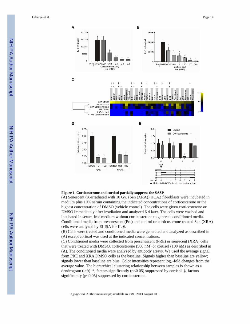
Figure 1. Corticosterone and cortisol partially suppress the SASP(A) Senescent (X-irradiated with 10 Gy, (Sen (XRA)) HCA2 fibroblasts were incubated inmedium plus 10% serum containing the indicated concentrations of corticosterone or thehighest concentration of DMSO (vehicle control). The cells were given corticosterone orDMSO immediately after irradiation and analyzed 6 d later. The cells were washed andincubated in serum-free medium without corticosterone to generate conditioned media.Conditioned media from presenescent (Pre) and control or corticosterone-treated Sen (XRA)cells were analyzed by ELISA for IL-6.(B) Cells were treated and conditioned media were generated and analyzed as described in(A) except cortisol was used at the indicated concentrations.(C) Conditioned media were collected from presenescent (PRE) or senescent (XRA) cellsthat were treated with DMSO, corticosterone (500 nM) or cortisol (100 nM) as described in(A). The conditioned media were analyzed by antibody arrays. We used the average signalfrom PRE and XRA DMSO cells as the baseline. Signals higher than baseline are yellow;signals lower than baseline are blue. Color intensities represent log2-fold changes from theaverage value. The hierarchical clustering relationship between samples is shown as adendrogram (left). *, factors significantly (p<0.05) suppressed by cortisol. ‡, factorssignificantly (p<0.05) suppressed by corticosterone.
Laberge et al. Page 14
Aging Cell. Author manuscript; available in PMC 2013 August 01.
NIH
-PA Author Manuscript
NIH
-PA Author Manuscript
NIH
-PA Author Manuscript
(D) Cells were infected with RAS- or MKK6-expressing lentiviruses. After selection, thecells were given DMSO-, 500 nM corticosterone (C1)- and 100 nM cortisol (C2) for 6 d.Conditioned media were generated as described above and analyzed by ELISA for IL-6. *,factors significantly different from DMSO-treated (p<0.05).(E) Cells were treated with 500 nM corticosterone for the indicated intervals (a–d, indicatedby the thick lines in the lower panel) before or after X-irradiation (XRA, indicated by thearrow). Conditioned media were prepared and analyzed by ELISA for IL-6 (upper panel). *,factors significantly different from DMSO-treated (p<0.05).
Laberge et al. Page 15
Aging Cell. Author manuscript; available in PMC 2013 August 01.
NIH
-PA Author Manuscript
NIH
-PA Author Manuscript
NIH
-PA Author Manuscript

Figure 2. Effect of glucocorticoids on the SASP depends on the glucocorticoid receptor(A) mRNA was extracted from presenescent (Mock) or senescent X-irradiated HCA2 cellstreated with DMSO, 500 nM corticosterone or 100 nM cortisol as described in the legend toFigure 1. Transcripts for IL-5, IL-6, IL-8, MMP-3, IL-1α, MCP-2, MCP-3 and GM-CSFwere quantified by quantitative PCR (normalized to tubulin). *, factors significantlydifferent from DMSO-treated (p<0.05).(B) mRNA was extracted from Pre and Sen (XRA) cells treated with DMSO, 500 nMcorticosterone (C1) or 100 nM cortisol (C2) as described above, and transcripts for the GRwere quantified by PCR (normalized to tubulin). Although GR mRNA levels tended to beslightly elevated in senescent cells, the increase was not statistically significant.(C) Pre and Sen (XRA) cells treated with DMSO, 500 nM corticosterone or 100 nM cortisolas described above were immunostained for GR 1, 4 and 7 d after X-irradiation.(D) Cells were infected with lentiviruses expressing shRNAs against GFP (control) or GR,and selected. Seven days after selection, mRNA was extracted and transcripts for GR werequantified by PCR (normalized to tubulin).(E) Total cell lysates were prepared from the shGFP- and shGR-expressing cells describedin (D), and analyzed by western blotting for GR and actin (control).
Laberge et al. Page 16
Aging Cell. Author manuscript; available in PMC 2013 August 01.
NIH
-PA Author Manuscript
NIH
-PA Author Manuscript
NIH
-PA Author Manuscript

(F) Cells infected with shGFP or shGR-expressing lentiviruses were X-irradiated and treatedimmediately thereafter with DMSO, 500 nM corticosterone or 100 nM cortisol. Conditionedmedia were collected 7 d later and analyzed by ELISA for IL-6.(G) Cells were treated as described in (F) except for the addition of RU-486 at the indicateddoses. Conditioned media were collected and analyzed by ELISA for IL-6 secretion.
Laberge et al. Page 17
Aging Cell. Author manuscript; available in PMC 2013 August 01.
NIH
-PA Author Manuscript
NIH
-PA Author Manuscript
NIH
-PA Author Manuscript

Figure 3. Glucocorticoids repress IL-1α expression(A) Presenescent (Pre) HCA2 cells were treated with DMSO, 500 nM corticosterone or 100nM cortisol for 24 h, or were induced to senesce by X-irradiation (Sen (XRA)) and givenDMSO, corticosterone or cortisol immediately thereafter. mRNA was extracted after theindicated intervals and transcripts for IL-1α were quantified by PCR (normalized totubulin).(B) mRNA extracted from cells described in (A) was used to quantify transcripts for IL-6(normalized to tubulin).(C) Pre and Sen (XRA) cells, prepared as described in (A), were immunostained for IL-1α.Sen (XRA) cells were immunostained 7 d after irradiation.
Laberge et al. Page 18
Aging Cell. Author manuscript; available in PMC 2013 August 01.
NIH
-PA Author Manuscript
NIH
-PA Author Manuscript
NIH
-PA Author Manuscript

Figure 4. Glucocorticoids impair the IL-1α/NF-κB pathway and suppress the ability of the SASPto induce tumor cell invasion(A) Total HCA2 cell lysates were prepared from presenescent (Pre) cells, or senescent cells(Sen (XRA)) cells treated with DMSO-, 500 nM corticosterone (C1)-, or 100 nM cortisol(C2) in the absence (left panel) or presence (right panel) of recombinant IL-1α protein(rIL-1α). The lysates were analyzed by western blotting for IRAK1, IkBα, RelA and actin(control).(B) After irradiation, Sen (XRA) cells were given DMSO, 500 nM corticosterone or 100 nMcortisol. Six d later, the cells were given recombinant IL-1α protein at the indicated doses inthe presence of the glucocorticoids in serum free media. Conditioned media were collected24 h later and analyzed by ELISA for IL-6.(C) Nuclear extracts were prepared from Pre cells, and Sen (XRA) cells treated with DMSO,500 nM corticosterone or 100 nM cortisol in the absence (left panel) or presence (rightpanel) of recombinant IL-1α protein (rIL-1α), and analyzed for NF-κB DNA bindingactivity.(D) Cells were infected with a lentivirus carrying an NF-κB-luciferase reporter construct,irradiated, and allowed to senesce. Immediately after irradiation, cells were treated withDMSO, 500 nM corticosterone or 100 nM cortisol, plus 0.5 μM RU-486 or 2.5 ng/ml IL-1α,as indicated. Seven d after irradiation, cells were trypsinized, counted, lysed and assayed forluciferase activity, which was normalized to cell number.(E) We prepared conditioned media from presenescent (Pre) cells or senescent cells (Sen(XRA) that had been treated with corticosterone (C1) or cortisol (C2) as described in thelegend to Figure 1. The conditioned media were then assayed for ability to stimulate T47Dhuman breast cancer cells to invade a basement membrane, as described in the ExperimentalProcedures.
Laberge et al. Page 19
Aging Cell. Author manuscript; available in PMC 2013 August 01.
NIH
-PA Author Manuscript
NIH
-PA Author Manuscript
NIH
-PA Author Manuscript