new polymeric carriers for controlled drug delivery following inhalation or injection
TRANSCRIPT

Biomaterials 23 (2002) 4425–4433
New polymeric carriers for controlled drug delivery followinginhalation or injection
Jie Fua, Jennifer Fiegela, Eric Kraulandb, Justin Hanesa,b,*aDepartment of Chemical Engineering, The Johns Hopkins University, 3400 N. Charles Street, 221 MD Hall, Baltimore, MD 21218, USA
bDepartment of Biomedical Engineering, The Johns Hopkins University, 3400 N. Charles Street, 221 MD Hall, Baltimore, MD 21218, USA
Received 14 January 2002; accepted 1 May 2002
Abstract
Inhalation is gaining increasing acceptance as a convenient, reproducible, and non-invasive method of drug delivery to the lung
tissue and/or the systemic circulation. However, sustained drug release following inhalation remains elusive, due in part to the lack
of appropriate materials designed specifically for use in the lungs to control the release of bioactive compounds. To address this
problem, we have synthesized a new family of ether-anhydride copolymers composed entirely of FDA-approved monomers,
including polyethylene glycol (PEG). Sebacic acid, a hydrophobic monomer, was copolymerized with PEG in order to produce
water-insoluble polymers capable of providing continuous drug release kinetics following immersion in an aqueous environment.
Various amounts of PEG (5�50% by mass) were incorporated into the backbone of the new polymers to allow tuning of particle
surface properties for potentially enhanced aerosolization efficiency and to decrease particle clearance rates by phagocytosis in the
deep lung. The preparation of large porous particles with these new polymers was systematically approached, utilizing central
composite design, to develop improved particle physical properties for deep lung delivery. Microparticles containing model drugs
were made with sizes suitable for deposition in various regions of the lung following inhalation as a dry powder. Due to such
properties as surface erosion (leading to continuous drug release profiles), erosion times ranging from hours to days (allowing
control over drug delivery duration), and ability to incorporate up to 50% PEG in their backbone, these new systems may also find
application as ‘‘stealth’’ carriers for therapeutic compounds following intravenous injection. r 2002 Elsevier Science Ltd. All rights
reserved.
Keywords: Pulmonary drug delivery; Controlled release; Biodegradable polymers; Lung; Inhalation
1. Introduction
We are currently living through a revolution inbiotechnology that is producing an abundance of potentnew protein, peptide, and DNA-based drugs. Asignificant challenge facing scientists and engineers isthe development of new delivery methods that maximizethe therapeutic effect and convenience of administrationof these new drugs [1].
Man has inhaled drugs for medicinal, recreational,and other purposes for centuries. Today’s smokers, drugabusers, and asthmatics know that inhaled drugs actquickly, minimize the dose required, and are non-invasive. In fact, inhalation of aerosolized drugs hasbecome a well-established means of treating localized
disease states within the lung, including the millions ofpeople in the US that use fast-acting inhaled b2-agonistsas treatment for unexpected asthma attacks [2]. It hasrecently been demonstrated that the lung may be anideal site for the non-invasive delivery of therapeuticmolecules, including peptides and proteins, to thesystemic circulation as well [3–12]. Insulin, calcitonin,interferons, parathyroid hormone, and leuprolide areexamples of proteins in clinical studies for systemicaction following inhalation [10,13–16]. The lung is anattractive route for drug delivery owing to its enormoussurface area for absorption (B100–140m2) [17], highlypermeable epithelium compared with the gastrointest-inal tract [18–20], and favorable environment forprotein drugs compared to the low pH and highprotease levels associated with oral delivery. In addition,pulmonary drug delivery avoids first pass hepaticmetabolism and is generally more acceptable to patientsthan an injection.
*Corresponding author. Tel.: +1-410-516-3484; fax: +1-410-516-
5510.
E-mail address: [email protected] (J. Hanes).
0142-9612/02/$ - see front matter r 2002 Elsevier Science Ltd. All rights reserved.
PII: S 0 1 4 2 - 9 6 1 2 ( 0 2 ) 0 0 1 8 2 - 5

Although promising, delivery of therapeutics to thelungs faces several anatomical and physiological chal-lenges [7]. To deposit in the lungs, drugs must traverse acomplex lung structure that is heterogeneous in geome-try and environment from patient to patient. Oncedeposited, natural clearance methods, including the‘‘mucociliary escalator’’, work to expel particles fromthe upper airways [7], while alveolar macrophagesrapidly (often within minutes) engulf particles between1 and 5 mm that reach the deep lungs [7]. Additionaldrug loss can occur in the inhaler device due toinefficient aerosolization, or in the mouth, throat, andupper airways due to sub-optimal aerosol characteristicsor improper coordination of aerosol activation andbreathing [21]. Consequently, aerosol design is vital tomaximize delivery efficiency and eliminate irreproduci-bility that can limit the practicality of new pulmonarytherapies.
Pulmonary drug delivery methods have traditionallyfocused on one of two strategies: (i) drug suspension/dissolution in liquid aerosol drops and (ii) mixtures ofdry drug particulates with dry carrier particles typicallycomposed of sugars. These methods, capable ofdelivering medicine quickly to the bloodstream or localtissue, have been studied for treatments rangingfrom asthma and pain relief [23] to influenza [24].Although effective as immediate relief therapies, aninability to achieve sustained drug delivery with tradi-tional methods has limited the scope of inhaledmedicines [22,25].
The use of controlled release polymeric systems is anapproach that holds promise for improving the durationand effectiveness of inhaled drugs, for both local andsystemic action [21]. Micrometer- and nanometer-sizedpolymeric systems have been used to deliver preciseamounts of drugs, including proteins and genes, overprolonged times to local tissues or the systemiccirculation following injection [1]. Initial studies withpolymeric aerosol systems showed that properly en-gineered, large porous particles (LPP) were also capableof delivering bioactive insulin to the blood of rats andcontrol glucose levels for 96 h [26]. The previous longestsustained delivery of insulin to the blood via the lungswas only 6 h, using liposomes that were intratracheallyinstilled into rat lungs [27]. Since then, only limitedexamples of polymeric aerosol systems have beenreported. For example, respirable poly(lactic-co-glyco-lic) acid (PLGA) microspheres containing rifampicin forthe treatment of tuberculosis have been studied in aguinea pig model [28,29]. Cationic polymers, such aspolyethyleneimine (PEI) and poly-l-lysine (PLL), com-plexed with DNA have also been tested in the airways asa method to achieve transient gene expression [30–32].Although promising, transient gene expression wouldalso require frequent administration to maintain atherapeutic effect [33,34]. Properly designed new poly-
meric aerosols, with the ability to target various regionsof the lung, should prove beneficial for prolonged non-invasive treatment of both lung disorders, such asasthma or cystic fibrosis, and diseases requiring drugdelivery to the systemic circulation.
Most previous studies of polymeric pulmonary drugdelivery have utilized PLGA since it is readily availableand has a long history of safety in humans.However, PLGA has many limitations as a carrier fordrugs in the lungs. First, small PLGA microspheresdegrade over the period of weeks to months, buttypically deliver drugs for a shorter period of time[26,38]. Such a pattern would lead to an unwantedbuild-up of polymer in the lungs upon repeat adminis-tration. Second, bulk degradation of PLGA micro-spheres creates an acidic core, which can damage pHsensitive drugs such as peptides and proteins [39].Surface eroding polymers, such as polyanhy-drides, lessen the effect of acidic build-up by increaseddiffusion rates of soluble fragments away from theparticle [40]. Third, PLGA microspheres have hydro-phobic surfaces, which result in sub-optimal particleflight into the deep lung (due to particle agglomerationby van der Waals forces) [41]. Additionally, hydro-phobic surfaces lead to rapid opsonization (proteinadsorption), resulting in a rapid clearance by alveolarphagocytic cells [42].
Two areas of focus in our laboratory include: (i) thesynthesis of novel biomaterials as potential drug carriersand (ii) the design and optimization of polymericmaterials into aerosol particles with desired physicalproperties for efficient and sustained drug delivery in thelungs. In this paper, we first describe the synthesis of anew class of polymers for pulmonary delivery, thepolyether-anhydrides. The polymers are composed ofthe monomers sebacic acid (SA) and polyethylene glycol(PEG). Sebacic acid is FDA-approved for treatment inhuman brain tumors while PEG is approved fornumerous medical applications. The safety of SA andPEG demonstrated in other tissues should improve theirchances for approval in pulmonary applications. Poly-ether-anhydrides have significantly shorter degradationtimes, ranging from hours to many days dependingon composition, and thus may be more appropriate forpulmonary delivery than existing polymers, such asPLGA. PEG was incorporated into the polymer back-bone to reduce the interparticle adhesion forces anddecrease the density of polymer aerosols, as well as torender the particles less susceptible to phagocytosis [43].We then describe the production and optimization ofpolymer microparticulate aerosols, with a focus onunusually large, low-density polymeric systems. Suchsystems can be designed to target specific regions ofthe lung, and therefore allow controlled drug deliveryto lung, or to the systemic circulation via the lung[26,35].
J. Fu et al. / Biomaterials 23 (2002) 4425–44334426

2. Materials and methods
2.1. Materials
All chemicals were purchased from Sigma-Aldrich(St. Louis, MO) unless otherwise noted. Sebacic acidwas recrystallized three times from ethanol. Aceticanhydride was purified by distillation. Toluene andchloroform (J.T. Baker, Phillipsburg, NJ) were refluxedover and distilled from calcium hydride. Poly(ethyleneglycol) biscarboxymethyl ether (Mn ¼ 600) was dried bylyophilization. Cadmium acetate, polyvinyl alcohol(88mol% hydrolyzed, Mw ¼ 25 kDa, Polysciences Inc.,Warrington, PA), bovine serum albumin (BSA), pyr-idine, phosphatidylcholine, and other reagents wereused as received without further purification.
2.2. The synthesis and characterization of poly(sebacic
anhydride-co-peg)
2.2.1. Sebacic acid (SA) prepolymer
SA (10.0 g) was refluxed in 100ml acetic anhydrideunder dry nitrogen for 15min, cooled to roomtemperature, and dried using a rotary evaporator. Thecrude prepolymer was recrystallized from dry toluene,washed with 1:1 anhydrous ethyl ether:petroleum ether(Fisher, Fair Lawn, NJ), and dried by vacuum.
2.2.2. Poly(ethylene glycol) (PEG) prepolymer
Polyoxyethylene dicarboxylic acid (10.0 g) was re-fluxed in 200ml acetic anhydride for 30min undernitrogen and evaporated to dryness using a rotaryevaporator. The residue was extracted with anhydrousether and dried under vacuum.
2.2.3. Polymer synthesis
A family of ether-anhydride copolymers was synthe-sized by melt polycondensation of SA and PEGprepolymers under high vacuum, as previously de-scribed [36]. The polymers were precipitated fromchloroform into petroleum ether and dried by vacuum.Molecular weight was monitored by gel permeationchromatography (GPC) (JASCO AS-1555, Tokyo,Japan) with three columns in series (Waters, Milford,MA; Styragel guard column, 4.6mm I.D. � 30mm; HR3 column, 4.6mm I.D. � 300mm; HR 4 column,4.6mm I.D.� 300mm) and polystyrene as standards(Fluka, Milwaukee, WI). The chemical structure ofpoly(PEG:SA) is shown in Fig. 1. Structure was
confirmed by 1H NMR recorded in CDCl3 on a VarianUNITY-400MHz spectrophotometer (Palo Alto, CA)and FT-IR with potassium bromide pellets on a Perkin-Elmer 1600 series spectrophotometer (Wellesley, MA).
2.3. Preparation of poly(sebacic anhydride-co-PEG)
microparticles
Microparticles were prepared using a double-emul-sion solvent-evaporation method [36]. The primarywater-in-oil emulsion was created by probe sonication(Sonics and Materials Inc., Newtown, CT) of 100 mlaqueous solution (+/� bovine serum albumin) in 4mlpolymer solution in methylene chloride (+/� phospha-tidylcholine). The primary emulsion was then pouredinto 100ml of 1% (wt/vol) poly(vinyl alcohol) (PVA)solution and homogenized (Silverson Machines Inc.,East Longmeadow, MA) at 6000 rpm for 1min to formthe double emulsion. Microspheres were stirred for 3 hto allow hardening, then collected by centrifugation,washed twice with deionized water, resuspended in 10mlwater, and freeze-dried.
2.4. Microparticle optimization via central composite
design
The effects of five microparticle preparation para-meters (homogenization speed of second emulsion,polymer concentration in methylene chloride (oil phase),PVA concentration in outer water phase, phosphatidyl-choline concentration in oil phase, water/oil ratio inprimary emulsion; see Table 1) on microparticle size,density and aerodynamic diameter were analyzed usinga half-replicate central composite design (CCD) [37]. Ina general CCD experiment, K input variables areassigned a center point value designated as 0, and ahigh and low value equidistant on either side of thecenter, (designated 1 and –1, respectively) called thecorner points. In this design, 2K experiments wereperformed on all combinations of corner points. Anadditional 2K experiments were performed on starpoints, which were a level 72k=4 for one variable andlevel 0 for all other variables. The remaining experi-ments were performed on center points, in which allvariables are kept at level 0. Data was collected fromeach experiment and quadratic relationships weredeveloped between all K input variables. Statisticalsignificance (po0:05) was determined by analysis ofvariance (ANOVA) for each response variable (size,density, and aerodynamic diameter).
2.5. Characterization of polymeric microparticles
The mass-average size distribution of microparticleswas determined using a Coulter Multisizer IIe (Beck-man-Coulter Inc., Fullerton, CA). Approximately 2ml
(OC(CH2)8C)y(OCCH2O(CH2CH2O)nCH2C)x
O O OO
Fig. 1. Chemical structure of poly(PEG:SA).
J. Fu et al. / Biomaterials 23 (2002) 4425–4433 4427

of isoton II solution was added to 5–10mg micropar-ticles. The solution was briefly vortexed to suspend themicroparticles and then added dropwise to 100ml isotonII solution until the coincidence of particles was between8% and 10%. Greater than 100,000 particles were sizedfor each batch of microparticles to determine the meanparticle size and size distribution. The bulk density ofthe particles was determined by tap density as describedpreviously [26]. Degradation studies of poly(PEG:SA)were accomplished by placing a known amount(B10mg) of microparticles in 1.0ml phosphate bufferedsaline (0.1m, pH 7.4), incubated at 371C under rotaryagitation. At predetermined time intervals, the molecu-lar weight of the polymer within microparticles wasmonitored by GPC.
3. Results and discussion
3.1. Synthesis of polyether-anhydrides
Poly(ether-anhydrides) were synthesized by meltpolycondensation of sebacic anhydride and poly(ethy-lene glycol) biscarboxylmethyl under high vacuum [36].
Copolymers of various compositions were characterizedby 1H NMR and FT-IR. The 1H NMR (Fig. 2A)resonance line of the methylene protons of PEGappeared at 3.65 ppm, which indicated PEG wasincorporated into the sample. The three peaks at 2.44,1.65, and 1.32 ppm were attributed to the methyleneprotons of SA. Data from GPC (not shown) containedone peak corresponding to the molecular weight of thepolymer, with no peak corresponding to the weight offree PEG. NMR studies combined with GPC dataindicated that PEG chain was successfully copolymer-ized with SA. The actual weight percentage of PEG inthe polymer was estimated by the area ratio of the PEGprotons to methyl protons of SA. As shown in Table 2,the estimates were in good agreement with the feed ratioof PEG to SA added prior to polymerization. Thetypical anhydride IR double peaks appeared at B1813and B1742 cm�1 (Fig. 2B). The weight average mole-cular weight of poly(PEG:SA) ranged from >12kDawith 50% PEG to nearly 20 kDa with 10% PEG in thefeed. The present study showed that a polyanhydridemolecular weight above E10 kDa is sufficient forefficient preparation of microparticles capable of con-trolled drug delivery.
Table 1
Microparticle preparation variables and their levels in the central composite design
Levels in
CCD
Homogenization
speed (rpm)
Polymer conc.
(mg/ml)
Water/oil phase
ratio (% vol/vol)
PVA conc.
(mg/ml)
Phosphatidyl-choline conc.
(mg/ml)
�1.68 4000 25 1 1 0
�1 5000 56.25 7 13.25 2.5
0 6000 87.5 13 25.5 5
+1 7000 118.75 19 37.75 7.5
+1.68 8000 150 25 50 10
Fig. 2. (A) 1H NMR spectra of poly(PEG:SA) 10:90 (10% PEG). (B) FT-IR spectra of poly(PEG:SA).
J. Fu et al. / Biomaterials 23 (2002) 4425–44334428
3.2. Poly(ether-anhydrides) as aerosol drug carriers
Microsphere size and density are crucial to drypowder aerosol design since it has been shown thatparticles deposit in the lungs based on their aerodynamicdiameter. The aerodynamic diameter of a particle can bedescribed as the in-flight diameter a spherical particlewould possess assuming it had a density of 1 g/cm3. Aquantitative relationship for the aerodynamic diameter(da) of a spherical particle derived from Stokes Law [45]is found to be
da ¼ dffiffiffiffiffiffiffiffiffiffir=ra
p; ð1Þ
where d ¼ geometric diameter, r ¼ particle mass density(g/cm3), and ra ¼ water mass density (1 g/cm3). Eq. (1)shows that a spherical particles’ aerodynamic diameterrelates its density and diameter into one parameter.Important early work by Landahl and coworkersshowed that sedimentation and inertial impaction inthe mouth, throat, and lungs uniquely depends on theaerodynamic diameter [46]. Sedimentation and inertialimpaction are the two most important mechanisms ofdeposition in the lung of particles >1mm in diameter.
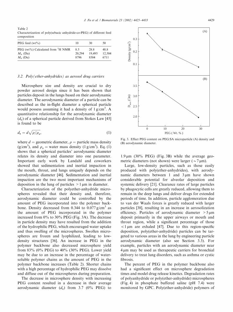
Characterization of the polyether-anhydride micro-spheres revealed that their density and, therefore,aerodynamic diameter could be controlled by theamount of PEG incorporated into the polymer back-bone. Density decreased from 0.344 to 0.077 g/cm3 asthe amount of PEG incorporated in the polymerincreased from 0% to 30% PEG (Fig. 3A). The decreasein particle density may have resulted from the additionof the hydrophilic PEG, which encouraged water uptakeand thus swelling of the microspheres. Swollen micro-spheres are frozen and lyophilized, leading to low-density structures [36]. An increase in PEG in thepolymer backbone also decreased microsphere yieldfrom 83% (0% PEG) to 40% (30% PEG). Lower yieldmay be due to an increase in the percentage of water-soluble polymer chains as the amount of PEG in thepolymer backbone increases (Table 2). Shorter chainswith a high percentage of hydrophilic PEG may dissolveand diffuse out of the microspheres during preparation.
The decrease in microsphere density with increasingPEG content resulted in a decrease in their averageaerodynamic diameter (da) from 3.7 (0% PEG) to
1.9 mm (30% PEG) (Fig. 3B) while the average geo-metric diameters (not shown) were larger (B7 mm).
Large, low-density particles, such as those easilyproduced with poly(ether-anhydrides), with aerody-namic diameters between 1 and 3 mm have shownconsiderable potential for alveolar deposition andsystemic delivery [21]. Clearance rates of large particlesby phagocytic cells are greatly reduced, allowing them toremain in the deep lungs and deliver drugs for extendedperiods of time. In addition, particle agglomeration dueto van der Waals forces is greatly reduced with largerparticles [38], resulting in an increase in aerosolizationefficiency. Particles of aerodynamic diameter >5 mmdeposit primarily in the upper airways or mouth andthroat region, while a significant percentage of thoseo1 mm are exhaled [47]. Due to this region-specificdeposition, poly(ether-anhydride) particles can be tar-geted to various areas in the lung by engineering particleaerodynamic diameter (also see Section 3.3). Forexample, particles with an aerodynamic diameter near4 mm may be used as therapeutic carriers for bronchialdelivery to treat lung disorders, such as asthma or cysticfibrosis.
The percent of PEG in the polymer backbone alsohad a significant effect on microsphere degradationtimes and model drug release kinetics. Degradation ratesof polyanhydride or poly(ether-anhydride) microspheres(Fig. 4) in phosphate buffered saline (pH 7.4) weremonitored by GPC. Poly(ether-anhydride) polymers of
Table 2
Characterization of poly(sebacic anhydride-co-PEG) of different feed
composition
PEG feed (wt%) 10 30 50
PEG (wt%) Calculated from 1H NMR 8.5 28.8 48.8
Mw (Da) 20,294 19,495 12,504
Mn (Da) 8796 8504 6711
0 10 20 30
2.0
2.5
3.0
3.5
0.1
0.2
0.3
(B)
Dae
ro (
µm)
PEG ( Wt. % )
(A)
Den
sity
(g/
cm3 )
Fig. 3. Effect PEG content on PEG:SA microparticle (A) density and
(B) aerodynamic diameter.
J. Fu et al. / Biomaterials 23 (2002) 4425–4433 4429
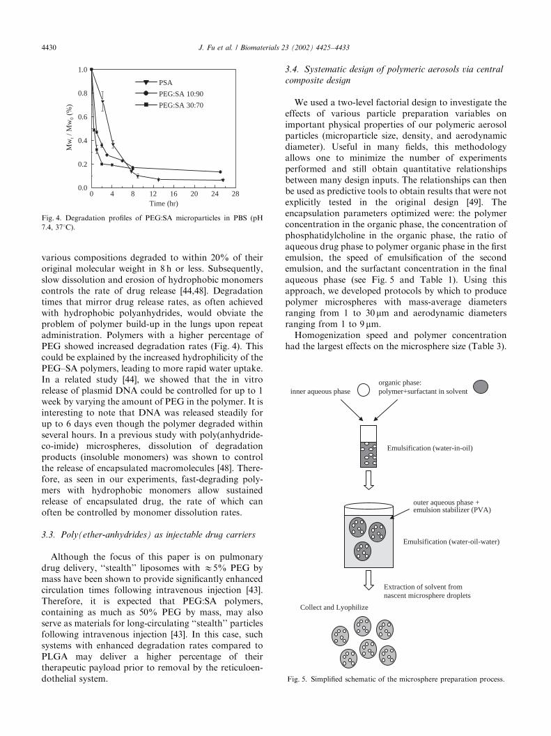
various compositions degraded to within 20% of theiroriginal molecular weight in 8 h or less. Subsequently,slow dissolution and erosion of hydrophobic monomerscontrols the rate of drug release [44,48]. Degradationtimes that mirror drug release rates, as often achievedwith hydrophobic polyanhydrides, would obviate theproblem of polymer build-up in the lungs upon repeatadministration. Polymers with a higher percentage ofPEG showed increased degradation rates (Fig. 4). Thiscould be explained by the increased hydrophilicity of thePEG–SA polymers, leading to more rapid water uptake.In a related study [44], we showed that the in vitrorelease of plasmid DNA could be controlled for up to 1week by varying the amount of PEG in the polymer. It isinteresting to note that DNA was released steadily forup to 6 days even though the polymer degraded withinseveral hours. In a previous study with poly(anhydride-co-imide) microspheres, dissolution of degradationproducts (insoluble monomers) was shown to controlthe release of encapsulated macromolecules [48]. There-fore, as seen in our experiments, fast-degrading poly-mers with hydrophobic monomers allow sustainedrelease of encapsulated drug, the rate of which canoften be controlled by monomer dissolution rates.
3.3. Poly(ether-anhydrides) as injectable drug carriers
Although the focus of this paper is on pulmonarydrug delivery, ‘‘stealth’’ liposomes with E5% PEG bymass have been shown to provide significantly enhancedcirculation times following intravenous injection [43].Therefore, it is expected that PEG:SA polymers,containing as much as 50% PEG by mass, may alsoserve as materials for long-circulating ‘‘stealth’’ particlesfollowing intravenous injection [43]. In this case, suchsystems with enhanced degradation rates compared toPLGA may deliver a higher percentage of theirtherapeutic payload prior to removal by the reticuloen-dothelial system.
3.4. Systematic design of polymeric aerosols via central
composite design
We used a two-level factorial design to investigate theeffects of various particle preparation variables onimportant physical properties of our polymeric aerosolparticles (microparticle size, density, and aerodynamicdiameter). Useful in many fields, this methodologyallows one to minimize the number of experimentsperformed and still obtain quantitative relationshipsbetween many design inputs. The relationships can thenbe used as predictive tools to obtain results that were notexplicitly tested in the original design [49]. Theencapsulation parameters optimized were: the polymerconcentration in the organic phase, the concentration ofphosphatidylcholine in the organic phase, the ratio ofaqueous drug phase to polymer organic phase in the firstemulsion, the speed of emulsification of the secondemulsion, and the surfactant concentration in the finalaqueous phase (see Fig. 5 and Table 1). Using thisapproach, we developed protocols by which to producepolymer microspheres with mass-average diametersranging from 1 to 30 mm and aerodynamic diametersranging from 1 to 9 mm.
Homogenization speed and polymer concentrationhad the largest effects on the microsphere size (Table 3).
0 4 8 12 16 20 24 280.0
0.2
0.4
0.6
0.8
1.0
PSA
PEG:SA 10:90
PEG:SA 30:70
Mw
t / M
w0
(%)
Time (hr)
Fig. 4. Degradation profiles of PEG:SA microparticles in PBS (pH
7.4, 371C).
inner aqueous phase organic phase:polymer+surfactant in solvent
Emulsification (water-in-oil)
outer aqueous phase + emulsion stabilizer (PVA)
Emulsification (water-oil-water)
Extraction of solvent from nascent microsphere droplets
Collect and Lyophilize
Fig. 5. Simplified schematic of the microsphere preparation process.
J. Fu et al. / Biomaterials 23 (2002) 4425–44334430

A modest increase in homogenization speed from 5000to 7000 rpm caused a decrease in the microsphere sizefrom 15.8 to 12.0 mm, whereas a decrease in polymerconcentration from 118.75 to 56.25mg/ml caused adecrease in microsphere size from 15.5 to 10.8 mm(Table 3). The ratio of aqueous drug phase to organicphase had little effect on microsphere diameter, with thediameter increasing from 13.0 to 14.0 mm with a ratioincrease from 7 to 19ml/ml. The surfactant (PVA)concentration in the final aqueous phase had an inverserelationship with microparticle size (Table 3). The PCconcentration did not have a significant effect on themicrosphere size. An example response surface showingthe effects of homogenization speed and polymerconcentration on microparticle size (Fig. 6) shows thatparticles from 9 to 19 mm could be produced (using a setwater/organic volume ratio of 0.13, PVA concentrationof 2.5% (wt/vol) and PC concentration of 5mg/ml). Theresponse surface can then serve as a predictive templateto control particle diameter, a critical parameter fortargeting aerosols to different regions of the lung.
The homogenization speed, polymer concentration,ratio of aqueous drug phase to organic phase, and PVAconcentration had no significant effect on microspheredensity (Table 3). As the PC concentration increased,however, the microparticle density decreased as much as
three-fold (Fig. 7). The response surface showing theeffects of polymer and PC concentration on micro-particle density shows that densities of 0.15–0.35 g/mlcan be achieved (with a set homogenization speed of6000 rpm, water/organic ratio of 0.13 and polymerconcentration of 87.5mg/ml). Lower densities can beachieved upon simultaneous optimization of all of theencapsulation parameters.
Homogenization speed and polymer concentrationalso had the largest effect on the aerodynamic diameterof microspheres (Table 3). As the homogenization speedincreased or the polymer concentration decreased, theaerodynamic diameter decreased. By increasing eitherthe PC or PVA concentration, a significant (po0:001 forPC concentration, po0:007 for PVA concentration)decrease in aerodynamic diameter was achieved;whereas an increase in the ratio of water to organicphase increased the aerodynamic diameter. Similarstudies, aided by CCD, on poly(ether-anhydride) micro-sphere formulations have been recently completed(J. Fiegel, J. Fu, J. Hanes, unpublished). We have alsoused CCD to optimize polymeric cationic particles forDNA delivery to the lungs (E. Krauland, J. Hanes,unpublished).
It should be noted that the ability to fully optimizenew formulations for pulmonary drug delivery has been
Fig. 6. Effect of polymer concentration and homogenization speed on
microparticle size.Fig. 7. Effect of polymer and PC concentrations on microparticle
density.
Table 3
Summary of effects of microparticle preparation parameters on important particle physical properties
Input variables (range) Effect on diameter Effect on density Effect on aerodynamic diameter
Significancen (diameter range) Significancen (density range) Significancen (aero. diameter range)
Homogenization
speed (5000–7000 rpm)
po0:001 (15.8–12.0mm) p ¼ 0:54 (not sig.) (0.190–0.179 g/cm3) po0:001 (6.95–9.03mm)
Polymer concentration
(56.25–118.75mg/ml)
po0:001 (10.8–15.5mm) p ¼ 0:77 (not sig.) (0.212–0.218 g/cm3) po0:001 (4.73–7.17mm)
Ratio of water/oil
phase (7–19ml/ml)
po0:001 (13.0–14.0mm) p ¼ 0:79 (not sig.) (0.186–0.191 g/cm3) po0:02 (5.57–6.13mm)
PVA concentration
(13.25–37.75mg/ml)
po0:001 (14.8–12.4mm) p ¼ 0:15 (not sig. (0.173–0.201 g/cm3) po0:007 (6.19–5.51mm)
PC concentration
(2.5–7.5mg/ml)
po0:09 (not sig.) (13.3–13.8mm) po0:001 (0.244–0.162 g/cm3) po0:001 (6.51–5.50mm)
np-values o0.05 are statistically significant.
J. Fu et al. / Biomaterials 23 (2002) 4425–4433 4431
limited by our inability to closely mimic the conditionsparticles encounter in various regions of the lung.Conventional ‘‘complete immersion’’ methods of parti-cle characterization (particles submersed in buffer) maygreatly overestimate the hydration, degradation, anddrug release kinetics of microparticles that deposit on athin fluid film on the lung surface. This is particularlyimportant in alveoli where the fluid thickness bathingthe epithelium is E0.07 mm [7], or two-orders ofmagnitude smaller than our particles. In collaborationwith Lehr and coworkers, we have begun utilizing air-interfaced lung epithelial cell monolayers that secretemucus or surfactant on their apical surface in particlecharacterization studies to more closely mimic the thinfluid layer found in vivo [50].
4. Conclusion
The use of polymeric systems to achieve controlleddrug delivery in the lung is still in the early stages ofdevelopment. New polymers specifically designed forpulmonary delivery are needed to overcome the limita-tions of currently available off-the-shelf polymerssuch as PLGA. We have reported the synthesisof a family of PEG-based poly(ether-anhydrides)that are aimed to correct several problems inherentwith PLGA. Further, we approached particle pre-paration systematically to engineer particle physicalproperties suitable for delivery to the various regions ofthe lung. Continued advances in material synthesis,particle engineering, particle characterization techniquesand mathematical modeling, should improve the like-lihood of the future development of suitable polymericcarriers for controlled drug delivery in the lungs. Suchsystems will likely find applications in both localtherapies (lung as target) and systemic therapies(delivery to the blood).
Acknowledgements
The authors are thankful to the Whitaker Foundation(grant RG-99-0046). Partial support for Jennifer Fiegelcame from a National Science Foundation GraduateFellowship (grant DGE-9616062).
References
[1] Langer R. Drug delivery and targeting. Nature 1998;392(6679):
5–10.
[2] www.lungusa.org/asthma/ cited 12/01.
[3] Wall DA. Pulmonary absorption of peptides and proteins. Drug
Del 1995;2:1–20.
[4] Niven RW, Lott FD, Cribbs JM. Pulmonary delivery of powder
and solutions containing granulocyte colony-stimulating factor
(rhg-CSF) after intratracheal instillation to the hamster. Pharm
Res 1993;10:1060–604.
[5] Patton J, Platz R. Pulmonary delivery of peptides and proteins for
systemic action. Adv Drug Del Rev 1992;8:179–96.
[6] Laube B, Benedict G, Dobs A. The lung as an alternative route of
delivery for insulin in controlling postprandial glucose levels in
patients with diabetes. Chest 1998;114:1734–9.
[7] Patton JS. Mechanisms of macromolecule absorption by the
lungs. Adv Drug Del Rev 1996;19:3–36.
[8] Wang J, Ben-Jebria A, Edwards DA. Inhalation of estradiol for
sustained systemic delivery. J Aerosol Med 1999;12:27–36.
[9] Choi WS, Murthy GGK, Edwards DA, Langer R, Klibanov AM.
Inhalation delivery of proteins from ethanol suspensions. Proc
Natl Acad Sci USA 2001;98:11103–7.
[10] Agu RU, Ugwoke MI, Armand M, Kinget R, Verbeke N. The
lung as a route for systemic delivery of therapeutic proteins and
peptides. Respir Res 2001;2:198–209.
[11] Pettis RJ, Hall I, Costa D, Hickey AJ. Aerosol delivery of muramyl
dipeptide to rodent lungs. AAPS Pharmsci 2000;2:U50–9.
[12] Patton JS. Deep-lung delivery of therapeutic proteins. Chemtech
1997;27:34–8.
[13] Damms B, Baines W. The cost of delivering drugs without
needles. Bio-Technology 1995;13:1438–40.
[14] Selam JL. Inhaled insulin: clinical results in type 2 diabetic
patients. Diabetes Metab 2001;27:S28–SA32.
[15] Patton JS, Bukar J, Nagarajan S. Inhaled insulin. Adv Drug Del
Rev 1999;35:235–47.
[16] Patton JS. Pulmonary delivery of drugs for bone disorders. Adv
Drug Del Rev 2000;42:239–48.
[17] Altiere RJ, Thompson DC. Physiology and pharmacology of the
airways. In: Hickey AJ, editor. Inhalation aerosols. New York:
Marcel Dekker, 1996. p. 233–72.
[18] Folkesson HG, Westrom BR, Karlsson BW. Permeability of the
respiratory tract to different-sized macromolecules after intra-
tracheal instillation in young and adult rats. Acta Physiol Scand
1990;139:347–54.
[19] Patton JS, Trinchero P, Platz R. Bioavailability of pulmonary
delivered peptides and proteins: a-interferon, calcitonins and
parathyroid hormones. J Controlled Release 1994;28:79–85.
[20] Patton JS. Inhalation: the other ‘‘oral’’ route for delivery of
molecules with low gastrointestinal bioavailability. Abstr Pap Am
Chem Soc 2000;219:175.
[21] Edwards DA, Abdelaziz B-J, Langer R. Recent advances in
pulmonary drug delivery using large, porous inhaled particles.
J Appl Phys 1998;85:379–85.
[22] Zeng X, Martin G, Marriott C. The controlled delivery of drugs
to the lung. Int J Pharm 1995;124:149–64.
[23] Dershwitz M, Walsh JL, Morishige RJ, Conners PM, Rubsamen
RM, Shafer SL, Rosow CE. Pharmacokinetics and pharmacody-
namics of inhaled versus intravenous morphine in healthy
volunteers. Anesthesiology 2000;93:619–28.
[24] Ehlers M, Silagy C, Fleming D, Freeman D. New approaches for
managing influenza in primary care. Clin Drug Invest
2001;21:443–52.
[25] Sanjar S, Matthews J. Treating systemic diseases via the lung.
J Aerosol Med 2001;14:S51–8.
[26] Edwards DA, Hanes J, Caponetti G, Hrkach J, Ben-Jebria A,
Eskew ML, Mintzes J, Deaver D, Lotan N, Langer R. Large
porous particles for pulmonary drug delivery. Science
1997;276:1868–71.
[27] Liu F, Shao Z, Kildsig D, Mitra A. Pulmonary delivery of free
and liposomal insulin. Pharm Res 1993;10:228–32.
[28] Suarez S, O’Hara P, Kazantseva M, Newcomer CE, Hopfer R,
McMurray DN, Hickey AJ. Respirable PLGA microspheres
containing rifampicin for the treatment of tuberculosis: screening
in an infectious disease model. Pharm Res 2001;18:1315–9.
J. Fu et al. / Biomaterials 23 (2002) 4425–44334432
[29] O’Hara P, Hickey AJ. Respirable PLGA microspheres containing
rifampicin for the treatment of tuberculosis: manufacture and
characterization. Pharm Res 2000;17:955–61.
[30] Fajac I, Allo JC, Souil E, Merten M, Pichon C, Figarella C,
Monsigny M, Briand P, Midoux P. Histidylated polylysine as a
synthetic vector for gene transfer into immortalized cystic fibrosis
airway surface and airway gland serous cells. J Gene Med
2000;2:268–378.
[31] Bragonzi A, Dina G, Villa A, Calori G, Biffi A, Bordignon C,
Assael BM, Conese M. Biodistribution and transgene expression
with nonviral cationic vector/DNA complexes in the lungs. Gene
Ther 2000;7:1753–60.
[32] Gautam A, Densmore CL, Golunski E, Xu B, Waldrep JC.
Transgene expression in mouse airway epithelium by aerosol gene
therapy with PEI-DNA complexes. Mol Ther 2001;3:551–6.
[33] Ferrari S, Pettenazzo A, Garbati N, Zacchello F, Behr JP, Scarpa
M. Polyethylenimine shows properties of interest for cystic fibrosis
gene therapy. BBA-Gene Struct Express 1999;1447:219–25.
[34] Matthews CB, Jenkins G, Hilfinger JM, Davidson BL. Poly-l-
lysine improves gene transfer with adenovirus formulated in
PLGA microspheres. Gene Ther 1999;6:1558–64.
[35] Hanes J, Evora C, Edwards DA, Langer R. Particles incorporat-
ing surfactants for pulmonary drug delivery. US Patent No.
5855913, 1999.
[36] Fu J, Fiegel J, Hanes J. Synthesis and characterization of PEG-
based ether-anhydride terpolymers: Novel polymers for pulmon-
ary drug delivery, in preparation.
[37] Peng PC. The design and analysis of scientific experiments.
Reading, MA: Addison-Wesley, 1967. p. 163–71.
[38] Batycky RP, Hanes J, Langer R, Edwards DA. A theoretical
model of erosion and macromolecular drug release from
biodegrading microspheres. J Pharm Sci 1997;86:1464–77.
[39] Mader K, Gallez B, Liu KJ, Swartz HM. Non-invasive in vivo
characterization of release processes in biodegradable polymers
by low-frequency electron paramagnetic resonance spectroscopy.
Biomaterials 1996;17:457–61.
[40] Shieh L, Tamada J, Chen I, Pang J, Domb A, Langer R. Erosion
of a new family of biodegradable Polyanhydrides. J Biomed
Mater Res 1994;28:1465–75.
[41] Visser J. Vanderwaals and other cohesive forces affecting powder
fluidization. Powder Technol 1989;58:1–10.
[42] Tabata Y, Ikada Y. Effect of the size and surface-charge of
polymer microspheres on their phagocytosis by macrophage.
Biomaterials 1988;9:356–62.
[43] Gref R, Minamitake Y, Peracchia MT, Trubetskoy V, Torchilin
V, Langer R. Biodegradable long-circulating polymeric nano-
spheres. Science 1994;263:1600–3.
[44] Fu J, Fiegel J, Hanes J. Large, light polyether-anhydride
microspheres: a new carrier for pulmonary drug delivery. Proc
Int Symp Controlled Rel Bioact Mater 2001;28:393–4.
[45] Bird RB, Stewart WE, Lightfoot EN. Transport phenomena. New
York: Wiley, 1960, p. 59.
[46] Landahl H. On the removal of air-borne droplets by the
human respiratory tract. I. Lung Bull Math Biophys 1950;12:
43–56.
[47] Darquenne C, Brand P, Heyder J, Paiva M. Aerosol dispersion
in human lung: comparison between numerical simulations
and experiments for bolus tests. J Appl Physiol 1997;83:
966–74.
[48] Hanes J, Chiba M, Langer R. Degradation of porous poly(anhy-
dride-co-imide) microspheres and implications for controlled
macromolecule delivery. Biomaterials 1998;19:163–72.
[49] Zeng XM, Martin GP, Marriott C. Tetrandrine delivery to the
lung: the optimization of albumin microsphere preparation by
central composite design. Int J Pharm 1994;109:135–45.
[50] Fiegel J, Ehrhardt C, Lehr C-M, Hanes J. Air-interfaced lung
epithelial cell monolayers for characterization of large light
polymer aerosols. Ann Biomed Eng 2001;29:S141.
J. Fu et al. / Biomaterials 23 (2002) 4425–4433 4433