new functionalities for paper-based sensors lead to...
TRANSCRIPT

AC09CH04-Crooks ARI 22 March 2016 11:50
RE V I E W
S
IN
AD V A
NC
E
New Functionalities forPaper-Based Sensors Lead toSimplified User Operation,Lower Limits of Detection,and New ApplicationsJosephine C. Cunningham, Paul R. DeGregory,and Richard M. CrooksDepartment of Chemistry, The University of Texas at Austin, Austin, Texas 78712-1224;email: [email protected]
Annu. Rev. Anal. Chem. 2016. 9:4.1–4.20
The Annual Review of Analytical Chemistry is onlineat anchem.annualreviews.org
This article’s doi:10.1146/annurev-anchem-071015-041605
Copyright c© 2016 by Annual Reviews.All rights reserved
Keywords
paper analytical device, point-of-care, microfluidic device
Abstract
In the last decade, paper analytical devices (PADs) have evolved into sophis-ticated yet simple sensors with biological and environmental applications inthe developed and developing world. The focus of this review is the techno-logical improvements that have over the past five years increased the appli-cability of PADs to real-world problems. Specifically, this review reports onadvances in sample processing, fluid flow control, signal amplification, andcomponent integration. Throughout, we have sought to emphasize advancesthat retain the main virtues of PADs: low cost, portability, and simplicity.
4.1
Review in Advance first posted online on March 30, 2016. (Changes may still occur before final publication online and in print.)
Changes may still occur before final publication online and in print
Ann
ual R
ev. A
nal.
Che
m. 2
016.
9. D
ownl
oade
d fr
om w
ww
.ann
ualr
evie
ws.
org
Acc
ess
prov
ided
by
Uni
vers
ity o
f T
exas
- A
ustin
on
05/1
7/16
. For
per
sona
l use
onl
y.

AC09CH04-Crooks ARI 22 March 2016 11:50
1. INTRODUCTION
The objective of the present review is to provide a snapshot of the field of paper analytical devices(PADs), particularly the thought processes that have led us to the current state of the art. The ad-vances in PADs we discuss here have made it possible to carry out increasingly sophisticated assaysusing inexpensive paper platforms. Over the last five years, a number of enabling technologicaladvances have been introduced to paper devices, including timed delivery of reagents (1–3), mag-netic concentration (4–7), signal amplification (4, 8–11), automatic on-device washing (1, 2, 10),hollow channels (4, 12–14), and blood separation (15–17). The powerful yet simple PAD shown inFigure 1, known as the NoSlip (discussed in Section 7), is an example of a paper device that inte-grates several of these individual advances for detection of proteins in the low picomolar range (14).Although many of the technologies discussed in this review are common in more sophisticated as-say systems, some clever science and engineering has been required to introduce them into simplepaper devices costing less than US$2, which is a benchmark for low-cost point-of-care applications.
The PAD field is moving very quickly, and the specific advances we discuss here will likely besuperseded by even more clever and functional systems within just a few years. We hope, however,that the general principles we discuss will provide guidance to future researchers who will developthe tools necessary for PADs to become a routine part of health care systems worldwide.
The PADs discussed in this review were developed within the last five years and can performquantitative assays using a handheld reader such as a camera phone, portable potentiostat, digitalmultimeter, commercial glucose meter, or stopwatch. Specifically excluded from this article are
a
b cc
Figure 1Photographs of a sophisticated electrochemical paper analytical device (PAD) known as the NoSlip. (a) TheNoSlip and its 3D-printed holder. (b) The NoSlip being inserted into its holder. (c) The fully assembled PADconnected to an electrochemical measurement device (not pictured).
4.2 Cunningham · DeGregory · Crooks
Changes may still occur before final publication online and in print
Ann
ual R
ev. A
nal.
Che
m. 2
016.
9. D
ownl
oade
d fr
om w
ww
.ann
ualr
evie
ws.
org
Acc
ess
prov
ided
by
Uni
vers
ity o
f T
exas
- A
ustin
on
05/1
7/16
. For
per
sona
l use
onl
y.
AC09CH04-Crooks ARI 22 March 2016 11:50
important closely related topics for which excellent recent reviews already exist. These includethe history (18–21; see also 22, 23 for more information on lateral flow assays) and fabrication(19, 20, 24–28) of PADs as well as the various detection methods (18, 22, 24, 27, 29) used byPADs. In addition, several very fine recent reviews contain subject matter that to some degreeoverlaps with that discussed here. These include a comprehensive review about the fabricationand applications of chemical sensing PADs by Henry and coworkers (28) and a perspective onrecent technological and engineering advances by Abbas and coworkers (30). The specific focushere on improved device functionality, underpinned by increasingly sophisticated design conceptsthat are intended to simplify the operation of PADs by end users, distinguishes the present reviewfrom earlier reports.
2. MULTIDIMENSIONAL PAPER DEVICES
Multidimensionality in paper devices enables multiplexing, reduces size, and incorporates addi-tional sample processing functions. For example, the invention of two-dimensional (2D) PADsby Whitesides and coworkers (31) made it possible to carry out separations (15, 16, 32–34) andsimultaneously detect multiple analytes using a single sample reservoir (11, 35–44). The subse-quent development of three-dimensional (3D) PADs led to even more sophisticated operations,such as on-device delivery of reagents (4, 8, 45–49), controlled flow rates (45, 47, 50–52), fluidicswitches (3, 53–55), and the ability to incorporate microscale objects into assays (4, 7, 45). Nu-merous publications have focused on the basic operating principles and fabrication methods for2D and 3D PADs (56, 57), and therefore they will be described only briefly here.
2.1. Two-Dimensional Paper Analytical Devices
Two-dimensional PADs date back to 1949, when Muller & Clegg (58) used an embossing tool toform patterns of hydrophobic and hydrophilic domains on a piece of paper. However, Whitesidesand coworkers (31) ushered in the modern era of 2D PADs in 2007. 2D PADs are easily fabricatedby patterning a single piece of paper (often chromatography paper) with a hydrophobic material,such as photoresist or wax, to define hydrophilic reservoirs and microfluidic channels. Fluid flowis usually driven by capillary forces present within the hydrophilic, fiber-containing regions ofthe PAD. Often, assay-specific reagents are spotted onto predefined regions of the paper afterpatterning (2, 40). For real-world applications, some sort of device packaging is also provided tomake the PAD functional in the hands of a user (33, 59–62).
A wide variety of detection modalities have been applied to 2D PADs. For example, colorimetricassays have been developed to test for amino acids (63), bacteria (35, 64), biomarkers for cancer(11), lung (36) and liver (17, 65) function, gases (66–68), infectious diseases (69), ions (37, 38,70–72), heavy metals (39–41, 73–80), small molecules (42, 72, 74, 81–83), pH (37, 44, 84), andproteins (1, 85, 86). In all of these cases, complexing agents, nanoparticles, or enzymes wereused to generate a visible color change. Electrochemical methods have also been used to detecta wide range of targets on 2D PADs, including antibiotics (87), gases (88, 89), metals (90), pH(91), and proteins (10, 92). Physical parameters, such as force (93), infrared (94) and UV (95)light, strain (96), and temperature and humidity (97), have also been detected electrochemically. Anumber of specific electrochemical techniques have been adapted to 2D PADs, including relativeresistivity, impedance, amperometry, conductivity, and voltammetry (linear, cyclic, square wave,and differential pulse).
In the sections below, we highlight two specific examples of 2D PADs that, taken together,typify some of the interesting advances that are characteristic of the field. The first is a 2D PAD
www.annualreviews.org • New Functionalities for Paper-Based Sensors 4.3
Changes may still occur before final publication online and in print
Ann
ual R
ev. A
nal.
Che
m. 2
016.
9. D
ownl
oade
d fr
om w
ww
.ann
ualr
evie
ws.
org
Acc
ess
prov
ided
by
Uni
vers
ity o
f T
exas
- A
ustin
on
05/1
7/16
. For
per
sona
l use
onl
y.

AC09CH04-Crooks ARI 22 March 2016 11:50
for the detection of a protein marker for malaria, developed by Yager and coworkers (98); thesecond is a device for the detection of glucose, developed by Laiwattanapaisal and coworkers (32).
2.2. Three-Dimensional Paper Analytical Devices
The fabrication of 3D PADs involves additional complexities that need not be considered for 2Ddevices. Foremost among these challenges is the requirement for fluidic communication betweenlayers of the device, but providing for even compression of the layers and more complex packagingare also important.
There are two common methods for fabricating 3D PADs. The first was devised by Whitesidesand coworkers (57) in 2008 and uses multiple pieces of wax-patterned paper that are aligned andstacked upon one another. This important advance stimulated others to think about constructingPADs in 3D, but the fabrication methodology used in this first publication is complex and doesnot lend itself to low-cost manufacturing due to the inherent difficulty of aligning and affixing theindividual layers. The second method, first developed by our group in 2011 (56), is much simpler,resolves the problems of cost, and provides additional benefits. In this case, a single piece of wax-patterned paper is folded using the principles of origami, such that the pre-patterned channelsself-align and the vias (required for vertical fluid flow) are in fluidic contact (56). Moreover, becausethe individual layers are not taped together, the device can be easily unfolded to read out assayresults within its interior layers. We call this family of devices oPADs, because they are assembledby origami. An example of an early-stage oPAD is shown in Figure 2.
The three most prominent detection strategies used for 3D PADs are colorimetry, electro-chemistry, and time-based measurements. For colorimetry using 3D PADs, assays have beendescribed for bacteria (99), a virus (100), metals (79, 101, 102), small molecules (5, 32, 50, 103–105), anions (106), and proteins (46, 50, 56, 104). Electrochemistry has been used to detect targetsincluding DNA (6, 62, 107), heavy metals (101, 108), small molecules (45, 109), gases (110), ions(111, 112), and proteins (7, 9, 60, 61, 113, 114). Time-based measurements have been demon-strated for enzymes (8) and small molecules (8, 47, 108, 115).
aa bb
Figure 2Photographic illustration of the origami fabrication process. (a) A sheet of chromatography paper is wax-patterned with the multipleorigami paper analytical device (oPAD) designs. Each individual oPAD device is cut from the sheet and briefly heated so that the waxpenetrates the thickness of the paper; the paper is then folded, compressed, and used for detection. (b) A partially unfolded oPAD that(prior to unfolding) had different colored dyes added to its inlets for demonstration purposes.
4.4 Cunningham · DeGregory · Crooks
Changes may still occur before final publication online and in print
Ann
ual R
ev. A
nal.
Che
m. 2
016.
9. D
ownl
oade
d fr
om w
ww
.ann
ualr
evie
ws.
org
Acc
ess
prov
ided
by
Uni
vers
ity o
f T
exas
- A
ustin
on
05/1
7/16
. For
per
sona
l use
onl
y.

AC09CH04-Crooks ARI 22 March 2016 11:50
2.3. Hybrid Paper Analytical Devices
Each of the materials commonly used for constructing microfluidic devices (paper, glass, andplastic) has advantages and disadvantages, and the degree to which they are used depends on theintended application. For paper devices, the primary advantages are ease of fabrication, low cost,and low power requirements (a pump is usually not required). Other materials provide more con-trollable flow characteristics, smaller and more precise channel dimensions, better control oversurface properties, transparency (for optical detection), and rigidity. Accordingly, for some pa-per fluidic applications, there may be advantages associated with introducing a second or thirdmaterial. We consider here only those hybrid devices in which paper is the dominant or func-tional material. The most common paper-based hybrids are those that combine paper with eitherpolydimethylsiloxane (PDMS) or a hard plastic.
2.3.1. Paper and polydimethylsiloxane. The combination of paper and PDMS can be advan-tageous for incubations spanning multiple hours. Paper-based spot arrays cost less and are morebiodegradable than plastic 96-well plates (116); however, wax-patterned chromatography paperloses some of its structural integrity and fluid confinement ability when in the presence of aqueoussolutions for periods of more than approximately 20 min. To resolve this problem, Peltonen andcoworkers (117) infused a cellulose matrix with PDMS to create microarrays that can hold a largervolume of liquid for at least 50 min. After 50 min, evaporation becomes a problem. To addressthis, Funes-Huacca and coworkers (118) used paper as a portable, self-contained culture chamberand added a PDMS lid to supply the bacteria with oxygen while simultaneously preventing evapo-ration of the growth medium. Clearly there are some advantages of combining paper and PDMS,but of course those advantages must outweigh the additional cost and complexity of hybrid devicefabrication.
2.3.2. Paper and plastic. The integration of plastic into PADs has been shown to provide addi-tional functionality including sophisticated valving (53), reduced rates of evaporation (119), andenhanced rigidity (59, 119). For example, Lutz and coworkers (33) combined paper and plastic formultiplexed detection of a malaria antigen and a salmonella antibody in patient plasma samples.In this case, the plastic reduced the evaporation rate and increased durability, whereas the paperregulated flow, stored predried reagents, metered fluid volume, filtered the sample, and acted asa batch mixer (see Section 4.5).
For electrochemical detection, carbon ink electrodes printed on PADs using a stencil (4, 7,120) or screen (108, 121) are satisfactory for most applications. However, the porous nature ofchromatography paper can lead to electrodes that are nonuniform, insufficiently conductive, or notrobust enough for particular applications. Electrodes printed on plastic can resolve these problems(34, 61, 122), are commercially available, and have been incorporated into PADs (32, 123–126).
3. HOLLOW CHANNELS
The cellulose fiber network usually present in PAD channels (we refer to these as paper channels)can be advantageous or disadvantageous depending upon the application. There are three commonproblems with paper channels. First, the mesh size in typical paper networks (∼10 µm) does notallow unhindered flow of micrometer-scale objects, such as microbeads or bacteria (127). Second,the high surface area of cellulose fibers can lead to a significant degree of nonspecific absorption.Third, the rate of fluid flow through paper is retarded by the presence of the cellulose fibers (50).
www.annualreviews.org • New Functionalities for Paper-Based Sensors 4.5
Changes may still occur before final publication online and in print
Ann
ual R
ev. A
nal.
Che
m. 2
016.
9. D
ownl
oade
d fr
om w
ww
.ann
ualr
evie
ws.
org
Acc
ess
prov
ided
by
Uni
vers
ity o
f T
exas
- A
ustin
on
05/1
7/16
. For
per
sona
l use
onl
y.
AC09CH04-Crooks ARI 22 March 2016 11:50
Paper-filled channel Hollow channel
Hydrophilic layer
Hollow area
Paper
Wax
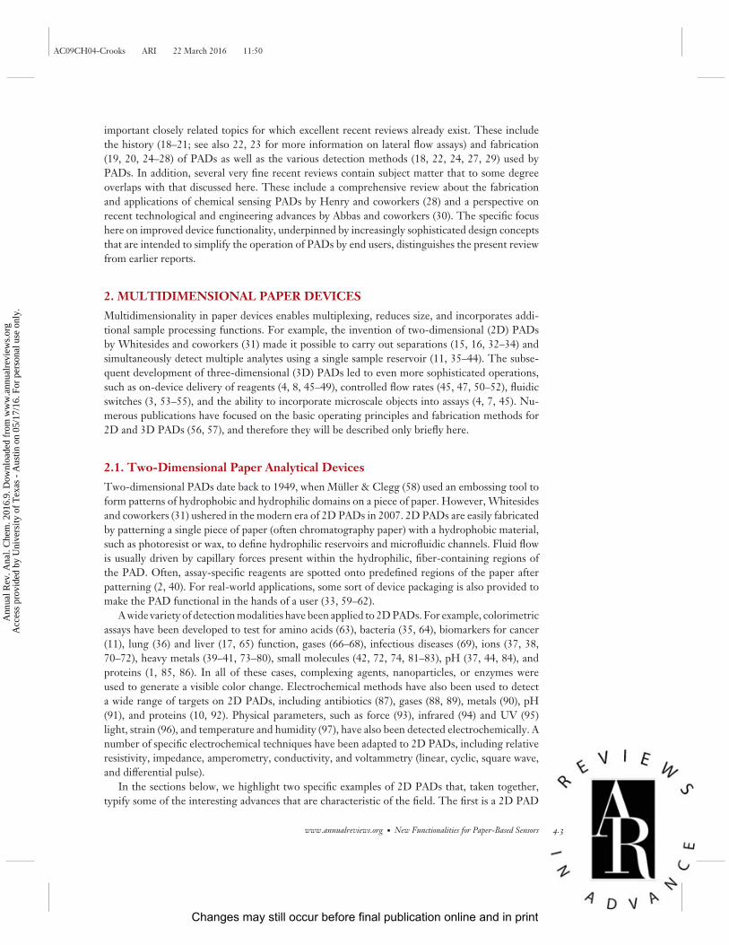
Figure 3Comparison of paper-filled and hollow channels. Wax-patterned boundaries define both types of channels.The difference is that the cellulose matrix has been removed from the hollow channel. The bottom layer ofthe hollow channel is rendered hydrophilic so that fluid can flow by capillary action. Figure adapted withpermission from Reference 50.
All three of these deficiencies can be resolved by simply removing the paper within a channel(Figure 3).
The fabrication of hollow channels is very simple and involves patterning a paper substratewith wax to define the channel network and then removing the cellulose content from the channelsusing a razor blade or laser cutter. To ensure capillary flow, however, it is also necessary to providea hydrophilic wall within the channel (50). If all four walls are impregnated with wax or anotherhydrophobic substance, then a pump is required for fluid flow (128).
We recently developed specific methods for creating hollow channels and also described someof their characteristics (13, 50). For example, their flow rate can be controlled by balancing capillaryand pressure forces (50). Indeed, the pressure resulting from even a single drop of liquid (∼10 µL)is sufficient to induce fast, laminar flow through hollow channels. Moreover, the flow rate in hollowchannels is typically about sevenfold higher than in conventional paper channels (50). Researchershave taken advantage of hollow or open channels to detect small molecules (12, 13, 50), metal ions(102), the toxin ricin (7), DNA (6), and bovine serum albumin (50).
4. CONTROLLED FLUID ACTUATION AND MANIPULATION
There are numerous detection strategies that require sequential addition of reagents, timed incu-bation, mixing, washing, or a combination thereof. These are, of course, routine operations forlarge-scale laboratory instruments and even for plastic and glass microfluidic devices, but until justa few years ago they were unachievable in PADs. In this section, we describe recent approachesfor overcoming the obstacles associated with integrating these types of sophisticated operationsinto simple paper devices.
4.1. Slipping
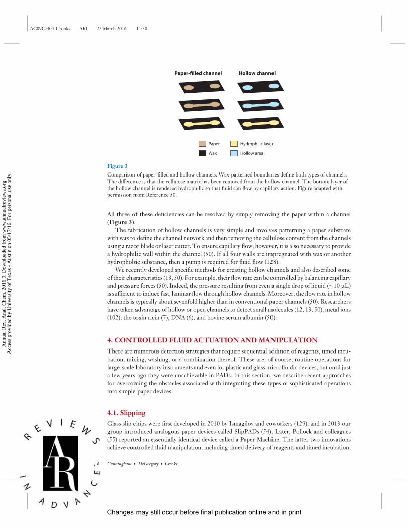
Glass slip chips were first developed in 2010 by Ismagilov and coworkers (129), and in 2013 ourgroup introduced analogous paper devices called SlipPADs (54). Later, Pollock and colleagues(55) reported an essentially identical device called a Paper Machine. The latter two innovationsachieve controlled fluid manipulation, including timed delivery of reagents and timed incubation,
4.6 Cunningham · DeGregory · Crooks
Changes may still occur before final publication online and in print
Ann
ual R
ev. A
nal.
Che
m. 2
016.
9. D
ownl
oade
d fr
om w
ww
.ann
ualr
evie
ws.
org
Acc
ess
prov
ided
by
Uni
vers
ity o
f T
exas
- A
ustin
on
05/1
7/16
. For
per
sona
l use
onl
y.
AC09CH04-Crooks ARI 22 March 2016 11:50
Step 1
Top view Side view
Slip
Step 2
Slip
Slip
Slip
Step 3
Figure 4Schematic illustrations outlining the operation of a slipping paper analytical device (SlipPAD) for parallelfluid manipulation. This SlipPAD consists of two wax-patterned layers. Black dashed lines in the top viewshighlight the bottom layers and their patterned fluidic channels and reservoirs. Figure reprinted withpermission from Reference 54.
by slipping one sheet of wax-patterned paper into alignment with another. The SlipPAD is par-ticularly versatile in this regard, because it can be used for high-throughput parallel reactions orfor sequential addition of multiple reagents.
Figure 4 is a schematic illustration showing the operation of a simple SlipPAD our groupdevised for generating calibration curves and performing concurrent assays. In Step 1, coloredreagents are loaded into defined channels, and then in Step 2 the slip layer is moved so that anarray of 285 paper wells is simultaneously loaded with the reagent. In Step 3, the paper wells areisolated from the filling channels. The SlipPAD approach has also enabled controlled movementof reagents at the time of need in biosensing applications (4, 6, 7, 107).
4.2. Channeling
The strategic arrangement of paper channels provides a means for controlled delivery of pre-driedreagents and increased assay automation on PADs (1, 2, 33, 98, 130–132). In the case of Figure 5,for example, channel length controls the timing of reagent delivery to the reaction zone for amalaria assay. Specifically, detection antibodies labeled with gold nanoparticles, washing buffer,and gold enhancement solution were predried on the individual timing channels. By simulta-neously applying fixed volumes of sample to each of the three fluid application zones, the predriedreagents were rehydrated and delivered by fluidic wicking to the test and control lines. In Figure 5,the contents of the shortest (far right) channel (containing gold-labeled detection antibodies) werefirst to reach the test and control lines, followed by the wash solution from the middle channel andthe gold enhancement solution from the far left channel. This judicious arrangement of channelsis advantageous, because it provides a simple way to complete multiple assay steps with minimaluser intervention.
www.annualreviews.org • New Functionalities for Paper-Based Sensors 4.7
Changes may still occur before final publication online and in print
Ann
ual R
ev. A
nal.
Che
m. 2
016.
9. D
ownl
oade
d fr
om w
ww
.ann
ualr
evie
ws.
org
Acc
ess
prov
ided
by
Uni
vers
ity o
f T
exas
- A
ustin
on
05/1
7/16
. For
per
sona
l use
onl
y.

AC09CH04-Crooks ARI 22 March 2016 11:50
Goldenhancement
solutions
Gold-labeleddetectionantibody
Test
(Wash)
Control
1 cm
Figure 5Schematic illustration of a 2D paper network for assaying Plasmodium falciparum histidine-rich protein 2(PfHRP2). Text labels and colored dots indicate the location of each reagent. Controlled delivery of reagentsand wash buffer to the detection zone is achieved by varying the channel length. The red drop indicates theaddition of the sample, and the blue drops represent buffer. Figure reprinted with permission fromReference 1.
4.3. Delaying
An alternative method for timed delivery of reagents on PADs is the use of a fluidic delay, suchas a one-way fluidic delay switch (51), shunt (133), or dissolvable reagent (134), that slows ortemporarily inhibits capillary flow. For example, Fu and coworkers (133) integrated an absorbentsink into a PAD channel with the intent of slowing capillary flow by absorbing some of the fluid.Additionally, Phillips and coworkers (8, 47, 52) developed a novel detection strategy, wherein ananalyte converts a predried hydrophobic blocking material in the channel to a hydrophilic formthat facilitates flow. The analyte concentration is either directly or inversely proportional to theadditional time required for the analyte to flow from the inlet to the outlet in comparison to areference channel.
4.4. Switching
The manipulation of flow in 3D PADs can also be achieved using a fluidic switch that initiates flowon demand. The switching function can be achieved by using a fluidic diode (135), a physicallymoveable paper flap that acts as a valve (38), or a press-and-flow button (3). For example, thepress-and-flow button (Figure 6) can be pressed to initiate flow at the discretion of the user.Specifically, in this example, a stylus is used to press the single-use “on” button, which activates aconnection between two paper channels that were previously separated by a hollow, hydrophobicgap. Whitesides and coworkers (3) initiated flow with a single-use “on” switch to detect glucose,proteins, ketones, and nitrite in artificial urine.
4.5. Mixing
Fluid mixing is an important operation for many types of assays. This is because flow in hollow andpaper channels is generally laminar (50, 136). Accordingly, mixing of fluidic streams occurs onlyby diffusion and is, therefore, slow. Only two examples have thus far been published to addressthe need for mixing. In one case, Yager and coworkers (33) demonstrated a batch mixer on animmunoassay card by using an air permeable vent that allowed air bubbles into the mixing chamberto induce convection. Yeo and coworkers (137) accomplished uniform mixing on a PAD usingsurface acoustic wave energy. There is clearly a need for more work in this area.
4.8 Cunningham · DeGregory · Crooks
Changes may still occur before final publication online and in print
Ann
ual R
ev. A
nal.
Che
m. 2
016.
9. D
ownl
oade
d fr
om w
ww
.ann
ualr
evie
ws.
org
Acc
ess
prov
ided
by
Uni
vers
ity o
f T
exas
- A
ustin
on
05/1
7/16
. For
per
sona
l use
onl
y.

AC09CH04-Crooks ARI 22 March 2016 11:50
5 mm
Paper
Photoresist
1 mm TapeGap GapPaper
Blue dye
Compressedpaper
Compressedpaper
a
b
c
d
Figure 6Top view and cross sections of a channel containing a fluidic “on” button for use with a programmable paperanalytical device constructed of paper and tape. (a) A fully assembled device; the cross-sectional view isobtained by cutting the device along the dashed line in the top view. (b) The device shown in panel a afteradding 10 μL of aqueous blue dye to the left side of the channel. (c) The device in panel b after compressingthe top layer of paper with a ballpoint pen to connect the gap. (d ) The device shown in panel c after the dyehas flowed past the fluidic “on” button. Figure reprinted with permission from Reference 3.
5. NANO- AND MICROSIZED OBJECTS
Nano- and microsized objects can improve analyte detection in PADs in several important ways.First, recognition agents, such as antibodies or aptamers, can be anchored to mobile particle sur-faces, leading to a high number of recognition elements compared to planar surfaces. Accordingly,these nano- and microsized surfaces provide higher binding efficiencies for solution-phase targetsdue to their increased binding capacity (138). Second, the speed of target binding can be enhancedon particles, compared to stationary surfaces, due to their mobility. Third, particles can be usedto amplify detection signals on PADs, thereby eliminating the need for enzymatic amplification.
Over the past few years, there have been many reports of the use of nanoparticles for colori-metric (1, 33, 35, 39, 63, 69, 74, 75, 79, 81, 84, 139–141) and electrochemical (4–7, 10, 35, 51,61, 62, 102, 142, 143) detection on both 2D and 3D PADs. Nanotubes, -rods, or -wires com-prised of carbon (60, 61, 87–89, 91, 102, 111, 113), zinc oxide (96), gold (86, 142), platinum (144),nickel (144), or copper (144) have been integrated into paper-based sensors to enhance detection.Microsized objects that have been used with PADs include magnetic and nonmagnetic microbeads(45) and microcapsules (145, 146).
www.annualreviews.org • New Functionalities for Paper-Based Sensors 4.9
Changes may still occur before final publication online and in print
Ann
ual R
ev. A
nal.
Che
m. 2
016.
9. D
ownl
oade
d fr
om w
ww
.ann
ualr
evie
ws.
org
Acc
ess
prov
ided
by
Uni
vers
ity o
f T
exas
- A
ustin
on
05/1
7/16
. For
per
sona
l use
onl
y.

AC09CH04-Crooks ARI 22 March 2016 11:50
a
b
Separation zone Separation zoneDetection zone
Figure 7Top-view photographs of a dumbbell-shaped paper analytical device (PAD) for whole blood separation.(a) The device prior to the addition of blood; the arrows indicate the location of blood filters and thedirection of flow. (b) The PAD after the addition of blood. The filters exclude the red blood cells while theplasma travels to the electrochemical detection zone at the center. Figure reprinted with permission fromReference 32.
6. FILTRATION AND SEPARATION
The properties of chromatography paper provide a natural avenue for filtration and separation onPADs, as has been demonstrated in experiments involving molecules (34, 147) and viruses (100).However, chromatography paper is not effective for removing interferences, such as blood cells.Therefore, detection in blood samples on PADs requires a separation membrane (15–17, 32, 33,49, 65). For example, a dumbbell-shaped electrochemical PAD (Figure 7) quantitatively assayedglucose in blood plasma that was isolated from whole blood using a membrane (32). In Figure 7a,the separation zones contain blood separation membranes, whereas the detection zone containschromatography paper overlaid onto a three-electrode electrochemical cell. The operation of thedevice involves depositing a sample of whole blood onto the two separation zones (Figure 7b).The membranes trap the red and white blood cells while the blood plasma continues to flow to-ward the detection zone. The symmetrical movement of blood plasma toward the central reservoirensures uniform flow over the electrodes. In this PAD, glucose was detected amperometricallyusing a commercial Prussian blue–modified, screen-printed carbon working electrode that mea-sured the amount of peroxide resulting from the reaction between glucose and glucose oxidase.
More highly resolved separations on PADs can be achieved using electrophoresis (148, 149).For example, our group demonstrated the separation of proteins using a low-voltage oPAD elec-trophoretic device (oPAD-Ep) (148). The separation channel in the oPAD-Ep was constructed bypaper folding (origami) and was specifically designed to require only low voltages. We anticipatethat active separation components, such as the oPAD-Ep, will eventually be integrated into morecomplex, multifunctional PAD designs in the future.
7. EXTRACTION AND CONCENTRATION
Additional sample processing is often required when sensing in realistic sample matrices, suchas blood, serum, natural waters, or urine. Incorporating extraction or preconcentration functionsinto PADs can lead to new applications, lower limits of detection (LODs), shortened assay times,
4.10 Cunningham · DeGregory · Crooks
Changes may still occur before final publication online and in print
Ann
ual R
ev. A
nal.
Che
m. 2
016.
9. D
ownl
oade
d fr
om w
ww
.ann
ualr
evie
ws.
org
Acc
ess
prov
ided
by
Uni
vers
ity o
f T
exas
- A
ustin
on
05/1
7/16
. For
per
sona
l use
onl
y.
AC09CH04-Crooks ARI 22 March 2016 11:50
Analyte
AgNP
Gold electrode Wax patterned paper Hemi-channel
Indicator dye BufferCarbon electrode
Magneticbead
Magnet
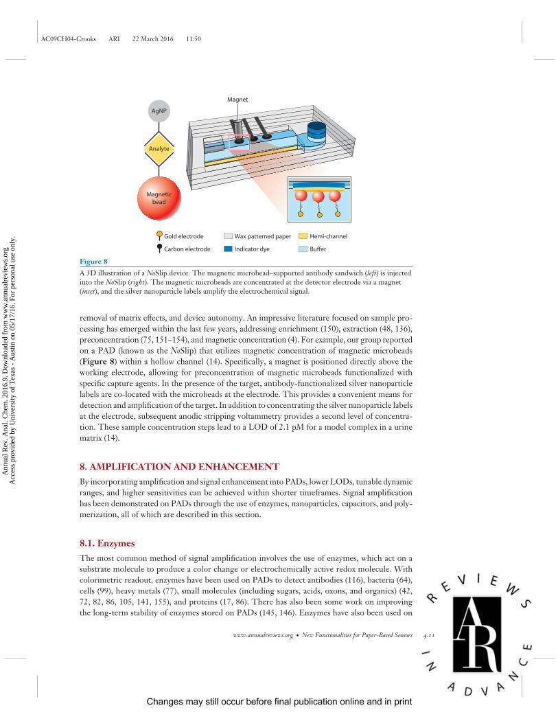
Figure 8A 3D illustration of a NoSlip device. The magnetic microbead–supported antibody sandwich (left) is injectedinto the NoSlip (right). The magnetic microbeads are concentrated at the detector electrode via a magnet(inset), and the silver nanoparticle labels amplify the electrochemical signal.
removal of matrix effects, and device autonomy. An impressive literature focused on sample pro-cessing has emerged within the last few years, addressing enrichment (150), extraction (48, 136),preconcentration (75, 151–154), and magnetic concentration (4). For example, our group reportedon a PAD (known as the NoSlip) that utilizes magnetic concentration of magnetic microbeads(Figure 8) within a hollow channel (14). Specifically, a magnet is positioned directly above theworking electrode, allowing for preconcentration of magnetic microbeads functionalized withspecific capture agents. In the presence of the target, antibody-functionalized silver nanoparticlelabels are co-located with the microbeads at the electrode. This provides a convenient means fordetection and amplification of the target. In addition to concentrating the silver nanoparticle labelsat the electrode, subsequent anodic stripping voltammetry provides a second level of concentra-tion. These sample concentration steps lead to a LOD of 2.1 pM for a model complex in a urinematrix (14).
8. AMPLIFICATION AND ENHANCEMENT
By incorporating amplification and signal enhancement into PADs, lower LODs, tunable dynamicranges, and higher sensitivities can be achieved within shorter timeframes. Signal amplificationhas been demonstrated on PADs through the use of enzymes, nanoparticles, capacitors, and poly-merization, all of which are described in this section.
8.1. Enzymes
The most common method of signal amplification involves the use of enzymes, which act on asubstrate molecule to produce a color change or electrochemically active redox molecule. Withcolorimetric readout, enzymes have been used on PADs to detect antibodies (116), bacteria (64),cells (99), heavy metals (77), small molecules (including sugars, acids, oxons, and organics) (42,72, 82, 86, 105, 141, 155), and proteins (17, 86). There has also been some work on improvingthe long-term stability of enzymes stored on PADs (145, 146). Enzymes have also been used on
www.annualreviews.org • New Functionalities for Paper-Based Sensors 4.11
Changes may still occur before final publication online and in print
Ann
ual R
ev. A
nal.
Che
m. 2
016.
9. D
ownl
oade
d fr
om w
ww
.ann
ualr
evie
ws.
org
Acc
ess
prov
ided
by
Uni
vers
ity o
f T
exas
- A
ustin
on
05/1
7/16
. For
per
sona
l use
onl
y.

AC09CH04-Crooks ARI 22 March 2016 11:50
aa d
bb
cc
eA
Figure 9(a–d ) Photographs of an origami paper analytical device outfitted with an on-device supercapacitor for signalamplification. (a,b) The device is constructed by origami folding. (c) The folded device is clamped to a circuitboard with a binder clip. (d ) The clamped device is placed into a cassette and connected to a digitalmultimeter. (e) Circuit diagram of the electronic setup. Figure adapted with permission from Reference 51.
PADs for electrochemical detection of biomarkers (9–11, 60, 61, 113), heavy metals (108), andsmall molecules (including sugars and toxins) (5, 108).
8.2. Nanoparticles
A particularly advantageous amplification approach involves the use of metal nanoparticles ascharge carriers (4) or catalysts (1, 98, 131) for electrolytic or electroless deposition, respectively.In comparison to enzymes, there are two main advantages to using nanoparticles for amplification.First, there are many straightforward methods known for immobilizing recognition elements (e.g.,antibodies or DNA) onto nanoparticles. Second, nanoparticles do not require long reaction timesor have limited stability, which are common problems with enzymes. As discussed in the previoussection, our group has shown that silver nanoparticles can be detected on PADs in the highfemtomolar (4) to low picomolar (7, 14) range, which is well matched to many biomarkers.
8.3. Capacitors
A capacitor can be coupled with a PAD fluidic network to amplify electronic signals by storingand then releasing charge to achieve gain (35, 45, 51, 156). For example, Huang and coworkers(51) fabricated a capacitor by drawing thin film graphite electrodes with a pencil and dipping
4.12 Cunningham · DeGregory · Crooks
Changes may still occur before final publication online and in print
Ann
ual R
ev. A
nal.
Che
m. 2
016.
9. D
ownl
oade
d fr
om w
ww
.ann
ualr
evie
ws.
org
Acc
ess
prov
ided
by
Uni
vers
ity o
f T
exas
- A
ustin
on
05/1
7/16
. For
per
sona
l use
onl
y.
AC09CH04-Crooks ARI 22 March 2016 11:50
the electrodes in a sulfuric acid and polyvinyl alcohol gel electrolyte. Figure 9 demonstrates theorigami folding (Figure 9a–b) and compression (Figure 9c) of the device, along with the cassetteinto which the oPAD is inserted (Figure 9d ). An on-device fluidic delay switch controls theactuation of the capacitor (Figure 9e). In the presence of target DNA, chemically modified goldnanoparticle labels participate in a photoelectrochemical process that results in the supercapacitorbeing charged. The PAD was able to detect DNA at low femtomolar concentrations by utilizinga sandwich assay.
8.4. Polymerization and Depolymerization
Kang and coworkers (11) showed that a polymerization reaction could be used to significantly am-plify a binding event on a 2D PAD. Specifically, target capture triggered a radical polymerizationreaction, and the resulting polymer provided multiple binding sites to enhance the signal resultingfrom a standard electrochemical enzyme-linked immunosorbent assay (ELISA). The ELISA wascarried out by sequential addition of antibodies, a blocking protein, and polymerization reagent,with thorough washing between each addition. Using this method, an LOD of 10 pg/mL wasachieved for detection of cancer biomarkers.
Depolymerization has also been used to amplify signals. For example, Phillips and coworkersshowed that an enzyme target could be detected in the low- to mid-femtomolar range using veryclever chemistry and a 3D PAD (8). The target concentration was determined by measuring thetime required for depolymerization of a hydrophobic gate arising from the presence of peroxidegenerated during detection.
9. CONCLUSIONS AND OUTLOOK
The movement toward smarter paper platforms outfitted with all device components necessaryfor sophisticated sensing chores is quickly becoming a reality. Goals for the coming years includefurther simplification of design and construction, integration of new functions such as separationsand preconcentration, and minimization of user intervention. At the present time, a numberof specific problems require attention. These include adding on-board storage of reagents andensuring their stability; figuring out how to efficiently resolvate those reagents at the time of need;eliminating nonspecific adsorption; and purifying matrices, such as blood, prior to analysis. All ofthis needs to be accomplished without introducing too much cost, or the advantage of using paperas a platform is lost.
A few other important points not discussed in detail thus far should be mentioned. If papersensors are to be used in the developing world or by the lay public in the developed world, theneither visual readout or cheap, foolproof electronic readers are required. The obvious choice forthe latter is cell phones, but how exactly these will be connected to the sensors, how the sensors willbe powered, and how wireless communication of results will be secured remain open questions.
At this stage in their development, it is difficult to say what commercial paper-based sensorsare going to look like in four or five years. It is certain, though, that individuals will need to takemore control over their own health care needs in the coming years. Home management of chronicdisease is a good example. In the United States, this is, in part, a consequence of the AffordableCare Act, which has added millions of newly insured people to the health care system without acorresponding increase in the number of doctors. In many parts of the developing world, nationalhealth care systems either do not exist or are so underfunded that they might as well not exist. Inboth cases, the ability of an individual to routinely check his or her biomarkers and transmit theresults wirelessly to distant clinics will be immensely valuable.
www.annualreviews.org • New Functionalities for Paper-Based Sensors 4.13
Changes may still occur before final publication online and in print
Ann
ual R
ev. A
nal.
Che
m. 2
016.
9. D
ownl
oade
d fr
om w
ww
.ann
ualr
evie
ws.
org
Acc
ess
prov
ided
by
Uni
vers
ity o
f T
exas
- A
ustin
on
05/1
7/16
. For
per
sona
l use
onl
y.

AC09CH04-Crooks ARI 22 March 2016 11:50
Two final points should be mentioned. First, inexpensive personal diagnostic devices will beable to provide early warning of massive contagions, such as the Ebola outbreak of 2014, as wellas information about how such diseases spread. Second, lifetime testing by individuals, whichprovides personal baselines for key biomarkers, may lead to early detection of cancer and otherdeadly diseases.
DISCLOSURE STATEMENT
The authors are not aware of any affiliations, memberships, funding, or financial holdings thatmight be perceived as affecting the objectivity of this review.
LITERATURE CITED
1. Fridley GE, Le H, Yager P. 2014. Highly sensitive immunoassay based on controlled rehydration ofpatterned reagents in a 2-dimensional paper network. Anal. Chem. 86(13):6447–53
2. Apilux A, Ukita Y, Chikae M, Chailapakul O, Takamura Y. 2013. Development of automated paper-baseddevices for sequential multistep sandwich enzyme-linked immunosorbent assays using inkjet printing.Lab Chip 13(1):126–35
3. Martinez AW, Phillips ST, Nie Z, Cheng C-M, Carrilho E, et al. 2010. Programmable diagnostic devicesmade from paper and tape. Lab Chip 10(19):2499–504
4. Scida K, Cunningham JC, Renault C, Richards I, Crooks RM. 2014. Simple, sensitive, and quantitativeelectrochemical detection method for paper analytical devices. Anal. Chem. 86(13):6501–7
5. Ge S, Liu W, Ge L, Yan M, Yan J, et al. 2013. In situ assembly of porous Au-paper electrode andfunctionalization of magnetic silica nanoparticles with HRP via click chemistry for Microcystin-LRimmunoassay. Biosens. Bioelectron. 49:111–17
6. Li X, Scida K, Crooks RM. 2015. Detection of hepatitis B virus DNA with a paper electrochemicalsensor. Anal. Chem. 87(17):9009–15
7. Cunningham JC, Scida K, Kogan MR, Wang B, Ellington AD, Crooks RM. 2015. Paper diagnosticdevice for quantitative electrochemical detection of ricin at picomolar levels. Lab Chip 15(18):3707–15
8. Lewis GG, Robbins JS, Phillips ST. 2013. Point-of-care assay platform for quantifying active enzymesto femtomolar levels using measurements of time as the readout. Anal. Chem. 85(21):10432–39
9. Li L, Xu J, Zheng X, Ma C, Song X, et al. 2014. Growth of gold-manganese oxide nanostructures ona 3D origami device for glucose-oxidase label based electrochemical immunosensor. Biosens. Bioelectron.61:76–82
10. Wu Y, Xue P, Kang Y, Hui KM. 2013. Paper-based microfluidic electrochemical immunodevice inte-grated with nanobioprobes onto graphene film for ultrasensitive multiplexed detection of cancer biomark-ers. Anal. Chem. 85(18):8661–68
11. Wu Y, Xue P, Hui KM, Kang Y. 2014. A paper-based microfluidic electrochemical immunodeviceintegrated with amplification-by-polymerization for the ultrasensitive multiplexed detection of cancerbiomarkers. Biosens. Bioelectron. 52:180–87
12. Fosdick SE, Anderson MJ, Renault C, DeGregory PR, Loussaert JA, Crooks RM. 2014. Wire, mesh,and fiber electrodes for paper-based electroanalytical devices. Anal. Chem. 86(7):3659–66
13. Renault C, Anderson MJ, Crooks RM. 2014. Electrochemistry in hollow-channel paper analytical de-vices. J. Am. Chem. Soc. 136(12):4616–23
14. Cunningham JC, Kogan MR, Tsai Y-J, Luo L, Richards I, Crooks RM. 2015. Paper-based sensor forelectrochemical detection of silver nanoparticle labels by galvanic exchange. ACS Sens. 1(1):40–47
15. Songjaroen T, Dungchai W, Chailapakul O, Henry CS, Laiwattanapaisal W. 2012. Blood separation onmicrofluidic paper-based analytical devices. Lab Chip 12(18):3392–98
16. Yang X, Forouzan O, Brown TP, Shevkoplyas SS. 2012. Integrated separation of blood plasma fromwhole blood for microfluidic paper-based analytical devices. Lab Chip 12(2):274–80
4.14 Cunningham · DeGregory · Crooks
Changes may still occur before final publication online and in print
Ann
ual R
ev. A
nal.
Che
m. 2
016.
9. D
ownl
oade
d fr
om w
ww
.ann
ualr
evie
ws.
org
Acc
ess
prov
ided
by
Uni
vers
ity o
f T
exas
- A
ustin
on
05/1
7/16
. For
per
sona
l use
onl
y.
AC09CH04-Crooks ARI 22 March 2016 11:50
17. Vella SJ, Beattie P, Cademartiri R, Laromaine A, Martinez AW, et al. 2012. Measuring markers ofliver function using a micropatterned paper device designed for blood from a fingerstick. Anal. Chem.84(6):2883–91
18. Nery E, Kubota L. 2013. Sensing approaches on paper-based devices: a review. Anal. Bioanal. Chem.405(24):7573–95
19. Martinez AW, Phillips ST, Whitesides GM, Carrilho E. 2009. Diagnostics for the developing world:microfluidic paper-based analytical devices. Anal. Chem. 82(1):3–10
20. Jeong S-G, Kim J, Nam J-O, Song YS, Lee C-S. 2013. Paper-based analytical device for quantitativeurinalysis. Int. Neurourol. J. 17(4):155–61
21. Then WL, Garnier G. 2013. Paper diagnostics in biomedicine. Rev. Anal. Chem. 32(4):269–9422. Yetisen AK, Akram MS, Lowe CR. 2013. Paper-based microfluidic point-of-care diagnostic devices. Lab
Chip 13(12):2210–5123. Chin CD, Linder V, Sia SK. 2012. Commercialization of microfluidic point-of-care diagnostic devices.
Lab Chip 12(12):2118–3424. Liana DD, Raguse B, Gooding JJ, Chow E. 2012. Recent advances in paper-based sensors. Sensors
12(9):11505–2625. Mace C, Deraney R. 2014. Manufacturing prototypes for paper-based diagnostic devices. Microfluid.
Nanofluid. 16(5):801–926. Rozand C. 2014. Paper-based analytical devices for point-of-care infectious disease testing. Eur. J. Clin.
Microbiol. Infect. Dis. 33(2):147–5627. Li X, Ballerini DR, Shen W. 2012. A perspective on paper-based microfluidics: current status and future
trends. Biomicrofluidics 6(1):01130128. Cate DM, Adkins JA, Mettakoonpitak J, Henry CS. 2015. Recent developments in paper-based microflu-
idic devices. Anal. Chem. 87(1):19–4129. Maxwell EJ, Mazzeo AD, Whitesides GM. 2013. Paper-based electroanalytical devices for accessible
diagnostic testing. MRS Bull. 38(4):309–1430. Ahmed S, Bui M-PN, Abbas A. 2016. Paper-based chemical and biological sensors: engineering aspects.
Biosens. Bioelectron. 77:249–6331. Martinez AW, Phillips ST, Butte MJ, Whitesides GM. 2007. Patterned paper as a platform for inexpen-
sive, low-volume, portable bioassays. Angew. Chem. Int. Ed. 46(8):1318–2032. Noiphung J, Songjaroen T, Dungchai W, Henry CS, Chailapakul O, Laiwattanapaisal W. 2013. Elec-
trochemical detection of glucose from whole blood using paper-based microfluidic devices. Anal. Chim.Acta 788:39–45
33. Lafleur L, Stevens D, McKenzie K, Ramachandran S, Spicar-Mihalic P, et al. 2012. Progress towardmultiplexed sample-to-result detection in low resource settings using microfluidic immunoassay cards.Lab Chip 12(6):1119–27
34. Carvalhal RF, Simao Kfouri M, de Oliveira Piazetta MH, Gobbi AL, Kubota LT. 2010. Electrochemicaldetection in a paper-based separation device. Anal. Chem. 82(3):1162–65
35. Li C-Z, Vandenberg K, Prabhulkar S, Zhu X, Schneper L, et al. 2011. Paper based point-of-care testingdisc for multiplex whole cell bacteria analysis. Biosens. Bioelectron. 26(11):4342–48
36. Li X, Tian J, Shen W. 2010. Quantitative biomarker assay with microfluidic paper-based analyticaldevices. Anal. Bioanal. Chem. 396(1):495–501
37. Lopez-Ruiz N, Curto VF, Erenas MM, Benito-Lopez F, Diamond D, et al. 2014. Smartphone-basedsimultaneous pH and nitrite colorimetric determination for paper microfluidic devices. Anal. Chem.86(19):9554–62
38. Li X, Tian J, Shen W. 2010. Progress in patterned paper sizing for fabrication of paper-based microfluidicsensors. Cellulose 17(3):649–59
39. Ratnarathorn N, Chailapakul O, Henry CS, Dungchai W. 2012. Simple silver nanoparticle colorimetricsensing for copper by paper-based devices. Talanta 99:552–57
40. Mentele MM, Cunningham J, Koehler K, Volckens J, Henry CS. 2012. Microfluidic paper-based ana-lytical device for particulate metals. Anal. Chem. 84(10):4474–80
41. Cate DM, Nanthasurasak P, Riwkulkajorn P, L’Orange C, Henry CS, Volckens J. 2014. Rapid detectionof transition metals in welding fumes using paper-based analytical devices. Ann. Occup. Hyg. 58(4):413–23
www.annualreviews.org • New Functionalities for Paper-Based Sensors 4.15
Changes may still occur before final publication online and in print
Ann
ual R
ev. A
nal.
Che
m. 2
016.
9. D
ownl
oade
d fr
om w
ww
.ann
ualr
evie
ws.
org
Acc
ess
prov
ided
by
Uni
vers
ity o
f T
exas
- A
ustin
on
05/1
7/16
. For
per
sona
l use
onl
y.
AC09CH04-Crooks ARI 22 March 2016 11:50
42. Dungchai W, Chailapakul O, Henry CS. 2010. Use of multiple colorimetric indicators for paper-basedmicrofluidic devices. Anal. Chim. Acta 674(2):227–33
43. Sameenoi Y, Panymeesamer P, Supalakorn N, Koehler K, Chailapakul O, et al. 2012. Microfluidicpaper-based analytical device for aerosol oxidative activity. Environ. Sci. Technol. 47(2):932–40
44. Karita S, Kaneta T. 2014. Acid-base titrations using microfluidic paper-based analytical devices. Anal.Chem. 86(24):12108–14
45. Liu H, Xiang Y, Lu Y, Crooks RM. 2012. Aptamer-based origami paper analytical device for electro-chemical detection of adenosine. Angew. Chem. Int. Ed. 51(28):6925–28
46. Schonhorn JE, Fernandes SC, Rajaratnam A, Deraney RN, Rolland JP, Mace CR. 2014. A devicearchitecture for three-dimensional, patterned paper immunoassays. Lab Chip 14(24):4653–58
47. Noh H, Phillips ST. 2010. Fluidic timers for time-dependent, point-of-care assays on paper. Anal. Chem.82(19):8071–78
48. Govindarajan AV, Ramachandran S, Vigil GD, Yager P, Bohringer KF. 2012. A low cost point-of-care viscous sample preparation device for molecular diagnosis in the developing world; an example ofmicrofluidic origami. Lab Chip 12(1):174–81
49. Jarujamrus P, Tian J, Li X, Siripinyanond A, Shiowatana J, Shen W. 2012. Mechanisms of red bloodcells agglutination in antibody-treated paper. Analyst 137(9):2205–10
50. Renault C, Li X, Fosdick SE, Crooks RM. 2013. Hollow-channel paper analytical devices. Anal. Chem.85(16):7976–79
51. Wang Y, Ge L, Wang P, Yan M, Ge S, et al. 2013. Photoelectrochemical lab-on-paper device equippedwith a porous Au-paper electrode and fluidic delay-switch for sensitive detection of DNA hybridization.Lab Chip 13(19):3945–55
52. Noh H, Phillips ST. 2010. Metering the capillary-driven flow of fluids in paper-based microfluidicdevices. Anal. Chem. 82(10):4181–87
53. Toley BJ, Wang JA, Gupta M, Buser JR, Lafleur LK, et al. 2015. A versatile valving toolkit for automatingfluidic operations in paper microfluidic devices. Lab Chip 15(6):1432–44
54. Liu H, Li X, Crooks RM. 2013. Paper-based slippad for high-throughput chemical sensing. Anal. Chem.85(9):4263–67
55. Connelly JT, Rolland JP, Whitesides GM. 2015. “Paper machine” for molecular diagnostics. Anal. Chem.87(15):7595–7601
56. Liu H, Crooks RM. 2011. Three-dimensional paper microfluidic devices assembled using the principlesof origami. J. Am. Chem. Soc. 133(44):17564–66
57. Martinez AW, Phillips ST, Whitesides GM. 2008. Three-dimensional microfluidic devices fabricatedin layered paper and tape. PNAS 105(50):19606–11
58. Muller RH, Clegg DL. 1949. Automatic paper chromatography. Anal. Chem. 21(9):1123–2559. Cuartero M, Crespo GA, Bakker E. 2015. Paper-based thin-layer coulometric sensor for halide deter-
mination. Anal. Chem. 87(3):1981–9060. Wang P, Ge L, Yan M, Song X, Ge S, Yu J. 2012. Paper-based three-dimensional electrochemical
immunodevice based on multi-walled carbon nanotubes functionalized paper for sensitive point-of-caretesting. Biosens. Bioelectron. 32(1):238–43
61. Ge S, Ge L, Yan M, Song X, Yu J, Huang J. 2012. A disposable paper-based electrochemical sensor withan addressable electrode array for cancer screening. Chem. Commun. 48(75):9397–99
62. Lu J, Ge S, Ge L, Yan M, Yu J. 2012. Electrochemical DNA sensor based on three-dimensional foldingpaper device for specific and sensitive point-of-care testing. Electrochim. Acta 80:334–41
63. Tseng S-C, Yu C-C, Wan D, Chen H-L, Wang LA, et al. 2012. Eco-friendly plasmonic sensors: using thephotothermal effect to prepare metal nanoparticle-containing test papers for highly sensitive colorimetricdetection. Anal. Chem. 84(11):5140–45
64. Jokerst JC, Adkins JA, Bisha B, Mentele MM, Goodridge LD, Henry CS. 2012. Development of apaper-based analytical device for colorimetric detection of select foodborne pathogens. Anal. Chem.84(6):2900–7
65. Pollock NR, Rolland JP, Kumar S, Beattie PD, Jain S, et al. 2012. A paper-based multiplexed transaminasetest for low-cost, point-of-care liver function testing. Sci. Transl. Med. 4(152):152ra129
4.16 Cunningham · DeGregory · Crooks
Changes may still occur before final publication online and in print
Ann
ual R
ev. A
nal.
Che
m. 2
016.
9. D
ownl
oade
d fr
om w
ww
.ann
ualr
evie
ws.
org
Acc
ess
prov
ided
by
Uni
vers
ity o
f T
exas
- A
ustin
on
05/1
7/16
. For
per
sona
l use
onl
y.

AC09CH04-Crooks ARI 22 March 2016 11:50
66. Xu M, Bunes BR, Zang L. 2011. Paper-based vapor detection of hydrogen peroxide: colorimetric sensingwith tunable interface. ACS Appl. Mater. Interfaces 3(3):642–47
67. Eaidkong T, Mungkarndee R, Phollookin C, Tumcharern G, Sukwattanasinitt M, Wacharasindhu S.2012. Polydiacetylene paper-based colorimetric sensor array for vapor phase detection and identificationof volatile organic compounds. J. Mater. Chem. 22(13):5970–77
68. Maruo YY, Akaoka K, Nakamura J. 2010. Development and performance evaluation of ozone detectionpaper using azo dye orange I: effect of pH. Sens. Actuators B 143(2):487–93
69. Veigas B, Jacob JM, Costa MN, Santos DS, Viveiros M, et al. 2012. Gold on paper-paper platform forAu-nanoprobe TB detection. Lab Chip 12(22):4802–8
70. Bhakta SA, Borba R, Taba M Jr, Garcia CD, Carrilho E. 2014. Determination of nitrite in saliva usingmicrofluidic paper-based analytical devices. Anal. Chim. Acta 809:117–22
71. Kim S, Jung E, Kim MJ, Pyo A, Palani T, et al. 2012. A simple, fast, and easy assay for transitionmetal-catalyzed coupling reactions using a paper-based colorimetric iodide sensor. Chem. Commun.48(70):8751–53
72. Klasner S, Price A, Hoeman K, Wilson R, Bell K, Culbertson C. 2010. Paper-based microfluidic devicesfor analysis of clinically relevant analytes present in urine and saliva. Anal. Bioanal. Chem. 397(5):1821–29
73. Rattanarat P, Dungchai W, Cate DM, Siangproh W, Volckens J, et al. 2013. A microfluidic paper-basedanalytical device for rapid quantification of particulate chromium. Anal. Chim. Acta 800:50–55
74. Cate DM, Dungchai W, Cunningham JC, Volckens J, Henry CS. 2013. Simple, distance-based mea-surement for paper analytical devices. Lab Chip 13(12):2397–404
75. Apilux A, Siangproh W, Praphairaksit N, Chailapakul O. 2012. Simple and rapid colorimetric detectionof Hg(II) by a paper-based device using silver nanoplates. Talanta 97:388–94
76. Das P, Ghosh A, Bhatt H, Das A. 2012. A highly selective and dual responsive test paper sensor ofHg2+/Cr3+ for naked eye detection in neutral water. RSC Adv. 2(9):3714–21
77. Hossain SMZ, Brennan JD. 2011. β-Galactosidase-based colorimetric paper sensor for determinationof heavy metals. Anal. Chem. 83(22):8772–78
78. Zhang Y, Li X, Li H, Song M, Feng L, Guan Y. 2014. Postage stamp-sized array sensor for the sensitivescreening test of heavy-metal ions. Analyst 139(19):4887–93
79. Chen G-H, Chen W-Y, Yen Y-C, Wang C-W, Chang H-T, Chen C-F. 2014. Detection of mercury(II)ions using colorimetric gold nanoparticles on paper-based analytical devices. Anal. Chem. 86(14):6843–49
80. Cate DM, Noblitt SD, Volckens J, Henry CS. 2015. Multiplexed paper analytical device for quantificationof metals using distance-based detection. Lab Chip 15(13):2808–18
81. Evans E, Moreira Gabriel EF, Benavidez TE, Tomazelli Coltro WK, Garcia CD. 2014. Modificationof microfluidic paper-based devices with silica nanoparticles. Analyst 139(21):5560–67
82. Cha R, Wang D, He Z, Ni Y. 2012. Development of cellulose paper testing strips for quick measurementof glucose using chromogen agent. Carbohydr. Polym. 88(4):1414–19
83. Salles MO, Meloni GN, de Araujo WR, Paixao TRLC. 2014. Explosive colorimetric discriminationusing a smartphone, paper device and chemometrical approach. Anal. Methods 6(7):2047–52
84. Pourreza N, Golmohammadi H. 2015. Application of curcumin nanoparticles in a lab-on-paper deviceas a simple and green pH probe. Talanta 131:136–41
85. Wang W, Wu W-Y, Wang W, Zhu J-J. 2010. Tree-shaped paper strip for semiquantitative colorimetricdetection of protein with self-calibration. J. Chromatogr. A 1217(24):3896–99
86. Zhou M, Yang M, Zhou F. 2014. Paper based colorimetric biosensing platform utilizing cross-linkedsiloxane as probe. Biosens. Bioelectron. 55:39–43
87. Wu X, Kuang H, Hao C, Xing C, Wang L, Xu C. 2012. Paper supported immunosensor for detectionof antibiotics. Biosens. Bioelectron. 33(1):309–12
88. Yun S, Kim J. 2010. Multi-walled carbon nanotubes-cellulose paper for a chemical vapor sensor. Sens.Actuators B 150(1):308–13
89. Mirica KA, Weis JG, Schnorr JM, Esser B, Swager TM. 2012. Mechanical drawing of gas sensors onpaper. Angew. Chem. Int. Ed. 51(43):10740–45
90. Apilux A, Dungchai W, Siangproh W, Praphairaksit N, Henry CS, Chailapakul O. 2010. Lab-on-paperwith dual electrochemical/colorimetric detection for simultaneous determination of gold and iron. Anal.Chem. 82(5):1727–32
www.annualreviews.org • New Functionalities for Paper-Based Sensors 4.17
Changes may still occur before final publication online and in print
Ann
ual R
ev. A
nal.
Che
m. 2
016.
9. D
ownl
oade
d fr
om w
ww
.ann
ualr
evie
ws.
org
Acc
ess
prov
ided
by
Uni
vers
ity o
f T
exas
- A
ustin
on
05/1
7/16
. For
per
sona
l use
onl
y.

AC09CH04-Crooks ARI 22 March 2016 11:50
91. Lei KF, Lee K-F, Yang S-I. 2012. Fabrication of carbon nanotube-based pH sensor for paper-basedmicrofluidics. Microelectron. Eng. 100:1–5
92. Jagadeesan KK, Kumar S, Sumana G. 2012. Application of conducting paper for selective detection oftroponin. Electrochem. Commun. 20:71–74
93. Liu X, Mwangi M, Li X, O’Brien M, Whitesides GM. 2011. Paper-based piezoresistive MEMS sensors.Lab Chip 11(13):2189–96
94. Gimenez AJ, Yaannez-Limoon JM, Seminario JM. 2011. Paper-based photoconductive infrared sensor.J. Phys. Chem. C 115(38):18829–34
95. Gimenez AJ, Yaannez-Limoon JM, Seminario JM. 2010. ZnO-paper based photoconductive UV sensor.J. Phys. Chem. C 115(1):282–87
96. Gullapalli H, Vemuru VSM, Kumar A, Botello-Mendez A, Vajtai R, et al. 2010. Flexible piezoelectricZnO-paper nanocomposite strain sensor. Small 6(15):1641–46
97. Mahadeva SK, Yun S, Kim J. 2011. Flexible humidity and temperature sensor based on cellulose-polypyrrole nanocomposite. Sens. Actuators A 165(2):194–99
98. Fu E, Liang T, Spicar-Mihalic P, Houghtaling J, Ramachandran S, Yager P. 2012. Two-dimensionalpaper network format that enables simple multistep assays for use in low-resource settings in the contextof malaria antigen detection. Anal. Chem. 84(10):4574–79
99. Jin S-Q, Guo S-M, Zuo P, Ye B-C. 2015. A cost-effective Z-folding controlled liquid handling microflu-idic paper analysis device for pathogen detection via ATP quantification. Biosens. Bioelectron. 63:379–83
100. Larsson PA, G Puttaswamaiah S, Ly C, Vanerek A, Hall JC, Drolet F. 2013. Filtration, adsorption andimmunodetection of virus using polyelectrolyte multilayer-modified paper. Colloids Surf. B 101:205–9
101. Rattanarat P, Dungchai W, Cate D, Volckens J, Chailapakul O, Henry CS. 2014. Multilayer paper-baseddevice for colorimetric and electrochemical quantification of metals. Anal. Chem. 86(7):3555–62
102. Zhang L, Wang Y, Ma C, Wang P, Yan M. 2015. Self-powered sensor for Hg2+ detection based onhollow-channel paper analytical devices. RSC Adv. 5(31):24479–85
103. Jayawardane BM, McKelvie ID, Kolev SD. 2012. A paper-based device for measurement of reactivephosphate in water. Talanta 100:454–60
104. Sechi D, Greer B, Johnson J, Hashemi N. 2013. Three-dimensional paper-based microfluidic device forassays of protein and glucose in urine. Anal. Chem. 85(22):10733–37
105. Chen X, Chen J, Wang F, Xiang X, Luo M, et al. 2012. Determination of glucose and uric acid withbienzyme colorimetry on microfluidic paper-based analysis devices. Biosens. Bioelectron. 35(1):363–68
106. Mu X, Xin X, Fan C, Li X, Tian X, et al. 2015. A paper-based skin patch for the diagnostic screening ofcystic fibrosis. Chem. Commun. 51(29):6365–68
107. Cunningham JC, Brenes NJ, Crooks RM. 2014. Paper electrochemical device for detection of DNA andthrombin by target-induced conformational switching. Anal. Chem. 86(12):6166–70
108. Nie Z, Nijhuis CA, Gong J, Chen X, Kumachev A, et al. 2010. Electrochemical sensing in paper-basedmicrofluidic devices. Lab Chip 10(4):477–83
109. Santhiago M, Henry CS, Kubota LT. 2014. Low cost, simple three dimensional electrochemical paper-based analytical device for determination of p-nitrophenol. Electrochim. Acta 130:771–77
110. Dossi N, Toniolo R, Pizzariello A, Carrilho E, Piccin E, et al. 2012. An electrochemical gas sensor basedon paper supported room temperature ionic liquids. Lab Chip 12(1):153–58
111. Novell M, Parrilla M, Crespo GA, Rius FX, Andrade FJ. 2012. Paper-based ion-selective potentiometricsensors. Anal. Chem. 84(11):4695–4702
112. Lan W-J, Zou XU, Hamedi MM, Hu J, Parolo C, et al. 2014. Paper-based potentiometric ion sensing.Anal. Chem. 86(19):9548–53
113. Zang D, Ge L, Yan M, Song X, Yu J. 2012. Electrochemical immunoassay on a 3D microfluidic paper-based device. Chem. Commun. 48(39):4683–85
114. Zhang L, Yang W, Yang Y, Liu H, Gu Z. 2015. Smartphone-based point-of-care testing of salivaryα-amylase for personal psychological measurement. Analyst 140(21):7399–406
115. Arena A, Donato N, Saitta G, Bonavita A, Rizzo G, Neri G. 2010. Flexible ethanol sensors on glossypaper substrates operating at room temperature. Sens. Actuators B 145(1):488–94
116. Cheng C-M, Martinez AW, Gong J, Mace CR, Phillips ST, et al. 2010. Paper-based ELISA. Angew.Chem. Int. Ed. 49(28):4771–74
4.18 Cunningham · DeGregory · Crooks
Changes may still occur before final publication online and in print
Ann
ual R
ev. A
nal.
Che
m. 2
016.
9. D
ownl
oade
d fr
om w
ww
.ann
ualr
evie
ws.
org
Acc
ess
prov
ided
by
Uni
vers
ity o
f T
exas
- A
ustin
on
05/1
7/16
. For
per
sona
l use
onl
y.

AC09CH04-Crooks ARI 22 March 2016 11:50
117. Maattanen A, Fors D, Wang S, Valtakari D, Ihalainen P, Peltonen J. 2011. Paper-based planar reactionarrays for printed diagnostics. Sens. Actuators B 160(1):1404–12
118. Funes-Huacca M, Wu A, Szepesvari E, Rajendran P, Kwan-Wong N, et al. 2012. Portable self-containedcultures for phage and bacteria made of paper and tape. Lab Chip 12(21):4269–78
119. Mensah ST, Gonzalez Y, Calvo-Marzal P, Chumbimuni-Torres KY. 2014. Nanomolar detection limitsof Cd2+, Ag+, and K+ using paper-strip ion-selective electrodes. Anal. Chem. 86(15):7269–73
120. Godino N, Gorkin R, Bourke K, Ducree J. 2012. Fabricating electrodes for amperometric detection inhybrid paper/polymer lab-on-a-chip devices. Lab Chip 12(18):3281–84
121. Dungchai W, Chailapakul O, Henry CS. 2009. Electrochemical detection for paper-based microfluidics.Anal. Chem. 81(14):5821–26
122. Santhiago M, Wydallis JB, Kubota LT, Henry CS. 2013. Construction and electrochemical charac-terization of microelectrodes for improved sensitivity in paper-based analytical devices. Anal. Chem.85(10):5233–39
123. Rattanarat P, Dungchai W, Siangproh W, Chailapakul O, Henry CS. 2012. Sodium dodecyl sulfate-modified electrochemical paper-based analytical device for determination of dopamine levels in biologicalsamples. Anal. Chim. Acta 744:1–7
124. Delaney JL, Hogan CF, Tian J, Shen W. 2011. Electrogenerated chemiluminescence detection in paper-based microfluidic sensors. Anal. Chem. 83(4):1300–1306
125. Tan SN, Ge L, Wang W. 2010. Paper disk on screen printed electrode for one-step sensing with aninternal standard. Anal. Chem. 82(21):8844–47
126. Shi J, Tang F, Xing H, Zheng H, Lianhua B, Wei W. 2012. Electrochemical detection of Pb and Cd inpaper-based microfluidic devices. J. Braz. Chem. Soc. 23:1124–30
127. Gribble CM, Matthews GP, Laudone GM, Turner A, Ridgway CJ, et al. 2011. Porometry, porosimetry,image analysis and void network modelling in the study of the pore-level properties of filters. Chem. Eng.Sci. 66(16):3701–9
128. Glavan AC, Martinez RV, Maxwell EJ, Subramaniam AB, Nunes RMD, et al. 2013. Rapid fabrication ofpressure-driven open-channel microfluidic devices in omniphobic RF paper. Lab Chip 13(15):2922–30
129. Liu W, Chen D, Du W, Nichols KP, Ismagilov RF. 2010. Slipchip for immunoassays in nanolitervolumes. Anal. Chem. 82(8):3276–82
130. Lutz BR, Trinh P, Ball C, Fu E, Yager P. 2011. Two-dimensional paper networks: programmable fluidicdisconnects for multi-step processes in shaped paper. Lab Chip 11(24):4274–78
131. Fu E, Kauffman P, Lutz B, Yager P. 2010. Chemical signal amplification in two-dimensional papernetworks. Sens. Actuators B 149(1):325–28
132. Fu E, Lutz B, Kauffman P, Yager P. 2010. Controlled reagent transport in disposable 2D paper networks.Lab Chip 10(7):918–20
133. Toley BJ, McKenzie B, Liang T, Buser JR, Yager P, Fu E. 2013. Tunable-delay shunts for papermicrofluidic devices. Anal. Chem. 85(23):11545–52
134. Lutz B, Liang T, Fu E, Ramachandran S, Kauffman P, Yager P. 2013. Dissolvable fluidic time delays forprogramming multi-step assays in instrument-free paper diagnostics. Lab Chip 13(14):2840–47
135. Chen H, Cogswell J, Anagnostopoulos C, Faghri M. 2012. A fluidic diode, valves, and a sequential-loading circuit fabricated on layered paper. Lab Chip 12(16):2909–13
136. Osborn JL, Lutz B, Fu E, Kauffman P, Stevens DY, Yager P. 2010. Microfluidics without pumps:reinventing the T-sensor and H-filter in paper networks. Lab Chip 10(20):2659–65
137. Rezk AR, Qi A, Friend JR, Li WH, Yeo LY. 2012. Uniform mixing in paper-based microfluidic systemsusing surface acoustic waves. Lab Chip 12(4):773–79
138. Chou J, Wong J, Christodoulides N, Floriano P, Sanchez X, McDevitt J. 2012. Porous bead-baseddiagnostic platforms: bridging the gaps in healthcare. Sensors 12(11):15467
139. Dungchai W, Sameenoi Y, Chailapakul O, Volckens J, Henry CS. 2013. Determination of aerosoloxidative activity using silver nanoparticle aggregation on paper-based analytical devices. Analyst138(22):6766–73
140. Ornatska M, Sharpe E, Andreescu D, Andreescu S. 2011. Paper bioassay based on ceria nanoparticles ascolorimetric probes. Anal. Chem. 83(11):4273–80
www.annualreviews.org • New Functionalities for Paper-Based Sensors 4.19
Changes may still occur before final publication online and in print
Ann
ual R
ev. A
nal.
Che
m. 2
016.
9. D
ownl
oade
d fr
om w
ww
.ann
ualr
evie
ws.
org
Acc
ess
prov
ided
by
Uni
vers
ity o
f T
exas
- A
ustin
on
05/1
7/16
. For
per
sona
l use
onl
y.
AC09CH04-Crooks ARI 22 March 2016 11:50
141. Luckham RE, Brennan JD. 2010. Bioactive paper dipstick sensors for acetylcholinesterase inhibitorsbased on sol-gel/enzyme/gold nanoparticle composites. Analyst 135(8):2028–35
142. Sun G, Liu H, Zhang Y, Yu J, Yan M, et al. 2015. Gold nanorods-paper electrode based enzyme-free electrochemical immunoassay for prostate specific antigen using porous zinc oxide spheres–silvernanoparticles nanocomposites as labels. New J. Chem. 39:6062–67
143. Li L, Kong Q, Zhang Y, Dong C, Ge S, Yu J. 2015. A 3D electrochemical immunodevice based on aporous Pt-paper electrode and metal ion functionalized flower-like Au nanoparticles. J. Mater. Chem. B3(14):2764–69
144. Mostafalu P, Sonkusale S. 2015. A high-density nanowire electrode on paper for biomedical applications.RSC Adv. 5(12):8680–87
145. Savolainen A, Zhang Y, Rochefort D, Holopainen U, Erho T, et al. 2011. Printing of polymer micro-capsules for enzyme immobilization on paper substrate. Biomacromolecules 12(6):2008–15
146. Guerrero MP, Bertrand F, Rochefort D. 2011. Activity, stability and inhibition of a bioactive paperprepared by large-scale coating of laccase microcapsules. Chem. Eng. Sci. 66(21):5313–20
147. Shiroma LY, Santhiago M, Gobbi AL, Kubota LT. 2012. Separation and electrochemical detection ofparacetamol and 4-aminophenol in a paper-based microfluidic device. Anal. Chim. Acta. 725:44–50
148. Luo L, Li X, Crooks RM. 2014. Low-voltage origami-paper-based electrophoretic device for rapidprotein separation. Anal. Chem. 86(24):12390–97
149. Ge L, Wang S, Ge S, Yu J, Yan M, et al. 2014. Electrophoretic separation in a microfluidic paper-based analytical device with an on-column wireless electrogenerated chemiluminescence detector. Chem.Commun. 50(43):5699–702
150. Tian J, Kannangara D, Li X, Shen W. 2010. Capillary driven low-cost V-groove microfluidic devicewith high sample transport efficiency. Lab Chip 10(17):2258–64
151. Gong MM, Zhang P, MacDonald BD, Sinton D. 2014. Nanoporous membranes enable concentrationand transport in fully wet paper-based assays. Anal. Chem. 86(16):8090–97
152. Moghadam BY, Connelly KT, Posner JD. 2014. Isotachophoretic preconcentration on paper-basedmicrofluidic devices. Anal. Chem. 86(12):5829–37
153. Moghadam BY, Connelly KT, Posner JD. 2015. Two orders of magnitude improvement in detectionlimit of lateral flow assays using isotachophoresis. Anal. Chem. 87(2):1009–17
154. Rosenfeld T, Bercovici M. 2014. 1000-fold sample focusing on paper-based microfluidic devices. LabChip 14(23):4465–74
155. Yuan J, Gaponik N, Eychmuller A. 2012. Application of polymer quantum dot-enzyme hybrids in thebiosensor development and test paper fabrication. Anal. Chem. 84(11):5047–52
156. Ge L, Wang P, Ge S, Li N, Yu J, et al. 2013. Photoelectrochemical lab-on-paper device based on anintegrated paper supercapacitor and internal light source. Anal. Chem. 85(8):3961–70
4.20 Cunningham · DeGregory · Crooks
Changes may still occur before final publication online and in print
Ann
ual R
ev. A
nal.
Che
m. 2
016.
9. D
ownl
oade
d fr
om w
ww
.ann
ualr
evie
ws.
org
Acc
ess
prov
ided
by
Uni
vers
ity o
f T
exas
- A
ustin
on
05/1
7/16
. For
per
sona
l use
onl
y.