new cytoplasmic dynein functions in light of the
TRANSCRIPT
^ 3 frSZGJi
NEW CYTOPLASMIC DYNEIN FUNCTIONS IN LIGHT
OF THE DROSOPHILA LABORCf MUTATION
Ph.D. THESIS
ISTVÁN BELECZ
UNIVERSITY OF SZEGED
FACULTY OF MEDICINE
DEPARTMENT OF BIOLOGY
Szeged, 2001

fc 5 5 G
CONTENTS CONTENTS 2
PUBLICATIONS 4
ABBREVIATIONS S
1. SUMMARY 6
2. INTRODUCTION 7
3. MATERIALS AND METHODS 10
Strains 10
Complementation analyses 10
Preparation of whole-mount embryos for immunofluorescence 10
Immunostaining of stage 14 oocytes 11
Electron microscopy 11
4. RESULTS 12
The gaieties of LahoreP 12
Oogenesis in LaborcP females 13
Hie first meiotic division 13
The second meiotic division 13
Cleveage cycles in the LaborcP eggs 1S
The first cleavage mitotic spindle in LaborcP eggs 15
Cleavage cycles in the LaborcP eggs 1S
The unfertilized LaborcP eggs 17
Ultrastructural analysis of the unfertilized LaborcP eggs 19
5. DISCUSSION 20
LaborcP is a dominant negative mutation in the dhc gene 20
Maternal function of the Lahore protein 20
The motor proteins 21
The dynein subunits 22
Heavy chains 22
Intermediate chains 23
Light intermediate chains 24
Light chains 24
Dynactin 24
Regulation of dynein motor functions 25
Association of motors with specific cargoes 25
Cytoplasmic dynein functions 26
Centrosome separation during interphase 26
Centrosome separation during cell division 28
Chromosome segregation and the role of dynein at the kinetochre 29
Recruitment of centrosome proteins 30
Dynein in spindle assembly 33
Dynein at the spindle poles 35

3
Centrosome replication 36
Hie centrosome cycle in mammalian cells 36
Centrosome cycles during die embryonic divisions in Drosophila 37
6. PERSPECTIVES 41
7. REFERENCES 42
8. ACKNOWLEDGEMENTS 48
4
PUBLICATIONS I. István Belecz, Cayetano Gonzalez, Jaakko Puro and János Szabad Dominant-negative mutant dynein allows spontaneous centrosome assembly, uncouples chromosome and centrosome cycles
Current Biology 11: 136-140 (2001). Impact factor: 8.733
II. Mónika Lippai, László Tirián, Imre Boros, József Mihály, Miklós Erdélyi, István Belecz, Endre Máthé, János Pósfai, Adam Nagy, Andor Udvardy, Efrosyni Paraskeva, Dirk Görlich and János Szabad
The Ketel gene encodes the Drosophila homologue of importin-p
Genetics 156: 1889-1900 (2000). Impact factor: 4.221
ül. Belecz István és Szabad János
A dineinek: motorok a sejtmozgásban és az anyagszállításban Biokémia 22: 53-60(1998).
5
ABBREVIATIONS
AAA: ATP-ases associated with cellular
activities
ATP: adenosin triphosphate
CNN: centrosomin
CP190: centrosomal protein of 190 kDa
Cdc: „cell division cycle" protein kinase
Cdk: ciklin dependent kinase
Dhc64C=dhc: cytoplasmic dynein heavy
chain gene of Drosophila
dhc': loss-of-function dhc allele
DAPI: DNA staining dye
DMla: mouse monoclonal anti tubulin
antibody
Dp+: duplication
EMS: ethyl methanesulfonate
FCS: fetal calf serum
FITC: fluorescein isotiocyanate
fs/Fs: recessive/dominant female sterile
mutation
gnu: „giant nuclei" recessive female sterile
mutation
HC: heavy chain
IC: intermediate chain
LIC: light intermediate chain
mel: recessive maternal effect lethal
mutation
mRNA: „messenger" ribonucleic acid
MT: microtubule
MTOC: microtubule organizing cener
Ned: nonclaret disjunctional
Nod: no distributive segregation
NuMA: nuclear protein that associates with
the mitotic apparatus
PBS: phosphate buffered saline
png: „pan gu" recessive female sterile
mutation
plu: „plutonium" recessive female sterile
mutation
Ran: Ras-like nuclear protein
Ran BP1: Ran binding protein
Ras: Rous sarcoma protein
RCC1: regulator of chromatin condensation
TAAB: embedding medium for electron
microscopy
WD: tryptophan, aspartic acid
ZwlO: kinetochore protein
6
I. SUMMARY LaborcP is a dominant female sterile mutation of Drosophila melanogaster. LaborcP identifies the
Drosophila cytoplasmic dynein heavy chain (Dhc64C) gene. The Ph.D. dissertation is based on
analysis of (i) the Laborc°-associated mutant phenotypes and (ii) genetic characterization of
LaborcP. The Ph.D. dissertation presents in light of the LaborcP-associated mutant phenotypes
novel cytoplasmic dynein functions.
1. Centrosomes with incomplete centrioles assemble instantaneously and spontaneously upon
egg activation in the unfertilized LaborcP eggs. Since centrosomes never assemble in wild
type unfertilized eggs we concluded involvement of the cytoplasmic dynein in a mechanism
that prevents de novo assembly of centrosomes in wild type unfertilized eggs.
2. In fertilized LaborcP eggs the first mitotic spindles contain multiple centrosomes prior to
completion of the first cleavage division, a phenomenon never described in any other system
previously. Presence of multiple centrosomes in spindle poles of the first mitotic division in the
LaborcP eggs is probably the consequence of precocious centrosome replication and separation.
It most likely stems from elimination of a mechanism - in presence of the LaborcP-encoded mutant molecules - that prevent centrosome replication prior to completion of mitosis and
suggest exertion of the negative control through cytoplasmic dynein.
3. In fertilized LaborcP eggs, the centrosomes detach from the nuclear envelope following the
first cleavage division and the centrosome cycles proceed as in wild type, and while the
centrosomes nucleate microtubule asters the chromosome cycles cease, nuclei degenerate. The
mutant phenotype clearly shows involvement of cytoplasmic dynein in establishing connection
between the centrosome and the chromosome cycles. Apparently the mutant centrosomes with
LaborcP-encoded heavy chains are not able to maintain the connection.
4. LaborcP is a gain-of-function mutation of dominant negative type: the LaborcP/+/+ females
(with one mutant and two wild type alleles) produce a few offspring. Slight reduction of sterility
of the LaborcP/+/Dp+ females clearly shows participation of the LaborcP encoded and the
normal gene products in the same pathway and imply the dominant negative nature of LaborcP i.e. the LaborcP-encoded mutant gene product impede function of the normal gene product.
5. When paternally derived LaborcP - like the labored loss-of-fimction alleles - behaves as a
recessive zygotic lethal mutation: the LaborcP/- hemizygotes, just like the laborcr/~ ones perish
at the beginning of larval life. Although the LaborcP allele is expressed during zygotic
development, when paternally derived, it does not disrupt development of the LaborcP/+ females and males. LaborcP causes difficulties only during the onset of embryogenesis in
combination with a maternally provided partner present in the egg cytoplasm. Apparently
LaborcP identifies maternal function of an essential zygotic gene.

7
2. INTRODUCTION Upon the commencement of embryogenesis, the egg cytoplasm must contain all the
components essential for early embryogenesis. Since there is little if any zygotic gene expression
during early embryogenesis the vast majority of the embryogenesis controlling factors are
deposited into the egg cytoplasm during oogenesis by the female, and apparently their synthesis
is under maternal control. The corresponding maternal genes engaged in the initiation of
embryogenesis represent some of the maternal-effect genes. The exact mechanism of the
initiation of embryogenesis is yet poorly understood. Many developmental biologists have
focused their efforts on model systems in which embryonic development is easier to study than
in mammals. The fruit fly Drosophila melanogaster is an excellent experimental organism for
studying maternal effects. Following meiotic divisions, the first 13 mitotic cycles are nuclear
divisions that occur synchronously in a shared cytoplasm. The cleavage cycles oscillate rapidly
between the S and M phases without detectable gap phases. DNA replication and mitosis take
only about 8-10 min during cycles 1-10, and gradually lenghten to 21 min by cycle 13. The
speed of the cleavage cycles is facilitated by stockpiles of maternally provided materials. The
zygotic genome is not transcribed before cycle 11, and transcription of most genes does not
begin until cycle 14 (Orr-Weaver, 1994). Genetic dissection - gene identification and
characterization through mutations - may be used to identify genes responsible for maternal
effect. Genetic dissection implies first identification and understanding the function of single
components and the subsequent reconstruction of the process. Mutations that block the initiation
of embryogenesis are likely to identify the desired key components. Analysis of maternal effect
lethal (mel) mutations, a special class of female sterile (fs) mutations, provided major
contribution to our knowledge about the genes contributing to the regulation of embryogenesis in
Drosophila melanogaster (Wieschaus, 1996). Analysis of the mel mutations promoted, among
others, the understanding of cell differentiation along the anterior-posterior and the dorsal-
ventral coordinates of the Drosophila embryo (Anderson and Niisslein-Volhard, 1986; Nüsslein-
Volhard et al., 1987). Females homozygous for a mel mutation deposit normal-looking and
fertilized eggs. However, embryogenesis does not commence or leads to the formation of
abnormal embryos.
The true maternal effect genes are rare. It is rather common that early embryogenesis is
governed by genes that have both maternal and zygotic functions and, in fact about 90% of the fs mutations are weak (hypomorph) alleles of genes with essential zygotic functions (Schüpbach
and Wieschaus, 1989). The complete loss-of-function (amorph) mutations in genes with both
maternal and zygotic functions result in zygotic lethality and hence do not allow readily analysis
of the maternal function, and go undetected ^Screens for mel mutations. Genes with both early ^ %
%

8
embryonic and zygotic functions may in principle be identified by dominant female sterile (Fs) mutations that disrupt the maternal-effect, however permit the zygotic functions, ie. females
carrying an Fs mutation can develop to normal but sterile adults.
Although several features of embryonic cleavage cycle control have been revealed in
Drosophila, a number of components and their functions remain to be elucidated. An example of
the unknown mechanisms is the system that ensures coordinated regulation of the chromosome
and the centrosome cycles during cleavage divisions. Knowing that most, if not all of the factors
required during early embryogenesis are maternally provided, we isolated 75 dominant female
sterile (Fs) mutations of Drosophila melanogaster hoping that a few will reveal new aspects of
the commencement of embryogenesis. LahoreD is one of the Fs mutations (Erdélyi and Szabad,
1989). As described below, LahoreP identifies the cytoplasmic dynein heavy chain gene (Belecz et
al., 2001). Cytoplasmic dynein, a minus-end-directed microtubule motor, has been implicated in
a broad range of microtubule (MT) dependent activities dining mitosis, meiosis and interphase
(Karki and Holzbaur, 1999). In Drosophila and mammalian cells dynein is required for spindle
formation and function. The formation of spindle poles in both centrosomal and acentrosomal
spindles is driven by a common group of noncentrosomal accessory proteins including NuMA,
cytoplasmic dynein, and dynactin (Gaglio et al., 1997). Dynein has been shown to play key role
in centrosome separation (Vaisberg et al., 1993) and, in general, several findings underline the
role of dynein in centrosome organization and function. Dynein appears to transport
pericentriolar components to the centrosome during both interphase and mitosis. The dynein-
transported molecules include dynactin, y-tubulin and pericentrin during interphase, dynactin and
NuMA during mitosis (Quintyne et aL, 1999). Dynein is also necessary for attachment and
migration of centrosomes along the nuclear envelope during interphase/prophase, as well as in
the maintenance of centrosome attachment to mitotic spindle poles (Robinson et al., 1999).
Cytoplasmic dynein functions at the kinetochor to coordinate chromosome separation and/or
poleward movement at anaphase onset (Starr et al., 1998). During Drosophila oogenesis,
cytoplasmic dynein is required to orient the cystocyte divisions and consequently oocyte
determination (McGrail and Hays, 1997). During interphase, cytoplasmic dynein mediates
movement of membranous vesicles, such as perinuclear positioning of the Golgi apparatus, ER
to Golgi transport, and retrograde axonal transport (Karki and Holzbaur, 1999).
Cytoplasmic dynein is a multisubunit protein that contains two heavy chains and multiple
intermediate, light intermediate and light chains. The cytoplasmic dynein complex works
together with a second multiprotein complex, dynactin. Dynactin is required for most of the
dynein mediated cellular activities, and is believed to function as an adapter complex that allows
dynein to bind cargo (Schroer, 1996).

9
In this dissertation I report novel cytoplasmic dynein functions as revealed by mutant
phenotypes associated with LaborcP, a dominant female sterile mutation of Drosophila (Belecz
et al., 2001). (1) In fertilized LahoreD eggs, deposited by LaborcP/+ females, multiple
centrosomes appear at the spindle poles of the first cleavage division, indicating involvement of the
cytoplasmic dynein in a mechanism that in wild type prevents centrosome replication prior to
completion of mitosis. (2) The centrosomes detach from the cleavage nuclei following the initial
cleavage divisions, replicate, separate and organize asters of MTs as in wild type, i.e. the
chromosome and the centrosome cycles are uncoupled. While the centrosome cycles proceed as in
wild type the few forming cleavage nuclei degenerate demonstrating that cytoplasmic dynein
function is necessary for the coupling of the chromosome and centrosome cycles. (3) In
unfertilized LaboréD eggs, centrosomes with incomplete centrioles form without accompanying
chromosome replication, showing involvement of cytoplasmic dynein in the mechanism that
prevents de novo centrosome and centriole assembly in wild type unfertilized eggs.

10
3. MATERIALS AND METHODS Strains
LaborcD [= Fs(3)Laborc] is an EMS-induced dominant female sterile mutation (Erdélyi and
Szabad, 1989). The laboré revertant (loss-of-fimction recessive) alleles were isolated following
X-ray and EMS mutagenesis of the Laboré3 allele (Erdélyi and Szabad, 1989). The Drosophila
cultures were kept on 2S°C on standard cornmeal agar media.
Complementation analyses
Drosophila females heterozygous for the laboré revertant alleles and males heterozygous for
loss-of-fimction alleles of the genes in the 64C cytological region [including mutant alleles of the
Dhc64C (dhc) gene (Gepner et al., 1996)] were mated. In showing allelism between loss-of-
fimction dhc" mutant and the laboré alleles we made use of the P(dhc+)x, an X-linked transgene
with a genomic segment that contains the entire dhc gene (Gepner et al., 1996).
Preparation of whole-mount embryos for immunofluorescence
Embryos were collected for up to 3 h from wild type (Canton-S) and Laboré°/+ females and
dechorionated using a 50% bleach solution. Alternatively embryos were squeezed out from the
uteri of the above females during CO2 narcosis and dechorionated manually on a double face
Scotch tape. After dechorionization, embryos were rinsed in distilled water and fixed in
heptane/methanol (1:1). Heptane is used to permeabilize the vitelline membrane. After
heptane/methanol treatment the embryos were transferred to fresh methanol and fixed for
additional 30 min. Fixed embryos were rehydrated for 2xl0-min periods in PBS containing
0.1% Triton X-100 (PBT). Before antibody labeling, embryos were blocked for 1 h at room
temperature in PBT containing 10% fetal calf serum (FCS) (PBT-FCS). All antibodies were
diluted in PBT-FCS with RNase (1 mg/ml) and incubations were performed at room temperature
for up to 3h or at 4 °C for up to 18 h. After each antibody incubation, embryos were rinsed at 15-
20-min intervals for 1-2 h in PBT-FCS at room temperature. Microtubules were labeled using the
mouse DM1-a anti-P-tubulin monoclonal antibody (Sigma) diluted 1:400 and a FITC-conjugated
goat anti-mouse secondary antibody (Jackson ImmunoResearch Labs) diluted 1:500. There was
no taxol used to stabilize MTs. Centrosomes were labeled using the Rbl88 rabbit polyclonal
antibody against the CP 190 centrosomal protein diluted 1:400 (Whitfield et al., 1988) and with
the anti-centrosomin (CNN) rabbit polyclonal antibody diluted 1:400 (Heuer et al., 1995) and
Texas red-conjugated goat anti-rabbit secondary antibody diluted 1:500 (Jackson
ImmunoResearch Labs). DNA was stained with TOTO-3 (Molecular Probes). Optical sections

11
were collected with a Leica TCS NT and with a Zeiss LSM 410 confocal microscope. After
labeling, embryos were mounted in Vectashield (Vector) or glycerol containing PBS and 1
mg/ml p-phenylenediamine.
Immunostaining of stage 14 oocytes
Ovaries were dissected in absolute methanol and transferred to a 10 ml plastic tube containing 2 ml of fresh methanol. About 10-20 single ovaries prepared in this way were sonicated with a sonifier B-12 from Branson Sonic Power Company fitted with a cone shaped probe of ~3-4 mm in diameter at the bottom. Sonication was applied in five cycles of lsec each. Oocytes without chorion and vitelline membrane were transferred to fresh methanol and kept at room temperature for a further 2h (Tavosanis et al., 1997). Immunostaining was carried out as described above.
Electron microscopy
For transmission electron microscopy the embryos were dechorionated with bleach, transferred
to heptane-glutaraldehyde, shaked for 1 min, then fixed in glutaraldehyde for 2 hours. After
rinsing in 0,1 M cacodylate buffer (pH 7,2) the embryos were postfixed in 1% osmium tetroxide
for 1 h and dehydrated in a graded series of ethanol and bulk stained in 1% uranyl acetate for lh.
After treatment with aceton the embryos were embedded in TAAB 812 and polimerized at 60 °C
for 12h. Random and serial cut sections using a Leica ultramicrotome (Ultracut S) were collected
on one hole copper grids and stained with uranyl acetate and lead citrate. Sections were analyzed
and photographed with a Philips CM 10 electron microscope at lOOkV.

12
4. RESULTS THE GENETICS OF LABORC f LaborP is one of the EMS-induced dominant female sterile mutations (Erdélyi and Szabad,
1989). Lahore0 is folly penetrant and expressive (Erdélyi and Szabad, 1989). Remarkably the
LaborP-associated defects are reduced in the Laborc°/+/Dp+ females who carry an additional
wild type copy of the LaborP-identified gene: offspring develop from about one percent of the
eggs. Slight fertility of the LaborPMDp+ females is a clear indication that LaborcP is a gain-of-
unction mutation and the mutant phenotypes are brought about by LaborcP-encoded mutant
protein molecules (Muller, 1932). The gain-of-function nature of LaborcP is also confirmed by
the feet that loss-of-function recessive laborcr alleles can be generated through second
mutagenesis of LaboréD (Erdélyi and Szabad, 1989). Slight fertility of the LaborPl+lDp+
females shows the dominant negative nature of LaborcP and implies participation of the mutant
and the normal gene products in the same pathway. LaborcP was mapped to the left arm of the
3rd chromosome between the interval delineated by the ru-h recessive marker mutations, in the
61F7-8 and 66D11-12 cytological region (Erdélyi and Szabad, 1989). Of the duplications that
cover different segments of the ru-h interval only the Dp(3;3)BK7 duplication (mentioned
above as Dp+) relieved the LaborcP-associated sterility. Dp(3;3)BK7 not only revealed the
nature of LaborcP but also showed that the LaborcP-identified gene resides within the 64C-64D
cytological region. We than carried out complementation analyses between the laborcr alleles
and representative mutant alleles of the genes that reside in the 64C-64D region (Lindsley and
Zimm, 1992). The laborcr alleles complement all the mutant alleles except the dfcc"mutant alleles
of the cytoplasmic dynein heavy chain gene Dhc64C (dhc.; Gepner et al., 1996) showing that
LaborcP is a gain-of-function allele of dhc~. This conclusion is further supported by the feet that
a dhc+ transgene overcomes lethality associated with the over twenty /aöorc7í//ic~combinations
we tested: the dhc \ laborcr/dhc~ zygotes are fully viable and fertile. When paternally derived,
LaborcP behaves as the labord (or the dhc~) zygotic lethal mutant alleles: the LaborcP 1-, like the
labord I- or the dhct- hemizygotes, die at the beginning of larval life, and the perishing larvae
with different genotypes are indistinguishable. The former findings illustrate (i) zygotic
requirement of the dhc gene and (ii) that the paternally derived LaborcP allele does not function
during the cellular stages of development.

13
OOGENESIS IN LABORCP FEMALES
Oogenesis is largely normal in the Lahore0 females. A normal looking oocyte develops in all of
the egg chambers. The meiotic divisions appear largely normal in the egg primordia. In a number
of cases however defective first and second meiotic divisions appear. Despite the meiotic defects
described below, meiosis is completed in the majority of the cases as revealed by the presence of
the normal looking polar body nuclei in the Lahore0 eggs.
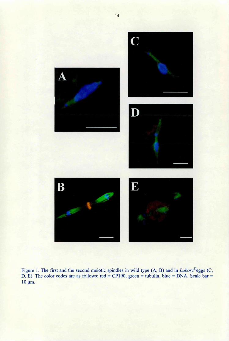
The first meiotic division The first meiotic spindle forms in Drosophila in a centrosome independent way (Matthies et al., 1996). Microtubules are nucleated in the vicinity of chromatin. The first meiotic spindle is indistinguishable from wild type in the Lahore0 eggs deposited by the Lahore0 females. The wild type first meiotic spindle has tapered poles without centrosomes, and contains highly compacted chromatin at the metaphase plate, with precociously separating fourth chromosomes (Fig. 1 A). In a number of Lahore0 eggs the spindle poles are divergent suggesting involvement of cytoplasmic dynein in spindle pole focusing (Fig. 1C). In other cases, nondisjunction of the chromosomes take place (Fig. ID), supporting that cytoplasmic dynein plays role in chromosome segregation.
The second meiotic division
The meiosis II spindle of Drosophila consist of two tandem spindles with anastral distal poles
and an aster-associated spindle pole body between the central poles (Fig. IB). The central
spindle pole contains centrosomal proteins. The central spindle pole body of the second meiotic
spindle in Lahore0 eggs is significantly larger than in wild type (Fig. IE). Altough this
phenotype is rare (2 out of 10), it indicates involvement of cytoplasmic dynein in assembly of the
central spindle pole body of the second meiotic spindle. As in wild type, there are no
centrosomes visible in the Lahore0 eggs in the meiotic divisions (Fig. 4D).
14
c
Figure 1. The first and the second meiotic spindles in wild type (A, B) and in Lahore0cggs (C, D, E). The color codes are as follows: red = CP 190, green = tubulin, blue = DNA. Scale bar = 10 pm.
15
CLEVEAGE CYCLES IN THE LABORCP EGGS
The first cleavage mitotic spindles in Labore0 eggs
The LaborcP/+ females produce normal numbers of normal looking so-called LaborcP eggs.
In fertilized LaborcP eggs the first cleavage mitosis forms as in wild type with one centrosome at
each spindle pole (Fig. 2A). In wild type the centrosomes do not replicate prior to completion of
mitosis. In fertilized LaborcP eggs, however, the centrosomes begin to replicate prior to
accomplishment of mitosis, as shown by presence of multiple centrosomes at the spindle poles
(Fig. 2E). This observation probably refers to a mechanism that prevents centrosome replication
prior to completion of mitosis. Cytoplasmic dyneins with LaborcP-encoded mutant heavy chain
ignore the mechanism.
Cleavage cycles in the LaborcP eggs Following the first and/or the second cleavage divisions, the centrosomes detach form the nuclear envelopes, replicate and the daughter centrosomes separate as in wild type (Fig. 2F, G). The usually four cleavage nuclei stop dividing. They remain deep down in the egg cytoplasm and eventually degenerate. Meanwhile the centrosome cycles proceed as in wild type and about two hours following fertilization the entire LaborcP egg cortex is populated with free centrosomes (Fig. 2H). The centrosomes nucleate microtubule asters that appear slightly larger as compared to wild type. Detachment of the centrosomes from the nuclear envelopes shows involvement of cytoplasmic dynein in centrosome attachment to the nucleus as was described by Robinson et al., 1999. The uncoupled chromosome and the centrosome cycles in the LaborcP eggs imply involvement of the cytoplasmic dynein in linking the chromosome and the centrosome cycles together.

16
A E
* *
Figure 2. Initiation of cleavage cycles in fertilized wild type (A-D) and in Lahore0 eggs (E-H). In wild type, there is one centrosome at each spindle pole (A). Multiple centrosomes emerge at poles of the first cleavage spindle in the Lahore0 eggs (E). Wild type (B and C) and Lahore0 eggs (F and G) with two and four nuclei. While characteristic spindles appear in wild type eggs (D), CSs detach from the nuclei, populate and nucleate MT asters in cortex of the Lahore0 eggs. Color codes are as in Fig. 1. Scale bar = 10 pm.

17
THE UNFERTILIZED LABORCP EGGS
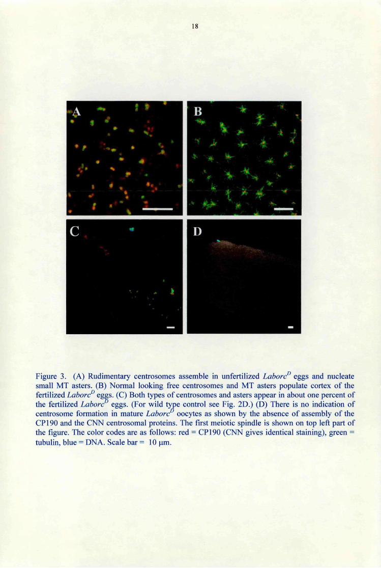
In Drosophila, cytoplasm of the wild type egg primordia contains a pool of dispersed centrosomal components without functional centrosomes. Upon egg activation, i.e. dining egg transfer from the ovaries through the oviduct to the outer world, some of the centrosome componenets assemble and form the central spindle pole body of the second meiotic spindle, irrespectively of fertilization. Four haploid nuclei form in a few minutes, and one of the nuclei will become the female pronucleus in the fertilized eggs, the other three will form the polar bodies. In unfertilized eggs all the four meiotic products will form polar body nuclei. Centrosomes of the embryos form when the sperm-derived centrioles recruit centrosome components form the egg cytoplasm. Centrosomes do not form in the unfertilized wild type eggs and development does not proceed beyond the four haploid nuclei stage (Foe et al., 1993). Contrary to the wild type eggs, in unfertilized LahoreD eggs the egg cytoplasm was full with centrosomes as revealed by the presence of the CP 190 and the CNN centrosomal proteins. The centrosomes nucleated small MT asters (Fig. 3A). However, unlike centrosomes in the fertilized LahoreD eggs (Fig. 3B), the centrosomes were slightly reduced in size and nucleated only small asters. It is important to note that there were no other nuclei than the four haploid products of the two meiotic divisions in the unfertilized LaborcP eggs.
We also noticed in a small number (1%) of the fertilized LaborcP eggs two separate groups of multiple centrosomes of both fertilized and unfertilized types (Fig. 3C.). While the anterior centrosome group might have originated form the sperm basal body, centrosomes in the another group might have assembled as in the unfertilized eggs.
The formation of centrosomes in the unfertilized LaborcP eggs clearly shows that dynein is involved in the mechanism that prevents de novo centrosome assembly in the unfertilized wild type eggs.
18
Figure 3. (A) Rudimentary centrosomes assemble in unfertilized LahoreD eggs and nucleate small MT asters. (B) Normal looking free centrosomes and MT asters populate cortex of the fertilized LahoreD eggs. (C) Both types of centrosomes and asters appear in about one percent of the fertilized LahoreD eggs. (For wild type control see Fig. 2D.) (D) There is no indication of centrosome formation in mature Lahore° oocytes as shown by the absence of assembly of the CP190 and the CNN centrosomal proteins. The first meiotic spindle is shown on top left part of the figure. The color codes are as follows: red = CP 190 (CNN gives identical staining), green = tubulin, blue = DNA. Scale bar = 10 pm.

19
Ultrastructural analysis of the unfertilized LahoreD eggs
To find out whether or not centrosomes in the unfertilized LahoreD eggs contained centrioles we
carried out an ultrastructural analysis. Apparently incomplete centrioles form, and their number
appears similar to the number of the centrosomes detected by immunofluorescence. However the
centrioles are rudimentary: the so-called carthweel structure in the middle of the centriole
appears normal in size and organization and the MT doublets are missing (Fig. 4B).
Figure 4. EM photographs of centriole cross-sections. (A) Wild-type cleavage embryo. (B) Rudimentary centrioles (arrows) in unfertilized LahoreD eggs. Note the lack of centriole MTs. Scale bar = 100 nm.

20
5. DISCUSSION
LABORŐ9 IS A DOMINANT NEGATIVE MUTATION IN THE dhc GENE Lahore10 was mapped to the left arm of the third chromosome (Erdélyi and Szabad, 1989).
Duplication and deficiency mapping located Laboré3 to the 64C cytological region. The laboré loss-of-function revertant alleles are zygotic lethal mutations and were generated through second
mutagenesis of the Laboré3 allele (Erdélyi and Szabad, 1989). To find out whether Laboré0 and
the laboré alleles identify an already known gene, we carried out complementation analyses
with lethal alleles of genes in the 64C region. Since the laboré alleles did not complement the
dhc~ mutations, that identify the Dhc64C cytoplasmic dynein heavy chain gene (Gepner et al.,
1996) we concluded that Laboré0 was an allele of that gene. This conclusion is further supported
by the finding that the P(dhc+)x transgene (Gepner et al., 1996) overcomes lethality of the
laboré/dhc" combinations.
When combined with tandem duplications that include the 64C region, about 1% of the
LaborcD/+/Dp+ females produced a few offspring. This result clearly shows that Laboré0 is a
gain-of-fiinction mutation i.e. the Laboré3-related defects are brought about by mutant gene
product. It furthermore implies that Laboré3 is a dominant negative mutation and implies that the
Laboré-encoded mutant and the normal gene products participate in the same process.
Maternal function of the Lahore protein
Interestingly the Laboré3 mutation does not interfere with viability, oogenesis and male fertility.
The toxic effect of the Laboré3-encoded mutant protein is manifested only in the embryos.
Because Laboré3 was induced with EMS, it is most likely a point mutation, which does not
affect expression of the Laboré3 allele, similarly to the KetelD mutation (Lippai et al. 2000;
Tirián et al., 2000). Laboré3 thus identifies maternal gene function of a zygotic gene: the egg
cytoplasm - the maternal dowry - of the wild type females contains dynein heavy chain protein
to match the unique needs of the cleavage stages of embryogenesis during which there is little if
any zygotic gene expression (Foe et al., 1993). The 13 cleavage cycles last only 8-10 minutes
each and require special factors to accomplish the fastest known eukaryotic cycles in the absence
of zygotic gene expression.
When paternally derived, Laboré3 behaves as the laboré (or the dhc") zygotic lethal alleles:
the Laboré3!-, like the laboré!- or the dhc7- hemizygotes, die at the beginning of larval life
and the different types of perishing larvae are indistinguishable. The former findings illustrate (i)
zygotic requirement of the Dhc64C gene and (ü) that the paternally derived Laboré0 allele does
21
not function during the cellular stages of development. In fact LaborcP is most likely expressed
in cells of the zygotes as the normal Dhc64C gene (Hays et al., 1994; Li et al., 1994).
Nonetheless LaborcP does not possess toxic effects since the LaborcP 1+ females and males are
viable and the males are fertile. The most likely explanation for the restricted action of LaborcP during the cleavage cycles is that LaborcP identifies maternal function of the zygoticaUy
essential Dhc64C gene and the deleterious effects are brought about following interaction of the
Lahore0-encoded mutant protein with a maternally provided partner present only in the egg
cytoplasm. Similar phenomenon was first described for the Ketel° alleles (Lippai et al., 2000;
Tiridn et al., 2000).
Although the mutant molecule might well be present in the somatic cells, it can not exert any
toxic effects in the absence of the appropriate partner. The mutant LaborcP protein is "simply"
nonfunctional without its partner, explaining the recessive loss-of-function phenotype in the
LaborcP I- zygotes. While functional dynein molecules form, the cytoplasmic dynein heavy chains
associate with light, light-intermediate and intermediate chain subunits. When in action,
cytoplasmic dyneins have been known to associate with cargoes, MTs, the dynactin complexes
and molecules that regulate dynein functions (Karki and Holzbaur, 1999). We propose that
association of the LaborcP encoded mutant cytoplasmic dynein heavy chain molecules with a
maternally provided dynein component-„designed" for embryogenesis-leads to the formation of
mutant cytoplasmic dynein and to defects characteristic of the LaborcP females. As described in
the coming chapters, there are plenty of candidates since the cytoplasmic dynein complexes are
composed from different types of molecules. The above mentioned maternally provided partner
awaits to be identified. In summary, the LaborcP alleles identify the maternal function of a both
maternally and zygotically required gene function.
THE MOTOR PROTEINS
Dyneins, kinesins and myosins represent the three major classes of molecular motors that move
along cytoskeletal elements (Fig. S). Dyneins move toward the minus end of the MTs, and most
kinesins move toward the plus end of the MTs (Belecz and Szabad, 1998). Myosins move along
the actin microfilaments. The kinesins and myosins have a motor domain that contains both
ATPase and filament-binding sites attached to an a-helical lever arm and neck region. Structural
analysis of the kinesin and myosin heads revealed striking similarity in the motor domains of the
kinesin and myosin enzymes (Kull et al., 1996; Rayment et al., 1993). The similarity implies that
the general mechanisms by which the motor proteins generate force while move along their
respective filament systems, are related at a fundamental mechanism and raised the possibility
that all cytoskeletal motors function similarly. However, one thorn remained in this rather
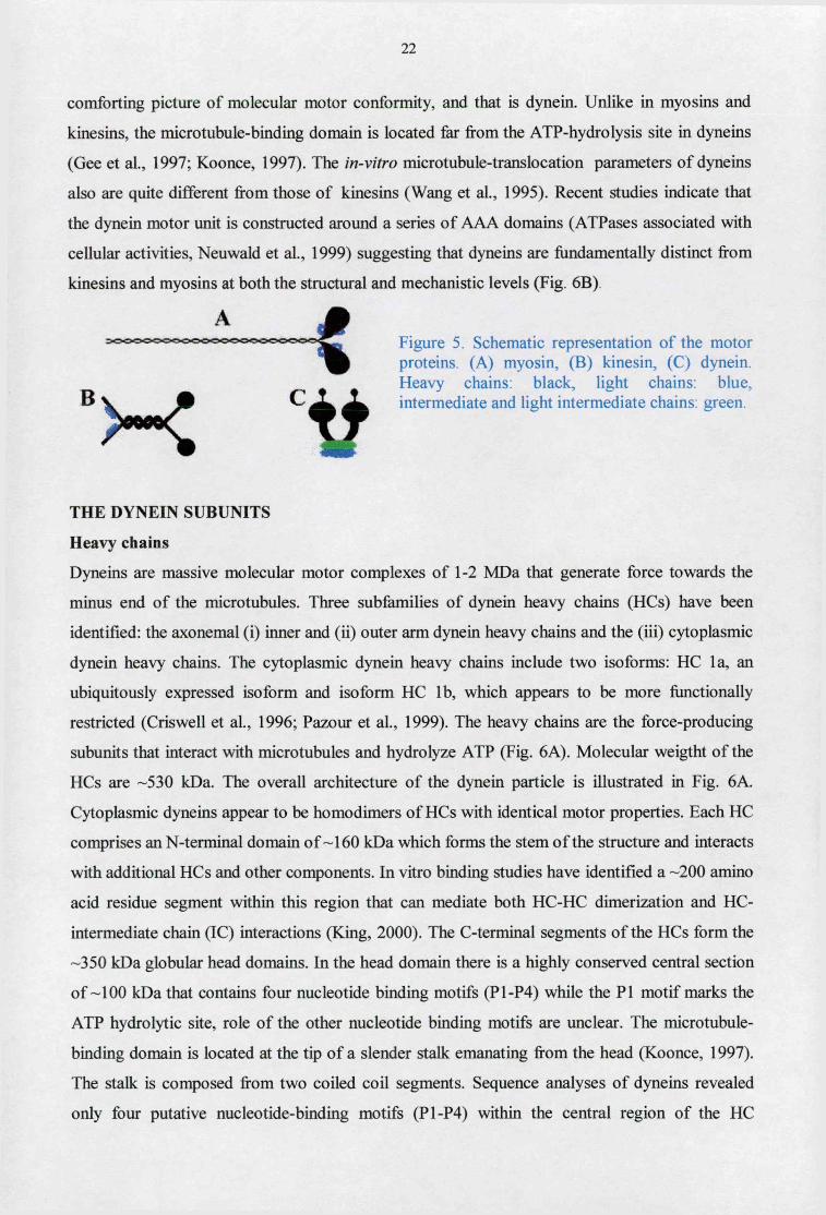
22
comforting picture of molecular motor conformity, and that is dynein. Unlike in myosins and
kinesins, the micro tubule-binding domain is located far from the ATP-hydro lysis site in dyneins
(Gee et al, 1997; Koonce, 1997). The in-vitro microtubule-translocation parameters of dyneins
also are quite different from those of kinesins (Wang et al., 1995). Recent studies indicate that
the dynein motor unit is constructed around a series of AAA domains (ATPases associated with
cellular activities, Neuwald et al., 1999) suggesting that dyneins are fundamentally distinct from
kinesins and myosins at both the structural and mechanistic levels (Fig. 6B)
THE DYNEIN SUBUNITS
Heavy chains
Dyneins are massive molecular motor complexes of 1-2 MDa that generate force towards the
minus end of the microtubules. Three subfamilies of dynein heavy chains (HCs) have been
identified: the axonemal (i) inner and (ii) outer arm dynein heavy chains and the (iii) cytoplasmic
dynein heavy chains. The cytoplasmic dynein heavy chains include two isoforms: HC la, an
ubiquitously expressed iso form and iso form HC lb, which appears to be more functionally
restricted (Criswell et al., 1996; Pazour et al., 1999). The heavy chains are the force-producing
subunits that interact with microtubules and hydrolyze ATP (Fig. 6A). Molecular weigtht of the
HCs are -530 kDa. The overall architecture of the dynein particle is illustrated in Fig. 6A.
Cytoplasmic dyneins appear to be homodimers of HCs with identical motor properties. Each HC
comprises an N-terminal domain o f -160 kDa which forms the stem of the structure and interacts
with additional HCs and other components. In vitro binding studies have identified a -200 amino
acid residue segment within this region that can mediate both HC-HC dimerization and HC-
intermediate chain (IC) interactions (King, 2000). The C-terminal segments of the HCs form the
-350 kDa globular head domains. In the head domain there is a highly conserved central section
o f -100 kDa that contains four nucleotide binding motifs (P1-P4) while the PI motif marks the
ATP hydrolytic site, role of the other nucleotide binding motifs are unclear. The microtubule-
binding domain is located at the tip of a slender stalk emanating from the head (Koonce, 1997).
The stalk is composed from two coiled coil segments. Sequence analyses of dyneins revealed
only four putative nucleotide-binding motifs (P1-P4) within the central region of the HC
Figure 5. Schematic representation of the motor proteins. (A) myosin, (B) kinesin, (C) dynein. Heavy chains: black, light chains: blue, intermediate and light intermediate chains: green. c

23
(Gibbons et al., 1991). However, more recent studies clearly indicate that the dynein motor also
contains six AAA domains plus an unrelated seventh unit at the C terminus (Fig. 6B; Neuwald et
al., 1999). The first four AAA units correspond to the previously identified motifs. The fifth and
sixth AAA domains had not been noted originally, because they lack intact consensus P-loop
motifs. The coiled coil and microtubule-binding region that protrudes from the main head
(Koonce, 1997) is located between the fourth and fifth AAA domains. Three-dimensional
reconstructions of the dynein motor domain from high-resolution negative stain images reveal a
13.5 nm diameter spheroidal structure composed of seven globular subdomains arranged around
a central cavity (Samso et al., 1998, Fig. 6B).
A MT-binding
Cargo Attachment
an unrelated C-terminal from King, 2000).
Intermediate chains
Cytoplasmic dyneins contain two ICs of -70-80 kDa which are are located at the base of the
soluble dynein particle. The ICs are members of the WD-repeat (tryptophan, aspartic acid)
protein family. ICs apparently act directly in the attachment of the dynein motor to its cargo.
Cytoplasmic dynein IC composition is complex and molecular and biochemical studies have
revealed a plethora of isoforms derived from differential phosphorylation and alternative splicing
(King, 2000). The IC isoforms are differentially expressed during development of specific cell
types (King, 2000). Although functional significance of the IC isoform diversity remains
unknown, one obvious possibility is that different versions of ICs are capable of interacting
either with different proteins or with the same protein but with varying affinities. ICs interact
directly with the pi50 Glued component of dynactin (Karki and Holzbaur, 1995). The dynactin
complex is an additional multimeric structure that acts as an activator of dynein-based motility
(King and Schroer, 2000; see below).
subdomain and the
Figure 6. Organization of the dynein motor domain. (A) Generic model of a dynein particle. The C-terminal portion of each HC forms a globular head containing the ATPase sites and has a small stalk-like structure that terminates in a microtubule-binding globular unit. The base of the dynein particle consists of the N-terminal regions of the HCs and a series of accessory proteins that function in cargo binding and might also have regulatory roles. (B) Model for the organization of an individual HC illustrating the heptameric structure of the head. Note that almost the entire globular motor unit consists of the six AAA domains,
coiled-coil microtubule-binding region. (Adapted
24
Light intermediate chains The 50-60 kDa light intermediate chain (LIC) dynein proteins contain an ATP-binding motif and
are distantly related to ABC transporters (Hughes et aL, 1995). However, there is as yet no direct
evidence that they actually bind nucleotide and their precise location within the dynein particle
has yet to be resolved. The dynactin independent interaction between pericentrin and
cytoplasmic dynein is specifically mediated by LIC (Purohit et al., 1999)
Light chains 1. Dynein LC8 LC8 belongs to a group of very highly conserved proteins present in distantly related organisms
as mammals, nematodes and plants. LC8 protein is a stoichiometric component of both brain
cytoplasmic dynein and myosin (King, 2000). The LC8 protein is present in many, seemingly
unrelated enzyme systems. Drosophila partial loss-of-function alleles lead to morphogenetic
defects in bristle and wing development, female sterility and also cause alterations in axonal
guidance during development. Total loss-of-function alleles result in embryonic lethality through
the induction of apoptotic pathways (Phillis et al., 1996). The LC8 associated phenotypes were
originally interpreted as being caused by defects in cytoplasmic dynein; however, it now seems
more likely that certain of the defects derive from the disruption of other enzyme activities.
2. The Tctexl dynein light chain family The diverse group of Tctexl dynein light chains was first identified in cytoplasmic dynein
(King, 2000). Examination of the current databases revealed several additional members of the
family including the rp3 protein that is also a demonstrable component of cytoplasmic dynein
(King, 2000). The Tctexl and rp3 light chains are differentially regulated in both a
developmental and tissue-specific manner. LCs may play a role in the binding of particular
cargoes or in differentially regulating some essential aspect of dynein function (King, 2000).
3. The roadblock/LC7 light chain family The most recent class of dynein LC reported contains closely related homologues that are present
in both flagellar and cytoplasmic dyneins (King, 2000). Mutations in the Drosophila roadblock gene lead to defects in axonal transport and mitosis suggesting that members of the
roadblock/LC7 light chain family play essential roles in dynein function.
DYNACTIN
Cytoplasmic dynein works in conjunction with a second multiprotein complex, dynactin (Fig. 7).
Dynactin is generally believed to function as an adapter that allows dynein to bind cargo.

25
Dynactin consist of 11 different subunits (Schroer, 1996; Allan, 2000). Dynactin has two distinct
structural domains, an actin like miniflament backbone and a flexible projecting sidearm. Dynein
binds the dynactin sidearm subunit, pl50Glued. The distal end of the pl50Glued sidearm also
contains a pair of MT binding sites. Transient MT binding by dinactin allows the dynein motor
to move more processively (King and Schroer, 2000).
REGULATION OF DYNEIN MOTOR FUNCTIONS
Dynein motor functions are under exquisite and precise control both in the cytoplasm and the
flagellum. How might dynein-based motor activity be controlled? A number of possibilities for
which there is some evidence exist including the following: direct
phosphorylation/dephosphorylation of the HCs, phosphorylation of ICs and LCs that result in an
alteration in motor activity and ligand-induced conformational changes in LCs directly
associated with the HCs (King, 2000).
Association of motors with specific cargoes
Mechanisms that account for the targeting of dyneins and kinesins to cargoes may differ. For
kinesins, the multiple cellular functions are provided at least in part by multiple kinesin-related
heavy chain polypeptides and associated light chains (reviewed by Goldstein, 1993; Moore and
Endow, 1996). Sequence differences outside of the motor domain of the kinesin heavy chains
contribute to the targeting of distinct kinesins to specific functions, either directly or by
association with other proteins. For example, in Drosophila, the kinesin-like protein Nod
contains a DNA-binding motif in the nonmotor domain that localizes it to chromosomes during
female meiosis (Ashfar et al., 1995). The mechanisms that target the dyneins to their cargoes are
less clear. The intermediate, light-intermediate, and light chain subunits are located in a position
Micro tubu le
Figure 7. Model of the dynactin complex and its proposed interaction with microtubules and cytoplasmic dynein (light blue). Dynactin subunits are: 8 or 9 xArpl (actin-related protein 1; pink); 4 or 5 xdynamitin (green); 2 xpl50 Glued (orange); 2 x p24 (brown); one each of p62, conventional actin, Arpll , capping protein a and B , p27 and p25 (yellow). ? indicates unknown cargo attacment factors. (Adapted from Allan, 2000)

26
to interact with other cellular components; their assembly and regulation may mediate the
targeting of cytoplasmic dyneins to specific cargoes (see review by King, 2000). The different
subunits of dynactin are also believed to function as an adapter that allows dynein to bind
different cargoes.
CYTOPLASMIC DYNEIN FUNCTIONS Cytoplasmic dyneins perform a broad range of cellular functions, including chromosome
segregation, spindle formation, nuclear migration, Golgi positioning, retrograde membrane
transport, and functioning in the endocytic pathway (Karki and Holzbaur, 1999). Only those
functions are discussed below, which are important for interpretation of the Laboré3 phenotype.
During the meiotic divisions Laboré3 results in unfocused spindle poles, chromosome
nondisjunction, and over-assembly of the central spindle pole body of the second meiotic spindle
in a number of cases. During the cleavage divisions Laboré3 uncouples the chromosome and the
centrosome cycles and leads to centrosome migration, attachment and (probably) centrosome
replication defects.
Centrosome separation during interphase For proper CS separation, centrosomes must move (tightly associated with the nucleus) until
they are diametrically opposed on the nucleus. One possible mechanism for centrosome
separation is that plus end directed kinesins exert pushing forces on overlapping microtubules
emanating from the two centrosomes. In the second mechanism, pulling forces generated by
minus end directed motors acting on astral microtubules in front of the moving centrosomes.
Dynein could generate such pulling forces by being anchored in the cytoplasm or at the cell
cortex. Alternatively pulling forces may result from dynein molecules anchored on the nucleus
(Fig. 8). The latter model is attractive because it explains both how centrosomes separate and
how they remain tightly associated with the nucleus. In the latter scenario, the minus ends of
astral microtubules, along with the centrosome, are pulled when they encounter anchored
cytoplasmic dynein on the nucleus. Longer astral microtubules encounter more anchored motors,
and thus experience a stronger pulling force than shorter ones. After centrosome duplication,
microtubules extending away from the centrosomes along the nucleus are long, whereas those
projecting the other centrosome are short. Thus, lenght dependent forces may ensure that
daughter centrosomes move away from each other until such pulling forces are balanced, which
occurs, when they are diametrically opposed. It has been shown recently, that cytoplasmic
dynein is required for centrosome attachment to the nucleus and centrosome migration along the
nuclear envelope during the embryonic cleveage divisions in Drosophila (Robinson et al., 1999).

27
Figure 8. Possible model of dynein-dependent separation of daughter centrosomes. Chromatin: blue, centrosomes: red, MTs: green. Cytoplasmic dynein molecules that interact with astral microtubules are shown in dark shading, others in
light shading.
In the Labor? eggs following the first or the second cleavage divisions, the centrosomes detach
from the nuclear envelopes showing involvement of cytoplasmic dynein in centrosome
anchorage to the nucleus (Belecz et al., 2001; Fig. 3E, F). The detached centrosomes continue
replication, nucleate MT asters and start migrating to the egg cortex. By the time the
centrosomes reach the egg cortex, they nucleate enormous microtubule asters and about two
hours following fertilization the entire cortex of the Labor? egg cytoplasm contains free
centrosomes (Fig. 3G, H).
The size of the free centrosomes is normal and they appear to be fully functional because all of
them nucleate MT asters. The usually four cleavage nuclei stop dividing. They remain deep
down in the egg cytoplasm and degenerate eventually (Fig. 3F). In DAPI stained preparations,
fragmented chromatin of the degenerating nuclei appear as giant polyploid nuclei as reported
earlier (Erdélyi and Szabad, 1989). It is unclear how Labor? leads to the cessation of the
chromosome cycle. Apparently the chromosome and the centrosome cycles are uncoupled in the
Labor? eggs implying an involvement of the cytoplasmic dynein in linking the two types of
cycles together.
As in wild type fertilized eggs, the blocks on chromosome and centrosome cycles are removed
in fertilized eggs of females homozygous for either of the female sterile mutations plu, pug or
gnu (Fig. 9). In the plu and png eggs the centrosome cycles cease and only a few asters appear
adjacent to the large polyploid nuclei (Shamanski and Orr-Weaver, 1991). The plutónium gene
encodes a small ankyrin repeat protein with a direct role in coupling S and M phases during
cleavage divisions and pan gu gene function is required for plutónium activities (Elfring et al.,
1997). In fertilized gnu eggs, however, both cycles proceed and while large polyploid nuclei
form inside the egg cytoplasm centrosomes populate the entire egg cortex (Freeman et al., 1986).
Evidently harmony of the chromosome and the centrosome cycles is disrupted in the gnu eggs.
Molecular function of the giant nuclei gene is not known. The chromosome and the centrosome
cycles can also be uncoupled by aphidicolin (Raff and Glover, 1989).
LahoreD is a new and unique addition to the above mutations. In fertilized LahoreD eggs the
chromosome replication is released, however while it comes to a standstill after usually two
rounds of replication the centrosome cycles proceed as in wild type. Since LahoreD is a gain of

28
function allele of the Dhc64C gene, the above observations illustrate involvement of cytoplasmic
dynein in establishing harmony of the centrosome and chromosome cycles. Analysis of eggs of
double mutant females should reveal the genetic hierarchy among the above genes (Glover,
1991).
UNFERTILIZED EGGS FERTILIZED EGGS
plu pan gu gnu
CHROMOSOME CYCLE CENTROSOME CYCLE
"T"
X Labore
plu pangu gnu
X )( )( )( •«wwwrarowisar*
X )( Labore
»
gnu
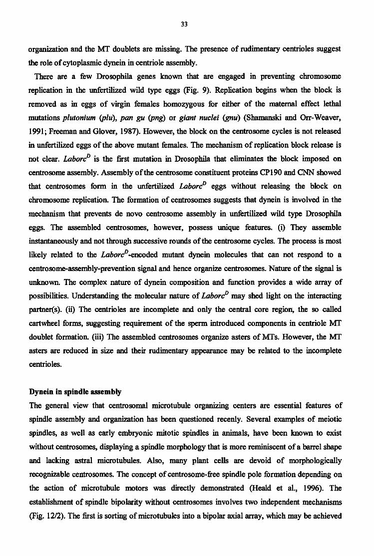
Figure 9. Mutations that eliminate the block on chromosome and/or centrosome cycles in unfertilized or in fertilized eggs leading to the formation of polyploid nuclei and/or free centrosomes that nucleate MT asters. (Dashed line represents the formation of rudimentary centrosomes and MT asters.)
Centrosome separation during cell division
Cytoplasmic dyneins are essential during the separation of the centrosomes in prophase. It
appears that by metaphase dynein activity may no longer be required to maintain the separation
of the spindle poles: the spindle apparatus, that is composed from bundles of kinetochore
microtubules and the overlapping polar microtubules originating from the opposite spindle poles,
have already stabilized the spacing of the centrosomes (Vaisberg et al., 1993)
There are several ways in which a minus end directed microtubule motors achieve centrosome
separation (Fig. 10). One model for centrosome separation resembles to some extent the situation
in axonemes, where ciliary dyneins act between parallel doublet microtubules. If a dynein
molecule is temporarily attached to a fixed point on one microtubule while it slides along a
microtubule from the other pole, the two closely spaced centrosomes will be forced apart. Such
sliding would lead to a gathering of microtubules into the space between the centrosomes,
forming a central spindle (Fig. 10A). Other microtubule motors, like the plus end directed
kinesins, might then bind to the interdigitating microtubules stabilizing the spindle and further
separating the poles (Fig. 10B). Another mechanism for spindle pole separation during anaphase
B is when dyneins (anchored to the cell membrane) exert a pulling force on the poles via the
astral microtubules (Fig. 10C). The first cleavage spindle in LahoreD eggs is shorter normal (Fig
2E) providing in vivo evidence for the role of cytoplasmic dynein in centrosome separation
during the cleavage divisions.

29
B
^ ^ J w I / \ J Figure 10. Cytoplasmic dynein's possible ~~/i ^ » ® r o l e s in daughter centrosome separation
s ^ J 1 during cell division. Dyneins: blue, kinesins: lilac, MTs: green, and centrosomes: red.
Chromosome segregation and the role of dynein at the kinetochre
Cytoplasmic dynein is the only known kinetochore assiciated protein capable of driving
chromosome movement towards the centrosomes. However dynein functions at the kinetochores
are ambiguous. Immunolocalization of dynein to the kinetochores of tissue culture cells,
combined with the analysis of kinetochore microtubule polarity, first suggested a potential role
of dynein in providing the force for chromosome movements along the mitotic spindle (Pfarr et
al., 1990; Steuer et al., 1990). Functional evidence that supports such a role for dynein is limited
to the recent observation in Tetrahymena that micronuclear chromosomes fail to segregate in cell
lines in which the cytoplasmic dynein gene, DYH1, is knocked out (Lee et al., 1999). More
recent reports of dynein and dynactin dynamics at the kinetochore suggest that both types of
molecules may function to mediate microtubule binding at the kinetochore (Starr et al., 1998;
Walczak et al., 1998). However the bulk of the dynein leaves the kinetochore very early in
mitosis, soon after the kinetochores begin to attach to microtubules (King et al., 2000). The
possible functions of the dynein fraction that left the kinetochores is therefore limited to the
initial attachment and movement of chromosomes and/or to a role in monitoring the attachment
state of kinetochores. The remaining dynein molecules at the kinetochore may be sufficient for
pulling the chromosomes during anaphase to the opposite poles. There are two kinetochore
proteins, ZW10 and Rod, which are essential to localize dynein to the kinetochore (Starr et al.,
1998). Model for the ZWIO/Rod-dependent targeting of dynein to the kinetochore is shown in
Fig. 11
In summary, dynein at the kinetochore can not be uniquely required for chromosome
microtubule attachments or movements before anaphase onset. Dynein might participate in the
checkpoint mechanisms that sense bipolar tension across the centromere, delaying anaphase
onset until all the chromosomes are properly aligned on the metaphase plate. Alternatively,
dynein might be required at the kinetochore to supplement and/or coordinate other microtubule
motors in moving chromosomes to the poles during anaphase (Sharp et al., 2000). During the

30
first meiotic division in LahoreD eggs in a number of cases nondisjunction of the chromosomes
take place (Fig. ID), supporting that cytoplasmic dynein plays some role in chromosome
segregation.
Figure. 11. A complex containing ZW10 and Rod proteins (two kinetochore proteins), as well as potential unknown additional components (?), is associated with the fibrous corona of the prometaphase kinetochore. Direct interactions between ZW10 and the p50 subunit of the dynactin complex then bring dynactin to the kinetochore. Dynactin in turn recruits cytoplasmic dynein to the kinetochore,
providing one possible contact between the kinetochore and microtubules. (Adapted from Starr et al., 1998)
jP„r* Dynactin CZ Z) Cytoplasmic Dynein
R O D -ZW10 F i b r o u s
C o r o n a
Recruitment of centrosome proteins
There is now good evidence for microtubule-dependent (Kuriyama, 1982; Balczon et al., 1999;
this manuscript) and microtubule-independent (Moritz et al., 1998; Khodjakov and Rieder, 1999)
mechanisms for the recruitment of proteins into the centrosomes. Dynein-mediated and passive
diffusion mechanisms represent parallel pathways for centrosome assembly. It is possible that
one pathway predominates over the other in certain biological systems or at different stages of
the cell cycle. In embryonic systems, for example, high levels of centrosome proteins (Gard et
al., 1990) may be sufficient to drive the initial stages of microtubule-independent recruitment
onto centrioles, although dynein-mediated transport becomes a major contributor at later times.
Dynein has been shown to transport pericentriolar components to the centrosome during both
interphase and mitosis. The dynein-transported molecules include dynactin, y-tubulin and
pericentrin during interphase, dynactin and NuMA during mitosis (Quintyne et al., 1999). The
LahoreD mutation revealed cytoplasmic dynein dependent transport of centrosomal components
during the assembly of the central spindle pole body of the second meiotic spindle, and in
unfertilized eggs, after the completion of meiosis.
The LahoreD mutation leads to enlargement of the central spindle pole body of the second
meiotic spindle
The meiosis II spindle of Drosophila oocytes is distinctive in structure, consisting of two tandem
spindles with anastral distal poles and an aster-associated spindle pole body between the central
poles (Fig. IB). Assembly of the anastral/astral meiosis II spindle occurs by reorganization of the

31
meiosis I spindle, without breakdown of the meiosis I spindle (Endow and Komma, 1998). The
unusual disk- or ring-shaped central spindle pole body forms de novo in the center of the
elongated meiosis I spindle, followed by formation of the central spindle poles. y-Tubulin
transiently localizes to the central spindle pole body, implying that the body acts as a
microtubule nucleating center for assembly of the central poles. Localization of y-tubulin to the
meiosis n spindle is dependent on the kinesin like minus end directed microtubule motor protein,
Nonclaret disjunctional (Ned; Endow and Komma, 1998). The central spindle pole body of the
second meiotic spindle in Lahore° eggs is significantly larger than in wild type (Fig. IE).
Altough this phenotype is rare (2 out of 10), it indicates the redundant involvement of
cytoplasmic dynein in the assembly of the central spindle pole body of the second meiotic
spindle, and suggests that the LahoreD encoded protein transports more than usual CP 190 into
the central spindle pole body. We suppose, that the LahoreD encoded protein cannot respond
properly to a negative regulatory signal which results in enlargement of the central spindle pole
body. The negative regulatory signal may prevent dynein molecules to assemble more than usual
centrosomal components to the central spindle pole body. Because after oocyte activation
translation of maternally provided mRNA-s commences the above mentioned negative
regulatory signal must already be present at the time when the second meiotic division
progresses.
The tandemly oriented second meiotic spindles detach from the central spindle pole body
showing that dynein (probably with other minus end directed MT motors, such as Ned) is
required to anchor the second meiotic spindle poles to the central spindle pole body. Roles for
cytoplasmic dynein in the assembly of the central spindle pole body and in the attachment of the
inner spindle poles to the central spindle pole body has not been shown in Drosophila previously.
In addition to the pericentriolar components dynactin, y-tubulin, pericentral, dynactin and NuMA
which have been previously shown to be transported by dynein to the MTOC-s, our results
suggest that CP190 is also transported actively by cytoplasmic dynein to the central spindle pole
body of the second meiotic spindle.
In unfertilized LaborP eggs centrosomes with incomplete centrioles assemble During the development of fertilized eggs, centrosome inheritance must be precisely controled
because if both gametes contribute functional centrosomes, the zygote will have an abnormal
spindle. In Drosophila, as in most animal species, egg cells do not carry centrosomes that are
lost during oogenesis (Schatten, 1994). Most centrosome/centriole components are maternally
supplied and are dispersed in the egg cytoplasm (Schatten, 1994). Eggs are activated while they
travel form the ovaries through the oviduct: the two meiotic divisions are completed and

32
translation of the maternally provided mRNAs commences (Foe et al., 1993). In absence of
fertilization a thus for unknown mechanism prevents both chromosome replication and
centrosome formation.
Centrosomes of the embryos derive from the sperm that introduces during fertilization a
centriole pair (Foe et al., 1993). The centriole replicates and, as generally believed, recruits
centrosome components from the egg cytoplasm to form functional centrosomes.
In Drosophila, some of the centrosome components assemble upon egg activation and form the
central spindle pole body of the second meiotic spindles, irrespectively of fertilization (Puro,
1991; RiparbeUi et al., 1997). However, centrosomes never form in the unfertilized wild type
eggs and development does not proceed beyond the four haploid nuclei stage (Foe et al., 1993).
Contrary to wild type, centrosomes assemble in cytoplasm of the unfertilized Laboré eggs (Fig.
3A). The centrosomes nucleate small asters of MTs. The feet that there are no centrosomes in the
mature Laboré oocytes (Fig. 3D) and a few minutes later (after the completion of meiosis) there
are lots of centrosomes in cytoplasm of the newly deposited unfertilized Laboré eggs shows
that centrosomes did not multiply in an exponential fashion - through repeated replication and
separation - as in fertilized wild type and Laboré eggs but the rather assembled instantaneously
shortly after egg deposition.
Examination of unfertilized Laboré eggs revealed a previously unknown mechanism that
prevents de novo assembly of centrosomes in wild type unfertilized eggs after oocyte activation.
Because Laboré identifies the cytoplasmic dynein heavy chain gene, we conclude involvement
of the cytoplasmic dynein in the above mechanism. Our hypothesis is that the Laboré encoded
proteins cannot respond to a yet unknown negative regulatory signal and assemble centrosomal
proteins into centrosomes.
Both rudimentary, characteristic for the unfertilized Laboré eggs, and normal centrosomes
appear only in about 1 percent of the fertilized Laboré eggs (Fig. 3C), suggesting that
fertilization changes egg cytoplasm chemistry such that centrosome and MT components do not
assemble spontaneously but rather „await" for the nucleating activities of the sperm-derived
centrosomes.
To find out whether or not centrosomes in the unfertilized Laboré eggs contained centrioles
we carried out an ultrastructural analysis. Embryonic centrioles posess nine doublet MTs,
together with an internal structure called the cartwheel (Debec et al., 1999, Fig. 4A). In the
unfertilized Laboré eggs apparently incomplete centrioles form, and their number appears
similar to the number of the centrosomes detected by immunofluorescence (Fig. 4B). However
the centrioles are rudimentary: the central carthweel of the centriole appears normal in size and
33
organization and the MT doublets are missing. The presence of rudimentary centrioles suggest
the role of cytoplasmic dynein in centriole assembly.
There are a few Drosophila genes known that are engaged in preventing chromosome
replication in the unfertilized wild type eggs (Fig. 9). Replication begins when the block is
removed as in eggs of virgin females homozygous for either of the maternal effect lethal
mutations plutónium (plu), pan gu (png) or giant nuclei (gnu) (Shamanski and Orr-Weaver,
1991; Freeman and Glover, 1987). However, the block on the centrosome cycles is not released
in unfertilized eggs of the above mutant females. The mechanism of replication block release is
not clear. LaborcP is the first mutation in Drosophila that eliminates the block imposed on
centrosome assembly. Assembly of the centrosome constituent proteins CP 190 and CNN showed
that centrosomes form in the unfertilized LahoreD eggs without releasing the block on
chromosome replication. The formation of centrosomes suggests that dynein is involved in the
mechanism that prevents de novo centrosome assembly in unfertilized wild type Drosophila
eggs. The assembled centrosomes, however, possess unique features, (i) They assemble
instantaneously and not through successive rounds of the centrosome cycles. The process is most
likely related to the Lahore0-encoded mutant dynein molecules that can not respond to a
centrosome-assembly-prevention signal and hence organize centrosomes. Nature of the signal is
unknown. The complex nature of dynein composition and function provides a wide array of
possibilities. Understanding the molecular nature of LaborcP may shed light on the interacting
partner(s). (ii) The centrioles are incomplete and only the central core region, the so called
cartwheel forms, suggesting requirement of the sperm introduced components in centriole MT
doublet formation, (üi) The assembled centrosomes organize asters of Mis. However, the MT
asters are reduced in size and their rudimentary appearance may be related to the incomplete
centrioles.
Dynein in spindle assembly
The general view that centrosomal microtubule organizing centers are essential features of
spindle assembly and organization has been questioned recenly. Several examples of meiotic
spindles, as well as early embryonic mitotic spindles in animals, have been known to exist
without centrosomes, displaying a spindle morphology that is more reminiscent of a barrel shape
and lacking astral microtubules. Also, many plant cells are devoid of morphologically
recognizable centrosomes. The concept of centrosome-free spindle pole formation depending on
the action of microtubule motors was directly demonstrated (Heald et al., 1996). The
establishment of spindle bipolarity without centrosomes involves two independent mechanisms
(Fig. 12/2). The first is sorting of microtubules into a bipolar axial array, which may be achieved

34
by plus end-directed, multimeric motors that can promote anti-parallel microtubule sliding and
axial alignment. Candidates for such an activity are the tetrameric motors of the BimC kinesin
family (Kashina et al., 1996). The second is the bundling of the oriented microtubules into poles,
involving the minus end-directed, microtubule motor cytoplasmic dynein. Live observations of
meiotic spindle formation in Drosophila oocytes (Matthies et al., 1996) have revealed that the
spindles form by an "inside-out" mechanism in which microtubules reorganize around the mass
of chromatin (Fig. 12/2). The process may involve the action of chromatin-bound, plus end-
directed kinesin-like proteins the chromokinesins (Wang and Adler, 1995, Vernos et al., 1995).
With the microtubule minus ends oriented away from the chromatin in the developing spindles,
the organization of the microtubules into bipolar spindles may then be achieved by the action of
multivalent, minus end-directed microtubule motor complexes that can tether parallel-oriented
microtubules into bundles and stabilize converging microtubules into poles (Fig. 12/2). As
Matthies et al. (1996) showed, in Drosophila oocytes the process is clearly dependent on
presence of the minus end-directed motor Ned, although there seem to be other motor proteins
with redundant functions involved.
1A IB 2A 2B
multivalent pfciv end-direvicd iroxTiHubnle minors • NtiMAAJ>tv*iaA1ytuilm eompli»*.?«
• chromatin- hmmd plusend-dtrevied mtcrotoluile rnnws multivalent plus .'{Hi-tluciVJ microuibtik- motors
• N u M A ,'itvnci ntitvnactm complexes Figure 12/1. Spindle formation in centrosome-containing cells. (A) Microtubules are nucleated from the duplicated centrosomes with their growing plus ends pointing away from the centrosomes. Microtubules are captured by the kinetochores of the chromosomes. Multivalent plus end-directed motors of the BimC family may be involved in the separation of the two centrosomes and the establishment of a symmetric spindle axis (big arrows). (B) In the mature spindle, microtubule minus ends disconnect from the centrosomes and are anchored to the body of the spindle by complexes of NuMA/dynein/dynactin. (The chromosomes are indicated in blue.) Figure 12/2. Spindle formation in centrosome-ffee cells. (A) Spindle formation is driven by chromatin-associated, plus end-directed microtubule motors, orienting chromatin-attached microtubules with their minus ends outward (arrows). Multivalent plus end-directed microtubule motors of the BimC family can interconnect antiparallel microtubules and establish a bipolar organization of the spindle by moving the microtubule ends apart. (B) During spindle pole formation complexes composed of NuMA, dynein, and dynactin induce convergent arrays of microtubules at the spindle poles and provide stability to the spindle by tethering the microtubule minus ends. (Adapted from Merdes and Cleveland, 1997)

35
Recent reports indicate that the small GTPase Ran (Ras-like nuclear protein), which plays a key
role in nuclear transport, also has a role in microtubule nucleation and in spindle assembly
(Kahana and Cleveland, 2000, Fig. 13). Ran in its GTP-bound form promotes the formation of
microtubules. Following nuclear envelope breakdown, chromatin-bound RCC1 converts
RanGDP to RanGTP which stimulate tubulin polymerization and promote spindle organization.
Chromosomal influence on spindle assembly is apparent in both centrosomal and acentrosomal
cells.
RanGTP WtBBSt: RanGDP
Dynein at the spindle poles
Dynein acts as a microtubule tethering factor at the spindle poles, irrespective of the presence or
absence of centrosomes. Microtubule tethering into poles is mediated by a large complex
containing NuMA, dynein, and dynactin, using the motor activity of dynein to power the
complex toward the microtuble minus ends and the distinct microtubule binding sites on NuMA
(Merdes et al., 2000) and the associated pl50 dynactin component (Karki and Holzbaur, 1995) to
provide the needed crosslinking. Microtubule tethering to spindle poles in centrosomal spindles
is needed because up to 75% of the interpolar microtubules do not connect directly to the
centrosome but end within a distance of>l pm thereof (Mastronarde et al., 1993). Moreover,
removal of the centrosome by micromanipulation does not grossly affect spindle integrity
(Nicklas et al., 1989). A plausible model for what keeps these microtubules in place invokes the
NuMA complex, which is distributed in a broad, crescent-shaped area between the centrosome
and the spindle microtubule bundles, rather than focused directly at the centrosome (Merdes et
al., 2000). NuMA thus is likely to be one of the connecting molecules that anchor the large
number of free microtubule minus ends to the microtubules still directly nucleated by the
centrosome. In a number of Labor? eggs the first meiotic spindle poles are divergent showing
involvement of cytoplasmic dynein in spindle pole focusing (Fig. 1C), which have been
RanGAP RanBPI
RanBPM
Figure 13. Model showing the proposed effects of chromosomal RCC1 on RanGTP/RanGDP levels in the surrounding cytoplasm. RCC1 converts RanGDP to RanGTP close to the chromosomes, while cytoplasmic RanGAP together with RanBPI leads to GTP hydrolysis by Ran, generating a gradient of RanGTP that promotes microtubule nucleation and growth towards the chromosomes. (Adapted from Heald and Weis, 2000)
/

36
described in other model systems. The above role for cytoplasmic dynein has not been shown in
Drosophila female meiosis previously.
CENTROSOME REPLICATION
Analysis of the early celeavage divisions in Laboré3 eggs suggests new and unexpected
cytoplasmic dynein functions in centrosome replication, therefore I briefly discuss recent
knowledge about centrosome replication in different systems.
The centrosome cycle in mammalian cells The cell cycle is an ordered progression of events that leads to division of one cell into two, each
with an identical copy of the genome. In normal divisions, both the chromosomal DNA and the
centrosome must be duplicated once and only once per cycle. The structure of DNA, with its
complementary strands, provides a simple mechanism for duplication. Centrosomes having no
DNA must employ some other mechanism of duplication. What happens during centrosome
replication (for reviews see Urbani and Stearns, 1999; Zimmermann et al., 1999)? In the G1
phase of the cell cycle, there is one centrosome per cell, consisting of a pair of centrioles and the
associated pericentriolar material. The replication process begins at the Gl-S transition, at
approximately the same time that DNA replication is initiated (Fig. 14). The visible
manifestation in the centrosome is that the centrioles move apart from each other (Fig. 14-16).
Once separated, new centrioles start to grow orthogonal to the parental centrioles. By G2, there
are two centrosomes lying side by side, each with a pair of centrioles within. Centrosome
duplication is semi-conservative, in that each centrosome of the duplicated pair has one old and
one new centriole. Typical somatic cells must have an existing centriole to create a new
centriole, although there are several well-characterized situations in both animal and plant cells
in which basal body/centriole formation occurs de novo. The lack of a fundamental requirement
for an existing centriole suggests that new centrioles are not strictly templated by old centrioles,
and it is not known how the very regular structure of the centriole is propagated. At the G2-M
transition the duplicated centrosomes move to opposite poles of the nuclear envelope (Fig. 14).
Migration of the daughter centrosomes is dependent on the action of microtubule motor proteins
(see above). As the nuclear envelope breaks down, microtubules from the centrosomes interact
with the chromosomes, and with overlapping microtubules from the opposite pole, creating the
bipolar spindle. Segregation of chromosomes, followed by cytokinesis results in daughter cells
with a single centrosome. How is centrosome duplication controlled so that it happens at the
right time, and happens only once per cell cycle? Recent experiments have shown that cyclin E
and its associated kinase Cdk2 are required for centrosome duplication. Activity of the cyclin E -

37
Cdk2 complex reaches a peak in activity at the Gl -S transition, and is also responsible for
initiating DNA replication, consistent with the similar timing of DNA and centriole replication.
In vitro experiments have shown that separation of the centrioles is the first step in centrosome
duplication requiring cyclin E-Cdk2 activity, although the relevant substrates of the kinase in
centriole separation are not yet known. An interesting difference between DNA replication and
centrosome duplication is that DNA replication has a stringent once-and-only-once control that
involves a mechanism termed 'licensing', which depends on selective access of replication
factors to the DNA. Centrosome duplication seems to be less tightly controlled, as in both
embryonic and somatic systems it is possible to have multiple rounds of centrosome duplication
within one cycle if the time spent in S phase is extended artificially (Urbani and Stearns, 1999).
Centrosome cycles during the embryonic divisions in Drosophila
Cell divisions
Distinct steps in the regulation of the postblastoderm centrosome cycles have been described
lately (Vidwans et al., 2000; Fig. 15) Activation of Cdc20 //zzy-dependent degradation triggers
the metaphase-anaphase transition and allows exit from mitosis. As cells progress through
mitosis, mother and daughter centrioles disengage and separate and centrosomes split into two.
Unlike mammalian cells, each interphase cell contains two centrosomes. The postblastoderm
mitoses (of cycles 14 and 15) are followed immediately by the S phase, during which
procentrioles form. G2 cells enter mitosis upon developmentally controlled expression of Cdc25
string (Widvans et al. 2000). Completion of daughter centriole assembly occurs as cells progress
to metaphase. Cdc25 string is required for completion of daughter centriole assembly, Cdc20
fizzy is required for timely centriole disengagement and mitotic cyclin/Cdkl needs to be
downregulated for procentriole formation (Widvans et al. 2000).
Figure 14. The centrosome duplication cycle. After cell division, each animal cell has one centrosome that contains a pair of centrioles (green and blue). The centrioles separate during the G1 phase. During S phase, a 'procentriole' (light green and light blue) forms at each centriole. The originally small procentrioles grow longer during the G2 phase and migrate to form the poles of the mitotic spindle (bright blue) during mitosis. The cell divides to make two cells that each contain one centrosome. (Adapted from Urbani and Stearns, 1999)

38
Metaphase Anaphase ccm+'-y I
II— ! i . ü ,
•— » C
G2 S Phase
Figure 15. The eentrosome cycle of the postblastoderm Drosophila embryo. Chromatin: blue, centrosomes: red, and centrioles as segmented cylinders. (Adapted from Widvans et al. 2000)
Cleavage divisions
The eentrosome cycle of the early Drosophila embryo is characterized by precocious separation
of the centrioles before their replication (Fig. 16). This peculiarity is perhaps a characteristic
feature of the syncytial divisions, which consist essentially of rapid alternate M and S phases
(Foe at al., 1993). In a typical somatic cell division cycle, nucleation of the daugter centriole
usually precedes the final separation of the parent centrioles (that is, the duplication of the
eentrosome as observable at the optical level). The most reliable and detailed description of the
eentrosome cycle in the syncytial Drosophila embryo has been done by Debec et al. (1999) who
used energy filtering transmission electron microscopy. During the first part of mitosis, and until
metaphase, the two centrioles are attached to each other by a fibrillar link. During anaphase, the
link disappears and the two centrioles start to separate. During late telophase, the two separated
centrioles bind to the newly formed nuclear envelope. Duplication of the centrosomes occurs
concomittantly. The separation and the migration of the two centrosomes, then constituted of
only one centriole, takes place during most of interphase. During interphase the centrosomes are
attached to the nuclei by a fibrogranular connection. The duplication of the centrioles occurs
only at the end of interphase. The two centrosomes that lie at opposite poles of the nuclei are
then composed of two centrioles, and the connections with the nuclei disappear. Soon after, the
mitotic spindle is established with a centriolar duplex at each pole. The speed of the syncytial
divisions allow a very short time, a few minutes, to assemble a daughter centriole, which is
remarkable for such a complex molecular structure. With an interphase consisting only S phase,
the early Drosophila embryo has to generate and separate two centriolar duplexes in a minimum
of time. If the signal triggering the duplication of the centrioles is generated during S phase, a
precocious separation of the parent centrioles may reduce the generation time of the new
centrosomes. The singularity of the eentrosome cycle in the early Drosophila embryo can then be

39
related to the extreme shortening of the interphase. As described below, Lahore° probably affects
centrosome replication during the early cleavage divisions.
Figure 16. The centrosome cycle of the early Drosophila embryo. Chromatin: blue, centrosomes: red, centrioles: black rectangles, spindle envelope: brown, and MTs: green. (Redrawn after Debec et al., 1999)
Lahore° results in the formation of gonomerie spindles with multiple centrosomes
In fertilized Lahore° eggs the first cleavage mitosis (that is gonomerie in Drosophila) forms as in
wild type with one centrosome at each spindle pole (Fig. 3A; Foe et al., 1993). In wild type the
centrosomes do not replicate prior to completion of mitosis (Foe et al., 1993). In fertilized
Lahore° eggs, however, multiple centrosomes developed at the spindle poles suggesting that the
centrosomes begin to replicate prior to accomplishment of mitosis (Belecz et al., 2001).
Although the centrosomes are sometimes smaller and variable in size than normal, they are
probably functional since they nucleate MTs. Analysis of the first cleavage spindles suggests
previously unknown involvement of the cytoplasmic dynein in the prevention of centrosome
replication and/or in the coupling of chromosome and centrosome cycle. Which of the following
possibilities lead to the formation of multiple centrosomes at the poles of the first cleavage
spindle is not clear at present,
(i) Precocious replication of the centrosomes.
We propose that the multiple centrosome phenotype shed light on an unknown mechanism that
prevents centrosome replication prior to completion of mitosis. Cytoplasmic dyneins with
Lahore0-encoded mutant heavy chain do not respond to a negative control on centrosome
replication, allow and perhaps even contribute to centrosome replication before completion of
the cleavage cycle. It is generally believed, that proper centrosome replication requires the
replication of centrioles. Therefore detection of centrioles at the Lahore0 gonomerie spindle
poles would be crucial to prove this possibility. Unfortunately there are no Drosophila anti-
centriole antibodies available yet. Electron microscopic detection of the centrioles at the
gonomerie spindle poles would be feasible theoretically to elucidate the former possibility.
Altough the size of the multiple centrosomes is uneven, at later stages all the centrosomes

40
become identical, normally shaped and probably functional suggesting the presence of centrioles
in all of the Lahore0 centrosomes.
(ii) Assembly of extra centrosomes without centrioles. It is also possible however, that
uncontrolled mutant dyneins assemble pericentriolar components without centrioles inside. This
hypothesis is supported by the assembly of the oversized central spindle pole body of the second
meiotic spindle in Lahore° eggs.
(iii) Centrosome fragmentation. The amount of pericentriolar components at the spindle poles is
more, than required for a normal sized centrosome. Altough assembly of a large centrosome
followed by it's fragmentation may cause the fragmented centrosome phenotype, we do not
favor this hypothesis, because such oversized centrosomes were never observed, and probably
acentriolar centrosome pieces would not be able to nucleate wild type looking MT asters later
on.
(iv) „Perdurance" of oocyte centrosomes. Because no centrosomes are present in Lahore0
oocytes (see above) the perdurance possibility can be excluded.
(v) Uncoupling of the chromosome and the centrosome cycles along with delayed chromosome
cycles or with fester than normal centrosome cycles can in principle also account for the multiple
centrosomes at poles of the first cleavage spindle in the fertilised Lahore0 eggs. The possibility
of speeded up centrosome cycle is rather unlikely since centrosomes appear in the egg cortex at
about the same time following fertilization as in wild type. Although the exact mechanism of
multiple centrosome formation is not clear, the Lahore0-associated mutant phenotypes clearly
show involvement of cytoplasmic dynein in establishment of harmony between the chromosome
and the centrosome cycles. The above detailed new cytoplasmic dynein functions could not have
been revealed with loss-of-function dynein mutations, because loss-of-function dynein heavy
chain mutations are zygotic lethal (Gepner et al., 1996). With gain-of-function female sterile
mutations new functions of essential zygotic genes were elucidated.

41
6. PERSPECTIVES The ,faborc story" is far from being complete. Determining the nucleotide sequence of the
LahoreD identified mutant cytoplasmic dynein heavy chain gene could shed light on the nature of
Lahore0. As discussed above, Lahore0 is most likely a point mutation. Identification of the point
mutation may help to reveal the new - cleavage division restricted - regulator of cytoplasmic
dynein (altough identification of a point mutation in the enormous 16,6 kb dhc gene [cDNA=14,3
kb] will not be easy). Development of Drosophila anti-centriolar antibodies, immunostaining of
the Lahore0 first cleavage spindle should elucidate whether cytoplasmic dynein does indeed
have a role in centrosome replication. Alternatively with energy filtering transmission electron
microscopy one may determine the exact number of centrioles in the Lahore0 first cleavage
spindle, also a more detailed description of the rudimentary centrioles in unfertilized LaborcP
eggs may be given. Hopefully technical development of the confocal microscopes will allow real
time imaging of the early embrionic divisions, which is not possible at present.

42
7. REFERENCES Allan V: Dynactin. CurrBiol, 2000,10:432 Anderson KA and Nfisslein-Volhard C: Dorsal group genes of Drosophila, pp. 177-194 in:
Gametogenesis and the Early Embryo, 1986, edited by J.G. Gall, New York
Afshar K, Scholey J, Hawley RS: Identification of the chromosome localization domain of the
Drosophila nod kinesin-like protein. J Cell Biol, 1995, 131:833-43
Balczon R, Varden CE, Schroer TA: Role for microtubules in centrosome doubling in Chinese
hamster ovary cells. CellMotil Cytoskeleton, 1999,42:60-72
Belecz I, Gonzalez C, Puro J, Szabad J: Dominant-negative mutant dynein allows
spontaneous centrosome assembly, uncouples chromosome and centrosome cycles.
Current Biology, 2001,11:136-140
Belecz I és Szabad J: A dineinek: motorok a sejtmozgásban és az anyagszállításban. Biokémia, 1998,22:53-60
Criswell PS, Ostrowski LE, Asai DJ: A novel cytoplasmic dynein heavy chain: expression of
DHClb in mammalian ciliated epithelial cells. JCellSci, 1996,109 :1891-8
Debec A, Marcaillou C, Bobinnec Y, Borot C: The centrosome cycle in syncytial Drosophila embryos analyzed by energy filtering transmission electron microscopy. Biol Cell, 1999,
91:379-391
Elfring LK, Axton JM., Fenger DD, Page, AW, Carminati JL, Orr-Weaver TL: Drosophila
PLUTONIUM protein is a specialized cell cycle regulator required at the onset of
embryogenesis. Mol Biol Cell, 1997, 8:583-93
Endow SA and Komma DJ: Assembly and dynamics of an anastrakastral spindle: the meiosis
H spindle of Drosophila oocytes. J Cell Sci, 1998,111:2487-95
Erdélyi M and Szabad J: Isolation and characterization of dominant female sterile mutations of
Drosophila melanogaster. I. Mutations on the third chromosome. Genetics, 1989,122:
111-127
Foe VE, Odell GM, Edgar BA: Mitosis and morphogenesis in the Drosophila embryo: point
and counterpoint. In The development of Drosophila melanogaster. Edited by Bate M, Martinez Arias A. New York: Cold Spring Harbor Laboratory Press; 1993;149-300
Freeman M, Nfisslein-Volhard C, Glover DM: The dissociation of nuclear and centrosomal
division in gnu, a mutation causing giant nuclei in Drosophila. CeU, 1986,46:457-468
Freeman, M and Glover DM: The gnu mutation of Drosophila causes inappropriate DNA
synthesis in unfertilised and fertilised eggs. Genes Dev, 1987,1:924-930
Gaglio T, Dionne MA, Compton DA: Mitotic spindle poles are organized by structural and
motor proteins in addition to centrosomes. J Cell Biol, 1997, 138:1055-1066

43
Gard DL, Hafezi S, Zhang T, Doxsey SJ: Centrosome duplication continues in cycloheximide-
treated Xenopus blastulae in the absence of a detectable cell cycle. Cell Biol, 1990,
110:2033-42
Gee MA, Heuser JE, Vallee RB: An extended microtubule-binding structure within the dynein
motor domain. Nature, 1997, 390:636-9
Gepner J., Li MG, Ludmann S, Kortas C, Boylan K, Iyadurai SJ, McGrail M, Hays TS:
Cytoplasmic dynein function is essential in Drosophila melanogaster. Genetics, 1996,
142:865-878
Glover DM: Mitosis in the Drosophila embryo - in and out of control. Trends in Genetics,
1991,7:105-138
Gibbons IR, Gibbons BH, Mocz G, Asai DJ: Multiple nucleotide-binding sites in the sequence
of dynein beta heavy chain. Nature, 1991, 352:640-3
Goldstein LS: With apologies to Scheherazade: tails of 1001 kinesin motors. Annu Rev Genet,
1993,27:319-51
Hartman JJ, Mahr J, McNally K, Okawa K, Iwamatsu A, Thomas S, Cheesman S, Heuser
J, Vale RD, McNally FJ:Katanin, a microtubule-severing protein, is a novel AAA ATPase
that targets to the centrosome using a WD40-containing subunit. Cell, 1998,93:277-87
Hays TS, Porter ME, McGrail M, Grissom P, Gosch P, Fuller MT, Mcintosh JR: A
cytoplasmic dynein motor in Drosophila: identification and localization during
embryogenesis. J Cell Sei, 1994,107:1557-1569
Heald R, Tournebize R, Blank T, Sandaltzopoulos R, Becker P, Hyman A, Karsenti E:
Self-organization of microtubules into bipolar spindles around artificial chromosomes in
Xenopus egg extracts. Nature, 1996, 382:420-5
Heald R and Weis K: Spindles get the ran around. Trends Cell Biol, 2000,10:1-4
Heuer JG, Li K, Kaufman TC: The Drosophila homeotic target gene centrosomin (enri)
encodes a novel centrosomal protein with leucine zippers and maps to a genomic region
required for midgut morphogenesis. Development 1995,121:3861-3876
Heuser J, Vale RD, McNally FJ: Katanin, a microtubule-severing protein, is a novel AAA
ATPase that targets to the centrosome using a WD40-containing subunit. Cell, 1998,
93:277-87
Hughes SM, Vaughan KT, Herskovits JS, Vallee RB: Molecular analysis of a cytoplasmic
dynein light intermediate chain reveals homology to a family of ATPases. J Cell Sei, 1995,
108:17-24

44
Kahana JA and Cleveland DW: Beyond nuclear transport. Ran-GTP as a determinant of
spindle assembly J Cell Biol, 1999,146:1205-1210
Karki S and Holzbaur E: Cytoplasmic dynein and dynactin in cell division and intracellular
transport. Curr Opin Cell Biol, 1999,11:45-53
Karki S, Holzbaur EL: Affinity chromatography demonstrates a direct binding between
cytoplasmic dynein and the dynactin complex. J Biol Chem, 1995,48:28806-11
Kashina AS, Scholey JM, Leszyk JD, Saxton WM: An essential bipolar mitotic motor.
Nature, 1996,384:225-32
Khodjakov A and Rieder CL:The sudden recruitment of gamma-tubulin to the centrosome at
the onset of mitosis and its dynamic exchange throughout the cell cycle, do not require
microtubules. J Cell Biol, 1999,146:585-96
King JM, Hays TS, Nicklas RB: Dynein is a transient kinetochore component whose binding is
regulated by microtubule attachment, not tension. J Cell Biol, 2000, 151:739-48
King SM: The dynein microtubule motor. Biochim Biophys Acta, 2000,1496:60-75
King SJ and Schroer TA: Dynactin increases the processivity of the cytoplasmic dynein motor. Nature Cell Biol, 2000,1:20-4
Koonce MP: Identification of a microtubule-binding domain in a cytoplasmic dynein heavy chain. J Biol Chem, 1997,272:19714-8
Kull FJ, Sablin EP, Lau R, Fletterick RJ, Vale RD: Crystal structure of the kinesin motor
domain reveals a structural similarity to myosin. Nature, 1996,380:550-5
Kuriyama R: Effect of colcemid on the centriole cycle in Chinese hamster ovary cells. J Cell Sci, 1982, 53:155-71
Lee S, Wisniewski JC, Dentler WL, Asai DJ: Gene knockouts reveal separate functions for
two cytoplasmic dyneins in Tetrahymena thermophila. MolBiol Cell, 1999,10:771-84
Li MG, McGrail M, Serr M, Hays TS: Drosophila cytoplasmic dynein, a microtubule motor
that is asymmetrically localized in the oocyte. J Cell Biol, 1994, 126:1475-1494
Lindsley DL and Zimm, GG: The genome of Drosophila melanogaster. Academic Press, San
Diego and London, 1992
Lippai M, Tirian L, Boros I, Mihaly J, Erdelyi M, Belecz I, Mathe E, Posfai J, Nagy A, Udvardy A, Paraskeva E, Gorlich D, Szabad J: The Ketel gene encodes a Drosophila
homologue of importin-beta. Genetics, 2000,156:1889-900
Mastronarde DN, McDonald KL, Ding R, Mcintosh JR: Interpolar spindle microtubules in
PTK cells. J Cell Biol, 1993, 123:1475-89

45
Matthies HJ, McDonald HB, Goldstein LS, Theurkauf WE: Anastral meiotic spindle
morphogenesis: role of the non-claret disjunctional kinesin-like protein. J Cell Biol, 1996, 134:455-64
McGrail M and Hays TS: The microtubule motor cytoplasmic dynein is required for spindle
orientation during germline cell divisions and oocyte differentiation in Drosophila. Development, 1997,124:2409-2419
Merdes A and Cleveland DW: Pathways of spindle pole formation: different mechanisms;
conserved components. J Cell Biol, 1997,138:953-6 Merdes A, Heald R, Samejima K, Earnshaw WC, Cleveland DW: Formation of spindle poles
by dynein/dynactin-dependent transport ofNuMA. J Cell Biol, 2000,149:851-62 Moore JD and Endow SA: Kinesin proteins: a phylum of motors for microtubule-based
motility. Bioessays, 1996,18:207-19 Moritz M, Zheng Y, Alberts BM, Oegema K: Recruitment of the gamma-tubulin ring complex
to Drosophila salt-stripped centrosome scaffolds. J Cell Biol, 1998,142:775-86 Muller HJ: Further studies on the nature and causes of gene mutations. Proc. 6th Int. Congr.
Genetics 1932, Brooklyn Botanic Gardens U.S.A. 1:213-255 Neuwald AF, Aravind L, Spouge JL, Koonin EV:AAA+: A class of chaperone-like ATPases
associated with the assembly, operation, and disassembly of protein complexes. Genome Res, 1999,9:27-43
Nicklas RB, Lee GM, Rieder CL, Rupp G: Mechanically cut mitotic spindles: clean cuts and
stable microtubules. J Cell Sci, 1989,94:415-23 Nusslein-Volhard C, Frohnhofer HG, Lehmann R:. Determination of anteroposterior polarity
in Drosophila. Science, 1987,238:1675-1681 Orr- Weaver TL:. Developmental modification of the Drosophila cell cycle. Trends Genet,
1994,10:321-327 Pazour GJ, Dickert BL, Witman GB: The DHClb (DHC2) isoform of cytoplasmic dynein is
required for flagellar assembly. J Cell Biol, 1999,144:473-81 Pfarr CM, Coue M, Grissom PM, Hays TS, Porter ME, Mcintosh JR: Cytoplasmic dynein is
localized to kinetochores during mitosis. Nature, 1990,345:263-5 Phillis R, Statton D, Caruccio P, Murphey RK: Mutations in the 8 kDa dynein light chain
gene disrupt sensory axon projections in the Drosophila imaginal CNS. Development, 1996,122:2955-63
Puro J: Differential mechanism governing segregation of and univalent in oocytes and
spermatocytes of Drosophila melanogaster.Chromosoma, 1991,100:205-314

46
Purohit A, Tynan SH, Vallee R, Doxsey SJ: Direct interaction of pericentral with cytoplasmic
dynein light intermediate chain contributes to mitotic spindle organization. J Cell Biol, 1999,147:481-92
Quintyne NJ, Gill SR, Eckley CL, Crego DM, Compton DA, Schroer TA: Dynactin is
required for microtubule anchoring at centrosomes. J Cell Biol, 1999,147:321-334
Raff JW and Glover DM: Centrosomes, and not nuclei, initiate pole cell formation in
Drosophila embryos. Cell, 1989,57:611-619
Rayment I, Holden HM, Whittaker M, Yohn CB, Lorenz M, Holmes KC, Milligan RA: Structure of the actin-myosin complex and its implications for muscle contraction.
Science, 1993,261:58-65
Riparbelli, MG, Whitfield W, Dallai R, Callaini G: Assembly of the zygotic centrosome in
the fertilized Drosophila egg. Mech Develop, 1997,65:135-144
Robinson JT, Wojcik EJ, Sanders MA, McGrail M, Hays TS: Cytoplasmic dynein is required
for the nuclear attachment and migration of centrosomes during mitosis in Drosophila. J Cell Biol, 1999, 146:597-608
Samso M, Radermacher M, Frank J, Koonce MP: Structural characterization of a dynein
motor domain. JMolBiol, 1998,276:927-37
Schatten G: The centrosome and its mode of inheritance: the reduction of the centrosome dining
gametogenesis and its restoration during fertilisation. Dev Biol, 1994.165:299-335
Schroer T: Structure and function of dynactin. Semin Cell Dev Biol, 1996,7:321-328
Sch&pbach T and Wieschaus E: Female sterile mutations on the second chromosome of
Drosophila melanogaster. 1. Maternal effect mutations. Genetics, 1989,121:101-117
Shamanski FL and Orr-Weaver TL: The Drosophila plutonium and pan gu genes regulate
entry into S phase at fertilisation. Cell, 1991,66:289-300
Sharp DJ, Rogers GC, Scholey JM: Cytoplasmic dynein is required for poleward chromosome
movement during mitosis in Drosophila embryos.7Vafwre Cell Biol, 2000,12:922-30
Starr DA, Williams BC, Hays TS, Goldberg ML: ZW10 helps recruit dynactin and dynein to
the kinetochore. J Cell Biol, 1998, 142:763-774
Steuer ER, Wordeman L, Schroer TA, Sheetz MP: Localization of cytoplasmic dynein to
mitotic spindles and kinetochores. Nature, 1990,345:266-8
Tavosanis G, Llamazares S, Goulielmos G, Gonzalez C: Essential role for y-tubulin in the
acentriolar female meiotic spindle of Drosophila. The EMBO Journal, 1997, 16:1809-
1819
47
Tirián L, Puro J, Erdélyi M, Boros I, Papp B, Lippai M, Szabad J:The Ketel° dominant
negative mutations identify maternal function of the Drosophila importin-p gene required
for cleavage nuclei formation. Genetics, 2000, 156:1901-12
Urbani L and Stearns T: The centrosome. Curr Biol, 1999,9:315-7
Vaisberg EA, Koonce MP, Mcintosh JR: Cytoplasmic dynein plays a role in mammalian
mitotic spindle formation. J Cell Biol, 1993, 123:849-858
Vernos I, Raats J, Hirano T, Heasman J, Karsenti E, Wylie C: Xklpl, a chromosomal
Xenopus kinesin-like protein essential for spindle organization and chromosome
positioning. Cell, 1995,81:117-27
Vidwans SJ, Wong ML, O'Farrell PH: Mitotic regulators govern progress through steps in the
centrosome duplication cycle. J Cell Biol, 1999,147:1371-8
Walczak CE, Vernos I, Mitchison TJ, Karsenti E, Heald R: A model for the proposed roles
of different microtubule-based motor proteins in establishing spindle bipolarity. Curr Biol, 1998,16:903-13
Wang Z and Adler R: Chromokinesin: a DNA-binding, kinesin-like nuclear protein. J Cell
Biol, 1995, 128:761-8 Wang Z, Khan S, Sheetz MP: Single cytoplasmic dynein molecule movements:
characterization and comparison with kinesin. Biophys J, 1995,69:2011-23 Whitfield WG, Millar SE, Saumweber H., Frasch V, Glover DM: Cloning of a gene
encoding an antigen associated with the centrosome in Drosophila. J Cell Sci, 1988, 89:467-480
Wieschaus E: Embryonic transcription and the control of developmental pathways. Genetics, 1996,142:5-10
Zimmerman W, Sparks CA, Doxsey SJ: Amorphous no longer: the centrosome comes into
focus. Curr Opin Cell Biol, 1999,11:122-8

48
8. ACKNOWLEDGEMENTS I would like to express my sincere gratitude to Professor Dr. János Szabad, head of
the Biology Department, University of Szeged for his scientific guidance and for the
encouragement and support he gave me since I joined his laboratory. I am indebted to Dr.
Salud Llamazares, Dr. Cayetano Gonzalez and Professor Dr. Jaakko Puro who shared their
knowledge with me.
I specially thank to László Tirián and Dr. Elena Rebollo for their help and for the
interesting discussions. I am greatful to all the members of the Szabad and Gozalez lab.