new approaches in small animal imaging h alfke department of radiology university of marburg
Post on 20-Dec-2015
213 views
TRANSCRIPT

New approaches in small animal imaging
H Alfke
Department of Radiology
University of Marburg

Background
Animal models widely used in biomedical research
More than 90% of animals used are mice Disease models for longitudinal studies more
efficient Demand for phenotyping of transgenic disease
models Non-invasive imaging studies very valuable tool

Evolutionary relationships
http://informatics.jax.org/silver/contents.shtml

Molecular imaging in animals
In vivo Ex vivo Data
ReporterBlack box
Transparent box
HistopathologyPCR etc.
Dissected tissueTime delay
Less predictive
Intact animalReal timePredictive

Physiology
Species Weight Blood volume
Heart rate Resp. rate
Human 70 000 g 7000 ml 60 bpm 20 pm
Rat 500 g 30 ml 350 bpm 100 pm
Mice 20 g 5 ml 600 bpm 160 pm

Voxel size and signal to noise ratio
10 x 10 mm1 x 1 mm
SNR x 1000!

What resolution do we need?
To have the same spatial resolution as in clinical imaging: mm3 -> 100µm3
For more functional analysis the Basic Functional Unit (BFU) is important:– BFU = the smallest aggregation of cells within an
organ that functions like the organ– Size: 100µm3

Imaging modalities
X-rays:– Radiography– Computed tomography (CT)
Ultrasound (US) Optical imaging Magnetic resonance tomography (MRI) Positron emission tomography (PET) Single photon emission tomography (SPET)

Basic principle
Planar– Fast, small data sets
Tomography– Internal structures– Quantitative
Volumetric acquisition Volumetry
X-rays
Widely used in clinical routine Fastest imaging method 3D data acquisition and quantification possible
with computed tomography (CT) High resolution and sensitivity limited by use of
ionising radiation Low intrinsic tissue contrast

Planimetric imaging

CT rat lung
Clinical scanner (Siemens Volume Zoom), slice thickness: 500µm

Dedicated small animal CT
© SkyScan Inc.

Possible improvements
X-ray source– Reduced focal spot size– Quasi monochromatic X-rax
X-ray imaging detectors– Larger arrays of smaller detectors
Spiral CT Better reconstruction algorithm
Ultrasound
Widely used in clinical routine No ionising radiation Very high spatial resolution in small objects
possible Real time imaging
– Guidance of intervention Functional information (heart pulsation, blood
flow)

Ultrasound
© Turnbull, New York

Optical imaging

Fluorescence reflectance imaging (FRI)
Fast imaging technique Good for near surface
structures Sensitivity dependend
on absorption and background fluorescence

Disadvantages of FRI
Not quantitative
Sensitivity varies with wave length

NIRF
Imaging within the „NIRF-window“ (700 – 900 nm)
Tissue penetration in the cm range

Fluorescent-mediated molecular tomography (FMT)
© Ntziachristos, CMIR

FMT
In vivo FMTof 9L gliosarkomasin mice brain
© Ntziachristos, CMIR
Magnetic resonance imaging
Best overall imaging method High intrinsic tissue contrast Morphologic, functional, and molecular imaging Relatively low sensitivity

Small animal coil
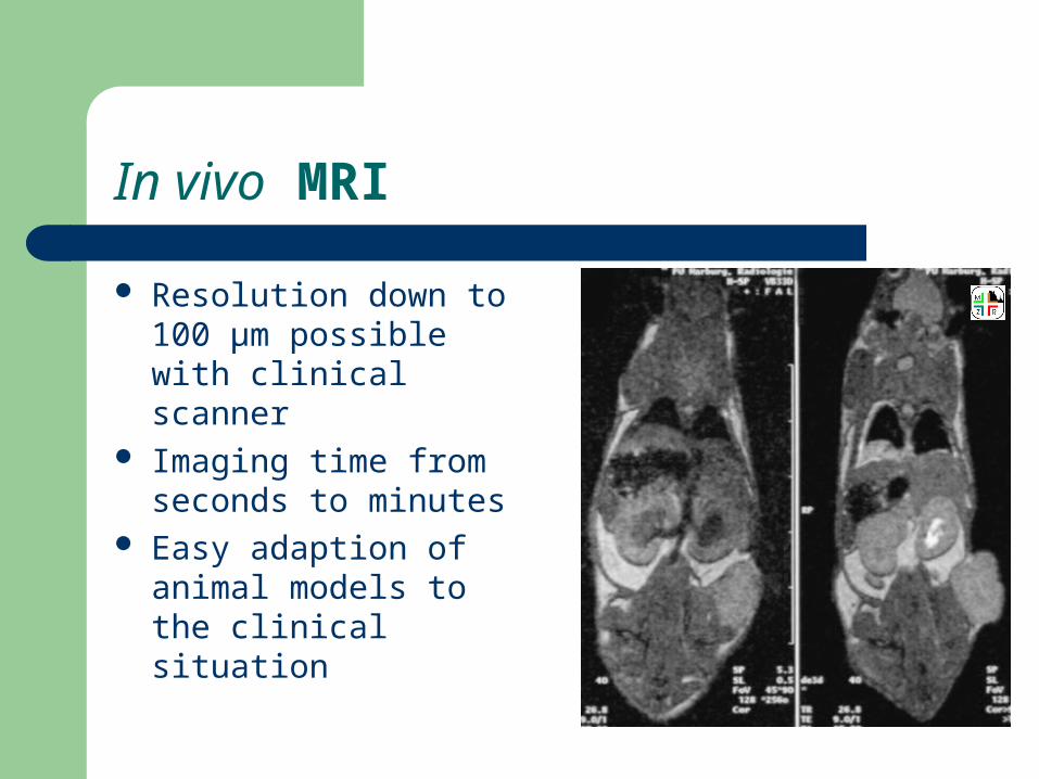
In vivo MRI
Resolution down to 100 µm possible with clinical scanner
Imaging time from seconds to minutes
Easy adaption of animal models to the clinical situation

Quantification of tumour perfusion

Parametric display
Dynamic analysis

Targeted MRI contrast media
Sipkins et al. Detection of tumor angiogenesis in vivo by a2ß3-targeted magnetic resonance imaging. Nat Med 1998;4:623–626


3D data analysis

Volumetric analysis

Dedicated systems

High field MR
Heart-MRI of new born mice

MR-Spectroscopy
Auricchio A et al. PNAS 2001;98:5205-10

Positron emission tomography

Characteristics of PET
„Electronic“ collimation (coincidence) Short halfe lifes of nuklids High costs (PET camera, cyclotron) Physical limitation of resolution:
– 1 mm for 18F– Some mm for other nuklid
Advantage: Organic elements like 11C, 15O

Radionuclides
Nuklid T ½
18F 2h
11C 20min
15O 2min
124I 4d
86Y 15h
68Ga 68min

Transgene expression imaging with PET
Chatziioannu AF Eur J Nucl Med 29 (2002) 98

Single photon emission tomography (SPET or SPECT)
Absorptive collimator Szintillation detector and
photo multiplyer Rotation of detector or
object necessary

Radionuclides
Nuklid T ½ Energy
99mTc 6h 140keV
111In 2.8d 245/171keV
67Ga 3.3d 93/185keV
123I 13h 159keV
131I 8d 364/284keV

SPET probes

Single Pinhole SPET
Spatial resolution ~ de + de (b/l)
– < 1mm achievable for near (1-2cm) subjects
Sensitivity = de cos3 / (16b2)
– Best close to the pinhole
King MA et al. J Cell Biochem S39 (2002) 221


SPET

Possible improvements
Higher sensitivity:– Multi-hole designs– Better detectors
Higher resolution:– Small pinhole designs resolution (< 0,1 mm)

Single pinhole vs multihole (7)
20 min 5 min

Comparison of imaging technologies
Technique Resolution Sensitivity Depth Time
MRI 10-100µm µ-mMol No limit Min
CT 50µm m-cMol No limit Sec
US <50µm mMol mm Sec
PET 1-2mm p-nMol No limit Min
SPET < 1mm p-nMol No limit Min
FRI 1-2mm p-nMol < 1cm Sec
FMT 1-2mm p-nMol < 10cm Sec

Recent developments and future directions
Multi-modality imaging or image co-registration Iterative image reconstruction algorithms Technical improvements
– Better detectors für SPET and PET– Improvements in coil design for MRI– Sequence adaptation for small animals for MRI– CT and MRI for high throughput screening
New reporter probes (contrast agents) New animal models

Multi-modality imaging
CT/PET MRI/PET CT/SPET FMT/MRT
SPET/CT: 125I labeled Herceptin®,© Iwata K et al.
No one imaging modality can provide all the information(structure, function, molecular processes) in one image!

Image fusion (co-registration)

Co-registration: MRT-PET
Comparison of 18F-FDG-PET and MRI in hamster
After i.p injection of human GW39 colon cancer cells
Lewis JS et al. Cancer Res 62 (2002) 445
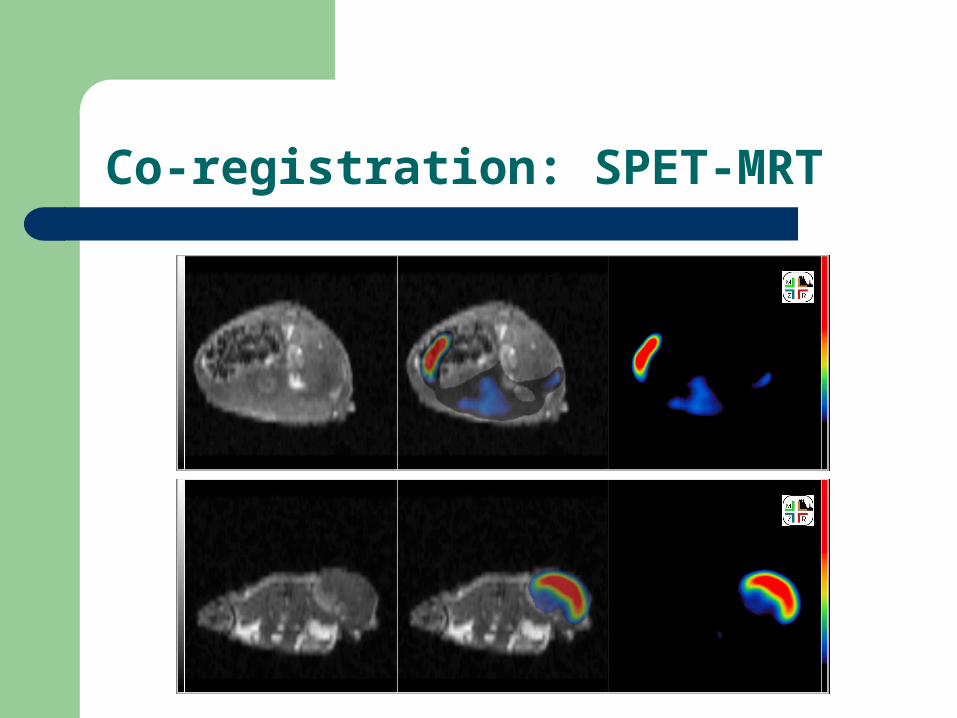
Co-registration: SPET-MRT

Image reconstruction
Chatziioannu AF Eur J Nucl Med 29 (2002) 98

New contrast agents
Aime S et al. JMRI 16 (2002) 394

New animal models
Giana P et al. Nat Med 9 (2003) 82