new antimicrobial compounds from a marine-derived bacillus sp
TRANSCRIPT
ORIGINAL ARTICLE
New antimicrobial compounds from a marine-derivedBacillus sp.
Mohamad Abdul Mojid Mondol1, Fakir Shahidullah Tareq2,3, Ji Hye Kim2, Min Ah Lee2, Hyi-Seung Lee2,Jong Seok Lee2, Yeon-Ju Lee2 and Hee Jae Shin2,3
In the course of our ongoing research for antimicrobial agents, we isolated five new compounds, three 24-membered
macrolactones, macrolactins X–Z (1–3) and two hydroxy unsaturated fatty acids, linieodolides A (5) and B (6), together with
a known metabolite, macrolactinic acid (4), from the culture broth of a marine Bacillus sp. The structures of these metabolites
were determined on the basis of their spectroscopic data interpretation. Their absolute configurations were addressed by
modified Mosher’s method and literature data review. Compounds 1–6 showed moderate to potent inhibitory activities
(8B128 mg ml�1) against selected pathogenic microorganisms.
The Journal of Antibiotics (2013) 66, 89–95; doi:10.1038/ja.2012.102; published online 5 December 2012
Keywords: antimicrobials; linieodolide; macrolactins; marine microorganisms
INTRODUCTION
Marine microorganisms, which are taxonomically diverse and geneti-cally special, have powerful potential in producing novel bioactivecompounds.1 Lately, marine bacteria have drawn much interest owingto their unique secondary metabolites, which differ significantly fromthose of their terrestrial counterparts.2,3 Marine microorganisms arehighly diversified and may have unique metabolic pathway because ofextreme living conditions resulting in a potential for the discovery ofnovel drugs and remain mostly unexplored.4
Discovery of new antimicrobial drugs are decreasing steadily,whereas infectious diseases caused by drug-resistant pathogens areemerging day by day.5,6 Therefore, new antimicrobial drugs areurgently needed to treat these infectious diseases. We have alsoreported new antimicrobial compounds from a marine sedimentBacillus sp.7–9 During our continuous screening for new metaboliteswith antimicrobial activities, we could isolate five new (1–3, 5 and 6)and one known (4) antimicrobial compounds from the fermentationbroth of a marine-derived Bacillus sp. 09ID194. Here, we reportisolation, structure elucidation and antimicrobial activity of thesecompounds (1–6).
RESULTS
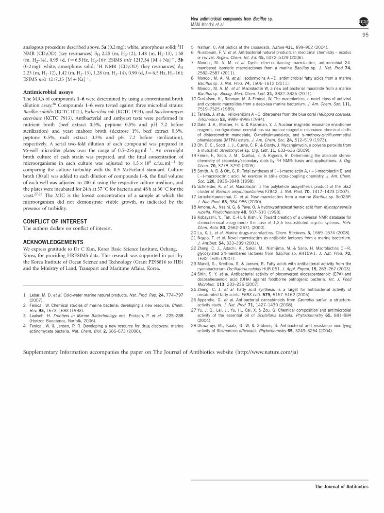
Structure elucidation of macrolactin X (1)Macrolactin X (1) was isolated as an amorphous solid. Its molecularformula, C24H34O6, was determined by high-resolution electrosprayionization mass spectrometry (HRESIMS) (m/z 441.2249 [MþNa]þ ), which suggested eight degrees of unsaturation. The IR
spectrum of 1 displayed characteristic absorption bands for hydroxy(3482 cm�1), carbonyl (1691 cm�1) and epoxide (1250 cm�1) func-tionalities. UV absorptions at 232 and 261 nm indicated the presenceof an extended conjugation system in the molecule. This suppositionwas supported by the 10 sp2 carbon signals in the 13C NMRspectrum. In total, 24 carbon resonances were observed in the 13CNMR spectrum, which were ascribed to one quaternary carbonyl,10 sp2, six oxymethine, six methylene and one methyl carbons byanalysis of an HSQC spectrum. One carbonyl and five double bondsaccounted for six degrees of unsaturation and the remaining twodegrees of unsaturation were ascribed to the existence of two cyclicring systems in 1. 1H and 13C NMR signals clearly indicated that 1belongs to macrolactin family.10
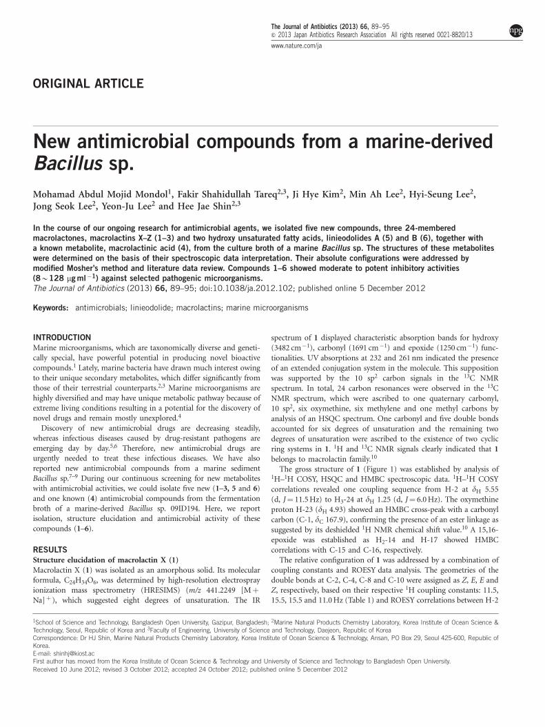
The gross structure of 1 (Figure 1) was established by analysis of1H–1H COSY, HSQC and HMBC spectroscopic data. 1H–1H COSYcorrelations revealed one coupling sequence from H-2 at dH 5.55(d, J¼ 11.5 Hz) to H3-24 at dH 1.25 (d, J¼ 6.0 Hz). The oxymethineproton H-23 (dH 4.93) showed an HMBC cross-peak with a carbonylcarbon (C-1, dC 167.9), confirming the presence of an ester linkage assuggested by its deshielded 1H NMR chemical shift value.10 A 15,16-epoxide was established as H2-14 and H-17 showed HMBCcorrelations with C-15 and C-16, respectively.
The relative configuration of 1 was addressed by a combination ofcoupling constants and ROESY data analysis. The geometries of thedouble bonds at C-2, C-4, C-8 and C-10 were assigned as Z, E, E andZ, respectively, based on their respective 1H coupling constants: 11.5,15.5, 15.5 and 11.0 Hz (Table 1) and ROESY correlations between H-2
1School of Science and Technology, Bangladesh Open University, Gazipur, Bangladesh; 2Marine Natural Products Chemistry Laboratory, Korea Institute of Ocean Science &Technology, Seoul, Republic of Korea and 3Faculty of Engineering, University of Science and Technology, Daejeon, Republic of KoreaCorrespondence: Dr HJ Shin, Marine Natural Products Chemistry Laboratory, Korea Institute of Ocean Science & Technology, Ansan, PO Box 29, Seoul 425-600, Republic ofKorea.E-mail: [email protected] author has moved from the Korea Institute of Ocean Science & Technology and University of Science and Technology to Bangladesh Open University.
Received 10 June 2012; revised 3 October 2012; accepted 24 October 2012; published online 5 December 2012
The Journal of Antibiotics (2013) 66, 89–95& 2013 Japan Antibiotics Research Association All rights reserved 0021-8820/13
www.nature.com/ja
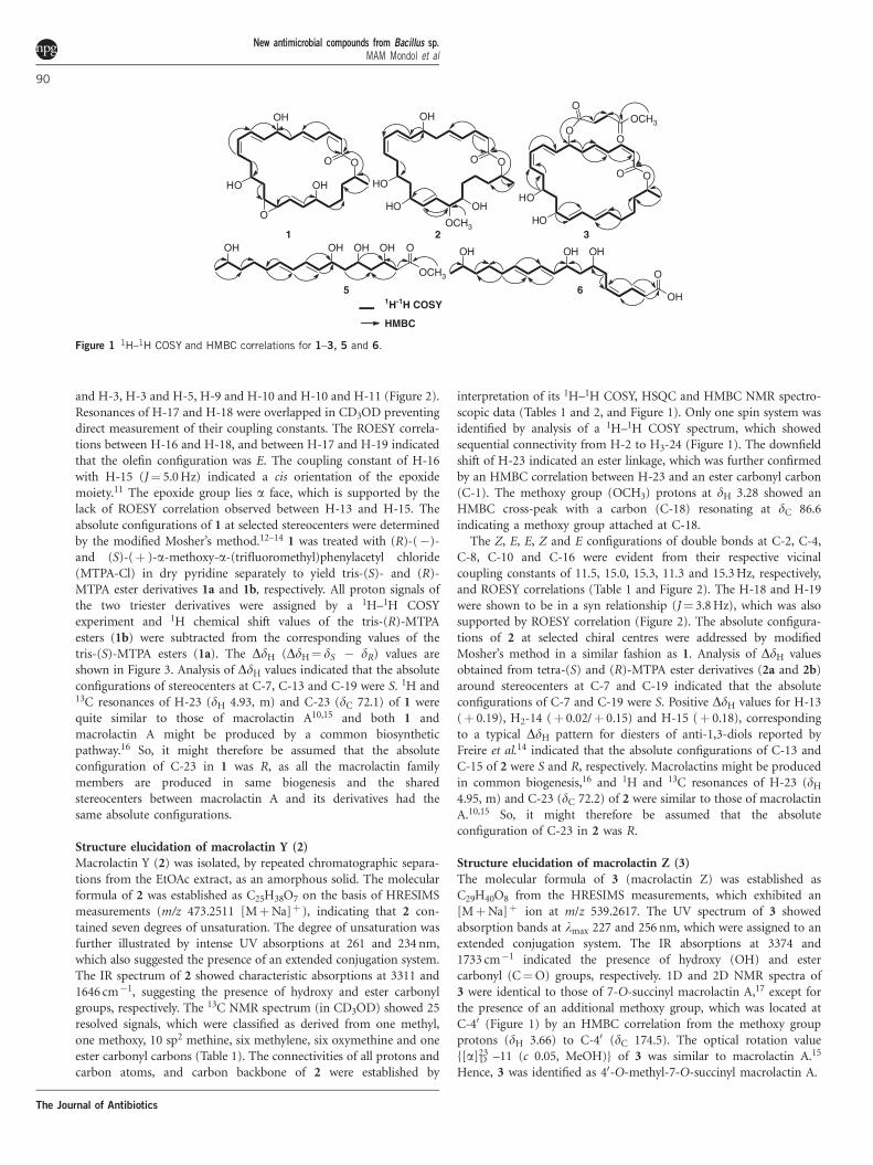
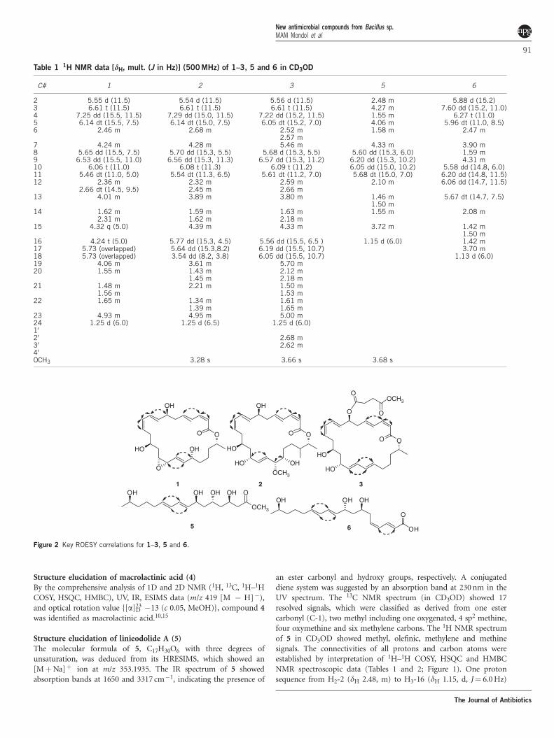
and H-3, H-3 and H-5, H-9 and H-10 and H-10 and H-11 (Figure 2).Resonances of H-17 and H-18 were overlapped in CD3OD preventingdirect measurement of their coupling constants. The ROESY correla-tions between H-16 and H-18, and between H-17 and H-19 indicatedthat the olefin configuration was E. The coupling constant of H-16with H-15 (J¼ 5.0 Hz) indicated a cis orientation of the epoxidemoiety.11 The epoxide group lies a face, which is supported by thelack of ROESY correlation observed between H-13 and H-15. Theabsolute configurations of 1 at selected stereocenters were determinedby the modified Mosher’s method.12–14 1 was treated with (R)-(�)-and (S)-(þ )-a-methoxy-a-(trifluoromethyl)phenylacetyl chloride(MTPA-Cl) in dry pyridine separately to yield tris-(S)- and (R)-MTPA ester derivatives 1a and 1b, respectively. All proton signals ofthe two triester derivatives were assigned by a 1H–1H COSYexperiment and 1H chemical shift values of the tris-(R)-MTPAesters (1b) were subtracted from the corresponding values of thetris-(S)-MTPA esters (1a). The DdH (DdH¼ dS � dR) values areshown in Figure 3. Analysis of DdH values indicated that the absoluteconfigurations of stereocenters at C-7, C-13 and C-19 were S. 1H and13C resonances of H-23 (dH 4.93, m) and C-23 (dC 72.1) of 1 werequite similar to those of macrolactin A10,15 and both 1 andmacrolactin A might be produced by a common biosyntheticpathway.16 So, it might therefore be assumed that the absoluteconfiguration of C-23 in 1 was R, as all the macrolactin familymembers are produced in same biogenesis and the sharedstereocenters between macrolactin A and its derivatives had thesame absolute configurations.
Structure elucidation of macrolactin Y (2)Macrolactin Y (2) was isolated, by repeated chromatographic separa-tions from the EtOAc extract, as an amorphous solid. The molecularformula of 2 was established as C25H38O7 on the basis of HRESIMSmeasurements (m/z 473.2511 [MþNa]þ ), indicating that 2 con-tained seven degrees of unsaturation. The degree of unsaturation wasfurther illustrated by intense UV absorptions at 261 and 234 nm,which also suggested the presence of an extended conjugation system.The IR spectrum of 2 showed characteristic absorptions at 3311 and1646 cm�1, suggesting the presence of hydroxy and ester carbonylgroups, respectively. The 13C NMR spectrum (in CD3OD) showed 25resolved signals, which were classified as derived from one methyl,one methoxy, 10 sp2 methine, six methylene, six oxymethine and oneester carbonyl carbons (Table 1). The connectivities of all protons andcarbon atoms, and carbon backbone of 2 were established by
interpretation of its 1H–1H COSY, HSQC and HMBC NMR spectro-scopic data (Tables 1 and 2, and Figure 1). Only one spin system wasidentified by analysis of a 1H–1H COSY spectrum, which showedsequential connectivity from H-2 to H3-24 (Figure 1). The downfieldshift of H-23 indicated an ester linkage, which was further confirmedby an HMBC correlation between H-23 and an ester carbonyl carbon(C-1). The methoxy group (OCH3) protons at dH 3.28 showed anHMBC cross-peak with a carbon (C-18) resonating at dC 86.6indicating a methoxy group attached at C-18.
The Z, E, E, Z and E configurations of double bonds at C-2, C-4,C-8, C-10 and C-16 were evident from their respective vicinalcoupling constants of 11.5, 15.0, 15.3, 11.3 and 15.3 Hz, respectively,and ROESY correlations (Table 1 and Figure 2). The H-18 and H-19were shown to be in a syn relationship (J¼ 3.8 Hz), which was alsosupported by ROESY correlation (Figure 2). The absolute configura-tions of 2 at selected chiral centres were addressed by modifiedMosher’s method in a similar fashion as 1. Analysis of DdH valuesobtained from tetra-(S) and (R)-MTPA ester derivatives (2a and 2b)around stereocenters at C-7 and C-19 indicated that the absoluteconfigurations of C-7 and C-19 were S. Positive DdH values for H-13(þ 0.19), H2-14 (þ 0.02/þ 0.15) and H-15 (þ 0.18), correspondingto a typical DdH pattern for diesters of anti-1,3-diols reported byFreire et al.14 indicated that the absolute configurations of C-13 andC-15 of 2 were S and R, respectively. Macrolactins might be producedin common biogenesis,16 and 1H and 13C resonances of H-23 (dH
4.95, m) and C-23 (dC 72.2) of 2 were similar to those of macrolactinA.10,15 So, it might therefore be assumed that the absoluteconfiguration of C-23 in 2 was R.
Structure elucidation of macrolactin Z (3)The molecular formula of 3 (macrolactin Z) was established asC29H40O8 from the HRESIMS measurements, which exhibited an[MþNa]þ ion at m/z 539.2617. The UV spectrum of 3 showedabsorption bands at lmax 227 and 256 nm, which were assigned to anextended conjugation system. The IR absorptions at 3374 and1733 cm�1 indicated the presence of hydroxy (OH) and estercarbonyl (C¼O) groups, respectively. 1D and 2D NMR spectra of3 were identical to those of 7-O-succinyl macrolactin A,17 except forthe presence of an additional methoxy group, which was located atC-40 (Figure 1) by an HMBC correlation from the methoxy groupprotons (dH 3.66) to C-40 (dC 174.5). The optical rotation value{[a]23
D –11 (c 0.05, MeOH)} of 3 was similar to macrolactin A.15
Hence, 3 was identified as 40-O-methyl-7-O-succinyl macrolactin A.
O
HO
O
OH
OH
O
O
HO
O
OH
HO
OCH3
O
HO
HO
O1
OOCH3
O
O
OCH3
OOHOHOHOH
OH
O
OHOHOH
1 2
HMBC
1H-1H COSY
3
5 6
OH
Figure 1 1H–1H COSY and HMBC correlations for 1–3, 5 and 6.
New antimicrobial compounds from Bacillus sp.MAM Mondol et al
90
The Journal of Antibiotics
Structure elucidation of macrolactinic acid (4)By the comprehensive analysis of 1D and 2D NMR (1H, 13C, 1H–1HCOSY, HSQC, HMBC), UV, IR, ESIMS data (m/z 419 [M � H]�),and optical rotation value {[a]23
D �13 (c 0.05, MeOH)}, compound 4was identified as macrolactinic acid.10,15
Structure elucidation of linieodolide A (5)The molecular formula of 5, C17H30O6 with three degrees ofunsaturation, was deduced from its HRESIMS, which showed an[MþNa]þ ion at m/z 353.1935. The IR spectrum of 5 showedabsorption bands at 1650 and 3317 cm�1, indicating the presence of
an ester carbonyl and hydroxy groups, respectively. A conjugateddiene system was suggested by an absorption band at 230 nm in theUV spectrum. The 13C NMR spectrum (in CD3OD) showed 17resolved signals, which were classified as derived from one estercarbonyl (C-1), two methyl including one oxygenated, 4 sp2 methine,four oxymethine and six methylene carbons. The 1H NMR spectrumof 5 in CD3OD showed methyl, olefinic, methylene and methinesignals. The connectivities of all protons and carbon atoms wereestablished by interpretation of 1H–1H COSY, HSQC and HMBCNMR spectroscopic data (Tables 1 and 2; Figure 1). One protonsequence from H2-2 (dH 2.48, m) to H3-16 (dH 1.15, d, J¼ 6.0 Hz)
Table 1 1H NMR data [dH, mult. (J in Hz)] (500 MHz) of 1–3, 5 and 6 in CD3OD
C# 1 2 3 5 6
2 5.55 d (11.5) 5.54 d (11.5) 5.56 d (11.5) 2.48 m 5.88 d (15.2)3 6.61 t (11.5) 6.61 t (11.5) 6.61 t (11.5) 4.27 m 7.60 dd (15.2, 11.0)4 7.25 dd (15.5, 11.5) 7.29 dd (15.0, 11.5) 7.22 dd (15.2, 11.5) 1.55 m 6.27 t (11.0)5 6.14 dt (15.5, 7.5) 6.14 dt (15.0, 7.5) 6.05 dt (15.2, 7.0) 4.06 m 5.96 dt (11.0, 8.5)6 2.46 m 2.68 m 2.52 m
2.57 m1.58 m 2.47 m
7 4.24 m 4.28 m 5.46 m 4.33 m 3.90 m8 5.65 dd (15.5, 7.5) 5.70 dd (15.3, 5.5) 5.68 d (15.3, 5.5) 5.60 dd (15.3, 6.0) 1.59 m9 6.53 dd (15.5, 11.0) 6.56 dd (15.3, 11.3) 6.57 dd (15.3, 11.2) 6.20 dd (15.3, 10.2) 4.31 m10 6.06 t (11.0) 6.08 t (11.3) 6.09 t (11.2) 6.05 dd (15.0, 10.2) 5.58 dd (14.8, 6.0)11 5.46 dt (11.0, 5.0) 5.54 dt (11.3, 6.5) 5.61 dt (11.2, 7.0) 5.68 dt (15.0, 7.0) 6.20 dd (14.8, 11.5)12 2.36 m
2.66 dt (14.5, 9.5)2.32 m2.45 m
2.59 m2.66 m
2.10 m 6.06 dd (14.7, 11.5)
13 4.01 m 3.89 m 3.80 m 1.46 m1.50 m
5.67 dt (14.7, 7.5)
14 1.62 m2.31 m
1.59 m1.62 m
1.63 m2.18 m
1.55 m 2.08 m
15 4.32 q (5.0) 4.39 m 4.33 m 3.72 m 1.42 m1.50 m
16 4.24 t (5.0) 5.77 dd (15.3, 4.5) 5.56 dd (15.5, 6.5 ) 1.15 d (6.0) 1.42 m17 5.73 (overlapped) 5.64 dd (15.3,8.2) 6.19 dd (15.5, 10.7) 3.70 m18 5.73 (overlapped) 3.54 dd (8.2, 3.8) 6.05 dd (15.5, 10.7) 1.13 d (6.0)19 4.06 m 3.61 m 5.70 m20 1.55 m 1.43 m
1.45 m2.12 m2.18 m
21 1.48 m1.56 m
2.21 m 1.50 m1.53 m
22 1.65 m 1.34 m1.39 m
1.61 m1.65 m
23 4.93 m 4.95 m 5.00 m24 1.25 d (6.0) 1.25 d (6.5) 1.25 d (6.0)10
20 2.68 m30 2.62 m40
OCH3 3.28 s 3.66 s 3.68 s
O
HO
HO
O
O
OCH3
O
O
O
HO
O
OH
OH
O
O
HO
O
OH
HO
OCH3
OCH3
OOHOHOHOH
OH
O
OHOHOH
1 2 3
5 6
OH
Figure 2 Key ROESY correlations for 1–3, 5 and 6.
New antimicrobial compounds from Bacillus sp.MAM Mondol et al
91
The Journal of Antibiotics
was established by analysis of a 1H–1H COSY spectral data. An HMBCcross-peak from the OCH3 protons (dH 3.68, s) to C-1 supported theconnectivity of OCH3 group to a carbonyl carbon, C-1.
The disubstituted C-8/C-9 and C-10/C-11 double bonds of 5 wereelucidated to have E configuration by the large vicinal coupling
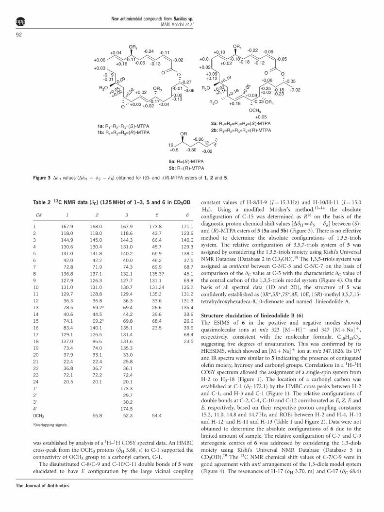
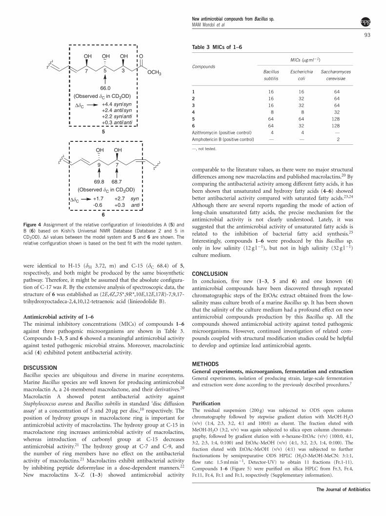
constant values of H-8/H-9 (J¼ 15.3 Hz) and H-10/H-11 (J¼ 15.0Hz). Using a modified Mosher’s method,12–14 the absoluteconfiguration of C-15 was determined as R18 on the basis of thediagnostic proton chemical shift values [DdH¼ dS � dR] between (S)-and (R)-MTPA esters of 5 (5a and 5b) (Figure 3). There is no effectivemethod to determine the absolute configurations of 1,3,5-triolssystem. The relative configuration of 3,5,7-triols system of 5 wasassigned by considering the 1,3,5-triols moiety using Kishi’s UniversalNMR Database (Database 2 in CD3OD).19 The 1,3,5-triols system wasassigned as anti/anti between C-3/C-5 and C-5/C-7 on the basis ofcomparison of the dC value at C-5 with the characteristic dC value ofthe central carbon of the 1,3,5-triols model system (Figure 4). On thebasis of all spectral data (1D and 2D), the structure of 5 wasconfidently established as (3R*,5R*,7S*,8E, 10E, 15R)-methyl 3,5,7,15-tetrahydroxyhexadeca-8,10-dienoate and named linieodolide A.
Structure elucidation of linieodolide B (6)The ESIMS of 6 in the positive and negative modes showedquasimolecular ions at m/z 323 [M�H]� and 347 [MþNa]þ ,respectively, consistent with the molecular formula, C18H28O5,suggesting five degrees of unsaturation. This was confirmed by itsHRESIMS, which showed an [MþNa]þ ion at m/z 347.1826. Its UVand IR spectra were similar to 5 indicating the presence of conjugatedolefin moiety, hydroxy and carbonyl groups. Correlations in a 1H–1HCOSY spectrum allowed the assignment of a single-spin system fromH-2 to H3-18 (Figure 1). The location of a carbonyl carbon wasestablished at C-1 (dC 172.1) by the HMBC cross peaks between H-2and C-1, and H-3 and C-1 (Figure 1). The relative configurations ofdouble bonds at C-2, C-4, C-10 and C-12 corroborated as E, Z, E andE, respectively, based on their respective proton coupling constants:15.2, 11.0, 14.8 and 14.7 Hz, and ROEs between H-2 and H-4, H-10and H-12, and H-11 and H-13 (Table 1 and Figure 2). Data were notobtained to determine the absolute configurations of 6 due to thelimited amount of sample. The relative configuration of C-7 and C-9stereogenic centres of 6 was addressed by considering the 1,3-diolsmoiety using Kishi’s Universal NMR Database (Database 5 inCD3OD).19 The 13C NMR chemical shift values of C-7/C-9 were ingood agreement with anti arrangement of the 1,3-diols model system(Figure 4). The resonances of H-17 (dH 3.70, m) and C-17 (dC 68.4)
Table 2 13C NMR data (dC) (125 MHz) of 1–3, 5 and 6 in CD3OD
C# 1 2 3 5 6
1 167.9 168.0 167.9 173.8 171.1
2 118.0 118.0 118.6 43.7 123.6
3 144.9 145.0 144.3 66.4 140.6
4 130.6 130.4 131.0 45.7 129.3
5 141.0 141.8 140.2 65.9 138.0
6 42.0 42.2 40.0 46.2 37.5
7 72.8 71.9 74.3 69.9 68.7
8 136.8 137.1 132.1 135.37 45.1
9 127.9 126.3 127.7 131.1 69.8
10 131.0 131.0 130.7 131.34 135.2
11 129.7 128.8 129.4 135.3 131.2
12 36.3 36.8 36.3 33.6 131.3
13 78.5 69.2a 69.4 26.6 135.4
14 40.6 44.5 44.2 39.6 33.6
15 74.1 69.2a 69.8 68.4 26.6
16 83.4 140.1 135.1 23.5 39.6
17 129.1 126.5 131.4 68.4
18 137.0 86.6 131.6 23.5
19 73.4 74.0 135.3
20 37.9 33.1 33.0
21 22.4 22.4 25.8
22 36.8 36.7 36.1
23 72.1 72.2 72.4
24 20.5 20.1 20.1
10 173.3
20 29.7
30 30.2
40 174.5
OCH3 56.8 52.3 54.4
aOverlapping signals.
O
R2O
O
OR1
OR3
O
-0.02
-0.11
-0.13
-0.24
-0.06-0.11
+0.16
+0.04
+0.06
+0.03
-0.19-0.01
0.00
+0.05
+0.02
+0.0
2
+0.03
+0.02
+0.02-0.17
-0.04
-0.02-0.13
-0.27-0.01 -0.68
1a: R1=R2=R3=(S )-MTPA1b: R1=R2=R3=(R )-MTPA
O
R2O
O
OR1
OR4R3O
OCH3
-0.05
-0.09
-0.12
-0.22
-0.18-0.10
+0.02
+0.10
+0.01
+0.02
+0.09+0.12
+0.19
+0.02+0.15
+0.18
+0.18
+0.0
5
+0.09-0.03
-0.25-0.02
-0.06
-0.18-0.23
-0.05
-0.02
+0.052a: R1=R2=R3=R4=(S )-MTPA
2b: R1=R2=R3=R4=(R )-MTPAOR
+0.5 -0.30
-0.06
-0.0216 12
5a: R=(S )-MTPA
5b: R=(R )-MTPA
Figure 3 DdH values (DdH ¼ dS � dR) obtained for (S)- and -(R)-MTPA esters of 1, 2 and 5.
New antimicrobial compounds from Bacillus sp.MAM Mondol et al
92
The Journal of Antibiotics
were identical to H-15 (dH 3.72, m) and C-15 (dC 68.4) of 5,respectively, and both might be produced by the same biosyntheticpathway. Therefore, it might be assumed that the absolute configura-tion of C-17 was R. By the extensive analysis of spectroscopic data, thestructure of 6 was established as (2E,4Z,7S*,9R*,10E,12E,17R)-7,9,17-trihydroxyoctadeca-2,4,10,12-tetraenoic acid (linieodolide B).
Antimicrobial activity of 1–6The minimal inhibitory concentrations (MICs) of compounds 1–6against three pathogenic microorganisms are shown in Table 3.Compounds 1–3, 5 and 6 showed a meaningful antimicrobial activityagainst tested pathogenic microbial strains. Moreover, macrolactinicacid (4) exhibited potent antibacterial activity.
DISCUSSION
Bacillus species are ubiquitous and diverse in marine ecosystems.Marine Bacillus species are well known for producing antimicrobialmacrolactin A, a 24-membered macrolactone, and their derivatives.20
Macrolactin A showed potent antibacterial activity againstStaphylococcus aureus and Bacillus subtilis in standard ‘disc diffusionassay’ at a concentration of 5 and 20mg per disc,10 respectively. Theposition of hydroxy groups in macrolactone ring is important forantimicrobial activity of macrolactins. The hydroxy group at C-15 inmacrolactone ring increases antimicrobial activity of macrolactins,whereas introduction of carbonyl group at C-15 decreasesantimicrobial activity.21 The hydroxy group at C-7 and C-9, andthe number of ring members have no effect on the antibacterialactivity of macrolactins.21 Macrolactins exhibit antibacterial activityby inhibiting peptide deformylase in a dose-dependent manners.22
New macrolactins X–Z (1–3) showed antimicrobial activity
comparable to the literature values, as there were no major structuraldifferences among new macrolactins and published macrolactins.20 Bycomparing the antibacterial activity among different fatty acids, it hasbeen shown that unsaturated and hydroxy fatty acids (4–6) showedbetter antibacterial activity compared with saturated fatty acids.23,24
Although there are several reports regarding the mode of action oflong-chain unsaturated fatty acids, the precise mechanism for theantimicrobial activity is not clearly understood. Lately, it wassuggested that the antimicrobial activity of unsaturated fatty acids isrelated to the inhibition of bacterial fatty acid synthesis.25
Interestingly, compounds 1–6 were produced by this Bacillus sp.only in low salinity (12 g l�1), but not in high salinity (32 g l�1)culture medium.
CONCLUSION
In conclusion, five new (1–3, 5 and 6) and one known (4)antimicrobial compounds have been discovered through repeatedchromatographic steps of the EtOAc extract obtained from the low-salinity mass culture broth of a marine Bacillus sp. It has been shownthat the salinity of the culture medium had a profound effect on newantimicrobial compounds production by this Bacillus sp. All thecompounds showed antimicrobial activity against tested pathogenicmicroorganisms. However, continued investigation of related com-pounds coupled with structural modification studies could be helpfulto develop and optimize lead antimicrobial agents.
METHODS
General experiments, microorganism, fermentation and extractionGeneral experiments, isolation of producing strain, large-scale fermentation
and extraction were done according to the previously described procedures.7
PurificationThe residual suspension (200 g) was subjected to ODS open column
chromatography followed by stepwise gradient elution with MeOH-H2O
(v/v) (1:4, 2:3, 3:2, 4:1 and 100:0) as eluent. The fraction eluted with
MeOH-H2O (3:2, v/v) was again subjected to silica open column chromato-
graphy, followed by gradient elution with n-hexane-EtOAc (v/v) (100:0, 4:1,
3:2, 2:3, 1:4, 0:100) and EtOAc-MeOH (v/v) (4:1, 3:2, 2:3, 1:4, 0:100). The
fraction eluted with EtOAc-MeOH (v/v) (4:1) was subjected to further
fractionations by semipreparative ODS HPLC (H2O-MeOH-MeCN: 3:1:1,
flow rate: 1.5 ml min�1, Detector-UV) to obtain 11 fractions (Fr.1-11).
Compounds 1–6 (Figure 5) were purified on silica HPLC from Fr.3, Fr.4,
Fr.11, Fr.4, Fr.1 and Fr.1, respectively (Supplementary information).
Table 3 MICs of 1–6
Compounds
MICs (mg ml�1)
Bacillus
subtilis
Escherichia
coli
Saccharomyces
cerevisiae
1 16 16 64
2 16 32 64
3 16 32 64
4 8 8 32
5 64 64 128
6 64 32 128
Azithromycin (positive control) 4 4 —
Amphotericin B (positive control) — — 2
—, not tested.
79
69.8 68.7
OHOH
(Observed �C in CD3OD)
Δ�C+1.7 +2.7 syn-0.6 +0.3 anti
OCH3
OOHOHOH
(Observed �C in CD3OD)
66.0
Δ�C +4.4 syn/syn+2.4 anti/syn+2.2 syn/anti+0.3 anti/anti
5
6
357
Figure 4 Assignment of the relative configuration of linieodolides A (5) and
B (6) based on Kishi’s Universal NMR Database (Database 2 and 5 in
CD3OD). Dd values between the model system and 5 and 6 are shown. The
relative configuration shown is based on the best fit with the model system.
New antimicrobial compounds from Bacillus sp.MAM Mondol et al
93
The Journal of Antibiotics
Physicochemical Properties
Macrolactin X (1). Amorphous solid; [a]23D �60 (c 0.05, MeOH); UV
(MeOH) lmax (log e) 200 (4.12), 232 (4.30) and 261 (4.09) nm; IR (MeOH)
nmax 3482 (br), 2922, 1691, 1250, 1195, 1038 cm�1; 1H and 13C NMR data
(CD3OD) (Tables 1 and 2); HRESIMS m/z 441.2249 [MþNa]þ (calcd for
C24H34O6Na, 441.2253).
Macrolactin Y (2). Amorphous solid; [a]23D �84 (c 0.05, MeOH); UV
(MeOH) lmax (log e) 200 (4.14), 234 (4.45) and 261 (4.25) nm; IR (MeOH)
nmax 3311 (br), 2921, 1646, 1409, 1104, 1024 cm�1; 1H and 13C NMR data
(CD3OD) (Tables 1 and 2); HRESIMS m/z 473.2511 [MþNa]þ (calcd for
C25H38O7Na, 473.2515).
Macrolactin Z (3). White, amorphous solid; [a]23D �11 (c 0.05, MeOH); UV
(MeOH) lmax (log e) 200 (3.90), 227 (4.18) and 256 (3.83) nm; IR (MeOH)
nmax 3374 (br), 2929, 1733, 1169, 990 cm�1; 1H and 13C NMR data (CD3OD)
(Tables 1 and 2); HRESIMS m/z 539.2617 [MþNa]þ (calcd for C29H40O8Na,
539.2621).
Linieodolide A (5). White, amorphous solid; [a]23D �34 (c 0.05, MeOH); UV
(MeOH) lmax (log e) 201 (4.03), 230 (4.23) and 256 (3.28) nm; IR (MeOH)
nmax 3317 (br), 2925, 1650, 1413, 1017 cm�1; 1H and 13C NMR data (CD3OD)
(Tables 1 and 2); HRESIMS m/z 353.1953 [MþNa]þ (calcd for C17H30O6Na,
353.1940).
Linieodolide B (6). White, amorphous solid; [a]23D �62 (c 0.05, MeOH); UV
(MeOH) lmax (log e) 231 (4.26) and 259 (4.05) nm; IR (MeOH) nmax 3340
(br), 2928, 1623, 1456, 1039 cm�1; 1H and 13C NMR data (CD3OD) (Tables 1
and 2); HRESIMS m/z 347.1826 [MþNa]þ (calcd for C18H28O5Na,
347.1834).
Tris-(S)-MTPA esters (1a) of 1. Compound 1 (0.8 mg) was dissolved in 150ml
pyridine and stirred at room temperature for 10 min. For preparation of the
tris-(S)-MTPA esters (1a) of 1, 20ml (R)-(�)- a-methoxy-a-(trifluoromethyl)-
phenylacetyl chloride (MTPA-Cl) was added to the reaction vial and the
mixture was stirred at room temperature for 16 h. Completion of the reaction
was monitored by LC/MS. The reaction mixture was dried in vacuo and
redissolved in EtOAc, washed with H2O, and purified on a silica HPLC using
5% MeOH in CH2Cl2 as eluent to obtain 1a (0.5 mg). All proton signals of the
triesters derivative (1a) were assigned by a 1H–1H COSY experiment. 1a:
Amorphous solid; 1H NMR (CD3OD) dH 5.52 (d, J¼ 11.5 Hz, H-2), 6.40
(t, J¼ 11.5 Hz, H-3), 6.84 (dd, J¼ 15.5, 11.5 Hz, H-4), 5.76 (dt, J¼ 15.5,
7.5 Hz, H-5), 2.48 (m, H2-6), 5.55 (m, H-7), 5.66 (dd, J¼ 15.5, 7.5 Hz, H-8),
6.44 (dd, J¼ 15.5, 11.0 Hz, H-9), 5.98 (t, J¼ 11.0 Hz, H-10), 5.40 (dt, J¼ 11.0,
5.0 Hz, H-11), 1.66 (m, H-12b), 2.14 (m, H-12a), 4.07 (m, H-13), 1.73 (m,
H-14b), 2.58 (m, H-14a), 5.52 (m, H-15), 4.55 (t, J¼ 4.5 Hz, H-16), 5.82
(overlapped, H-17), 5.82 (overlapped, H-18), 4.60 (m, H-19), 1.52 (m, H-20),
1.27 (m, H-21b), 1.30 (m, H-21a), 1.55 (m, H2-22), 4.50 (m, H-23), 1.20 (d,
J¼ 6.0 Hz, H3-24), 3.47 (OCH3, s), 3.53 (OCH3, s), 3.54 (OCH3, s), 7.34B7.56
(15 H, m); ESIMS m/z 1089.90 [MþNa]þ .
Tris-(R)-MTPA esters (1b) of 1. In an entirely analogous way, the tris-(R)-
MTPA esters (1b) was obtained using (S)-(þ )-a-methoxy-a-(trifluoro-
methyl)phenylacetyl chloride (MTPA-Cl). 1b (0.4 mg): amorphous solid; 1H
NMR (CD3OD) dH 5.54 (d, J¼ 11.5 Hz, H-2), 6.51 (t, J¼ 11.5 Hz, H-3), 6.97
(dd, J¼ 15.5, 11.5 Hz, H-4), 6.00 (dt, J¼ 15.5, 7.5 Hz, H-5), 2.54 (m, H2-6),
5.66 (m, H-7), 5.50 (dd, J¼ 15.5, 7.5 Hz, H-8), 6.40 (dd, J¼ 15.5, 11.0 Hz,
H-9), 5.92 (t, J¼ 11.0 Hz, H-10), 5.37 (dt, J¼ 11.0, 5.0 Hz, H-11), 1.85 (m,
H-12b), 2.15 (m, H-12a), 4.07 (m, H-13), 1.68 (m, H-14b), 2.56 (m, H-14a),
5.50 (m, H-15), 4.53 (t, J¼ 5.0 Hz, H-16), 5.80 (overlapped, H-17), 5.80
(overlapped, H-18), 5.43 (m, H-19), 1.56 (m, H-20), 1.29 (m, H-21b), 1.43
(m, H-21a), 1.54 (m, H2-22), 4.77 (m, H-23), 1.88 (d, J¼ 6.0 Hz, H3-24), 3.49
(OCH3, s), 3.50 (OCH3, s), 3.54 (OCH3, s), 7.36B7.60 (15 H, m); ESIMS m/z
1089.82 [MþNa]þ .
Tetra-(S)- and (R)-MTPA esters (2a and 2b) of 2. Tetra-(S)-and (R)-MTPA
ester derivatives of 2 were prepared from (R)-(�)-and (S)-(þ )-MTPA-Cl,
respectively, according to the procedure described above. All 1H NMR signals
were assigned by a 1H–1H COSY experiment. 2a (0.5 mg): white, amorphous
solid; 1H NMR (CD3OD) dH 5.51 (d, J¼ 11.5 Hz, H-2), 6.46 (t, J¼ 11.5
Hz, H-3), 7.18 (dd, J¼ 15.0, 11.5 Hz, H-4), 5.82 (dt, J¼ 15.0, 7.5 Hz, H-5),
2.46 (m, H2-6), 5.58 (m, H-7), 5.70 (dd, J¼ 15.3, 5.5 Hz, H-8), 6.46 (dd,
J¼ 15.3, 11.3 Hz, H-9), 5.06 (t, J¼ 11.3 Hz, H-10), 5.25 (dt, J¼ 11.3, 6.5 Hz,
H-11), 2.39 (m, H-12b), 2.48 (m, H-12a), 5.17 (m, H-13), 1.58 (m, H-14b),
1.83 (m, H-14a), 5.42 (m, H-15), 5.46 (dd, J¼ 15.3, 4.5 Hz, H-16), 5.49 (dd,
J¼ 15.3, 4.5 Hz, H-17), 3.80 (dd, J¼ 8.2, 3.8 Hz, H-18), 5.18 (m, H-19), 1.48
(m, H-20b), 1.58 (m, H-20a), 2.23 (m, H2-21), 1.35 (m, H-22b), 1.40 (m,
H-22a), 5.00 (m, H-23), 1.16 (d, J¼ 6.5 Hz, H3-24), 3.25 (OCH3, s), 3.45
(OCH3, s), 3.48 (OCH3, s), 3.50 (OCH3, s), 3.54 (OCH3, s), 7.32B7.62 (20 H,
m); ESIMS m/z 1338.51 [MþNa]þ . 2b (0.45 mg): white, amorphous solid;1H NMR (CD3OD) dH 5.56 (d, J¼ 11.5 Hz, H-2), 6.55 (t, J¼ 11.5 Hz, H-3),
7.30 (dd, J¼ 15.0, 11.5 Hz, H-4), 6.04 (dt, J¼ 15.0, 7.5 Hz, H-5), 2.64
(m, H2-6), 5. 68 (m, H-7), 5. 68 (dd, J¼ 15.3, 5.5 Hz, H-8), 6.36 (dd,
J¼ 15.3, 11.3 Hz, H-9), 6.05 (t, J¼ 11.3 Hz, H-10), 5.23 (dt, J¼ 11.3, 6.5 Hz,
H-11), 2.30 (m, H-12b), 2.36 (m, H-12a), 4.98 (m, H-13), 1.56 (m, H-14b), 1.
68 (m, H-14a), 5.23 (m, H-15), 5.58 (dd, J¼ 15.3, 4.5 Hz, H-16), 5.44 (dd,
J¼ 15.3, 4.5 Hz, H-17), 3.71 (dd, J¼ 8.2, 3.8 Hz, H-18), 5.21 (m, H-19), 1.73
(m, H-20b), 1.60 (m, H-20a), 2.29 (m, H2-21), 1.53 (m, H-22b), 1.63 (m,
H-22a), 5.95 (m, H-23), 1.18 (d, J¼ 6.5 Hz, H3-24), 3.20 (OCH3, s), 3.47
(OCH3, s), 3.52 (OCH3, s), 3.54 (OCH3, s), 3.55 (OCH3, s), 7.35B7.60 (20 H,
m); ESIMS m/z 1338.40 [MþNa]þ .
Tetra-(S)- and (R)-MTPA esters (5a and 5b) of 5. Tetra-(S)- and (R)-MTPA
esters of 5 were prepared from (R)-(�)-and (S)-(þ )-MTPA-Cl by an
O
HO
O
OH
OH
17
13
19
24
O
O
HO
O
OH
17
1324
HO
OCH3
19
1 2
O
HO
HO
O1
7
1324
19
O
OCH3
O
O1′
4′
O
HO
OH1
7
13
19
OH
HO
4
OH
3
3
24
OCH3
OOHOHOHOH
1715
5
OH
O
OHOHOH
1
1017
6
OH
Figure 5 Chemical structures of antimicrobial compounds 1–6.
New antimicrobial compounds from Bacillus sp.MAM Mondol et al
94
The Journal of Antibiotics
analogous procedure described above. 5a (0.2 mg): white, amorphous solid; 1H
NMR (CD3OD) (key resonances) dH 2.25 (m, H2-12), 1.48 (m, H2-13), 1.58
(m, H2-14), 0.95 (d, J¼ 6.5 Hz, H3-16); ESIMS m/z 1217.34 [MþNa]þ . 5b
(0.2 mg): white, amorphous solid; 1H NMR (CD3OD) (key resonances) dH
2.23 (m, H2-12), 1.42 (m, H2-13), 1.28 (m, H2-14), 0.90 (d, J¼ 6.5 Hz, H3-16);
ESIMS m/z 1217.35 [MþNa]þ .
Antimicrobial assaysThe MICs of compounds 1–6 were determined by using a conventional broth
dilution assay.26 Compounds 1–6 were tested against three microbial strains:
Bacillus subtilis (KCTC 1021), Escherichia coli (KCTC 1923), and Saccharomyces
cerevisiae (KCTC 7913). Antibacterial and antiyeast tests were performed in
nutrient broth (beef extract 0.3%, peptone 0.5% and pH 7.2 before
sterilization) and yeast maltose broth (dextrose 1%, beef extract 0.3%,
peptone 0.5%, malt extract 0.3% and pH 7.2 before sterilization),
respectively. A serial two-fold dilution of each compound was prepared in
96-well microtiter plates over the range of 0.5–256mg ml�1. An overnight
broth culture of each strain was prepared, and the final concentration of
microorganisms in each culture was adjusted to 1.5� 108 c.f.u. ml�1 by
comparing the culture turbidity with the 0.5 McFarland standard. Culture
broth (30ml) was added to each dilution of compounds 1–6, the final volume
of each well was adjusted to 200ml using the respective culture medium, and
the plates were incubated for 24 h at 37 1C for bacteria and 48 h at 30 1C for the
yeast.27,28 The MIC is the lowest concentration of a sample at which the
microorganism did not demonstrate visible growth, as indicated by the
presence of turbidity.
CONFLICT OF INTERESTThe authors declare no conflict of interest.
ACKNOWLEDGEMENTSWe express gratitude to Dr C Kun, Korea Basic Science Institute, Ochang,
Korea, for providing HRESIMS data. This research was supported in part by
the Korea Institute of Ocean Science and Technology (Grant PE98816 to HJS)
and the Ministry of Land, Transport and Maritime Affairs, Korea.
1 Lebar, M. D. et al. Cold-water marine natural products. Nat. Prod. Rep. 24, 774–797(2007).
2 Fenical, W. Chemical studies of marine bacteria: developing a new resource. Chem.Rev. 93, 1673–1683 (1993).
3 Laatsch, H. Frontiers in Marine Biotechnology. eds. Proksch, P. et al. 225–288(Horizon Bioscience, Norfolk, 2006).
4 Fenical, W. & Jensen, P. R. Developing a new resource for drug discovery: marineactinomycete bacteria. Nat. Chem. Biol. 2, 666–673 (2006).
5 Nathan, C. Antibiotics at the crossroads. Nature 431, 899–902 (2004).6 Nussbaum, F. V. et al. Antibacterial natural products in medicinal chemistry – exodus
or revival. Angew. Chem. Int. Ed. 45, 5072–5129 (2006).7 Mondol, M. A. M. et al. Cyclic ether-containing macrolactins, antimicrobial 24-
membered isomeric macrolactones from a marine Bacillus sp. J. Nat. Prod 74,
2582–2587 (2011).8 Mondol, M. A. M. et al. Ieodomycins A�D, antimicrobial fatty acids from a marine
Bacillus sp. J. Nat. Prod. 74, 1606–1612 (2011).9 Mondol, M. A. M. et al. Macrolactin W, a new antibacterial macrolide from a marine
Bacillus sp. Bioorg. Med. Chem. Lett. 21, 3832–3835 (2011).10 Gustafson, K., Rohman, M. & Fenical, W. The macrolactins, a novel class of antiviral
and cytotoxic macrolides from a deep-sea marine bacterium. J. Am. Chem. Soc. 111,
7519–7525 (1989).11 Tanaka, J. et al. Helioxenicins A�C: diterpenes from the blue coral Heliopora coerulea.
Tetrahedron 53, 9989–9996 (1994).12 Dale, J. A., Mosher, H. S. & Kashman, Y. J. Nuclear magnetic resonance enantiomer
reagents, configurational correlations via nuclear magnetic resonance chemical shiftsof distereometric mandelate, O-methylmandelate, and a-methoxy-a-trifluoromethylphenylacetate (MTPA) esters. J. Am. Chem. Soc. 24, 512–519 (1973).
13 Oh, D. C., Scott, J. J., Currie, C. R. & Clardy, J. Mycangimycin, a polyene peroxide froma mutualist Streptomyces sp. Org. Lett. 11, 633–636 (2009).
14 Freire, F., Seco, J. M., Quinoa, E. & Riguera, R. Determining the absolute stereo-chemistry of secondary/secondary diols by 1H NMR: basis and applications. J. Org.Chem. 70, 3778–3790 (2005).
15 Smith, A. B. & Ott, G. R. Total syntheses of (�)-macrolactin A, (þ )-macrolactin E, and(�)-macrolactinic acid: An exercise in stille cross-coupling chemistry. J. Am. Chem.Soc. 120, 3935–3948 (1998).
16 Schneider, K. et al. Macrolactin is the polyketide biosynthesis product of the pks2cluster of Bacillus amyloliquefaciens FZB42. J. Nat. Prod. 70, 1417–1423 (2007).
17 Jaruchoktaweechai, C. et al. New macrolactins from a marine Bacillus sp. Sc026P.J. Nat. Prod. 63, 984–986 (2000).
18 Arnone, A., Nasini, G. & Pava, O. A hydroxytetradecatrienoic acid from Mycosphaerellarubella. Phytochemistry 48, 507–510 (1998).
19 Kobayashi, Y., Tan, C.-H. & Kishi, Y. Toward creation of a universal NMR database forstereochemical assignment: the case of 1,3,5-trisubstituted acyclic systems. Helv.Chim. Acta. 83, 2562–2571 (2000).
20 Lu, X. L. et al. Marine drugs-macrolactins. Chem. Biodivers. 5, 1669–1674 (2008).21 Nagao, T. et al. Novel macrolactins as antibiotic lactones from a marine bacterium.
J. Antibiot. 54, 333–339 (2001).22 Zheng, C. J., Adachi, K., Sakai, M., Nishijima, M. & Sano, H. Macrolactins O�R,
glycosylated 24-membered lactones from Bacillus sp. AH159-1. J. Nat. Prod. 70,
1632–1635 (2007).23 Mundt, S., Kreitlow, S. & Jansen, R. Fatty acids with antibacterial activity from the
cyanobacterium Oscillatoria redekei HUB 051. J. Appl. Phycol. 15, 263–267 (2003).24 Shin, S. Y. et al. Antibacterial activity of bioconverted eicosapentaenoic (EPA) and
docosahexaenoic acid (DHA) against foodborne pathogenic bacteria. Int. J. FoodMicrobiol. 113, 233–236 (2007).
25 Zheng, C. J. et al. Fatty acid synthesis is a target for antibacterial activity ofunsaturated fatty acids. FEBS Lett. 579, 5157–5162 (2005).
26 Appendio, G. et al. Antibacterial cannabinoids from Cannabis sativa: a structure-activity study. J. Nat. Prod. 71, 1427–1430 (2008).
27 Yu, J. Q., Lei, J., Yu, H., Cai, X. & Zou, G. Chemical composition and antimicrobialactivity of the essential oil of Scutellaria barbata. Phytochemistry 65, 881–884(2004).
28 Oluwatuyi, M., Kaatz, G. W. & Gibbons, S. Antibacterial and resistance modifyingactivity of Rosmarinus officinalis. Phytochemistry 65, 3249–3254 (2004).
Supplementary Information accompanies the paper on The Journal of Antibiotics website (http://www.nature.com/ja)
New antimicrobial compounds from Bacillus sp.MAM Mondol et al
95
The Journal of Antibiotics