neutrophils exhibit rapid agonist-induced increases in ... agents that bypass physiologically...
TRANSCRIPT

Rapid agonist-induced O-GlcNAcylation
1
Neutrophils Exhibit Rapid Agonist-Induced
Increases in Protein-Associated O-GlcNAc
Zachary T. Kneass and Richard B. Marchase
University of Alabama at Birmingham, Department of Cell Biology, 1918 University Boulevard,
MCLM 690, Birmingham, AL, 35294
Corresponding Author: Richard B. Marchase Phone: (205) 934-1294 Fax: (205) 934-0950 E-mail: [email protected]
JBC Papers in Press. Published on August 20, 2004 as Manuscript M407911200
Copyright 2004 by The American Society for Biochemistry and Molecular Biology, Inc.
by guest on May 15, 2018
http://ww
w.jbc.org/
Dow
nloaded from

Rapid agonist-induced O-GlcNAcylation
2
Running Title
Rapid agonist-induced O-GlcNAcylation
by guest on May 15, 2018
http://ww
w.jbc.org/
Dow
nloaded from

Rapid agonist-induced O-GlcNAcylation
3
Abbreviations
O-GlcNAc: O-linked β-N-acetylglucosamine
UDP-GlcNAc: UDP-N-acetylglucosamine
OGT: UDP-GlcNAc: polypeptide O-β-N-acetylglucosaminyltransferase (EC 2.4.1.94) GlcNH2: Glucosamine
GalNH2: Galactosamine
GlcNH2 6-P: Glucosamine 6-phosphate
fMLF: formyl-methionine-leucine-phenylalanine tripeptide
PMN: Neutrophil or polymorphonuclear leukocyte
HBP: Hexosamine biosynthesis pathway
HBSS: Hank’s buffered salt solution
PBS: Phosphate buffered saline
HPLC: High performance liquid chromatography
OPA: O-pthalaldehyde
DMEM: Dulbecco’s modification of Eagle’s medium
BSA: Bovine serum albumin
by guest on May 15, 2018
http://ww
w.jbc.org/
Dow
nloaded from

Rapid agonist-induced O-GlcNAcylation
4
Summary
A variety of cytoplasmic and nuclear proteins can be modified on serine and threonine residues
by O-linked β-N-acetylglucosamine (O-GlcNAc), although the effects of this modification on
protein and cellular functions are not completely defined. The sugar donor for the O-GlcNAc
transferase that catalyzes this posttranslational modification is UDP-N-acetylglucosamine (UDP-
GlcNAc), a product of the hexosamine biosynthesis pathway (HBP). Here, the dynamics of the
O-GlcNAc modification are examined in the physiological context of agonist-induced signal
transduction using neutrophils. Formylated Met-Leu-Phe (fMLF) is shown to stimulate a rapid
and transient increase in protein O-GlcNAcylation in both immunoblot and immunofluorescence
imaging assays using O-GlcNAc-specific antibodies. In HPLC analyses of HBP metabolic
activity, short-term exposure to an exogenous substrate of the HBP, glucosamine (GlcNH2),
leads to increased GlcNH2 6-phosphate and then UDP-GlcNAc levels. The GlcNH2 treatments
also increase O-GlcNAcylation and augment the aforementioned fMLF-associated increase. In
functional assays, GlcNH2 pre-treatment selectively augments fMLF-induced chemotaxis but has
little effect on respiratory burst activity. Furthermore, augmenting levels of O-GlcNAc in the
absence of agonist is sufficient to stimulate chemotaxis. These data demonstrate that neutrophils
possess a functionally significant O-GlcNAcylation pathway that is robustly induced by
stimulation with agonist. We propose that O-GlcNAcylation plays an important role in rapid and
dynamic neutrophil signal transduction, especially with respect to chemotaxis.
by guest on May 15, 2018
http://ww
w.jbc.org/
Dow
nloaded from

Rapid agonist-induced O-GlcNAcylation
5
Introduction
The addition of O-glycosidic N-acetylglucosamine (GlcNAc) in β linkage to Ser/Thr
residues (O-GlcNAc) is a dynamic posttranslational modification that occurs on numerous
cytoplasmic and nuclear proteins and is distinct from complex carbohydrates synthesized in the
secretory pathway (1,2). Proteins modified in this manner include nuclear pore components
(2,3), transcription factors (4,5), cytoskeleton-associated proteins and signaling enzymes (6,7).
Early work on this modification suggested that it was dynamic and inducible and led to
the prediction that O-GlcNAc would have a regulatory role analogous to phosphorylation (8-10).
The cloning and characterization of enzymes responsible for the addition, O-GlcNAc transferase
(OGT), and removal, neutral β-N-acetylglucosaminidase, of O-GlcNAc moieties supported this
idea (11-13). However, it has only been over the last few years that studies have begun to
elucidate functional roles for O-GlcNAc and have uncovered mechanisms through which it may
modulate cellular signaling pathways, and thereby cellular function, in health and disease. For
example, altered O-GlcNAcylation has been extensively investigated with respect to its role in
the pathogenesis of diabetes mellitus (6,14-16) and may play a role in the etiology of certain
cancers (17,18) and Alzheimer disease (19,20).
Many of the approaches used to investigate O-GlcNAc function have relied on the
manipulation of hexosamine biosynthesis. Approximately 2-5% of the total glucose transported
into cells feeds into the hexosamine biosynthesis pathway (HBP) to form glucosamine (GlcNH2)
6-phosphate (GlcNH2 6-P) and ultimately UDP-N-acetylglucosamine (UDP-GlcNAc), the sugar
donor for OGT (21). OGT activity has been found to be sensitive to relatively small changes in
substrate availability over a wide range of substrate concentrations and, additionally, recognizes
by guest on May 15, 2018
http://ww
w.jbc.org/
Dow
nloaded from

Rapid agonist-induced O-GlcNAcylation
6
different acceptor proteins at different concentrations of UDP-GlcNAc (22). Thus, as several
studies have shown, when cells in culture (4,14,23) or tissues in vivo (15) are exposed to
hyperglycemic conditions they exhibit enhanced levels of O-GlcNAc that can be blocked by
HBP inhibitors. This indicates that elevated levels of glucose lead to enhanced HBP flux and
UDP-GlcNAc formation and thereby enhanced O-GlcNAc. Exogenous GlcNH2, which is not
normally present in the cellular environment at appreciable levels, is also transported into cells
and preferentially metabolized through this pathway, resulting in enhanced O-GlcNAc (6,21,23).
Proponents of O-GlcNAc as an important protein modification theorize that O-GlcNAc
regulates critical aspects of protein biology, including protein stability, subcellular localization
and protein-protein interactions. The current body of research in this area supports these ideas,
and several reviews have recently emerged expounding their various facets (1,7,24). However,
important questions remain. Foremost among these is whether O-GlcNAc can serve as a rapid,
highly inducible signal transduction mechanism akin to phosphorylation, the archetypal
regulatory mechanism for the coupling of extracellular signals to specific cell responses.
The bulk of the work on the functional significance of O-GlcNAc has relied on long-term
treatments, hours or days, with either high glucose (4,14,15) or GlcNH2 (6,23), or the use of
pharmacological agents (6,25-27) that inhibit specific enzymes in the pathways affecting O-
GlcNAcylation to manipulate this protein modification. Lymphocytes provided early evidence
that O-GlcNAcylation can be a dynamic process, as pharmacological mitogens were shown to
induce changes in cellular O-GlcNAc over times as short as one hour (9). In addition, certain
pharmacological agents that bypass physiologically relevant agonist- and receptor-specific
signaling mechanisms induce relatively rapid changes in O-GlcNAc. In particular,
pharmacological treatments with the calcium ionophore A23187 and the protein phosphatase
by guest on May 15, 2018
http://ww
w.jbc.org/
Dow
nloaded from
Rapid agonist-induced O-GlcNAcylation
7
inhibitor okadaic acid have been shown to induce changes in O-GlcNAc within one minute,
suggesting that O-GlcNAc responses may indeed proceed quite rapidly (28). These examples are
often used as corroborative evidence of O-GlcNAc’s potential as a signaling mechanism that is
analogous to phosphorylation. There is, however, only limited evidence of physiological
agonist-induced, receptor-mediated changes in O-GlcNAcylation. Specifically, insulin infusions
over several hours lead to increases in O-GlcNAc, although it is likely that these responses are
due to an increase in glucose transport and a slowly progressive increase in HBP flux rather than
a direct effect of insulin on the O-GlcNAcylation machinery (6,29,30).
Neutrophils (polymorphonuclear leukocytes or PMNs) respond to a large and diverse
group of stimuli over a range of times, resulting in a variety of metabolic and functional
responses. These responses, which include degranulation, phagocytosis, chemotaxis, a
respiratory burst and alterations in gene expression, ultimately lead to microbe killing. Within
this context PMNs have served as useful models for studying the signal transduction mechanisms
involved in cellular stimulation. One of the most commonly used PMN stimuli, the chemotactic
tripeptide formyl-methionine-leucine-phenylalanine (fMLF), binds to cell-surface receptors and
induces protein phosphorylation within tens of seconds (31-33). These phosphorylation events
are central to a diverse set of signaling pathways, leading, for example, to chemotaxis and the
production of reactive oxygen species (34-38).
This study was initiated to examine the relevance of O-GlcNAc in cellular responsiveness
to external stimuli using an established PMN model. We report here that PMNs possess a
functionally significant HBP and associated O-GlcNAcylation mechanism. In doing so, we also
provide evidence of rapid and robust agonist-induced changes in O-GlcNAcylation. We show
that PMNs respond rapidly to both GlcNH2 and agonist, leading to similar and additive increases
by guest on May 15, 2018
http://ww
w.jbc.org/
Dow
nloaded from

Rapid agonist-induced O-GlcNAcylation
8
in protein O-GlcNAcylation. Our data support the premise that protein O-GlcNAcylation is a
highly dynamic signaling mechanism capable of rapidly transducing receptor-associated signals
to influence cellular function.
by guest on May 15, 2018
http://ww
w.jbc.org/
Dow
nloaded from

Rapid agonist-induced O-GlcNAcylation
9
Experimental Procedures
PMN Isolation—Whole blood from volunteers was obtained by venipuncture and layered onto a
double discontinuous gradient formed with equal volumes of Histopaque-1077 (Sigma) over
Histopaque-1119 (Sigma) (39). The blood was centrifuged at 700 x g for 30 min. Granulocytes
were collected from the 1077/1119 interface and washed in Hank’s buffered salt solution
buffered with 10 mM HEPES, pH 7.4 (HBSS). Contaminating red blood cells were lysed by
incubation in 0.15 M NH4Cl/1 mM KHCO3/ 0.1 mM EDTA for 5 min at 37 °C. The
granulocytes were then washed twice in HBSS and resuspended in HBSS. The resulting samples
were greater than 95% PMNs as indicated by the polymorphic nature of their nuclei (see
immunofluorescence microscopy below). Cell viability was assessed through Trypan Blue
(Sigma) exclusion.
Immunoblotting—2x10⁶ PMNs were treated as indicated, directly lysed in 5X sample buffer (0.3
M Tris⋅HCl/5% SDS/50% glycerol/0.025% bromophenol blue/5% mercaptoethanol) and boiled
for 5 min. 7.5x10⁵ cell equivalents per lane (~60 µg protein) were separated by SDS-PAGE (40)
and transferred to Immobilon-P (Millipore). Immunoblotting was performed using a rapid
immunodetection method for Immobilon-P (Millipore Tech Note TN051). Briefly, the
membranes were equilibrated in methanol and air dried. The dry membrane was incubated with
a 1:1000 dilution of anti-O-GlcNAc antibody CTD110.6 (41) (Covance) in 1% casein/phosphate
buffered saline (PBS) (Pierce) with 0.01% Tween-20 for 2 hr and then washed three times in
PBS. To demonstrate O-GlcNAc-specific immunoreactivity, 10 mM GlcNAc was added during
the primary antibody incubation (41). The membrane was then incubated with a 1:5000 dilution
by guest on May 15, 2018
http://ww
w.jbc.org/
Dow
nloaded from

Rapid agonist-induced O-GlcNAcylation
10
of horseradish peroxidase-conjugated goat anti-mouse IgM (Calbiochem) in 1% casein/PBS with
0.01% Tween-20 for 1 hr. After further washing in PBS the immunoblots were developed with
enhanced chemiluminescence (SuperSignal West Pico; Pierce).
Immunofluorescence Microscopy—PMNs at 1.25x10⁶ cells/ml were treated as indicated. The
cells were fixed by adding an equal volume of 6% formaldehyde (Tousimis)/PBS for 30 min and
resuspended in PBS. The cells were cytospun onto glass coverslips at 500 x g for 5 min. The
coverslips were post-fixed in 3% formaldehyde/PBS for 10 min and washed with PBS. The
PMNs were permeablized with methanol (-20 °C) for 2 min. After washing, the coverslips were
blocked for 30 min at room temperature in 1% casein/PBS with 0.2% Tween-20 and then
incubated with a 1:250 dilution of anti-O-GlcNAc antibody CTD110.6 in 1% casein/PBS with
0.2% Tween-20 for 60 min at 37 °C. To demonstrate O-GlcNAc-specific immunoreactivity, 100
mM GlcNAc was added during the primary antibody incubation. After washing, the coverslips
were then blocked for 10 min at room temperature with 10% normal goat serum (Sigma)/PBS
with 0.2% Tween-20 and then incubated with a 1:250 dilution of Alexa Fluor 594-cojugated goat
anti-mouse IgM (Molecular Probes) in 10% normal goat serum/PBS with 0.2% Tween-20 for 45
min at room temperature. The coverslips were finally washed and mounted with 9:1
glycerol/PBS. Hoechst 33258 (Molecular Probes), 1:1000 dilution of 10 mg/ml stock, was used
when indicated to counterstain nuclei (>95% of cells stained had polymorphic nuclei consistent
with PMN nuclear morphology). Quantification of fMLF-induced O-GlcNAc label by
fluorescence intensity was performed using Image J (NIH).
by guest on May 15, 2018
http://ww
w.jbc.org/
Dow
nloaded from

Rapid agonist-induced O-GlcNAcylation
11
GlcNH2 6-P and UDP-GlcNAc Determinations—5x10⁶ PMNs were treated as indicated and
analyzed for GlcNH2 6-P and UDP-GlcNAc as previously described (42). Briefly, the cells were
centrifuged, and the pellet extracted with 100 µl 0.3 M perchloric acid. The extract was
centrifuged for 10 min at 4 °C at 14,000 x g, and 200 µl of 1:4 trioctylamine:1,1,2-trichloro-
trifluoroethane (freon) was mixed with the resulting supernatant. The mixture was centrifuged
for 5 min at 4 °C at 14,000 x g, and the aqueous phase (top) was stored at -80 °C for up to one
week. For HPLC determination of UDP-GlcNAc the aqueous phase was run on a strong anion
exchange column (Partisil SAX; Thermo) with the detector set to 262 nm. The flow rate was 2
ml/min, with the mobile phases consisting of 5 mM NH4H2PO4, pH 2.8 and 750 mM NH4H2PO4,
pH 3.7 for buffers A and B, respectively, with a linear gradient from 0% to 100% buffer B over
40 min. The retention time for UDP-GlcNAc was ~17 min. For the HPLC determination of
GlcNH2 6-P 100 µl of the aqueous phase was mixed with 200 µl of 6 mM O-pthalaldehyde
(OPA) (Sigma)/1% ethanol/0.2% mercaptoethanol/0.2 M boric acid, pH 9.7 for 1 min followed
by neutralization with 400 µl of 100 mM NaH2PO4. The sample was then loaded onto a C-18
column (Ultrasphere; Beckman Coulter) with the detector set to 340 nm. The flow rate was 0.5
ml/min, with the mobile phases consisting of 90% 16.7 mM NaH2PO4, pH 7.2/5% iso-
propanol/5% acetonitrile and 76% 19.7 mM NaH2PO4, pH 7.2/12% iso-propanol/12%
acetonitrile for buffers A and B, respectively, with a linear gradient from 0% to 100% B over 15
min followed by a linear gradient of 100% to 0% buffer B over 10 min. The retention time for
GlcNH2 6-P was ~9.5 min. The data were analyzed and quantified as area under the curve by
System Gold Nouveau software (Beckman Coulter). For determination of nmol UDP-
GlcNAc/mg protein and pmol GlcNH2 6-P/mg protein, UDP-GlcNAc (Sigma) and GlcNH2 6-P
(Sigma) standards were run to establish molar reference values. PMN cell equivalents lysed in
by guest on May 15, 2018
http://ww
w.jbc.org/
Dow
nloaded from

Rapid agonist-induced O-GlcNAcylation
12
1% NP-40/0.5% sodium deoxycholate/50 mM Tris-HCl, pH 7.4/150 mM NaCl were assayed
using the Lowry method (43) for protein concentration determination.
Chemotaxis Assays—2 x 10⁵ PMNs were pre-treated as indicated, washed, resuspended in
DMEM/5% BSA and then placed in the upper filter plate of a MultiScreen-MIC chemotaxis
plate (Millipore) with 3 µm membrane pores. The lower receiver plate contained DMEM and
100 µM fMLF where indicated. Basal chemotaxis was assessed in the absence of fMLF. The
chemotaxis chambers were placed in a 37 ºC incubator for 45 min. The filter plate was then
carefully removed and migrating cells counted by microscopic examination of receiver plates
(44).
Respiratory Burst Assays—PMN respiratory burst activity was assessed by luminol-dependent
chemiluminescence (45). The PMNs were pretreated where indicated, and all samples were
rapidly washed before being assayed (the presence of free GlcNH2 in the chemiluminescence
buffer was found to inhibit chemiluminescence readings). Samples were prepared by mixing 106
washed PMNs into HBSS/10 µM luminol/100 µM sodium azide/10 U/ml horseradish
peroxidase. Chemiluminescence was monitored at room temperature in a tube-based
luminometer (Optocomp 1; MGM Instruments) without stirring or shaking. Baseline readings
were established over 30 sec followed by stimulation with fMLF as indicated.
by guest on May 15, 2018
http://ww
w.jbc.org/
Dow
nloaded from

Rapid agonist-induced O-GlcNAcylation
13
Results
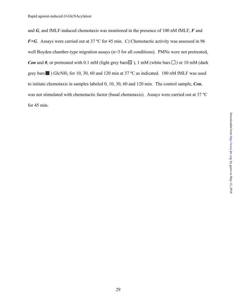
PMNs display rapid agonist-induced changes in O-GlcNAc. The formylated peptide fMLF is
a well-established and widely used PMN agonist. It rapidly activates a diverse group of signal
transduction pathways by engaging a specific G-protein-coupled receptor (33). We used fMLF
to assess whether receptor-associated stimulation could signal protein O-GlcNAcylation. 2 min
of stimulation with 100 nM fMLF led to increased protein-associated O-GlcNAc as determined
by immunoblot analyses of whole cell extracts with the O-GlcNAc-specific antibody CTD110.6
(Fig. 1A). A variety of proteins with molecular weights above ~50 kDa were observed, with
some variation in the banding pattern and/or intensity of some bands from experiment to
experiment, as might be expected for primary PMN isolates. Additional analyses revealed that
O-GlcNAcylation begins within 1 min of stimulation and progresses through 5 min (Fig. 1B). In
each experiment the specificity of CTD110.6 immunoreactivity was established by competitively
blocking antibody binding with free GlcNAc.
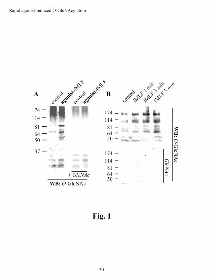
Although the O-GlcNAc-specific antibody CTD110.6 has been successfully used
previously in immunoblot and Elisa-based assays (41), it has not been evaluated with respect to
immunocytochemical microscopy. We developed a protocol using CTD110.6 in a fluorescence-
based immunocytochemical assay as an alternate and complementary method of examining
changes in O-GlcNAc modifications. 100 nM fMLF increased O-GlcNAc over 2 min of
stimulation (Fig. 2A). We obtained low background, antibody-specific signal using
formaldehyde fixation followed by methanol permeation. The permeation agent was a critical
variable in developing this assay, as commonly used agents such as Triton and acetone yielded
high non-specific backgrounds (data not shown). The agonist-induced CTD110.6 signal was
by guest on May 15, 2018
http://ww
w.jbc.org/
Dow
nloaded from

Rapid agonist-induced O-GlcNAcylation
14
specific for protein-associated O-GlcNAc, as it was competitively blocked by free GlcNAc.
These results paralleled our immunoblot data.
CTD110.6 labeled both the nucleus and cytoplasm in resting and stimulated cells. The
cells displayed two predominant patterns of label: a less intense, diffuse and relatively
homogenous label and a comparatively intense, non-homogenous, punctate label that, in contrast
to a Hoechst counter-stain that clearly defined the polymorphic multi-lobed PMN nucleus, was
restricted to the cytoplasm. The punctate cytoplasmic label displayed the major increase in
fluorescence intensity upon agonist stimulation (Fig.2B). Labeling of the nucleus was more
diffuse and considerably less intense than the punctate cytoplasmic label, although it too
increased with stimulation. This was somewhat unexpected as a variety of O-GlcNAcylated
proteins are known to be nuclear, and a more intense nuclear signal might be anticipated (7). It
is not clear whether this was a property of the particular labeling protocol used, a feature of O-
GlcNAc-modified proteins in PMNs, or a reflection of a specific property of the antibody and/or
the cell.
Of further interest, this assay also proved to be a sensitive means of assessing the
temporal dynamics of agonist-induced O-GlcNAcylation. Increases in protein O-GlcNAcylation
were evident within 30 sec of stimulation with fMLF and returned to near resting levels after 10
min (Fig. 3). This provided further support for the notion that O-GlcNAc is a rapidly inducible
post-translation protein modification.
PMNs possess a functional HBP that can use exogenous GlcNH2 to form the metabolic
precursors for protein O-GlcNAcylation. The HBP plays an integral role in O-GlcNAc
signaling, providing UDP-GlcNAc, the necessary substrate to drive OGT activity and O-
by guest on May 15, 2018
http://ww
w.jbc.org/
Dow
nloaded from
Rapid agonist-induced O-GlcNAcylation
15
GlcNAcylation (21). Having established that PMNs posses a robust and rapidly inducible O-
GlcNAcylation mechanism, we next asked what the effects of increasing flux through the HBP
would have in PMNs. GlcNH2 is an exogenous substrate for the HBP that is commonly used to
drive HBP-dependent O-GlcNAcylation (6,21,23). On entering a cell it is phosphorylated by
hexokinase to form GlcNH2 6-P, thus directly entering the HBP. Further processing yields UDP-
GlcNAc, the HBP product necessary for O-GlcNAcylation.
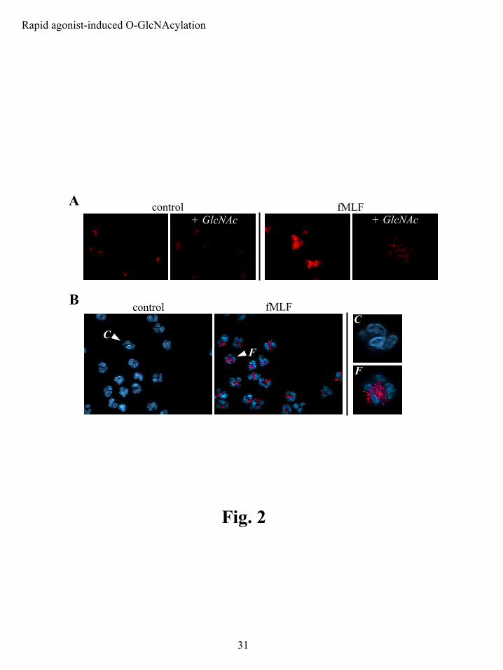
We examined GlcNH2 6-P and UDP-GlcNAc levels in GlcNH2-treated PMNs by HPLC.
Resting GlcNH2 6-P levels were about 20-fold less than UDP-GlcNAc levels, ~0.06 pmol/mg
protein versus ~1.4 nmol/mg protein (Fig. 4). UDP-GlcNAc was the predominant sugar
nucleotide in PMNs and was a major phospho-nucleotide, along with ADP, ATP and UDP-
glucose (data not shown). This suggests that a relatively abundant pool of UDP-GlcNAc is
important for normal PMN physiology and function. Short duration treatments with 10 mM
GlcNH2 led to the production of both GlcNH2 6-P and UDP-GlcNAc. GlcNH2 6-P levels rose
following GlcNH2 treatment, doubling over 2 min and increasing ~8-fold by 60 min (Fig. 4A),
indicating that GlcNH2 rapidly entered the cells and was quickly phosphorylated by hexokinase.
The increase in UDP-GlcNAc levels clearly followed the rise in GlcNH2 6-P (Fig. 4B), as might
be expected for HBP processing. Although the percent increase in UDP-GlcNAc was
significantly smaller than that for GlcNH2 6-P at any given time (for example, 60% versus 700%
after 60 min), the molar increase in UDP-GlcNAc was greater than that for GlcNH2 6-P (an
increases of 0.83 vs. 0.42 nmol/mg protein after 60 min). No drop in cellular ATP levels was
observed for GlcNH2 treatment (data not shown). In fMLF-treated cells there was no apparent
change in GlcNH2 6-P or UDP-GlcNAc levels with stimulation (data not shown).
by guest on May 15, 2018
http://ww
w.jbc.org/
Dow
nloaded from

Rapid agonist-induced O-GlcNAcylation
16
GlcNH2 increases protein-associated O-GlcNAc under the same conditions that increase
UDP-GlcNAc. We next determined if the observed GlcNH2-induced changes in HBP flux were
associated with alterations in O-GlcNAc. Short duration GlcNH2 treatment, 10 mM for 30 min,
enhanced protein O-GlcNAc in whole cell extracts as determined by immunoblotting with the O-
GlcNAc-specific antibody CTD110.6 (Fig. 5A). A group of proteins bearing O-GlcNAc with a
wide range of molecular weights above ~50 kDa was observed, with some variation in the
banding pattern from experiment to experiment. The overall banding pattern was similar to that
induced by fMLF (refer to Figs. 1A and 1B). The specificity of these immunoblots in identifying
proteins modified through GlcNH2-specific metabolism was confirmed by pre-treatment with 10
mM galactosamine (GalNH2), a stereoisomer of GlcNH2, which did not lead to increased O-
GlcNAc in the same time frame. The specificity of CTD110.6 for detecting O-GlcNAcylated
proteins was again established through competitive binding assays using free GlcNAc.
To investigate the relationship between increased UDP-GlcNAc and protein O-GlcNAc
in our system, aliquots from the same PMN preparation were taken for each experimental
condition (Fig. 5B). These experiments used PMNs from a single donor and cell isolation,
eliminating day-to-day and inter-donor variation. We found that significant changes in GlcNH2-
induced protein O-GlcNAcylation corresponded with only modest increases in UDP-GlcNAc.
UDP-GlcNAc levels and O-GlcNAcylation were dependent on the GlcNH2 dose and pre-
treatment duration: higher doses and longer pre-treatments enhanced UDP-GlcNAc and O-
GlcNAc, while lower doses and shorter pre-treatments were either without effect or produced
more modest increases. There was an apparent threshold for robust O-GlcNAcylation at 60 min
of treatment for all concentrations tested, although this did not clearly reflect a defined level of
UDP-GlcNAc. However, there was a trend for progressively elevated UDP-GlcNAc to
by guest on May 15, 2018
http://ww
w.jbc.org/
Dow
nloaded from

Rapid agonist-induced O-GlcNAcylation
17
correspond to progressively enhanced O-GlcNAc (for the representative experiment shown in
Fig. 5B this was especially evident for the 10 and 1 mM GlcNH2 conditions).
We used our previously described fluorescence-based immunocytochemical assay again
as an alternate and complementary method of evaluating GlcNH2-induced O-GlcNAcylation.
Cells treated with 10 mM GlcNH2 for 30 min showed a significantly increased fluorescent signal
versus untreated controls (Fig. 5C). The signal was specific for O-GlcNAc, as it was
competitively blocked by free GlcNAc. These results paralleled and confirmed our immunoblot
data. Interestingly, the GlcNH2-associated increase in CTD110.6 label was, as it was by
immunoblot, grossly similar to that observed for fMLF (refer to Figs. 2A and 2B). The effects of
GlcNH2 were most readily apparent in the cytoplasm, with a relatively large increase in the
punctate label. The more diffuse and homogenous labeling pattern, present in the cytosol and the
nucleus, was also increased by GlcNH2 treatment.
GlcNH2 treatment amplifies fMLF-induced protein O-GlcNAcylation. We next asked if
GlcNH2 was effective in conjunction with fMLF as a means of manipulating agonist-associated
O-GlcNAcylation. Combined GlcNH2 and agonist treatments were additive for O-
GlcNAcylation, with short duration GlcNH2 pre-treatments, which do not significantly increase
O-GlcNAc by themselves (refer to Fig. 5B), leading to noticeably increased fMLF-induced O-
GlcNAc (Fig. 6). These additive effects were most clearly evident at higher GlcNH2
concentrations. These experiments also confirmed that the pattern of O-GlcNAcylation due to
fMLF approximated that observed for GlcNH2.
by guest on May 15, 2018
http://ww
w.jbc.org/
Dow
nloaded from
Rapid agonist-induced O-GlcNAcylation
18
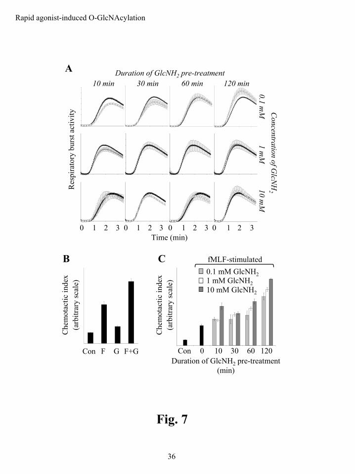
GlcNH2 selectively modulates PMN functional outputs. Having shown that GlcNH2 pre-
treatment can be used to modulate fMLF-induced O-GlcNAcylation, we attempted to elucidate
what effect(s) this had on fMLF-associated function. This would provide an initial determination
of whether changes in O-GlcNAc have a signaling role. We examined the effect of GlcNH2
treatment on agonist-induced respiratory burst (defined by the production of reactive oxygen
species) and chemotactic activities. GlcNH2 pre-treatments of various durations, 10-120 min,
and concentrations, 0.1-10 mM GlcNH2, had little effect (in all cases statistically non-significant,
p>0.05) on the respiratory burst elicited by 100 nM fMLF in luminol-dependent assays (Fig. 7A).
There was a trend toward reduced and slower onset activity for short duration GlcNH2 pre-
treatments, 10 and 30 min, and somewhat enhanced and faster onset activity for long duration
pre-treatments, 120 min. The GlcNH2 treatments did not affect cell viability or morphology
(data not shown).
An initial assessment of the effect of short duration GlcNH2 pre-treatment, 10 mM for 30
min, on chemotaxis revealed that GlcNH2 significantly increases both basal and fMLF-induced
activity (60% and 63% respectively) in simple 45 min Boyden chamber-type assays (Fig. 7B).
These data supported the possibility that GlcNH2 selectively modulates only certain aspects of
neutrophil physiology. We further defined GlcNH2-enhanced chemotaxis by altering both the
duration of GlcNH2 pre-treatment and the concentration of GlcNH2 used (Fig. 7C). The
chemotactic response was dependent on the GlcNH2 dose and pre-treatment duration: higher
doses and longer pre-treatments clearly enhanced chemotactic activity toward fMLF, while lower
doses and shorter pre-treatments were without significant effect. Significantly enhanced
chemotaxis was observed for all GlcNH2 doses tested by 120 min of pre-treatment, with 10 mM
GlcNH2 requiring as little as 10 min of pre-treatment and 1 mM requiring approximately 60 min.
by guest on May 15, 2018
http://ww
w.jbc.org/
Dow
nloaded from

Rapid agonist-induced O-GlcNAcylation
19
Discussion
Protein O-GlcNAcylation is emerging as an important post-translational modification that
regulates protein function. However, the physiological contexts in which rapid changes in the
level of O-GlcNAc occur are only beginning to be elucidated. Changes in O-GlcNAcylation are
implicated in the pathogenesis of diabetes mellitus, a disease characterized by chronic
hyperglycemia (6,14,15), and have also been suggested to serve as a nutrient sensor, in which O-
GlcNAc signals are modulated through nutrient availability (46,47). Recently, work by Zhang
has suggested that O-GlcNAc may couple proteasomes to the general metabolic state of the cell
(48), and Zachara has provided evidence that O-GlcNAc modulates stress-activated signaling
networks and may be integral to cellular survival responses (49). In addition, O-GlcNAcylation
is also often compared to phosphorylation (8-10), which is fundamental in the regulation of a
variety of signaling pathways. The central feature of many receptor-mediated signaling events,
in which extracellular agonists engage cognate receptors to activate signaling pathways, involves
rapid alterations in protein phosphorylation. As yet, there is only limited evidence to suggest
such a role for O-GlcNAc (6,29,30). Using a PMN model and fMLF as an extracellular
stimulus, we have found that O-GlcNAc can be rapidly induced in a receptor-dependent manner
and consequently may act as a regulatory signal for downstream functional responses.
The receptor-derived signal for many functional responses is rapidly transduced,
occurring over tens of seconds. Our data show changes in O-GlcNAc within this time frame. In
conjunction with modified chemotactic responsiveness as a result of O-GlcNAc-inducing
treatments, these data support a role for this post-translational modification in rapidly dynamic
signal transduction. fMLF rapidly and robustly increased O-GlcNAc on a large number of
by guest on May 15, 2018
http://ww
w.jbc.org/
Dow
nloaded from

Rapid agonist-induced O-GlcNAcylation
20
proteins. The rate at which this modification was induced was evident in both
immunofluorescence microscopy and immunoblots assays, in which fMLF caused an easily
detected increase in CTD110.6 immunoreactivity within 30 sec and 1-2 min respectively.
Furthermore, a number of proteins were modified by O-GlcNAc in response to fMLF. Our
immunofluorescence analysis of the CTD110.6 label revealed that the immunoreactivity was
predominantly cytoplasmic with the most concentrated labeling being associated with punctate
structures in the cell. It is not clear whether this pattern is truly reflective of O-GlcNAc
distribution or is in part a consequence of the methodology, as a variety of nucleus-associated
proteins are known to be O-GlcNAcylated, including nuclear pore proteins and transcription
factors (7). It may be that the large size of the CTD110.6 IgM prevents it from efficiently
penetrating the nucleus, even in fixed cells. Nonetheless, the label we detected was shown to be
specific for O-GlcNAc by competition with free GlcNAc, providing evidence for the validity of
the assay in assessing the temporal dynamics of O-GlcNAcylation. In addition, GlcNH2-induced
increases in O-GlcNAc, in both immunoblot and immunofluorescence analyses, revealed similar
results to those observed for fMLF. As a whole, these data provide substantive evidence of
agonist-induced O-GlcNAcylation and represent the first evidence that such changes occur over
short intervals.
We felt it essential to establish that exogenous GlcNH2 would be metabolized by the HBP
in a comparable time frame. Short-term GlcNH2 treatments increased the levels of HBP
metabolites, first GlcNH2 6-P and then UDP-GlcNAc. UDP-GlcNAc formation was dependent
on the time and concentration of GlcNH2 treatment, with treatments at high GlcNH2
concentrations, 10 mM, significantly increasing UDP-GlcNAc within 30 min while lower
concentrations required 60-120 min. Robust increases in O-GlcNAcylation occurred at 60 min
by guest on May 15, 2018
http://ww
w.jbc.org/
Dow
nloaded from

Rapid agonist-induced O-GlcNAcylation
21
GlcNH2 treatment for most concentrations tested, although 10 mM GlcNH2 increased O-GlcNAc
within 30 min. Thus, when compared to the respective UDP-GlcNAc levels, there is a parallel
between increases in UDP-GlcNAc and O-GlcNAcylation. Interestingly, at 100 µM GlcNH2
increases in O-GlcNAcylation correlated with increases in UDP-GlcNAc of only 20-25%. These
modest increases are likely relevant though, as OGT’s protein acceptor specificity has been
shown to be sensitive to small changes in UDP-GlcNAc (22). In addition, the observed levels of
UDP-GlcNAc reflect net changes and not just UDP-GlcNAc synthesis, and thus do not take into
account its utilization, which may be significant as the length of GlcNH2 treatment increases.
Furthermore, the observed levels may not accurately reflect localized concentration changes that
could occur in particular cellular compartments. This is relevant because cytosolic levels of
UDP-GlcNAc are thought to be 10-20 times lower than ER/Golgi levels (50,51).
We also combined GlcNH2 and agonist treatments, and found additive increases in O-
GlcNAc, in particular for several high molecular weight proteins in the short duration GlcNH2
pre-treatments. These additive changes varied by protein, but clearly suggest that GlcNH2 might
modify agonist-induced signal transduction by altering the level of O-GlcNAcylation induced by
the agonist itself. This also suggests how GlcNH2 might alter agonist-induced functional
responses such as chemotaxis.
By understanding the conditions under which UDP-GlcNAc and O-GlcNAc levels can be
manipulated through HBP metabolism, we have begun to establish conditions for understanding
how O-GlcNAc affects PMN function. The same conditions that altered HBP metabolism, UDP-
GlcNAc formation and O-GlcNAcylation, also affected chemotactic but not respiratory burst
activity. This suggests that protein O-GlcNAc modulates certain aspects of agonist-induced
by guest on May 15, 2018
http://ww
w.jbc.org/
Dow
nloaded from
Rapid agonist-induced O-GlcNAcylation
22
signaling. Furthermore, the diverse signaling pathways that lead from fMLF-associated receptor
engagement to chemotactic and respiratory burst activity are different (34,52-54).
In conclusion, our results support the theory that O-GlcNAc is a rapidly induced signal
that operates in the context of physiological receptor-mediated signal transduction. The
ubiquitous nature of this post-translational modification, the large numbers of target proteins that
are either suggested or known to be modified by it and its similarities to phosphorylation in
modulating protein function suggest that it may play a general role in dynamic signaling in
response to cellular stimulation.
by guest on May 15, 2018
http://ww
w.jbc.org/
Dow
nloaded from

Rapid agonist-induced O-GlcNAcylation
23
Acknowledgements
We thank Sherry Johnson for administrative support and Pam Bounelis for insightful input. We
additionally thank Mary Ann Accavitti and the UAB Epitope Recognition and Immunodetection
Core for the production of the CTD110.6 monoclonal antibody and Albert Tousson and the UAB
High Resolution Imaging Facility for technical expertise with respect to immunofluorescence
microscopy. This work was supported by NIH DK55647 and the Juvenile Diabetes Research
Foundation.
by guest on May 15, 2018
http://ww
w.jbc.org/
Dow
nloaded from

Rapid agonist-induced O-GlcNAcylation
24
References
1. Wells, L., Vosseller, K., and Hart, G. W. (2001) Science 291, 2376-2378. 2. Holt, G. W., Haltiwanger, R. S., Torres, C.-R., and Hart, G. W. (1987) J. Biol. Chem.
262, 14847-14850 3. Ding, M., and Vandre, D. D. (1996) J. Biol. Chem. 271, 12555-12561 4. Gao, Y., Miyazaki, J., and Hart, G. W. (2003) Arch. Biochem. Biophys. 415, 155-163 5. Hiromura, M., Choi, C. H., Sabourin, N. A., Jones, H., Bachvarov, D., and Usheva, A.
(2003) J. Biol. Chem. 278, 14046-14052 6. Vosseller, K., Wells, L., Lane, M. D., and Hart, G. W. (2002) Proc. Natl. Acad. Sci. U S
A 99, 5313-5318 7. Wells, L., Whalen, S. A., and Hart, G. W. (2003) Biochem. Biophys. Res. Commun. 302,
435-441 8. Hart, G. W., Greis, K. D., Dong, L. Y., Blomberg, M. A., Chou, T. Y., Jiang, M. S.,
Roquemore, E. P., Snow, D. M., Kreppel, L. K., Cole, R. N., and et al. (1995) Adv. Exp. Med. Biol. 376, 115-123
9. Kearse, K. P., and Hart, G. W. (1991) Proc. Natl. Acad. Sci. USA 88, 1701-1705 10. Hart, G. W., Haltiwanger, R. S., Holt, G. D., and Kelly, W. G. (1989) Annu. Rev.
Biochem. 58, 841-874 11. Kreppel, L. K., Blomberg, M. A., and Hart, G. W. (1997) J. Biol. Chem. 272, 9308-9315 12. Gao, Y., Wells, L., Comer, F. I., Parker, G. J., and Hart, G. W. (2001) J. Biol. Chem. 276,
9838-9845 13. Iyer, S. P., and Hart, G. W. (2003) Biochemistry 42, 2493-2499 14. Du, X. L., Edelstein, D., Rossetti, L., Fantus, I. G., Goldberg, H., Ziyadeh, F., Wu, J., and
Brownlee, M. (2000) Proc. Natl. Acad. Sci. USA 97, 12222-12226 15. Liu, K., Paterson, A. J., Chin, E., and Kudlow, J. E. (2000) Proc. Natl. Acad. Sci. USA
97, 2820-2825 16. Hawkins, M., Barzilai, N., Liu, R., Hu, M., Chen, W., and Rossetti, L. (1997) J. Clin.
Invest. 99, 2173-2182 17. Chou, T. Y., Dang, C. V., and Hart, G. W. (1995) Proc. Natl. Acad. Sci. USA 92, 4417-
4421 18. Slawson, C., Pidala, J., and Potter, R. (2001) Biochim. Biophys. Acta. 1537, 147-157 19. Yao, P. J., and Coleman, P. D. (1998) J. Neurosci. 18, 2399-23411 20. Griffith, L. S., Mathes, M., and Schmitz, B. (1995) J. Neurosci. Res. 41, 270-278 21. Marshall, S., Nadeau, O., and Yamasaki, K. (2004) J. Biol. Chem. 824-831 22. Kreppel, L. K., and Hart, G. W. (1999) J. Biol. Chem. 274, 32015-32022 23. Parker, G. J., Lund, K. C., Taylor, R. P., and McClain, D. A. (2003) J. Biol. Chem. 278,
10022-10027 24. Wells, L., and Hart, G. W. (2003) FEBS. Lett. 546, 154-158 25. Haltiwanger, R. S., Grove, K., and Philipsberg, G. A. (1998) J. Biol. Chem. 273, 3611-
3617 26. Konrad, R. J., Mikolaenko, I., Tolar, J. F., Liu, K., and Kudlow, J. E. (2001) Biochem. J.
356, 31-41
by guest on May 15, 2018
http://ww
w.jbc.org/
Dow
nloaded from

Rapid agonist-induced O-GlcNAcylation
25
27. Konrad, R. J., Zhang, F., Hale, J. E., Knierman, M. D., Becker, G. W., and Kudlow, J. E. (2002) Biochem. Biophys. Res. Commun. 293, 207-212
28. Griffith, L. S., and Schmitz, B. (1999) Eur. J. Biochem. 262, 824-831 29. Walgren, J. L., Vincent, T. S., Schey, K. L., and Buse, M. G. (2003) Am. J. Physiol.
Endocrinol. Metab. 284, E424-E434 30. Majumdar, G., Harmon, A., Candelaria, R., Martinez-Hernandez, A., Raghow, R., and
Solomon, S. S. (2003) Am. J. Physiol. Endocrinol. Metab. 285, E584-591 31. Andrews, P. C., and Babior, B. M. (1983) Blood 61, 333-340 32. Andrews, P. C., and Babior, B. M. (1984) Blood 64, 883-890 33. Omann, G. M., Allen, R. A., Bokoch, G. M., Painter, R. G., Traynor, A. E., and Sklar, L.
A. (1987) Physiol. Rev. 67, 285-322 34. Downey, G. P., Butler, J. R., Tapper, H., Fialkow, L., Saltiel, A. R., Rubin, B. B., and
Grinstein, S. (1998) J. Immunol. 160, 434-443 35. Babior, B. M. (1988) Arch. Biochem. Biophys. 264, 361-367 36. Nick, J. A., Avdi, N. J., Young, S. K., Knall, C., Gerwins, P., Johnson, G. L., and
Worthen, G. S. (1997) J. Clin. Invest. 99, 975-986 37. Chen, Q., Powell, D. W., Rane, M. J., Singh, S., Butt, W., Klein, J. B., and McLeish, K.
R. (2003) J. Immunol. 170, 5302-5308 38. Caldwell, S. E., Cassidy, L. F., and Abramson, J. S. (1988) J. Immunol. 140, 3560-3567 39. English, D., and Anderson, B. (1974) J. Immunol. Methods 5, 40. Laemmli, U. K. (1970) Nature 227, 680-685 41. Comer, F. I., Vosseller, K., Wells, L., Accavitti, M. A., and Hart, G. W. (2001) Anal.
Biochem. 293, 169-177 42. Robinson, K. A., Weinstein, M. L., Lindenmayer, G. E., and Buse, M. G. (1995)
Diabetes 44, 1438-1446 43. Lowry, O. H., Rosebrough, N. J., Farr, A. L., and Randall, R. J. (1951) J. Biol. Chem.
193, 265-275 44. Frevert, C. W., Wong, V. A., Goodman, R. B., Goodwin, R., and Martin, T. R. (1998) J.
Immunol. Methods 213, 41-52 45. Wymann, M. P., von Tscharner, V., Deranleau, D. A., and Baggiolini, M. (1987) Anal.
Biochem. 165, 371-378 46. McClain, D. A. (2002) J. Diabetes. Complications. 16, 72-80 47. Wells, L., Vosseller, K., and Hart, G. W. (2003) Cell. Mol. Life Sci. 60, 222-228 48. Zhang, F., Su, K., Yang, X., Bowe, D. B., Paterson, A. J., and Kudlow, J. E. (2003) Cell
115, 715-725 49. Zachara, N., O'Donnell, N., Cheung, W., Mercer, J., Marth, J., and Hart, G. W. (2004) J.
Biol. Chem. M403773200 50. Waldman, B. C., and Rudnick, G. (1990) Biochemistry 29, 44-52 51. Traynor, A. J., Hall, E. T., Walker, G., Miller, W. H., Melancon, P., and Kuchta, R. D.
(1996) J. Med. Chem. 39, 2894-2899 52. Zu, Y. L., Qi, J., Gilchrist, A., Fernandez, G. A., Vazquez-Abad, D., Kreutzer, D. L.,
Huang, C. K., and Sha'afi, R. I. (1998) J. Immunol. 160, 1982-1989 53. Heuertz, R. M., Tricomi, S. M., Ezekiel, U. R., and Webster, R. O. (1999) J. Biol. Chem.
274, 17968-17974 54. Hallet, M. B., and Lloyds, D. (1997) The molecular and ionic signaling of neutrophils.
Molecular Biology Intelligence Unit (Bioscience, L., Ed.), Chapman and Hill, Austin
by guest on May 15, 2018
http://ww
w.jbc.org/
Dow
nloaded from

Rapid agonist-induced O-GlcNAcylation
26
Figure Legends
Figure 1: PMNs display rapid agonist-induced protein O-GlcNAcylation as assessed by O-
GlcNAc-specific immunoblots. Immunoblots were performed using the anti-O-GlcNAc
antibody CTD110.6. The specificity of the antibody for protein O-GlcNAc was determined by
adding 10 mM GlcNAc to the primary antibody dilution buffer (indicated as + GlcNAc). A)
PMNs were left unstimulated (control) or stimulated with agonist, 100 nM fMLF, for 2 min. B)
PMNs were left unstimulated (control) or stimulated with 100 nM fMLF for 1, 3 or 5 min.
Figure 2: Immunofluorescence microscopy reveals rapid agonist-induced protein O-
GlcNAcylation. Immunofluorescence labeling was performed using the anti-O-GlcNAc
antibody CTD110.6. The specificity of this antibody for protein O-GlcNAc was assessed by
adding 100 mM GlcNAc to the primary antibody dilution buffer (indicated as + GlcNAc). A)
PMNs were left unstimulated (control) or stimulated with 100 nM fMLF for 2 min. Two
predominant labeling patterns were evident: a relatively intense punctate label and a less intense,
diffuse and homogenous label. B) The more intense, punctate O-GlcNAc label was limited to
the cytoplasm, as was evident when the cells were counterstained using the nuclear stain Hoechst
33258. PMNs were left unstimulated (control) or stimulated with 100 nM fMLF for 2 min.
Higher magnification images of selected cells revealed a punctate label that was localized
exclusively to the cytoplasm (the arrows in the low magnification images that are labeled as C
and F, control and fMLF, respectively, are the individual cells shown in the higher magnification
views).
by guest on May 15, 2018
http://ww
w.jbc.org/
Dow
nloaded from

Rapid agonist-induced O-GlcNAcylation
27
Figure 3: Immunofluorescence microscopy is an effective means of determining the
temporal dynamics of agonist-induced protein O-GlcNAcylation. Immunofluorescence
labeling was performed using the anti-O-GlcNAc antibody CTD110.6, and Hoechst 33258 was
used as a nuclear counterstain. fMLF induced robust cytoplasmic O-GlcNAcylation within 30
sec that continued for at least 5 min, after which it appeared to decrease toward resting levels.
PMNs were left unstimulated (C) or stimulated with 100 nM fMLF for various times ranging
from 30 sec (F 30sec) to 10 min (F 10min). Inset shows quantification of fMLF-induced O-
GlcNAc label by fluorescence intensity (n = number of cells examined for each condition, *
indicates p<0.05 in comparison to control as evaluated by Student’s t-test).
Figure 4: PMNs metabolize exogenous GlcNH2 into metabolic precursors that are used in
the pathway leading to protein O-GlcNAcylation. A) GlcNH2 6-P levels in PMNs treated
with GlcNH2. PMNs were left untreated (C) or treated with 10 mM GlcNH2 for 2, 10, 30 or 60
min at 37 ºC. OPA-conjugated acid extracts (n=3 for each condition) were analyzed on a C-18
column using HPLC. B) UDP-GlcNAc levels in PMNs treated with GlcNH2. PMNs were left
untreated (C) or treated with 10 mM GlcNH2 for 2, 10, 30 or 60 min at 37 ºC. Acid extracts (n=3
for each condition) were analyzed on a strong anion exchange column using HPLC.
Figure 5: GlcNH2 increases protein O-GlcNAc under the same conditions that increase
UDP-GlcNAc. A) Immunoblots were performed using the anti-O-GlcNAc antibody CTD110.6.
The specificity of the antibody for protein O-GlcNAc was assessed by adding 10 mM GlcNAc to
the primary antibody dilution buffer (indicated as + GlcNAc). Substrate driven O-
GlcNAcylation was assessed by adding 10 mM GlcNH2 or 10 mM GalNH2 for 30 min at 37 ºC.
by guest on May 15, 2018
http://ww
w.jbc.org/
Dow
nloaded from

Rapid agonist-induced O-GlcNAcylation
28
Control PMNs were left untreated. B) Single matched UDP-GlcNAc (HPLC) and O-
GlcNAcylation (immunoblots with CTD110.6) analyses for PMNs treated with 0.1, 1 or 10 mM
GlcNH2 for 10, 30, 60 and 120 min each at 37 ºC. Control cells were left untreated. C)
Immunofluorescence labeling was performed using the anti-O-GlcNAc antibody CTD110.6.
The specificity of this antibody for protein O-GlcNAc was assessed by adding 100 mM GlcNAc
to the primary antibody dilution buffer (indicated as + GlcNAc). PMNs remained untreated, left,
or were treated with 10 mM GlcN for 30 min at 37 ºC, right.
Figure 6: GlcNH2 treatment modifies fMLF-induced protein O-GlcNAcylation.
Immunoblots were performed using the anti-O-GlcNAc antibody CTD110.6. PMNs were
pretreated as indicated with 10, 1 or 0.1 mM GlcNH2 for 10, 30, 60 and 120 min at 37 ºC. The
cells were then stimulated with 100 nM fMLF for 2 min (indicated by F for the non-pretreated
sample). The control sample, C, was not stimulated and was not pretreated.
Figure 7: GlcNH2 modulates PMN chemotaxis but not respiratory burst activity. A)
Respiratory burst activity was assessed by luminol-dependent chemiluminescence. PMNs (n=3
for all conditions) were pretreated with 0.1, 1 or 10 mM GlcNH2 for 10, 30, 60 and 120 min at 37
ºC and then stimulated with 100 nM fMLF (open circles, ○). Control samples were not
pretreated with GlcNH2, but were stimulated with 100 nM fMLF (closed circles, ●). In all cases
fMLF stimulation was preceded by 30 sec of baseline recording. B) Chemotactic activity was
assessed in Boyden chamber-type migration assays (n=3 for all conditions). PMNs did not
receive a pretreatment, Con and F, or were pretreated with 10 mM GlcNH2 30 min at 37 ºC, G
and F+G. Basal levels of chemotaxis were monitored in the absence of chemotactic factor, Con
by guest on May 15, 2018
http://ww
w.jbc.org/
Dow
nloaded from
Rapid agonist-induced O-GlcNAcylation
29
and G, and fMLF-induced chemotaxis was monitored in the presence of 100 nM fMLF, F and
F+G. Assays were carried out at 37 ºC for 45 min. C) Chemotactic activity was assessed in 96
well Boyden chamber-type migration assays (n=3 for all conditions). PMNs were not pretreated,
Con and 0, or pretreated with 0.1 mM (light grey bars ), 1 mM (white bars ) or 10 mM (dark
grey bars ) GlcNH2 for 10, 30, 60 and 120 min at 37 ºC as indicated. 100 nM fMLF was used
to initiate chemotaxis in samples labeled 0, 10, 30, 60 and 120 min. The control sample, Con,
was not stimulated with chemotactic factor (basal chemotaxis). Assays were carried out at 37 ºC
for 45 min.
by guest on May 15, 2018
http://ww
w.jbc.org/
Dow
nloaded from
agon
ist-fM
LF
contr
ol
agon
ist-fM
LF
816450
114174
contr
ol
37
+ GlcNAcWB: O-GlcNAc
AfM
LF 3 min
fMLF 1
min
fMLF 5
min
contr
ol
816450
114174
816450
114174 +
GlcNAc
WB:O
-GlcN
Ac
B
Fig. 1
Rapid agonist-induced O-GlcNAcylation
30
by guest on May 15, 2018
http://ww
w.jbc.org/
Dow
nloaded from
control fMLF+ GlcNAc+ GlcNAc
A
B control
C
fMLF
F
C
F
Fig. 2
Rapid agonist-induced O-GlcNAcylation
31
by guest on May 15, 2018
http://ww
w.jbc.org/
Dow
nloaded from

C F 30sec F 1min
F 3minF 2min
F 10min
F 7.5min
F 5min
Fig. 3
Rapid agonist-induced O-GlcNAcylation
32
Con (n=40)
F 0.5 (n=36)
F 1 (n=48)
F 2 (n=21)
F 3 (n=40)
F 4 (n=38)
F 5 (n=47)
F 7.5 (n=31)
F 10 (n=39)
Prot
ein O
-Glc
NA
c(f
luor
esce
nt in
tens
ity)
Duration 100 nM fMLF stimulation (min)
**
** *
* *
by guest on May 15, 2018
http://ww
w.jbc.org/
Dow
nloaded from
10 mM GlcNH2 treatment(min)
1.21.51.82.12.4
UD
P-G
lcN
Ac
(n
mol
/mg
prot
ein)
C 2 10 30 60
B
10 mM GlcNH2 treatment(min)
Glc
NH
26-
P(p
mol
/mg
prot
ein)
0
0.15
0.30
0.45
C 2 10 30 60
A0.60
Fig. 4
Rapid agonist-induced O-GlcNAcylation
33
by guest on May 15, 2018
http://ww
w.jbc.org/
Dow
nloaded from

816450
114174
37
contr
olGalN
H 2
GlcNH 2
contr
olGalN
H 2
GlcNH 2
+ GlcNAc
WB:O-GlcNAc
C 10 30 60 120
0.1 mM GlcNH230022515075
0
A
10 mM GlcNH2
C 10 30 60 120Time (min)
UD
P-G
lcN
Ac
(per
cent
con
trol)
175
836248
32
1 mM GlcNH2
C 10 30 60 120
WB
: O-G
lcN
Ac
B
C
+ GlcNAc+ GlcNAc
Control GlcNH2
Fig. 5
Rapid agonist-induced O-GlcNAcylation
34
by guest on May 15, 2018
http://ww
w.jbc.org/
Dow
nloaded from

175
8362
C F 10 30 60 120 10 30 60 120 10 30 60 120
(min GlcNH2 pre-treatment)
10 mM GlcNH2
1 mM GlcNH2
0.1 mM GlcNH2
fMLF + GlcNH2 pre-treatment
Fig. 6
Rapid agonist-induced O-GlcNAcylation
35
by guest on May 15, 2018
http://ww
w.jbc.org/
Dow
nloaded from
10 min 30 min 60 minDuration of GlcNH2 pre-treatment
120 minR
espi
rato
ry b
urst
act
ivity
0 1Time (min)
2 3 0 1 2 3 0 1 2 3 0 1 2 3
0.1 mM
1 mM
10 mM
Concentration of G
lcNH
2
A
Con 0 10 30 60 120
Che
mot
actic
inde
x(a
rbitr
ary
scal
e)
0.1 mM GlcNH21 mM GlcNH210 mM GlcNH2
Duration of GlcNH2 pre-treatment (min)
fMLF-stimulatedC
Con F G F+G
B
Che
mot
actic
inde
x(a
rbitr
ary
scal
e)
Fig. 7
Rapid agonist-induced O-GlcNAcylation
36
by guest on May 15, 2018
http://ww
w.jbc.org/
Dow
nloaded from

Zachary T. Kneass and Richard B. MarchaseNeutrophils exhibit rapid agonist-induced increases in protein-associated O-GlcNAc
published online August 20, 2004J. Biol. Chem.
10.1074/jbc.M407911200Access the most updated version of this article at doi:
Alerts:
When a correction for this article is posted•
When this article is cited•
to choose from all of JBC's e-mail alertsClick here
by guest on May 15, 2018
http://ww
w.jbc.org/
Dow
nloaded from