neurotrophic activity of honokiol on the cultures of fetal rat cortical neurons
TRANSCRIPT

Neurotrophic Activity of Honokiol on the Cultures of Fetal RatCortical Neurons
Yoshiyasu Fukuyama,a,* Kousuke Nakade,a Yuka Minoshima,a Ritsuko Yokoyama,a
Haifeng Zhaia and Yasuhide Mitsumotob
aInstitute of Pharmacognosy, Faculty of Pharmaceutical Sciences, Tokushima Bunri University, Yamashiro-cho,Tokushima 770-8514, Japan
bNeurodegenerative Disease Research Group, Second Institute of New Drug Research, Otsuka Pharmaceutical Co. Ltd.,Tokushima 771-0192, Japan
Received 10 January 2002; accepted 9 February 2002
Abstract—Honokiol, a main biphenyl neolignan of the traditional crude medicine, Magnoliae cortex, was found to show neuro-trophic activity on the cultures of rat cortical neurons at concentration from 0.1 to 10 mM. In the cortical neurons cultured inserum-free medium supplemented with B27, honokiol could promote neurite outgrowth. In addition, the survival and growth ofneurons were significantly enhanced by adding honokiol to the primary cultures in serum-free medium supplemented with N2. Itsneurotrophic activity was comparable to 40 ng mL�1 of bFGF at concentration of 10 mM. # 2002 Elsevier Science Ltd. All rightsreserved.
Introduction
Simple biphenyl neolignans, honokiol (1) and magnolol(2), are the main constituents of the stem bark of Mag-nolia obovata Thunb1 and Magnolia officinalis Rhed,which have been used as a traditional Chinese medicinefor the treatment of thrombotic stroke, nervous dis-turbance, and gastrointestinal complaints.2 Compounds1 and 2 were shown to exhibit a central depressant, withsuccessively higher doses eliciting muscle relaxation,sedation, sleeping, and anesthesia.3,4 The dihydro-genated derivative of 1, in particular, showed ansignificant anxiolytic-like activity.5 In addition to theseCNS activities, a number of biological activities ofhonokiol and magnolol have been documented.6 In thispaper, we report an intriguing neurotrophic and neuro-protective effects of honokiol (1) on the primarycultures of fetal rat cortical neurons in two differentserum-free mediums, and thereby significance of 1 canbe jumped up to another useful field of CNS (Fig. 1).
Results and Discussion
In the previous paper,7 we reported the structures ofneurotrophic sesquiterpenes linked with magnolol and/or honokiol isolated from the stem bark of M. obovata.As the neuronal cells used for screening active com-pounds were cultured in the serum-containing medium,8
the issue on indirect effects of unknown components inthe serum remained to be unsolved. Thus, first, the pre-sent cell cultures were performed using 18-day fetal ratcortical neurons in the serum-free Neurobasal Medium(NBM) supplemented with B27.9,10 Honokiol (1) had astriking effect on the morphological differentiation of
0960-894X/02/$ - see front matter # 2002 Elsevier Science Ltd. All rights reserved.PI I : S0960-894X(02 )00112-9
Bioorganic & Medicinal Chemistry Letters 12 (2002) 1163–1166
Figure 1. Structures of honokiol (1) and magnolol (2).
*Corresponding author. Tel.: +81-88-622-9611x5911; fax: +81-88-655-3051; e-mail: [email protected]

cortical neurons as shown in Figure 2. After 7 days incell cultures, neurons in cultures (c, d, and e) containing0.1�10 mM of 1 were found to extend very long neur-ites, and more prominent and darker staining neuronalsomata in comparison with those of control culture (a)containing 0.5% EtOH. In addition, one of the fiberswas much longer and showed more neurite branchesthan those of the control cultures. Basic fibrobrastgrowth factor (bFGF) is well known as a trophic factorto enhance neurite extension and increase neuronal sur-vival.11 Adding 40 ng mL�1 of bFGF to this cell culturecould significantly enhance neurite extension as shownin Figure 2(b). On the other hand, magnolol (2) wasfound to promote neurite extension at concentration of1.0–10 mM as shown in Figure 2(f). However, its effect onneuronal morphology was not comparable to that of 1.
The morphometric analysis of the neurotrophic effect of1 was carried out by measuring the longest neuritelength of each neuron using Lumina Vision and Mac-SCOPE software.12 The results were shown in Figure 3.The length of neurites were found to be increased in thecultures treated with 1. The neurite length was increasedin a dose-dependent manner, and reached to the max-imum peak at 10 mM.13 It was striking that the neuritelength was 1.8 times greater than in control and theeffect of 1 was much more potent than bFGF. On theother hand, magnolol (2) was found to have weak effecton neurite extension according to the morphometricanalysis as shown in Figure 3. This prominent differenceof trophic effects caused by 1 and 2 is presumablyresponsible for the position of aryl–aryl bond formed
between two units of allylphenol, and is consistent withthe fact that 1 shows other CNS related activities moreeffectively than 2. 14,15
In general, the culture conditions without serum are notenough for most neurons to survive for a long period.NBM/B27, however, supplies a high neuronal survivalunder serum-free condition. The survival of corticalneurons can be maintained in this medium within 7 dayseven if a final cell density is decreased up to 9000 cellscm�2.10,16 Thus, NBM/B27 is useful for in vitro studiesof neuronal development, pharmacology, and effects ofgrowth factors. In contrast, it is not good at screeningneuroprotective effects as measured by neuronal survi-val and viability. We observed that most neurons diedwithin 5 days in the primary cultures plated at cell den-sity less than 2�105 cells cm�2 in the case of usingDMEM/N2 medium as shown in Figure 4(a). There-fore, we have decided that this serum-free mediumappropriates to the cell cultures to see whether honokiolcan protect neuronal death. In the experiment, cellsuspensions were initially plated at a density of 2�105
cells cm�2 and cultures were fixed 3 days after additionof honokiol (1) (0.01, 0.1, 1.0, 10, and 100 mM). Theresults were shown in Figures 4 and 5. The control cul-ture (a) in the absence of 1 carried only a small numberof survival neurons. In contrast, the cultures (b) and (c)in the presence of 1 not only increased the number ofsurvival neurons but also enhanced neurite extensionand wide neurite branching in a concentration-depen-dent manner. In particular, the culture (c) containing 10�M of 1 showed fantastic neuronal survival and growth.
Figure 2. Enhancement of neurite outgrowth by honokiol (1) and magnolol (2) in primary cultures of E18 SD rat cortical neurons in NBM/B27.After the neuronal cells (9000 cells cm�2) cultured for 6 days in the presence of 0.5% EtOH, 1, and 2 were fixed by 4% paraformaldehyde-PBS, theimmunohistochemical staining for the microtuble associated protein-2 (MAP-2) were performed. Pictures: (a) 0.5% EtOH, (b) 40 ng mL�1 bFGF,(c) 0.1 mM honokiol, (d) 1.0 mM honokiol, (e) 10 mM honokiol, and (f) 10 mM magnolol.
1164 Y. Fukuyama et al. / Bioorg. Med. Chem. Lett. 12 (2002) 1163–1166
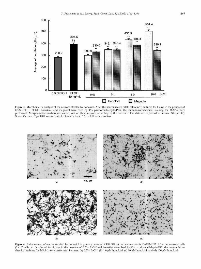
Figure 3. Morphometric analysis of the neurons effected by honokiol. After the neuronal cells (9000 cells cm�2) cultured for 6 days in the presence of0.5% EtOH, bFGF, honokiol, and magnolol were fixed by 4% paraformaldehyde-PBS, the immunohistochemical staining for MAP-2 wereperformed. Morphometric analysis was carried out on these neurons according to the criteria.12 The data are expressed as means�SE (n=80);Student’s t-test: ##p<0.01 versus control; Dunnet’s t-test: **p <0.01 versus control.
Figure 4. Enhancement of neurite survival by honokiol in primary cultures of E18 SD rat cortical neurons in DMEM/N2. After the neuronal cells(2�105 cells cm�2) cultured for 4 days in the presence of 0.5% EtOH and honokiol were fixed by 4% paraformaldehyde-PBS, the immunohisto-chemical staining for MAP-2 were performed. Pictures: (a) 0.5% EtOH, (b) 1.0 mM honokiol, (c) 10 mM honokiol, and (d) 100 mM honokiol.
Y. Fukuyama et al. / Bioorg. Med. Chem. Lett. 12 (2002) 1163–1166 1165
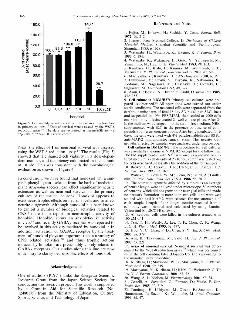
Next, the effect of 1 on neuronal survival was assessedusing the WST 8 reduction assay.17 The results (Fig. 5)showed that 1 enhanced cell viability in a dose-depen-dent manner, and its potency culminated in the summitat 10 mM. This was consistent with the morphologicalevaluation as shown in Figure 4.
In conclusion, we have found that honokiol (1), a sim-ple biphenyl lignan, isolated from the bark of medicinalplant Magnolia species, can effect significantly neuriteextension as well as neuronal survival in the primarycultures of rat cortical neurons. Honokiol appears toexert neurotrophic effects on neuronal cells and to affectneurite outgrowth. Although honokiol has been knownto exhibit a number of biological activities related toCNS,6 there is no report on neurotrophic activity ofhonokiol. Honokiol shows an anxiolytic-like activityin vivo,18 and recently GABAA receptor was reported tobe involved in this activity mediated by honokiol.19 Inaddition, activation of GABAA receptor by the treat-ment of honokiol plays an important role in a variety ofCNS related activities,20 and thus trophic actionsinduced by honokiol are presumably closely related toGABAA receptors. Our studies along this line are nowunder way to clarify neurotrophic effects of honokiol.
Acknowledgements
One of authors (R.Y.) thanks the Sasagawa ScientificResearch Grant from the Japan Science Society forconducting this research project. This work is supportedby a Grant-in Aid for Scientific Research (No.12480175) from the Ministry of Education, Culture,Sports, Science, and Technology of Japan.
References and Notes
1. Fujita, M.; Itokawa, H.; Sashida, Y. Chem. Pharm. Bull.1972, 20, 212.2. Juangsu New Medical College. In Dictionary of ChineseMaterial Medica; Shanghai Scientific and Technological:Shanghai, 1985; p 1628.3. Watanabe, H.; Watanabe, K.; Hagino, K. J. Pharm. Dyn.1983, 6, 184.4. Watanabe, K.; Watanabe, H.; Goto, Y.; Yamaguchi, M.;Yamamoto, N.; Hagino, K. Planta Med. 1983, 49, 103.5. Kuribara, H.; Kishi, E.; Kimura, M.; Weintraub, S. T.;Maruyama, Y. Pharmacol., Biochem. Behav. 2000, 67, 597.6. Maruyama, Y.; Kuribara, H. CNS Drug Rev. 2000, 6, 35.7. Fukuyama, Y.; Otoshi, Y.; Miyoshi, K.; Nakamura, K.;Kodama, M.; Nagasawa, M.; Hasegawa, T.; Okazaki, H.;Sugawara, M. Tetrahedron 1992, 48, 377.8. Asou, H.; Isasaki, N.; Hirano, S.; Dahl, D. Brain Res. 1985,332, 355.9. Cell culture in NBM/B27: Primary cell cultures were pre-pared as described.16 All operations were carried out understerile conditions. The neuronal cells were separated from thecerebral hemispheres of fetal 18 day SD rat (Japan SLC, Inc.)and suspended in 10% FBS/MEM, then seeded at 9000 cellscm�2 into poly-l-lysine-coated 24 well-culture plates. After 24h, the medium was changed into the serum-free medium, NBMsupplemented with B27, in the presence or absence of com-pounds at different concentrations. After being incubated for 6days, the cells were fixed with 4% paraformaldehyde/PBS foranti-MAP-2 immunohistochemical stain. The neurite out-growths affected by samples were analyzed under microscope.
Cell culture in DMEM/N2: The procedure for cell cultureswas essentially the same as NBM/B27 except for the followings:DMEM supplemented with N221 was used as a serum-free cul-tured medium; a cell density of 2�105 cells cm�2 was plated on;the cells were fixed 3 days after the addition of the test samples.10. Brewer, G. J.; Torricelli, J. R.; Evege, E. K.; Price, P. J. J.Neurosci. Res. 1993, 35, 567.11. Walicke, P.; Cowan, W. M.; Ueno, N.; Baird, A.; Guille-min, R. Proc. Natl. Acad. Sci. U.S.A. 1986, 83, 3012.12. The neurite outgrowths affected by samples as an averageof neurite length were analyzed under microscope. 80 numbersof neurons, which did not grow on or near glial cells and madeno network-formation to more than two cells, and were wellstained with anti-MAP-2, were selected for measurements ofeach sample. Length of the longest neurite extended from acell body was measured and calculated by using LuminaVision and MacSCOPE software.13. All neuronal cells were killed in the cultures treated with100 mM of 1.14. Tsai, T. H.; Westly, J.; Lee, T. F.; Chen, C. F.; Wang,L. C. H. Planta Med. 1995, 61, 477.15. Hou, Y. C.; Chao, P. D.; Chen, S. Y. Am. J. Chin. Med.2000, 28, 379.16. Abe, K.; Takayanagi, M.; Saito, H. Jpn. J. Pharmacol.1990, 53, 221.17. Assay of neuronal survival: Neuronal survival was deter-mined by the WST-8 reduction assay,22 which was performedusing the cell counting kit-8 (Dojindo Co. Ltd.) according tothe manufacturer’s procedure.18. Kuribara, H.; Stavinoha, W. B.; Maruyama, Y. J. Pharm.Pharmacol. 1998, 50, 819.19. Maruyama, Y.; Kuribara, H.; Kishi, E.; Weintraub, S. T.;Ito, Y. J. Pharm. Pharmacol. 2001, 53, 721.20. Wang, A. J.; Nielsen, M. Pharmacology 2001, 63, 34.21. Cestelli, A.; Savettieri, G.; Ferraro, D.; Vitale, F. Dev.Brain. Res. 1985, 22, 219.22. Tominaga, H.; Ushiyama, M.; Ohseto, F.; Sasamoto, K.;Hamamoto, T.; Suzuki, K.; Watanabe, M. Anal. Commun.1999, 36, 47.
Figure 5. Cell viability of rat cortical neurons enhanced by honokiolin primary cultures. Effects of survival were assessed by the WST-8reduction assay.22 The data are expressed as means�SE (n=4);**p<0.015, ***p<0.005 versus control.
1166 Y. Fukuyama et al. / Bioorg. Med. Chem. Lett. 12 (2002) 1163–1166