neuropathology of neurodegenerative diseases in toronto
TRANSCRIPT

Neuropathology of neurodegenerative diseases in Toronto: Accomplishments and challenges
Toronto Dementia Research Alliance
David MunozHead, Pathology, St. Michael’s Hospital
Professor, Laboratory Medicine and Pathobiology, University of Toronto

Human Resources
8.5 Neuropathologists in Toronto 3 UHN 1.5 Sunnybrook 2 Hospital for Sick Children 1Mount Sinai 1 St. Michael’s
2 Primary interest in dementia L-N H, DGM

Canadian Brain Tissue Bank 1
Established 1982 Initially managed by the Canadian Neurological Coalition
1982-1992: National scope SupportMedical Research CouncilOntario Mental Health Foundation Canadian Neurological Coalition
Budget 150K-200K/yr 120 brains/yr Staff: 2 Office staff, 1Tissue Coordinator1Medical Director

Canadian Brain Tissue Bank 2
1992-2000 Reduced budget to 25% of prior Reduced donors No detailed frozen tissue dissections
2000-2010 (gradual) No donations No staff Tissue stored in freezers at UHN Retrievals continue as part of collaborative studies

CBTB: Ongoing international collaborations

CBTB current status
Collection of >300 frozen & histological brain samples of well clinically characterized patients
Interested &dedicated neuropathologists
Good facilities
Lack of stable funding
Assets Liabilities

CBTB: Potential
Pathological confirmation of presumed diagnosis for numerous ongoing clinical studies in Toronto Source of donations
Fresh frozen tissue for basic researchers Further delineation of neurodegenerative diseases
through clinico-pathological correlation studies Stable funding only missing factor

Ongoing Intra-city collaborations 1
STRUCTURAL CORRELATE OF FOCAL SIGNS IN ALZHEIMER’S
DISEASE

Unresolved questions
► Clinical profile of patients with PPA and AD pathology
► In cases with AD pathology, pathological differences between cases with PPA symptoms without PPA symptoms
► Is the focal presentation related to Selective regional distribution of plaques and tangles Additional presence of a different lesion

ALL CASESPathological Diagnosis:► Alzheimer´s Disease► Abundant NP & NFT ► Braak stage V-VI► Typical distribution
Bielchowsky x 4

AT8Parietal
Cortical & GWJ
ATAC: Argyrophilic Thorny Astrocyte Clusters7 out of 8 cases

tau
Eccentric nucleus
Stout perikarya
Coarse dense clumps
Thorny outline
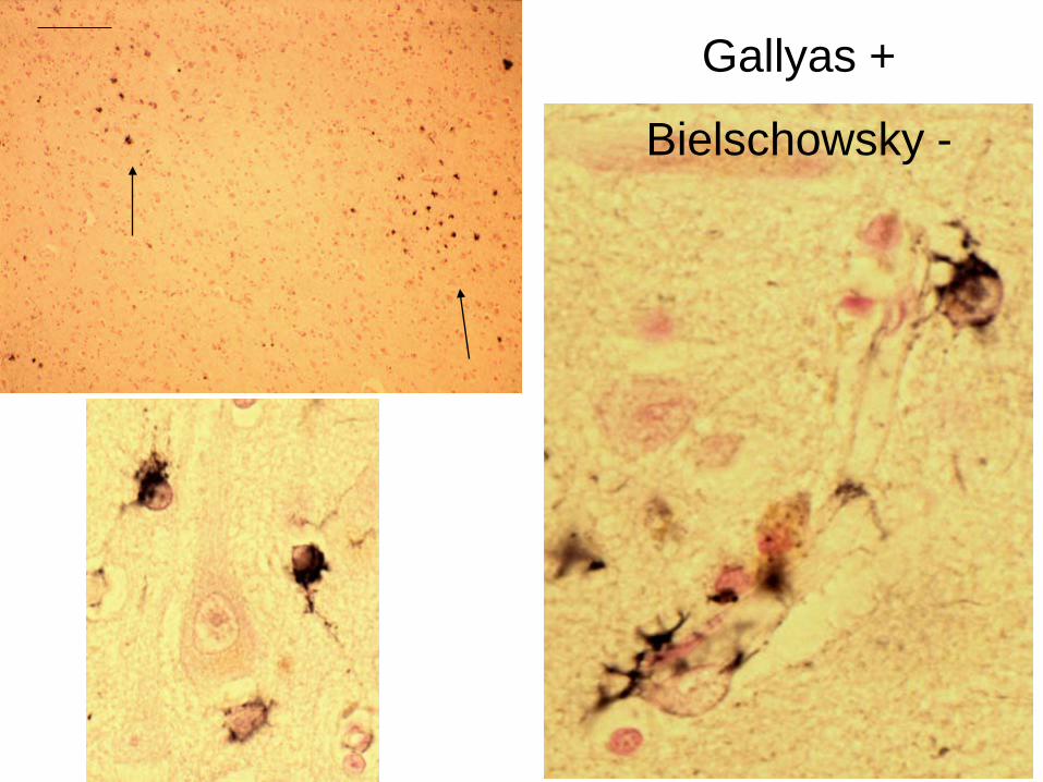
Gallyas +
Bielschowsky -

ATAC characterization
Cerebral cortex Subcortical WM (near
GWJ) Absent subcortical GM
Myelin pallor (leukoaraiosis)
Gliosis Infarcts NFT density Plaque density
Location Unrelated to

CONCLUSIONS :ATAC
► ATAC are a common, but not universal co-substrate of PPA in AD
► ATAC differ from ATA in normal aging Location, most Gallyas +
► Intensification of aging changes vs independent phenomenon Neuronal NFT:
►Aging ≈ AD ►Aging ≠ PSP, CBD
► Hypothesis: ATAC represent a marker of a process responsible for or contributing to the prominent focal clinical manifestations in AD

External Confirmation
Finds aphasia in AD poorly related to NFT, plaque distribution. ATAC in subset of cases

Ongoing collaboration
Structural substrate of focal signs in AD Black, Bilbao (Sunnybrook) Munoz (St. Michael’s)

Ongoing Intra-city collaborations 2
FUSOPATHIESRelationship between aFTLD-U, NIFID, & BIBD
Black, Bilbao (Sunnybrook)Munoz (St. Michael’s)

What we need
Stable funding to support a Brain Bank Strengthen clinical studies by confirming diagnosis Improved QA, basic-clinical link
Histological studies Biochemical studies Imaging correlation Strengthen genetic studies
We have the capability. With funding support as an outcome of this alliance we will be in a position to re-start this program