neuronal migration mechanisms in development and disease...
TRANSCRIPT

Available online at www.sciencedirect.com
Neuronal migration mechanisms in development and diseaseManuel Valiente and Oscar Marın
Neuronal migration is a fundamental process that determines
the final allocation of neurons in the nervous system,
establishing the basis for the subsequent wiring of neural
circuitry. From cell polarization to target identification, neuronal
migration integrates multiple cellular and molecular events that
enable neuronal precursors to move across the brain to reach
their final destination. In this review we summarize novel
findings on the key processes that govern the cell biology of
migrating neurons, describing recent advances in their
molecular regulation in different migratory pathways of the
brain, spinal cord, and peripheral nervous system. We will also
review how this basic knowledge is contributing to a better
understanding of the etiology and pathophysiology of multiple
neurological syndromes in which neuronal migration is
disrupted.
Address
Instituto de Neurociencias, Consejo Superior de Investigaciones
Cientıficas & Universidad Miguel Hernandez, 03550 Sant Joan d’Alacant,
Spain
Corresponding author: Marın, Oscar ([email protected])
Current Opinion in Neurobiology 2010, 20:68–78
This review comes from a themed issue on
Development
Edited by Francois Guillemot and Oliver Hobert
Available online 5th January 2010
0959-4388/$ – see front matter
# 2009 Elsevier Ltd. All rights reserved.
DOI 10.1016/j.conb.2009.12.003
IntroductionInstructed by extracellular cues, the activation of gui-
dance receptors and their downstream signaling pathways
enable newborn neurons to migrate through the devel-
oping nervous system until they reach their destination.
The migratory cycle of neurons involves leading process
dynamics, through which directional migration is
achieved, and somal translocation, which involves the
movement of perinuclear material and organelles as well
as the nucleus. Different classes of neurons adjust and
modify this basic migratory cycle depending on the
specific requirements of their migratory pathway, which
may also change through time. For example, pyramidal
cells transit through at least three different phases during
their migration toward the cortical plate [1] (Figure 1).
During these stages, pyramidal cells adopt different
morphologies, each of which probably involves distinct
Current Opinion in Neurobiology 2010, 20:68–78
molecular requirements and whose disruption causes
specific neurological syndromes [2,3].
Recent work has shed light on the molecular mechanisms
regulating each of the different aspects of the migratory
cycle of neurons, the topic of the present review. We will
also summarize recent findings illustrating how abnormal
neuronal migration may cause neurological disorders.
The transcriptional mechanisms regulating neuronal
migration will not be addressed in this article, as it has
recently been reviewed elsewhere [4,5].
Leading process dynamicsReal time imaging experiments have begun to shed light
into the dynamic behavior of the leading process in
migrating neurons. Somehow surprisingly, we have dis-
covered that the leading process of migrating neurons has
relatively distinct morphologies in different classes of
neurons, probably reflecting their adaptation to specific
microenvironments. In contrast to the simplicity of the
single leading process reported for radially migrating
pyramidal cells [6], tangentially migrating neurons dis-
play very elaborated leading process morphologies. This
is the case, for example, of precerebellar neurons, cortical
interneurons, or immature enteric neurons, all of which
have branched leading processes [7–10]. In these cells,
leading process branches are stabilized or retracted in
binary decisions that determine the direction followed by
the nucleus during nucleokinesis [11�] (Figure 2), and this
process is intimately linked to their directional guidance
[8,11�,12�]. Thus, guidance cues influence the frequency
and orientation at which new leading process branches
emerge, which allow migrating neurons to rapidly change
direction without having to reorient pre-existing
branches. This guidance mechanism is different from
that described for growing axons, in which growth cone
steering determine the direction of movement [13].
It has been suggested that leading process branching
represents a general, default strategy for neuronal
migration, which might be suppressed under certain
circumstances, for example, during glial-guided radial
migration [14]. Consistent with this view, the leading
process of pyramidal cells become progressively unipolar
as neurons transit from the progenitor region toward the
intermediate zone (Figure 1), where they start actively
interacting with radial glial fibers [1,3]. Moreover,
preventing the normal interaction of pyramidal cells
with radial glial fibers increases the number of leading
process branches in these cells [15��] (Figure 1). One
of the proteins that may regulate this process is the
small GTP-binding protein Rnd2 [16��]. Rnd2 silencing
www.sciencedirect.com

Neuronal migration mechanisms in development and disease Valiente and Marın 69
Figure 1
Regulators of cell morphology during glial-guided migration. Radial glia cells present in the ventricular zone (VZ) are the progenitors of cortical
projection neurons and extend a long basal process that ends in the pial basement membrane over the marginal zone (MZ). Newborn neurons polarize
and begin their migration close to the progenitor radial glia cell (1). At the subventricular zone (SVZ), however, migrating neurons halt their progression
toward the cortical plate (CP) and modify their morphology (2), becoming multipolar cells. Cells at this state are characterized by the generation of
multiple processes all around the cell and can make small tangential displacements. Eventually, cells polarize again, acquire a clear bipolar
morphology (3), and restart their migration through the intermediate zone (IZ) and into the CP. During this later phase, projection neurons migrate
closely attached to the basal process of radial glia cells. Interaction with the cellular scaffold of radial glia is mediated, among other molecules, by
connexins Cx26 and Cx43. The coupling of the nucleus (n) and the centrosome (c) is also schematically depicted in these drawings. The distance
between the nucleus and centrosome (white dashed line) changes during migration owing to the forward displacement of the centrosome and the
saltatory movement of the nucleus. Knockdown of several regulators of the morphology of pyramidal cells impairs their migration. For example, RNAi
against Cx43 or Cx26 disrupts the progression of projection neurons from the multipolar stage, probably owing to a failure in their ability to interact with
radial glial fibers. Of note, cells that manage to progress toward the CP develop a leading process with multiple branches. Rnd2 knockdown causes a
similar phenotype, although it is presently unclear how this small GTP-binding protein (expressed by cells in the SVZ/IZ transition) controls this
process. Other proteins that regulate the stability of microtubules (e.g., p600, MARK2/Par-1, and Lkb1) are essential for the progression of neurons
from the multipolar stage into glial-guided migration. Loss of p600 leads to abnormal development of the leading process, which becomes thin and
wavy. Reduction in the levels of MARK2/Par-1 or Lkb1 causes different defects in centrosome–nucleus coupling. Axons of migrating and positioned
neurons were not drawn for simplicity. For axonal phenotypes see corresponding references.
perturbs the transition of pyramidal cells from the multi-
polar stage to the unipolar stage, and even those cells that
manage to polarize in the absence of Rnd2 have many
more leading process branches than normal cells
(Figure 1).
Leading process stability depends on a system of longi-
tudinal microtubules that links the leading edge of the
cell with the soma. This system not only provides struc-
www.sciencedirect.com
tural support to the leading process, but also allows the
flow of vesicles required for intracellular communication.
This process seems to be directly regulated by p600 (also
known as Ubr4), a novel microtubule associated protein
(MAP) that interacts with the endoplasmic reticulum
(ER). Knockdown of p600 in pyramidal cells leads to a
prominent reduction in acetylated tubulin and an almost
complete loss of the ER in the leading process, which
acquires a wavy appearance (Figure 1) and disrupts
Current Opinion in Neurobiology 2010, 20:68–78

70 Development
Figure 2
The cell biology of neuronal migration. (A) Schematic drawing of representative structures in a migrating interneuron. A high magnification of the
nucleus and associated cytoskeleton and organelles is also depicted. Note that actin and Myosin II are enriched at the rear part of the nucleus and at
the proximal aspect of the leading process. (B) Schematic of the migratory cycle of cortical interneurons. The cycle starts with the extension of a
branch of the leading process (green arrow) and the forward movement of the perinuclear dilatation or swelling, containing the Golgi apparatus and the
centrosome (blue arrow). Extension and retraction of different branches of the leading process coexist until one of them is selected. At this time, the
nucleus moves forward until the branch point (yellow arrow), which represents the main static point for the cell in the entire cycle. Non-selected
processes retract while a new process starts to grow, and the cycle is repeated. (C) A high magnification schema of nucleus and cytoskeleton during
nucleokinesis. tyr-Tub, tyrosinated tubulin; ace-tub, acetylated tubulin; and t, time.
migration [17]. These experiments reinforce the notion
that microtubule stability and membrane trafficking are
required for leading process maintenance.
The signaling mechanisms that control leading process
branching are presently unknown, although recent work
in cerebellar granule cells suggests that this process is
modulated by intracellular Ca2+ and cAMP levels. Thus,
experimentally increasing the concentration of intracellu-
lar Ca2+ from extracellular sources or internal organelle
deposits or stimulating the adenylyl cyclase promotes
leading process branching, while reducing the intracellu-
lar Ca2+ concentration or inhibiting PKA decreases its
frequency [12�]. These results suggest that guidance cues
regulating intracellular Ca2+ and cAMP levels are likely
regulators of leading process branching during neuronal
migration.
Molecular regulation of nucleokinesisWhile the leading process determines the direction in
which the neuron moves, effective neuronal migration is
only achieved by the subsequent translocation of the cell
soma, including the small organelles and the nucleus.
Soma translocation occurs in two consecutive phases in
most neurons studied so far, as initially described in
Current Opinion in Neurobiology 2010, 20:68–78
cerebellar granule cells [18]. First, a cytoplasmic dilata-
tion or swelling appears in the proximal part of the leading
process, toward which the centrosome and the Golgi
apparatus moves. Second, the nucleus advances forward
and invades the cytoplasmic dilatation (Figure 2).
The movement of centrosome and nucleus is highly
dependent on the integrity of a rich network of micro-
tubules with different post-transcriptional modifications
that extends between both [18,19��,20,21�,22]. Most
microtubules surrounding the nucleus are tyrosinated,
which makes them highly dynamic. By contrast, micro-
tubules at the anterior pole of the nucleus, near the
centrosome, are acetylated and therefore more stable.
Although the function of these two types of microtubules
is not completely understood, they seem to play different
roles in somal translocation [20,21�].
It has been hypothesized that the microtubule network
surrounding the nucleus is connected with the centro-
some, which is the main microtubule-organizing center
(MTOC) in these cells. Consequently, it was suggested
that the microtubule-dependent pulling forces on the
nucleus converge at the centrosome [18,23,24]. Recent
experiments, however, have put this hypothesis into
www.sciencedirect.com

Neuronal migration mechanisms in development and disease Valiente and Marın 71
question [21�]. Analysis of migrating granule cells in
organotypic slices, a preparation that resembles the
microenvironment found in vivo, suggests that most peri-
nuclear microtubules are not anchored to the centrosome
but rather extend into the leading process. In addition,
the authors frequently observed that the nucleus in these
cells advance forward pass the centrosome, which would
argue against a role of this structure in pulling the
nucleus. Further experiments would be needed to elu-
cidate whether these observations represent a specializ-
ation of granule cells or indeed define a general principle
for migrating neurons.
MAPs and related motor proteins are important regulators
of the movement of organelles. RNAi experiments have
shown that Lissencephaly 1 (Lis1) and the related motor
protein dynein are both independently required during
centrosome progression and nucleokinesis [19��]. In
addition, several proteins involved in cell polarity have
been described to play important roles in coordinating the
movement of the centrosome and the nucleus in each
migratory cycle. Par6a, for instance, localizes to the
centrosome and is required for its forward movement
[18]. Similarly, loss of MARK2 (microtubule affinity-
regulating kinase 2, also known as Par-1) perturbs centro-
some dynamics and blocks the transition of radially
migrating pyramidal cells through the multipolar stage
(Figure 1) [25�]. MARK2 levels are inversely related to
the stability of microtubules, and so loss of MARK2
function may impair migration owing to excessive micro-
tubule stabilization. Interestingly, the lissencephaly-
associated gene Doublecortin (DCX) is a substrate of
MARK2, and loss of DCX function increases microtubule
dynamics [26�]. Another polarity protein, LKB1 (a Ser/
Thr kinase related to Par-4), may also be involved in
regulating the transition of migrating neurons through the
multipolar stage (Figure 1) [27�]. Knockdown of LKB1 in
migrating neurons impairs neuronal migration, probably
owing to interfering with centrosome–nucleus coupling
[27�]. It should be noted, however, that genetic loss of
Lkb1 function does not lead to layering defects in the
cerebral cortex [28], and so the precise involvement of
this protein in neuron migration requires further exam-
ination. Nevertheless, disruption of Lkb1 leads to
exuberant leading process branching, which is likely to
perturb neuronal migration [27�,28,29].
Independently of the precise function of MAPs and
associated motor proteins in somal translocation, it is
unlikely that they alone provide the forces required for
the movement of these structures. Several studies have
shown that the motor protein Myosin II is enriched
behind the nucleus of migrating neurons and that
pharmacological blocking its ATPase activity inhibits
nucleokinesis, suggesting that rear contraction of acto-
myosin fibers propels the nucleus [30,31]. Recent work
suggests that F-actin and Myosin II motors are also
www.sciencedirect.com
enriched in the proximal region of the leading process
of migrating cerebellar granule cells [32�]. Rapid F-actin
turnover powered by Myosin II in this region appears to
contribute to the forward movement of the centrosome,
which in turn facilitates the subsequent displacement of
the nucleus [32�]. These results are nevertheless contro-
versial, because previous experiments in pyramidal cells
led to the suggestion that centrosome movement is inde-
pendent of Myosin II function [19��].
Recent studies suggest that signals emanating from the
cell surface influence centrosome–nucleus coupling, and
therefore somal translocation. During cerebellar devel-
opment, Sema6A is expressed in the deep aspect of the
external granule layer, where it promotes the inward
migration of granule cells through its PlexinA2 receptor
[33��]. Interestingly, disruption of Sema6A/PlexinA2 sig-
naling impairs centrosome–nucleus coupling, which leads
to defective migration [33��]. Although the precise mech-
anisms linking PlexinA2 activation with the cytoskeleton
are currently unknown, these results nicely illustrate that
guidance and somal displacement are probably coordi-
nated downstream of surface receptors during neuronal
migration.
Adhesion during neuronal migrationCell migration requires the dynamic regulation of
adhesion complexes between migrating cells and sur-
rounding extracellular matrix (ECM) proteins. In many
cell types, this process involves integrin-mediated
adhesion [34], but the function of this signaling system
in neuronal migration has remained controversial. In the
CNS, cell adhesion dynamics have been best character-
ized during glial-guided migration in developing cortical
structures [35]. Early in vitro studies emphasized a role for
neuronal a3b1 integrin receptors in mediating the inter-
action of migrating pyramidal neurons with radial glial
cells [36]. Subsequent analyses of mouse mutants have in
turn focused the attention to radial glial cells, as these
cells and their endfeet anchorage to the meningeal base-
ment membrane are dramatically affected in the absence
of b1 integrin signaling [37–39]. According to this later
view, integrin function is only indirectly required for
migrating neurons, which would otherwise migrate nor-
mally in the absence of integrin-mediated adhesion. As
migration on two-dimensional substrates naturally over-
emphasizes the role of adhesion, it is very likely that invivo studies may offer a much more precise idea of the
contribution of integrin signaling during neuronal
migration. Nevertheless, mouse genetic analyses may
have overlooked subtle defects in the motility of integrin
mutant neurons [40], which may only cause a minor delay
in migration and not a major disruption of cortical layer-
ing. Consistent with this notion, two recent studies
suggest that integrin signaling cell-autonomously influ-
ence neuronal migration. Conditional deletion of
neuronal laminin g1, a component of the ECM deposited
Current Opinion in Neurobiology 2010, 20:68–78

72 Development
around migrating neurons and radial glial fibers that binds
integrin receptors, disrupts the migration of pyramidal
neurons [41]. In addition, specific removal of a3 integrins
from tangentially migrating interneurons perturbs their
normal distribution as they invade the embryonic cortex,
suggesting that these receptors are required to recognize
specific guidance cues present in the migratory route of
interneurons [42].
Recent studies have provided a new perspective to the
role of cell–cell adhesions during radial migration. The
Gap junction proteins Connexin 26 and 43 are expressed
at the contact points between radial glial fibers and
migrating neurons during glial-guided migration
[15��,43] (Figure 1). Gap junctions are best known for
their channel capabilities, which allow the electrical and
chemical coupling of cells or the release of small mol-
ecules to the extracellular space. During radial migration,
however, Gap junctions provide dynamic adhesive con-
tacts between neurons and radial glial fibers that enable
the stabilization of the leading process of migrating
neurons along radial glial processes [15��]. Very little is
known about the mechanisms regulating Gap adhesion
dynamics during radial migration, but recent work
suggests that the C-terminal tail of connexins is required
in this process [44�]. This domain has been shown to
regulate mobility in other cell types, and it is known to
interact with several cytoskeleton proteins, such as zona
occludens-1 (ZO-1) and N-cadherin.
News from the Reelin frontReelin is crucial for the lamination of cortical structures
[45], but the molecular mechanisms underlying its action
remain unclear [46]. Genetic studies have positioned
Reelin, ApoER2, Vldlr and Dab1 into a common signaling
pathway that leads to the phosphorylation of Dab1 in
migrating neurons (Figure 3), an event that is required for
normal layering of the cortex [47,48]. ApoER2 and Vldlr
are the canonical Reelin receptors, but their relative
contribution to neuronal migration is controversial.
Recent experiments suggest that the neuronal positioning
of late-born pyramidal neurons is largely attributable to
ApoER2. On the contrary, lack of Vldlr causes ectopic
accumulation of pyramidal neurons into layer I, which
suggest that both phenotypes (layering and invasion) are
molecularly distinguishable [49�] (Figure 3).
The mechanisms underlying Reelin function in neuronal
migration are only beginning to be elucidated. Recent
experiments have shown that Dab1 tyrosine phosphoryl-
ation sites have distinct signaling properties in vivo [50]
(Figure 3). Two of the putative tyrosine phosphorylation
sites, named ab sites (Y185 and Y198), are sufficient for
the recruitment of Src-family kinases (SFKs), phosphoryl-
ation of Dab1 at these sites, stimulation of Akt/PI3K
signaling, and targeting activated Dab1 for degradation.
The ab sites seem also important for the subsequent
Current Opinion in Neurobiology 2010, 20:68–78
phosphorylation of another two tyrosine residues, the
cd sites (Y220 and Y232). Previous work has shown that
phosphorylation of the cd tyrosines is necessary to termi-
nate glial-guided migration, because neurons expressing
non-phosphorylatable forms of these residues (Y220F and
Y232F mutants, respectively) fail to downregulate the
expression of a3 integrin in the cortical plate, and con-
sequently remain attached to the radial glia [51]. This
function might be mediated by other elements in the
signaling cascade, such as the adaptor proteins Crk and
Crk-like (CrkL) and the Ras family guanine nucleotide
exchange factor C3G, because their recruitment to the
Dab1 complex also requires intact cd sites [50]. Moreover,
loss of these proteins causes phenotypes that are similar to
those described in reeler [52�,53] (Figure 3). In sum, Dab1
seems to operate both as an activator and scaffold protein
through the phosphorylation of the ab and cd domains,
respectively, and both functions are required for normal
cortical development in vivo.
Recent work has revealed additional elements in the
signaling cascade downstream of Dab1. Biochemical
experiments suggest that activated Dab1 prevents the
degradation of the intracellular, active form of Notch in
migrating pyramidal neurons [54��]. It is presently unclear
how this process works, since Reelin signaling also pro-
motes the degradation of Dab1 as part of a feedback loop
that exists in the pathway [55,56,57��]. In any case,
Notch-ICD is cell-autonomously required for the normal
positioning of neurons both in the neocortex and in the
dentate gyrus [54��,58�], suggesting that one of the
possible functions of Reelin signaling in cortical devel-
opment would be to modulate Notch signaling (Figure 4).
This would be consistent with the hypothesis that Reelin
functions as a signal that incites migrating neurons to
adapt their cytoskeleton during glial-guided migration
[59]. In Drosophila growth cones, for instance, Notch
controls actin cytoskeletal dynamics through its inter-
action with Abl [60]. Consistently, pyramidal cells lacking
Notch display abnormal leading processes [54��].
Halting migrationsUpon arrival to their final destination, neurons cancel
their migratory program and continue their differentiation
into mature neurons. Understanding the mechanisms
through which neurons perceive that they have reached
their target position has been a puzzling question for a
long time, but recent studies have begun to elucidate this
issue [61,62]. It has been suggested that early patterns of
activity generated in the target region may influence this
process. Migrating interneurons, for example, sense ambi-
ent GABA and glutamate in their way to the cortex using
GABAA and AMPA/NMDA receptors [63]. At early stages
of cortical development, both neurotransmitters seem to
depolarize migrating cells, inducing intracellular Ca2+
transients that promote cell movement. Upon arrival to
the cortex, interneurons upregulate the expression of the
www.sciencedirect.com

Neuronal migration mechanisms in development and disease Valiente and Marın 73
Figure 3
The genetics of Reelin signaling. Schematic drawings of the distribution of cortical pyramidal cells in wild type mice and in different mouse mutants
related to the Reelin signaling pathway. In wild type mice, early born pyramidal cells split the preplate in two layers, the marginal zone (MZ, light green
circles) and the subplate (sp, dark green circles), to form the cortical plate. Here, subsequent cohorts of pyramidal cells are organized according to an
inside-out sequence to progressively form layers VI to II (blue, yellow and red circles). The absence of Reelin (reeler mice), both Reelin receptors
(AporER2 and Vldlr) or the intracellular adaptor protein Dab1 all cause the same phenotype: the preplate does not split and the layering pattern is
grossly inverted. Mouse mutants for individual Reelin receptors have milder phenotypes than double mutants and reveal specific differences in their
function. Loss of ApoER2 leads to a phenotype that is similar to that found in the double receptor mutant, although milder. On the contrary, the
absence of Vldlr primarily affects the late-born pyramidal cells, which end up migrating all the way to the basement membrane in the MZ. Although
layering has been investigated in these mice, the preplate state was not specifically addressed; the schema thus depicts the most likely situation. The
function of Dab1 involves tyrosine phosphorylation by the Src-family kinases Fyn and Src. Mice with point mutations in all tyrosine residues
phosphorylated in Dab1 upon Reelin activation (Dab15F/5F) or with simultaneous loss of both kinases (Fyn�/�; Src�/�) reproduce the phenotype found
in reeler or Dab1 mutants (scramble, yotari or Dab1�/� mice). When point mutations only affect the so-called ab residues (Y185, Y198), the phenotype
is very similar to complete loss of Dab1. By contrast, point mutation of the cd residues (Y220, Y232) does not impair preplate splitting and causes a
relatively milder layering phenotype than that found in Dab1 mutants. Biochemical experiments suggest that Crk, Crk-like (CrkL) and C3G are
downstream effectors of the Reelin signaling pathway, whose activation is dependent on Dab1 phosphorylation at the cd residues. Mice that are
homozygous for C3G hypomorphic alleles (C3Ggt/gt) fail to split the preplate and have inverted layering, but in addition show disruption of the basal
lamina (red line) and many neurons fail to migrate altogether. Conditional deletion of both Crk and CrkL using a Nestin-Cre mouse line leads prominent
cortical defects, with preplate splitting failure and roughly inverted layering. It should be noted that the layering defects in both C3Ggt/gt and Crk�/
�;CrkL�/� double mutants requires a more detailed examination. Apo, ApoER2; IZ, intermediate zone; SVZ, subventricular zone; Vl, Vldlr; and VZ,
ventricular zone.
potassium/chloride (K+/Cl�) exchanger KCC2, which
modifies their response to GABA from depolarizing to
hyperpolarizing. This later event leads to a reduction in
the Ca2+ influx, and since this is ultimately needed for
movement, the net result is that neurons end up halting
their migration [64��]. According to this model, KCC2
expression determines the end of the migratory period for
cortical interneurons, although the underlying mechan-
isms remain to be clarified. To this end, it would be
www.sciencedirect.com
important to understand the dynamics of KCC2 expres-
sion in interneurons, as these cells tend to invade the
cortex in different waves according to their birth date [65].
Migration and diseaseNeurological syndromes caused by disruption of neuronal
migration are among the most devastating human dis-
orders (Table 1). They typically arise during mid-
embryonic stages and are mostly due to genetic causes
Current Opinion in Neurobiology 2010, 20:68–78
74 Development
Figure 4
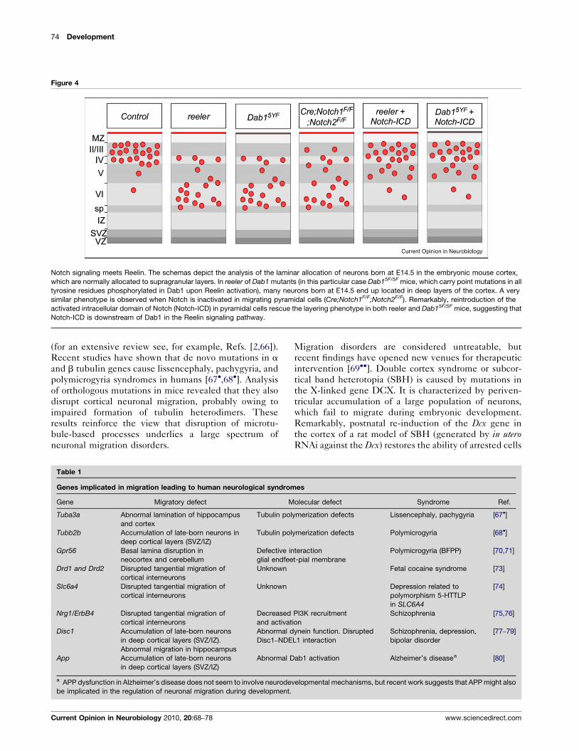
Notch signaling meets Reelin. The schemas depict the analysis of the laminar allocation of neurons born at E14.5 in the embryonic mouse cortex,
which are normally allocated to supragranular layers. In reeler of Dab1 mutants (in this particular case Dab15F/5F mice, which carry point mutations in all
tyrosine residues phosphorylated in Dab1 upon Reelin activation), many neurons born at E14.5 end up located in deep layers of the cortex. A very
similar phenotype is observed when Notch is inactivated in migrating pyramidal cells (Cre;Notch1F/F;Notch2F/F). Remarkably, reintroduction of the
activated intracellular domain of Notch (Notch-ICD) in pyramidal cells rescue the layering phenotype in both reeler and Dab15F/5F mice, suggesting that
Notch-ICD is downstream of Dab1 in the Reelin signaling pathway.
(for an extensive review see, for example, Refs. [2,66]).
Recent studies have shown that de novo mutations in a
and b tubulin genes cause lissencephaly, pachygyria, and
polymicrogyria syndromes in humans [67�,68�]. Analysis
of orthologous mutations in mice revealed that they also
disrupt cortical neuronal migration, probably owing to
impaired formation of tubulin heterodimers. These
results reinforce the view that disruption of microtu-
bule-based processes underlies a large spectrum of
neuronal migration disorders.
Table 1
Genes implicated in migration leading to human neurological syndrom
Gene Migratory defect M
Tuba3a Abnormal lamination of hippocampus
and cortex
Tubulin pol
Tubb2b Accumulation of late-born neurons in
deep cortical layers (SVZ/IZ)
Tubulin pol
Gpr56 Basal lamina disruption in
neocortex and cerebellum
Defective in
glial endfee
Drd1 and Drd2 Disrupted tangential migration of
cortical interneurons
Unknown
Slc6a4 Disrupted tangential migration of
cortical interneurons
Unknown
Nrg1/ErbB4 Disrupted tangential migration of
cortical interneurons
Decreased
and activat
Disc1 Accumulation of late-born neurons
in deep cortical layers (SVZ/IZ).
Abnormal migration in hippocampus
Abnormal d
Disc1–NDE
App Accumulation of late-born neurons
in deep cortical layers (SVZ/IZ)
Abnormal D
a APP dysfunction in Alzheimer’s disease does not seem to involve neurodev
be implicated in the regulation of neuronal migration during development.
Current Opinion in Neurobiology 2010, 20:68–78
Migration disorders are considered untreatable, but
recent findings have opened new venues for therapeutic
intervention [69��]. Double cortex syndrome or subcor-
tical band heterotopia (SBH) is caused by mutations in
the X-linked gene DCX. It is characterized by periven-
tricular accumulation of a large population of neurons,
which fail to migrate during embryonic development.
Remarkably, postnatal re-induction of the Dcx gene in
the cortex of a rat model of SBH (generated by in uteroRNAi against the Dcx) restores the ability of arrested cells
es
olecular defect Syndrome Ref.
ymerization defects Lissencephaly, pachygyria [67�]
ymerization defects Polymicrogyria [68�]
teraction
t-pial membrane
Polymicrogyria (BFPP) [70,71]
Fetal cocaine syndrome [73]
Depression related to
polymorphism 5-HTTLP
in SLC6A4
[74]
PI3K recruitment
ion
Schizophrenia [75,76]
ynein function. Disrupted
L1 interaction
Schizophrenia, depression,
bipolar disorder
[77–79]
ab1 activation Alzheimer’s diseasea [80]
elopmental mechanisms, but recent work suggests that APP might also
www.sciencedirect.com

Neuronal migration mechanisms in development and disease Valiente and Marın 75
to migrate into their proper layers in the cortex. Moreover,
this intervention rescues the phenotypic deficits linked to
this model of SBH, including the epilepsy that is also
associated to the human disorder [69��].
Mutations in GPR56, an orphan G-protein-coupled re-
ceptor (GPCR), cause another type of polymicrogyria
(BFPP), a severe disorder characterized by mental retar-
dation and seizures. Analysis of the cerebellum of GPR56
mouse mutants has revealed major morphological
abnormalities [70], reminiscent of those described in
human patients. Intriguingly, GPR56 seems to be respon-
sible for the regulation of adhesion to the basal lamina
both in the cerebral cortex and cerebellum [70,71],
suggesting a crucial role in the maintenance of the pial
basement membrane and morphogenesis.
Multiple lines of evidence suggest that a loss of excitatory/
inhibitory balance in specific brain circuitries may be
responsible for some of the symptoms observed in several
psychiatric disorders, such as schizophrenia, anxiety, or
depression [72]. Defects in the migration of cortical inter-
neurons are a likely source of abnormal inhibitory tone in
the developing cortex, and several neurotransmitters
linked to human pathologies have been shown to influence
this process. For example, dopamine receptor activation
influences interneuron migration [73]. Similarly, mouse
mutants for the serotonin transporter gene Slc6a4, which
in humans has been associated with an increased risk of
depression, have defects in the distribution of cortical
interneurons. These mice have high levels of extracellular
serotonin, and experimental evidence suggests that sero-
tonin activation of 5-HT6 receptors in cortical interneurons
decreased their migration [74].
ConclusionsOur knowledge of the mechanisms underlying the cell
biology of migration is rapidly expanding, in particular in
relation to nucleokinesis. There are, however, important
questions that remain to be addressed. For example, we
know very little about the mechanisms coordinating the
guidance of the leading process with the movement of the
nucleus. We also ignore the mechanisms that allow the
coordinated allocation of different classes of neurons to
the same region of the brain, as it is the case for the
cerebral cortex. Finally, several genes linked to important
neurological and neuropsychiatric disorders, such as
NRG1 [75,76], DISC1 [77–79], or the b-amyloid precursor
protein APP [80], are required for neuronal migration, but
we still ignore the relevance of these findings in the
disease process. Detailed analysis of all these questions
should expand our knowledge of this central process in
the development of the nervous system.
AcknowledgementsWe are grateful to members of the Marın, Rico, and Borrell labs for helpfuldiscussions and comments. We apologize to colleagues whose work is not
www.sciencedirect.com
cited in this review. Regrettably, space was too limited to cover more than afew selected topics and to cite all significant original articles. Work in ourlaboratory is supported by grants from Spanish Government SAF2008-00770, CONSOLIDER CSD2007-00023, and the EURYI scheme award(see www.esf.org/euryi) to OM. MV was supported by a fellowship from theGeneralitat Valenciana CTBPRA/2005/021.
References and recommended readingPapers of particular interest, published within the annual period ofreview, have been highlighted as:
� of special interest�� of outstanding interest
1. Kriegstein AR, Noctor SC: Patterns of neuronal migration in theembryonic cortex. Trends Neurosci 2004, 27:392-399.
2. Francis F, Meyer G, Fallet-Bianco C, Moreno S, Kappeler C,Socorro AC, Tuy FP, Beldjord C, Chelly J: Human disorders ofcortical development: from past to present. Eur J Neurosci2006, 23:877-893.
3. LoTurco JJ, Bai J: The multipolar stage and disruptions inneuronal migration. Trends Neurosci 2006, 29:407-413.
4. Chedotal A, Rijli F: Transcriptional regulation of tangentialneuronal migration in the developing forebrain. Curr OpinNeurobiol 2009, 19:139-145.
5. Nobrega-Pereira S, Marın O: Transcriptional control of neuronalmigration in the developing mouse brain. Cereb Cortex 2009,19(Suppl. 1):i107-i113.
6. Rakic P: Mode of cell migration to the superficial layers of fetalmonkey neocortex. J Comp Neurol 1972, 145:61-83.
7. Marın O, Rubenstein JLR: A long, remarkable journey:tangential migration in the telencephalon. Nat Rev Neurosci2001, 2:780-790.
8. Watanabe H, Murakami F: Real time analysis of pontine neuronsduring initial stages of nucleogenesis. Neurosci Res 2009,64:20-29.
9. Hao MM, Anderson RB, Kobayashi K, Whitington PM, Young HM:The migratory behavior of immature enteric neurons. DevNeurobiol 2009, 69:22-35.
10. Ward ME, Jiang H, Rao Y: Regulated formation and selection ofneuronal processes underlie directional guidance of neuronalmigration. Mol Cell Neurosci 2005, 30:378-387.
11.�
Martini FJ, Valiente M, Lopez Bendito G, Szabo G, Moya F,Valdeolmillos M, Marın O: Biased selection of leading processbranches mediates chemotaxis during tangential neuronalmigration. Development 2009, 136:41-50.
In this manuscript, the authors demonstrate that tangentially migratinginterneurons (and probably many other cell types throughout the nervoussystem) continuously branch their leading processes as part of theirmigratory cycle. They also show that interneurons use binary choicesof leading process branches instead of leading process steering toachieve directional migration.
12.�
Kumada T, Jiang Y, Kawanami A, Cameron DB, Komuro H:Autonomous turning of cerebellar granule cells in vitro byintrinsic programs. Dev Biol 2009, 326:237-249.
This study shows that intrinsic programs may dictate the behavior ofmigrating neurons in the absence of external cues, and that the laterinfluence migration by modulating Ca2+ influx and cAMP levels.
13. Lin AC, Holt CE: Local translation and directional steering inaxons. EMBO J 2007, 26:3729-3736.
14. Marın O, Valdeolmillos M, Moya F: Neurons in motion: sameprinciples for different shapes? Trends Neurosci 2006,29:655-661.
15.��
Elias LA, Wang DD, Kriegstein AR: Gap junction adhesion isnecessary for radial migration in the neocortex. Nature 2007,448:901-907.
This paper reveals that the adhesive properties of Gap junctions mediatethe dynamic interaction between pyramidal neurons and radial glial fibersduring glial-guided migration. RNAi experiments suggest that both Cx26and Cx43 seem to be required for pyramidal cell migration. The precise
Current Opinion in Neurobiology 2010, 20:68–78

76 Development
mechanisms through which connexins interact with the cytoskeletonremain to be elucidated, although the authors suggest that signalingthrough the carboxy-terminal domains of connexins is not required in thisprocess. This later result is in conflict with reports from a differentlaboratory (see annotation in Ref. [44�]).
16.��
Heng JI, Nguyen L, Castro DS, Zimmer C, Wildner H, Armant O,Skowronska-Krawczyk D, Bedogni F, Matter JM, Hevner R et al.:Neurogenin 2 controls cortical neuron migration throughregulation of rnd2. Nature 2008, 455:114-118.
Besides providing the first example of the transcriptional regulation of thespatio-temporal expression of a small GTP-binding protein, this paperdemonstrates that Rnd2 is required for the radial migration of corticalpyramidal cells.
17. Shim SY, Wang J, Asada N, Neumayer G, Tran HC, Ishiguro K,Sanada K, Nakatani Y, Nguyen MD: Protein 600 is a microtubule/endoplasmic reticulum-associated protein in CNS neurons. JNeurosci 2008, 28:3604-3614.
18. Solecki DJ, Model L, Gaetz J, Kapoor TM, Hatten ME: Par6asignaling controls glial-guided neuronal migration. NatNeurosci 2004, 7:1195-1203.
19.��
Tsai JW, Bremner KH, Vallee RB: Dual subcellular roles for LIS1and dynein in radial neuronal migration in live brain tissue. NatNeurosci 2007, 10:970-979.
The authors provide evidence that LIS1 and dynein are both required forthe movement of the centrosome and the nucleus, and that their functionin both processes is independent. Their experiments also suggest thatmyosin contributes to nuclear movement, but is dispensable for centro-some migration. This later finding is in conflict with recent experimentsfrom the Hatten laboratory (see annotation in Ref. [32�]).
20. Baudoin JP, Alvarez C, Gaspar P, Metin C: Nocodazole-inducedchanges in microtubule dynamics impair the morphology anddirectionality of migrating medial ganglionic eminence cells.Dev Neurosci 2008, 30:132-143.
21.�
Umeshima H, Hirano T, Kengaku M: Microtubule-based nuclearmovement occurs independently of centrosome positioningin migrating neurons. Proc Natl Acad Sci U S A 2007,104:16182-16187.
This manuscript dissociates microtubule pulling forces from the centro-some, by arguing that most microtubules that form the perinuclear cagedo not converge at the centrosome but rather extend into the leadingprocess.
22. Collin L, Schlessinger K, Hall A: APC nuclear membraneassociation and microtubule polarity. Biol Cell 2008,100:243-252.
23. Tsai LH, Gleeson JG: Nucleokinesis in neuronal migration.Neuron 2005, 46:383-388.
24. Higginbotham HR, Gleeson JG: The centrosome in neuronaldevelopment. Trends Neurosci 2007, 30:276-283.
25.�
Sapir T, Sapoznik S, Levy T, Finkelshtein D, Shmueli A, Timm T,Mandelkow EM, Reiner O: Accurate balance of the polaritykinase MARK2/Par-1 is required for proper cortical neuronalmigration. J Neurosci 2008, 28:5710-5720.
The authors show that reducing or increasing MARK2 levels disruptsmultipolar to bipolar transition of migrating pyramidal neurons in a kinase-independent manner, probably owing to altered microtubule dynamics.MARK2 kinase function is subsequently required for the proper migrationof pyramidal cells into the cortical plate.
26.�
Sapir T, Shmueli A, Levy T, Timm T, Elbaum M, Mandelkow EM,Reiner O: Antagonistic effects of doublecortin and MARK2/Par-1 in the developing cerebral cortex. J Neurosci 2008,28:13008-13013.
This paper suggests that MARK2 and DCX have opposite functions inregulating microtubule stability. Thus, while MARK2 function seems todestabilize microtubules, DCX reduces microtubule dynamics. Knock-down of both proteins have different effects in centrosome movement:while loss of MARK2 prevents centrosome motility, reduction of DCXlevels leads to rapid, disorganized movement of this structure.
27.�
Asada N, Sanada K, Fukada Y: LKB1 regulates neuronalmigration and neuronal differentiation in the developingneocortex through centrosomal positioning. J Neurosci 2007,27:11769-11775.
Using RNAi experiments, the authors show that LKB1 is required for theradial migration of pyramidal neurons. Thus, besides its role in establish-
Current Opinion in Neurobiology 2010, 20:68–78
ing cell polarity in these cells (see Ref. [28] for an elegant analysis of thefunction of LKB1 in this context), LKB1 seem to participate in themolecular machinery required for centrosome–nucleus coupling duringmigration.
28. Barnes AP, Lilley BN, Pan YA, Plummer LJ, Powell AW, Raines AN,Sanes JR, Polleux F: LKB1 and SAD kinases define a pathwayrequired for the polarization of cortical neurons. Cell 2007,129:549-563.
29. Shelly M, Cancedda L, Heilshorn S, Sumbre G, Poo MM: Lkb1/strad promotes axon initiation during neuronal polarization.Cell 2007, 129:565-577.
30. Bellion A, Baudoin JP, Alvarez C, Bornens M, Metin C:Nucleokinesis in tangentially migrating neurons comprisestwo alternating phases: Forward migration of the Golgi/centrosome associated with centrosome splitting and myosincontraction at the rear. J Neurosci 2005, 25:5691-5699.
31. Schaar BT, McConnell SK: Cytoskeletal coordination duringneuronal migration. Proc Natl Acad Sci U S A 2005,102:13652-13657.
32.�
Solecki DJ, Trivedi N, Govek EE, Kerekes R, Gleason S,Hatten ME: Myosin II motors and F-Actin dynamics drive thecoordinated movement of the centrosome and soma duringCNS glial-guided neuronal migration. Neuron 2009, 63:63-80.
Experimental evidence suggests that actomyosin forces are required forthe forward movement of the centrosome. This result disputes previousfindings on the role of myosin in this process (see annotation in Ref.[19��]).
33.��
Renaud J, Kerjan G, Sumita I, Zagar Y, Georget V, Kim D,Fouquet C, Suda K, Sanbo M, Suto F et al.: Plexin-A2 and itsligand, Sema6a, control nucleus–centrosome coupling inmigrating granule cells. Nat Neurosci 2008, 11:440-449.
This is the first example of the regulation of centrosome–nucleus couplingtriggered by receptor signaling in response to a guidance cue.
34. Lauffenburger DA, Horwitz AF: Cell migration: a physicallyintegrated molecular process. Cell 1996, 84:359-369.
35. Hatten ME: Central nervous system neuronal migration. AnnuRev Neurosci 1999, 22:511-539.
36. Anton ES, Kreidberg JA, Rakic P: Distinct functions of a3 anda(v) integrin receptors in neuronal migration and laminarorganization of the cerebral cortex. Neuron 1999, 22:277-289.
37. Graus-Porta D, Blaess S, Senften M, Littlewood-Evans A,Damsky C, Huang Z, Orban P, Klein R, Schittny JC, Muller U: b1-Class integrins regulate the development of laminaeand folia in the cerebral and cerebellar cortex. Neuron 2001,31:367-379.
38. Halfter W, Dong S, Yip YP, Willem M, Mayer U: A critical functionof the pial basement membrane in cortical histogenesis. JNeurosci 2002, 22:6029-6040.
39. Belvindrah R, Graus-Porta D, Goebbels S, Nave KA, Muller U: b1Integrins in radial glia but not in migrating neurons areessential for the formation of cell layers in the cerebral cortex.J Neurosci 2007, 27:13854-13865.
40. Schmid RS, Shelton S, Stanco A, Yokota Y, Kreidberg JA,Anton ES: a3b1 Integrin modulates neuronal migration andplacement during early stages of cerebral corticaldevelopment. Development 2004, 131:6023-6031.
41. Chen ZL, Haegeli V, Yu H, Strickland S: Cortical deficiency oflaminin g1 impairs the AKT/GSK-3b signaling pathway andleads to defects in neurite outgrowth and neuronal migration.Dev Biol 2009, 327:158-168.
42. Stanco A, Szekeres C, Patel N, Rao S, Campbell K, Kreidberg JA,Polleux F, Anton ES: Netrin-1-a3b1 integrin interactionsregulate the migration of interneurons through the corticalmarginal zone. Proc Natl Acad Sci U S A 2009, 106:7595-7600.
43. Cina C, Bechberger JF, Ozog MA, Naus CC: Expression ofconnexins in embryonic mouse neocortical development. JComp Neurol 2007, 504:298-313.
44.�
Cina C, Maass K, Theis M, Willecke K, Bechberger JF, Naus CC:Involvement of the cytoplasmic c-terminal domain of
www.sciencedirect.com

Neuronal migration mechanisms in development and disease Valiente and Marın 77
Connexin43 in neuronal migration. J Neurosci 2009,29:2009-2021.
This paper provides genetic support to the idea that connexin-basedadhesions mediate the association between pyramidal cells and radialglial cells during glial-guided migration. In addition, the authors provideevidence that the C-terminal domain of Cx43 is required for neuronalmigration, which contradicts previous findings (see annotation in Ref.[15��]).
45. D’Arcangelo G, Miao GG, Chen SC, Soares HD, Morgan JI,Curran T: A protein related to extracellular matrix proteinsdeleted in the mouse mutant reeler. Nature 1995, 374:719-723.
46. Soriano E, Del Rıo JA: The cells of Cajal-Retzius: still a mysteryone century after. Neuron 2005, 46:389-394.
47. Rice DS, Curran T: Role of the Reelin signaling pathway incentral nervous system development. Annu Rev Neurosci 2001,24:1005-1039.
48. Howell BW, Herrick TM, Hildebrand JD, Zhang Y, Cooper JA:Dab1 tyrosine phosphorylation sites relay positional signalsduring mouse brain development. Curr Biol 2000, 10:877-885.
49.�
Hack I, Hellwig S, Junghans D, Brunne B, Bock HH, Zhao S,Frotscher M: Divergent roles of ApoER2 and Vldlr in themigration of cortical neurons. Development 2007,134:3883-3891.
Detailed analysis of the developing cortex in ApoER2 and Vldlr mousemutants reveal that both Reelin receptors may have partially not over-lapping functions.
50. Feng L, Cooper JA: Dual functions of Dab1 during braindevelopment. Mol Cell Biol 2009, 29:324-332.
51. Sanada K, Gupta A, Tsai LH: Disabled-1-regulated adhesion ofmigrating neurons to radial glial fiber contributes to neuronalpositioning during early corticogenesis. Neuron 2004,42:197-211.
52.�
Park TJ, Curran T: Crk and Crk-like play essential overlappingroles downstream of Disabled-1 in the Reelin pathway. JNeurosci 2008, 28:13551-13562.
This paper provides genetic support to the function of the adaptorproteins Crk and CrkL downstream of Dab1 and upstream of C3G andAkt phosphorylation in the Reelin signaling pathway.
53. Voss AK, Britto J, Dixon MP, Sheikh BN, Collin C, Tan SS,Thomas T: C3g regulates cortical neuron migration, preplatesplitting and radial glial cell attachment. Development 2008,135:2139.
54.��
Hashimoto-Torii K, Torii M, Sarkisian MR, Bartley CM, Shen J,Radtke F, Gridley T, Sestan N, Rakic P: Interaction betweenReelin and Notch signaling regulates neuronal migration in thecerebral cortex. Neuron 2008, 60:273-284.
This elegant paper presents evidence that links the Reeling signalingpathway with Notch signaling in migrating pyramidal cells. The authorsdemonstrate that Notch-ICD is cell-autonomously required by pyramidalcells for radial migration. Moreover, Notch-ICD levels are reduced inreeler, and re-expression of Notch-ICD in reeler mutant neurons rescuethe migration phenotype. It is suggested that phosphorylated Dab1blocks the degradation of Notch-ICD, although at this point it is unclearhow regulation of Dab1 (whose degradation is also induced by Reelin, seeRef. [57��]) influences this process.
55. Arnaud L, Ballif BA, Cooper JA: Regulation of protein tyrosinekinase signaling by substrate degradation during braindevelopment. Mol Cell Biol 2003, 23:9293-9302.
56. Bock HH, Jossin Y, May P, Bergner O, Herz J: Apolipoprotein Ereceptors are required for Reelin-induced proteasomaldegradation of the neuronal adaptor protein Disabled-1. J BiolChem 2004, 279:33471-33479.
57.��
Feng L, Allen NS, Simo S, Cooper JA: Cullin 5 regulates Dab1protein levels and neuron positioning during corticaldevelopment. Genes Dev 2007, 21:2717-2730.
Reelin induces the degradation of Dab1 as part of a negative feedbackloop in the signaling pathway. This papers shows that this process ismediated by the E3 ubiquitin ligase component Cullin 5 (Cul5). Thisprotein binds to phosphorylated Dab1 and targets it for degradation.Loss of Cul5 in migrating neurons causes an accumulation of active Dab1protein and defective cortical layering, with many neurons accumulatingat the top of the cortical plate and even in the marginal zone. The results
www.sciencedirect.com
demonstrate that downregulation of Dab1 is important for proper corticaldevelopment.
58.�
Sibbe M, Forster E, Basak O, Taylor V, Frotscher M: Reelin andNotch1 cooperate in the development of the dentate gyrus. JNeurosci 2009, 29:8578-8585.
Along with Ref. [54��], this study shows that Reelin signaling interacts withNotch in controlling neuronal positioning in the developing cerebralcortex, in this case in the dentate gyrus.
59. D’Arcangelo G, Nakajima K, Miyata T, Ogawa M, Mikoshiba K,Curran T: Reelin is a secreted glycoprotein recognized by theCR-50 monoclonal antibody. J Neurosci 1997, 17:23-31.
60. Crowner D, Le Gall M, Gates MA, Giniger E: Notch steersdrosophila ISNB motor axons by regulating the Abl signalingpathway. Curr Biol 2003, 13:967-972.
61. Cobos I, Borello U, Rubenstein JLR: Dlx transcription factorspromote migration through repression of axon and dendritegrowth. Neuron 2007, 54:873-888.
62. Yokota Y, Ring C, Cheung R, Pevny L, Anton ES: Nap1-regulatedneuronal cytoskeletal dynamics is essential for the finaldifferentiation of neurons in cerebral cortex. Neuron 2007,54:429-445.
63. Lujan R, Shigemoto R, Lopez-Bendito G: Glutamate and GABAreceptor signalling in the developing brain. Neuroscience 2005,130:567-580.
64.��
Bortone D, Polleux F: KCC2 expression promotes thetermination of cortical interneuron migration in avoltage-sensitive calcium-dependent manner. Neuron 2009,62:53-71.
This paper shows that cortical interneurons terminate migration uponexpression of KCC2, which induces cell hyperpolarization and blockingCa2+ influx.
65. Lopez-Bendito G, Sanchez-Alcaniz JA, Pla R, Borrell V, Pico E,Valdeolmillos M, Marin O: Chemokine signaling controlsintracortical migration and final distribution of GABAergicinterneurons. J Neurosci 2008, 28:1613-1624.
66. Kerjan G, Gleeson JG: Genetic mechanisms underlyingabnormal neuronal migration in classical lissencephaly.Trends Genet 2007, 23:623-630.
67.�
Keays DA, Tian G, Poirier K, Huang GJ, Siebold C, Cleak J,Oliver PL, Fray M, Harvey RJ, Molnar Z et al.: Mutations ina-tubulin cause abnormal neuronal migration in mice andlissencephaly in humans. Cell 2007, 128:45-57.
See annotation to Ref. [68�].
68.�
Jaglin XH, Poirier K, Saillour Y, Buhler E, Tian G, Bahi-Buisson N,Fallet-Bianco C, Phan-Dinh-Tuy F, Kong XP, Bomont P et al.:Mutations in the b-tubulin gene TUBB2B result inasymmetrical polymicrogyria. Nat Genet 2009, 41:746-752.
Along with Ref. [67�], this paper shows that mutations in tubulin genesimpair neuronal migration in mice and cause neurological disorders inhumans.
69.��
Manent JB, Wang Y, Chang Y, Paramasivam M, LoTurco JJ: Dcxreexpression reduces subcortical band heterotopia andseizure threshold in an animal model of neuronal migrationdisorder. Nat Med 2009, 15:84-90.
This remarkable study shows that postnatal expression of Dcx canreactivate the migration of neurons whose movement was arrested duringembryogenesis in a rat model subcortical band heterotopia. Theseexperiments suggest potential therapies for otherwise untreatable neu-rological disorders.
70. Koirala S, Jin Z, Piao X, Corfas G: GPR56-regulated granule celladhesion is essential for rostral cerebellar development. JNeurosci 2009, 29:7439-7449.
71. Li S, Jin Z, Koirala S, Bu L, Xu L, Hynes RO, Walsh CA, Corfas G,Piao X: GPR56 regulates pial basement membrane integrityand cortical lamination. J Neurosci 2008, 28:5817-5826.
72. Di Cristo G: Development of cortical GABAergic circuits and itsimplications for neurodevelopmental disorders. Clin Genet2007, 72:1-8.
73. Crandall JE, McCarthy DM, Araki KY, Sims JR, Ren JQ, Bhide PG:Dopamine receptor activation modulates GABA neuron
Current Opinion in Neurobiology 2010, 20:68–78

78 Development
migration from the basal forebrain to the cerebral cortex. JNeurosci 2007, 27:3813-3822.
74. Riccio O, Potter GB, Walzer C, Vallet P, Szabo G, Vutskits L, Kiss J,Dayer A: Excess of serotonin affects embryonic interneuronmigration through activation of the serotonin receptor 6. MolPsychiatry 2009, 14:280-290.
75. Flames N, Long JE, Garratt AN, Fischer TM, Gassmann M,Birchmeier C, Lai C, Rubenstein JLR, Marın O: Short- and long-range attraction of cortical GABAergic interneurons byNeuregulin-1. Neuron 2004, 44:251-261.
76. Gambarotta G, Garzotto D, Destro E, Mautino B, Giampietro C,Cutrupi S, Dati C, Cattaneo E, Fasolo A, Perroteau I: ErbB4expression in neural progenitor cells (st14a) is necessary tomediate neuregulin-1b1-induced migration. J Biol Chem 2004,279:48808-48816.
Current Opinion in Neurobiology 2010, 20:68–78
77. Kamiya A, Kubo K-i, Tomoda T, Takaki M, Youn R, Ozeki Y,Sawamura N, Park U, Kudo C, Okawa M et al.: A schizophrenia-associated mutation of Disc1 perturbs cerebral cortexdevelopment. Nat Cell Biol 2005, 7:1167-1178.
78. Duan X, Chang JH, Ge S, Faulkner RL, Kim JY, Kitabatake Y,Liu XB, Yang CH, Jordan JD, Ma DK et al.: Disrupted-in-schizophrenia 1 regulates integration of newly generatedneurons in the adult brain. Cell 2007, 130:1146-1158.
79. Meyer KD, Morris JA: Disc1 regulates granule cell migrationin the developing hippocampus. Hum Mol Genet 2009,18:3286-3297.
80. Young-Pearse TL, Bai J, Chang R, Zheng JB, LoTurco JJ,Selkoe DJ: A critical function for b-amyloid precursor protein inneuronal migration revealed by in utero RNA interference. JNeurosci 2007, 27:14459-14469.
www.sciencedirect.com