neurological. common neurological disorders in children hydrocephalus neural tubes defects bacterial...
TRANSCRIPT
Neurological
Common Neurological Disorders in Children
HydrocephalusNeural Tubes DefectsBacterial MeningitisGuillain-Barre SyndromeReye’s SyndromeSeizures
Structural Defects
Hydrocephalus“Water on the Brain”Imbalance between the production and absorption of CSF. Often from congenital CNS malformationsResults in rapid head enlargement in infantsCan lead to irreversible neurological damage

INFANT Hydrocephalus
Early Signs
Projectile vomiting not associated with feedingScalp veins become prominent**Shrill, high pitched cry**Increasing irritability
Infant Hydrocephalus Later signs & symptoms
•Bulging anterior fontanel and a head circumference that increases at an abnormal rate
•Enlargement of the forehead
•Depressed eyes rotated downward: “sunset eyes” (pupils sink downward)
Hydrocephalus Signs & symptoms
in older Child
No enlargement of head (skull is closed)Begins with generalized neuro symptoms
HA in morningNauseaVomiting
Followed by signs of increased ICP
Relief of hydrocephalus
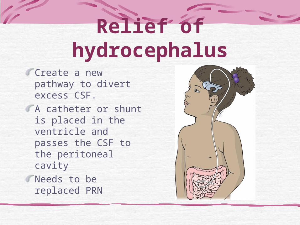
Create a new pathway to divert excess CSF. A catheter or shunt is placed in the ventricle and passes the CSF to the peritoneal cavityNeeds to be replaced PRN
Post-op Nursing Care Shunt Placement
Keep child flat unless ICP is present the bed slightly elevatedSlowly increase HOB over few daysSupport head when moving childPain managementVital Signs
Post-op Nursing Care Shunt Placement
Observe for signs of increasing ICP – neurologic assessmentObserve for abdominal distentionStrict I & O AntibioticsMeticulous skin careSupport family
Discharge Management Post Shunt Placement
Teach parents to monitor for shunt complications:
Headache, progressive or worseningDrowsiness or inappropriate sleepiness during the day, irritability Nausea, vomiting Personality changes or changes in school performance Fever Redness or swelling along the shunt tract

Neural Tube Disorders
Defects of closure of neural tube during fetal developmentCongenital (present at birth)Believed to be caused by genetic or environmental factors, but exact etiology is unknown
Common in women with poor folic acid intake before and during pregnancy
Nursing ImplicationsAdvise all women to adhere to routine screening/diagnostic testingAdvise all women capable of becoming pregnant to consume
0.4 mg of folic acid daily

Neural Tube Disorders
Types:Spina Bifida
OccultaCystica
MeningoceleMyelomeningocele
Spina BifidaMost common CNS defectCaused by failure of neural tube to close at some point along spinal columnTypes:
spina bifida occulta spina bifida cystica
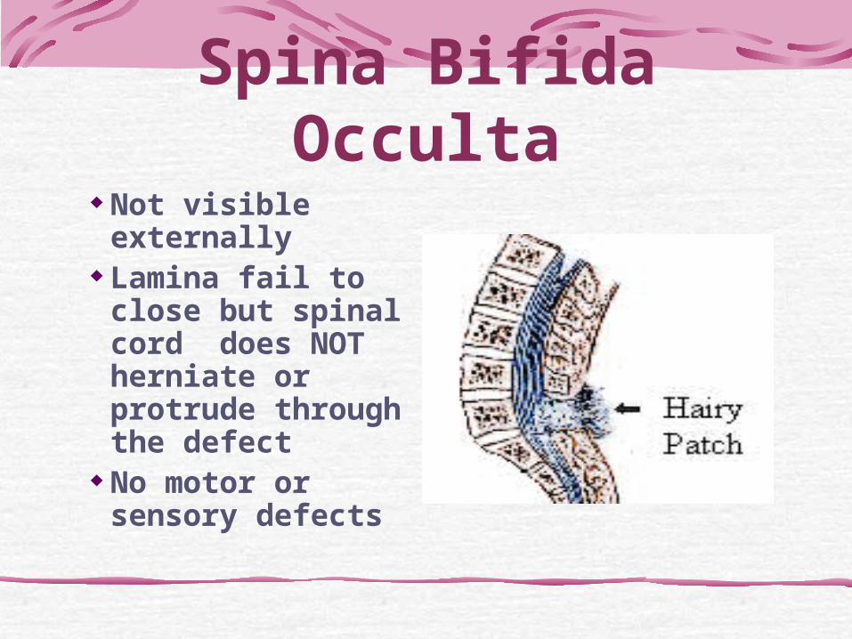
Spina Bifida Occulta
Not visible externally
Lamina fail to close but spinal cord does NOT herniate or protrude through the defect
No motor or sensory defects
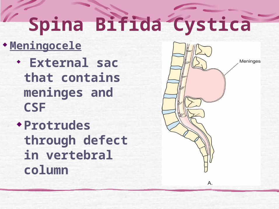
Spina Bifida Cystica Meningocele
External sac that contains meninges and CSF
Protrudes through defect in vertebral column
Meningocele Not associated
with neurologic deficit – good prognosis
Hydrocephalus may be an associated finding, or aggravated after repair
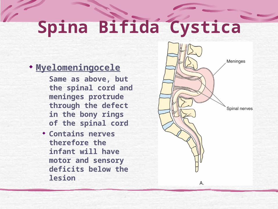
Spina Bifida Cystica
MyelomeningoceleSame as above, but the spinal cord and meninges protrude through the defect in the bony rings of the spinal cord
Contains nerves therefore the infant will have motor and sensory deficits below the lesion
Myelomeningocele
Visible at birth, most often in the lumbaosacral area
Covered with a very fragile thin membrane/sac which can tear easily, allowing CSF to leak out
Nursing Interventions
• Protect the sac from injury• Keep free from infection
• Position: prone or side lying • Cover sac with sterile, moist non-
adherent dressing, sterile technique imperative
• Parents need emotional support & education regarding short and long term needs of infant
Nursing Interventions• Surgical repair usually within first 24 hours
– observe for early signs of infection: elevated temp, irritability, lethargy, nuchal rigidity
– observe for signs of increasing ICP (may indicate hydrocephalus)
Habilitation
Emphasizes constructive use of ‘normal’ parts of body & minimizes the disabilities making the child as self-helpful as is possible in the activities of daily livingMajor problems: incontinence, constipation, obesity or malnutrition
Neurologic Infections
Meningitis
•Acute inflammation of the cerebral meninges as a result of a bacterial or viral infection
Bacterial MeningitisFollows 2-3 days of upper respiratory infection •Haemophilus influenzae type b was the most common cause of bacterial meningitis in children prior to the use of the Hib conjugate vaccine
• May be caused by Strep pneumoniae in child < 24months (not fully vaccinated yet)
• Meningococcal predominantly in school-age children & adolescents (vaccine preventable)
Bacterial MeningitisSigns & SymptomsAbrupt onset of feverChillsIncreasing irritabilityHeadacheConvulsionsBlurred visionCranial nerve paralysisOpisthotonic position
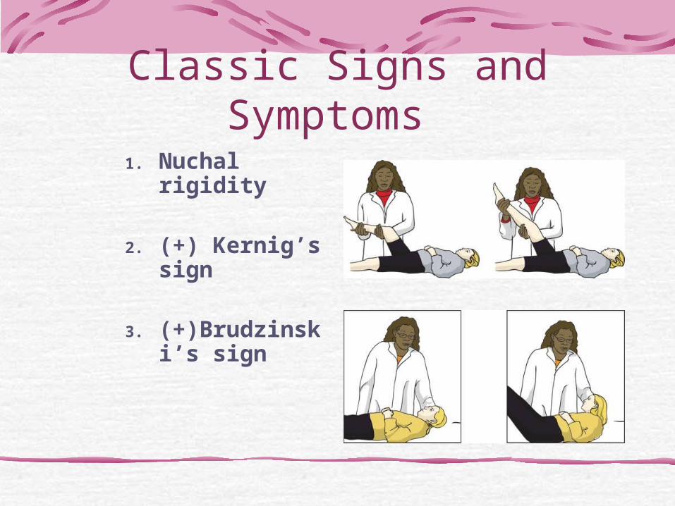
Classic Signs and Symptoms
1. Nuchal rigidity
2. (+) Kernig’s sign
3. (+)Brudzinski’s sign
Signs & Symptomsin Newborn
Above slide’s signs plus:• poor suck• weak cry• lethargy• can lead to sudden shock, seizures, apnea
• bulging fontanel
Bacterial MeningitisDiagnosis:
lumbar puncture to analyze CSF increased WBC’s and protein; decreased glucose (bacteria
feed on glucose)
Treatment: ABX x 10 days, IV or Intrathecal Respiratory isolation x 24 hours while on ABX Maintenance of optimum hydration Maintenance of ventilation Reduction of increased ICP Management of bacterial shock Control of seizures Prophylactic ABX for family members

ComplicationsLife Threatening ConditionIf Survival:
Hearing lossBlindnessParesisIntellectual impairment
Nursing ManagementFrequently assess vital signs, LOC, neurologic assessment to identify changes in the child’s condition.Measure head circumference frequently- risk for hydrocephalus.Monitor intake and output
Nursing ManagementPromote comfort • reduced stimulation (dim lights, quiet
room) Side-lying positionIdentify parents’ concerns, provide supportPrevention is a major role for nurses.
Encourage parents to get their infants and children fully immunized!
Guillain-Barre Syndrome
• Immune-mediated disease of motor weakness that is often associated with viral or bacterial infection of respiratory or GI tract or vaccine administration
• Adults have increased susceptibility, can affect children usually ages 4-10
• Inflammation of nerve fibers, impairs nerve conduction though demyleination
• Ascending paralysis from lower extremities
Initial SymptomsPeripheral neuritis occurs several days after primary infectionMuscle tendernessTendon reflexes decreased or absentParesthesia & crampsProximal symmetric muscle weaknessUrinary incontinence or retentionDecreased swallowing & respiratory efforts-may lead to respiratory failure
TreatmentWait for disease to stabilize Intravenous immune globulin IVIG Physical TherapyRarely fatal, often residual paralysis

Nursing CareMonitoring respiratory statusManaging autonomic nervous system dysfunctionPreventing complications associated with immobilityProviding emotional supporttTeaching the parents how tocare for the child after discharge.
Reye’s Syndrome A life threatening acute encephalitisOccurs after viral infection if tx’d w/ aspirinHigh Mortality RateEducation efforts has helped to reduce incidence (use Tylenol or Ibuprofen not ASA)
Reye’s Syndrome
Begins with mild viral infection that worsens w/i 24-48 hours
LethargyVomitingFollowed by
AgitationAnorexiaCombativenessConfusion leading to stupor, coma, seizures, respiratory arrest
Reye’s SyndromeLabs
Increased:Liver enzymesSerum ammoniaPT, PTTWBC
Decreased:Serum glucose

Reye’s SyndromeNursing Care
If Survival in PICUMonitor:
Neurological statusRespiratory effortHypoglycemiaCerebral edema
Seizures
SeizuresInvoluntary contraction of muscle caused by abnormal electrical brain impulsesThey are episodic and abruptOften triggered by environmental of physiological stimuli Exact location of the electrical foci and the number of brain cells involved determines the nature of the seizure (sterotypical)
Types of SeizuresNonrecurrent – Acute
Febrile episodesDrugsMetabolic alterations
Recurrent – Chronic (Epilepsy)
Idiopathic (primary) epilepsy
Epilepsy secondary to trauma, hemorrhage, infections, congenital defects
Seizures: 2 categories
PartialSimpleComplex
Only 1 area of brain involvedSymptoms are associated with the area affectedNo LOC or consciousness is impaired
GeneralizedInfantile spasmsFebrileAbsenceTonic Clonic
Entire brainUsually have loss of consciousnessMay have auraPostical
Partial Seizures
Simple Partial Seizures Complex Partial Seizures

Simple Partial Seizures• Seizure is short, lasts < 30 seconds• No loss of consciousness, aura, postical
stateSeizure Activity:
Abnormal motor activity• One extremity or part of extremity, uncontrolled
movement
Abnormal sensory activityNumbness, tingling, paresthesia or pain starting in 1 area of body, may spread to other parts of bodyMay include abnormal auditory, olfactory and visual sensations

Complex Partial SeizuresSeizure is longer, 30 seconds-5 minutesConsciousness is impaired immediatelyMay have slight aura
Seizure Activity:Sudden change in postureAbnormal motor activity, twitching, loss of tone, tingling or numbnessAutomatisms-lip smacking, chewing, suckingCircumoral pallor
Afterward: drowsiness
Generalized SeizuresInfantile spasms
AbsenceTonic Clonic
Febrile
Infantile Spasms
Age: 4 months to 2 years• Occur in clusters 5-150/day,
worse at night• Altered consciousness
Abrupt flexion/extension of neck, trunk, extremitiesEye rollingMay have permanent cognitive & developmental delays
Absence Seizure
• Lasts 5-10 seconds, multiple times a day 50-100 per day
• Seizure is a brief loss of consciousness• Appears to look like a staring spell
Rhythmic blinking & twitching of mouth or armMistaken for daydreaming or behavior problemsInterferes with learning1/3 of children will grow out of them by adolescence
Tonic Clonic(Grand Mal Seizure)4 stages of Seizure1. Prodromal:
Drowsiness, dizziness, malaise, lack of coordination, “not himself”
2. Aura:May precede seizure, reflects portion of brain where seizure originates
Tonic-clonic stage3. Tonic-Clonic:
Tonic: 20 seconds, all muscles cx (rigid), child falls to ground, LOC, respiratory muscles affected, grunting, airway compromisedClonic: 20-30 seconds, jerky muscle contract & relax rapidly, froth or bloody sputum, urinary or bowel incontience
4. Postictal:Appears to relax, semi-conscious, sound sleep for 1-4h, no recollection of event
Acute Febrile SeizureDue to increased temperature > 102 F (but may occur as low as 100 F)Higher fever=higher risk
Occur between 6 months and 5 years, with a peak incidence between 18 and 24 months of ageTonic-clonic patternLasts 15-20 seconds

Epilepsy
• Chronic disorder with recurrent seizures in children 3 and older
• Symptoms depend on type of seizure
• No association with illness, injury
• Seizure may be triggered by something
Epilepsy Management Anticonvulsants-monotherapy is
desired Dosage increased as child grows Control the seizures or reduce
their frequency Discover and correct the cause
when possible, know triggers Help child live a normal life
Epilepsy Management
Instruct parents on importance of giving meds to achieve therapeutic drug levels
Med can be withdrawn when child is seizure free for 2 yrs with normal EEG
TAPER! Gradually decreased over 1-2 weeks
TriggersChanges in dark-light patternsSudden loud noises, specific voicesSudden or startling movementsExtreme changes in temperatureDehydration, fatigueHyperventilationHypoglycemiaCaffeine, insufficient protein in diet
Status Epilepticus(Intractable Seizures)Continuous seizure activity lasting > 30 minutes or a series of seizures from which the child does not regain a premorbid level of consciousness
If A Patient is Admitted for Seizure Activity or has PMH
of SeizuresEnsure IV access and PatencyCheck MD orders for Seizure MedicationCheck that medication is on the UnitCheck Suction at bedside, ambu bag, mask and 02 tubing, SaO2 is available and working!Are Side Rails Padded?ID band- correct? Indicate Seizure Risk?Know how to initiate emergency or rapid response

Nursing Management during Seizures
1. Maintain Patent AirwayPlace nothing in the child’s mouth during
a seizure Loose teeth may be knocked out and aspirated.
Position side so secretions can drainPulse oximetry reading (SpO2) Oxygen for < 95%- use maskSuction prn
Nursing Management during Seizures
Ensure Safety•If OOB gently assist to floor•Bed in lowest position •Stay near child•Protect head from injury
Nursing Management during Seizures
Administer MediationInitial order is intravenous medicationsIV push slowly Benzodiazopene IV push slowly to
avoid apneaDiazepam (Valium) or lorazepam
(Ativan)Followed by anticonvulsant

Nursing Management during Seizures
Observe and RecordType of seizure activityVital SignsTime seizure started and stopped
Dilantin Toxicity: nystagmus, ataxia, decresed mental capacity
Low levels: seizure activity
S/E: gingival hyperplagia (discuss oral hygiene), drowsiness, thrombpcytopenia, leukopenia, increased liver enzymes
Nursing responsibility:Monitor CBC, LFT, therapeutic drug levels
Practice Questions!
A 10-year old is diagnosis is Guillain-Barre Syndrome. It would be imperative for the nurse to inform the physician after observing which of the following?
1. Weak muscle tone in the feet2. Weak muscle tone in the legs3. Increasing hoarseness and cough4. Tingling in the hands
A 4-year-old is being evaluated for hydrocephalus. The nurse notes which of the following as an early sign of hydrocephalus in a child?
1. Bulging fontanels2. Rapid enlargement of the head3. Shrill, high-pitched cry4. Early morning headache
A child with a history of a seizure was admitted 2 hours ago. The history indicates fever, chills, and vomiting for the past 3-4 hours. In report the nurse is told that the child had a positive Brudzinski’s sign. The nurse infers this is most likely caused by:
1. Increased intracranial pressure2. Meningeal irritation3. Encephalitis4. Intraventricular hemorrhage
A nurse is assessing a new admission. The 6-month-old infant displays irritability, bulging fontanels, and setting-sun eyes. The nurse would suspect:
1. Hydrocephelus2. Hypertension3. Skull fracture4. Myelomeningocele
An 8-year-old client with a ventriculoperitoneal shunt was admitted for shunt malfunction. He presents with symptoms of increased intracranial pressure. The mechanism of the development of his symptoms is most probably related to:
1. Increased flow of CSF2. Increased reabsorption of CSF3. Obstructed flow of CSF4. Decreased production of CSF
The nurse is taking a history of a child admitted for EEG testing to determine seizure activity. The parent reports that the child has “odd” behavior, including periods of lip smacking, and muscle twitching . The nurse suspects:
1.Simple Partial Seizures2.Complex Partial Seizures3.Absence Seizures4.Tonic-Clonic Seizures