neuroendocrine tumor of the breast: is it primary or … · 2015-12-29 · by providing a platform...
TRANSCRIPT

CASE REPORT PEER REVIEWED | OPEN ACCESS
www.edoriumjournals.com
International Journal of Case Reports and Images (IJCRI)International Journal of Case Reports and Images (IJCRI) is an international, peer reviewed, monthly, open access, online journal, publishing high-quality, articles in all areas of basic medical sciences and clinical specialties.
Aim of IJCRI is to encourage the publication of new information by providing a platform for reporting of unique, unusual and rare cases which enhance understanding of disease process, its diagnosis, management and clinico-pathologic correlations.
IJCRI publishes Review Articles, Case Series, Case Reports, Case in Images, Clinical Images and Letters to Editor.
Website: www.ijcasereportsandimages.com
Neuroendocrine tumor of the breast: Is it primary or metastatic?
Ornela A. Dervishaj, Alexandra S. Renzi, Romulo Genato, Philip Q. Xiao, Armand P. Asarian
ABSTRACT
Introduction: Neuroendocrine breast carcinoma is a rare form of tumor that may be either primary or metastatic. Case Report: We present the case of a patient who was referred to our outpatient office with the diagnosis of a triple negative invasive breast carcinoma on an ultrasound-guided core needle biopsy. The patient subsequently underwent a lumpectomy with sentinel lymph node biopsy. Postoperative pathology revealed neuroendocrine carcinoma of the breast with sentinel lymph node biopsy negative for carcinoma. The tumor cells were positive for AE1/AE3, chromogranin, synaptophysin and CD-56 but negative for estrogen, progesterone and HER2. To exclude a primary carcinoma elsewhere, we obtained a whole body PET scan which showed a mildly hypermetabolic mesenteric mass in the midline with curvilinear calcification. Computed tomography scan of the abdomen and pelvis with oral and IV contrast showed a mass-like soft tissue within the terminal ileum with thickening of the cecum. An octreotide scan of the whole body revealed abnormal activity in the midline of the lower abdomen 6 hours and 24 hours after administration of octreotide. Colonoscopy showed a nodular friable mass in the ileocecal valve extending from the terminal ileum. Biopsy from the colonoscopy demonstrated a neuroendocrine carcinoma. The patient subsequently underwent a right hemicolectomy with primary anastomosis. Postoperative pathology was concordant with a neuroendocrine carcinoma and 9 of the 20 lymph nodes were positive for carcinoma. We performed a literature review to explore the reported incidence, diagnosis and treatment of this rare tumor metastasizing to the breast. Conclusion: Differentiating between primary and metastatic tumor of the breast represents a challenge, albeit an important one, as the first disease can subject the patient to the morbidities of a mastectomy with axillary node dissection and the other is locally controlled.
(This page in not part of the published article.)

International Journal of Case Reports and Images, Vol. 7 No. 1, January 2016. ISSN – [0976-3198]
Int J Case Rep Images 2016;7(1):34–39. www.ijcasereportsandimages.com
Dervishaj et al. 34
CASE REPORT OPEN ACCESS
Neuroendocrine tumor of the breast: Is it primary or metastatic?
Ornela A. Dervishaj, Alexandra S. Renzi, Romulo Genato, Philip Q. Xiao, Armand P. Asarian
ABSTRACT
Introduction: Neuroendocrine breast carcinoma is a rare form of tumor that may be either primary or metastatic. Case Report: We present the case of a patient who was referred to our outpatient office with the diagnosis of a triple negative invasive breast carcinoma on an ultrasound-guided core needle biopsy. The patient subsequently underwent a lumpectomy with sentinel lymph node biopsy. Postoperative pathology revealed neuroendocrine carcinoma of the breast with sentinel lymph node biopsy negative for carcinoma. The tumor cells were positive for AE1/AE3, chromogranin, synaptophysin and CD-56 but negative for estrogen, progesterone and HER2. To exclude a primary carcinoma elsewhere, we obtained a whole body PET scan which showed a mildly hypermetabolic mesenteric mass in the midline with curvilinear calcification. Computed tomography scan of the abdomen and pelvis with oral and IV contrast showed a mass-like soft tissue within the terminal ileum with thickening of the cecum. An octreotide scan of the whole body revealed abnormal activity
Ornela A. Dervishaj1, Alexandra S. Renzi1, Romulo Genato1, Philip Q. Xiao2, Armand P. Asarian1
Affiliations: 1Department of Surgery, The Brooklyn Hospital Center, Icahn School of Medicine at Mount Sinai, Brooklyn, NY 11201; 2Department of Pathology and Laboratory Medicine, The Brooklyn Hospital Center, Icahn School of Medicine at Mount Sinai, Brooklyn, NY 11201.Corresponding Author: Philip Xiao, MD, FCAP Chairman, Department of Pathology, The Brooklyn Hospital Center, Brooklyn, NY 11201; Ph: (718) 250 8207; Fax: (718) 250 8056; Email: [email protected]
Received: 17 June 2015Accepted: 21 August 2015Published: 01 January 2016
in the midline of the lower abdomen 6 hours and 24 hours after administration of octreotide. Colonoscopy showed a nodular friable mass in the ileocecal valve extending from the terminal ileum. Biopsy from the colonoscopy demonstrated a neuroendocrine carcinoma. The patient subsequently underwent a right hemicolectomy with primary anastomosis. Postoperative pathology was concordant with a neuroendocrine carcinoma and 9 of the 20 lymph nodes were positive for carcinoma. We performed a literature review to explore the reported incidence, diagnosis and treatment of this rare tumor metastasizing to the breast. Conclusion: Differentiating between primary and metastatic tumor of the breast represents a challenge, albeit an important one, as the first disease can subject the patient to the morbidities of a mastectomy with axillary node dissection and the other is locally controlled.
Keywords: Breast cancer, Colorectal metastasis, Neuroendocrine tumor
How to cite this article
Dervishaj OA, Renzi AS, Genato R, Xiao PQ, Asarian AP. Neuroendocrine tumor of the breast: Is it primary or metastatic? Int J Case Rep Images 2016;7(1):34–39.
doi:10.5348/ijcri-201607-CR-10594
INTRODUCTION
Neuroendocrine tumors (NET) are low grade malignant neoplasms that occur most frequently in the gastrointestinal tract (74%) and respiratory system (25%)
CASE REPORT PEER REVIEWED | OPEN ACCESS
International Journal of Case Reports and Images, Vol. 7 No. 1, January 2016. ISSN – [0976-3198]
Int J Case Rep Images 2016;7(1):34–39. www.ijcasereportsandimages.com
Dervishaj et al. 35
[1]. When discovered in the gastrointestinal tract they are most commonly seen in the small bowel [2]. When NET of the breast is diagnosed, the question remains whether the tumor is a primary versus metastatic disease. Subtle yet important differences in radiological and pathological presentations may aid in the diagnosis. In our case, the patient had an initial presentation of NET within the breast and ultimate work-up revealed a primary elsewhere.
CASE REPORT
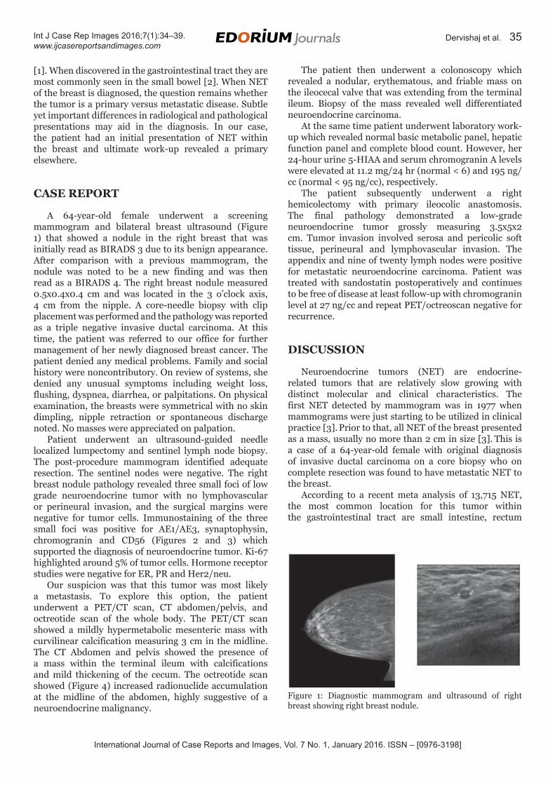
A 64-year-old female underwent a screening mammogram and bilateral breast ultrasound (Figure 1) that showed a nodule in the right breast that was initially read as BIRADS 3 due to its benign appearance. After comparison with a previous mammogram, the nodule was noted to be a new finding and was then read as a BIRADS 4. The right breast nodule measured 0.5x0.4x0.4 cm and was located in the 3 o’clock axis, 4 cm from the nipple. A core-needle biopsy with clip placement was performed and the pathology was reported as a triple negative invasive ductal carcinoma. At this time, the patient was referred to our office for further management of her newly diagnosed breast cancer. The patient denied any medical problems. Family and social history were noncontributory. On review of systems, she denied any unusual symptoms including weight loss, flushing, dyspnea, diarrhea, or palpitations. On physical examination, the breasts were symmetrical with no skin dimpling, nipple retraction or spontaneous discharge noted. No masses were appreciated on palpation.
Patient underwent an ultrasound-guided needle localized lumpectomy and sentinel lymph node biopsy. The post-procedure mammogram identified adequate resection. The sentinel nodes were negative. The right breast nodule pathology revealed three small foci of low grade neuroendocrine tumor with no lymphovascular or perineural invasion, and the surgical margins were negative for tumor cells. Immunostaining of the three small foci was positive for AE1/AE3, synaptophysin, chromogranin and CD56 (Figures 2 and 3) which supported the diagnosis of neuroendocrine tumor. Ki-67 highlighted around 5% of tumor cells. Hormone receptor studies were negative for ER, PR and Her2/neu.
Our suspicion was that this tumor was most likely a metastasis. To explore this option, the patient underwent a PET/CT scan, CT abdomen/pelvis, and octreotide scan of the whole body. The PET/CT scan showed a mildly hypermetabolic mesenteric mass with curvilinear calcification measuring 3 cm in the midline. The CT Abdomen and pelvis showed the presence of a mass within the terminal ileum with calcifications and mild thickening of the cecum. The octreotide scan showed (Figure 4) increased radionuclide accumulation at the midline of the abdomen, highly suggestive of a neuroendocrine malignancy.
The patient then underwent a colonoscopy which revealed a nodular, erythematous, and friable mass on the ileocecal valve that was extending from the terminal ileum. Biopsy of the mass revealed well differentiated neuroendocrine carcinoma.
At the same time patient underwent laboratory work-up which revealed normal basic metabolic panel, hepatic function panel and complete blood count. However, her 24-hour urine 5-HIAA and serum chromogranin A levels were elevated at 11.2 mg/24 hr (normal < 6) and 195 ng/cc (normal < 95 ng/cc), respectively.
The patient subsequently underwent a right hemicolectomy with primary ileocolic anastomosis. The final pathology demonstrated a low-grade neuroendocrine tumor grossly measuring 3.5x5x2 cm. Tumor invasion involved serosa and pericolic soft tissue, perineural and lymphovascular invasion. The appendix and nine of twenty lymph nodes were positive for metastatic neuroendocrine carcinoma. Patient was treated with sandostatin postoperatively and continues to be free of disease at least follow-up with chromogranin level at 27 ng/cc and repeat PET/octreoscan negative for recurrence.
DISCUSSION
Neuroendocrine tumors (NET) are endocrine-related tumors that are relatively slow growing with distinct molecular and clinical characteristics. The first NET detected by mammogram was in 1977 when mammograms were just starting to be utilized in clinical practice [3]. Prior to that, all NET of the breast presented as a mass, usually no more than 2 cm in size [3]. This is a case of a 64-year-old female with original diagnosis of invasive ductal carcinoma on a core biopsy who on complete resection was found to have metastatic NET to the breast.
According to a recent meta analysis of 13,715 NET, the most common location for this tumor within the gastrointestinal tract are small intestine, rectum
Figure 1: Diagnostic mammogram and ultrasound of right breast showing right breast nodule.

International Journal of Case Reports and Images, Vol. 7 No. 1, January 2016. ISSN – [0976-3198]
Int J Case Rep Images 2016;7(1):34–39. www.ijcasereportsandimages.com
Dervishaj et al. 36
and stomach, respectively. Not the appendix as was previously believed [2]. The most common NET to have metastasized at time of diagnosis is that of the cecum, pancreas and small intestine (81.5%, 71.9%, and 58.3%, respectively). The most frequent sites of metastasis, excluding the lymph nodes (89.8%), are the liver (44.1%), lung (13.6%), peritoneum (13.6%), and pancreas (6.8%) [2]. It is unusual to find this rare tumor in the breast as a metastasis. NET metastases do not favor any particular quadrant of the breast, but they do occur more commonly on the right breast than the left [3]. In the review by Kalisher et al., the authors report on 59 cases of neuroendocrine tumors in literature. Thirty-eight of these were primary neuroendocrine tumors of the breast, while only nine were metastases [3].
The most common cancers to metastasize to the breast in decreasing frequency are contralateral breast carcinoma, malignant melanoma, prostate, lung, and renal cell carcinoma [3]. Primary breast NET comprise <2% of all primary breast cancers (Table 1) [1, 4–6]. In 2003, the World Health Organization (WHO) classified primary NET of the breast as tumors with expression of one or more immunohistochemical markers (neuron specific enolase, chromogranin A, and synaptophysin) in at least 50% of the tumor cells [1].
Our patient’s initial finding was an oval-shaped hypoechoic lesion which was initially thought to be benign due to absence of irregularity. Multiple case reports have reported an initial benign appearing mammogram with a well-circumscribed mass with no calcifications [1, 3, 5]. In a review of 1845 breast cancers, Belgin et al. found 5 NETs of the breast. On mammogram, four of them appeared round, while only one patient had an irregularly shaped lesion.
Studies have demonstrated that diagnosing NET of breast on core needle biopsy may be difficult [7]. Angarita et al. also report a case of a patient who was diagnosed with IDC that was postoperatively found to have a NET, in their case a primary neuroendocrine tumor of the breast.
Fine needle aspiration (FNA) is also a common tool for the diagnosis of malignancy. However while it may help identify malignant cells, it has often proven difficult in distinguishing NET from other breast cancers [5].
Moreover, due to identification of malignant cells on FNA, it has sometimes led to an immediate mastectomy without further characterization of the cancer [5].
On gross pathology, metastatic NET tends to be less fixed to the surrounding tissues than primary tumors and is located in the subcutaneous tissue rather than breast tissue [3]. NET consists of a uniform cell population with abundant eosinophilic cytoplasm and nuclei with stippled (‘‘salt and pepper’’) chromatin [1]. Invasive ductal carcinoma (IDC), on the other hand, usually consists of atypical cells with occasional to numerous mitoses which is unlike the uniform round cells seen in NET [1, 3].
Lobular carcinoma in situ (LCIS) may be confused with NET because of the uniform cells with round nuclei lying in well-defined islands [3]. However, LCIS has no
Figure 2: Microscopic examination reveals that tumor is composed of insular, trabecular monotonous small round cells showing peripheral palisading with moderate finely granular cytoplasm, small nucleoli, salt and pepper chromatin. Mitotic figures are present (H&E stain, x200).
Figure 3: Immunohistochemical stain for synaptophysin is diffusely strong positive (H&E stain, x200).
Figure 4: Octreotide scan showed increased radionuclide accumulation at the midline of the abdomen, highly suggestive of a neuroendocrine malignancy.

International Journal of Case Reports and Images, Vol. 7 No. 1, January 2016. ISSN – [0976-3198]
Int J Case Rep Images 2016;7(1):34–39. www.ijcasereportsandimages.com
Dervishaj et al. 37
fibrosis and it preserves the pattern of markedly dilated terminal ducts filled with slightly dyshesive cells [3].
Infiltrating lobular carcinoma (ILC) can be distinguished from metastatic NET, since NET rarely “Indian file” [3].
Histologic confirmation of NET can be made by a positive argyrophil reaction and evidence of small membrane-bound neurosecretory granules [8]. Although the solid nests found in NET of the breast are also present in DCIS/LCIS and IDC/ILC, both of the latter can be excluded when considering E-cadherin positivity and p63 negativity of the palisading cells, respectively [6].
Once NET is diagnosed by histopathologic features, the question remains whether the tumor is a primary versus metastatic NET to the breast. The definite feature that makes the diagnosis of primary is intraductal component of NET, as metastatic NET has no intraductal component
[3, 4]. Based on this finding, we advise screening of other sites using different imaging modalities once the diagnosis of metastatic NET is suspected.
Chromogranin A (CGA), chromogranin B (CGB), and synaptophysin (SYP) are considered the most sensitive and specific NET markers but they are relatively nonspecific when it comes to differentiating between primary versus metastatic NET to the breast [4, 6]. For breast tumor markers, more than 90% of diagnosed primary NET cases can express estrogen receptors (ER) while up to 75–80% express progesterone receptors (PR) [6]. However, presence of ER/PR is not sensitive nor specific enough to differentiate between primary versus metastatic NET to the breast. To the best of our
knowledge, we could not find a study that reported the presence of these markers in NET of the bowel. Hence we can assume that the presence of these markers in a case of NET of the breast can help exclude a metastasized NET from the bowel. However, there have been cases of bronchopulmonary NET which have been reported to be positive for ER/PR and metastasized to the breast. In one report of pulmonary NET, 40% of specimens expressed ER, and 70% expressed PR [1, 2, 4, 9]. Moreover, when these tumor markers are found to be negative, one has to assume that the tumor could be a metastasis from the bowel, lung or a primary of the breast.
Richter-Ehrenstein et al. looked at the expression of Gross cystic disease fluid protein (GCDFP-15), mammaglobin, and TTF-1 (thyroid transcription factor 1) to help assist in differentiation of a primary versus metastatic NET to the breast. In primary NET, they found that GCDFP-15 was expressed in 6 out of 9, mammaglobin was positive in 4 out of 9, and TTF-1 was not expressed in any of the breast tumors. One of these patients was found to have a primary in the midgut, and this was the only patient that had 0% expression of ER/PR and HER2. In evaluation of the 99 patients with primary NET of gut, they found no expression of GCDFP-15, mammaglobin, or TTF-1. Other studies have reported similar results using these tumor markers [1, 10–12].
It is the difference in treatment of primary versus metastatic NET to the breast that is most worrisome about misdiagnoses. Prior to lumpectomy and radiation treatment, a simple or radical mastectomy was the
Table 1: Different characteristics of primary versus metastatic neuroendocrine tumor of the breast
Feature Primary Metastatic
Prevalence 0.27–2% 0.4–0.7%
Age at diagnosis 6th or 7th decade of life [1, 2, 3] 5th decade of life most common
Symptoms of carcinoid syndrome
No cases reported to date depends on primary
Axillary involvement Commonly involved in tumors >3 cm in size Not reported in literature
Radiology –US: irregular margin, hypoechoic, homogenous texture, round nodule, absence of cystic component [1, 2] –MRI: homogeneous lobulated tumors with early contrast enhancement, suggesting malignancy [1, 2]
- Mammogram: most common appear as benign, well circumscribed nodules, absent micro-calcifications,
Gross Pathology pale-yellow colored, fleshy or firm with either smooth or irregular margins [1, 2].
Mets tend to be less fixed to surrounding tissues and located in subcutaneous tissue adjacent to breast tissue [5]
Histological Markers –strongly + ER/PR, Expression of GCDFP-15, mammaglobin and lack of TTF-1 , + for synaptophysin/chromogranin A
–negative for ER/PR, GCDFP-15, mammaglobin, or TTF-1 +synaptophysin and chromogranin A
Treatment same recommendations as invasive ductal carcinoma local excision only
Survival Similar to Luminal Type A invasive ductal carcinoma Depends on primary source
International Journal of Case Reports and Images, Vol. 7 No. 1, January 2016. ISSN – [0976-3198]
Int J Case Rep Images 2016;7(1):34–39. www.ijcasereportsandimages.com
Dervishaj et al. 38
standard treatment of primary carcinoid tumors, even in cases of correct diagnoses [3, 8]. This is because primary carcinoid tumor of the breast behaves similarly to IDC and hence is treated in the same manner. Whereas in cases of metastatic NET to the breast, an excision of the lesion is thought to be sufficient treatment and that radiation to the breast is not necessary [13].
CONCLUSION
In order to determine the appropriate treatment for our patient, we needed to know if there was a primary elsewhere which led to the eventual discovery of original tumor in the ileocecum. Our work-up included a PET/CT scan, octreotide scan and colonoscopy. All three studies helped us find and confirm the site of the primary as well as obtain tissue for diagnosis. We recommend the use of all three in the workup of a neuroendocrine tumor.
*********
Author ContributionsOrnela A. Dervishaj – Substantial contributions to conception and design, Acquisition of data, Analysis and interpretation of data, Drafting the article, Revising it critically for important intellectual content, Final approval of the version to be publishedAlexandra S. Renzi – Analysis and interpretation of data, Revising it critically for important intellectual content, Final approval of the version to be publishedRomulo Genato – Analysis and interpretation of data, Revising it critically for important intellectual content, Final approval of the version to be publishedPhilip Q. Xiao – Analysis and interpretation of data, Revising it critically for important intellectual content, Final approval of the version to be publishedArmand P. Asarian – Analysis and interpretation of data, Revising it critically for important intellectual content, Final approval of the version to be published
GuarantorThe corresponding author is the guarantor of submission.
Conflict of InterestAuthors declare no conflict of interest.
Copyright© 2016 Ornela A. Dervishaj et al. This article is distributed under the terms of Creative Commons Attribution License which permits unrestricted use, distribution and reproduction in any medium provided the original author(s) and original publisher are properly credited. Please see the copyright policy on the journal website for more information.
REFERENCES
1. Richter-Ehrenstein C, Arndt J, Buckendahl AC, et al. Solid neuroendocrine carcinomas of the breast: metastases or primary tumors? Breast Cancer Res Treat 2010 Nov;124(2):413–7.
2. Modlin IM, Lye KD, Kidd M. A 5-decade analysis of 13,715 carcinoid tumors. Cancer 2003 Feb 15;97(4):934–59.
3. Harrist TJ, Kalisher L. Breast metastasis: an unusual manifestation of a malignant carcinoid tumor. Cancer 1977 Dec;40(6):3102–6.
4. Alkaied H, Harris K, Azab B, Dai Q. Primary neuroendocrine breast cancer, how much do we know so far? Med Oncol 2012 Dec;29(4):2613–8.
5. Günhan-Bilgen I, Zekioglu O, Ustün EE, Memis A, Erhan Y. Neuroendocrine differentiated breast carcinoma: imaging features correlated with clinical and histopathological findings. Eur Radiol 2003 Apr;13(4):788–93.
6. López-Bonet E, Alonso-Ruano M, Barraza G, Vazquez-Martin A, Bernadó L, Menendez JA. Solid neuroendocrine breast carcinomas: incidence, clinico-pathological features and immunohistochemical profiling. Oncol Rep 2008 Dec;20(6):1369–74.
7. Angarita FA, Rodríguez JL, Meek E, Sánchez JO, Tawil M, Torregrosa L. Locally-advanced primary neuroendocrine carcinoma of the breast: case report and review of the literature. World J Surg Oncol 2013 Jun 5;11:128.
8. Jablon LK, Somers RG, Kim PY. Carcinoid tumor of the breast: treatment with breast conservation in three patients. Ann Surg Oncol 1998 Apr-May;5(3):261–4.
9. Chan JK, Suster S, Wenig BM, Tsang WY, Chan JB, Lau AL. Cytokeratin 20 immunoreactivity distinguishes Merkel cell (primary cutaneous neuroendocrine) carcinomas and salivary gland small cell carcinomas from small cell carcinomas of various sites. Am J Surg Pathol 1997 Feb;21(2):226–34.
10. Sapino A, Righi L, Cassoni P, Papotti M, Gugliotta P, Bussolati G. Expression of apocrine differentiation markers in neuroendocrine breast carcinomas of aged women. Mod Pathol 2001 Aug;14(8):768–76.
11. Sica G, Wagner PL, Altorki N, et al. Immunohistochemical expression of estrogen and progesterone receptors in primary pulmonary neuroendocrine tumors. Arch Pathol Lab Med 2008 Dec;132(12):1889–95.
12. Fritzsche FR, Thomas A, Winzer KJ, et al. Co-expression and prognostic value of gross cystic disease fluid protein 15 and mammaglobin in primary breast cancer. Histol Histopathol 2007 Nov;22(11):1221–30.
13. Kaltsas GA, Putignano P, Mukherjee JJ, et al. Carcinoid tumours presenting as breast cancer: the utility of radionuclide imaging with 123I-MIBG and 111In-DTPA pentetreotide. Clin Endocrinol (Oxf) 1998 Nov;49(5):685–9.
International Journal of Case Reports and Images, Vol. 7 No. 1, January 2016. ISSN – [0976-3198]
Int J Case Rep Images 2016;7(1):34–39. www.ijcasereportsandimages.com
Dervishaj et al. 39
Access full text article onother devices
Access PDF of article onother devices

EDORIUM JOURNALS AN INTRODUCTION
Edorium Journals: On Web
About Edorium JournalsEdorium Journals is a publisher of high-quality, open ac-cess, international scholarly journals covering subjects in basic sciences and clinical specialties and subspecialties.
Edorium Journals www.edoriumjournals.com
Edorium Journals et al.
Edorium Journals: An introduction
Edorium Journals Team
But why should you publish with Edorium Journals?In less than 10 words - we give you what no one does.
Vision of being the bestWe have the vision of making our journals the best and the most authoritative journals in their respective special-ties. We are working towards this goal every day of every week of every month of every year.
Exceptional servicesWe care for you, your work and your time. Our efficient, personalized and courteous services are a testimony to this.
Editorial ReviewAll manuscripts submitted to Edorium Journals undergo pre-processing review, first editorial review, peer review, second editorial review and finally third editorial review.
Peer ReviewAll manuscripts submitted to Edorium Journals undergo anonymous, double-blind, external peer review.
Early View versionEarly View version of your manuscript will be published in the journal within 72 hours of final acceptance.
Manuscript statusFrom submission to publication of your article you will get regular updates (minimum six times) about status of your manuscripts directly in your email.
Our Commitment
Most Favored Author programJoin this program and publish any number of articles free of charge for one to five years.
Favored Author programOne email is all it takes to become our favored author. You will not only get fee waivers but also get information and insights about scholarly publishing.
Institutional Membership programJoin our Institutional Memberships program and help scholars from your institute make their research accessi-ble to all and save thousands of dollars in fees make their research accessible to all.
Our presenceWe have some of the best designed publication formats. Our websites are very user friendly and enable you to do your work very easily with no hassle.
Something more...We request you to have a look at our website to know more about us and our services.
We welcome you to interact with us, share with us, join us and of course publish with us.
Browse Journals
CONNECT WITH US
Invitation for article submissionWe sincerely invite you to submit your valuable research for publication to Edorium Journals.
Six weeksYou will get first decision on your manuscript within six weeks (42 days) of submission. If we fail to honor this by even one day, we will publish your manuscript free of charge.
Four weeksAfter we receive page proofs, your manuscript will be published in the journal within four weeks (31 days). If we fail to honor this by even one day, we will publish your manuscript free of charge and refund you the full article publication charges you paid for your manuscript.
This page is not a part of the published article. This page is an introduction to Edorium Journals and the publication services.