neurodynamic evaluation of the sciatic nerve during neural
TRANSCRIPT

Neurodynamic evaluation of the sciatic nerve
during neural mobilisation: ultrasound imaging
assessment of sciatic nerve movement and the
clinical implications for treatment
Richard F. Ellis
A thesis submitted to AUT University in fulfilment of the requirements for the degree
of Doctor of Philosophy (PhD)
2011
Department of Physiotherapy
School of Rehabilitation and Occupation Studies
Primary supervisor: Associate Professor Dr. Wayne Hing


i
Table of Contents List of Figures ..................................................................................................... ix
List of Tables ....................................................................................................... xi
Attestation of Authorship.................................................................................. xii
Declaration of Co-authored works.................................................................. xiii
Publications and conference presentations .................................................... xiv
PUBLISHED PEER-REVIEWED ARTICLES.......................................................XIV
ARTICLES CURRENTLY SUBMITTED AND UNDER PEER-REVIEW..........XIV
CONFERENCE PRESENTATIONS. ...................................................................... XV
Acknowledgements ......................................................................................... xviii
Abstract ............................................................................................................. xxi
Chapter One. Introduction ................................................................................ 1
1.1 BACKGROUND ...................................................................................................1
1.2 AIMS AND OBJECTIVES OF THIS DOCTORAL RESEARCH.......................3
1.3 SIGNIFICANCE OF THIS DOCTORAL RESEARCH .......................................4
1.4 THESIS PRESENTATION ...................................................................................5
Chapter Two. Literature review ....................................................................... 7
2.1 INTRODUCTION ..................................................................................................7
2.2 NEURODYNAMIC FEATURES OF THE PERIPHERAL NERVOUS
SYSTEM.......................................................................................................................8
2.2.1 The ability of a nerve to move and slide. ...................................................10

ii
2.2.2 The ability of a nerve to withstand stretch. ................................................12
2.2.3 The ability of a nerve to withstand compression. ......................................15
2.3 BIOMECHANICAL FEATURES OF NERVE EXCURSION...........................17
2.3.1 The nervous system is a continuous system...............................................17
2.3.2 The sequence of nerve excursion. ..............................................................18
2.3.3 Nerve excursion is greatest the closer to the axis of joint motion. ............21
2.3.4 Increased neural tension will reduce nerve excursion and joint range.......23
2.3.5 Convergence and tension points. ...............................................................25
2.3.6 Regional peripheral nerve excursion..........................................................27
2.3.6.1 Upper limb nerve movement. .....................................................27
2.3.6.2 Lower limb nerve movement. .....................................................28
2.4 CLINICAL IMPLICATIONS OF IMPAIRED NERVE MOVEMENT.............31
2.4.1 Neural mechanosensitivity. ........................................................................32
2.4.2 Neural oedema leading to impaired nerve movement. ..............................34
2.4.3 Neural fibrosis leading to impaired nerve movement. ...............................35
2.5 NEURAL MOBILISATION................................................................................38
2.5.1 The influence of neural mobilisation upon impaired nerve excursion......40
2.5.1.1 Mechanical influences of neural mobilisation on nerve excursion.
.................................................................................................................40
2.5.1.2 Physiological influences of neural mobilisation on nerve
excursion. ................................................................................................42
2.5.1.3 Analgesic influences of neural mobilisation on nerve excursion.
.................................................................................................................43
2.5.2 Neural mobilisation techniques.................................................................44
2.5.2.1 ‘Sliders’. ......................................................................................44
2.5.2.2 ‘Tensioners’. ...............................................................................46

iii
2.5.2.3 ‘Sliders’ versus ‘tensioners’........................................................47
2.5.2.4 Neural mobilisation prescription. ...............................................48
2.6 ULTRASOUND IMAGING OF PERIPHERAL NERVE MOVEMENT ..........50
2.6.1 Principles and properties of ultrasound imaging.......................................50
2.6.1.1 The physics of ultrasound imaging. ...........................................51
2.6.1.2 Ultrasound imaging techniques. .................................................52
2.6.2 Ultrasound imaging of peripheral nerves..................................................53
2.6.2.1 Ultrasound imaging of the sciatic nerve. ....................................55
2.6.3 Ultrasound imaging assessment of peripheral nerve movement...............57
2.6.3.1 Frame-by-frame cross-correlation analysis. ..............................57
2.6.3.2 Real-time spectral Doppler ultrasound.......................................61
Chapter Three. Neural mobilisation: a systematic review of randomised
controlled trials with an analysis of therapeutic efficacy............................... 63
3.1 PRELUDE............................................................................................................63
3.2 ABSTRACT.........................................................................................................64
3.3 INTRODUCTION ...............................................................................................65
3.4 METHODS ..........................................................................................................66
3.4.1 Literature search strategy. .........................................................................66
3.4.2 Study selection. .........................................................................................67
3.4.3 Methodological quality assessment. .........................................................68
3.4.4 Analysis of therapeutic efficacy................................................................72
3.4.5 Clinical benefit. .........................................................................................73
3.5 RESULTS ............................................................................................................73
3.5.1 Selection of studies. ..................................................................................73
3.5.2 Methodological quality. ............................................................................74

iv
3.5.3 Study characteristics. ................................................................................75
3.5.4 Therapeutic efficacy..................................................................................75
3.5.5 Clinical benefit. .........................................................................................76
3.6 DISCUSSION ......................................................................................................83
3.6.1 Future research. .........................................................................................86
3.7 CONCLUSION....................................................................................................87
Chapter Four. Reliability of measuring sciatic and tibial nerve movement
with diagnostic ultrasound during a neural mobilisation technique ............ 89
4.1 PRELUDE............................................................................................................89
4.2 ABSTRACT.........................................................................................................90
4.3 INTRODUCTION ...............................................................................................91
4.4 MATERIALS AND METHODS.........................................................................92
4.4.1 Subjects. ....................................................................................................92
4.4.2 Equipment and procedures........................................................................93
4.4.2.1 Subject set-up. .............................................................................93
4.4.2.2 Diagnostic ultrasound parameters and imaging. .......................93
4.4.2.3 Frame-by-frame cross-correlation algorithm and calculation
software. ..................................................................................................96
4.4.3 Statistical analysis. ....................................................................................96
4.4.4 Ethics.........................................................................................................97
4.5 RESULTS ............................................................................................................97
4.6 DISCUSSION ....................................................................................................103
4.6.1 Limitations of the study. .........................................................................105
4.7 CONCLUSION..................................................................................................106

v
Chapter Five. The influence of increased nerve tension on sciatic nerve
excursion during a side-lying neural mobilisation exercise - a reliability
study .................................................................................................................. 107
5.1 PRELUDE..........................................................................................................107
5.2 ABSTRACT.......................................................................................................108
5.3 INTRODUCTION .............................................................................................109
5.4 METHODS ........................................................................................................111
5.4.1 Participants...............................................................................................111
5.4.2 Procedure. ................................................................................................112
5.4.3 Ultrasound imaging and analysis. ............................................................113
5.4.4 Statistical analysis. ...................................................................................115
5.5 RESULTS ..........................................................................................................115
5.6 DISCUSSION ....................................................................................................116
5.7 CONCLUSION..................................................................................................119
Chapter Six. Comparison of different neural mobilisation exercises upon
longitudinal sciatic nerve movement: an in-vivo study utilising ultrasound
imaging.............................................................................................................. 120
6.1 PRELUDE..........................................................................................................120
6.2 ABSTRACT.......................................................................................................121
6.3 INTRODUCTION .............................................................................................122
6.4 METHODS ........................................................................................................125
6.4.1 Participants..............................................................................................125
6.4.2 Participant set-up.....................................................................................126
6.4.3 Fastrak electrogoniometer.......................................................................127
6.4.4 Diagnostic US parameters and imaging..................................................129

vi
6.4.5 Frame-by-frame cross-correlation algorithm and calculation software. .130
6.4.6 US video selection criteria. .....................................................................131
6.4.7 Neural mobilisation exercises. ................................................................131
6.4.8 Statistical analysis. ..................................................................................133
6.5 RESULTS ..........................................................................................................134
6.6 DISCUSSION ....................................................................................................135
6.7 CONCLUSIONS................................................................................................140
Chapter Seven. In-vivo ultrasound assessment of sciatic nerve excursion
during neural mobilisation involving knee and ankle movement and the
influence of cervical flexion............................................................................. 142
7.1 PRELUDE..........................................................................................................142
7.2 ABSTRACT.......................................................................................................142
7.3 INTRODUCTION .............................................................................................144
7.4 MATERIALS AND METHODS.......................................................................146
7.4.1 Participants..............................................................................................146
7.4.2 Participant set-up.....................................................................................147
7.4.3 Joint ROM analysis. ................................................................................147
7.4.4 Ultrasound imaging.................................................................................148
7.4.5 Neural mobilisation exercises .................................................................149
7.4.6 Statistical analysis ...................................................................................151
7.5 RESULTS ..........................................................................................................152
7.6 DISCUSSION ....................................................................................................153
7.7 CONCLUSION..................................................................................................156

vii
Chapter Eight. Identifying the sequence of sciatic nerve excursion during
different neural mobilisation exercises: an in-vivo study utilising ultrasound
imaging.............................................................................................................. 158
8.1 PRELUDE..........................................................................................................158
8.2 ABSTRACT.......................................................................................................159
8.3 INTRODUCTION .............................................................................................160
8.4 METHODS ........................................................................................................164
8.4.1 Participants..............................................................................................164
8.4.2 Participant position. ................................................................................164
8.4.3 Joint ROM analysis. ................................................................................165
8.4.4 Neural mobilisation exercises. ................................................................166
8.4.5 Ultrasound imaging and ultrasound video selection criteria...................167
8.4.6 Frame-by-frame cross-correlation algorithm and calculation software. .168
8.4.7 Synchronisation of data collection systems. ...........................................169
8.4.8 Statistical analysis. ..................................................................................170
8.5 RESULTS ..........................................................................................................170
8.6 DISCUSSION ....................................................................................................177
8.7 CONCLUSION..................................................................................................180
Chapter Nine. Discussion and conclusions ................................................... 182
9.1 TOWARDS A BETTER UNDERSTANDING OF NEURAL MOBILISATION
...................................................................................................................................182
9.2 ULTRASOUND ASSESSMENT OF SCIATIC NERVE EXCURSION .........184
9.3 SCIATIC NERVE EXCURSION IN RESPECT TO DIFFERENT TYPES OF
NEURAL MOBILISATION.....................................................................................185
viii
9.4 THE BIOMECHANICS OF SCIATIC NERVE EXCURSION DURING
NEURAL MOBILISATION.....................................................................................187
9.4.1 The relevance of the continual system. ...................................................188
9.4.2 The effect of increased neural tension. ...................................................189
9.4.3 Exploiting the sequence of nerve excursion. ..........................................191
9.5 CLINICAL IMPLICATIONS............................................................................192
9.5.1 Recommendations for the prescription of sliders. ...................................193
9.5.2 Recommendations for the prescription of tensioners...............................194
9.6 STUDY LIMITATIONS....................................................................................194
9.7 FUTURE RESEARCH FOR THE DEVELOPMENT OF NEURAL
MOBILISATION......................................................................................................195
References......................................................................................................... 198
Appendices........................................................................................................ 241
APPENDIX 1............................................................................................................241
APPENDIX 2............................................................................................................244
APPENDIX 3............................................................................................................245
APPENDIX 4............................................................................................................247
APPENDIX 5............................................................................................................250
APPENDIX 6............................................................................................................251
APPENDIX 7............................................................................................................253
APPENDIX 8............................................................................................................254

ix
List of Figures Figure 1. Typical A) load-elongation & B) stress-strain curves for peripheral nerves....9
Figure 2. Sciatic nerve unfolding with passive knee extension ....................................20
Figure 3. Sliders and Tensioners ...................................................................................46
Figure 4. Ultrasound appearance of the sciatic nerve. ..................................................54
Figure 5. Detailed ultrasound appearance of peripheral nerves. ...................................55
Figure 6. Graphical representation of frame-by-frame cross-correlation analysis........58
Figure 7. Frame-by-frame cross-correlation analysis of sciatic nerve excursion .........60
Figure 8. ‘Slider’ neural mobilisation sequence and participant set-up position. .........93
Figure 9. Measurement of transverse movement of the sciatic nerve at the posterior
mid-thigh.........................................................................................................................94
Figure 10. Comparison of sciatic nerve movement at each scanning location ...........100
Figure 11. Direction of transverse sciatic nerve movement at the posterior mid-thigh.
.......................................................................................................................................101
Figure 12. Direction of transverse sciatic nerve movement at the popliteal crease. ...101
Figure 13. Bland-Altman graph (Difference vs. Average) for all trials measuring
anterior-posterior (AP) sciatic nerve movement at the posterior mid-thigh. ................102
Figure 14. Bland-Altman graph (Difference vs. Average) for all trials measuring
longitudinal sciatic nerve movement at the posterior mid-thigh...................................103
Figure 15. Participant set-up for side-lying neural mobilisation.................................112
Figure 16. Participant set-up for slump-sitting neural mobilisation exercises............127
Figure 17. Slump-sitting neural mobilisation exercises (Chapter Six). ......................132
Figure 18. Slump-sitting neural mobilisation exercises (Chapter Seven)...................150
Figure 19. Comparison of mean sciatic nerve excursion (mm) between all neural
mobilisation exercises. ..................................................................................................153

x
Figure 20. Slump-sitting neural mobilisation exercises (Chapter Eight). ...................166
Figure 21. Relationship of sciatic nerve excursion, knee and cervical movement during
TEKS.............................................................................................................................173
Figure 22. Relationship of sciatic nerve excursion, knee and cervical movement during
OEKS. ...........................................................................................................................174
Figure 23. Relationship of sciatic nerve excursion, knee and cervical movement during
KT. ................................................................................................................................175
Figure 24. Sciatic nerve excursion, knee and cervical ROM data for all neural
mobilisation exercises ...................................................................................................176

xi
List of Tables Table 1. In-vivo assessment of longitudinal nerve excursion in humans. .....................30
Table 2. The PEDro Scale ..............................................................................................70
Table 3. Randomised controlled trials of neural mobilisation as a treatment modality in
order of PEDro score.......................................................................................................71
Table 4. Number and percentage of the studies meeting each PEDro criteria ..............74
Table 5. Randomised controlled trials of neural mobilisation as a treatment modality 77
Table 6. Level of evidence for therapeutic efficacy per intervention type....................85
Table 7. Amount of sciatic nerve movement (mm) measured for each direction at each
scanning location.............................................................................................................99
Table 8. Intraclass correlation coefficients (ICC) of test-retest reliability for each
direction of nerve movement at each scanning location .................................................99
Table 9. Descriptive results for longitudinal sciatic nerve excursion across the neural
mobilisation exercises ...................................................................................................171
Table 10. Cervical, knee ROM and sciatic nerve excursion at each quarter during the
three second exercise performance ...............................................................................172

xii
Attestation of Authorship
I hereby declare that this submission is my own work and that, to the best of my
knowledge and belief, it contains no material previously published or written by another
person (except when explicitly defined in the Acknowledgements), nor material which
to a substantial extent has been submitted for the award of any other degree or diploma
of a university of institution of higher learning.
Signed:
Date: 5th September, 2011.


xiv
Publications and conference presentations
PUBLISHED PEER-REVIEWED ARTICLES.
Ellis, R. F., & Hing, W. A. (2008). Neural mobilization: a systematic review of
randomized controlled trials with an analysis of therapeutic efficacy. Journal of
Manual & Manipulative Therapy, 16(1), 8-22.
Ellis, R. F., Hing, W. A., Dilley, A., & McNair, P. (2008). Reliability of measuring
sciatic and tibial nerve movement with diagnostic ultrasound during a neural
mobilisation technique. Ultrasound in Medicine and Biology, 34(8), 1209-
1216.
Ellis, R. F., & Hing, W. A. (2008). Neural mobilization: A systematic review of
randomized controlled trials with an analysis of therapeutic efficacy.
[Mobilizacje struktur nerwowych – przegląd systematyczny badań z
randomizacją i grupą kontrolną oraz analiza skuteczności terapeutycznej].
Medical Rehabilitation. [Rehabilitacja Medyczna], 12(2), 34-44.
Ellis, R. F., & Hing, W. A. (2009). Neurale mobilisation: Systematischer review
randomisierter kontrollierter studien mit einer analyse der therapeutischen
wirksamkeit. Manuelletherapie, 2, 47-57.
ARTICLES CURRENTLY SUBMITTED AND UNDER PEER-REVIEW.
Ellis, R. F., Hing, W. A., & McNair, P. (2012). Comparison of different neural
mobilisation exercises upon longitudinal sciatic nerve movement: an in-vivo
study utilising ultrasound imaging. Journal of Orthopaedic and Sports
Physical Therapy.

xv
Ellis, R. F., Hing, W. A., Mawston, G. & McNair, P. J. (2012). In-vivo ultrasound
assessment of sciatic nerve excursion during neural mobilisation involving
knee and ankle movement and the influence of cervical flexion. Journal of
Manual and Manipulative Therapy.
Ellis, R. F., Hing, W. A., & McNair, P. J. (2012). Identifying the sequence of sciatic
nerve excursion during different neural mobilisation exercises: an in-vivo study
utilising ultrasound imaging. Clinical Biomechanics
CONFERENCE PRESENTATIONS.
The presenter is bolded.
Hing, W.A., & Ellis, R.F. (2006). Neural tissue mobility measurement by diagnostic
ultrasound. Paper presented at the meeting of the 2nd Indian National Manual
Therapy Conference., New Delhi, India.
Hing, W., & Ellis, R. (2007). Diagnostic ultrasound assessment of sciatic nerve
movement during neural mobilisation. Paper presented at the meeting of the
15th International World Confederation of Physical Therapy (WCPT)
Congress, Vancouver, Canada.
Ellis, R.F., Hing, W.A., & Allen, S. (2007). Diagnostic ultrasound assessment of sciatic
nerve movement during neural mobilisation - quantitative assessment and
reliability. Paper presented at the meeting of the New Zealand Manipulative
Physiotherapists Association (NZMPA) Biennial Scientific Conference,
Rotorua, New Zealand.

xvi
Ellis, R. F., & Hing, W. A. (2008). Measurement of sciatic nerve movement, using
ultrasound imaging, in a neural pre-loaded sitting position compared to an
unloaded prone position. Paper presented at the meeting of the International
Federation of Orthopaedic Manipulative Physical Therapists (IFOMPT)
Conference, Rotterdam, The Netherlands.
Ellis, R.F., Hing, W.A., & McNair, P.J. (2008). Measurement of sciatic nerve
movement, using ultrasound imaging, in a neural pre-loaded sitting position
compared to an unloaded prone position. Paper presented at the meeting of the
Physiotherapy New Zealand (PNZ) Conference, Dunedin, New Zealand.
Ellis, R. F., Hing, W. A., & Allen, S. (2008). Diagnostic ultrasound assessment of
sciatic nerve movement during neural mobilisation - quantitative assessment
and reliability. Paper presented at the meeting of the Australia and New
Zealand Association of Clinical Anatomists, Auckland, New Zealand.
Ellis, R. F., Milne, A., Tuck, K., Hing, W. A., & McNair, P. (2009). The reliability of
measuring knee range of movement in response to submaximal pain created by
increased neural tension during a modified slump test. Paper presented at the
meeting of the New Zealand Manipulative Physiotherapists Association
(NZMPA) Biennial Conference, Rotorua, New Zealand.
Ellis, R.F., Eason, T., Edwards, C., Hing, W.A., & McNair, P.J. (2010). Assessing the
clinical validity of neurodynamic tests. Paper presented at the meeting of the
Neurodynamics and the Neuromatrix, Nottingham, United Kingdom.
Ellis, R. F., Hing, W. A., & McNair, P. J. (2011). Comparison of different neural
mobilisation exercises upon longitudinal sciatic nerve movement: an in-vivo
study utilising ultrasound imaging. Paper presented at the meeting of the New

xvii
Zealand Manipulative Physiotherapists Association (NZMPA) Biennial
Conference, Rotorua, New Zealand.
This presentation won the award for the “Best Podium Presentation” at this
conference.
xviii
Acknowledgements
The reality of completing a PhD is that my efforts would not have been possible
without the help of several people, involved at different stages throughout the process. I
am very grateful for all those who have contributed to this PhD and would like to take
this opportunity to thank them.
The two people who I owe the greatest thanks are my primary supervisor,
Associate Professor Wayne Hing and my secondary supervisor, Professor Peter McNair.
Everyone who undertakes a PhD does so naïve to the process. I have been extremely
fortunate to have been guided and mentored by two very experienced researchers.
Wayne has been involved right from the start, when I initially enrolled in the Masters
programme. He was instrumental in encouraging me to become a member of the
academic staff at AUT University and also encouraged me to pursue this PhD. Wayne
has always had an “open door” for questions, discussion, coffee, and edits (with some
email traffic past midnight!). Peter’s guidance has also been important. His comment
and advice in regard to research design, interpretation and thesis development has been
invaluable. Meetings with Peter have been challenging. He pushes you to find the
answers to issues that he raises and you always leave with more work than when you
went in!
Thanks also to Dr. Andrew Dilley. Andrew and his research team developed the
ultrasound analysis software that I used throughout this thesis. After emailing him for
his advice, Andrew sent me the analysis software and instructed me in its use without
reservation. He was very obliging with further advice as I implemented and learned to
use the software. I am grateful for his contribution as a co-author in one of the initial
publications from this PhD.
xix
Thanks to Physiotherapy New Zealand (PNZ), New Zealand Manipulative
Physiotherapists Association (NZMPA), Maurice and Phyllis Paykel Scholarship Trust
and AUT University for funding support.
To the many staff at AUT University that have helped me during this PhD, I am
grateful. In particular, thanks to Dr. Grant Mawston and David Rice for their technical
expertise and advice in using additional research equipment. Thanks also to Scott
Allen, for tuition in ultrasonography, and the Horizon Radiology team.
I have been fortunate to have had many research students involved in different
aspects of this doctoral research. Tamara Robinson, Sarah Pelham, Peter and Keryn
Matheson, Kelly Tuck, Alyssa Milne, Tim Eason, Chris Edwards, Amanda Bunn, Sia
Blom, Trish Knight and Sandee Hui have all made varied contributions of which I am
very grateful. They have all worked hard and have been (or at least seemed to have
been) enthusiastic about the topic I enjoy. I hope they have learned a lot about research
and may be inspired to indulge any burgeoning research urges in the future! Thanks
also to Dave Mabbott for drawing several of the illustrations used throughout this thesis
and within several of the journal papers.
I am forever grateful for the support of my family. My brothers and sister have
been a constant support. My parents have encouraged me throughout my life, and
especially throughout my education, culminating with the completion of this PhD. I
take the mantel of the family’s highest academic qualification from my Mum. I know
that many of the highs and lows I have experienced, she also went through when
completing her Masters. It has been great to share these experiences with her and take
her advice. My Dad has closely been involved in the presentation of this thesis. He
took on the task of proof-reading, looking for errors in spelling, grammar and
formatting. Dad made many valuable edits and suggestions of better ways to express
many aspects. Inadvertently he has now emerged a student of neurodynamics!
xx
Lastly, this thesis is dedicated to my own family, my beautiful wife Kate and my
children, Jake and Lilah. Although the kids may not have directly contributed to the
PhD, they were a constant source of enjoyment and fun during times when Dad was not
“writing his book”. I owe a great debt of thanks to Kate. She endured most throughout
this process: periods of time when I was away on writing retreats, late nights when I
was writing in the garage, weekends away at conferences, grumpy rants and frustration.
Kate kept me sane, kept our family safe and well and completely supported me
throughout this PhD. Without your love and support, Kate, this would not have been
possible.
xxi
Abstract
Neural mobilisation is a physiotherapeutic tool that is used to directly influence
peripheral nerve mechanics, in particular the neurodynamic features of the peripheral
nervous system. Neurodynamics refers to the integrated biomechanical and
neurophysiological features of the nervous system. It is believed that many common
peripheral nerve disorders have underlying features of neurodynamic dysfunction as
part of their clinical aetiology, for example a loss of the ability of a nerve to glide and
slide against adjacent tissues. Neural mobilisation offers an intervention which aims to
restore optimal neurodynamics.
The first aim of this thesis was to collate the randomised controlled trials
(RCTs) that have assessed neural mobilisation in order to evaluate the methods and
strength of evidence of their findings. A systematic review was conducted which also
focused on identifying methodological robustness and consistencies. Prior to this
systematic review, there has been no previous systematic review published that has
examined neural mobilisation. The results showed that there was a lack of RCTs that
have assessed the therapeutic efficacy of neural mobilisations, particularly for neural
mobilisation employed for lower limb nerve disorders. Secondly, the studies that were
identified lacked consistency and had methodological weaknesses. None of these
studies directly assessed nerve movement.
One of many issues apparent from the review was the lack of research which has
utilised a tool that examines and quantifies the biomechanical features of peripheral
nerve movement during neural mobilisation. This issue could be resolved through
ultrasound imaging (USI) allowing real-time, in-vivo assessment of peripheral nerve
mechanics. An initial aim of this thesis was to investigate the intra-rater reliability of

xxii
using USI to quantify sciatic nerve movement during neural mobilisation. Although the
reliability of this technique has been assessed within the upper limb (median nerve), this
has not been done for nerves of the lower limb. The findings of the reliability studies of
this thesis indicated that there was excellent reliability (Intraclass Correlation
Coefficient (ICC) ≥ 0.75) in the assessment of longitudinal sciatic nerve excursion
which is consistent with previous studies which examined upper limb nerves.
The next study examined whether different types of neural mobilisation resulted
in different amounts of sciatic nerve excursion. Theoretically different neural
mobilisation exercises will influence nerve excursion differently, and this has been
determined for the median nerve. However, this situation has not been explored in the
lower limb. It was found that neural mobilisation exercises designed to maximise nerve
excursion (‘sliders’) resulted in significantly greater nerve excursion compared to those
exercises designed to elongate peripheral nerves (‘tensioners’). This finding was
consistent with studies conducted in the upper limb. These findings have important
clinical ramifications as identifying which neural mobilisation exercises maximise nerve
excursion will guide exercise selection.
The final two studies examined the two specific biomechanical features of
sciatic nerve excursion during neural mobilisation, namely the influence of added nerve
tension and the sequence of nerve excursion. Several key features were observed.
Firstly, that sciatic nerve excursion was greatest when closer to the axis of joint rotation
which induced the movement. Secondly, that additional neural tension, obtained from
adding cervical flexion to the slump-sitting neural mobilisation exercises, was
insufficient to alter sciatic nerve excursion consistently. Thirdly, that sciatic nerve
excursion shows a specific sigmoidal sequence of excursion. These findings provided a

xxiii
biomechanical perspective to support both theoretical models regarding nerve
movement and clinical commentary concerning the use of neural mobilisation.
The findings of this thesis are relevant for the future design of clinical trials
which will further examine the therapeutic efficacy of neural mobilisation. USI, as a
tool to assess nerve movement in-vivo and real-time, is reliable and will enhance
assessment of nerve mechanics in nerve disorders. Its use as an outcome measure for
clinical trials is warranted. The design and choice of neural mobilisation exercises to
influence nerve excursion can now be more specific. Ultimately this will allow more
accurate assessment of the therapeutic efficacy of neural mobilisation.
1
Chapter One. Introduction
1.1 BACKGROUND
It has been theorised that adverse nerve mechanics has a significant role in many
peripheral neuropathies, with impaired peripheral nerve movement having been
implicated (Dilley, Odeyinde, Greening, & Lynn, 2008; Erel et al., 2003; Greening,
Dilley, & Lynn, 2005; Greening et al., 2001; Greening et al., 1999; Hough, Moore, &
Jones, 2007a; Nakamichi & Tachibana, 1995; Rozmaryn et al., 1998; Szabo, Bay,
Sharkey, & Gaut, 1994; Wilgis & Murphy, 1986). Subsequently, neural mobilisation
has emerged as a means of improving nerve movement. Neural mobilisation, which
describes therapeutic exercises designed to influence peripheral nerve mechanics and
physiology, initially focused at mitigating the effects of adverse neural tension
(Shacklock, 2005b). However since the mid-1980’s more theoretical perspectives of
how neural mobilisation may be clinically beneficial have emerged. Subsequently a
new term, neurodynamics (Shacklock, 1995b) was promoted.
A key premise of neurodynamics is the interdependent relationship between
nerve mechanics and nerve physiology (Shacklock, 1995a, 2005a). Contemporary
views regarding the use of neural mobilisation reflect the ability to influence nerve
mechanics in order to reduce extrinsic and intrinsic mechanical pressures (Bertolini,
Silva, Trindade, Ciena, & Carvalho, 2009; Brown et al., 2011; Butler, 2000; Butler,
Shacklock, & Slater, 1994; Gifford, 1998; Herrington, 2006; Kitteringham, 1996;
Shacklock, 2005a).
Initially, much of the evidence to support the use of neural mobilisation was
anecdotal (Medina McKeon & Yancosek, 2008). To date, many of the perceived

2
benefits of neural mobilisation are founded in theory and have not been directly
supported with research evidence (Beneciuk, Bishop, & George, 2009).
Much of the research to date that has examined neural mobilisation has used
measures of clinical improvement (i.e. pain scales, joint range of movement (ROM),
functional outcomes measures, etc.) in order to judge efficacy and provide validation
that neural mobilisation can directly influence peripheral nerves. Furthermore, the
initial studies that have examined the influence of neural mobilisation on nerve
movement were conducted using cadavers (Coppieters & Alshami, 2007; Coppieters &
Butler, 2008). However, such studies had a limited scope for providing support for
theoretical concepts regarding nerve mechanics due to the limitations of cadaveric
research.
As most of the early concepts of neurodynamics and neural mobilisation were
based on theory and cadaveric research primarily, the specific exercises utilised in the
early clinical trials were chosen without a sound basis having been provided by in-vivo
experimental studies. In fact several authors (Akalin et al., 2002; Bardak et al., 2009;
Baysal et al., 2006; Heebner & Roddey, 2008; Pinar, Enhos, Ada, & Gungor, 2005)
stated that the neural mobilisation exercises utilised in their studies were chosen based
on those used in previous research.
With the rapid development of modern imaging techniques, it has been possible
to assess peripheral nerve mechanics both in-vivo in humans and real-time. Ultrasound
imaging (USI) provides an effective, low risk and low cost method of real-time
assessment of nerve mechanics. USI analysis therefore provides an accessible research
tool to enable the systematic assessment of neural mobilisation techniques that are
designed and utilised to deliberately manipulate and exploit nerve movement (Hough,
Moore, & Jones, 2000b). This has lead to studies which have specifically examined the

3
influence of neural mobilisation upon upper limb nerve mechanics with USI
(Coppieters, Hough, & Dilley, 2009; Echigo et al., 2008). Different types of neural
mobilisation exercises have been examined in the upper limb. For example, techniques
to enhance nerve sliding have been shown to produce more median nerve excursion
compared to tensioning techniques (Coppieters et al., 2009).
However, the fact that upper limb nerves were the initial focus, further research
is required for lower limb nerves to explore whether the findings seen within the upper
limb are apparent in the lower limb. Caution must be made when making inferences
regarding lower limb nerve mechanics from studies conducted in the upper limb. The
kinematics and functional use of the upper compared to lower limbs is very different.
To date, there is no research that has analysed lower limb nerve movement in-vivo in
humans in response to neural mobilisation.
Therefore, the intention of this thesis was to examine sciatic nerve mechanics in
healthy participants. In-vivo analysis of nerve mechanics in humans’ is in its infancy.
Before this field of research can be expanded into clinical populations, it is imperative
to initially assess normal sciatic nerve mechanics in response to neural mobilisation.
This thesis provides a series of studies that have addressed the issues raised above, with
specific reference to the lower limb and with an overall aim to provide experimental
evidence to support or refute the theoretical concepts concerning neural mobilisation.
1.2 AIMS AND OBJECTIVES OF THIS DOCTORAL RESEARCH
The aims of this research were:
(1) To collate the research that has been conducted regarding the use of neural
mobilisation in order to assess randomised controlled trials (RCTs) that

4
have examined the therapeutic efficacy of neural mobilisation and evaluate
the strength of evidence to support the use of neural mobilisation. A
systematic review of RCTs was conducted to examine the available clinical
research regarding neural mobilisation.
(2) To investigate the reliability of using USI to quantify sciatic nerve excursion
during mobilisation exercises. Two intra-rater reliability studies were
conducted, using USI and frame-by-frame cross-correlation analysis, to
quantify sciatic nerve movement during a slump-sitting neural mobilisation
exercise and a side-lying neural mobilisation exercise.
(3) To investigate whether different types of neural mobilisation exercises result
in different amounts of sciatic nerve excursion. Specific analysis of the
amount of sciatic nerve excursion was conducted during different types of
neural mobilisation exercises.
(4) To investigate whether different biomechanical features (the proximity of
joint movement, additional neural tension and sequence of nerve excursion)
varied in respect to sciatic nerve excursion in response to slump-sitting
neural mobilisation exercises.
1.3 SIGNIFICANCE OF THIS DOCTORAL RESEARCH
The findings from this research have significance for professionals who are
involved in the prevention and management of peripheral nerve disorders, particularly
those affecting the sciatic nerve and its associated nerve tracts. Information from this
research will increase understanding of the amount of sciatic nerve excursion that can
be expected in healthy people in response to neural mobilisation exercises.

5
Furthermore, the amount of sciatic nerve excursion during different types of neural
mobilisation exercises (i.e. sliders and tensioners) will allow more specific and
appropriate selection of neural mobilisation exercises. In addition to improved exercise
selection, enhanced understanding of sciatic nerve biomechanics, in response to slump-
sitting neural mobilisation, will provide clinicians’ greater knowledge of biomechanical
effects associated with the movement of nerves in the lower limb.
1.4 THESIS PRESENTATION
This thesis is presented in a paper based style. Initially a general and narrative
literature review discusses the relevant background regarding neurodynamics, the
different neurodynamic and biomechanical features of the peripheral nervous system
(PNS) and the clinical implication of impaired nerve movement, neural mobilisation
and the use of USI to assess nerve motion. Thereafter the thesis is comprised of the
following papers:
1. “Neural mobilisation: a systematic review of randomised controlled trials
with an analysis of therapeutic efficacy” (Chapter Three).
2. “Reliability of measuring sciatic and tibial nerve movement with diagnostic
ultrasound during a neural mobilisation technique” (Chapter Four).
3. “The influence of increased nerve tension on sciatic nerve excursion during a
side-lying neural mobilisation exercise - a reliability study” (Chapter Five).
4. “Comparison of different neural mobilisation exercises upon longitudinal
sciatic nerve movement: an in-vivo study utilising ultrasound imaging”
(Chapter Six).

6
5. “In-vivo ultrasound assessment of sciatic nerve excursion during neural
mobilisation involving knee and ankle movement and the influence of
cervical flexion” (Chapter Seven).
6. “Identifying the sequence of sciatic nerve excursion during different neural
mobilisation exercises: an in-vivo study utilising ultrasound imaging”
(Chapter Nine).
Thereafter, a summary of the overall thesis is provided, conclusions are drawn
and areas for future research are presented.
7
Chapter Two. Literature review
2.1 INTRODUCTION
Contemporary theories regarding the potential benefit of neural mobilisation
reflect the likelihood that many different inter-related influences including
biomechanical and neurophysiological effects occur. More specifically, there is a
widely held belief that neural mobilisation can be used to enhance nerve movement for
clinical nerve disorders where nerve movement is perceived to be impaired (Akalin et
al., 2002; Bardak et al., 2009; Bialosky et al., 2009b; Brown et al., 2011; Coppieters &
Alshami, 2007; Fahrni, 1966; George, 2002; González-Iglesias, Huijbregts, Fernández-
de-las-Peñas, & Cleland, 2010; Kitteringham, 1996; Medina McKeon & Yancosek,
2008; Oskay et al., 2010; Pinar et al., 2005; Rozmaryn et al., 1998; Szabo et al., 1994).
However, most of the clinical trials that have been conducted to assess the influence of
neural mobilisation have not analysed their influence on nerve movement.
In spite of the fact that there are many different underlying mechanisms of
neural mobilisation, it is the focus of the following literature review to evaluate the
biomechanical features of the PNS that allows peripheral nerves to respond to body
movement. Furthermore, the implications of impaired nerve movement are discussed.
Contemporary theories and research evidence is discussed in regard to healthy and
pathologic situations. Finally the background to the use of USI in the assessment of
nerve movement is discussed. The detail within this literature review is important to
create an understanding of the intent and objectives of this thesis.

8
2.2 NEURODYNAMIC FEATURES OF THE PERIPHERAL NERVOUS
SYSTEM
2.2.1 Neurodynamics.
Scientists and medical professionals have been interested in the mechanical
properties and function of the PNS since the late 1800’s, beginning with discussion of
clinical tests of neural tissue sensitivity such as the Laségue sign and the straight-leg
raise (SLR) test (Dyck, 1984; Woodhall & Hayes, 1950). Impaired nerve motion was
implicated in various clinical conditions and was described as ‘adverse neural tension’
(Breig, 1978).
Neurodynamics as a specific concept was presented in a seminal paper published
in 1995 (Shacklock, 1995b). Neurodynamics refers to the integrated biomechanical,
physiological and mechanical functions of the nervous system (Butler, 2000, 2005;
Shacklock, 1995a, 1995b, 2005a; Walsh, 2005). The nervous system has a vital role as
a bidirectional transport system carrying information to and from different body systems
in order to perceive, process and activate human movement. In doing so, the nervous
system must therefore be able to cope with the mechanical and physiological stresses
that are imposed upon it from neighbouring tissues in order to operate effectively.
Specialised interdependent neuroanatomical and neurophysiological features allow the
nervous system to maintain optimum function.
“If it cannot move, glide and stretch, then the nervous system’s cardinal function
of conduction will be useless” (Butler, 2000, p.98). For a peripheral nerve to function
properly it must maintain an undisturbed connection to neuronal cell bodies within the
central nervous system and must also maintain a continuous blood supply (Lundborg,
1975). Therefore the PNS must simultaneously cope with body movement and dissipate

9
mechanical force by adapting to elongation and compression which allow independent
movement in relation to its surrounding tissues.
To ensure that significant or adverse increases in neural tension are generally
avoided, peripheral nerves are well designed to cope with movement and elongation
(Bertelli, Tumilasci, Mira, & Loda, 1993; Dilley, Lynn, Greening, & DeLeon, 2003;
Dilley, Summerhayes, & Lynn, 2007; Hall, Zusman, & Elvey, 1998; Kwan, Wall,
Massie, & Garfin, 1992; Marshall, 1883; Sunderland, 1990). For example the toe-
region of both the load-elongation and stress-strain curves for a peripheral nerve (Figure
1) suggest that there is an in-situ slack which allows a nerve initially to respond to
elongation without a significant increase in resistance, force or stress. Further
elongation results in a linear increase in force/stress until a time where the structural
integrity of the nerve fails and permanent deformation occurs (Beel, Groswald, &
Luttges, 1984; Kwan et al., 1992; Li & Shi, 2007; Rydevik et al., 1990; Topp & Boyd,
2006).
This image has been removed by the author of this thesis due to copyright reasons
Topp & Boyd (2006). Structure and biomechanics of peripheral nerves: nerve responses to physical stresses and implications for physical therapist practice. Physical Therapy, 86(1), p. 101.
There are several essential neurodynamic features of the nervous system. It is
essential to the development of this thesis to discuss the relevance of these
neurodynamic features in order to understand neural movement and mechanics.
Although it is easier to discuss each of the neurodynamic features in isolation, it is
important to note that these features are interdependent.
Figure 1. Typical A) load-elongation & B) stress-strain curves for peripheral nerves
10
2.2.1 The ability of a nerve to move and slide.
From an extrinsic perspective, the PNS must be able to move and slide in
relation to, and independently of, its surrounding tissues (Butler, 2000; Butler &
Gifford, 1989b; Dilley et al., 2003; Shacklock, 2005a). The relationship between the
PNS and the surrounding tissues is commonly referred to as the ‘mechanical interface’
(Butler, 1989, 2000; Shacklock, 1995b, 2005a; Slater, Butler, & Shacklock, 1994).
Although the mechanical interface, also known as the ‘nerve bed’ or ‘neural container’,
is not a direct layer or feature of the specific anatomy of the PNS, it must be taken into
account when discussing nerve biomechanics and nerve pathology.
Excursion of a nerve refers to the movement of a nerve in respect to the
surrounding mechanical interface (Boyd, Puttlitz, Gan, & Topp, 2005; Byl, Puttlitz, Byl,
Lotz, & Topp, 2002; Dilley et al., 2003; Erel et al., 2003; McLellan & Swash, 1976;
Topp & Boyd, 2006). This sliding capacity allows the nerve to adapt to changes in
position and length of the nerve bed imposed by limb movements (Abe, Doi, & Kawai,
2005; Dilley et al., 2007; Erel et al., 2003; McLellan & Swash, 1976; Szabo et al., 1994;
Wilgis & Murphy, 1986).
It has been suggested that nerve excursion, in all planes, allows dissipation of
tension in an attempt to equalise pressure along the length of the nerve tract (Breig &
Troup, 1979b; Dilley et al., 2003; McLellan & Swash, 1976; Shacklock, 2005a; Szabo
et al., 1994; Topp & Boyd, 2006; Walsh, 2005). Significant mechanical forces can be
generated as peripheral nerves slide against the mechanical interface. For example,
passive finger and wrist flexion has been shown to generate significant frictional forces
from the flexor tendons relative to the median nerve (Szabo et al., 1994).
The routes of most peripheral nerve trunks fall beyond the movement plane of
many joints (Beith, Robins, & Richards, 1995; Phillips, Smit, De Zoysa, Afoke, &

11
Brown, 2004). Dissipation of tensile forces via elongation and sliding must occur
relatively evenly throughout the length of the nerve. Therefore theories suggest that if
nerves are not able to slide freely then the portion of the nerve closest to the axis of joint
movement has greater tensile forces imposed on it (Dilley et al., 2008; Erel et al., 2003;
Greening et al., 2001; Hunter, 1991, 1996). The ability to slide may also protective
against increases in intraneural pressure caused by excesses of tension (Butler, 2000;
Dilley et al., 2003; McLellan, 1975; McLellan & Swash, 1976; Shacklock, 2005a;
Szabo et al., 1994).
The neural connective tissue layers have specialised roles in facilitating nerve
excursion. The mesoneurium is the external sheath, or adventitia, surrounding the
whole nerve. This allows sliding of the nerve relative to the mechanical interface
(Butler, 2000; George & Smith, 1996; Lundborg & Dahlin, 1996; Mackinnon, 2002;
Rath & Millesi, 1990; Shacklock, 2005a). The external epineurium allows nerve
sliding in relation to the mesoneurium and nerve bed (George & Smith, 1996;
Mackinnon, 2002; Rydevik, Lundborg, Olmarker, & Myers, 2001).
Neural sliding is believed to also occur internally as the individual fascicles and
fibres slide against each other (Abe et al., 2005; Butler, 1989; Shacklock, 2005a; Walsh,
2005). This internal movement is facilitated by the internal epineurium and
endoneurium (Butler, 2000; Gifford, 1998; Millesi, Zoch, & Rath, 1990; Shacklock,
2005a; Walsh, 2005).
Movement of the nervous system also aids nerve nutrition and removal of
metabolic wastes. Axoplasmic flow (otherwise known as axonal transport) exhibits
thixotropic properties. Thixotropy refers to the decreased viscosity of a fluid (i.e.
axoplasm) in response to movement (Baker, Ladds, & Rubinson, 1977; Coppieters,
Bartholomeeusen, & Stappaerts, 2004; Shacklock, 2005a). For example, repeated

12
movement of neural tissue in relation to the local mechanical interface, or vice versa,
may cause a decrease in the viscosity of axoplasm (Coppieters et al., 2004; Shacklock,
1995b, 2005a; Shacklock, Butler, & Slater, 1994). A ‘milking effect’ from nerve
movement is believed to occur which will exploit the thixotropic properties therefore
aiding axoplasmic flow, microcirculation (Akalin et al., 2002; Butler, 2000; Shacklock,
1995a, 2005a; Shacklock et al., 1994) and decreasing internal pressure within the nerve
(Akalin et al., 2002; Baysal et al., 2006).
2.2.2 The ability of a nerve to withstand stretch.
The ability of the PNS to dissipate tensile load is critical to maintain nerve
function (Coppieters et al., 2006; Phillips et al., 2004). As little as 5-10% increase in
nerve strain has been shown to impair intraneural vascularity (Ogata & Naito, 1986),
axoplasmic flow (Dahlin & McLean, 1986) and nerve conductance (Wall et al., 1992).
As little as 3% increase in nerve strain, in an already neurogenically inflamed nerve,
leads to ectopic nociceptive discharge and dyaesthetic pain (Dilley, Lynn, & Pang,
2005).
The neural connective tissue layers have an important role to play in nerve
elongation. The collagen fibres of the epineurium and endoneurium have an undulating
configuration which allows some slack to accommodate initial elongation forces
(Marshall, 1883; Sunderland, 1965; Topp & Boyd, 2006). The perineurium provides
the primary resistance to elongation/stretching (Bove, 2008; Kwan et al., 1992; Piña-
Oviedo & Ortiz-Hidalgo, 2008; Rydevik et al., 1990; Sunderland, 1990; Sunderland &
Bradley, 1961a). The perineurium can accept 18-22% strain before a peripheral nerve
structurally fails (Rydevik et al., 1990; Sunderland & Bradley, 1961a). The spinal nerve
roots do not have a perineurial layer and are therefore less resistant to elongation

13
compared to peripheral nerves (Singh, Kallakuri, Chen, & Cavanaugh, 2009;
Sunderland, 1965; Sunderland & Bradley, 1961b).
The protective ability of the perineurium has been shown in animal studies
which have deliberately induced an experimental neuritis. For instance, Dilley et al.
(2005) provided evidence of a small number of A and C fibres of the sciatic or peroneal
nerves in rats becoming mechanosensitive when exposed to elongation, in the presence
of a local neuritis. The proportion of fibres which exhibited mechanosensitivity
dramatically increased when a small cut was made in the perineurium. Although unsure
of the precise mechanism, Dilley et al. (2005) commented upon the important protective
role that the perineurium offered in response to mechanical load (i.e. stretch) under
conditions of local neurogenic inflammation.
Like other soft tissues, the neural connective tissues possess viscoelastic
properties (i.e. stress-relaxation, creep, etc.) which allow nerves to adapt to elongation
forces (Abe et al., 2005; Kwan et al., 1992; Lundborg & Rydevik, 1973; Phillips et al.,
2004; Sunderland & Bradley, 1961a; Wall, Kwan, Rydevik, Woo, & Garfin, 1991; Wall
et al., 1992). A mixture of both elastin and collagen fibres throughout the connective
tissue layers and the addition of a constant endoneurial fluid pressure contribute
significantly to the viscoelasticity of peripheral nerves (Phillips et al., 2004).
The vasa nervorum are vast vascular networks throughout all connective tissue
layers and capillary plexuses within each fascicle (Del Pinal & Taylor, 1990; Keir &
Rempel, 2005; Kerns, 2008; Lundborg & Dahlin, 1996; Lundy-Ekman, 2002; Rydevik
et al., 2001). The undulating and coiled course of these blood vessels allows
maintenance of vessel lumen size, and therefore blood flow, in response to length and
pressure changes during elongation (Bove, 2008; Bove & Light, 1997; Keir & Rempel,

14
2005; Kitteringham, 1996; Lundborg & Dahlin, 1996; Rempel, Dahlin, & Lundborg,
1999; Rempel & Diao, 2004).
It has been shown in animal studies, that as little as 8% elongation can cause a
decrease in intraneural blood flow (Lundborg & Rydevik, 1973), and greater than 15%
elongation can cause total stagnation of flow (Lundborg & Rydevik, 1973; Ogata &
Naito, 1986; Wall et al., 1992). Similar levels of nerve elongation (15%) have been
shown to result in a reduction of nerve root conductance (Rydevik, Brown, &
Lundborg, 1984; Sunderland & Bradley, 1961b) with a link to impairment in intraneural
microcirculation (Rydevik et al., 1984). It is the coiled path and viscoelastic nature of
the connective tissues, of both the nerve and vasa nervorum, which allows rapid
restoration of intraneural blood flow upon release of elongation forces (Keir & Rempel,
2005; Lundborg & Rydevik, 1973; Shacklock, 1995a).
Providing innervation to all neural connective tissue layers and the vasa
nervorum are the nervi nervorum (Bove, 2008; Bove & Light, 1995, 1997; Gifford,
1998; Hall & Elvey, 1999; Marshall, 1883; Sauer, Bove, Averbeck, & Reeh, 1999).
The nervi nervorum are unmyelinated nociceptors which are stimulated by excessive
mechanical and chemical stress (Bove & Light, 1995, 1997; Hall & Elvey, 1999; Sauer
et al., 1999). The nervi nervorum are mechanosensitive to elongation stress towards the
natural limits of length of the neural connective tissues (Bove & Light, 1997; Dilley et
al., 2005). Like the vasa nervorum, the nervi nervorum are coiled and undulating
allowing enough slack through their course to accommodate nerve elongation (Bove &
Light, 1997; George & Smith, 1996; Gifford, 1998; Kitteringham, 1996; Lundborg,
1975; Shacklock et al., 1994; Topp & Boyd, 2006).

15
2.2.3 The ability of a nerve to withstand compression.
Excessive compression may compromise neurophysiological features such as
axoplasmic flow, impulse conduction and intraneural blood flow. Decreases in the
cross-sectional area (CSA) of fibro-osseous tunnels (e.g. carpal tunnel and cubital
tunnel) result in significant increases in extraneural pressures during normal movements
of the wrist (Gelberman, Hergenroeder, & Hargens, 1981) and elbow (Gelberman et al.,
1998). For instance, the ulnar nerve is reduced in size by 50% to accommodate a 50%
reduction in the relative diameter of the ulnar groove at the elbow during movement
(Apfelberg & Larson, 1973; Gelberman et al., 1998). Similarly, the spinal canal has
been shown to reduce in diameter by 16% during spinal extension (Schonstrom,
Lindahl, Willen, & Hansson, 1989).
Increases in carpal tunnel pressures of between 40-50mmHg have been shown to
reduce the sensory and motor conductance of the median nerve by up to 40%
(Gelberman, Szabo, Hargens, Yaru, & Minteer-Convery, 1983). Pressures as little as
30mmHg in the carpal tunnel have been shown to elicit signs of paraesthesia
(Gelberman et al., 1981). Research by Gelberman et al. (1981) suggested that the
pressure within the carpal tunnel of healthy humans rose from 3mmHg to 30mmHg
(wrist in neutral) and 90-100mmHg (wrist in full flexion or extension) in humans with
carpal tunnel syndrome (CTS) (Gelberman et al., 1981). Therefore, the necessity of the
nervous system to accommodate compression, which is forced upon it by a decrease in
the diameter of the surrounding mechanical interfaces, is vital.
The epineurium provides the primary resistance to compression (Bove & Light,
1997; Sunderland, 1990), acting as a shock-absorber to dissipate compressive forces
(Rydevik, Lundborg, & Bagge, 1981; Sunderland, 1990). Both connective tissue
density (Armstrong, Castelli, Evans, & Diaz-Perez, 1984; Keir & Rempel, 2005;

16
Rempel et al., 1999; Rydevik et al., 2001; Sunderland, 1965; Sunderland & Bradley,
1952) and fascicle numbers (Gifford, 1998; Mackinnon, 2002; Sladjana, Ivan, &
Bratislav, 2008; Sunderland, 1965, 1990; Sunderland & Bradley, 1949) have been
shown to be greater at regions of increased mechanical load, i.e. adjacent to a joint.
Both of these situations afford the PNS greater protection from compressive forces due
to the relative increase in connective tissue volume (Gifford, 1998; Mackinnon, 2002;
Sladjana et al., 2008; Sunderland, 1965, 1990; Sunderland & Bradley, 1949).
Compressive forces, along with the associated increases in intraneural pressure,
have the potential to mechanically occlude intraneural vessels. As the vasa nervorum
form large plexuses and anastamoses throughout all structural levels of a peripheral
nerve, local compression can cause focal ischaemia but may not necessarily have a
profound influence on nerve conductance, at least in the short term (Lundborg, 1975;
Olmarker, Holm, & Rydevik, 1990; Rydevik et al., 1991; Sunderland, 1990). For
example, local epineurial venous blood flow in animals has been shown to be impaired
at 20-30mmHg (Ogata & Naito, 1986; Rydevik et al., 1981), and total nerve ischaemia
occurred at 60-80mmHg (Ogata & Naito, 1986; Rydevik et al., 1981). However, nerve
conduction impairment was not seen until higher levels (75-100mmHg) of compression
in studies in animals (Olmarker et al., 1990; Rydevik et al., 1991). The implication is
that nerve conductance may be able to be maintained, in the short term at least, from the
collateral and branching vascular networks beyond the region of compression
(Lundborg, 1975; Olmarker et al., 1990; Rydevik et al., 1991; Sunderland, 1990).
As noted earlier, the nervi nervorum act as nociceptors in the presence of
excessive mechanical stress (Bove & Light, 1995, 1997; Hall & Elvey, 1999; Sauer et
al., 1999). Adverse nerve compression can be a key factor in the development of
neurogenic inflammation and subsequently the potentiation of ectopic sensory discharge

17
(Butler, 1989; Cleland, Hunt, & Palmer, 2004; Devor, 1991; Kobayashi et al., 2009;
Rydevik et al., 1984) and development of abnormal impulse generating sites (Butler,
2000; Devor, 1991). Some theories, however, suggest that adverse symptoms which are
believed to result from focal compression may not solely be as a result of
mechanosensitivity (Bove & Light, 1997; Greening, 2006). For example the nervi
nervorum may become sensitised in the presence of intraneural oedema or intraneural
ischaemia (Bove & Light, 1997; Dilley et al., 2005).
2.3 BIOMECHANICAL FEATURES OF NERVE EXCURSION
There are several important biomechanical features of the PNS that dictate the
way it moves against the mechanical interface. It is important to acknowledge and
understand the biomechanics of nerve movement when designing and prescribing neural
mobilisation exercises. Historically, neural mobilisation exercises have been designed
to target perceived neural tension or have been chosen, in some research trials, due to
historical precedents. A key premise of this thesis is to explore several of these
biomechanical features in order to have a better understanding of nerve movement
during neural mobilisation.
2.3.1 The nervous system is a continuous system.
“The nervous system as a whole is a mechanically and physiologically
continuous structure from the brain to the end terminals in the periphery” (Shacklock et
al., 1994, p. 21). That the nervous system is a continual system is very important when
considering the mechanical influences upon any region of the PNS can have mechanical
and/or physiological consequences elsewhere in the system (Beel et al., 1984; Breig &

18
Marions, 1963; Butler, 1989, 2000; Elvey & Hall, 2004; Gifford, 1998; Shacklock,
1995b, 2005a; Shacklock et al., 1994; Smith, 1956; Walsh, 2005).
For example, from studies performed on rhesus monkeys and unembalmed full-
term human foetuses it was observed that movement of the hip through flexion (with the
knee extended and ankle dorsiflexed) induced excursion of the spinal cord distally, from
traction imposed at the lumbosacral nerve roots, as far as the cerebellum (Smith, 1956).
From his cadaver and biomechanical studies, Breig (1978) showed that movement of
the cervical spine will influence movement at the lumbar dural sheath and vice versa.
Caudad movement of the dural sheath, within the vertebral canal, and spinal nerve root
has been shown in cadavers during SLR (Charnley, 1951; Falconer, McGeorge, &
Begg, 1948; Goddard & Reid, 1965; Inman & Saunders, 1941; Shacklock, 2007) and in
animals (Smith, 1956). Conversely, lumbar flexion will cause a cranial excursion of the
lumbar nerve roots (Shacklock, 2007; Smith, 1956).
The fact that the nervous system is a continual system has important
implications when considering assessment and treatment. Influence at one point in the
nervous system can have influence at more distant locations, with quite important
ramifications (Butler, 1989, 2000; Rydevik et al., 1984; Shacklock, 2005a).
2.3.2 The sequence of nerve excursion.
There is some evidence to suggest that nerve excursion follows a sigmoidal
pattern. Following analysis of studies which have examined the viscoelastic properties
(i.e. stress-strain, load-elongation relationships, etc.) of peripheral nerves per se, it has
been suggested that a nerve initially goes through a period of unfolding followed by
sliding, once the inherent slack of the nerve is taken up, which is followed by elongation
as the elastic limits of the nerve is reached (Millesi, 1986; Topp & Boyd, 2006).

19
Depending on the position of a limb and therefore the position of the nerve bed,
peripheral nerves and spinal nerve roots may be in unloaded positions, i.e. curled or
curved (Breig & Marions, 1963; Dilley et al., 2003; Dilley et al., 2007; Ko et al., 2006;
Sunderland, 1978), particularly those peripheral nerves that are situated on the concave
aspect of a joint (Shacklock, 2005a). This unloading or slack represents what Dilley
and colleagues have referred to as areas of high compliance (Dilley et al., 2003; Dilley
et al., 2007). From a biomechanical perspective, compliance is the inverse of stiffness
(Hall, Cacho, McNee, Riches, & Walsh, 2001; Walker & Cartwright, 2011). Stiffness
is represented by the slope of the linear portion of a load-elongation curve (Topp &
Boyd, 2006; Walker & Cartwright, 2011). The resting in-situ slack, as represented by
the initial toe-region of a load-deformation curve (Rydevik et al., 1990; Topp & Boyd,
2006), is perhaps a more suitable term than compliance.
Close to large joints, which have large axes of rotation, significant nerve slack
must exist (Dilley et al., 2003; Millesi, 1986; Phillips et al., 2004; Zoech, Reihsner,
Beer, & Millesi, 1991). For example, longitudinal excursion of the median (Dilley et
al., 2003) and ulnar (Dilley et al., 2007) nerves during abduction of the shoulder has
shown very little movement within the first 50° of abduction. However, beyond 50° of
abduction significantly greater median nerve excursion occured to allow the upper limb
to come away from the body (Dilley et al., 2003).
More recently, Dilley and colleagues have also shown, with USI, that peripheral
nerves that appear to be folded and slack at rest will straighten once exposed to joint
movement and therefore elongation (Dilley et al., 2003; Dilley et al., 2007). A similar
phenomenon has been observed during this doctoral research (Figure 2). Other areas of
nerve slack have been described at the elbow (Dilley et al., 2003; Dilley et al., 2007),
wrist (Wright, Glowczewskie, Wheeler, Miller, & Cowin, 1996) and hip (Charnley,

20
1951; Goddard & Reid, 1965; Inman & Saunders, 1941; Shacklock, 2005b, 2007).
Unfolding occurs at the many structural elements of a peripheral nerve including
individual axons, fascicles and the nerve trunk (Byl et al., 2002; Dilley et al., 2003;
Grewal, Varitimidis, Vardakas, Fu, & Sotereanos, 2000), before nerve sliding can occur
(Breig, 1978; Kwan et al., 1992; McLellan & Swash, 1976; Shacklock, 2005a;
Shacklock et al., 1994).
A) Curved sciatic nerve in-situ. B) Straight sciatic nerve following elongation as a result of knee extension. These images were taken at the level of the posterior mid-thigh and were collected as part of this doctoral research.
Following unfolding of the nerve and the respective connective tissue layers,
sliding and eventually elongation can occur (Breig, 1978; Kwan et al., 1992; Shacklock,
1995b, 2005a, 2007; Shacklock et al., 1994). As the slack of the neural connective
tissues is taken up, eventually there will be relatively less sliding and greater nerve
elongation (Charnley, 1951; Dilley et al., 2007; Kwan et al., 1992; McLellan & Swash,
1976; Shacklock, 1995b, 2005a; Topp & Boyd, 2006).
Figure 2. Sciatic nerve unfolding with passive knee extension

21
This sequence of nerve movement has been illustrated in several studies.
Human cadaveric experiments have shown that during SLR excursion of the sciatic
nerve initially started after some degree of hip flexion, which varied depending on
which research is examined [for example after 5° of hip flexion (Goddard & Reid,
1965) or following 15-30º of hip flexion (Breig & Troup, 1979b; Charnley, 1951;
Fahrni, 1966; Inman & Saunders, 1941)]. As the SLR continued, progressively less
actual sliding occurred, so that by greater than 70° of hip flexion movement diminished
and elongation occurred (Charnley, 1951; Gajdosik, LeVeau, & Bohannon, 1985;
Gifford, 1998; Goddard & Reid, 1965; Shacklock, 2005a). A similar trend was seen by
Ko et al. (2006) with excursion of the lumbosacral nerve roots during the SLR in
cadavers. The nerve roots remained slack and still during the first 30-40° of hip flexion,
then root excursion was evident with two-thirds having been completed at 60° hip
flexion, and beyond 70° hip flexion excursion reduced and elongation was evident.
Different aspects of sequential nerve excursion have also been demonstrated
with in-vivo analysis of the median nerve. For example, longitudinal median nerve
excursion in the upper arm, induced by extension of the wrist and fingers, did not
become significant until the wrist extended beyond neutral (Dilley et al., 2003). The
explanation was that significant excursion did not occur until the resting, in-situ slack of
the median nerve was taken up (Dilley et al., 2003).
2.3.3 Nerve excursion is greatest the closer to the axis of joint motion.
It is joint and body motion which changes the length of the nerve bed and
therefore induces peripheral nerve excursion and eventually elongation. Neurodynamic
theories suggest that the greatest amount of nerve movement will occur closest to the
axis of rotation that is initiating the movement (Dilley et al., 2003; Dilley et al., 2007;

22
Echigo et al., 2008; Goddard & Reid, 1965; McLellan & Swash, 1976; Zoech et al.,
1991). A nerve tract will not behave uniformly along its entire length as different
regions along a nerve will show differing responses in excursion and strain depending
on their position and relevant local anatomical relationships (Gifford, 1998;
Kleinrensink, Stoeckart, Vleeming, Snijders, & Mulder, 1995; Nee, Yang, Liang,
Tseng, & Coppieters, 2010; Phillips et al., 2004; Wright et al., 1996).
However, as the joint movement continues, or additional joint movements occur,
there will be a continuation of nerve sliding at more distant locations along the same
nerve. Nerve excursion is then progressively less compared to that which occurs at the
site of the original joint movement (Nee et al., 2010; Phillips et al., 2004; Shacklock,
2005a; Topp & Boyd, 2006). For example, in cadaveric research utilising the SLR test
significantly greater sciatic nerve excursion (at the level of the proximal posterior thigh)
was recorded during hip flexion, when compared to more distant locations such as the
spinal nerve roots at the level of the intervertebral foramen (Goddard & Reid, 1965) and
the tibial nerve two centimetres above the popliteal crease (Coppieters et al., 2006).
Dilley et al. (2003) observed that longitudinal median nerve movement, from in-
vivo ultrasound assessment in healthy participants, was significantly greater during wrist
extension (0 - 40°), when movement was measured at the forearm (4.2mm) compared to
in the upper arm (1.8mm). The same trend was shown for ulnar nerve excursion during
wrist extension (0 - 40°) with movement at the distal forearm (2.1mm) significantly
greater compared to the proximal forearm (1.1mm) again from in-vivo ultrasound
assessment in healthy participants (Dilley et al., 2007).
23
2.3.4 Increased neural tension will reduce nerve excursion and joint range.
Multiple joint movements and body position significantly influence the
relationship between nerve excursion and strain (Coppieters & Butler, 2008). Nerve
excursion that is present with single joint movement will diminish with additional joint
movements that increase tension (Coppieters & Alshami, 2007; Coppieters et al., 2006;
Coppieters et al., 2009; Wright et al., 1996). Significant increases in nerve strain will
occur once sliding diminishes and elongation occurs. It is during this elongation that
nerve tension increases exponentially (Bay, Sharkey, & Szabo, 1997; Kwan et al., 1992;
Millesi, Zoch, & Reihsner, 1995; Sunderland & Bradley, 1961a) and, potentially, a
similar incraese in intraneural pressures (Bay et al., 1997; Charnley, 1951; Kwan et al.,
1992; Millesi et al., 1995; Sunderland, 1990; Sunderland & Bradley, 1961a; Topp &
Boyd, 2006). For example, cadaver studies have demonstrated that tibial, medial and
lateral plantar (Alshami, Babri, Souvlis, & Coppieters, 2008) and median (Coppieters &
Alshami, 2007; Coppieters & Butler, 2008) nerve strain significantly increased with
additional joint movements that elongated the nerve bed. Tibial nerve strain
significantly increased, during ankle dorsiflexion, as the proximal aspect of the nerve
tract was elongated (by adding hip flexion and knee extension) (Alshami et al., 2008).
The same phenomenon was witnessed in the medial and lateral plantar nerves, whereby
nerve strain was significantly higher in a pre-tensioned position (ankle dorsiflexion)
compared to a unloaded position (ankle plantarflexion) (Alshami et al., 2008).
In-vivo analyses show a similar relationship between nerve strain/tension and
joint position. In participants undergoing total hip joint replacement, the recorded
sciatic nerve strain induced with knee extension was significantly greater when the hip
was positioned at 45° flexion compared to a neutral position (Fleming et al., 2003). In-
vivo measurement of median nerve strain, measured at the proximal forearm, was
24
greater when the shoulder was at 90° abduction (2% strain) compared to 45° abduction
(1.5% strain) (Dilley et al., 2003). Significantly less median nerve excursion at the level
of the wrist, induced by wrist extension, was observed when the median nerve was pre-
tensioned (elbow positioned in extension) compared to when it was unloaded (elbow
positioned in flexion) (Coppieters & Alshami, 2007; Coppieters & Butler, 2008). In
accordance with the decreased median nerve excursion seen in the pre-tensioned
position, Coppieters and Alshami (2007) noted that there was a matching increase in the
amount of nerve strain recorded.
This same relationship has also been observed in a pathological situation. For
example, the presence of a lumbar intervertebral disc (IVD) herniation will potentially
cause a mechanical deformation of the adjacent dural sheath. This increase in neural
tension may cause a decrease in spinal ROM and also spinal nerve root excursion
(Breig, 1978; Shacklock, 2007). Irrespective of symptom exacerbation, from a purely
mechanistic perspective, it is this increase in relative neural strain and tension that
potentially limits hip ROM during a SLR in the presence of an IVD herniation (Breig &
Troup, 1979b; Charnley, 1951; Falconer et al., 1948; Goddard & Reid, 1965; O'Connell,
1946; Woodhall & Hayes, 1950).
Recent studies have examined the influence of neural mobilisation upon nerve
mechanics in cadavers (Coppieters & Alshami, 2007; Coppieters & Butler, 2008) and
in-vivo (Coppieters et al., 2009; Echigo et al., 2008). Sliding techniques have been
shown to result in significantly more nerve excursion coupled with less strain compared
to tensioning techniques (Coppieters & Alshami, 2007; Coppieters & Butler, 2008).
Clinically the influence of additional neural strain and tension can be seen
during neurodynamic testing where joint ROM decreases as tension is progressively
added to the PNS. This is a key premise of neurodynamic testing. For example, studies

25
which have examined knee ROM during a slump test have concluded that significantly
less knee extension occurred with the addition of cervical flexion (Fidel, Martin,
Dankaerts, Allison, & Hall, 1996; Herrington, Bendix, Cornwell, Fielden, & Hankey,
2008; Johnson & Chiarello, 1997; Tucker, Reid, & McNair, 2007). The explanation
given for the reduction in knee extension was that cervical flexion imposed additional
tension upon the neuromeningeal structures at the spinal cord and nerve roots which led
to a reciprocal increase in tension further down to the sciatic nerve tract (Fidel et al.,
1996; Herrington et al., 2008; Johnson & Chiarello, 1997; Tucker et al., 2007; Yeung,
Jones, & Hall, 1997).
2.3.5 Convergence and tension points.
As the body moves, the tension that is imposed upon the PNS will differ
depending on the proximity of the nerve to the moving joints (Phillips et al., 2004). In
order to dissipate mechanical forces, peripheral nerve movement occurs along the
resultant pressure gradient, towards the site of joint movement (Phillips et al., 2004;
Shacklock, 1995b, 2005a; Topp & Boyd, 2006). Therefore, from each side of a joint,
the nerve will slide towards that joint axis allowing more constant movement between
the nerve and the mechanical interface for minimal force summation and avoidance of
excessive elongation stress (Butler, 1989, 2000; Shacklock, 2005a). This phenomenon
has been referred to as ‘convergence’ (Butler, 2000; Shacklock, 2005a; Topp & Boyd,
2006).
This pattern of nerve movement has been witnessed in many in-vivo studies in
humans. For example, the median nerve moved toward the wrist during wrist extension
(Dilley et al., 2003; Dilley et al., 2007; Echigo et al., 2008; Hough et al., 2000b;
Tuzuner et al., 2004), toward the elbow during elbow extension (Coppieters et al., 2009;
26
Dilley et al., 2003), toward the shoulder with shoulder abduction (Dilley et al., 2003) or
scapula protraction (Julius, Lees, Dilley, & Lynn, 2004) and toward the cervical spine
during contralateral cervical side-flexion (Coppieters et al., 2009; Dilley et al., 2003;
Julius et al., 2004). The ulnar nerve moved toward the wrist during wrist extension and
towards the elbow during elbow extension (Dilley et al., 2007).
Although this movement pattern appears to be consistent for the peripheral nerve
tracts of the limbs, the same cannot be said for the spinal cord and spinal nerve roots
(Butler, 2000; Shacklock, 2005a). Several studies have concluded that the
neuromeningeal tissues moved in opposite directions, in response to spinal movements,
at varied spinal segments (Reid, 1960; Smith, 1956; Yuan, Dougherty, & Marguiles,
1998). It is believed that relative regional spinal flexibility is greatest at C5-6, T6 and
L4-5 and therefore neural movement will occur relative to these points (Butler, 1989,
2000; Shacklock, 2005a). For example, the convergent movement of the spinal cord
towards C6, during cervical flexion, has been seen both in cadaveric studies of monkeys
(Smith, 1956) and also in-vivo from analysis of human neck movement using magnetic
resonance imaging (MRI) (Yuan et al., 1998). The same phenomenon has been
reported in the lumbar spine where lumbar nerve roots above L4/5 moved caudally and
those below L4/5 moved cephalically (Louis, 1981).
Butler (1989, 2000) has referred to these spinal regions as ‘tension points’.
Depending on a person’s individual flexibility, the locations of these regions may not be
consistent across a population (Butler, 1989, 2000). However, the phenomenon of
convergence will remain and, relative to both the areas of greater segmental movement
and the point of initial movement, sliding and elongation of the nervous system will
occur in relation to the pressure gradients created at these locations and during specific
movements.

27
2.3.6 Regional peripheral nerve excursion.
Contemporary research to assess peripheral nerve excursion has been greatly
enhanced with modern imaging techniques. For example, USI has become an
increasingly popular method as it allows real-time, in-vivo analysis of nerve movement
that is non-invasive (Dilley, Greening, Lynn, Leary, & Morris, 2001; Hough, Moore, &
Jones, 2000a; Hough et al., 2000b). The following sections detail specific regional
assessment of peripheral nerve movement. It is important to appreciate the amount of
nerve excursion that has been seen in order to provide context for studies which
examine nerve excursion in pathological groups or in response to neural mobilisation.
2.3.6.1 Upper limb nerve movement.
Longitudinal median nerve excursion in-vivo (Table 1) has been reported to
range from 0.1 - 15.3mm (Coppieters et al., 2009; Dilley et al., 2001; Dilley et al., 2003;
Echigo et al., 2008; Hough et al., 2000b; Julius et al., 2004; McLellan & Swash, 1976).
Cadaveric research of longitudinal median nerve excursion has shown a range of 0 -
23.8mm (Coppieters & Alshami, 2007; Coppieters & Butler, 2008; Szabo et al., 1994;
Wilgis & Murphy, 1986; Wright et al., 1996; Yoshii et al., 2008). Transverse nerve
excursion of the median nerve has been reported to range from 1.5 – 3.0mm in an in-
vivo study (Nakamichi & Tachibana, 1995) compared to 0.9 – 1.9mm in a cadaver study
(Yoshii et al., 2008).
Longitudinal excursion of the ulnar nerve in-vivo (Table 1) has been reported to
range from 0.1 - 4.0mm (Dilley et al., 2007) compared to 0 - 13.8mm reported in
cadaver studies (Coppieters & Butler, 2008; Grewal et al., 2000; Wilgis & Murphy,
1986; Wright, Glowczewskie, Cowin, & Wheeler, 2001).

28
To date, radial nerve excursion has not been quantified in-vivo. However,
Wright et al. (2005) analysed radial nerve excursion, in ten transthoracic cadaver arms,
at both the wrist and the elbow. At the wrist, excursion ranged from 0 - 4.3mm for
single joint movements and 1.8 - 7.6mm for multiple joint movements. This is
compared with excursion at the elbow, which ranged from 0 - 8.8mm for single joint
movements and 2.8 - 11.4mm for multiple joint movements (Wright, Glowczewskie,
Cowin, & Wheeler, 2005).
Wilgis and Murphy (1986) analysed excursion of the brachial plexus in 15 intact
cadaver arms. The range of movement (full flexion-extension arc at the shoulder joint)
was 11-17mm, with measurement occurring directly proximal to the shoulder joint. In-
vivo excursion of the brachial plexus has not been reported.
In summary, for the peripheral nerves of the upper limb there are limited
numbers of in-vivo analyses, restricted to the median and ulnar nerves. An upper value
of median and ulnar nerve excursion in-vivo, respectively, was 15.3mm and 4mm.
2.3.6.2 Lower limb nerve movement.
To date, there is a paucity of research which has quantified lower limb nerve
movement in-vivo. In-vivo analysis of femoral nerve excursion (Table 1) was examined
by Hsu et al. (2007) in response to different ranges of hip lateral rotation. A range of
0.99 - 2.63mm ventral and 0.68 - 1.22mm lateral excursion was seen with lateral hip
rotation ranging from 0 - 45°.
Following cadaveric studies, sciatic nerve excursion has been reported to range
from 3.47 – 46.8mm (Beith, Richards, & Robins, 1994; Coppieters et al., 2006;

29
Goddard & Reid, 1965) whilst tibial nerve excursion has been reported to range from
3.1 – 49.2mm (Beith et al., 1994; Coppieters et al., 2006).
In-vivo analysis of lumbo-sacral nerve root excursion has not been reported.
Longitudinal lumbosacral nerve root excursion, in cadaveric studies, has been reported
to range from 0.00 – 10mm (Breig & Troup, 1979b; Gilbert et al., 2007a, 2007b;
Goddard & Reid, 1965; Inman & Saunders, 1941; Ko et al., 2006; Smith, Massie,
Chesnut, & Garfin, 1993; Sunderland & Bradley, 1961b). Two studies analysed
transverse (perpendicular) excursion of the lumbosacral nerve roots in cadavers and
reported a range of 0.02 – 0.23mm, in response to the SLR test (Gilbert et al., 2007a,
2007b).
In summary, for the peripheral nerves of the lower limb there is less in-vivo
analyses available, restricted to the femoral nerve only. The upper limit of in-vivo
femoral nerve excursion has been shown to be 2.63mm.

30
Tab
le 1
. In-vivo
asse
ssm
ent o
f lon
gitu
dina
l ner
ve e
xcur
sion
in h
uman
s.
Loc
atio
n of
mea
sure
men
t
Ner
ve
Ass
essm
ent
met
hods
U
pper
ar
m
Fore
arm
W
rist
Jo
int m
ovem
ent
Lim
b po
sitio
n R
efer
ence
s
Med
ian
USI
5.
9mm
- 3.
5mm
-
Prot
ract
ion
of
scap
ulot
hora
cic
join
t Sh
ould
er 9
0° fl
exio
n an
d 20
° ab
duct
ion,
elb
ow e
xten
ded,
fo
rear
m su
pina
tion
45°,
hand
an
d fin
gers
neu
tral.
(Jul
ius e
t al.,
200
4)
U
SI
10.2
mm
+
Sim
ulta
neou
s elb
ow
exte
nsio
n an
d ip
sila
tera
l nec
k si
de-
flexi
on
Shou
lder
abd
ucte
d to
90°
and
ex
tern
al ro
tatio
n, w
rist i
n ne
utra
l, fin
gers
in e
xten
sion
.
(Cop
piet
ers e
t al.,
200
9)
U
SI
3m
m -
El
bow
ext
ensi
on fr
om
90° f
lexi
on to
neu
tral
Shou
lder
abd
ucte
d to
90°
, w
rist i
n ne
utra
l (D
illey
et a
l., 2
003)
U
SI, n
eedl
e el
ectro
de
mov
emen
t
7.4m
m +
2.
9mm
+ -
15.3
mm
+
Ex
tens
ion
of w
rist a
nd
finge
rs fr
om n
eutra
l
Shou
lder
in 2
0-45
° abd
uctio
n,
elbo
w e
xten
sion
, for
earm
80°
su
pina
tion
(Dill
ey e
t al.,
200
1; D
illey
et a
l., 2
003;
Ec
higo
et a
l., 2
008;
Hou
gh e
t al.,
20
00b;
McL
ella
n &
Sw
ash,
197
6)
Uln
ar
USI
0.8m
m -
El
bow
flex
ion
from
0°
to 9
0°
Shou
lder
abd
ucte
d to
90°
, w
rist i
n ne
utra
l (D
illey
et a
l., 2
007)
U
SI
4.
0mm
+
Ex
tens
ion
of w
rist a
nd
finge
rs fr
om n
eutra
l Sh
ould
er in
90°
abd
uctio
n an
d el
bow
90°
flex
ion
(Dill
ey e
t al.,
200
7)
U
SI
3.
0mm
+
Ex
tens
ion
of w
rist a
nd
finge
rs fr
om n
eutra
l Sh
ould
er a
t 40°
adbu
ctio
n an
d el
bow
ext
ende
d (D
illey
et a
l., 2
007)
Ner
ve
Ass
essm
ent
met
hods
L
ocat
ion
of m
easu
rem
ent
Ingu
inal
cre
ase
Join
t mov
emen
t L
imb
posi
tion
Ref
eren
ces
Fem
oral
U
SI
0.99
- 2.
63m
m v
entra
l 0.
68 -
1.22
mm
late
ral
Hip
ext
erna
l rot
atio
n 0
- 45°
Su
pine
lyin
g w
ith th
e hi
p at
15
° abd
uctio
n (H
su e
t al.,
200
7)
USI
: ultr
asou
nd im
agin
g. +
= d
ista
l ner
ve e
xcur
sion
. - =
pro
xim
al n
erve
exc
ursi
on

31
2.4 CLINICAL IMPLICATIONS OF IMPAIRED NERVE MOVEMENT
A loss of free excursion of the PNS has been reported in peripheral neuropathies
and certain clinical situations, for example non-specific arm pain (Dilley et al., 2008;
Greening et al., 2005; Greening et al., 2001; Greening et al., 1999; Wilgis & Murphy,
1986), carpal tunnel syndrome (Erel et al., 2003; Hough et al., 2007a; Hunter, 1996;
Nakamichi & Tachibana, 1995; Rozmaryn et al., 1998; Szabo et al., 1994), cubital
tunnel syndrome (Oskay et al., 2010) and whiplash (Greening et al., 2005). However,
controversy exists as to whether impaired nerve excursion exists in these clinical
conditions.
For example, there is conflicting evidence in that some studies report decreased
longitudinal nerve sliding in populations with various peripheral neuropathies (Erel et
al., 2003; Greening et al., 2001; Greening et al., 1999; Hough et al., 2007a; Nakamichi
& Tachibana, 1995; Valls-Sole, Alvarez, & Nunez, 1995). Others have found no
significant differences in nerve excursion between clinical populations and controls
(Dilley et al., 2008; Erel et al., 2003). Furthermore, several studies have shown a
significant reduction in transverse nerve excursion in certain clinical conditions (i.e.
CTS) (Erel et al., 2003; Nakamichi & Tachibana, 1995) which can be present without
change to longitudinal excursion (Erel et al., 2003). This situation is compounded by
the fact that comparative critical appraisal of this body of evidence is problematic in
that different methods of nerve excursion assessment have been used (i.e. different
imaging techniques and data analysis), different pathological populations have been
studied, different joint movement have been used to induce nerve excursion and
different participant positions have been used.
The sections of a peripheral nerve immediately adjacent to a region that is
unable to slide freely may be placed under more elongation load and stress as the nerve

32
bed moves (Dilley et al., 2008; Erel et al., 2003; Greening et al., 2001; Hunter, 1991,
1996; Szabo et al., 1994). Intrinsic shear trauma to the nerve itself is also possible when
a loss of sliding occurs (McLellan, 1975; Wilgis & Murphy, 1986). It is also believed
that the more nerve excursion is restricted, the greater the adverse mechanical and
physiological consequences, therefore a dose-dependent response is thought to be
apparent (Nakamichi & Tachibana, 1995).
In regard to the therapeutic use of neural mobilisation, it is widely believed that
exercises help to promote nerve excursion and in doing so must therefore have a
positive effect on neural mechanics and physiology. The following sections provide
background to clinical situations that may lead to impaired nerve movement.
2.4.1 Neural mechanosensitivity.
Walsh and Hall have stated that “neural tissue mechanosensitivity (local
tenderness over nerve trunks and pain in response to limb movements that elongate the
nerve) is a recognised feature of pain of neural origin” (Walsh & Hall, 2009b, p. 623).
Mechanosensitivity refers to the generation of impulses, in response to mechanical
stress (i.e. stretching, compression forces, etc), by the neural tissue (Boyd, Wanek,
Gray, & Topp, 2009; Dilley et al., 2005; Shacklock, 1995b; Walsh & Hall, 2009a,
2009b). Mechanical loads that are normally innocuous may trigger the activation of
nociceptive ectopic signals in sensitised neural tissue (Schmid et al., 2009; van der
Heide, Bourgoin, Eils, Garnevall, & Blackmore, 2006). Mechanosensitivity is a normal
physiological response which is considered a defence mechanism of the nervous system
(Boyd et al., 2009).

33
The underlying pathological basis of increased neural mechanosensitivity
remains contentious (Hall & Elvey, 2004). It is generally well accepted that the
stimulation and sensitisation of the nociceptive nervi nervorum is a feature of
mechanosensitivity (Bove & Light, 1997; Hall & Elvey, 2004; van der Heide et al.,
2006). It is thought that the nervi nervorum are stimulated in the presence of excessive
mechanical and/or chemical stimuli (Bove & Light, 1995, 1997; Fernández-de-las-
Peñas et al., 2009; Hall & Elvey, 1999; Sauer et al., 1999).
From a clinical perspective, neural mechanosensitivity is determined by the
response of neural tissues to elongation (which is induced by neurodynamic tests) and to
nerve palpation (Boyd et al., 2009; Dilley et al., 2005; Dilley et al., 2008; Greening et
al., 2005; Walsh & Hall, 2009a). Neurodynamic tests use sequences of joint motion in
order to progressively and selectively move and stretch a particular nerve tract (Byl et
al., 2002).
The underlying premise of neurodynamic testing is to progressively elongate the
nerve bed, and therefore the nerve. As this is done, the level of neural
mechanosensitivity can be judged in regard to the symptomatic response. The ulnar
nerve neurodynamic test has shown to increase strain in the ulnar nerve in the region of
8.6% (Byl et al., 2002) and 8.2% for the median nerve neurodynamic test (Byl et al.,
2002). Other authors suggest the median nerve may be exposed to 11% elongation
during median neurodynamic testing (Topp & Boyd, 2006). In the presence of an
experimentally induced local neuritis in rats, intact A and C fibres have been shown to
become mechanosensitive when exposed to elongation forces in excess of 3%
elongation (Dilley et al., 2005).

34
2.4.2 Neural oedema leading to impaired nerve movement.
Intraneural oedema is a common manifestation of nerve injury (Brown et al.,
2011; Troup, 1986). Repetitive irritation of a peripheral nerve may cause stimulation of
the nervi nervorum whose response is to release substance-P and calcitonin gene-related
peptide (Bove & Light, 1997; Shacklock, 2005a) resulting in increased local intraneural
blood flow (Zochodne & Ho, 1991).
Studies involving animals have shown dramatic increases in nerve CSA (52%),
indicative of intraneural oedema, as a result of focal compression injury (Beel et al.,
1984). Imaging (i.e. USI and MRI) studies have shown significant increases in median
nerve CSA in people with CTS compared to matched controls, particularly at the
proximal entrance to the carpal tunnel (Allmann et al., 1997; El Miedany, Aty, &
Ashour, 2004; Hammer, Hovden, Haavardsholm, & Kvien, 2006; Klauser et al., 2009;
Mondelli, Filippou, Aretini, Frediani, & Reale, 2008), the extent being correlated with
increased symptom severity (El Miedany et al., 2004). It is believed that swelling of a
nerve proximal to an entrapment site may lead to decreased nerve movement (Allmann
et al., 1997; McLellan & Swash, 1976; Shacklock, 2005a; Valls-Sole et al., 1995;
Wilgis & Murphy, 1986).
The mechanical forces sustained through various types of peripheral nerve
injury may cause disruption of the tight epithelial bonds within the walls of the
intraneural vessels. This may lead to increased permeability of the blood-nerve barrier
(Brown et al., 2011; Gifford, 1998; Kobayashi et al., 2009; Olmarker, Rydevik, &
Holm, 1989; Rempel & Diao, 2004; Totten & Hunter, 1991) and the perineurial
diffusion barrier (Keir & Rempel, 2005; Mackinnon, 2002; Rydevik & Lundborg, 1977;
Sunderland, 1978). Failure at these barriers can cause extravasation of fluid and

35
proteins resulting in intraneural oedema which may eventually lead to intraneural
fibrosis and impaired nerve function (Lundborg & Rydevik, 1973).
It is difficult for a peripheral nerve to cope with intraneural oedema as there is
no lymphatic drainage present within the endoneurial and perineurial spaces (George &
Smith, 1996; Keir & Rempel, 2005; Lundborg, 1975; Lundborg, Myers, & Powell,
1983; Oskay et al., 2010; Rempel & Diao, 2004; Rydevik et al., 1981). An intact blood-
nerve barrier also limits the dispersion of endoneurial fluid (Lundborg, 1975; Lundborg
et al., 1983; Mackinnon, 2002; Rydevik et al., 1981; Sunderland, 1978) which means
there is no capacity for drainage of oedema within the perineurium and endoneurium.
This can lead to what has been referred to as a “compartment syndrome in miniature”
(Lundborg et al., 1983; Mackinnon, 2002). Subsequently it can take a long time for
intraneural oedema to dissipate. In the rat sciatic nerve, intraneural oedema was still
present up to 28 days following external compression of 30mmHg applied for two hours
(Powell & Myers, 1986).
2.4.3 Neural fibrosis leading to impaired nerve movement.
Neural fibrosis can impair nerve sliding (Erel et al., 2010; Hunter, 1991, 1996;
Kobayashi, Shizu, Suzuki, Asai, & Yoshizawa, 2003; LaBan, Friedman, & Zemenick,
1986; Wilgis & Murphy, 1986) which may lead to mechanosensitivity due to
inappropriate dissipation of tensile forces (Beith et al., 1994; Breig & Marions, 1963;
Butler, 2000; Dilley et al., 2001; Dilley et al., 2003; Erel et al., 2003; Goddard & Reid,
1965; Kobayashi et al., 2010; Walsh, 2005).
An in-vivo study, by Boyd et al. (2005), involved rats that were exposed to a
compression injury. They exhibited significantly decreased sciatic nerve excursion and

36
increased nerve strain from seven days post-injury, compared to a sham injury group.
These authors suggested that changes in the mechanical properties of the injured nerve,
through alteration in endoneurial collagen organisation, lead to increased stiffness of the
nerve resulting in increased strain and decreased excursion when exposed to movement
(Boyd et al., 2005). However, nerve strain levels of the injured rats returned to a similar
level to the sham group at 21 days post-injury. The authors suggest that this shows
modification of the mechanical behaviour of the injured neural tissue over this period of
time (Boyd et al., 2005).
Extraneural fibrosis of the mesoneurium and epineurium can occur resulting in
internal scarring and adhesion of the nerve to adjacent tissues (Abe et al., 2005; Bove &
Light, 1997; Hunter, 1991, 1996; Mackinnon, 2002; Oskay et al., 2010; Rozmaryn et
al., 1998; Walsh, 2005). Intraneural fibrosis can result in the loss of internal fascicular
sliding and the loss of the undulations evident in the internal connective tissue layers
(Oskay et al., 2010; Sunderland, 1965; Walsh, 2005).
Both extraneural and intraneural fibrosis has been seen in studies involving
animals which have been exposed to highly repetitive reaching and grasping tasks.
Neural fibrosis following this type of mechanical stress is believed to occur in people
with idiopathic CTS which is linked to high-repetition work and vibrational forces
(Clark, Al-Shatti, Barr, Amin, & Barbe, 2004; Clark et al., 2003).
The presence of mast cells, granular tissue, fibroblasts and disorganised collagen
fibres have been observed, histologically, in the examination of extraneural fibrotic scar
tissue with adherent lumbosacral nerve roots (Kobayashi et al., 2010) and idiopathic
CTS (Clark et al., 2004; Clark et al., 2003; Ettema, Amadio, Zhao, Wold, & An, 2004).
It is speculated that tethering of peripheral nerves may have a negative impact on
intraneural microcirculation and perfusion (Burke, Ellis, McKenna, & Bradley, 2003;
37
Hunter, 1991, 1996) and in turn may lead to intraneural fibrosis (Abe et al., 2005;
Lundborg & Dahlin, 1996; Rozmaryn et al., 1998).
Two studies have shown that surgical removal of extraneural adhesions from
lumbosacral nerve roots, in the presence of IVD herniation, have resulted in significant
increases in nerve root excursion during a reverse-SLR (Kobayashi et al., 2003;
Kobayashi et al., 2010). These same studies have also shown that nerve root adhesions
can reduce both intraradicular blood flow (Kobayashi et al., 2003) and also radicular
action potential amplitude (Kobayashi et al., 2010). However, there is some contention
to the influence that extraneural fibrosis may have in regard to research which
concluded that there was no strong correlation evident between the size and shape of
intervertebral disc bulges, as seen on MRI, and limitation in hip ROM during SLR
(Thelander, Fagerlund, Fribery, & Larsson, 1992).
The ability of the PNS to slide against its mechanical interface is believed to
make the PNS vulnerable to friction, strain and fibrosis (Hunter, 1991, 1996; Keir &
Rempel, 2005; Mackinnon, 2002; McLellan, 1975; Wright et al., 2001). For example,
following experimentally induced peripheral nerve adhesion in rabbits, Abe et al. (2005)
observed marked increases in nerve strain during limb movement that was proportional
to the degree of neural fibrosis present.
Neural fibrosis has also been linked to neural compression. The potential for
decreased nerve excursion, in the presence of neural fibrosis, may in turn lead to greater
tractional forces, during limb movement, resulting in nerve compression (Abe et al.,
2005; Brown et al., 2011; Hunter, 1991, 1996; Wright et al., 2001) and nociceptive
stimulation resulting in neurogenic symptoms (Butler, 2000; Clark et al., 2003; Dilley et
al., 2005; Walsh, 2005). This could lead to uneven tension distribution, for example at
points close to the site of decreased nerve sliding (Breig & Marions, 1963; Dilley et al.,
38
2008; Hunter, 1991, 1996). It is possible that the scar tissue itself may eventually
become innervated and therefore be a primary source of neurogenic pain (Bove &
Light, 1997).
In summary, the presence of intraneural fibrosis leading to impaired nerve
excursion may increase both elongation and compressive forces at and near the site of
nerve injury. This series of events has the capacity to evolve into a self-perpetuating
vicious circle of pathophysiological events which ultimately impair the biomechanical
and physiological capacity of the nerve (Bove & Light, 1997)
2.5 NEURAL MOBILISATION
The concept of adverse neural tension was introduced by Breig (1978). On the
basis of adverse neural tension, further discussion regarding the use of techniques to
improve neural mobility evolved (Breig, 1978). In this regard, adverse neural tension
implied compromise of the normal compliance within neural tissues which detracted
from the ability to cope with strain (Hall et al., 1998). As a consequence, the early
premise of focused therapeutic intervention upon the nervous system often involved
stretching in order to combat perceived neural tension (Shacklock, 2005b). Some of the
initial neural mobilisation techniques were aggressive, involving end-range stretching of
the neural tissues which were often sustained (Fahrni, 1966; Kornberg & Lew, 1989;
Marshall, 1883; Shacklock, 1995a, 2005a).
There has been a shift away from describing neural mobilisation simply in
mechanical terms. Some were uncomfortable with the use of the phrase ‘adverse neural
tension’, as the strategy for dealing with tension, from a therapist’s perspective, is to

39
stretch (Hall et al., 1998; Shacklock, 2005a). This is a strategy that delicate and
sensitised neural tissue will not always tolerate well (Shacklock, 2005a).
Essentially, neural mobilisation techniques have been developed from
neurodynamic tests (Bertolini et al., 2009; Butler & Gifford, 1989b; Byl et al., 2002;
Coppieters et al., 2009; Maitland, 1985; Shacklock, 1995b). As neurobiology and
biomechanical knowledge has developed and progressed, along with a better
appreciation and acceptance of neurodynamics, neural mobilisation has evolved with
the introduction of more contemporary mobilisations, such as “sliding techniques” or
“tensioning techniques” (Coppieters & Alshami, 2007; Coppieters & Butler, 2008;
Coppieters et al., 2009; Echigo et al., 2008).
“In not considering mechanical treatment of the neuraxis in physiotherapy,
treatment of the target tissues alone may in some cases lead to ineffective and
temporary results” (Shacklock et al., 1994, p. 33). Treatment of neurodynamic
dysfunction can be overlooked or ignored by clinicians who lack knowledge regarding
peripheral nerve biomechanics and physiology. However it is vital that neurodynamic
function is not ignored in order to allow full recovery of many common musculoskeletal
pathologies (Butler et al., 1994). Mobilisation of the nervous system is now considered
an important adjunct in the treatment of musculoskeletal disorders (Herrington, 2006).
Neural mobilisation techniques provide a means of direct therapeutic application to the
nervous system (Shacklock, 1995a, 2005a).
The principle aim of neural mobilisation is to restore the dynamic balance
between the relative movement of neural tissues to their surrounding mechanical
interfaces, thereby reducing extrinsic pressures on the neural tissue and promotion of
optimum neurophysiologic function (Bertolini et al., 2009; Brown et al., 2011; Butler,

40
2000; Butler et al., 1994; Gifford, 1998; Herrington, 2006; Kitteringham, 1996;
Shacklock, 1995b, 2005a).
2.5.1 The influence of neural mobilisation upon impaired nerve excursion.
“The ultimate aim of treatment is to restore the patients’ range of nervous
system movement and stretch capabilities to normalise the sensitivity of the system”
(Butler et al., 1994, p. 693). It remains unclear as to whether neural mobilisation has a
treatment effect from influence of nerve mechanics, nerve physiology or other
mechanisms (Beneciuk et al., 2009; Brown et al., 2011), or whether in fact it is a
combination of these. It is easy to view neural mobilisation and neurodynamics purely
from a mechanistic perspective. This is inappropriate as the concept of neurodynamics
involves an intimate relationship between physical neural mechanics and consequent
physiological responses.
2.5.1.1 Mechanical influences of neural mobilisation on nerve excursion.
A loss of nerve mobility is believed to be a potential aetiological factor of many
peripheral neuropathies. Many authors have speculated that one of the primary benefits
of neural mobilisation is to promote nerve excursion in order to breakdown neural
fibrosis and tethering and restore optimal nerve mobility (Akalin et al., 2002; Bialosky
et al., 2009b; Brown et al., 2011; Coppieters & Alshami, 2007; Fahrni, 1966; George,
2002; González-Iglesias et al., 2010; Kitteringham, 1996; Medina McKeon &
Yancosek, 2008; Oskay et al., 2010; Pinar et al., 2005; Rozmaryn et al., 1998; Szabo et
al., 1994). The prophylactic use of exercises to promote nerve excursion following
surgery (i.e. carpal tunnel release surgery), to mitigate the potential for extraneural

41
fibrosis, has been advocated (Coppieters & Alshami, 2007; Fahrni, 1966; Hunter, 1991,
1996; Totten & Hunter, 1991), but also challenged (Scrimshaw & Maher, 2001).
The fundamental problem with this proposition is that there is very little
evidence to support the claim that neural mobilisation will improve nerve mobility in
the presence of impaired nerve excursion, for example following neural fibrosis and
adhesions. Furthermore, there is limited evidence (and that is controversial) to suggest
that loss of nerve mobility is in fact a factor in peripheral nerve disorders (see 2.4).
Theories suggest that neural mobilisation may also optimise the viscoelastic
properties of neural tissue and increase its extensibility (Beneciuk et al., 2009; Fidel et
al., 1996; Méndez-Sánchez et al., 2010; Shacklock, 1995b, 2005a; Talebi, Taghipour-
Darzi, & Norouzi-Fashkhami, 2010). It has been argued that an increase in the
elongation capacity and extensibility of neural tissue could potentially decrease the
amount of stress imposed during movement (Beneciuk et al., 2009; Fidel et al., 1996;
Herrington, 2006; Méndez-Sánchez et al., 2010). An in-vivo study by Fidel et al.
(1996), which examined changes in knee ROM following repeated passive knee
extension movements, consistent with several neural mobilisation exercises, resulted in
significantly increased knee extension (towards terminal knee extension) at both first
onset of symptoms and maximum tolerable symptoms during a slump test.
Similar findings were seen with an in-vivo study in asymptomatic participants
which compared a group receiving neural mobilisation (tensioners) compared to a sham
neural mobilisation group. Significant increases of elbow extension and significant
decreases in sensory descriptors, during a median nerve neurodynamic test, were seen in
the neural mobilisation group compared to the sham group, which were seen both
immediately and at one-week follow-up (Beneciuk et al., 2009). It has been suggested
42
that improvements in joint ROM may, in part, have some basis in improved nerve
compliance and elongation (Beneciuk et al., 2009; Fidel et al., 1996).
2.5.1.2 Physiological influences of neural mobilisation on nerve excursion.
Theories suggest that enhancing the sliding action of peripheral nerves against
their mechanical interface may in turn promote a mechanical ‘milking effect’ upon the
nerve (Akalin et al., 2002; Bardak et al., 2009; Baysal et al., 2006; Coppieters et al.,
2004; Coppieters & Butler, 2008; Rozmaryn et al., 1998; Seradge, Parker, Baer,
Mayfield, & Schall, 2002; Shacklock, 2005a). It is believed that this action has the
potential to exploit the thixotropic properties that peripheral nerves possess which may
assist axoplasmic flow (Baysal et al., 2006; Butler, 1989; Coppieters et al., 2004;
Coppieters & Butler, 2008; Dahlin & McLean, 1986; Heebner & Roddey, 2008; Ogata
& Naito, 1986; Oskay et al., 2010; Tal-Akabi & Rushton, 2000; Talebi et al., 2010;
Wall et al., 1992), intraneural circulation, removal of metabolic wastes and removal of
oedema (Akalin et al., 2002; Bardak et al., 2009; Baysal et al., 2006; Bialosky et al.,
2009b; Burke et al., 2003; Coppieters et al., 2004; Coppieters & Butler, 2008;
Coppieters, Stappaerts, Wouters, & Janssens, 2003b; Medina McKeon & Yancosek,
2008; Oskay et al., 2010; Rozmaryn et al., 1998; Talebi et al., 2010). As intraneural
oedema is believed to contribute to impaired nerve excursion (Allmann et al., 1997;
McLellan & Swash, 1976; Shacklock, 2005a; Valls-Sole et al., 1995; Wilgis & Murphy,
1986), improvement of these neurophysiological features may occur with neural
mobilisation thereby enhancing nerve excursion.
Brown et al. (2011) speculated that there may also be intraneural mechanical
effects which contribute to fluid dissipation. From cadaveric research, these authors
proposed that a mechanical pumping effect, generated by movement at the ankle joint,

43
of cyclical fascicular elongation and relaxation caused greater dispersion of injected dye
into the tibial nerve compared to a control limb without movement (Brown et al., 2011).
It is important therefore to appreciate that the likely physiological effects of neural
mobilisation occur through a combination of both intraneural and extraneural effects
(Brown et al., 2011).
2.5.1.3 Analgesic influences of neural mobilisation on nerve excursion.
There is a growing body of evidence, both in human and animal research, which
indicates that manual and manipulative therapy, including neural mobilisation
techniques have immediate endogenous hypoalgesic and sympathoexcitatory influences
(Beneciuk et al., 2009; Bialosky et al., 2009b; Cowell & Phillips, 2002; Paungmali,
Vicenzino, & Smith, 2003; Sterling et al., 2010; Wright, 1995; Wright & Vicenzino,
1995; Zusman, 2004). Theoretically, neural mobilisation may improve nerve movement
via an analgesic effect if mechanosensitivity is reduced. However, there is no direct
evidence to support this theory.
Vicenzino et al. (1996) conducted a RCT which examined the use of a cervical
lateral glide technique (which involves mobilisation of the cervical nerve roots) for the
treatment of people with lateral epicondyalgia. Their hypothesis was that for this
population, a manual therapy technique chosen deliberately to influence a body region
distant to the site of pain would show an immediate hypoalgesic response greater than
placebo or control groups. All participants had baseline impairments of a median nerve
neurodynamic test and therefore a neurodynamic component to their condition. The use
of the cervical lateral glide technique resulted in significant improvements in
neurodynamic testing (improvement in joint ROM), pain free grip strength and pain-
pressure thresholds in the treatment group over both the placebo and control groups.

44
The authors believed that the significant improvements of the measures of
mechanosensitivity achieved from using a non-noxious manual technique (involving
mobilisation of the local neural tissues), distant to the site of complaint, was due to
influence of centrally mediated descending inhibitory pathways (Vicenzino, Collins, &
Wright, 1996).
Sterling et al. (2010) conducted an RCT to assess the immediate effect of a
cervical lateral glide technique versus manual contact in a cohort of people with chronic
whiplash associated disorders. The outcome measures assessed included those
associated with measurement of central hyperexcitability, for example pain pressure
threshold, thermal pain thresholds and nociceptive flexion reflex. They concluded
significant reductions in nociceptive flexion reflex but not of the other two measures.
The authors believed that this shows further evidence of the neuromodulatory effects of
spinal manual therapy, in this case consistant with a neural mobilisation.
2.5.2 Neural mobilisation techniques.
2.5.2.1 ‘Sliders’.
Upon initial limb movement, the inherent slack of a peripheral nerve will be
taken up which then allows nerve sliding (either transverse or longitudinal) to occur
(Charnley, 1951; Dilley et al., 2003; Fahrni, 1966; Shacklock, 2005a). It is possible to
exploit this phenomenon when selecting neural mobilisation techniques. For example,
if a sliding problem is suspected, a ‘sliding’ technique applied with larger amplitude
within the inner to mid range of its movement is most likely to exploit nerve sliding
(Coppieters & Butler, 2008; Coppieters et al., 2009; Shacklock, 2005a).

45
A neurodynamic slider or sliding technique is a “…manoeuvre whose purpose is
to produce a sliding movement of neural structures relative to their adjacent tissue”
(Shacklock, 2005a, p. 22). Sliders provide a means of physically influencing the
excursion of a nerve, sliding along its nerve bed, without placing great tension upon it
(Coppieters & Alshami, 2007; Coppieters & Butler, 2008; Coppieters et al., 2009;
Herrington, 2006; Oskay et al., 2010; Shacklock, 2005a).
Sliders provide a combination of movements that elongate the nerve bed at one
end whilst simultaneously releasing tension from the other end (Beneciuk et al., 2009;
Butler, 2000; Coppieters & Alshami, 2007; Coppieters et al., 2004; Coppieters &
Butler, 2008; Coppieters et al., 2009; Fernández-de-las-Peñas et al., 2010; González-
Iglesias et al., 2010; Herrington, 2006; Shacklock, 2005a). This combination of
movements allows the nerve to slide along its tension gradient towards the end of the
tract to which tension is applied (Butler, 2000; Shacklock, 2005a). When this occurs,
nerve excursion is promoted without the proportional increase in nerve tension that is
associated with elongation or tensioning manoeuvres (Coppieters & Alshami, 2007;
Coppieters et al., 2004; Coppieters & Butler, 2008). It is believed that sliders may also
have an internal effect in promoting interfascicular and inter-fibre sliding (Oskay et al.,
2010).
Sliders have been further categorised as either being one-ended or two-ended
(Herrington, 2006; Shacklock, 2005a). A one-ended slider exploits nerve excursion by
utilising a joint movement at one end of the nerve tract through large, early-mid range
movements prior to when nerve elongation may occur (Shacklock, 2005a). An example
of a one-ended slider for the sciatic nerve and lumbo-sacral nerve roots would be to use
knee extension in sitting (from mid-range flexion towards extension), with the cervical
spine held in neutral or extension (Figure 3).

46
Two-ended sliders utilise combinations of joint movements by increasing
elongation at one end of the nerve bed whilst simultaneously releasing tension from a
distant aspect of the nerve bed (Herrington, 2006; Shacklock, 2005a). An example of a
two-ended slider for the sciatic nerve and lumbo-sacral nerve roots would be to utilise
simultaneous knee extension and cervical extension in sitting (Figure 3). Depending on
the combination of joint movements used, and exploiting the phenomenon of
convergence, sliders can be designed to encourage either proximal or distal peripheral
nerve excursion (Herrington, 2006; Shacklock, 2005a).
2.5.2.2 ‘Tensioners’.
A tensioner or tensioning technique applied with short or large amplitude
movement, within the mid–outer range is more likely to exploit nerve elongation and
tension (Coppieters & Butler, 2008; Coppieters et al., 2009; Shacklock, 2005a; Talebi et
al., 2010). A tensioner will utilise combinations of joint movements that will alternate
between elongating the nerve bed then releasing (Beneciuk et al., 2009; Coppieters et
Figure 3. Sliders and Tensioners
47
al., 2004; Coppieters & Butler, 2008; Coppieters et al., 2009; González-Iglesias et al.,
2010; Herrington, 2006).
Tensioners exploit the viscoelastic properties of the nervous system via
elongation through the length of the nerve tract (Herrington, 2006). Further to this,
tensioners have been shown to also create relative excursion of the peripheral nerve
(Coppieters & Butler, 2008; Coppieters et al., 2009) along with increased nerve strain
(Coppieters & Butler, 2008).
It is recommended that tensioners do not extend beyond the elastic limit of the
nerve and are not sustained for long periods (but rather oscillatory), as this may
jeopardise and damage the integrity of the nerve (Butler, 2000; Shacklock, 2005a;
Talebi et al., 2010). An example of a tensioner (Figure 3), targeted for the lumbo-sacral
nerve roots and sciatic nerve tract, is moving from a sitting position with the spine in
extension, hips and knees flexed and ankles plantarflexed (PF) into a slump-sitting
position, where cervical and trunk flexion is combined with knee extension and ankle
dorsiflexion (DF) (Herrington, 2006; Maitland, 1985).
2.5.2.3 ‘Sliders’ versus ‘tensioners’.
Cadaver (Coppieters & Butler, 2008) and in-vivo (Coppieters et al., 2009)
research has shown that the degree of nerve excursion is greatest with a slider compared
to a tensioner. Furthermore, cadaver research has shown that the amount of nerve
tension created is greater with tensioners compared to sliders (Coppieters & Butler,
2008). As sliders generate significantly more nerve excursion compared to tensioners
(Coppieters & Butler, 2008; Coppieters et al., 2009), sliders have been suggested to be
beneficial to increase neural excursion and also for reducing pain (Shacklock, 2005a).
As previously mentioned, neural mobilisation may have endogenous hypoalgesic effects

48
(Beneciuk et al., 2009; Bialosky et al., 2009b; Cowell & Phillips, 2002; Paungmali et
al., 2003; Wright, 1995; Wright & Vicenzino, 1995; Zusman, 2004).
It follows that for patients that have significant symptoms (i.e. have increased
mechanosensitivity) it is more judicious to use a sliding technique rather than a
tensioning technique (Herrington, 2006; Talebi et al., 2010; Walsh, 2005). Tensioners
are potentially more provocative compared to sliders due to the greater increase in
neural tension (Coppieters & Alshami, 2007; Coppieters & Butler, 2008).
Theories have suggested that tensioners may be used for the enhancement of the
viscoelastic function of a nerve in response to tension loads (Shacklock, 2005a; Talebi
et al., 2010), however there is no empirical evidence to support this. There is also a
potential for compromise of intraneural blood flow when a peripheral nerve is exposed
to tension, elongation or compression (Herrington, 2006; Ogata & Naito, 1986;
Shacklock, 2005a; Talebi et al., 2010). As they generate less tension within a nerve
compared to tensioners, sliders may potentially lead to less compromise of intraneural
blood flow (Herrington, 2006). Therefore there may be potential benefits in using
sliders instead of tensioners, with potentially less risk from neural tissue hypoxia and
avoidance of neural vascular compromise (Hall & Elvey, 1999; Herrington, 2006).
2.5.2.4 Neural mobilisation prescription.
As is the case with many therapeutic interventions used by physiotherapists, the
prescription and dosage is rarely defined and agreed (Bialosky, Bishop, Price,
Robinson, & George, 2009a; Walsh, 2005). This is no different for neural mobilisation
(Walsh, 2005). Many clinical decisions need to be taken into context when prescribing
exercises or deciding on dosage for any given technique.

49
A clinical reasoning model perpetually challenges these decisions and constantly
asks questions with regard to prescription and dosage. The position is no different when
considering neurodynamics and neural mobilisation. Shacklock (2008, personal
communication) has reiterated this point, stating that decisions regarding the dosage and
exercise prescription of neural mobilisation has not been specifically identified and
must be made upon sound clinical reasoning (Shacklock, 2008).
A clinical trial conducted by Scrimshaw and Maher (2001) examined the use of
neural mobilisation in the post-operative care of 81 patients undergoing lumbar spinal
surgery (including discectomy, laminectomy and fusion) and compared this to standard
care. These authors concluded that neural mobilisation was no more effective compared
to the standard care package, both immediately post-operative and up to twelve months
follow-up. Interestingly one of the discussion points was that the prescription of neural
mobilisation exercises may have not been optimal. These authors highlight the lack of
consensus regarding prescription of neural mobilisation, as having sought colleague
opinion upon conclusion of their study, “…some colleagues are firmly of the opinion
that the protocol failed because it was too vigorous, whereas a similar number are
convinced that the protocol was too gentle” (Scrimshaw & Maher, 2001, p. 2650).
It is judicious initially to prescribe neural mobilisation exercises to utilise ranges
of movement which do not exacerbate symptoms in order to mitigate a potential
increase in mechanosensitivity (Butler, 1989; Butler & Gifford, 1989a; Koury &
Scarpelli, 1994; Shacklock, 2005a; Totten & Hunter, 1991; Walsh, 2005). It would also
be preferable, in acute situations, to prescribe neural mobilisation exercises that
mechanically influence a region along the nerve tract distant to the hypothesised
dysfunctional region (Butler, 1989; Butler & Gifford, 1989a; Kostopoulos, 2004;
Shacklock, 2005a; Walsh, 2005).
50
As mechanosensitivity subsides, then neural mobilisation exercises can
incorporate challenges to tension and also can be moved closer to the suspected location
of nerve dysfunction (Butler & Gifford, 1989a; Kostopoulos, 2004; Koury & Scarpelli,
1994; Shacklock, 2005a; Talebi et al., 2010; Walsh, 2005), for example progressing
from a slider to a tensioner (Herrington, 2006; Talebi et al., 2010) or specifically
targeting certain limb positions and postures which may directly influence neural
tension and excursion.
2.6 ULTRASOUND IMAGING OF PERIPHERAL NERVE MOVEMENT
2.6.1 Principles and properties of ultrasound imaging.
USI is an extremely versatile and cost effective method of imaging (Chiou,
Chou, Chiou, Liu, & Chang, 2003; Gibbon, 1998; Hashimoto, Kramer, & Wiitala, 1999;
Heinemeyer & Reimers, 1999; Martinoli, Bianchi, & Derchi, 2000; Walker, Cartwright,
Wiesler, & Caress, 2004). It offers distinct advantages over other imaging methods in
the ability to measure both in real-time and in-vivo (Chiou et al., 2003; Echigo et al.,
2008; Hashimoto et al., 1999; Jeffery, 2003; Martinoli et al., 2000). Other advantages
that USI has over other imaging techniques are that it is portable, non-invasive and
eliminates recipient claustrophobia (Chiou et al., 2003; Hashimoto et al., 1999;
Martinoli et al., 2000; Walker et al., 2004).
An important feature of USI, which must be taken into account, is that it is
operator dependent (Beekman & Visser, 2003, 2004; Chiou et al., 2003; Martinoli et al.,
2000; Peer, Kovacs, Harpf, & Bodner, 2002). Potentially the validity of analysis and
accuracy of diagnosis is directly influenced by the skill of the operator, not only of their

51
technical skill in the application of the ultrasound but also of their knowledge of what is
being seen (i.e. anatomy and pathology) (Peer et al., 2002).
2.6.1.1 The physics of ultrasound imaging.
USI utilises energy signals created when a sound wave is transmitted and then
echoed back from a source (Jeffery, 2003). As the sound wave passes into and through
the target tissues, the wave is scattered, reflected and attenuated by these tissues in a
specific manner depending on the physical properties of the tissues (Bushong, 1999;
Jeffery, 2003). The amount of that sound wave that is reflected back (and therefore the
amount that propagates further through the tissues) is determined by the difference in
acoustic impedance of the various tissues (Kossoff, 2000). Acoustic impedance is
defined by the product of the tissue density multiplied by the velocity of sound wave
propagation through the tissue (Bushong, 1999; Kossoff, 2000).
Analysis can be made of the speed and intensity of the reflected signals (Jeffery,
2003). For example, the acoustic impedance of air is much less than soft tissues (i.e.
muscle) which in turn is much less than bone. Therefore virtually none of the sound
wave will be reflected back to the transducer when the signal passes through air. This
lack of reflection will be presented as a low-intensity grey-scale image, which appears
visually dull/black (otherwise known as hypoechoic).
Conversely, bone has much greater acoustic impedance and will reflect back a
greater proportion of the sound wave. Therefore a much brighter, high-intensity grey-
scale image is created, which will appear visually to be bright/white (otherwise known
as hyperechoic) (Kossoff, 2000).

52
2.6.1.2 Ultrasound imaging techniques.
There are several varieties of USI mode, including A-mode, B-mode, M-mode
and Doppler (Anderson & McDicken, 1999; Hashimoto et al., 1999; Jeffery, 2003;
Kossoff, 2000). B-mode ultrasound is the most widely used for medical imaging
(Anderson & McDicken, 1999; Harvey, Pilcher, Eckersley, Blomley, & Cosgrove,
2002a; Kossoff, 2000). B-mode, or ‘brightness’ mode, allows grey-scale imaging
whereby the image is represented by various shades of grey depending on the
amplitude, and therefore brightness displayed, of the echoed signals’ amplitude (Jeffery,
2003). B-mode grey-scale imaging allows real-time imaging, whereby a series of
images are rapidly displayed in sequence (in the range of 25-120 frames per second)
(Anderson & McDicken, 1999) allowing depiction of tissue motion (Anderson &
McDicken, 1999; Harvey et al., 2002a; Jeffery, 2003; Kossoff, 2000). A great benefit
of USI is that ultrasound travels very quickly through human tissues (on average 1540
metres/second) (Anderson & McDicken, 1999; Beekman & Visser, 2004; Bushong,
1999; Jeffery, 2003) therefore allowing echo creation, imaging and virtually
instantaneous analysis (Anderson & McDicken, 1999).
Doppler imaging analyses the shift in frequency of reflected signals. As
discussed earlier, different tissues will absorb and reflect more or less of the ultrasound
beam and also at different speeds. Doppler scanning will analyse the differences in
returning frequencies between the target tissues and will produce an image accordingly
(Bushong, 1999; Kossoff, 2000). This frequency shift is not only created by the relative
differences in the tissues physical properties, but is also relative to the speed with which
it is moving (Hough et al., 2000a; Jeffery, 2003; Kossoff, 2000).
The measurement of the frequency shift is the key principle in differentiating
Doppler imaging from B-mode imaging. Essentially in Doppler imaging the ultrasound

53
beam remains stationary and detects changes in velocity and amplitude of reflected
images as the tissues underneath move. However, B-mode generally detects changes in
amplitude of reflected signals as the ultrasound beam moves relative to static tissues
underneath (Kossoff, 2000). Doppler imaging is most commonly used in the
measurement of blood flow (Anderson & McDicken, 1999; Hough et al., 2000a).
2.6.2 Ultrasound imaging of peripheral nerves.
USI of peripheral nerves is widely reported in use both for clinical diagnostic
purposes (i.e. to identify nerve compression, trauma, pathology, etc.) (Beekman, van der
Plas, Uitdehaag, Schellens, & Visser, 2004; Beekman & Visser, 2003; Esselinckx et al.,
1986; Fornage, 1993; Graif, Seton, Nerubai, Horoszowski, & Itzchak, 1991;
Heinemeyer & Reimers, 1999) and also in the assessment of neural biomechanics
(Coppieters et al., 2009; Dilley et al., 2001; Dilley et al., 2003; Dilley et al., 2007; Erel
et al., 2003; Erel et al., 2010; Greening et al., 2005; Greening et al., 2001; Greening et
al., 1999; Hough et al., 2000a, 2000b).
Peripheral nerves are well visualised with USI (Figure 4), but more easily within
the upper limb compared to the lower limb (Beekman & Visser, 2004). Therefore it is
likely this has contributed to the majority of published papers regarding USI have
examined upper limb nerves. Peripheral nerves appear as hypoechoic tubes when
viewed longitudinally and hypoechoic round/oval sections when viewed transversely
(Beekman & Visser, 2004; Chiou et al., 2003; Fornage, 1988, 1993; Graif et al., 1991;
Hashimoto et al., 1999; Hough et al., 2000a; Wilkinson, Grimmer, & Massy-Westropp,
2001).
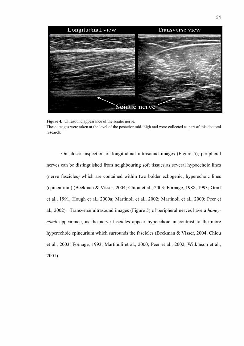
54
Figure 4. Ultrasound appearance of the sciatic nerve. These images were taken at the level of the posterior mid-thigh and were collected as part of this doctoral research.
On closer inspection of longitudinal ultrasound images (Figure 5), peripheral
nerves can be distinguished from neighbouring soft tissues as several hypoechoic lines
(nerve fascicles) which are contained within two bolder echogenic, hyperechoic lines
(epineurium) (Beekman & Visser, 2004; Chiou et al., 2003; Fornage, 1988, 1993; Graif
et al., 1991; Hough et al., 2000a; Martinoli et al., 2002; Martinoli et al., 2000; Peer et
al., 2002). Transverse ultrasound images (Figure 5) of peripheral nerves have a honey-
comb appearance, as the nerve fascicles appear hypoechoic in contrast to the more
hyperechoic epineurium which surrounds the fascicles (Beekman & Visser, 2004; Chiou
et al., 2003; Fornage, 1993; Martinoli et al., 2000; Peer et al., 2002; Wilkinson et al.,
2001).
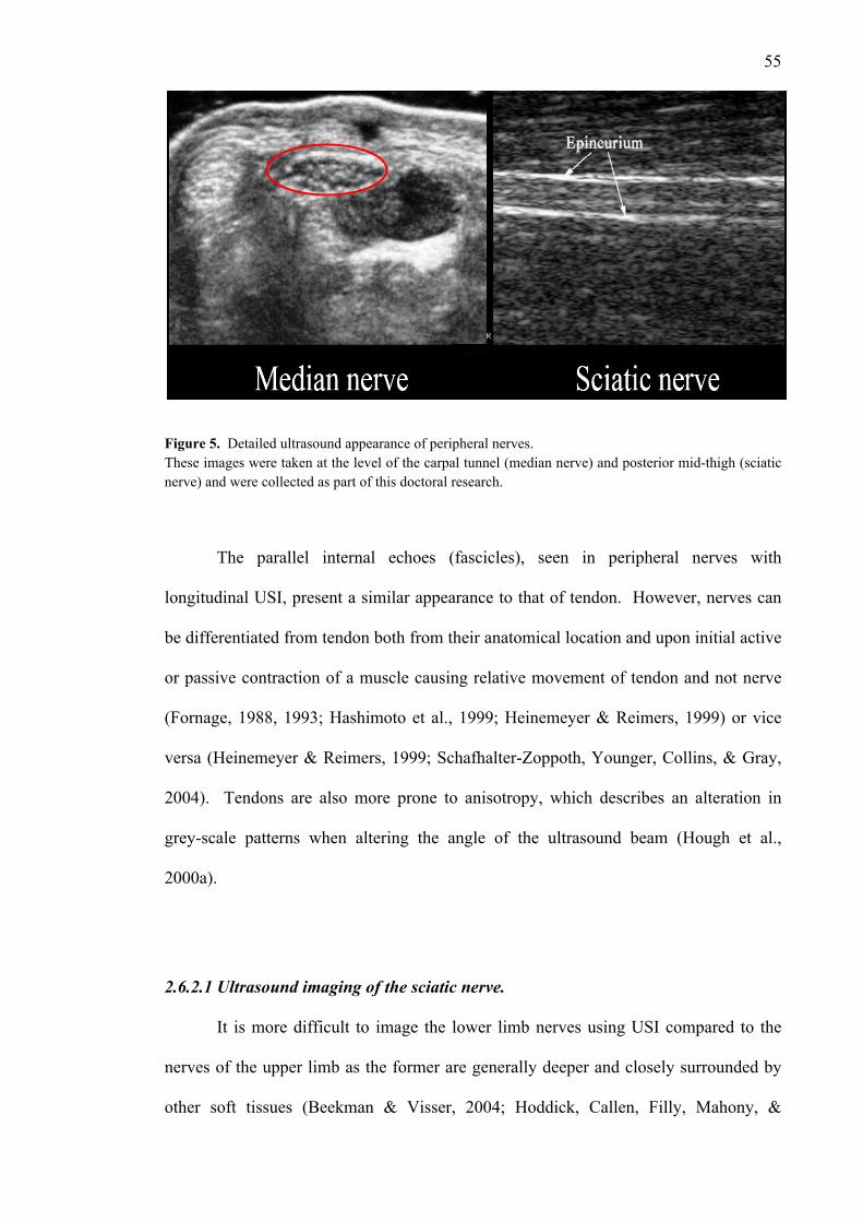
55
Figure 5. Detailed ultrasound appearance of peripheral nerves. These images were taken at the level of the carpal tunnel (median nerve) and posterior mid-thigh (sciatic nerve) and were collected as part of this doctoral research.
The parallel internal echoes (fascicles), seen in peripheral nerves with
longitudinal USI, present a similar appearance to that of tendon. However, nerves can
be differentiated from tendon both from their anatomical location and upon initial active
or passive contraction of a muscle causing relative movement of tendon and not nerve
(Fornage, 1988, 1993; Hashimoto et al., 1999; Heinemeyer & Reimers, 1999) or vice
versa (Heinemeyer & Reimers, 1999; Schafhalter-Zoppoth, Younger, Collins, & Gray,
2004). Tendons are also more prone to anisotropy, which describes an alteration in
grey-scale patterns when altering the angle of the ultrasound beam (Hough et al.,
2000a).
2.6.2.1 Ultrasound imaging of the sciatic nerve.
It is more difficult to image the lower limb nerves using USI compared to the
nerves of the upper limb as the former are generally deeper and closely surrounded by
other soft tissues (Beekman & Visser, 2004; Hoddick, Callen, Filly, Mahony, &

56
Edwards, 1984). Through most of the course of the sciatic nerve there is also a lack of
obvious anatomical landmarks which makes examination difficult (Bruhn, Van Geffen,
Gielen, & Scheffer, 2008). However, successful ultrasound imaging of the sciatic nerve
has been reported (Chan et al., 2006; Fornage, 1993; Graif et al., 1991; Heinemeyer &
Reimers, 1999; McCartney, Brauner, & Chan, 2004; Ricci, 2005; Schafhalter-Zoppoth
et al., 2004; Schwemmer et al., 2005; Schwemmer et al., 2004; Sinha & Chan, 2004;
Vloka, Hadžić, April, & Thys, 2001).
There is wide variance of locations reported for USI of the sciatic nerve and its
branches. Graif et al. (1991) describe the diameter of the sciatic nerve to range from 5 -
9mm and to be positioned between 2 - 5cm below the skin surface at the posterior mid-
thigh (PMT). Heinemeyer and Reimers (1999) scanned the sciatic nerve with
ultrasound “in the middle of the dorsal thigh” (p. 482) and the tibial nerve distal to its
bifurcation within the popliteal fossa. The accuracy of nerve identification at these
locations was reported as 74% (37 of 50 healthy participants) at the mid-dorsal thigh
and 20% (10 of 50 healthy participants) at the bifurcation (Heinemeyer & Reimers,
1999).
Other authors have noted higher rates of success, 72% (53 of 74 participants),
(Schwemmer et al., 2005) and 100%; 10 of 10 participants (Sinha & Chan, 2004), 15 of
15 participants (Bruhn et al., 2008) and 60 of 60 limbs (Ricci, 2005); for identification
of the sciatic nerve. Bruhn et al. (2008) state that the best location to visualise the
sciatic nerve is where the skin-nerve distance is the least (i.e. the nerve is closer to the
skin). They suggest that the optimal location for scanning is 5.4-10.8cm below the
subgluteal crease (Bruhn et al., 2008). Beyond the sciatic bifurcation, the tibial nerve
continues as a mid-line extension of the sciatic nerve. It is well visualised on ultrasound
imaging at the level of the popliteal crease within the popliteal fossa (Bruhn et al., 2008;

57
Heinemeyer & Reimers, 1999; McCartney et al., 2004; Ricci, 2005; Schwemmer et al.,
2005).
The addition of body movements can allow more easy visualisation and
differentiation of the sciatic nerve and its branches, a method recommended for the
assessment of the median nerve using USI (Coppieters et al., 2009; Echigo et al., 2008).
For example, Schafhalter-Zoppoth et al. (2004) found that by adding passive
dorsiflexion of the foot (when imaging the sciatic nerve with the knee in extension) the
tibial branch will move superficially (i.e. towards the skin) and medially. With the
addition of plantarflexion, the common peroneal branch will also move superficially
and laterally. These authors referred to this action as the “seesaw sign” and claim it
improves visualisation of the sciatic nerve branches. Schafhalter-Zoppoth et al. (2004)
also concluded that in longitudinal imaging, the tibial nerve branch of the sciatic nerve
will move towards the foot during ankle dorsiflexion.
2.6.3 Ultrasound imaging assessment of peripheral nerve movement.
2.6.3.1 Frame-by-frame cross-correlation analysis.
Methods of tracking grey speckle features of successive frames of ultrasound
footage, in order to quantify soft tissue motion, have been reported within the literature
(Bohs & Trahey, 1991; Hein & O'Brien, 1993; Korstanje, Selles, Henk, Hovius, &
Bosch, 2009; Korstanje, Selles, Stam, Hovius, & Bosch, 2010b). Dilley et al. (2001)
have developed a software package which uses speckle tracking to calulate nerve
motion. An ultrasound video loop can be broken into a succession of digital frames
which each depict individual pixels across a grey-scale from white to black. When the
successive frames are put together in a video sequence it is evident that the speckle

58
features (i.e. the individual pixels) have moved as a result of tissue movement. Dilley et
al. (2001) designed a computer programme that was able to calculate the relative
movement of selected pixels between frames by employing a cross-correlation
algorithm.
This image has been removed by the author of this thesis due to copyright reasons
Figure 6. Graphical representation of frame-by-frame cross-correlation analysis. A) Rectangular region of interest (ROI) within the median nerve. B) Enlargement of ROI in A. in the top panel then moved onwards by one frame in the bottom panel. C) Plot of the correlation coefficient against the pixel shift. Illustration from Coppieters, M. W., Hough, A. D., & Dilley, A. (2009). Different nerve-gliding exercises induce different magnitudes of median nerve longitudinal excursion: an in vivo study using dynamic ultrasound imaging. Journal of Orthopaedic and Sports Physical Therapy, 39(3), p. 167.
The algorithm enables calculation of the correlation coefficient between pixel
grey levels, for a selected rectangular frame, or region of interest (ROI), between two
adjacent ultrasound images (Figure 6, A). The coordinates of the second successive
ROI are offset along the horizontal ultrasound image plane and are shifted by one pixel
at a time within a predetermined range (i.e. -10 to 10 pixels) (Figure 6, B). The
correlation coefficient (measuring the degree to which two variables are linearly related)
is calculated against each individual pixel shift and therefore corresponds to the relative
movement between frames (Figure 6, C).
The peak of a quadratic equation, fitted to the maximum three correlation
coefficients, allows determination of sub-pixel accuracy. In doing so this further refines
the algorithm by more accurately assessing the maximum cross-correlation value. This
peak is equivalent to the pixel shift/movement between adjacent frames. Therefore this
algorithm establishes that the degree of pixel shift that has the highest associated

59
correlation coefficient corresponds to and represents the relative movement which is
measured.
Any relative vertical pixel shift is also taken into account in the cross-correlation
formula. For each horizontal cross-correlation analysis, between two adjacent frames,
vertical pixel shift is offset against the horizontal plane within a pre-set range (i.e. -5 to
+5 pixels) (Dilley et al., 2001). Relative movement between frames can be calculated
by this software for ultrasound footage of up to 100 frames.
This frame-by-frame cross-correlation software package can also take into
account any potential movement of the skin relative to the ultrasound transducer. To do
this, a relatively static structure or tissue within the field of the image is selected. The
selected tissue is often either a facial layer, interosseous membrane, bone or the
subcutaneous tissues. The same frame-by-frame cross-correlation analysis is performed
(as above); however this time the ROI becomes the static tissue. Any movement
detected within this analysis is then subtracted from that seen from the nerve. This
equation provides a more specific calculation of the actual nerve movement by
removing potential error which may arise from any relative movement of the ultrasound
transducer (Figure 7).
During initial analysis of this technique, Dilley et al. (2001) looked at the
reliability of measurement of several phantom controls (string and avian nerve) in
water-bath trials whereby the phantom controls were moved a specified distance and
velocity whilst ultrasound footage was captured in the longitudinal plane. Analysis was
then performed in-vivo measuring longitudinal median nerve excursion, at the forearm,
during wrist and finger movements. Graphical representation of the results determined
that the technique was reliable when comparing between 1-3 frame intervals. The
accuracy of measurement of the phantom controls was stated to be within 25
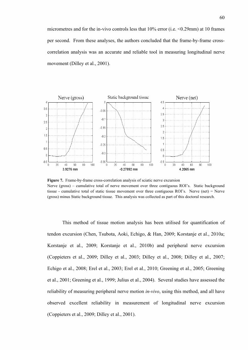
60
micrometres and for the in-vivo controls less that 10% error (i.e. <0.29mm) at 10 frames
per second. From these analyses, the authors concluded that the frame-by-frame cross-
correlation analysis was an accurate and reliable tool in measuring longitudinal nerve
movement (Dilley et al., 2001).
Figure 7. Frame-by-frame cross-correlation analysis of sciatic nerve excursion Nerve (gross) – cumulative total of nerve movement over three contiguous ROI’s. Static background tissue - cumulative total of static tissue movement over three contiguous ROI’s. Nerve (net) = Nerve (gross) minus Static background tissue. This analysis was collected as part of this doctoral research.
This method of tissue motion analysis has been utilised for quantification of
tendon excursion (Chen, Tsubota, Aoki, Echigo, & Han, 2009; Korstanje et al., 2010a;
Korstanje et al., 2009; Korstanje et al., 2010b) and peripheral nerve excursion
(Coppieters et al., 2009; Dilley et al., 2003; Dilley et al., 2008; Dilley et al., 2007;
Echigo et al., 2008; Erel et al., 2003; Erel et al., 2010; Greening et al., 2005; Greening
et al., 2001; Greening et al., 1999; Julius et al., 2004). Several studies have assessed the
reliability of measuring peripheral nerve motion in-vivo, using this method, and all have
observed excellent reliability in measurement of longitudinal nerve excursion
(Coppieters et al., 2009; Dilley et al., 2001).

61
2.6.3.2 Real-time spectral Doppler ultrasound.
Real-time spectral Doppler ultrasound measurement of longitudinal nerve
movement has also been described. The Doppler effect refers to a change in frequency
of a wave source (i.e. a moving musculoskeletal tissue) due to relative motion (Hough
et al., 2000a). This technique employs tracing of the phases of movement of each of the
spectral Doppler plots, which are represented by deflections of movement picked up by
changes in frequency of the refracted ultrasound wave. Calculations of the area under
the deflections yield velocity time integrals (VTI). These are represented along an x-
axis within the spectral Doppler sonogram. Deflections above and below the x-axis are
representative of the VTI’s and give a measurement of nerve movement either in a
proximal or distal longitudinal direction. Measurement of the area under each of the
deflections allows the VTI to be established (Hough et al., 2000a, 2000b).
Hough et al. (2000b) designed a study to specifically examine the test-retest
reliability of using spectral Doppler ultrasound measurement of longitudinal median
nerve movement during wrist extension. Measurement was taken over three tests on
both arms of sixteen healthy participants. Repeatability of test-retest measurement of
longitudinal median nerve movement was reported as having an intra-class correlation
coefficient (ICC) of 0.92 (95% CI: 0.87-0.96). The authors concluded that this was a
good level of test-retest repeatability for the assessment of median nerve excursion
(Hough et al., 2000b).
Hough and colleagues have, however, raised some concerns with regard to this
method. For example, imaging of peripheral nerves at sites where there may be
relatively more transverse neural movement may take the nerve outside of the spectral
Doppler image and therefore create image artefacts (Hough et al., 2000b). However,
the same limitation has been reported for other methods attempting to analyse

62
longitudinal tissue movement (Dilley et al., 2001) and is not unique to the Doppler
method.

63
Chapter Three. Neural mobilisation: a systematic review of randomised controlled trials with an analysis of therapeutic
efficacy
Ellis, R. F., & Hing, W. A. (2008). Neural mobilization: a systematic review of
randomized controlled trials with an analysis of therapeutic efficacy. Journal of
Manual & Manipulative Therapy, 16(1), 8-22.
This paper was awarded runner-up for The 2008 OPTP Award for Excellence in
a Published Review of the Literature (awarded by the Journal of Manual and
Manipulative Therapy). The article has also been translated and re-printed in Czech and
German journals of manual therapy.
Ellis, R., & Hing, W. (2008). Neural mobilization: A systematic review of randomized
controlled trials with an analysis of therapeutic efficacy. [Mobilizacje struktur
nerwowych – przegląd systematyczny badań z randomizacją i grupą kontrolną
oraz analiza skuteczności terapeutycznej]. Medical Rehabilitation.
[Rehabilitacja Medyczna], 12(2), 34-44.
Ellis, R., & Hing, W. (2009). Neurale mobilisation: Systematischer review
randomisierter kontrollierter studien mit einer analyse der therapeutischen
wirksamkeit. Manuelletherapie, 2, 47-57.

64
3.1 PRELUDE
In order to ascertain what research had been conducted regarding the use of
neural mobilisation, a systematic review of RCTs was conducted to examine the
available clinical research regarding neural mobilisation. Prior to this systematic review
there has been no previous systematic review published that has examined neural
mobilisation.
To date several clinical textbooks (Butler, 1991, 2000, 2005; Shacklock, 2005a)
and textbook chapters (Butler et al., 1994; Butler & Slater, 1994; Elvey & Hall, 2004;
Shacklock, 1995a) have been published which significantly emphasise the therapeutic
use of neural mobilisation. Through these sources, neural mobilisation has been
advocated and promoted. In respect to the thesis, this review enabled all previous RCTs
that have used neural mobilisation to be collated in order to review research
methodologies and resultant implications and to review whether there was available
evidence to support the wide theory base regarding neural mobilisation.
3.2 ABSTRACT
Neural mobilisation is a treatment modality used in relation to pathologies of the
nervous system. It has been suggested that neural mobilisation is an effective treatment
modality, although support of this suggestion is primarily anecdotal. The purpose of this
paper was to provide a systematic review of the literature pertaining to the therapeutic
efficacy of neural mobilisation. A search to identify randomised controlled trials
investigating neural mobilisation was conducted using the key words neural
mobilisation/mobilization, nerve mobilisation/mobilization, neural manipulative
physical therapy, physical therapy, neural/nerve glide, nerve glide exercises,
nerve/neural treatment, nerve/neural stretching, neurodynamics, and nerve/neural

65
physiotherapy. The titles and abstracts of the papers identified were reviewed to select
papers specifically detailing neural mobilisation as a treatment modality. The PEDro
scale, a systematic tool used to critique RCTs and grade methodological quality, was
used to assess these trials. Methodological assessment allowed an analysis of research
investigating therapeutic efficacy of neural mobilisation. Ten randomised clinical trials
(discussed in 11 retrieved articles) were identified that discussed the therapeutic effect
of neural mobilisation. This review highlights the lack in quantity and quality of the
available research. Qualitative analysis of these studies revealed that there is only
limited evidence to support the use of neural mobilisation. Future research needs to re-
examine the application of neural mobilisation with use of more homogeneous study
designs and pathologies; in addition, it should standardise the neural mobilisation
interventions used in the study.
Keywords: Neural Mobilisation, Neurodynamics, Randomised Controlled Trial,
Systematic Review, Therapeutic Efficacy
3.3 INTRODUCTION
In the past, neural tension was used to describe dysfunction of the peripheral
nervous system. More recently, there has been a shift away from a purely mechanical
rationale to include physiological concepts such as structure and function of the nervous
system. Neurodynamics is now a more accepted term referring to the integrated
biomechanical, physiological, and morphological functions of the nervous system
(Butler, 2000; Shacklock, 1995a, 1995b, 2005a). Regardless of the underlying
construct, it is vital that the nervous system is able to adapt to mechanical loads, and it
must undergo distinct mechanical events such as elongation, sliding, cross-sectional

66
change, angulation, and compression. If these dynamic protective mechanisms fail, the
nervous system is vulnerable to neural oedema, ischaemia, fibrosis, and hypoxia, which
may cause altered neurodynamics (Butler, 2000; Shacklock, 1995b).
When neural mobilisation is used for treatment of adverse neurodynamics, the
primary theoretical objective is to attempt to restore the dynamic balance between the
relative movement of neural tissues and surrounding mechanical interfaces, thereby
allowing reduced intrinsic pressures on the neural tissue and thus promoting optimum
physiologic function (Butler, 2000; Butler et al., 1994; Gifford, 1998; Kitteringham,
1996; Shacklock, 1995b, 2005a). The hypothesised benefits from such techniques
include facilitation of nerve gliding, reduction of nerve adherence, dispersion of noxious
fluids, increased neural vascularity, and improvement of axoplasmic flow (Butler, 2000;
Butler et al., 1994; Coppieters et al., 2003b; Gifford, 1998; Kitteringham, 1996;
Rozmaryn et al., 1998; Scrimshaw & Maher, 2001; Shacklock, 1995b, 2005a).
However, these etiological mechanisms for the clinically observed effects of neural
mobilisation still require robust validation. At present, the positive clinically observed
effect of neural mobilisation is mainly based on anecdotal evidence. Therefore, the
purpose of this paper was to systematically review and assess the therapeutic efficacy of
neural mobilisation for treatment of altered neurodynamics through evaluation of
appropriate randomised controlled trials (RCTs). It was hypothesised that the findings
might guide evidence-based practice in the clinical application of neural mobilisation.

67
3.4 METHODS
3.4.1 Literature search strategy.
A search to identify RCTs examining neural mobilisation was conducted in
March 2007. The following electronic databases were searched: MEDLINE via PubMed
(from 1966 onwards), Cumulative Index to Nursing and Allied Health Literature
(CINAHL) (from 1982 onwards), the Cochrane Controlled Trials Register in the
Cochrane Library (latest edition), SPORT-Discus (from 1830 onwards), Allied and
Complementary Medicine Database (AMED) (from 1985 onwards), Physiotherapy
Evidence Database (PEDro) (from 1953 onwards), ProQuest 5000 International,
ProQuest Health and Medical Complete, EBSCO MegaFile Premier, Science Direct
(from 1995 onwards) and Web of Science (from 1945 onwards).
The search strategy of these databases included terms and keywords related to
the intervention: neural mobilisation/mobilization, nerve mobilisation/mobilization,
neural manipulative physical therapy, physical therapy, neural/nerve glide, nerve glide
exercises, nerve/neural treatment, nerve/neural stretching, neurodynamics and
nerve/neural physiotherapy. Randomised controlled trial or RCT was the key term used
in relation to the methodology of the studies. The titles and/or abstracts of these
citations were reviewed to identify papers specifically detailing neural mobilisation
used as a treatment modality. The search was limited to studies written in or translated
to English and those utilising human subjects. There was no limitation regarding the
date the studies were published, other than the date limitations of each selected
database. In addition, the reference lists of each paper were searched to identify other
relevant papers.

68
3.4.2 Study selection.
The method for selection of relevant studies was consistent with suggested
guidelines for conducting systematic reviews (van Tulder, Furlan, Bombardier, &
Bouter, 2003). The following inclusion criteria were used to select relevant papers for
the review:
• Type of participant: participants older than 18, of either gender, and with a
clinical diagnosis consistent with neurodynamic dysfunction (musculoskeletal
conditions with symptoms of pain and/or paraesthesia indicative of compromise
of the peripheral nervous system)
• Type of study design: randomised controlled trials
• Type of intervention: use of a manual or exercise technique designed to have a
direct effect on neural tissue with the purpose of dynamically influencing (e.g.
sliding, stretching, moving, mobilising, etc.) the neural tissue
• Outcome measurements: at least one of the following outcome measurements
used to assess the status of the nervous system: pain rating (e.g. Visual Analogue
Scale [VAS], function-specific pain VAS (i.e. work- or sport-related pain), pain
and or range of movement (ROM) during neural tissue provocation tests
(NTPT), functional disability scores (e.g. Short-form McGill Pain
Questionnaire, Northwick Park Questionnaire, and Oswestry Disability Index).
3.4.3 Methodological quality assessment.
Three reviewers independently assessed the methodological quality of each
RCT. The PEDro Scale (Table 2), developed by The Centre of Evidence-Based
Physiotherapy (CEBP), was utilised to assess each paper (CEBP, 2006). The PEDro
Scale, an 11-item scale, is a validated, reliable, and versatile tool used to rate RCTs for
69
the PEDro Database (Clark et al., 1999; Maher, Sherrington, Herbert, Moseley, &
Elkins, 2003; Overington, Goddard, & Hing, 2006). The PEDro scale has been used as a
measure of methodological quality in many systematic literature reviews (Ackerman &
Bennell, 2004; Bleakley, McDonough, & MacAuley, 2004; Hakkennes & Keating,
2005; Harvey, Herbert, & Crosbie, 2002b; O'Shea, Taylor, & Paratz, 2004).
An overall score of methodological quality, or quality score (QS), was
determined for each paper by each of the three reviewers as a total of positive scores for
10 of the 11 items (i.e. N/10). Unlike the other items, Criterion One of the PEDro scale
relates to external validity and was not used in the final total PEDro score (CEBP, 2006;
Maher et al., 2003; Overington et al., 2006). A consensus method was used to discuss
and resolve discrepancies between the markings of each paper between the reviewers.
The agreed QS for each paper are included in Table 3.
The various items of the PEDro Score deal with different aspects of RCT
analysis including internal validity, external validity, and statistics. In order to allow
quantitative analysis of the methodological quality of a systematic review, van Tulder et
al. (2003) recommended the analysis of the internal validity criteria of any rating tool.
For the PEDro Scale, seven items relating to internal validity were identified. These
seven items include items 2, 3, and 5 through 9. An internal validity score (IVS) has
also been used in other systematic reviews (Reid & Rivett, 2005) to allow calculation of
the number of internal validity criteria met for that particular rating system and to
thereby give an assessment of methodological quality. It was decided to calculate an
IVS for this review based on the relevant internal validity criteria of the PEDro Scale.
The positive scores of each of these seven items were added together to calculate the
IVS.

70
Table 2. The PEDro Scale Modified from Maher, C. G., Sherrington, C., Herbert, R. D., Moseley, A. M., & Elkins, M. (2003). Reliability of the PEDro scale for rating randomised controlled trials. Physical Therapy, 83(3), 713-721.
Score Criteria No Yes
1. Eligibility criteria were specified * 2. Subjects randomly allocated to groups NO (0) YES (1) 3. Allocation was concealed NO (0) YES (1) 4. Groups similar at baseline regarding the most important prognostic factors NO (0) YES (1) 5. Blinding of all subjects NO (0) YES (1) 6. Blinding of all therapists who administered therapy NO (0) YES (1) 7. Blinding of all assessors who measured at least one outcome NO (0) YES (1) 8. Measures of at least one key outcome were obtained from more than 85% of initially allocated subjects NO (0) YES (1) 9. All subjects for whom outcome measures were available received treatment or control as allocated, or if this was not the case, at least one outcome measure analysed using “intention to treat” analysis NO (0) YES (1) 10. The results of between-group statistical comparisons are reported for at least one key outcome NO (0) YES (1) 11. The study provides both point measures and measures or variability for at least one key outcome NO (0) YES (1) Total N/10
* Criteria 1 score is not included in the overall PEDro rating.
To stratify methodological quality, the summated score of the 7-item IVS,
calculated from the initial PEDro score (QS), was divided into three categories. A study
of high methodological quality obtained IVS values of 6 - 7, a moderate quality
obtained IVS values between 4 - 5, and a limited quality was scored between 0 - 3. This
decision was made based on even cut-off points between 0 and 7.
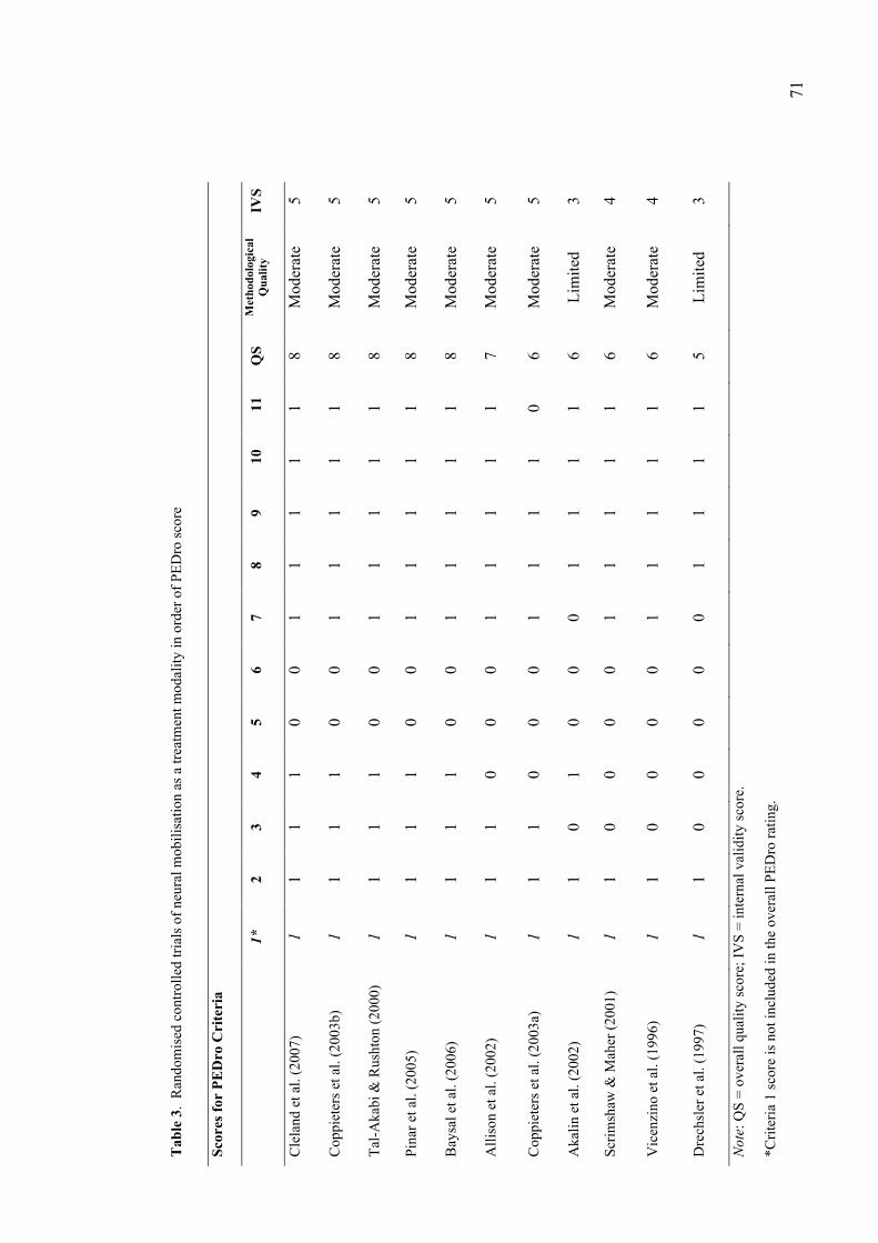
71
Tab
le 3
. R
ando
mis
ed c
ontro
lled
trial
s of n
eura
l mob
ilisa
tion
as a
trea
tmen
t mod
ality
in o
rder
of P
EDro
scor
e
Scor
es fo
r PE
Dro
Cri
teri
a
1*
2
3 4
5 6
7 8
9 10
11
Q
S M
etho
dolo
gica
l Q
ualit
y IV
S
Cle
land
et a
l. (2
007)
1
1 1
1 0
0 1
1 1
1 1
8 M
oder
ate
5
Cop
piet
ers e
t al.
(200
3b)
1 1
1 1
0 0
1 1
1 1
1 8
Mod
erat
e 5
Tal-A
kabi
& R
usht
on (2
000)
1
1 1
1 0
0 1
1 1
1 1
8 M
oder
ate
5
Pina
r et a
l. (2
005)
1
1 1
1 0
0 1
1 1
1 1
8 M
oder
ate
5
Bay
sal e
t al.
(200
6)
1 1
1 1
0 0
1 1
1 1
1 8
Mod
erat
e 5
Alli
son
et a
l. (2
002)
1
1 1
0 0
0 1
1 1
1 1
7 M
oder
ate
5
Cop
piet
ers e
t al.
(200
3a)
1 1
1 0
0 0
1 1
1 1
0 6
Mod
erat
e 5
Aka
lin e
t al.
(200
2)
1 1
0 1
0 0
0 1
1 1
1 6
Lim
ited
3
Scrim
shaw
& M
aher
(200
1)
1 1
0 0
0 0
1 1
1 1
1 6
Mod
erat
e 4
Vic
enzi
no e
t al.
(199
6)
1 1
0 0
0 0
1 1
1 1
1 6
Mod
erat
e 4
Dre
chsl
er e
t al.
(199
7)
1 1
0 0
0 0
0 1
1 1
1 5
Lim
ited
3
Not
e: Q
S =
over
all q
ualit
y sc
ore;
IVS
= in
tern
al v
alid
ity sc
ore.
*Crit
eria
1 sc
ore
is n
ot in
clud
ed in
the
over
all P
EDro
ratin
g.

72
3.4.4 Analysis of therapeutic efficacy.
When RCTs are heterogeneous, there is no available method to quantitatively
assess the relative benefit (or lack thereof) of one intervention versus another because
the studies may not be comparing similar patient populations or interventions. In
situations where the heterogeneity of primary studies prevents use of a quantitative
meta-analysis to summarise the results, recommendations are typically made based on a
qualitative assessment of the strength of the evidence (Reid & Rivett, 2005). The RCTs
reviewed for this paper were considered heterogeneous because they explored a variety
of pathologies and different types of neural mobilisation techniques. Consequently, a
quantitative meta-analysis was not appropriate and results were analysed in a qualitative
fashion. The qualitative assessment used within this review was an adaptation of those
used by several authors (Karjalainen et al., 2001; Reid & Rivett, 2005; van Tulder et al.,
2003) modified specifically to include the IVS obtained from the PEDro Scale:
• Level 1: Strong evidence: provided by generally consistent findings in multiple
RCTs of high quality.
• Level 2: Moderate evidence: provided by generally consistent findings in one
RCT of high quality and one or more of lower quality.
• Level 3: Limited evidence: provided by generally consistent findings in one
RCT of moderate quality and one or more low-quality RCTs.
• Level 4: Insufficient evidence: provided by generally consistent findings of one
or more RCTs of limited quality, or when no RCTs were available, or when
studies provided conflicting results.
73
3.4.5 Clinical benefit.
Lastly, to determine whether a clinical benefit for neural mobilisation could be
concluded, a ranking system similar to that used by Linton and van Tulder (2001) was
used. A positive effect was concluded if the intervention (i.e. neural mobilisation) was
statistically significantly more beneficial compared to the control for at least one key
outcome variable, a negative effect if the intervention was less effective than the
control, and a neutral effect was concluded where the intervention and control did not
statistically differ significantly for any of the outcome variables (Linton & van Tulder,
2001).
3.5 RESULTS
3.5.1 Selection of studies.
Ten RCTs, represented by 11 published articles (Akalin et al., 2002; Allison,
Nagy, & Hall, 2002; Baysal et al., 2006; Cleland, Childs, Palmer, & Eberhart, 2007;
Coppieters, Stappaerts, Wouters, & Janssens, 2003a; Coppieters et al., 2003b;
Drechsler, Knarr, & Snyder-Mackler, 1997; Pinar et al., 2005; Scrimshaw & Maher,
2001; Tal-Akabi & Rushton, 2000; Vicenzino et al., 1996), satisfied the inclusion
criteria following the electronic and manual reference list searches. The articles
published by Coppieters et al. (2003a, 2003b) are from the same subject group and were
thus classified as one RCT. These studies are summarised in Table 5.

74
3.5.2 Methodological quality.
The methodological quality for each paper, represented by the IVS, is detailed in
Table 3. Nine of 11 studies (Allison et al., 2002; Baysal et al., 2006; Cleland et al.,
2007; Coppieters et al., 2003a, 2003b; Pinar et al., 2005; Scrimshaw & Maher, 2001;
Tal-Akabi & Rushton, 2000; Vicenzino et al., 1996) reviewed were given an IVS of 4
or 5 and were of moderate methodological quality. Two of the studies (Akalin et al.,
2002; Drechsler et al., 1997) were given an IVS of 3, suggesting limited methodological
quality. Table 4 presents statistics relating to the percentage of each item that was
satisfied for an IVS score.
Table 4. Number and percentage of the studies meeting each PEDro criteria
PEDro Criteria Number meeting
criterion (N)
Percent meeting
criterion (%)
1. Eligibility criteria specified (yes/no) 11 100
2. Subjects randomly allocated to groups (yes/no) 11 100
3. Allocation was concealed (yes/no) 7 64
4. Groups similar at baseline (yes/no) 6 55
5. Subjects were blinded to group allocation (yes/no)
0 0
6. Therapists who administered therapy were blinded (yes/no)
0 0
7. Assessors were blinded (yes/no) 9 82
8. Minimum 85% follow-up (yes/no) 9 100
9. Intent to treat analysis for at least 1 key variable (yes/no)
9 100
10. Results of statistical analysis between groups reported (yes/no)
9 100
11. Point measurements and variability reported (yes/no)
10 91

75
All of the 11 studies satisfied the items relating to random allocation of subjects,
measures of one key outcome taken from greater than 85% of the population, use of
intention-to-treat analysis (where this was required due to a drop-out group), and results
of statistical analysis reported (items 2, 8, 9, and 10).
All 11 studies did not satisfy items 5 and 6, which relate to subject and therapist
blinding. Two studies (Akalin et al., 2002; Drechsler et al., 1997) did not satisfy item 7,
which relates to rater blinding. This suggests that these two studies lacked all three
forms of blinding (subject, therapist, and rater). The other 9 studies were single-blinded
(rater-blinded) studies. There was no clear trend established for item 4, which relates to
concealed allocation of subjects.
3.5.3 Study characteristics.
All ten RCTs used different methods of application of neural mobilisation (e.g.
cervical lateral glide, slump sliders, peripheral nerve sliders, etc.), and some studies
chose to combine these techniques with home-based neural mobilisation exercises.
There were also differing neurodynamic dysfunctions examined, including lateral
epicondyalgia, carpal tunnel syndrome, post-operative spinal surgery, non-radicular low
back pain, and neurogenic cervico-brachial pain syndrome. Therefore, all ten RCTs
were clinically and therapeutically heterogeneous, necessitating a qualitative analysis
for summarising the results. Table 5 contains details of study characteristics.
3.5.4 Therapeutic efficacy.
Of the 11 studies identified, 6 different categories or types of treatment were
identified (Table 6). Using the qualitative rating system, as mentioned earlier, it appears

76
there is limited evidence (Level 3) to support the use of neural mobilisation that
involves active nerve and flexor tendon gliding exercises of the forearm (Akalin et al.,
2002; Baysal et al., 2006; Pinar et al., 2005), cervical contralateral glides (Coppieters et
al., 2003a, 2003b; Vicenzino et al., 1996), and Upper Limb Tension Test 2b (ULTT2b)
mobilisation (Drechsler et al., 1997; Tal-Akabi & Rushton, 2000) in the treatment of
altered neurodynamics or neurodynamic dysfunction. There was inconclusive evidence
(Level 4) to support the use of neural mobilisation involving slump stretches (Cleland et
al., 2007) and combinations of neural mobilisation techniques (Allison et al., 2002;
Scrimshaw & Maher, 2001) in the treatment of altered neurodynamics or neurodynamic
dysfunction.
3.5.5 Clinical benefit.
Table 5 lists the study details of the 11 studies. More studies found a positive
effect (Akalin et al., 2002; Allison et al., 2002; Baysal et al., 2006; Cleland et al., 2007;
Coppieters et al., 2003a, 2003b; Pinar et al., 2005; Vicenzino et al., 1996) than a neutral
effect (Drechsler et al., 1997; Scrimshaw & Maher, 2001; Tal-Akabi & Rushton, 2000).

77
Tab
le 5
. R
ando
mis
ed c
ontro
lled
trial
s of n
eura
l mob
ilisa
tion
as a
trea
tmen
t mod
ality
Aut
hor
Patie
nt d
emog
raph
ics
Inte
rven
tion
Gro
up (I
G)
Com
pari
son
Gro
up (C
G)
Out
com
e R
esul
t IV
S Q
S
Cle
land
et a
l. (2
007)
N
=30
(9 m
ale,
21
fem
ale)
-A
ge ra
nge
18-6
0 ye
ars
-Mea
n ag
e (y
ears
) IG
40.
0 ( ±
12.2
), C
G 3
9.4
(±11
.3)
Dur
atio
n sy
mpt
oms (
wee
ks)
IG 1
4.5
(±8.
0), C
G 1
8.5
(±12
.5)
16 s
ubje
cts w
ith lo
w b
ack
pain
Sa
me
as c
ontro
l plu
s:
Slum
ped
stre
tchi
ng e
xerc
ise
(pos
ition
hel
d 30
seco
nds,
5 re
petit
ions
) H
ome
exer
cise
slu
mp
stre
tche
s (2
repe
titio
ns fo
r 30
seco
nds)
2
x w
eek
for 3
wee
ks
14 s
ubje
cts w
ith lo
w b
ack
pain
5-
min
ute
cycl
e w
arm
-up
Lum
bar s
pine
mob
ilisa
tion
(Pos
terio
r-an
terio
r m
obili
satio
ns to
hyp
omob
ile
lum
bar
segm
ents
, gra
de 3
-4)
Stan
dard
ised
exe
rcis
e pr
ogra
m (p
elvi
c til
ts,
brid
ging
, squ
ats,
quad
rupe
d al
tern
ate
arm
/leg
activ
ities
; 2
sets
10
repe
titio
ns e
ach
exer
cise
) 2
x w
eek
for 3
wee
ks
Out
com
es w
ere
mea
sure
d pr
e-
and
post
-trea
tmen
t 1.
Bod
y di
agra
m (f
or d
istri
butio
n of
sym
ptom
s)
2. N
umer
ic p
ain
ratin
g sc
ale
(NPR
S)
3. M
odifi
ed O
swes
try d
isab
ility
in
dex
(OD
I)
4. F
ear a
void
ance
bel
iefs
qu
estio
nnai
re
No
base
line
diff
eren
ces b
etw
een
grou
ps (p
> 0.
05).
At d
isch
arge
, pat
ient
s who
rece
ived
sl
umpe
d st
retc
hing
dem
onst
rate
d si
gnifi
cant
ly g
reat
er im
prov
emen
ts in
di
sabi
lity
(9.7
poi
nts o
n th
e O
DI,
p<
0.00
1), p
ain
(0.9
3 po
ints
on
the
NPR
S, p
=0.0
01),
and
cent
ralis
atio
n of
sym
ptom
s (p<
0.01
) tha
n pa
tient
s w
ho d
id n
ot.
The
betw
een-
grou
p co
mpa
rison
s su
gges
t tha
t slu
mp
stre
tchi
ng is
be
nefic
ial f
or im
prov
ing
shor
t-ter
m
disa
bilit
y, p
ain,
and
cen
tralis
atio
n of
sy
mpt
oms.
5 8
Bay
sal e
t al.
(200
6)
N=3
6 (3
6 fe
mal
e pa
tient
s –
all w
ith c
linic
al a
nd
elec
troph
ysio
logi
cal e
vide
nce
of C
TS
All
with
bila
tera
l inv
olve
men
t M
ean
age
– G
roup
1 4
7.8
± 5.
5; G
roup
2 5
0.1
± 7.
3;
Gro
up 3
51.
4 ±
5.2
Mea
n du
ratio
n of
sym
ptom
s (y
ears
) – G
roup
1 1
.5 ±
1.6
; G
roup
2 1
.4 ±
0.8
; Gro
up 3
1.
4 ±
0.8
8 ev
entu
al d
ropo
uts
- Gro
up 1
- (N
=12)
cus
tom
m
ade
neut
ral v
olar
splin
t (w
orn
for 3
wee
ks);
exer
cise
th
erap
y (n
erve
and
tend
on
glid
ing
exer
cise
s as d
escr
ibed
by
Tot
ten
& H
unte
r, 19
91) 5
se
ssio
ns d
aily
, eac
h ex
erci
se
repe
ated
10x
/ses
sion
–fo
r 3
wee
ks
- Gro
up 2
- (N
=12)
cus
tom
m
ade
neut
ral v
olar
splin
t (w
orn
for 3
wee
ks);
Ultr
asou
nd (1
5min
/ses
sion
to
palm
ar c
arpa
l tun
nel,
1MH
z,
1.0w
/cm
2, 1
:4, 5
cm2
trans
duce
r) 1
Rx/
day,
eve
ry 5
da
ys fo
r 3 w
eeks
(tot
al 1
5 R
x’s)
Expe
rimen
tal g
roup
s 1 a
nd 3
th
at in
corp
orat
ed n
erve
gl
idin
g ex
erci
ses a
nd a
co
mpa
rison
gro
up th
at d
id
not i
ncor
pora
te th
ese
exer
cise
s.
Com
paris
on b
etw
een
grou
ps
2 an
d 3
as th
e on
ly d
iffer
ence
in
inte
rven
tion
prog
ram
s was
th
at g
roup
3 u
sed
nerv
e gl
idin
g ex
erci
ses a
nd g
roup
2
did
not.
All
mea
sure
s pre
-Rx,
end
of R
x,
and
8 w
eeks
F/U
1.
pa
in (V
AS)
2.
Ti
nel’s
sign
3.
Ph
alen
’s si
gn
4.
mea
n st
atic
two-
poin
t di
scrim
inat
ion
– pu
lp o
f ra
dial
thre
e di
gits
5.
ha
nd-g
rip st
reng
th –
han
d-he
ld d
ynam
omet
er
6.
pinc
h st
reng
th –
bet
wee
n th
umb
and
little
fing
er –
dy
nam
omet
er
7.
sym
ptom
-sev
erity
scal
e qu
estio
nnai
re (1
1 ite
ms)
8.
fu
nctio
nal s
tatu
s sca
le
ques
tionn
aire
(8 it
ems)
9.
m
edia
n m
otor
ner
ve
cond
uctio
n –
mot
or d
ista
l
No
sign
ifica
nt d
iffer
ence
s bet
wee
n gr
oups
at t
he e
nd o
f Rx
and
8 w
eeks
fo
llow
-up
of a
ll m
easu
res o
f Tr
eatm
ent E
ffec
t (m
easu
res 1
, 5, 6
, 7,
8, 9
, 10)
W
ithin
gro
up c
ompa
rison
s sho
wed
si
gnifi
cant
impr
ovem
ent s
een
in a
ll 3
grou
ps in
Tin
el’s
and
Pha
len’
s sig
ns
at e
nd o
f Rx
and
8 w
eeks
follo
w-u
p Si
gnifi
cant
impr
ovem
ent s
een
in a
ll 3
grou
ps in
grip
and
pin
ch st
reng
th a
t 8
wee
ks fo
llow
-up.
N
o ch
ange
s see
n in
two-
pt
disc
rimin
atio
n W
ithin
-gro
up a
naly
sis s
how
ed
sign
ifica
nt im
prov
emen
t in
pain
,
4 8

78
- Gro
up 3
-(N
=12)
cus
tom
m
ade
neut
ral v
olar
splin
t (w
orn
for 3
wee
ks);
exer
cise
th
erap
y (n
erve
and
tend
on
glid
ing
exer
cise
s as d
escr
ibed
by
Tot
ten
& H
unte
r, 19
91) 5
se
ssio
ns d
aily
, eac
h ex
erci
se
repe
ated
10x
/ses
sion
–
cont
inue
d fo
r 3 w
eeks
; U
ltras
ound
(1
5min
utes
/ses
sion
to p
alm
ar
carp
al tu
nnel
, 1M
Hz,
1.
0w/c
m2,
1:4
, 5cm
2 tra
nsdu
cer)
1 R
x/da
y, e
very
5
days
for 3
wee
ks (t
otal
15
Rx’
s)
late
ncy
EMG
of a
bduc
tor
polli
cis
10.
sens
ory
dist
al la
tenc
y –
EMG
of
abd
ucto
r pol
licis
11
. ne
edle
EM
G o
f abd
ucto
r po
llici
s bre
vis –
look
ing
for d
ener
vatio
n
12.
patie
nt sa
tisfa
ctio
n su
rvey
(at
8wee
ks fo
llow
-up
only
)
sym
ptom
and
func
tiona
l sca
les o
f all
thre
e gr
oups
at e
nd-R
x an
d 8
wee
ks
follo
w-u
p G
roup
3 h
ad si
gnifi
cant
ly th
e be
st
resu
lts a
t 8 w
eeks
follo
w-u
p pa
tient
sa
tisfa
ctio
n qu
estio
nnai
re
Med
ian
sens
ory
dist
al la
tenc
y si
gnifi
cant
ly d
ecre
ased
in g
roup
s 1
and
3 at
end
-Rx
and
8 w
eeks
follo
w-
up
No
sign
ifica
nt c
hang
e se
en in
med
ian
mot
or d
ista
l lat
ency
of a
ll 3
grou
ps
P<0.
05
In su
mm
ary,
bet
wee
n-gr
oup
anal
ysis
re
veal
ed n
o di
ffer
ence
bet
wee
n gr
oups
, but
with
in-g
roup
ana
lysi
s sh
owed
that
all
grou
ps im
prov
ed a
st
atis
tical
ly si
gnifi
cant
am
ount
for a
m
ajor
ity o
f out
com
e m
easu
res.
Pi
nar e
t al.
(200
5)
N =
26 (f
emal
e)
Age
rang
e 35
-55
year
s D
urat
ion
of sy
mpt
oms (
mo)
C
G 4
7.6
(± 6
.8),
IG 4
9.6
(±
5.2)
14 p
atie
nts (
19 h
ands
) pa
tient
s dia
gnos
ed w
ith e
arly
-m
iddl
e st
ages
CTS
In
add
ition
to sp
lint w
earin
g an
d pa
tient
trai
ning
pro
gram
tre
ated
with
ner
ve g
lidin
g ex
erci
ses 1
0 re
petit
ions
5 se
ts
a da
y fo
r 10
wee
ks,
com
bine
d w
ith a
con
serv
ativ
e tre
atm
ent p
rogr
am
12 p
atie
nts (
16 h
ands
) pa
tient
s dia
gnos
ed w
ith e
arly
-m
iddl
e st
ages
CTS
Tr
eate
d in
vol
ar sp
lint i
n ne
utra
l wor
n da
y &
nig
ht fo
r 6-
wee
ks, t
hen
nigh
t onl
y fr
om
wee
k 6-
10, a
nd a
pat
ient
tra
inin
g pr
ogra
m fo
r the
m
odifi
catio
n of
func
tiona
l ac
tiviti
es (a
void
repe
titiv
e ac
tiviti
es, e
tc.)
with
a
cons
erva
tive
treat
men
t pr
ogra
m.
Und
erta
ken
befo
re a
nd a
fter a
10-
wee
k tre
atm
ent p
rogr
am.
1. T
inel
’s T
est
2. P
hale
n’s T
est
3. P
ain
(VA
S) o
ver a
day
4.
Mot
or F
unct
ion
– m
anua
l m
uscl
e te
stin
g, a
nd g
rip st
reng
th
(Jam
ar h
and
dyna
mom
eter
) 5.
Sen
sory
eva
luat
ion
(Sem
mes
-W
eins
tein
mon
ofila
men
t [SW
M]
&2-
poin
t dis
crim
inat
ion
test
[2
PD])
6.
Ele
ctro
phys
iolo
gica
l tes
t –
med
ian
& u
lnar
ner
ve. d
ista
l la
tenc
ies
Bet
wee
n-gr
oup
com
paris
ons f
or
thes
e sa
me
varia
bles
show
ed n
o st
atis
tical
ly si
gnifi
cant
diff
eren
ces
pre-
treat
men
t or p
ost-t
reat
men
t, so
th
e gr
oups
wer
e si
mila
r. B
oth
grou
ps m
ade
stat
istic
ally
si
gnifi
cant
impr
ovem
ents
in p
ain,
pi
nch
& g
rip st
reng
th, a
nd se
nsiti
vity
te
stin
g ac
cord
ing
to in
tra-g
roup
or
“with
in-g
roup
” an
alys
is (p
< 0.
05).
A
stat
istic
ally
sign
ifica
nt re
sult
favo
urin
g th
e in
corp
orat
ion
of n
eura
l gl
idin
g ex
erci
ses –
with
mor
e ra
pid
pain
redu
ctio
n, a
nd g
reat
er fu
nctio
nal
5 8

79
impr
ovem
ent e
spec
ially
in g
rip
stre
ngth
(p<
0.05
). Ta
bles
2 –
4 p
rovi
de p
ost-t
reat
men
t da
ta o
n el
ectro
phys
iolo
gica
l, Ti
nel’s
, an
d Ph
alen
’s te
st fi
ndin
gs.
Sinc
e al
l su
bjec
ts h
ad “
posi
tive/
path
olog
ic”
findi
ngs p
re-tr
eatm
ent,
the
auth
ors
coul
d us
e th
ese
2x2
cont
inge
ncy
tabl
es to
gen
erat
e a
num
ber n
eede
d to
tre
at to
see
whe
ther
ther
e w
as a
cl
inic
ally
impo
rtant
eff
ect f
avou
ring
neur
al g
lidin
g ex
erci
ses o
n th
ese
parti
cula
r out
com
es.
Cop
piet
ers e
t al
. (20
03b)
(R
efer
ence
s de
scrib
ed
toge
ther
due
to
pape
rs’
diff
eren
t ou
tcom
es o
n th
e sa
me
subj
ect s
ampl
e w
ith th
e sa
me
inte
rven
tion
tech
niqu
e).
Cop
piet
ers e
t al
. (20
03a)
N=2
0 (1
6 fe
mal
es, 4
mal
es)
Age
rang
e 35
- 65
year
s -M
ean
age
(yea
rs)
IG 4
9.1
(±14
.1),
CG
46.
6 (±
12.1
) -M
ean
dura
tion
of sy
mpt
oms
(mo)
IG
2.7
, CG
3.2
A
s abo
ve
10 su
bjec
ts w
ith b
rach
ial o
r ce
rvic
obra
chia
l neu
roge
nic
pain
R
ecei
ved
neur
al m
obili
satio
n tre
atm
ent (
cont
rala
tera
l glid
e of
cer
vica
l seg
men
t)
Cer
vica
l con
trala
tera
l glid
e C
5-T1
. Sev
eral
com
pone
nts
of th
e ne
ural
tens
ion
prov
ocat
ion
test
of t
he
med
ian
nerv
e (N
TPT1
) wer
e ap
plie
d.
Patie
nts i
n su
pine
rece
ived
a
late
ral t
rans
latio
n m
ovem
ent
10 su
bjec
ts w
ith b
rach
ial o
r ce
rvic
obra
chia
l neu
roge
nic
pain
R
ecei
ved
ultra
soun
d do
se o
f 0.
5 W
/cm²,
5 m
inut
es
sona
tion
time,
20%
size
of
head
5cm²,
freq
uenc
y 1M
Hz.
Pu
lsed
ultr
asou
nd fo
r 5
min
utes
ove
r the
mos
t pai
nful
ar
ea (0
.5 W
/cm²,
1MH
z,
treat
men
t hea
d 5c
m²).
A
rm w
as in
unl
oade
d po
sitio
n. U
ltras
ound
cho
sen
beca
use
it do
es n
ot in
volv
e an
y m
ovem
ent o
f per
iphe
ral
Out
com
es w
ere
mea
sure
d pr
e-
and
post
-trea
tmen
t 1.
Elb
ow e
xten
sion
RO
M d
urin
g N
TPT1
2.
Pai
n (V
AS)
3.
Sym
ptom
dis
tribu
tion
M
easu
rem
ents
take
n pr
e- a
nd p
ost-
tre
atm
ent
1. E
lbow
ext
ensi
on R
OM
dur
ing
NTP
T1
2. P
ain
inte
nsity
dur
ing
the
NTP
T1 V
AS
Sign
ifica
nt d
iffer
ence
s in
treat
men
t ef
fect
s bet
wee
n tw
o gr
oups
cou
ld b
e ob
serv
ed fo
r all
outc
ome
mea
sure
s (p≤0
.306
). Fo
r the
mob
ilisa
tion
grou
p, th
e in
crea
se in
elb
ow
exte
nsio
n fr
om 1
37.3
º to
156.
7º, t
he
43%
dec
reas
e in
are
a of
sym
ptom
di
strib
utio
n an
d de
crea
se in
pai
n fr
om 7
.3 to
5.8
wer
e si
gnifi
cant
(p≤0
003)
. For
ultr
asou
nd g
roup
, th
ere
wer
e no
sign
ifica
nt d
iffer
ence
s O
n th
e in
volv
ed si
de, t
he sh
ould
er
gird
le e
leva
tion
forc
e oc
curr
ed e
arlie
r an
d th
e am
ount
of f
orce
at t
he e
nd o
f th
e te
st w
as su
bsta
ntia
lly th
ough
not
si
gnifi
cant
ly g
reat
er o
n th
e un
invo
lved
side
at t
he c
orre
spon
ding
R
OM
. Tog
ethe
r with
a si
gnifi
cant
re
duct
ion
in p
ain
perc
eptio
n af
ter t
he
cerv
ical
mob
ilisa
tions
, a c
lear
5 5
8 6

80
away
from
thei
r inv
olve
d si
de, w
hile
mim
icki
ng
cerv
ical
side
flex
ion
or
rota
tion.
A
fter 2
tria
ls, 3
repe
titio
ns
wer
e pe
rfor
med
.
nerv
es.
tend
ency
tow
ard
norm
aliz
atio
n of
the
forc
e cu
rve
coul
d be
obs
erve
d,
nam
ely,
a si
gnifi
cant
dec
reas
e in
fo
rce
gene
ratio
n an
d a
dela
yed
onse
t. Th
e co
ntro
l gro
up d
emon
stra
ted
no
diff
eren
ces.
Alli
son
et a
l. (2
002)
N
=30
(20
fem
ales
, 10
m
ales
) A
ge ra
nge
18-7
5 ye
ars
Med
ian
dura
tion
of
sym
ptom
s (m
o)
NT
12
IQR
48
AT
72
IQR
72
CG
12
IQ
R 9
1
Neu
ral t
issu
e m
anua
l the
rapy
(N
T) —
Cer
vica
l lat
eral
glid
e,
shou
lder
gird
le o
scill
atio
n,
mus
cle
re-e
duca
tion,
hom
e m
obili
satio
n.
For 8
wee
ks.
Arti
cula
r tre
atm
ent g
roup
(A
T ) G
leno
hum
eral
join
t m
obili
satio
n, th
orac
ic
mob
ilisa
tion
and
hom
e ex
erci
se.
For 8
wee
ks.
Rec
eive
d no
inte
rven
tion
for
the
initi
al 8
wee
ks
(The
n at
the
end
of th
e st
udy
they
wer
e gi
ven
neur
al
treat
men
t as a
cro
ss-o
ver
prot
ocol
.)
Mea
sure
men
ts ta
ken
pre-
treat
men
t 4 w
eeks
into
trea
tmen
t an
d po
st -t
reat
men
t. 1.
McG
ill p
ain
ques
tionn
aire
2.
Nor
thw
ick
Park
que
stio
nnai
re
3. P
ain
(VA
S)
Bot
h in
terv
entio
n gr
oups
wer
e ef
fect
ive
in im
prov
ing
pain
inte
nsity
, pa
in q
ualit
y sc
ores
, and
func
tiona
l di
sabi
lity
leve
ls.
How
ever
, a g
roup
diff
eren
ce w
as
obse
rved
for t
he V
AS
scor
es a
t 8
wee
ks w
ith th
e “n
eura
l man
ual
ther
apy”
gro
up h
avin
g a
sign
ifica
ntly
lo
wer
scor
e.
5 7
Aka
lin e
t al.
(200
2)
N=3
6 (2
mal
e, 3
4 fe
mal
e)
Age
rang
e 38
-64
year
s M
ean
age
51.9
3 ±5
.1 y
ears
M
ean
grou
p ag
e (y
ears
) C
G 5
2.16
(±5.
6), I
G 5
1.7
(±5.
5)
Dur
atio
n of
sym
ptom
s (m
o)
CG
47.
6 (±
6.8
), IG
49.
6 (±
5.
2)
18 su
bjec
ts w
ith C
TS
Sam
e as
con
trol p
lus:
Te
ndon
glid
es in
5 p
ositi
ons.
Med
ian
nerv
e ex
erci
ses i
n 6
posi
tions
. (Ea
ch p
ositi
on w
as
mai
ntai
ned
for 5
seco
nds;
10
repe
titio
ns o
f eac
h ex
erci
se w
ere
done
5 ti
mes
a
day)
Fo
r 4 w
eeks
18 su
bjec
ts w
ith C
TS
Cus
tom
-mad
e ne
utra
l vol
ar
wris
t spl
int w
as in
stru
cted
to
be w
orn
all n
ight
and
dur
ing
the
day
as m
uch
as p
ossi
ble
fo
r 4 w
eeks
Und
erta
ken
pre-
treat
men
t and
8
wee
ks p
ost-t
reat
men
t 1.
Pha
len’
s sig
n 2.
Tin
el’s
sign
3.
2-p
oint
dis
crim
inat
ion
4. G
rip st
reng
th
5. P
inch
stre
ngth
6.
Sym
ptom
seve
rity
scor
e 7.
Fun
ctio
nal s
tatu
s sco
re
A p
atie
nt sa
tisfa
ctio
n in
vest
igat
ion
unde
rtake
n by
te
leph
one
8.3
(± 2
.5) m
onth
s po
st-tr
eatm
ent
At t
he e
nd o
f tre
atm
ent,
with
in-g
roup
an
alys
is sh
owed
a si
gnifi
cant
im
prov
emen
t was
obt
aine
d in
all
para
met
ers i
n bo
th g
roup
s. Th
e ne
rve
and
tend
on g
lide
grou
p ha
d sl
ight
ly
grea
ter s
core
s but
the
diff
eren
ce
betw
een
grou
ps w
as n
ot si
gnifi
cant
ex
cept
for l
ater
al p
inch
stre
ngth
. A
tota
l of 7
2% o
f the
con
trol g
roup
an
d 93
% n
erve
and
tend
on sl
ide
grou
p re
porte
d go
od o
r exc
elle
nt
resu
lts in
the
patie
nt sa
tisfa
ctio
n in
vest
igat
ion,
but
the
diff
eren
ce
betw
een
the
grou
ps w
as n
ot
sign
ifica
nt.
In su
mm
ary,
bot
h gr
oups
impr
oved
by
a st
atis
tical
ly si
gnifi
cant
am
ount
3 6

81
acco
rdin
g to
with
in-g
roup
ana
lysi
s co
mpa
ring
befo
re a
nd a
fter t
reat
men
t, bu
t exc
ept f
or la
tera
l pin
ch st
reng
th,
both
gro
ups i
mpr
oved
a si
mila
r am
ount
bec
ause
bet
wee
n-gr
oup
anal
ysis
reve
aled
no
stat
istic
ally
si
gnifi
cant
diff
eren
ces a
fter t
reat
men
t W
hile
pat
ient
satis
fact
ion
perc
enta
ges w
ere
high
er in
the
neur
al
mob
ilisa
tion
grou
p, th
is d
iffer
ence
be
twee
n gr
oups
was
not
stat
istic
ally
si
gnifi
cant
. Sc
rimsh
aw &
M
aher
(200
1)
N=8
1 (3
0 fe
mal
e, 5
1 m
ale)
M
ean
age
(yea
rs)
IG
55
(±17
), C
G 5
9 (±
16)
35 su
bjec
ts u
nder
goin
g lu
mba
r dis
cect
omy
(N=9
), fu
sion
(N=6
) or l
amin
ecto
my
(N=2
0)
Sam
e as
con
trol p
lus n
eura
l m
obili
satio
n ad
ded.
Ex
erci
ses w
ere
enco
urag
ed
for u
p to
6 w
eeks
pos
t-di
scha
rge
46 su
bjec
ts u
nder
goin
g lu
mba
r dis
cect
omy
(N=7
), fu
sion
(N=9
) or l
amin
ecto
my
(N=3
0)
Stan
dard
pos
t-ope
rativ
e ca
re
(exe
rcis
es fo
r low
er li
mb
and
trunk
) Ex
erci
ses w
ere
enco
urag
ed
for u
p to
6 w
eeks
pos
t- di
scha
rge
Mea
sure
d at
bas
elin
e, 6
wee
ks, 6
m
onth
s, an
d 12
mon
ths.
1. G
loba
l per
ceiv
ed e
ffec
t (G
PE)
2. P
ain
(VA
S)
3. M
cGill
pai
n qu
estio
nnai
re
4. Q
uébe
c D
isab
ility
Sca
le
5. S
traig
ht le
g ra
ise
6. T
ime
take
n to
retu
rn to
wor
k
All
patie
nts r
ecei
ved
the
treat
men
t as
allo
cate
d w
ith 1
2-m
onth
follo
w-u
p da
ta a
vaila
ble
for 9
4% o
f tho
se
rand
omis
ed. T
here
wer
e no
st
atis
tical
ly si
gnifi
cant
or c
linic
ally
si
gnifi
cant
ben
efits
pro
vide
d by
the
neur
al m
obili
satio
ns tr
eatm
ent f
or
any
outc
ome.
4 6
Tal-A
kabi
&
Rus
hton
(2
000)
N=2
1 A
ge ra
nge
29-8
5 ye
ars
Mea
n ag
e 47
.1 (±
14.8
) D
urat
ion
of sy
mpt
oms (
year
s)
2.3
(±2.
5, ra
nge
1-3)
A
ll su
bjec
ts a
re o
n th
e w
aitin
g lis
t for
surg
ery
Gro
up 1
: 7 su
bjec
ts w
ith
CTS
rec
eive
d U
LTT
2a
mob
ilisa
tion
Gro
up 2
: - 7
subj
ects
with
C
TS re
ceiv
ed c
arpa
l bon
e m
obili
satio
n (a
nter
ior-
post
erio
r and
or p
oste
rior-
ante
rior)
and
a fl
exor
re
tinac
ulum
stre
tch
Gro
up 3
7
subj
ects
with
CTS
rece
ived
no
inte
rven
tion
All
exce
pt P
RS
wer
e ta
ken
pre-
an
d po
st-tr
eatm
ent
1. S
ympt
oms d
iary
(24h
r VA
S)
2. F
unct
iona
l box
scal
e (F
BS)
3.
RO
M -w
rist f
lexi
on/e
xten
sion
4.
ULT
T2a
5. P
ain
relie
f sca
le (P
RS)
6.
Con
tinui
ng o
n to
hav
e su
rger
y
An
effe
ct o
f neu
ral m
obili
satio
n on
pa
in d
emon
stra
ted
a st
atis
tical
ly
sign
ifica
nt d
iffer
ence
bet
wee
n th
e 3
grou
ps (p
<0.0
1).
How
ever
, alth
ough
this
impr
ovem
ent
was
bet
ter t
han
no tr
eatm
ent,
it w
as
not s
uper
ior t
o th
e ef
fect
that
cou
ld
be a
chie
ved
with
car
pal b
one
mob
ilisa
tion,
with
no
stat
istic
al
diff
eren
ce in
eff
ectiv
enes
s of
treat
men
t dem
onst
rate
d be
twee
n th
e tw
o in
terv
entio
n gr
oups
.
5 8
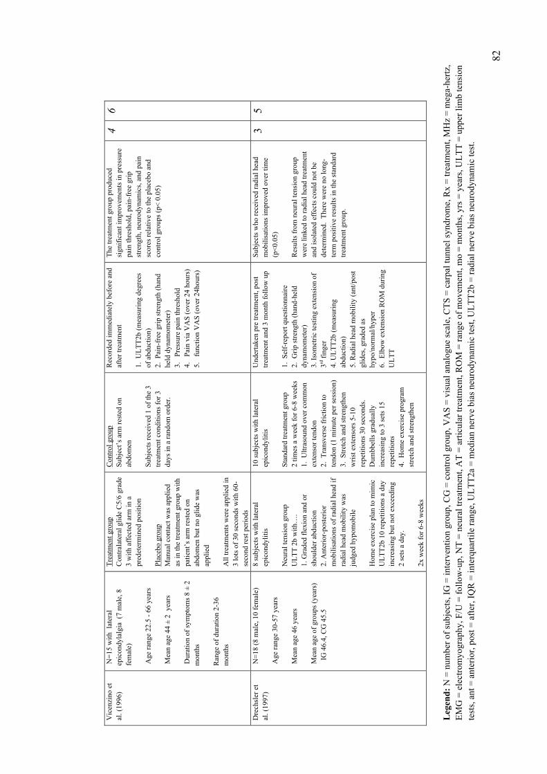
82
Vic
enzi
no e
t al
. (19
96)
N=1
5 w
ith l
ater
al
epic
ondy
lalg
ia (
7 m
ale,
8
fem
ale)
A
ge ra
nge
22.5
- 66
yea
rs
Mea
n ag
e 44
± 2
yea
rs
Dur
atio
n of
sym
ptom
s 8 ±
2
mon
ths
Ran
ge o
f dur
atio
n 2-
36
mon
ths
Trea
tmen
t gro
up
Con
trala
tera
l glid
e C
5/6
grad
e 3
with
aff
ecte
d ar
m in
a
pred
eter
min
ed p
ositi
on
Plac
ebo
grou
p M
anua
l con
tact
was
app
lied
as in
the
treat
men
t gro
up w
ith
patie
nt’s
arm
rest
ed o
n ab
dom
en b
ut n
o gl
ide
was
ap
plie
d A
ll tre
atm
ents
wer
e ap
plie
d in
3
lots
of 3
0 se
cond
s with
60-
seco
nd re
st p
erio
ds
Con
trol g
roup
Su
bjec
t’s a
rm re
sted
on
abdo
men
Su
bjec
ts re
ceiv
ed 1
of t
he 3
tre
atm
ent c
ondi
tions
for 3
da
ys in
a ra
ndom
ord
er.
Rec
orde
d im
med
iate
ly b
efor
e an
d af
ter t
reat
men
t 1.
ULT
T2b
(mea
surin
g de
gree
s of
abd
uctio
n)
2. P
ain-
free
grip
stre
ngth
(han
d he
ld d
ynam
omet
er)
3.
Pres
sure
pai
n th
resh
old
4.
Pain
via
VA
S (o
ver 2
4 ho
urs)
5.
fu
nctio
n V
AS
(ove
r 24h
ours
)
The
treat
men
t gro
up p
rodu
ced
sign
ifica
nt im
prov
emen
ts in
pre
ssur
e pa
in th
resh
old,
pai
n-fr
ee g
rip
stre
ngth
, neu
rody
nam
ics,
and
pain
sc
ores
rela
tive
to th
e pl
aceb
o an
d co
ntro
l gro
ups (
p< 0
.05)
4 6
Dre
chsl
er e
t al
. (19
97)
N=1
8 (8
mal
e, 1
0 fe
mal
e)
Age
rang
e 30
-57
year
s M
ean
age
46 y
ears
M
ean
age
of g
roup
s (ye
ars)
IG 4
6.4,
CG
45.
5
8 su
bjec
ts w
ith la
tera
l ep
icon
dylit
is
Neu
ral t
ensi
on g
roup
U
LTT
2b w
ith…
. 1.
Gra
ded
flexi
on a
nd o
r sh
ould
er a
bduc
tion
2. A
nter
ior-
post
erio
r m
obili
satio
ns o
f rad
ial h
ead
if ra
dial
hea
d m
obili
ty w
as
judg
ed h
ypom
obile
H
ome
exer
cise
pla
n to
mim
ic
ULT
T2b
10 re
petit
ions
a d
ay
incr
easi
ng b
ut n
ot e
xcee
ding
2
sets
a d
ay.
2x w
eek
for 6
-8 w
eeks
10 su
bjec
ts w
ith la
tera
l ep
icon
dylit
is
Stan
dard
trea
tmen
t gro
up
2 tim
es a
wee
k fo
r 6-8
wee
ks
1. U
ltras
ound
ove
r com
mon
ex
tens
or te
ndon
2.
Tra
nsve
rse
fric
tion
to
tend
on (1
min
ute
per s
essi
on)
3. S
tretc
h an
d st
reng
then
w
rist e
xten
sors
5-1
0 re
petit
ions
30
seco
nds.
Dum
bbel
ls g
radu
ally
in
crea
sing
to 3
sets
15
repe
titio
ns
4. H
ome
exer
cise
pro
gram
st
retc
h an
d st
reng
then
Und
erta
ken
pre
treat
men
t, po
st
treat
men
t and
3 m
onth
follo
w u
p 1.
Sel
f-re
port
ques
tionn
aire
2.
Grip
stre
ngth
(han
d-he
ld
dyna
mom
eter
) 3.
Isom
etric
test
ing
exte
nsio
n of
3rd
fing
er
4. U
LTT2
b (m
easu
ring
abdu
ctio
n)
5. R
adia
l hea
d m
obili
ty (a
nt/p
ost
glid
es, g
rade
d as
hy
po/n
orm
al/h
yper
6.
Elb
ow e
xten
sion
RO
M d
urin
g U
LTT
Subj
ects
who
rece
ived
radi
al h
ead
mob
ilisa
tions
impr
oved
ove
r tim
e (p
<0.0
5)
Res
ults
from
neu
ral t
ensi
on g
roup
w
ere
linke
d to
radi
al h
ead
treat
men
t an
d is
olat
ed e
ffec
ts c
ould
not
be
dete
rmin
ed.
Ther
e w
ere
no lo
ng-
term
pos
itive
resu
lts in
the
stan
dard
tre
atm
ent g
roup
.
3 5
Leg
end:
N =
num
ber o
f sub
ject
s, IG
= in
terv
entio
n gr
oup,
CG
= c
ontro
l gro
up, V
AS
= vi
sual
ana
logu
e sc
ale,
CTS
= c
arpa
l tun
nel s
yndr
ome,
Rx
= tre
atm
ent,
MH
z =
meg
a-he
rtz,
EMG
= e
lect
rom
yogr
aphy
, F/U
= fo
llow
-up,
NT
= ne
ural
trea
tmen
t, A
T =
artic
ular
trea
tmen
t, R
OM
= ra
nge
of m
ovem
ent,
mo
= m
onth
s, yr
s = y
ears
, ULT
T =
uppe
r lim
b te
nsio
n te
sts,
ant =
ant
erio
r, po
st =
afte
r, IQ
R =
inte
rqua
rtile
rang
e, U
LTT2
a =
med
ian
nerv
e bi
as n
euro
dyna
mic
test
, ULT
T2b
= ra
dial
ner
ve b
ias n
euro
dyna
mic
test
.

83
3.6 DISCUSSION
A search to identify RCTs investigating neural mobilisation yielded 11 studies
that met the inclusion criteria for this review. Analyses of these studies, using the
criteria of Linton and van Tulder (2001), indicated that 8 of the 11 studies (Akalin et al.,
2002; Allison et al., 2002; Baysal et al., 2006; Cleland et al., 2007; Coppieters et al.,
2003a, 2003b; Pinar et al., 2005; Vicenzino et al., 1996) concluded a positive benefit
from using neural mobilisation in the treatment of altered neurodynamics or
neurodynamic dysfunction. Three of the 11 studies (Drechsler et al., 1997; Scrimshaw
& Maher, 2001; Tal-Akabi & Rushton, 2000) concluded a neutral benefit, which
suggests that neural mobilisation was no more beneficial than standard treatment or no
treatment. Nine of the 11 studies (Allison et al., 2002; Baysal et al., 2006; Cleland et al.,
2007; Coppieters et al., 2003a, 2003b; Pinar et al., 2005; Scrimshaw & Maher, 2001;
Tal-Akabi & Rushton, 2000; Vicenzino et al., 1996) reviewed demonstrated moderate
methodological quality; the two remaining studies (Akalin et al., 2002; Drechsler et al.,
1997) yielded limited methodological quality. Studies exhibited weaknesses in random
allocation, intention to treat, concealed allocation, and blinding; consequently, our
ability to review and assess the therapeutic efficacy of neural mobilisation for treatment
of altered neurodynamics through evaluation of appropriate randomised controlled trials
was substantially limited.
Methodological weaknesses can lead to over- or under-estimations of actual
outcomes. For example, blinding can significantly eliminate bias and confounding, and
is essential in maintaining the robustness of a RCT. Blinding is difficult for use in
studies involving manual therapy (Beaton, Boers, & Wells, 2002; Salaffi, Stancati,
Silvestri, Ciapetti, & Grassi, 2004), and in this review only 9 of the 11 studies blinded
the raters. Some have argued that blinding for use in manual therapy studies is useful

84
(Beaton et al., 2002), although it is arguable that non-masked raters could bias outcome
findings.
The outcome measures used by the RCTs in this review also lacked
homogeneity. A battery of different scales was used, and findings are not transferable
across populations. One method used to standardise measures of success is the use of a
minimal clinically important difference score (MCID). MCID relates to the smallest
change in a clinical outcome measure, which correlates to a person feeling “slightly
better” than the initially recorded state (Salaffi et al., 2004). Findings can be
dichotomised into success or failure. In research that analyses the therapeutic benefit of
an intervention, the MCID is an important statistic, as it represents a
level of therapeutic benefit significant enough to change clinical practice (Beaton et al.,
2002). MCIDs are population- and pathology-specific, and they require analysis to
determine a properly computed value. To our knowledge, all or a majority of the
outcome scales used have not been evaluated for an MCID for the population examined
in our study.
Due to the heterogeneity in respect to the neural mobilisation interventions used
in these RCTs, it is difficult to make general conclusions regarding neural mobilisation
as a general therapeutic tool. Over all, six different categories or types of neural
mobilisation treatments were identified (Table 6). Of these, there was limited evidence
to support the use of active nerve and flexor tendon gliding exercises of the forearm
(Akalin et al., 2002; Baysal et al., 2006; Pinar et al., 2005), cervical contralateral glides
(Coppieters et al., 2003a, 2003b; Vicenzino et al., 1996), and Upper Limb Tension Test
2b (ULTT2b) mobilisation (Drechsler et al., 1997; Tal-Akabi & Rushton, 2000) in the
treatment of altered neurodynamics or neurodynamic dysfunction. There was
inconclusive evidence to support the use of slump stretches (Cleland et al., 2007) and

85
combinations of neural mobilisation techniques (Allison et al., 2002; Scrimshaw &
Maher, 2001) in the treatment of altered neurodynamics or neurodynamic dysfunction.
Table 6. Level of evidence for therapeutic efficacy per intervention type
Number Type of Intervention Studies per Intervention Evidence for Intervention
1 Slump stretches Cleland et al. (2007)
Insufficient (Level 4)
2 Active nerve and flexor tendon gliding exercises (forearm)
Baysal et al. (2006) Pinar et al. (2005) Akalin et al. (2002)
Limited (Level 3)
3 Cervical contralateral glide (nerve mobilisation)
Coppieters et al. (2003b) Coppieters et al. (2003a) Vicenzino et al. (1996)
Limited (Level 3)
4 Combination (neural tissue manual therapy, cervical lateral glide, and shoulder girdle oscillations)
Allison et al. (2002) Insufficient (Level 4)
5 Combination (Straight leg raise, knee flexion/extension, and passive cervical flexion)
Scrimshaw & Maher (2001) Insufficient (Level 4)
6 Upper limb tension test 2b (ULTT 2b) neural mobilisation
Tal-Akabi & Rushton (2000) Drechsler et al. (1997)
Limited (Level 3)
Future studies are needed and a larger, more comprehensive body of work is
required before conclusive evidence is available. Only 10 RCTs met the inclusion
criteria for this systematic review. Unfortunately, all were clinically heterogeneous in
that they looked at a number of different pathologies and different types of neural
mobilisation. This made quantitative analysis of therapeutic efficacy impossible. As
Reid and Rivett (2005) have stated, direct quantitative comparison, within the realms of
systematic review, is very difficult when pathologies, interventions, and outcome
measures are heterogeneous. For example, even for this review there were a number of

86
studies that looked at neural mobilisation in treatment for lateral epicondyalgia
(Drechsler et al., 1997; Vicenzino et al., 1996), carpal tunnel syndrome (Akalin et al.,
2002; Baysal et al., 2006; Pinar et al., 2005; Tal-Akabi & Rushton, 2000), and
cervicobrachial pain (Allison et al., 2002; Coppieters et al., 2003a, 2003b). The specific
neural mobilisation intervention differed between studies, making, in these cases, the
treatments too heterogeneous for statistical pooling.
With respect to the clinical implications of these findings, it is interesting to
note that generally all the RCTs that invesitgated neural mobilisation for upper quadrant
(i.e. cervical spine, shoulder girdle, and upper limb) problems, with the exception of one
study (Allison et al., 2002), concluded that there was limited evidence for therapeutic
efficacy. This is in direct contrast to studies that examined neural mobilisation for
lower quadrant (i.e. lumbar spine, pelvic girdle, and lower limb) problems (Allison et
al., 2002; Cleland et al., 2007; Scrimshaw & Maher, 2001) in that all provided
inconclusive evidence for therapeutic efficacy. From a more specific pathological
perspective, for neural mobilisation of cervical nerve roots, three papers supported the
use of cervical contralateral glide mobilisation. For neural mobilisation of the median
nerve in people with carpal tunnel syndrome, three papers supported the use of active
nerve and flexor tendon gliding exercises of the forearm (Akalin et al., 2002; Baysal et
al., 2006; Pinar et al., 2005).
3.6.1 Future research.
Considering the results of the extensive literature search carried out for this
review, there is an obvious paucity of research concerning the therapeutic use of neural
mobilisation. Not only is there a lack in quantity of such research, upon dissection of the

87
scarce research that is available, there is also a lack of quality. Future research should
look not only at similar pathologies but also at similar neural mobilisation techniques.
Another key feature of these studies is that only clinical outcome measures were
used. In the introduction, we discussed the biomechanical, physiological, and
morphological theories underlying neural mobilisation. One of the key theories for
using neural mobilisation is to exploit the mechanical effect that this form of
mobilisation has on the neural tissue and its mechanical interface. It is possible to use
objective in-vivo measurements of neural movement (i.e. glide, slide, stretch, etc.) via
real-time diagnostic ultrasound. It will be important to eventually substantiate clinical
improvements with objective measurement of neural movement. For example, recent
unpublished data have demonstrated that it is possible to visualise and quantify, with
reasonable reliability, sciatic nerve movement during neural mobilisation (Ellis, 2007).
As it has been postulated that an improvement in nerve mobility may explain any
perceived benefits of neural mobilisation, it would be relevant to make a comparison of
clinical measures with objective measures (e.g. ROM and neural mobility) in an in-vivo
situation in studies that examine neural mobilisation. Such a comparison may give clues
as to whether neural mobilisation is more likely to impose a mechanical effect or a
neurophysiological effect on the nervous system.
3.7 CONCLUSION
Neural mobilisation is advocated for treatment of neurodynamic dysfunction. To
date, the primary justification for using neural mobilisation has been based on a few
clinical trials and primarily anecdotal evidence. Following a systematic review of the
literature examining the therapeutic efficacy of neural mobilisation, 10 RCTs discussed
in 11 studies were retrieved. A majority of these studies concluded a positive
therapeutic benefit from using neural mobilisation. However, in consideration of their

88
methodological quality, qualitative analysis of these studies revealed that there is only
limited evidence to support the use of neural mobilisation. Future research needs to
examine more homogeneous studies (with regard to design, pathology, and
intervention), and we suggest that they combine clinical outcome measures with in-vivo
objective assessment of neural movement.
REFERENCES
References for this paper can be found in the Reference list at the back of the
published paper (see Appendix 9). The references for this paper have also been
included in the full reference list of this thesis.

89
Chapter Four. Reliability of measuring sciatic and tibial nerve movement with diagnostic ultrasound during a neural
mobilisation technique
Ellis, R. F., Hing, W. A., Dilley, A., & McNair, P. J. (2008). Reliability of measuring
sciatic and tibial nerve movement with diagnostic ultrasound during a neural
mobilisation technique. Ultrasound in Medicine and Biology, 34(8), 1209-1216.
4.1 PRELUDE
The systematic review highlighted the heterogeneity of research methodology
across those RCTs which have examined neural mobilisation. In particular,
methodological heterogeneity was evident in regard to the neural mobilisation exercises
that were selected. What was also apparent is that none of the RCTs assessed the
influence of the neural mobilisation exercises on in-vivo nerve movement, a key
premise of these techniques.
It is imperative to quantify nerve movement during neural mobilisation as they
are often used to influence nerve movement. With the availability of USI, in-vivo
assessment of peripheral nerve mechanics is now viable. Several studies have
concluded that this method is reliable in the assessment of median nerve movement
(Coppieters et al., 2009; Dilley et al., 2001). However, this has not been concluded for
the assessment of lower limb nerve movement.

90
The second aim of this thesis was to investigate the reliability of using USI to
quantify sciatic nerve excursion during a sitting neural mobilisation exercise designed to
influence the sciatic nerve. Establishing the high reliability of such methods in the
lower limb is important to validate its use as an outcome measure to quantify nerve
movement.
4.2 ABSTRACT
Diagnostic ultrasound provides a technique whereby real-time, in-vivo analysis
of peripheral nerve movement is possible. This study measured sciatic nerve movement
during a ‘slider’ neural mobilisation technique (ankle dorsiflexion/plantarflexion and
cervical extension/flexion). Transverse and longitudinal movement was assessed from
still ultrasound images and video sequences by using frame-by-frame cross-correlation
software.
Sciatic nerve movement was recorded in the transverse and longitudinal planes.
The amount of sciatic nerve movement was as much as 12.3mm and 8.6mm in each
respective plane. The reliability of ultrasound measurement of transverse sciatic nerve
movement was fair to excellent (ICC = 0.39 – 0.76) and excellent (ICC = 0.75) for
analysis of longitudinal movement. Therefore this study provides further evidence that
diagnostic ultrasound is a feasible, non-invasive, real-time, in-vivo method for analysis
of sciatic nerve movement.
Keywords
Ultrasound, sciatic nerve, neurodynamics, neural mobilisation, frame-by-frame
cross-correlation, reliability

91
4.3 INTRODUCTION
Diagnostic ultrasound presents an important tool in the study of neurodynamics.
Neurodynamics is a term that refers to the integrated biomechanical and physiological
functioning of the nervous system (Butler, 2000; Shacklock, 1995b, 2005a), i.e. the
ability for a peripheral nerve to slide and stretch in order to accommodate changes in its
bed length during joint movements.
Neural mobilisation is a clinical technique that attempts to, amongst other goals,
restore normal neurodynamics in conditions where nerve sliding may be affected.
Neural mobilisation exercises are well documented within the literature (Cleland et al.,
2007; Coppieters et al., 2004; Coppieters & Butler, 2008; Herrington, 2006; Scrimshaw
& Maher, 2001; Shacklock, 2005a; Totten & Hunter, 1991). A large number of studies
examining neural mobilisation have used cadavers (Beith et al., 1994; Coppieters &
Butler, 2008; LaBan, MacKenzie, & Zemenick, 1989; Szabo et al., 1994; Wright et al.,
1996). The dissection and removal of certain tissues, required for the examination of
neural mobilisation, and embalming techniques in cadavers may alter neural mechanics
compared to in-vivo. Diagnostic ultrasound therefore offers a potentially portable and
non-invasive method for the assessment of peripheral nerve movement. Diagnostic
ultrasound has a distinct advantage over other static imaging methods (i.e. magnetic
resonance imaging) with its ability to measure soft tissue movement both in real-time
and in-vivo (Chiou et al., 2003; Hashimoto et al., 1999; Jeffery, 2003; Martinoli et al.,
2000).
Several research groups have developed techniques to utilise real-time
ultrasound imaging to assess both transverse (Greening et al., 2001) and longitudinal
peripheral nerve movement in-vivo (Dilley et al., 2001; Hough et al., 2000b). Methods
for measuring longitudinal movement have utilised Doppler ultrasound (Hough et al.,

92
2000a, 2000b) as well as frame-by-frame cross-correlation analysis of high frequency
ultrasound image sequences (Dilley et al., 2001; Dilley et al., 2003). Most of the
previous studies that have used diagnostic ultrasound to measure peripheral nerve
movement have examined nerves in the upper limb (Dilley et al., 2003; Dilley et al.,
2007; Erel et al., 2003; Greening et al., 2005; Greening et al., 2001; Hough et al., 2000a,
2000b; Hough, Moore, & Jones, 2007b). In contrast, the present study uses diagnostic
ultrasound to measure movement of the sciatic nerve in response to a neural
mobilisation exercise, consisting of cervical extension combined with ankle
dorsiflexion. Calculations of longitudinal nerve motion were performed using frame-by-
frame cross-correlation analysis. The key aims of the present study were to assess the
reliability of measuring sciatic nerve movement, both in longitudinal and transverse
planes, using diagnostic ultrasound and frame-by-frame cross-correlation analysis.
4.4 MATERIALS AND METHODS
4.4.1 Subjects.
Twenty-seven subjects (fourteen females, thirteen males), with an age range
between 18 – 38 years (average = 22.82 years, SD 4.61), were included in this study.
Informed consent to participate in this study was given by all subjects. Subjects met the
inclusion criteria if they were healthy individuals between the ages of 18-40 and did not
have a history of significant/major trauma or surgery to the lumbar, hip, buttock
(gluteal) or hamstring (posterior thigh) regions, symptoms consistent with sciatic nerve
impairment (i.e. paraesthesias, weakness, etc.), or a positive slump test (a test which
determines dysfunction of the sciatic nerve and its associated branches) as described by
Butler (2000).
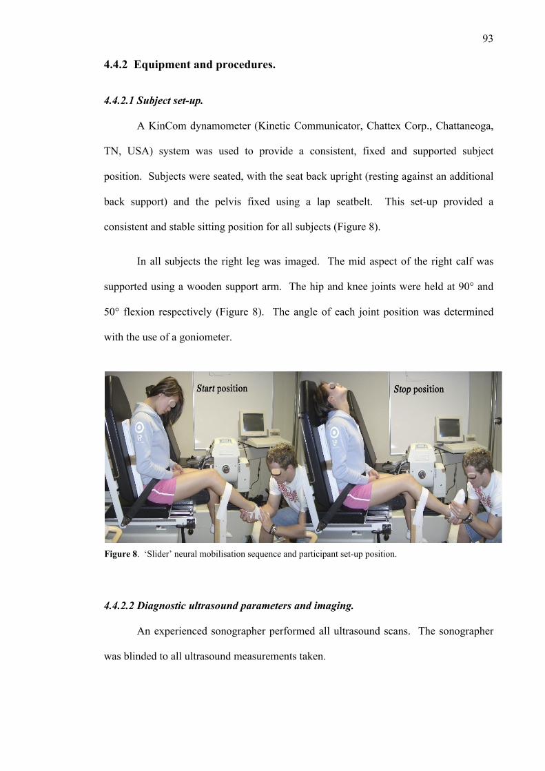
93
4.4.2 Equipment and procedures.
4.4.2.1 Subject set-up.
A KinCom dynamometer (Kinetic Communicator, Chattex Corp., Chattaneoga,
TN, USA) system was used to provide a consistent, fixed and supported subject
position. Subjects were seated, with the seat back upright (resting against an additional
back support) and the pelvis fixed using a lap seatbelt. This set-up provided a
consistent and stable sitting position for all subjects (Figure 8).
In all subjects the right leg was imaged. The mid aspect of the right calf was
supported using a wooden support arm. The hip and knee joints were held at 90° and
50° flexion respectively (Figure 8). The angle of each joint position was determined
with the use of a goniometer.
4.4.2.2 Diagnostic ultrasound parameters and imaging.
An experienced sonographer performed all ultrasound scans. The sonographer
was blinded to all ultrasound measurements taken.
Figure 8. ‘Slider’ neural mobilisation sequence and participant set-up position.

94
B-mode real time ultrasound scanning was performed using a Philips HD11
(Philips Medical Systems Company, Eindhoven, The Netherlands) ultrasound machine
with a 5-12 MHz, 55mm, linear array transducer. The sciatic nerve was scanned in both
the transverse and longitudinal planes at two separate locations, the posterior mid-thigh
(PMT) and popliteal crease (PC).
Figure 9. Measurement of transverse movement of the sciatic nerve at the posterior mid-thigh
1. Transverse movement: a static ultrasound image (Figure 9) was taken at the
Start position of the neural mobilisation exercise. At this position, the neck was in full
flexion (i.e. chin on the chest) with the ankle joint in full plantarflexion. Within the
ultrasound image, digital markers were placed at the lateral-medial and anterior-to-

95
posterior (AP) extremities/boundaries of the visualised nerve. Measurements of the
location of the nerve were recorded.
During the neural mobilisation, active extension of the neck, to a maximum
tolerable extension (e.g. 40-70° from neutral), was performed simultaneously with
passive dorsiflexion of the ankle until maximum tolerable dorsiflexion (e.g. 20-40° from
neutral). At this Stop position a second static ultrasound image was taken. Digital
markers were again placed at the lateral-medial and AP extremities of the visualised
nerve.
Digital callipers were used to measure the vertical/anterior-posterior distance
(‘AP’ movement) between the posterior markers on the Start and the Stop positions.
The same procedure was also used to measure the distance between the right side lateral
markers on the Start to the Stop position to represent the medial-lateral distance
(‘Lateral’ movement) (Figure 9).
A total of three successive measurements were taken at one minute intervals. All
measurements were taken by the same sonographer.
2. Longitudinal movement: an ultrasound cine-loop of the nerve in longitudinal
plane was recorded from the Start to the Stop position. The video loop was captured
over a one-second period at 40 frames per second. A total of three successive
measurements were recorded at one minute intervals.

96
4.4.2.3 Frame-by-frame cross-correlation algorithm and calculation software.
Each video loop of the longitudinal nerve movement was converted to digital
format (bitmaps) and analysed off-line using a method of frame-by-frame cross-
correlation analysis that was developed in Matlab (Mathworks, USA) by Dilley et al.
(2001). This method employs a cross-correlation algorithm to determine relative
movement between successive frames in a sequence of ultrasound images (Dilley et al.,
2001). From the initial frame of the sequence, three rectangular regions of interest
(ROI) were selected over the sciatic nerve. During the analysis, the program compares
the grey-scale values from the ROI’s between adjacent frames of the image sequence. In
the compared frame, the coordinates of the ROI are offset along the horizontal image
plane a pixel at a time within a predetermined range. A correlation coefficient is
calculated for each individual pixel shift. The peak of a quadratic equation fitted to the
maximum three correlation coefficients is equivalent to the pixel shift/movement
between adjacent frames (Dilley et al., 2001). Pixel shift measurements for the nerve
were offset against (subtracted from) pixel shifts measurements within the same
ultrasound field, from stationary structures (i.e. subcutaneous layers, bone, etc.). This
method allows for any slight movement of the ultrasound transducer to be eliminated
from the analysis.
4.4.3 Statistical analysis.
Intraclass correlation coefficients (ICC) were calculated as an indication of the
reliability of both transverse and longitudinal sciatic nerve movement analysis at the
PMT and PC. Bland-Altman plots have been used to provide graphical representation
of some of the key reliability findings.

97
As a measure of combined transverse nerve movements basic trigonometry
(Pythagoras’ theorem) was used to calculate the diagonal distance of nerve movement,
or hypotenuse distance, between the Start and Stop positions. This particular movement
was also quantified in terms of the direction of the movement (i.e. superomedial,
superolateral, inferomedial, inferolateral).
Paired T-tests were utilised to explore statistical differences of sciatic nerve
measurements between different movement directions and also between different
scanning locations.
4.4.4 Ethics.
Ethics approval was sought and approved by AUTEC (Auckland University of
Technology Ethics Committee) (see Appendix 3).
4.5 RESULTS
4.5.1 Transverse ultrasound imaging of lateral and anterior-to-posterior sciatic
nerve movement.
Data representing the transverse sciatic nerve movements measured at the PMT
and PC are presented in Table 7 and Figure 10.
At the PMT, the range of lateral movement following the neural mobilisation
was 0.01 – 12.30mm, with a mean value of 3.54mm (SEM ±1.18mm). This compares
to AP movement which ranged from 0.02 – 4.19mm with a mean value of 1.61mm
(SEM ±0.78mm). Paired T-tests concluded a statistically significant difference between
lateral and AP measurements at the PMT (P<0.05).

98
With respect to the direction of the nerve movement, in 78% of scans at the
PMT the nerve moved superiorly (i.e. towards the skin) and medially, 9% moved
superior-laterally, 11% inferior (i.e. away from the skin) and medially and 2% inferior-
lateral (Figure 11).
At the PC scanning location, the range of lateral movement, during the neural
mobilisation, was 0.31 – 10.90mm, with a mean value of 6.62mm (SEM ±1.10mm).
This compares to AP movement which ranged from 3.00 – 8.28mm with a mean value
of 3.26mm (SEM ±0.99mm). Paired T-tests concluded a statistically significant
difference between these measurements at the PC (P<0.05).
There was a significant reduction in lateral movement at the PMT (mean =
3.54mm) compared to the PC (mean = 6.62mm; P<0.05). There was also a significant
reduction in AP movement at the PMT (mean = 1.61mm) compared to the PC (mean =
3.26mm; P<0.05).
With respect to the direction of the nerve movement, in 100% of scans at the PC
the nerve moved superiorly (i.e. towards the skin) and medially (Figure 12).
Measurements of transverse sciatic nerve movement were possible across all
twenty-seven subjects.

99
Tab
le 7
. A
mou
nt o
f sci
atic
ner
ve m
ovem
ent (
mm
) mea
sure
d fo
r eac
h di
rect
ion
at e
ach
scan
ning
loca
tion.
A
ll fig
ures
repr
esen
tativ
e of
dat
a co
llect
ed fr
om 2
7 su
bjec
ts (n
=27)
asi
de fr
om th
ose
in It
alic
s (n=
3)
D
irec
tion
of S
ciat
ic N
erve
Mov
emen
t
Tra
nsve
rse
Lon
gitu
dina
l
AP
Lat
eral
Scan
ning
loca
tion
R
ange
M
ean
SEM
R
ange
M
ean
SEM
R
ange
M
ean
SEM
Post
erio
r mid
-thig
h (P
MT)
0.
02 -
4.19
1.
61
0.62
0.
01 -
12.3
0 3.
54
1.01
0.
45 -
8.61
3.
47
0.75
Popl
iteal
foss
a/cr
ease
(PC
) 3.
00 -
8.28
3.
26
0.85
0.
31 -
10.9
0 6.
62
1.38
0.
71 -
7.60
5.
22
0.48
A
bbre
viat
ions
: mm
= m
illim
etre
, n =
num
ber o
f sub
ject
s, A
P =
ante
rior-
to-p
oste
rior,
SEM
= st
anda
rd e
rror
of m
easu
rem
ent
Tab
le 8
. Int
racl
ass c
orre
latio
n co
effic
ient
s (IC
C) o
f tes
t-ret
est r
elia
bilit
y fo
r eac
h di
rect
ion
of n
erve
mov
emen
t at e
ach
scan
ning
loca
tion
All
figur
es re
pres
enta
tive
of d
ata
colle
cted
from
27
subj
ects
(n=2
7) a
side
from
thos
e in
ital
ics (
n=3)
D
irec
tion
of S
ciat
ic N
erve
Mov
emen
t
Tra
nsve
rse
Lon
gitu
dina
l
AP
Lat
eral
Sc
anni
ng lo
catio
n
ICC
95
% C
I IC
C
95%
CI
ICC
95
% C
I
Post
erio
r mid
-thig
h (P
MT)
0.
392
0.15
4 - 0
.625
0.
758
0.60
0 - 0
.871
0.
753
0.59
3 - 0
.868
Popl
iteal
foss
a/cr
ease
(PC
) 0.
560
0.33
5 - 0
.750
0.
697
0.51
0 - 0
.837
0.
970
0.73
2 - 0
.999
Abb
revi
atio
ns: m
m =
mill
imet
re, n
= n
umbe
r of s
ubje
cts,
AP
= an
terio
r-to
-pos
terio
r, SE
M =
stan
dard
err
or o
f mea
sure
men
t, C
I = c
onfid
ence
inte
rval
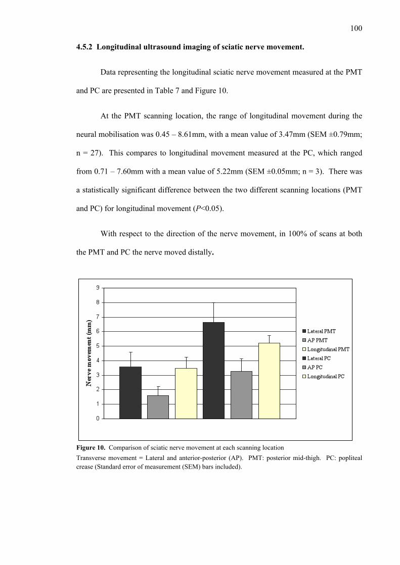
100
4.5.2 Longitudinal ultrasound imaging of sciatic nerve movement.
Data representing the longitudinal sciatic nerve movement measured at the PMT
and PC are presented in Table 7 and Figure 10.
At the PMT scanning location, the range of longitudinal movement during the
neural mobilisation was 0.45 – 8.61mm, with a mean value of 3.47mm (SEM ±0.79mm;
n = 27). This compares to longitudinal movement measured at the PC, which ranged
from 0.71 – 7.60mm with a mean value of 5.22mm (SEM ±0.05mm; n = 3). There was
a statistically significant difference between the two different scanning locations (PMT
and PC) for longitudinal movement (P<0.05).
With respect to the direction of the nerve movement, in 100% of scans at both
the PMT and PC the nerve moved distally.
Transverse movement = Lateral and anterior-posterior (AP). PMT: posterior mid-thigh. PC: popliteal crease (Standard error of measurement (SEM) bars included).
Figure 10. Comparison of sciatic nerve movement at each scanning location

101
Figure 11. Direction of transverse sciatic nerve movement at the posterior mid-thigh. SM = superior-medial, SL = superior-lateral, IM = inferior-medial, IL = inferior-lateral
Figure 12. Direction of transverse sciatic nerve movement at the popliteal crease. SM = superior-medial, SL = superior-lateral, IM = inferior-medial, IL = inferior-lateral

102
5.5.3 Reliability of diagnostic ultrasound assessment of sciatic nerve movement.
The reliability of sciatic nerve movement recorded at the PMT resulted in an
ICC, across three trials for each of the twenty-seven subjects, of 0.76 for lateral sciatic
movement, compared to 0.39 for AP movement (Table 10). Bland-Altman plots
representing these analyses (comparing the difference between the results plotted
against their average) are represented in Figure 17 and 7 respectively.
The reliability of sciatic nerve movement recorded at the PC resulted in an ICC
of 0.70 for lateral sciatic movement, compared to 0.56 for AP movement (Table 10).
The reliability of longitudinal sciatic nerve movement recorded at the PMT (n =
27) resulted in an ICC of 0.75 (Figure 18). However because data could only be
recorded from three subjects (n=3) for longitudinal movement at the PC is not
appropriate to calculate an ICC on such a small subject number (Table 10).
0 1 2 3 4-4
-3
-2
-1
0
1
2
3
4
Average (mm)
Dif
fere
nce (
mm
)
Figure 13. Bland-Altman graph (Difference vs. Average) for all trials measuring AP sciatic nerve movement at the PMT. Bolded line indicates Limits of Agreement (95%).

103
0.0 2.5 5.0 7.5 10.0-4
-3
-2
-1
0
1
2
3
4
Average (mm)
Dif
fere
nce (
mm
)
Figure 14. Bland-Altman graph (Difference vs. Average) for all trials measuring longitudinal sciatic nerve movement at the PMT. Bolded line indicates Limits of Agreement (95%).
5.6 DISCUSSION
Diagnostic ultrasound assessment of peripheral nerve movement is useful in
order to establish the extent of neural movement in both the normal and pathological
situation. In the present study, sciatic nerve movement was measured using diagnostic
ultrasound, a technique that has mainly been used to examine nerve movement in the
upper limb (i.e. median and ulnar nerves). This study sought to determine the reliability
of this technique for measuring sciatic nerve movement, and to make comment of
sciatic nerve movement during neural mobilisation.
Intraclass correlation coefficients were determined as a measure of reliability
(Table 10). Generally, an ICC below 0.40 represents poor reliability, 0.40 – 0.75
represents fair reliability and above 0.75 represents excellent reliability (Fleiss, 1986).
The reliability of the lateral component of the transverse nerve movement was
considered excellent at the PMT (ICC = 0.76). However, the reliability of the lateral

104
component at the PC and the AP component at both the PMT and PC was considered to
be fair (ICC = 0.39 - 0.69).
Diagnostic ultrasound has been described as operator dependent (Beekman &
Visser, 2003, 2004; Chiou et al., 2003; Martinoli et al., 2000; Peer et al., 2002) which
may account for the fair reliability result. Error can occur during placement of the
digital markers. It is therefore important for the sonographer to be consistent with the
placement of the digital markers and callipers between trials. Blinding of the
sonographer from the measurements themselves can help eliminate any measurement
bias.
The reliability of longitudinal nerve movement was considered excellent at the
PMT (ICC = 0.75). At the PC however, reliability analysis was performed in only three
subjects. In the remaining twenty-four subjects, it was not possible to obtain sequences
of longitudinal nerve movement, since the nerve moved beyond the field of the
ultrasound image during the neural mobilisation exercise, i.e. the nerve was no longer in
line with the transducer. Interestingly, a similar movement out of the image plane was
not seen at the PMT. The observed movement of the sciatic nerve out of the image
plane during measurement of longitudinal sliding at the PC, which is also consistent
with an increase in transverse movement at the PC compared to the PMT, may indicate
a region of high compliance behind the knee (i.e. a wavy nerve course). In the upper
limb there are regions of high compliance across major joints in both the median and
ulnar nerve that allow for large changes in their bed length during joint rotations (Dilley
et al., 2003; Dilley et al., 2007). Through the shoulder region for example, both the
median and ulnar nerves follow a wavy course, which accommodates an increase in
nerve bed length during shoulder abduction. The presence of a high compliant region
behind the knee is consistent with unloading of the sciatic nerve with the knee in a

105
flexed position (i.e. the knee was fixed in 50 degrees of flexion during the neural
mobilisation).
In the present study, movement of the sciatic nerve was generated with two
simultaneous movements, namely ankle plantarflexion-dorsiflexion and cervical
flexion-extension. The results demonstrated sciatic nerve movement in different
directional patterns depending upon the site and location (superior movement referring
to superficial nerve movement towards the skin and inferior movement referring to deep
nerve movement towards the femur). At the PMT (i.e. the most proximal site), distal
longitudinal nerve movement was coupled with superior-medial transverse movement in
78% of trials, superior-lateral transverse movement in 9% of trials, inferior-medial
transverse movement in 11% of trials and inferior-lateral transverse movement in 2% of
trials (Figure 11). This is in contrast to the trials recorded at the PC which showed
100% movement of the sciatic/tibial nerve distal longitudinal movement coupled with
superior-medial transverse movement (Figure 12). This finding could be explained by
the differing mechanical interface at each location. At the PC, within the popliteal
fossa, there is much more relative space for the nerve to more freely move compared to
the tighter mechanical interface afforded by the hamstring musculature at the PMT.
4.6.1 Limitations of the study.
This study utilised an ultrasound method which enabled measurement from two-
dimensional (2D) images. This method does not allow analysis of simultaneous
movement within multiple planes. This poses a limitation not only of this study, but of
all studies using 2D imaging which purport to accurately measure true and pure
longitudinal nerve movement using diagnostic ultrasound. It has been suggested that
the use of three (3D) and four-dimensional (4D) diagnostic ultrasound scanner may

106
present an opportunity to measure real-time soft-tissue movement in multiple planes
thereby giving a more accurate representation of actual movement (Deng et al., 2000).
4.7 CONCLUSION
The study concluded that using diagnostic ultrasound assessment and frame-by-
frame cross-correlation analysis to quantify sciatic nerve movement, it is possible to
provide excellent reliability for measurement of longitudinal sciatic nerve movement
during neural mobilisation. The reliability of the transverse movement of the sciatic
nerve was fair irrespective of location or direction.
REFERENCES
References for this paper can be found in the Reference list at the back of the
published paper (see Appendix 10). The references for this paper have also been
included in the full reference list of this thesis.

107
Chapter Five. The influence of increased nerve tension on sciatic nerve excursion during a side-lying neural mobilisation
exercise - a reliability study
5.1 PRELUDE
For the initial reliability study of this doctoral research a standardised, upright
sitting posture was utilised for the neural mobilisation exercise. The hip and knee joints
were held at 90° and 50° flexion respectively. In all participants the right leg was
imaged. Following this study, excellent reliability of measuring longitudinal sciatic
nerve movement at the posterior mid-thigh during simultaneous cervical extension and
ankle dorsiflexion (neural mobilisation), using high-resolution ultrasound and frame-by-
frame cross-correlation analysis, was concluded.
Subsequent to this initial study, consideration was given to the use of a side-
lying neural mobilisation for analysis of sciatic nerve excursion. Neural mobilisation
techniques undertaken in side-lying have been previously suggested for patients with
neurodynamic dysfunction (Cleland et al., 2004; Shacklock, 1995b, 2005a). Potentially
side-lying would offer another advantageous participant position with which to gain
easy access to the PMT for sciatic nerve scanning. Furthermore, the addition of nerve
tension was used during the neural mobilisation. This was done in order to see whether
increased nerve tension would influence sciatic nerve excursion.
The objective of this study was twofold. Firstly, to examine the influence of
added nerve tension upon sciatic nerve excursion, during a neural mobilisation exercise.
Secondly, to build on the initial reliability study (Chapter Four) to establish whether
similar levels of longitudinal sciatic movement were seen with equivalent levels of
reliability when using a side-lying neural mobilisation compared to a seated

108
mobilisation. Furthermore, added neural tension was used in order to see whether
additional nerve tension would reduce sciatic nerve excursion during the mobilisation.
5.2 ABSTRACT
Background: ultrasound imaging has been successfully used to measure the
amount of peripheral nerve movement. This study utilised USI to assess changes in the
amount of longitudinal sciatic nerve movement, when exposed to increased load, during
a side-lying neural mobilisation, in healthy asymptomatic participants. The reliability
of using USI to measure longitudinal sciatic nerve movement, in the modified slump
position, was also assessed.
Methods: Thirteen participants (five males and eight females) were involved
and were aged between 20 and 44 years. Longitudinal sciatic nerve movement was
recorded and measured using USI. The participants were placed in side-lying with their
hip and knee flexed to 90°. Longitudinal sciatic nerve movement was measured at three
positions of progressive neural tension (i.e. with greater knee extension and level of
stretch measured by a 10-point numeric rating scale (NRS) set at 0/10, 2/10 and 4/10
stretch). Sciatic nerve movement was induced with passive ankle movement from full
plantarflexion to full dorsiflexion coupled with simultaneous active cervical flexion to
extension. The reliability of measuring longitudinal sciatic nerve movement at each of
the three positions was also calculated.
Results: There were statistically significant differences (P<0.005) seen in the
mean amount of longitudinal sciatic nerve movement between positions NRS 0/10
(0.59mm) and NRS 2/10 (0.99mm) and also between positions NRS 2/10 (0.99mm) and
NRS 4/10 (0.52mm). The reliability (ICC (95% CI); SEM; MDC) of measuring

109
longitudinal sciatic nerve movement was for NRS 0/10 0.95 (0.88–0.99; 0.10mm;
0.28mm), for NRS 2/10 0.89 (0.70–0.97; 0.21mm; 0.59) and for NRS 4/10 0.94 (0.83–
0.98; 0.09; 0.24mm).
Discussion: The amount of sciatic nerve excursion, during a slide-lying neural
mobilisation exercise, varied depending on the amount of knee extension and therefore
sciatic nerve tension in the exercise position. The amount of sciatic nerve excursion
was less than seen in the previous study which utilised a seated exercise. Differences in
nerve mechanics, which may have been evident given the two different positions, likely
influenced the level of nerve excursion seen. However, the reliability of measuring
sciatic nerve excursion was excellent which is in line with the previous study.
5.3 INTRODUCTION
Neurodynamic theories suggest that nerve movement will diminish as greater
amounts of tension and load are placed upon the nerve tract. As the resting slack of a
peripheral nerve is taken up, eventually there will be relatively less nerve excursion and
greater elongation (Charnley, 1951; Dilley et al., 2007; Kwan et al., 1992; McLellan &
Swash, 1976; Shacklock, 1995b, 2005a; Topp & Boyd, 2006). In order to maximise
nerve excursion during neural mobilisation exercises, it is important to understand how
much nerve tension can be placed upon a nerve tract before the sliding capacity of that
nerve is compromised.
Human cadaveric experiments have shown that during a straight-leg raise (SLR),
excursion of the sciatic nerve initially started after the first 5° of hip flexion (Goddard &
Reid, 1965) or following 15-30º of hip flexion (Breig & Troup, 1979b; Charnley, 1951;
Fahrni, 1966; Inman & Saunders, 1941). As the SLR continued, progressively less

110
actual sliding occurred, so that by greater than 70° of hip flexion, sliding diminished
and elongation occurred (Charnley, 1951; Gajdosik et al., 1985; Gifford, 1998; Goddard
& Reid, 1965; Shacklock, 2005a).
An in-vivo analysis of longitudinal median nerve excursion has also examined
the effect of additional neural tension upon nerve movement. Less median nerve
excursion was seen during wrist extension with the elbow joint positioned in extension
(and therefore pre-loaded) compared to flexion. Likewise, median nerve excursion was
less during elbow extension when the wrist was positioned in neutral compared to 45°
flexion. Essentially the more tension that the median nerve was exposed to, the less
nerve excursion was seen (Dilley et al., 2003). This same phenomenon was witnessed
by examination of median nerve excursion in the forearm, with nerve movement
induced by wrist extension. Here, significant median nerve excursion was not
witnessed until the slack of the median nerve was taken up by extending the elbow
(Dilley et al., 2003).
Clinically the influence of additional neural tension can be seen during
neurodynamic testing where joint ROM decreases as tension is progressively added to
the PNS. For example, studies which have examined knee ROM during a slump test
have concluded that significantly less knee extension occurred with the addition of
cervical flexion (Fidel et al., 1996; Herrington et al., 2008; Johnson & Chiarello, 1997;
Tucker et al., 2007). The explanation given for the reduction in knee extension was that
cervical flexion imposed additional tension upon the neuromeningeal structures at the
spinal cord and nerve roots leading to a reciprocal increase in tension further down to
the sciatic nerve tract (Fidel et al., 1996; Herrington et al., 2008; Johnson & Chiarello,
1997; Tucker et al., 2007; Yeung et al., 1997).
111
There were two aims of this study. The first aim was to quantify the amount of
longitudinal sciatic nerve movement, using USI at the PMT, during a side-lying neural
mobilisation exercise with the lower limb positioned with different amounts of nerve
tension. It was hypothesised that nerve excursion would be reduced as tension increased
upon the sciatic nerve.
The second aim of this study was to further assess the reliability of using USI to
measure longitudinal sciatic nerve movement, during a side-lying neural mobilisation
exercise. It was hypothesised that the reliability of measurement of longitudinal sciatic
nerve movement would be similar to that seen from a previous study which utilised a
seated neural mobilisation exercise (see Chapter Four).
5.4 METHODS
5.4.1 Participants.
Fourteen participants (six males and eight females) volunteered for this study
and met the inclusion and exclusion criteria. The inclusion and exclusion criteria were
healthy individuals between the ages of 18-50 who did not have a history of
significant/major trauma or surgery to the lumbar, hip, buttock (gluteal) or hamstring
(posterior thigh) regions; symptoms consistent with sciatic nerve impairment (i.e.
paraesthesias, weakness, etc.); or a positive slump test, as described by Butler (2000).
All participants were asked to read an information sheet regarding the study and then
informed, written consent was obtained. Ethics approval was sought and approved by
AUTEC (Auckland University of Technology Ethics Committee) (see Appendix 3).

112
5.4.2 Procedure.
The participants were positioned in side-lying on a plinth. Their back and head
were aligned against a wall to ensure the spine was in a neutral position. The hips and
knees were positioned in 90° of flexion and these angles were measured using an
electrogoniometer. A sliding board was placed on a table adjacent to the plinth and the
participants were directed to place their left leg on the sliding board. An inflatable cuff
was used to adjust the height of the thigh to ensure the hip joint was in a neutral position
in the transverse plane, as measured using an inclinometer (Figure 15). The left leg was
imaged in all participants. The sciatic nerve was imaged at a point along the posterior
mid-thigh (PMT), halfway between the gluteal and popliteal creases.
The first ultrasound measurements were taken with the knee placed in 90° of
flexion. At this position participants were asked whether they could rate the amount of
stretch they perceived in their hamstring region on a numeric rating scale (NRS). A
Figure 15. Participant set-up for side-lying neural mobilisation

113
score of 0 represented “no stretch”. A score of 10 represented “the maximum tolerable
stretch”. Whilst in this first position (NRS 0/10 position), when asked, all participants
reported “no stretch”.
Over a three second period, sciatic nerve movement was initiated by passively
moving the ankle from a position of maximal plantarflexion to a position of maximal
dorsiflexion along with simultaneous active cervical flexion to extension. Three
ultrasound recordings of longitudinal sciatic nerve excursion at the PMT were taken for
three separate movements.
The participants were then asked to extend their knee to a position where a
stretch in the hamstring occurred that they rated as 2 out of 10 on the NRS (NRS 2/10
Position). Once again three ultrasound recordings were taken for three separate
movements using the same test movement as that detailed above.
Following measurement in this position the participants were asked to extend
their knee to a position where the stretch on the hamstring was rated as 4 out of 10 on
the NRS (NRS 4/10 Position). Finally three ultrasound recordings were taken for three
separate movements using the same test movement as that detailed above. The
sonographer was blinded to all ultrasound measurements taken.
5.4.3 Ultrasound imaging and analysis.
The ultrasound recordings were captured by the same sonographer. B-mode USI
was performed using a Philips HD11 (Philips Medical Systems Co., Eindhoven, The
Netherlands) ultrasound machine with a 5 to 12 MHz, 55mm linear array transducer.
The sciatic nerve was imaged at the PMT and an ultrasound cine-loop was used to

114
record the movement in the longitudinal plane. The images were recorded using a video
loop of three seconds in duration at 40 frames per second.
The video sequences were converted to a digital format (bitmaps). Each video
sequence was then analysed off-line using a method of frame-by-frame cross-
correlation analysis that was developed in Matlab (Mathworks, USA) by Dilley et al.
(2001). This method employs a cross-correlation algorithm to determine relative
movement between successive frames in a sequence of ultrasound images (Dilley et al.,
2001). During the analysis, the program compares the gray scale values of speckle
features from the regions of interest (ROI’s) within the nerve between adjacent frames
of the image sequence. In the compared frame, the coordinates of the ROI are offset
along the horizontal and vertical image planes a pixel at a time within a predetermined
range. A correlation coefficient is calculated for each individual pixel shift. The peak of
a quadratic equation, fitted to the maximum three correlation coefficients, allows
determination of sub-pixel accuracy. In doing so this further refines the algorithm by
more accurately assessing the maximum cross-correlation value. This peak is
equivalent to the pixel shift/movement between adjacent frames. Therefore this
algorithm establishes that the degree of pixel shift that has the highest associated
correlation coefficient corresponds to and represents the relative movement which is
measured (Dilley et al., 2001).
Pixel shift measurements for the nerve were offset against (subtracted from) pixel
shifts measurements within the same ultrasound field, from stationary structures (i.e.
subcutaneous layers, bone, etc.). This method allows for any slight movement of the
ultrasound transducer to be eliminated from the analysis. This method has proved to be
a highly reliable method of assessment of nerve motion (Coppieters et al., 2009; Dilley
et al., 2001).
115
5.4.4 Statistical analysis.
In regard to the first research aim (changes of nerve excursion in response to
increased nerve tension), mean longitudinal sciatic nerve excursion was calculated for
each of the test positions (NRS 0/10, NRS 2/10 and NRS 4/10). T-tests, with alpha
level set at 0.05, were then used to compare mean values of excursion at each position.
In regard to the second research aim (reliability of measurement), intraclass
correlation coefficients (ICC) were calculated for the test-retest measurements across
the three successive trials in each test position. The standard error of measurement
(SEM) and minimal detectable change (MDC) were calculated for each of the three
testing positions. All statistics were calculated using SPSS (version 14) statistical
analysis software.
5.5 RESULTS
Fourteen participants met the inclusion and exclusion criteria. The data from
one participant was eventually withdrawn as there was poor visualisation of the sciatic
nerve from the ultrasound footage. Of the thirteen remaining participants, there were
eight females and five males. The average age of the participants was 26.08 years
(range: 20-44 years).
The amount of longitudinal sciatic nerve movement was (mean±SD) 0.59mm ±
0.47mm for NRS 0/10, 0.99mm ± 0.65mm for NRS 2/10 and 0.52mm ± 0.36mm for
NRS 4/10 positions respectively.
There were statistically significant differences (P<0.005) seen in the mean
amount of longitudinal sciatic nerve movement between positions NRS 0/10 (0.59mm)

116
and NRS 2/10 (0.99mm) and also between positions NRS 2/10 (0.99mm) and NRS 4/10
(0.52mm).
The reliability (ICC (95% CI); SEM; MDC) of measuring longitudinal sciatic
nerve movement was for NRS 0/10 0.95 (0.88–0.99; 0.10mm; 0.28mm), for NRS 2/10
0.89 (0.70–0.97; 0.21mm; 0.59) and for NRS 4/10 0.94 (0.83–0.98; 0.09; 0.24mm).
5.6 DISCUSSION
This study sought to establish whether the amount of longitudinal sciatic nerve
excursion during a side-lying neural mobilisation exercise differed when additional
tension was added to the sciatic nerve. Tension to the sciatic nerve tract, as determined
by an increased subjective reporting of posterior thigh stretch, was added prior to
performing the neural mobilisation exercise by progressively extending the knee, whilst
in the side-lying position. The findings of this study showed that when no stretch was
felt (NRS 0/10) there was significantly less sciatic nerve excursion compared to when
stretch was rated at 2/10 on the NRS (0.59mm compared to 99mm respectively).
However, from increasing the posterior thigh stretch from 2/10 to 4/10 on the NRS there
was a significant reduction in sciatic nerve excursion (0.99mm compared to 0.52mm
respectively).
At the NRS 0/10 no stretch was reported by any of the participants. That being
the case, it was assumed that very little (if any) tension was placed upon the sciatic
nerve. When passive ankle dorsiflexion was performed there was a small amount of
sciatic nerve excursion recorded (0.59mm). This value suggested a small amount of
nerve movement occurred potentially as the slack of the resting slack of the nerve was
taken up.

117
To reach the NRS 2/10 position, the knee was passively extended which would
have caused increased tension on the sciatic nerve, along with other posterior thigh
structures. The increased nerve excursion seen at the NRS 2/10 position may be
explained due to nerve slack being reduced and increased nerve sliding occurring.
The third position moved the knee further into extension resulting in a greater
stretch being applied to the sciatic nerve (4/10 subjective feeling of stretch on a NRS).
With the additional nerve stretch, sciatic nerve excursion decreased from 0.99mm at the
NRS 2/10 position to 0.52mm at the NRS 4/10 position. This decrease in nerve
excursion may be explained as less sliding is occurring due to the presence of increased
nerve tension.
Of note is the significant reduction in sciatic nerve excursion seen from this
study compared to the initial reliability study (Chapter Four). The range of all data for
longitudinal sciatic nerve excursion, recorded at the PMT during the neural
mobilisation, in this side-lying study was 0.02mm–2.43mm (mean 0.70mm) compared
to the sitting based study (sitting) 0.45mm-8.61mm (mean 3.47mm). There are several
reasons why a large difference in sciatic nerve excursion was recorded between the two
positions (sitting versus side-lying) was seen.
Firstly, the knee position varied between studies which would have influenced
the amount of pre-load upon the sciatic nerve tract and therefore the amount of nerve
excursion. For the sitting based study (Chapter Four) the knee was held constant at 50°
flexion. For the current side-lying study (Chapter Five) the mean knee position for NRS
2/10 position was 45.92° flexion and for the NRS 4/10 position 34.85° (0° equals
terminal knee extension). The mean sciatic nerve excursion at the NRS 2/10 position
(the closest knee position to the 50° flexion used in the reliability study) was 0.99mm,
once again significantly less than the 3.47mm in the reliability study.

118
Secondly, the influence of gravity may have had a significant effect. The
influence of gravity upon the spinal cord, lumbo-sacral nerve roots and sciatic nerve
tract is different between a side-lying and a sitting position. Several studies, which have
examined the morphology and location of the spinal cord and cauda equina for lumbar
epidural and puncture procedures, have recorded that the position of the spinal cord and
cauda equina shift depending on the position a person adopts. This shift is gravity
dependent, for example the spinal cord and cauda equina has been shown to migrate
anteriorly (approximately 6.3mm at the conus medullaris) (Ranger, Irwin, Bunbury, &
Peutrell, 2008) and toward the ipsilateral side that the person was lying on
(approximately between 1.6 - 3.4mm at the lumbar nerve roots) (Ranger et al., 2008;
Takiguchi, Yamaguchi, Hashizume, & Kitajima, 2004a; Takiguchi, Yamaguchi, Okuda,
& Kitajima, 2004b; Takiguchi, Yamaguchi, Usui, Kitajima, & Matsuno, 2006;
Takiguchi, Yamaguchi, Tezuka, & Kitajima, 2009) which was exaggerated (i.e. greater
shift to the lower side) when the knees and hips were flexed (up to 6.1mm at the lumbar
nerve roots) (Takiguchi et al., 2004a; Takiguchi et al., 2009).
The potential exists that, in a side-lying position because of the documented
gravity-dependent shift in the neural tissues, the tension that is imposed on the lumbo-
sacral nerve roots and sciatic nerve tract was significantly different to that imposed in
sitting which subsequently led to the reduction in sciatic nerve excursion seen during
the side-lying study.
The second aim of this study was to test the reliability of measuring longitudinal
sciatic nerve excursion, using USI, during a neural mobilisation exercise performed in
side-lying. An ICC of below 0.40 represents poor reliability, 0.40 - 0.75 represents fair
reliability and above 0.75 represents excellent reliability (Fleiss, 1986). The reliability
of the nerve movement at all three positions was considered excellent (NRS 0/10

119
Position 0.95, NRS 2/10 Position 0.89, NRS 4/10 Position 0.94). The 95% CI’s
however were reasonably close in respect to their spread (between 0.70-0.99 over the
three positions). These ICC values indicate a higher level of reliability of measuring
sciatic nerve excursion compared to the seated neural mobilisation (ICC 0.75) (Chapter
Four). However, with the mean values of excursion being substantially smaller, this
may also have contributed to the high levels of reliability.
5.7 CONCLUSION
In agreement with current neurodynamic theories, this study suggests that the
addition of increased nerve tension, once the resting slack of the sciatic nerve was taken
up, resulted in decreased nerve excursion during a neural mobilisation exercise
performed in side-lying. The reliability of measuring longitudinal sciatic nerve
excursion, with USI, during a side-lying neural mobilisation exercise was excellent.
Compared to the side-lying position, the sitting position afforded a greater
potential for sciatic nerve excursion. Furthermore, as seated neural mobilisation
exercises, for the sciatic nerve tract, are more commonly used in clinical practice it was
decided to return to using a sitting position for future studies as part of this research.
REFERENCES
The references for this paper have been included in the full reference list of this
thesis.

120
Chapter Six. Comparison of different neural mobilisation exercises upon longitudinal sciatic nerve movement: an in-
vivo study utilising ultrasound imaging
6.1 PRELUDE
The previous papers concluded that the intra-rater reliability of USI and frame-
by-frame cross-correlation analysis to quantify in-vivo longitudinal sciatic nerve
excursion, during neural mobilisation exercises, was excellent. Therefore this method
provides a suitable tool to assess the amount of sciatic nerve excursion that occurs
during different types of neural mobilisation exercises.
Different neural mobilisation exercises have been proposed to have different
effects. Understanding these effects will be crucial for appropriate neural mobilisation
design and prescription. In regard to nerve excursion, sliders have been seen to result in
greater nerve excursion compared to tensioners (Coppieters & Alshami, 2007;
Coppieters & Butler, 2008; Coppieters et al., 2009). At present, this has only been
examined in-vivo within the upper limb (Coppieters et al., 2009).
This third aim of this thesis was to assess whether the amount of longitudinal
sciatic nerve excursion differed across different types of neural mobilisation exercises.
Accordingly a controlled laboratory study using a single-group, within-subject
comparisons was utilised to assess the amount of longitudinal sciatic nerve excursion
that occurred with different types of slump-sitting neural mobilisation exercises (two-
ended slider, one-ended slider and a tensioner). Previous research in the upper limb has
concluded that different amounts of median nerve excursion occurred during different
neural mobilisation exercises (Coppieters et al., 2009). This study is the first to

121
examine this situation in the lower limb. A paper from this chapter has been submitted
for publication in the “Journal of Orthopaedic and Sports Physical Therapy”.
6.2 ABSTRACT
Study Design: A controlled laboratory study using a single-group, within-subject
comparisons
Objectives: An in-vivo study to determine whether different types of neural
mobilisation exercises are associated with differing amounts of longitudinal sciatic
nerve excursion
Background: Recent research in the upper limb, of healthy participants, has concluded
that nerve excursion differs significantly between different types of neural mobilisation
exercises. This has not been examined in the lower limb. It is important to initially
examine the influence of neural mobilisation upon peripheral nerve excursion in healthy
people in order to appreciate peripheral nerve excursion in conditions where nerve
excursion may be compromised.
Methods: High-resolution ultrasound was used to assess sciatic nerve excursion at the
posterior mid-thigh. Four different neural mobilisation exercises were utilised in thirty-
one healthy participants. These neural mobilisation exercises used combinations of
knee extension and cervical flexion and extension. Frame-by-frame cross-correlation
analysis was used to calculate nerve excursion. A repeated-measures analysis of
variance and isolated means comparisons were used to analyse these data.
Results: Different neural mobilisation exercises induced significantly different
amounts of sciatic nerve excursion (P<0.001). The slider was associated with the
largest sciatic nerve excursion (3.2±2.0mm) and was significantly greater (P<0.02) than

122
seen with a single-joint mobilisation (2.6±1.4mm; P<0.02) and a tensioner
(2.6±1.5mm).
Conclusion: These findings support previous research which has examined median
nerve excursion associated with different neural mobilisation exercises. Appreciation of
such excursion provides support for theoretical perspectives of nerve motion being
generalisable to the lower limb.
Keywords: diagnostic ultrasound, nerve biomechanics, nerve sliding
6.3 INTRODUCTION
The use and description of neural mobilisation exercises to influence the
mechanical properties of peripheral nerves gained popularity through the late 1970’s
through to the mid 1980’s. However the underlying mechanisms associated with clinical
improvements following neural mobilisation remains unclear (Brown et al., 2011).
There are many theories which have been postulated including physiological effects [i.e.
removal of intraneural oedema (Brown et al., 2011; Butler, 2000; Coppieters et al.,
2004; Ellis & Hing, 2008; Shacklock, 2005a)], central effects [i.e. reduction of dorsal
horn and supraspinal sensitisation (Butler, 2000; Coppieters & Butler, 2008)] and
mechanical effects [i.e. enhanced nerve excursion (Butler, 2000; Coppieters & Alshami,
2007; Coppieters & Butler, 2008; Coppieters et al., 2009; Ellis & Hing, 2008; Méndez-
Sánchez et al., 2010; Shacklock, 2005a)].
Neural mobilisation exercises have been developed from neurodynamic tests
(Butler & Gifford, 1989b; Coppieters et al., 2009; Maitland, 1985; Shacklock, 1995b).
There is evidence to suggest that neurodynamic tests encourage peripheral nerve
movement through elongation of the nerve bed (Byl et al., 2002; Coppieters et al., 2006;

123
Dilley et al., 2003; Wilgis & Murphy, 1986; Wright et al., 2005). It is from this body of
evidence that neural mobilisation exercises have evolved in an attempt to
therapeutically maximise movement in conditions that nerve movement and elongation
is perceived to be compromised.
Until recently, concepts regarding the mechanical influence of neural
mobilisation upon the peripheral nervous system were grounded in theory rather than
concrete evidence. Subsequently, it is likely that many highlighted RCTs (Ellis & Hing,
2008), which have examined neural mobilisation, chose exercises on a theoretical and
pragmatic basis rather than specifically designing exercises to match clinical conditions.
Several studies which have examined neural mobilisation have stated specifically that
the exercises utilised were chosen based solely on those used in previous research
(Akalin et al., 2002; Bardak et al., 2009; Baysal et al., 2006; Heebner & Roddey, 2008;
Pinar et al., 2005). Now that a growing body of evidence is emerging regarding the
mechanical effects that neural mobilisation exercises have upon peripheral nerves, it is
imperative to design exercises to specifically suit certain conditions based on an
understanding that specific mechanical features can be enhanced. However, before this
can be achieved, more attention must be given to understanding the influence of neural
mobilisation exercises upon normal nerve excursion.
Recent studies have examined the influence of neural mobilisation upon nerve
mechanics in cadavers (Coppieters & Alshami, 2007; Coppieters & Butler, 2008) and
subsequently in-vivo research has been undertaken (Coppieters et al., 2009; Echigo et
al., 2008; Ellis, Hing, Dilley, & McNair, 2008). Coppieters et al. (Coppieters et al.,
2009) examined the difference in median nerve excursion between different types of
nerve-gliding exercises (including sliders and tensioners). Sliders utilise combinations
of joint movements to encourage peripheral nerve sliding/excursion by increasing

124
elongation at one end of the nerve bed, therefore increasing tension in the nerve, whilst
simultaneously releasing tension from a distant aspect of the nerve bed (Beneciuk et al.,
2009; Butler, 2000; Coppieters & Alshami, 2007; Coppieters & Butler, 2008;
Coppieters et al., 2009; Herrington, 2006; Shacklock, 2005a). In doing so, excursion is
promoted without significant increase in nerve tension that is associated with elongation
or tensioning exercises (Coppieters & Alshami, 2007; Coppieters & Butler, 2008). In
contrast, tensioners utilise combinations of joint movements that elongate the nerve bed
from both ends in an attempt to stretch the neural connective tissues (Butler, 2000;
Coppieters & Butler, 2008; Coppieters et al., 2009; Herrington, 2006; Shacklock,
2005a).
The conclusion has been that, of the nerve-gliding exercises that were assessed,
sliders produce greater amounts of median nerve excursion compared to tensioners
(Coppieters & Alshami, 2007; Coppieters & Butler, 2008; Coppieters et al., 2009). It
has also been shown that there is a difference between certain types of neural
mobilisation exercises, i.e. significantly less nerve excursion induced from nerve-
gliding exercises initiated at one point along the nerve bed versus sliders (Coppieters et
al., 2009).
Neural mobilisation exercises, derived from neurodynamic tests such as the
slump test or straight-leg raise test, for lumbar spine and lower limb clinical conditions
have been advocated in clinical texts (Butler, 2000, 2005; Maitland, 1985; Shacklock,
2005a) and following clinical trials (Bertolini et al., 2009; Cleland et al., 2007;
Herrington, 2006; Kornberg & Lew, 1989; Schafer, Hall, Muller, & Briffa, 2011;
Webright, Randolph, & Perrin, 1997). However, to the authors’ knowledge, no
previous research has been published which has examined in-vivo measurement of
sciatic nerve excursion in normal healthy participants during different types of neural

125
mobilisation exercises. Such work would support theoretical perspectives that to date
have only received experimental support from studies in the upper limb. The
generalisability of such theory would be enhanced if it could be shown that similar
trends exist in the lower limb where anatomical structures and motion are different.
The aim of this study was to examine longitudinal sciatic nerve excursion at the
posterior mid-thigh (PMT), using high-resolution ultrasound (US), during slump-sitting
neural mobilisation exercises. This research was conducted with healthy participants in
order to assess normal nerve excursion. In line with previous studies (Coppieters &
Alshami, 2007; Coppieters & Butler, 2008; Coppieters et al., 2009) it was hypothesised
that sliders would induce greater longitudinal sciatic nerve excursion compared to
tensioners and that sliders will induce greater longitudinal sciatic nerve excursion
compared to nerve-gliding exercises utilising single joint movements.
6.4 METHODS
6.4.1 Participants.
Thirty-one healthy participants (22 females, 9 males; range 21-61 years;
mean±SD age 29±9 years, height 170.5cm±7.5, weight 68.6kg±13, BMI 23.4±3) were
included in this study. Participants met the inclusion criteria if they were healthy
individuals over the age of 18 years and did not have symptoms suggestive of sciatic
nerve dysfunction.
Participants were excluded if they had a history of significant/major trauma or
surgery to the lumbar, hip, buttock (gluteal) or hamstring (posterior thigh) regions,
symptoms consistent with sciatic nerve impairment (i.e. paraesthesias, weakness, etc.),
or a positive slump test (a test which determines mechanosensitivity of the sciatic nerve

126
and its associated branches) as described by Butler (2000). Participants were also
excluded if they had a neurological condition or other disorders which might alter the
function of the nervous system, for example diabetes.
Based upon a pilot study of 15 participants, a power analysis and sample size
calculation was performed using G*Power 3 (Erdfelder, Faul, & Buchner, 1996; Faul,
Erdfelder, Lang, & Buchner, 2007). The dependant variable used for this analysis was
sciatic nerve excursion, between the different neural mobilisation exercises, with power
set at 0.8 and the α=0.05, the number of participants required was 21. Following the
pilot study and power analysis, a review of the procedures and methods used was
conducted. From this it was deemed that the procedures were appropriate and no
changes were necessary. Prior to the power analysis, participant recruitment had
already found another 16 participants. Based on the conclusion that no procedural
changes were to be made, the data of the 15 participants from the pilot study were
pooled with the remaining 16 participants.
Participants were provided with both written and verbal information concerning
the testing procedures. Informed consent was given by all participants. The study was
approved by the Auckland University of Technology Ethics Committee (AUTEC) (see
Appendix 8).
6.4.2 Participant set-up.
A Biodex system 3 isokinetic dynamometer (Biodex Medical, Shirley, NY,
USA) was used to provide a consistent, fixed and supported sitting position and enable
standardised passive joint movement to facilitate the neural mobilisation exercises.
Participants were positioned on the Biodex seat and asked to adopt a slumped spinal

127
position. This involved a sitting position where the spine was relaxed forwards into a
flexed position through the length of their spine. Once in the slump position, the trunk
of each participant was brought forwards until the right hip joint was at 90° flexion, as
measured by a universal goniometer. The slump position was maintained with contact
of the sternum against a 45cm diameter swiss ball which was placed on the participants
lap. A seatbelt was then utilised to maintain this position (Figure 16).
6.4.3 Fastrak electrogoniometer.
Range of movement (ROM) at the cervical spine and knee joints was measured
with a 3-Space Fastrak (Polhemus Inc., Colchester, Vermont, USA) electromagnetic
Figure 16. Participant set-up for slump-sitting neural mobilisation exercises.
128
motion tracking system. The Fastrak is a device that tracks the position of up to four
separate sensors, in real-time within six-degrees of freedom across three dimensions, in
respect to a source unit which emits a low-intensity electromagnetic field. The position
and orientation of each of the sensors (angular and linear displacement) within the
electromagnetic field is recorded at a set frequency (30 samples per second when using
four sensors). The Fastrak system has been shown to be accurate to within ±0.2° when
recording spinal motion (Pearcy & Hindle, 1989).
For cervical Fastrak measurements, Sensor 1 was placed at the middle of the
forehead (in line with the bridge of the nose) (Amiri, Jull, & Bullock-Saxton, 2003;
Dall'Alba, Sterling, Treleaven, Edwards, & Jull, 2001; Jasiewicz, Treleaven, Condie, &
Jull, 2007; Sterling, Jull, Carlsson, & Crommert, 2002; Trott, Pearcy, Ruston, Fulton, &
Brien, 1996) using an adjustable elastic headband. Sensor 2 was placed over the
spinous process of C7, and secured with double sided tape and tape over the top of the
sensor. For knee Fastrak measurements both sensors were secured (using double sided
tape) on the lateral aspect of the leg, Sensor 1 100mm above the lateral joint line and
Sensor 2 100mm below the joint line (Bullock-Saxton, Wong, & Hogan, 2001).
In order to avoid potential interference from other metallic objects within the
electromagnetic field, the electromagnetic source unit was secured to a wooden pole and
elevated from the ground as recommended by previous studies utilising the Fastrak
system (Bullock-Saxton et al., 2001; Hemmerich, Brown, Smith, Marthandam, & Wyss,
2006). The supporting pole was placed far enough in front of the participant and other
metal equipment to ensure no interference. Prior calibration involved placing all four
sensors, in pairs, against a universal goniometer which was moved through a set angle.
No interference of the Fastrak system was evident.

129
The Fastrak was linked to a computer which recorded the signals being
transmitted from the sensors. LabVIEW 2009 (Version 9.0f2, National Instruments,
Austin, TX, USA) computer software was utilised to allow real-time visualisation and
recording of the motion trace. The joint angle data, as recorded from the cervical spine
and knee were then synchronised off-line to the recorded US sequences.
6.4.4 Diagnostic US parameters and imaging.
Excursion of the sciatic nerve was assessed at the level of the PMT (half-way
between the gluteal crease and popliteal crease). Ellis et al. (2008) previously
concluded high levels of reliability (two-way mixed, intraclass correlation coefficient
(ICC) of 0.753) for measurement of longitudinal sciatic nerve excursion at this location.
Initial transverse imaging at the PMT allowed localisation of the sciatic nerve. Once
identified, the US transducer was rotated into the longitudinal plane. This method of
peripheral nerve location is recommended (Coppieters et al., 2009; Echigo et al., 2008).
A sonographer with five years experience performed all US scans. During the US
sequence selection and cross-correlation analysis the researcher was blinded to the
participant (including their relevant demographic data), the recording session and the
neural mobilisation exercise that was tested.
B-mode real time US scanning was performed using a Philips iU22 (Philips
Medical Systems Company, Eindhoven, The Netherlands) US machine with a 12-5
MHz, 50mm, linear array transducer. An US sequence of the nerve in a longitudinal
plane was recorded for each exercise trial. The video sequence was captured over a
three second period at a capture rate of 30 frames per second.

130
6.4.5 Frame-by-frame cross-correlation algorithm and calculation software.
Each video sequence was converted to a digital format (bitmaps). The image size
for each of the frames was 800x600 pixels. ImageJ (Version 1.42, National Institute of
Health, Maryland, USA) digital image analysis software was used to calculate the
image resolution and also the scale conversion for pixels to millimetres. Image
resolution varied between 7.3 – 10.4 pixels/millimetre depending on the depth of US
penetration required to capture the sciatic nerve.
Each video sequence was then analysed off-line using a method of frame-by-
frame cross-correlation analysis that was developed in Matlab (Mathworks, USA) by
Dilley et al. (2001). This method employs a cross-correlation algorithm to determine
relative movement between successive frames in a sequence of US images (Dilley et al.,
2001). During the analysis, the program compares the gray scale values of speckle
features from the regions of interest (ROI’s) within the nerve between adjacent frames
of the image sequence. In the compared frame, the coordinates of the ROI are offset
along the horizontal and vertical image planes a pixel at a time within a predetermined
range. A correlation coefficient is calculated for each individual pixel shift.
The peak of a quadratic equation, fitted to the maximum three correlation
coefficients, allows determination of sub-pixel accuracy. In doing so this further refines
the algorithm by more accurately assessing the maximum cross-correlation value. This
peak is equivalent to the pixel shift/movement between adjacent frames. Therefore this
algorithm establishes that the degree of pixel shift that has the highest associated
correlation coefficient corresponds to and represents the relative movement which is
measured (Dilley et al., 2001).
Pixel shift measurements for the nerve were offset against (subtracted from)
pixel shifts measurements within the same US field, from stationary structures (i.e.

131
subcutaneous layers, bone, etc.). This method allows for any slight movement of the
US transducer to be eliminated from the analysis. This method has proved to be a
highly reliable method of assessment of nerve motion (Coppieters et al., 2009; Dilley et
al., 2001; Ellis et al., 2008).
6.4.6 US video selection criteria.
Each US video sequence was reviewed several times. To be selected for
analysis, the video sequence must have had clear pixilation and clear identification of
the sciatic nerve throughout the three second duration. The sciatic nerve must have
stayed within the longitudinal plane of the US transducer, and therefore stayed within
the US image. Of the video sequences that met these criteria, two were randomly
chosen for each of the eight neural mobilisations per participant for cross-correlation
analysis.
During the US sequence selection and cross-correlation analysis the researcher
was blinded to the participant (including their relevant demographic data), the recording
session and the neural mobilisation exercise that was tested.
6.4.7 Neural mobilisation exercises.
1A. Slider mobilisation: Simultaneous passive knee extension (from 80º flexion
to 20° flexion - loading of the sciatic nerve caudally via the tibial nerve) with active
cervical extension (from full comfortable cervical flexion to full comfortable cervical
extension - unloading of nervous system cranially).

132
1B. Single-joint mobilisation: Passive knee extension (from 80º flexion to 20°
flexion - loading of the sciatic nerve caudally via the tibial nerve). Each participant was
instructed to look straight ahead in order to maintain their cervical spine in a neutral
position.
1C. Single-joint mobilisation: active cervical flexion (from full comfortable
cervical extension to full comfortable cervical flexion - loading of nervous system
cranially). The knee was held stationary at 80° of knee flexion.
1D. Tensioner mobilisation: Simultaneous passive knee extension (from 80º
flexion to 20° flexion - loading of the sciatic nerve caudally via the tibial nerve) with
active cervical flexion (from full comfortable cervical extension to full comfortable
cervical flexion - loading of nervous system cranially).
1A. Slider mobilisation. 1B. and 1C. Single-joint mobilisations. 1D. Tensioner mobilisation
Figure 17. Slump-sitting neural mobilisation exercises (Chapter Six).

133
In order to specifically limit movement to the knee and cervical spine, used for
each of the neural mobilisations, a rigid thermoplastic ankle foot orthosis (AFO), set at
neutral (0° DF), was worn each participant to eliminate the potential influence of ankle
movement.
Joint movement occurred at an angular velocity of 20°/second, as set by the
Biodex. The order in which the exercises were completed was randomly assigned
(blinded opaque envelope selection) to avoid any possible order effects. Each
participant completed all four exercises. Two repetitions of each movement were
performed as practice/familiarisation trials. Then a further three repetitions of each
movement were performed for data collection. There was a one minute rest period
between each mobilisation exercise.
6.4.8 Statistical analysis.
To test the study hypotheses, repeated-measures analysis of variance (ANOVA)
was utilised to assess differences in the dependant variable: longitudinal sciatic nerve
excursion. Post-hoc pairwise comparisons of means were then performed using the
Bonferroni test (Ottenbacher, 1991). Subsequently the readjusted alpha level was set
was at 0.008.
The intra-rater reliability of measuring longitudinal sciatic nerve excursion from
US footage using cross-correlation software was also examined. A two-way mixed
intraclass correlation coefficient (ICC2,1), with 95% confidence intervals and standard
error of measurement (SEM) were calculated to determine the reliability of
measurement.

134
Mean and range calculations were performed to establish the cervical and knee
ROM for each of the neural mobilisations. Paired t-tests (α=0.05) were used to
calculate whether any differences were present for the total cervical spine and knee
ROM recordings between each of the neural mobilisations exercises.
6.5 RESULTS
Of the thirty-one participants that were enrolled into the study, one participant
was excluded on the basis that the sciatic nerve was not able to be maintained within the
longitudinal scanning plane during data recording.
For neural mobilisations that involved cervical flexion (1C and 1D) the mean
range of cervical motion was 127.8°±7.6° (38.7°±9.6° extension through to 89.1°±13.1°
flexion). For the slider mobilisation (1A) that utilised cervical extension the mean
range of cervical motion was 134.3°±0.2° extension (77.0°±6.2° flexion through to
57.2°±4.8° extension). There was no significant difference in the overall range of
cervical spine movement between exercises using cervical extension (127.8°±7.6°)
versus cervical flexion (134.3°±0.2°) (P=0.2).
The repeated measures ANOVA showed that there was a significant difference
across the neural mobilisation exercises (P<0.0001). Using paired t-tests for individual
comparisons, there were statistically significant differences in sciatic nerve excursion
between the slider mobilisation and tensioner mobilisation (3.2mm vs. 2.6mm; P<0.02),
between the slider mobilisation and both single-joint mobilisations (1A 3.2mm vs. 1B
2.6mm = P<0.02; 1A 3.2mm vs. 1C -0.1mm = P<0.01). There was no significant
difference between 1B versus 1D.

135
The reliability of measuring sciatic nerve excursion across all trials for all four
neural mobilisations combined was excellent with an ICC of 0.95 (95% CI, 0.93 – 0.96;
SEM, 0.2mm).
6.6 DISCUSSION
In support of the study hypotheses, differences in the amount of longitudinal
sciatic nerve excursion exist between different types of neural mobilisation exercises.
The slider mobilisation generated significantly more sciatic nerve excursion compared
to the tensioner mobilisation. Furthermore, the slider mobilisation also generated
significantly greater sciatic nerve excursion compared to the single-joint mobilisations
(1B and 1C). Overall these findings are generally in agreement with other work
(Coppieters & Alshami, 2007; Coppieters & Butler, 2008; Coppieters et al., 2009) that
has examined these techniques in the upper limb and thus provide evidence that the
theoretical construct of nerve excursion during joint motion is also occurring in the
lower limb. More specifically this theory states that a slider mobilisation is designed to
maximise nerve excursion by simultaneously elongating the nerve bed from one end
whilst releasing tension from the other. It has been postulated that a cumulative effect
is then created resulting in greater nerve excursion (Coppieters et al., 2009). A single-
joint mobilisation deliberately utilises movement from one end of the nerve bed only
and therefore does not have the capacity to exploit any potential cumulative effect.
It is notable that no significant difference was seen between the single-joint
mobilisation 1B (2.6mm) versus the tensioner mobilisation (2.6mm). It is expected that
full extension of the cervical spine would allow a release of neural tension via the spinal
cord and spinal nerve roots (Breig, 1978; Breig & Marions, 1963; Shacklock, 2007)
which potentially allows more excursion of the sciatic nerve. However, it is also
136
notable that very little nerve excursion was generated with the opposite movement, full
cervical flexion (1C). The tensioner mobilisation involved simultaneous elongation of
the nerve bed from both ends. However, the inclusion of cervical flexion did not
significantly decrease the amount of sciatic nerve excursion, which was expected,
having elongated the proximal aspect of the nerve bed. In a similar manner the single-
joint mobilisation generated at the cervical spine (1C) produced minimal sciatic nerve
excursion (-0.1±0.1mm).
It is not surprising that isolated cervical flexion did not cause a significant
amount of sciatic nerve excursion. In view of the nervous system being a continuum,
cervical flexion has been suggested to increase tension within the lumbar nerve roots
resulting in an unfolding of resting slack within the nerve root (Breig & Marions, 1963;
Breig & Troup, 1979a; Shacklock, 2007). Cervical flexion has been shown in research
involving cadavers to cause 3-4mm cranial movement of spinal nerve roots within the
thoracic spine (Breig & Marions, 1963). Cervical flexion has been widely advocated as
an additional manoeuvre to the slump test in order to further tension the neural system
leading to further neural sensitisation and symptom reproduction (Breig & Troup,
1979a; Butler, 2000, 2005; Shacklock, 2005a). However, it is unclear as to whether the
increased neural sensitisation stems from an increase in neural tension, neural excursion
and/or both. Hall, Zusman and Elvey (Hall et al., 1998) reported that the addition of
cervical flexion during a straight-leg raise test (SLR) test did not show any significant
change in hip flexion to first onset of manually perceived resistance in either a healthy
or a lumbar radiculopathy group. These authors concluded that cervical flexion did not
have a significant effect of neural tissue compliance during the SLR (Hall et al., 1998).
Although nerve movement is greatest closer to the axis of joint rotation, it is generally
accepted that nerve excursion becomes less the further movement is occurring from the
joint (Coppieters et al., 2006; Echigo et al., 2008; Ellis et al., 2008; Hall et al., 1998;

137
Shacklock, 2005a), which supports the theory that the entire nervous system is
continuous. Our findings are consistent with this theory in that very little sciatic nerve
excursion was evident, from full cervical flexion.
Previous studies that assessed nerve excursion during neural mobilisation
exercises, in cadavers (Coppieters & Alshami, 2007) and in-vivo (Coppieters et al.,
2009), have observed greater amounts of median nerve excursion, both in absolute
terms and between the different mobilisations, compared to the present study of sciatic
nerve excursion. It is difficult to make direct comparisons in the current study due to
the different kinematics between movement of the upper and lower limbs (e.g. in
relation to joint ranges of movement). In respect to certain techniques, similarities can
be seen between upper limb and lower limb data. For example, sciatic nerve excursion
during the tensioner (2.6mm) is close to that seen for the median nerve during a
tensioner (1.8mm) (Coppieters et al., 2009).
At this time, the clinical implication of the small magnitude of nerve excursion
during neural mobilisation exercises is unclear. The key factor which limits further
scrutiny of these values is the controversy that exists regarding whether reduced nerve
excursion is a factor in peripheral nerve disorders and whether, if evident, neural
mobilisation may influence a potential loss of nerve excursion. Although the magnitude
of excursion between the different techniques appears small, given the accuracy of
measurement (as seen from the ICC and SEM values), these values reflect true
differences.
The one other study that has examined sciatic nerve excursion, using in-vivo US
assessment, found similar levels of sciatic nerve excursion at the PMT (Ellis et al.,
2008). However, direct comparison to this previous study is limited as differing
methods were utilised. An alternative means of comparison for in-vivo nerve excursion

138
values, during neural mobilisation, is from studies performed in the upper limb
(Coppieters et al., 2009; Echigo et al., 2008). From these studies, the mean median
nerve excursion range that was recorded was 3.4 - 10.2 mm (Coppieters et al., 2009)
and 0.8 - 3.0 mm (Echigo et al., 2008) respectively, with different methods and neural
mobilisation exercises utilised.
In the current study, of the total data that was collected regarding longitudinal
sciatic nerve movement, the maximum excursion of the sciatic nerve was 8.2mm.
Direct comparisons of the present study to the available evidence are difficult, due to
methodological variations. It is not possible to directly compare cadaveric measures of
nerve excursion to the in-vivo situation as methods of tissue dissection and preservation
have the potential to alter nerve mechanics. Unfortunately, there is a paucity of
research which has examined the excursion of the sciatic nerve in-vivo. However, a
study utilising cadavers by Beith et al. (1995) examined the increase in length of the
nerve bed of the sciatic-tibial-medial plantar nerve tract and found a 42.2 ± 2.4mm
(mean ± SD) increase in nerve bed length when moving the knee from 90° flexion to
terminal extension. The same nerve bed increased by 6.8mm ± 0.69mm from 20° PF to
plantargrade (0°) (Beith et al., 1995).
More recently, in an in-vivo study, Ellis et al. (2008) reported 3.5mm of
longitudinal sciatic nerve movement at the PMT during full range ankle PF through to
DF. Although the level of sciatic nerve excursion was greater here compared to the
present study, it is difficult to directly compared the two results due to key
methodological differences. For example, Ellis et al. (2008) used an upright sitting
position (i.e. no slump) compared to the present study. Also the present study limited
the knee ROM at -20° from terminal extension. Although speculative, if the knee had
been taken into terminal extension sciatic nerve excursion may have been greater.

139
The neural mobilisation techniques used in this study utilised a combination of
passive knee movement and active cervical spine movement. It may have been ideal to
have used movements at the knee and cervical spine that were either both active or
passive. In regard to using active knee movement, there are technical difficulties with
using US with an actively moving limb, particularly at the PMT as active movement of
the hamstrings (even in an eccentric capacity) makes it difficult to maintain US
transducer contact. Implementing passive cervical spine movement through a
standardised range would have been logistically difficult. However, the range of active
cervical spine movement used between the different neural mobilisation exercises was
not statistically different. This indicated consistency of cervical spine movement
between participants across the different exercises.
It is clinically relevant to employ specific exercises that maximise their aims.
For example, in conditions where nerve excursion is believed to be compromised it
would be logical to utilise a neural mobilisation designed to maximise nerve excursion
(e.g. a slider). Subsequently, for clinical situations which may be irritable or acute it
would be logical to utilise neural mobilisation that still challenges nerve excursion, but
to a lesser extent which may decrease the potential adverse influence of excessive
neural tension (e.g. a nerve-gliding exercise utilising a single-joint mobilisation).
In light of the small difference in sciatic nerve excursion between the different
neural mobilisations, other potential features, which were not examined in this study,
may influence nerve excursion. For example, nerve strain may have a direct influence
upon nerve excursion, as has been seen in a cadaver study where increased median
nerve strain resulted in decreased nerve excursion (Coppieters & Butler, 2008).
Likewise, symptom reproduction may also limit joint ROM and subsequently nerve
excursion. Furthermore, based on these findings, it may be such that due to the small
140
differences seen it may be that more generic exercises are provide the same influence
and features beyond those that are mechanical may be relevant. These features, along
with further analysis of nerve mechanics, are worthy of consideration in regard to future
research which examines neural mobilisation design. Future research that investigates
the therapeutic efficacy of neural mobilisation should select specific exercises based on
a better understanding of the neural mechanical effects that are likely to be exploited.
6.7 CONCLUSIONS
The findings of this study provide evidence that lower limb nerve excursion
follows theory related to such movements. Different types of neural mobilisation
exercises induced significantly different amounts of longitudinal sciatic nerve
movement. A slider mobilisation produced the most nerve excursion compared to
single-joint mobilisations and a tensioner mobilisation. It is important to take this
information regarding nerve mechanics, together with similar findings from related
research in the upper limb, to enable more specific design and prescription of neural
mobilisation to specifically suit certain peripheral nerve disorders.
Key Points:
Findings: Different types of neural mobilisation exercises produce different
amounts of longitudinal sciatic nerve excursion. The use of cervical extension during a
slump slider significantly increases sciatic nerve excursion.
Implications: Knowledge regarding the varied amounts of sciatic nerve
excursion that was seen with different exercises will be important to aid in specific
design of neural mobilisation exercises to suit certain peripheral nerve disorders,

141
particularly if further research concludes that impaired nerve excursion is evident. This
study does not comment on the clinical efficacy of these techniques.
Caution: Details regarding the amount of sciatic nerve excursion can only be
inferred from the slump-sitting exercises that were utilised. Data on sciatic nerve
excursion is only relevant to recordings taken at the level of the posterior mid-thigh and
may be different at regions beyond that location.
REFERENCES
The references for this paper have been included in the full reference list
of this thesis.

142
Chapter Seven. In-vivo ultrasound assessment of sciatic nerve excursion during neural mobilisation involving knee and
ankle movement and the influence of cervical flexion
7.1 PRELUDE
The previous paper sought to conclude the amount of sciatic nerve excursion
that can be induced with different types of slump-sitting neural mobilisation exercises
(sliders and tensioners). However, the study did not seek to examine more specific
biomechanical features of the sciatic nerve excursion, for example relative nerve
excursion at different locations along a peripheral nerve and the effect of additional
nerve tension.
The fourth aim of this thesis was to investigate the different biomechanical
features of sciatic nerve excursion in response to slump-sitting neural mobilisation
exercises. Neurodynamic theories suggest that peripheral nerve excursion will occur
greatest the closer to the axis of joint rotation and also that nerve excursion will be
directly influenced by changes in neural tension (as seen in Chapter Five).
However, these biomechanical features have not been assessed for sciatic nerve
excursion during slump-sitting neural mobilisation exercises that specifically utilise
cervical flexion and those that do not. A paper from this chapter has been submitted for
publication in “Clinical Biomechanics”.
7.2 ABSTRACT
Background: Peripheral nerve excursion is directly influenced by the location
of the joint that is inducing the movement and the relative tension imposed upon the
neural system. These factors are important when designing neural mobilisation

143
exercises in order to maximise nerve excursion in some treatments. The evidence
suggests that peripheral nerve excursion is greatest closest to the axis of rotation and is
directly influence by neural tension. However there is a lack of research to document
these effects for the sciatic nerve in-vivo.
Methods: A single-group, within subject study was conducted utilising high-
resolution ultrasound imaging, and frame-by-frame cross-correlation analysis to assess
sciatic nerve excursion. Imaging of the sciatic nerve was at the level of the posterior
mid-thigh. Different neural mobilisation exercises, performed in slump-sitting, were
examined inducing movement at the knee or ankle and with or without cervical flexion.
Findings: A one-ended knee slider produced significantly greater (P<0.0001)
distal sciatic nerve excursion (mean 2.63mm (SD 1.44)) compared to a one-ended ankle
slider (-0.17mm, SD 0.48). The addition of cervical flexion to a one-ended ankle slider
resulted in a significant increase in proximal sciatic nerve excursion from 0.17mm (SD
0.48) to 0.39mm (SD 0.50) (P<0.0001). However there was no significant change
(P=0.70) in sciatic nerve excursion with the addition of cervical flexion to the one-
ended knee slider (2.63mm (SD 1.43) versus 2.56mm (SD 1.49).
Interpretation: For neural mobilisation exercises designed to mobilise the
sciatic nerve, in slump-sitting greater nerve excursion will occur using movement at the
knee joint. However, the position of the cervical spine (whether in neutral or flexed)
does not significantly alter distal sciatic nerve excursion.
Keywords: neural mobilisation, nerve biomechanics, diagnostic ultrasound

144
7.3 INTRODUCTION
Neural mobilisation attempts to restore movement between neural tissues and
their surrounding mechanical interfaces thereby reducing extrinsic and intrinsic
pressures to promote optimum neural function (Brown et al., 2011; Butler, 2000; Butler
et al., 1994; Ellis et al., 2008; Gifford, 1998; Herrington, 2006; Kitteringham, 1996;
Shacklock, 1995b, 2005a). As neural mobilisation aims to influence nerve movement,
an understanding of nerve mechanics is important. However at present there is scant
evidence which has examined the biomechanical effect of neural mobilisation upon
nerve excursion. Such work could be utilised to guide the choice and design of neural
mobilisation exercises.
Several important biomechanical features of the nervous system need to be
considered when assessing neural mobilisation. That the nervous system is a continual
system requires careful clinical reasoning for the most appropriate technique to be
chosen. For example, theories suggest that the greatest amount of nerve excursion will
be induced closest to the axis of rotation that is initiating the movement (Boyd et al.,
2005; Dilley et al., 2008; Dilley et al., 2007; Echigo et al., 2008; Ellis et al., 2008;
Goddard & Reid, 1965; Julius et al., 2004; McLellan & Swash, 1976; Wilgis &
Murphy, 1986). However as joint movement continues, there will be a continuation of
nerve excursion at more distant locations although progressively less the further from
the axis of joint movement (Dilley et al., 2003; Dilley et al., 2008; Echigo et al., 2008;
Ellis et al., 2008; Julius et al., 2004; Reid, 1960; Wright et al., 2001).
A further consideration is the influence of nerve tension. Nerve excursion that is
present with single joint movement will diminish with additional joint movements that
increase tension (Coppieters & Alshami, 2007; Coppieters et al., 2006; Coppieters et al.,
2009; Wright et al., 1996). With increasing tension the elastic limits of the neural

145
connective tissues are challenged resulting in a decrease in relative excursion
(Coppieters et al., 2006; Dilley et al., 2003; Dilley et al., 2007; Kwan et al., 1992).
The clinical implication of relative tension versus excursion is significant when
utilising different types of neural mobilisation. The potential exists for unwanted
cumulative increases in tension or excursion in regions beyond the targeted area may
occur (Alshami et al., 2008; Coppieters & Butler, 2008; Topp & Boyd, 2006). For
example, the inclusion of additional joint movements (i.e. from the cervical spine)
which increase nerve tension, during a neural mobilisation exercise, may be
contraindicated in acute conditions (Coppieters & Butler, 2008). The location or
sequence of joint movement will determine the balance between nerve excursion versus
nerve strain during a neural mobilisation exercise (Coppieters & Alshami, 2007;
Coppieters & Butler, 2008). It is reasonable to suggest that ensuring this balance is
appropriate for a specific condition may dictate the potential therapeutic efficacy of
neural mobilisation.
The objective of this study was to utilise high-resolution USI to examine
specific biomechanical effects of different neural mobilisation exercises upon
longitudinal sciatic nerve excursion. Guided by a premise established in the upper limb
that greater nerve excursion occurs closest to the axis of joint rotation, the first
hypothesis was that sciatic nerve excursion, measured at the posterior mid-thigh (PMT),
will be greater for neural mobilisation exercises that utilise the knee joint compared to
those utilising the ankle joint. Furthermore, on the basis that increased neural tension
will decrease the relative amount of nerve excursion, the second hypothesis was that
increased neural tension via the addition of cervical flexion to neural mobilisation
exercises will decrease the amount of sciatic nerve excursion irrespective of the joint
movement used to induce sciatic nerve excursion (i.e. knee or ankle movements).

146
7.4 MATERIALS AND METHODS
7.4.1 Participants
Thirty healthy participants (21 females, 9 males) (range 21-61 years; mean (SD)
age 29 (9) years, height 170.3cm (7.6), weight 68.4kg (13.2), BMI 23.4 (3.1))
volunteered for this study. The inclusion criteria stipulated that participants did not have
sciatic nerve dysfunction so that normal sciatic nerve mechanics could be assessed. The
exclusion criteria were a history of significant/major trauma or surgery to the lumbar,
hip, buttock (gluteal) or hamstring (posterior thigh) regions, symptoms consistent with
sciatic nerve impairment (i.e. paraesthesias, weakness, etc.), a positive slump test (as
described by Butler, 2000) and conditions which may alter the function of the nervous
system, for example, diabetes.
A power analysis and sample size calculation was performed using G*Power 3,
online power analysis software (Erdfelder et al., 1996; Faul et al., 2007) on the data of
fifteen participants. The dependent variable was mean sciatic nerve excursion between
the exercises generated at the knee (one-ended knee slider, OEKS) and generated at the
ankle (one-ended ankle slider, OEAS). To establish a significant difference for this
variable, with the power set at 0.8 and alpha level set at 0.05 the number of participants
required was four. Following the pilot study and power analysis, a review of the
procedures and methods used was conducted. From this it was deemed that the
procedures were appropriate and no changes were necessary. Prior to the power
analysis, participant recruitment had already found another 15 participants. Based on
the conclusion that no procedural changes were to be made, the data of the 15
participants from the pilot study were pooled with the remaining 15 participants.
Participants were provided with both written and verbal information concerning
the testing procedures. Informed consent was given by all participants. The study was

147
approved by the Auckland University of Technology Ethics Committee (AUTEC) (see
Appendix 8).
7.4.2 Participant set-up
A Biodex system 3 isokinetic dynamometer (Biodex Medical, Shirley, NY,
USA) was used to provide the passive knee and ankle movements during the neural
mobilisation exercises. Each participant was asked to sit on the Biodex seat and relax,
flexing through their spine to adopt a slumped position. This position was maintained
with contact of the sternum against a 45cm diameter Swiss ball which was placed on the
participants lap. A seatbelt was utilised to maintain this position.
7.4.3 Joint ROM analysis.
ROM at the cervical spine, knee and ankle joints was measured with a 3-Space
Fastrak (Polhemus Inc., Colchester, Vermont, USA) electromagnetic motion tracking
system. The Fastrak system monitors the position of four separate sensors in respect to
a source unit emitting a low-intensity electromagnetic field. The Fastrak system has
been shown to be accurate to within 0.2° when recording spinal motion (Pearcy &
Hindle, 1989).
For the cervical spine, sensor 1 was placed at the middle of the forehead (in
line with the bridge of the nose) using an adjustable elastic headband and sensor 2 was
placed over the spinous process of C7 (Amiri et al., 2003; Dall'Alba et al., 2001;
Jasiewicz et al., 2007). For the knee, both sensors were placed on the lateral aspect of
the leg, sensor 1 was 100mm above the lateral joint line and sensor 2 was 100mm below
the joint line (Bullock-Saxton et al., 2001; Laprade & Lee, 2005). For the ankle, sensor

148
1 was placed on the tibia, mid-way between the lateral malleolus and lateral knee joint
line, and sensor 2 was placed along the lateral aspect of the base of the foot just
posterior to the base of the 5th metatarsal.
LabVIEW 2009 (Version 9.0f2, National Instruments, Austin, TX, USA)
computer software was utilised to allow real-time visualisation and recording of the
motion trace from the Fastrak. The joint angle data recorded from the cervical, knee and
ankle were then synchronised off-line with the recorded ultrasound (US) sequences.
7.4.4 Ultrasound imaging
Excursion of the sciatic nerve was assessed at the level of the PMT (half-way
between the gluteal crease and popliteal crease), a position that has high reliability when
measuring sciatic nerve excursion (Ellis et al., 2008). Initial transverse imaging at the
PMT allowed localisation of the sciatic nerve. Once identified, the US transducer was
rotated into the longitudinal plane (Coppieters et al., 2009; Echigo et al., 2008). An
experienced sonographer performed all US scans. B-mode real time US scanning was
performed using a Philips iU22 (Philips Medical Systems Company, Eindhoven, The
Netherlands) US machine with a 12-5MHz, 50mm, linear array transducer. An US
sequence of the nerve in longitudinal plane was recorded for each exercise trial. Each
US video sequence was three seconds long captured at 30 frames per second.
Each USI video sequence was converted to digital format (bitmaps). The image
size for each of the frames was 800x600 pixels. ImageJ (Version 1.42, National Institute
of Health, Maryland, USA) digital image analysis software was used to calculate the
image resolution and the scale conversion for pixels to millimetres.
149
Each video sequence was analysed using frame-by-frame cross-correlation
analysis software developed in Matlab (Mathworks, USA). This method employs a
cross-correlation algorithm to determine relative movement of speckle features between
successive frames in a sequence of US images (Dilley et al., 2001). This method has
proved to be a highly reliable method of assessment of nerve motion (Coppieters et al.,
2009; Dilley et al., 2001; Ellis et al., 2008).
Each US video sequence was reviewed for clear pixilation and clear
identification of the sciatic nerve throughout the three second duration. Of the video
sequences that met these criteria, two were randomly chosen for each of the four neural
mobilisations, per participant, for cross-correlation analysis.
7.4.5 Neural mobilisation exercises
Four neural mobilisation exercises were assessed (Figure 18). These were:
1. One-ended knee slider (OEKS): passive knee extension from 80º knee
flexion to 20° flexion. The cervical spine was maintained steady by
instructing each participant to look ahead and to fix their gaze upon a
stationary object.
2. One-ended ankle slider (OEAS): passive ankle dorsiflexion (DF) from 30°
plantarflexion (PF) to neutral (0° DF). The cervical spine was maintained
steady by instructing each participant to look ahead and to fix their gaze
upon a stationary object.
3. Knee Tensioner (KT): Simultaneous passive knee extension from 80º flexion
to 20° flexion with active cervical flexion from full extension to full flexion.

150
4. Ankle tensioner (AT): Simultaneous passive ankle DF from 30° PF to
neutral (0° DF) with active cervical flexion from full extension to full
flexion.
OEKS: 1-ended knee slider; KT: knee tensioner; OEAS: 1-ended ankle slider; AT: ankle tensioner
For the OEKS and KT the Biodex moved the knee joint between 80° flexion and
20° flexion (0° equivalent to terminal knee extension). Knee joint movement was set at
a joint angular velocity of 20°/second. The ankle/foot was held in a rigid thermoplastic
ankle foot orthosis (AFO), set at 90°. This was standardised across all participants over
all trials.
Figure 18. Slump-sitting neural mobilisation exercises (Chapter Seven).

151
For the OEAS and AT, the Biodex passively moved the ankle from 30° PF to 0°,
at a velocity of 10°/second. The knee joint was held at 50° flexion by the adjustable
arm of the Biodex. Once again, this was standardised for all participants over all trials.
A randomisation procedure was utilised for the order of exercises undertaken.
Each participant completed all four exercises. Two repetitions of each movement were
performed as practice/familiarisation trials. Then a further three repetitions of each
movement were performed for data collection with a one minute rest period between
each mobilisation exercise.
All saved US sequences were transferred to a computer. To ensure blinding of
the measurements, the researcher who carried out all of the US sequence analyses was
blinded to knowledge concerning both the participant and exercises.
7.4.6 Statistical analysis
Repeated-measures analysis of variance (ANOVA) was utilised to detect
differences in sciatic nerve excursion across the four neural mobilisations. To
specifically test the primary study hypothesis, paired t-tests were used to calculate
whether any differences were seen between the OEKS versus the OEAS. To
specifically test the secondary study hypothesis, paired t-tests were used to calculate
whether any differences were seen between the OEKS versus the KT and OEAS versus
the AT. The alpha level was set at 0.05.

152
7.5 RESULTS
For neural mobilisations that involved cervical flexion (KT and AT) the mean
range of cervical motion was 134.3° (SD 1.8°) (from 38.7° (SD 11.6°) extension to
96.5° (SD 13.4°) flexion).
A positive value indicated nerve excursion in a distal direction (towards the
knee) whilst a negative value indicated excursion in a proximal direction (towards the
hip). A significant difference in the amount of sciatic nerve excursion between all four
neural mobilisation exercises (P<0.0001) was found. A statistically significant
difference was seen in sciatic nerve excursion between the OEKS (2.63mm (SD 1.43))
versus OEAS (-0.17mm (SD 0.48)) (P<0.0001). There was a statistically significant
difference in sciatic nerve excursion between the OEAS (-0.17mm (SD 0.48)) and AT (-
0.39mm (SD 0.50)) (P<0.0001). With the addition of cervical flexion sciatic nerve
excursion between the OEKS and KT was unchanged (P=0.70) (Figure 19).
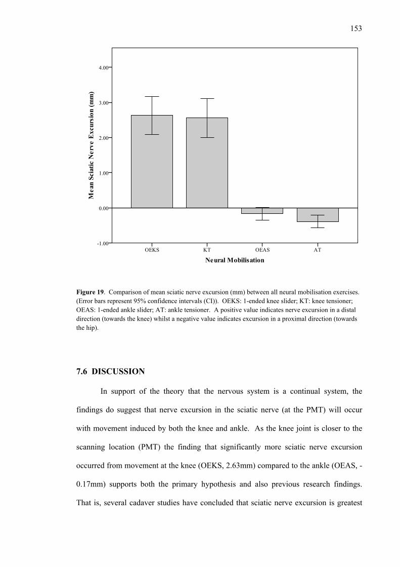
153
Figure 19. Comparison of mean sciatic nerve excursion (mm) between all neural mobilisation exercises. (Error bars represent 95% confidence intervals (CI)). OEKS: 1-ended knee slider; KT: knee tensioner; OEAS: 1-ended ankle slider; AT: ankle tensioner. A positive value indicates nerve excursion in a distal direction (towards the knee) whilst a negative value indicates excursion in a proximal direction (towards the hip).
7.6 DISCUSSION
In support of the theory that the nervous system is a continual system, the
findings do suggest that nerve excursion in the sciatic nerve (at the PMT) will occur
with movement induced by both the knee and ankle. As the knee joint is closer to the
scanning location (PMT) the finding that significantly more sciatic nerve excursion
occurred from movement at the knee (OEKS, 2.63mm) compared to the ankle (OEAS, -
0.17mm) supports both the primary hypothesis and also previous research findings.
That is, several cadaver studies have concluded that sciatic nerve excursion is greatest

154
the closer to the axis of joint rotation (Coppieters et al., 2006; Goddard & Reid, 1965;
Reid, 1960; Smith, 1956). However this phenomenon has not been confirmed, prior to
this study, in-vivo when examining sciatic nerve excursion induced from movements at
the knee and ankle.
To the authors’ knowledge, no previous research has examined the influence of
increased neural tension, imposed by the addition of cervical flexion, upon sciatic nerve
excursion during neural mobilisation exercises in slump-sitting. A significant
difference in nerve excursion was seen when cervical flexion was added to the ankle
exercise (OEAS -0.17mm versus AT -0.39mm). However no significant difference was
seen in nerve excursion when cervical flexion was added to the knee exercise (OEKS
2.63mm versus KT 2.56mm). Therefore it has been possible to accept the secondary
hypothesis in regard mobilisations utilising ankle movement, however the hypothesis
has been rejected in regard to mobilisations utilising knee movement.
It has been postulated that progressive addition of neural load will cause less
nerve excursion and more nerve tension to occur (Coppieters et al., 2006; Dilley et al.,
2003; Kwan et al., 1992; Topp & Boyd, 2006). In-vivo studies have concluded that
knee extension, during a slump test, decreases when cervical flexion is added to the test
(Fidel et al., 1996; Herrington et al., 2008; Johnson & Chiarello, 1997; Tucker et al.,
2007; Yeung et al., 1997) however these studies have not examined nerve motion
directly. The explanation given for the reduction in knee extension is that the addition
of cervical flexion imposes additional tension upon the neuromeningeal structures at the
spinal cord and nerve roots leading to a reciprocal increase in tension further down to
the sciatic nerve tract (Fidel et al., 1996; Herrington et al., 2008; Johnson & Chiarello,
1997; Tucker et al., 2007; Yeung et al., 1997). Others (Breig, 1978; Breig & Marions,
1963; Breig & Troup, 1979b) have observed that cervical flexion increases general

155
neural tension to an extent whereby excursion of the lumbosacral nerve roots has been
seen in cadaver studies. It is possible that the non-significant decrease in nerve
excursion seen by incorporating cervical flexion in the KT occurred as further tension
was being imposed on an already loaded system which would have not allowed
significant changes in nerve excursion. If this had been the case then sufficient tension
may not have been added to result in a decrease in nerve excursion.
The ankle joint movement used during the OEAS and AT (30° DF) was half that
used at the knee joint for the OEKS and KT (60° extension). This smaller joint range
may not have exposed the sciatic nerve tract to significant tension. Several studies have
reported significant reductions in either hip flexion angle (during a straight-leg raise
test) (Boland & Adams, 2000; Gajdosik et al., 1985; Herrington et al., 2008) or knee
extension angle (during a slump test) (Johnson & Chiarello, 1997; Yeung et al., 1997)
when the ankle is held in DF compared to when relaxed in PF. These studies all used
end-range DF as the test position, which is significantly more than the range used in the
present study. It is possible that the smaller ankle ROM used in the present study did
not start to challenge nerve mobility to the same extent as seen for knee movement.
This may have resulted in less neural tension generated.
Of note is that the values of sciatic nerve excursion recorded from OEAS and
AT are negative. Negative values correspond to nerve excursion towards the hip joint
(proximal excursion). Several authors have suggested that cervical flexion (Breig &
Marions, 1963; Breig & Troup, 1979b; Lew, Morrow, Lew, & Fairbank, 1994;
Shacklock, 2007; Troup, 1986) and general spinal flexion (Inman & Saunders, 1941;
Penning & Wilmink, 1981; Shacklock, 2007) induces cephalic movement of the lumbar
nerve roots. The increased sciatic nerve excursion seen in the AT, with movement of
the nerve towards the hip, may have resulted from cephalically directed tension and

156
movement generated with cervical flexion via the spinal cord, lumbosacral nerve roots
and sciatic nerve.
A limitation of this study is the possibility that greater nerve excursion occurred
during the OEKS due to the greater ROM used at the knee compared to the ankle for the
OEAS. It was not possible to standardise the ROM at both joints to the same range due
to concerns over limiting the potential of excessive neural stretch that may have
occurred with ankle DF beyond a neutral position. As slump-sitting positions increase
general load on the nervous system (Fidel et al., 1996; Slater et al., 1994) to avoid
potential adverse effects of excessive increases in neural tension, the Biodex
dynamometer was set to ranges below that of maximum knee and ankle joint
movement. Clinically it would be common practice to use a range of joint movement
during a neural mobilisation exercise up to or just prior to a level which would induce
clinical symptoms (Shacklock, 2005a). The exercises used in this study did not attempt
to use ranges of movement whereby clinical symptoms were reported and so must be
viewed in context. A further limitation of this study was the use of different velocities
in regard to the joint movement (20°/second for the knee and 10°/second for the ankle).
This was done to ensure that the total joint movements were conducted over the same
testing period (three seconds). It cannot be discounted that differences in joint angular
velocity may influence nevre excursion.
7.7 CONCLUSION
Significantly greater sciatic nerve excursion is generated in slump-sitting neural
mobilisation exercises, at the level of the posterior mid-thigh, when using knee as
opposed to ankle movements. This supports previous research which reports that
peripheral nerve excursion is greatest in the region closest to the joint movement used.
157
There does not appear to be a significant difference in distal sciatic nerve excursion
when adopting either a neutral or a flexed cervical position during slump-sitting neural
mobilisation exercises.
REFERENCES
The references for this paper have been included in the full reference list of this
thesis.

158
Chapter Eight. Identifying the sequence of sciatic nerve excursion during different neural mobilisation exercises: an
in-vivo study utilising ultrasound imaging
8.1 PRELUDE
Although the previous two studies (Chapters Six and Seven) have quantified
varied amounts of sciatic nerve excursion during different neural mobilisation exercises,
neither study has examined whether there is a specific sequence of sciatic nerve
excursion. The identification of a sequence of sciatic nerve excursion will be important
for the specific design of neural mobilisation exercises, particularly those that are
intended to maximise nerve excursion.
It has been suggested that peripheral nerves demonstrate a sigmoidal pattern of
excursion when exposed to movement from the body (Topp & Boyd, 2006). A sequence
of peripheral nerve excursion has been observed in the lower limb of cadavers (Breig &
Troup, 1979b; Charnley, 1951; Fahrni, 1966; Goddard & Reid, 1965; Inman &
Saunders, 1941) and in-vivo in the upper limb (Dilley et al., 2003; Dilley et al., 2008;
Dilley et al., 2007). The assessment of a sequence of sciatic nerve excursion, during
neural mobilisation, has not been examined. An important aspect of the fourth aim of
this thesis was to examine whether a sequence of sciatic nerve excursion could be
identified during different slump-sitting neural mobilisation exercises.
The observation of a sequence of nerve excursion in the lower limb would be
important to the design and prescription of neural mobilisation exercises. It would
allow an appreciation of the point, during the performance of neural mobilisation
exercises, that optimum nerve excursion is occurring. A paper from this chapter has
been submitted for publication in the “Journal of Orthopaedic and Sports Physical
Therapy”.

159
8.2 ABSTRACT
Study Design: A controlled laboratory study using a single-group, within-
subject comparison.
Objectives: To determine whether the sciatic nerve exhibits a sigmoidal
sequence of excursion in-vivo during different types of neural mobilisation exercises.
Background: Research suggests that peripheral nerves exhibit a sigmoidal
sequence of excursion during limb movements. Initially slack is taken up before nerve
excursion occurs followed by a period of elongation. Knowledge of such a sequence
during neural mobilisation exercises will enhance their design to maximise nerve
excursion.
Methods: High-resolution ultrasound imaging of sciatic nerve excursion was
synchronised with cervical and knee joint range of movement data during the
performance of three different neural mobilisation exercises in thirty healthy
participants. Imaging of the sciatic nerve was at the level of the posterior mid-thigh.
Graphical and descriptive analysis was utilised to identify the sequence of nerve
excursion during each neural mobilisation exercise.
Results: A sigmoidal sequence of nerve excursion was identified for the two-
ended slump slider and slump tensioner exercises but not the one-ended slump slider.
The point of greatest nerve excursion, for all exercises, occurred once 73-80% of each
exercise had been completed (during a three second exercise period). Once 67% of the
exercise was complete, the amount of nerve excursion was significantly greater for the
two-ended slump slider compared to the one-ended slump slider (P<0.05). Once 90%
of each exercise was complete, the amount of nerve excursion was significantly greater
for the two-ended slump slider compared to the slump tensioner (P<0.05).

160
Conclusion: These findings support previous cadaveric research that sciatic
nerve excursion exhibits a sigmoidal sequence during a two-ended slump slider and
slump tensioner neural mobilisation exercises. Appreciation of the sequence of nerve
excursion during different neural mobilisation exercises will enhance their prescription
for conditions where nerve excursion is compromised.
Keywords: ultrasound imaging, nerve biomechanics, nerve sliding
8.3 INTRODUCTION
In order to cope with mechanical stresses that are imposed from adjacent tissues,
the nervous system must possess specific biomechanical features to maintain the
integrity of its neurophysiological function (Kwan et al., 1992). These biomechanical
features ensure that the nervous system can operate optimally within an environment
where it is constantly challenged by movements and postures of the body. One of these
biomechanical features is the ability of a peripheral nerve to glide and slide against its
surrounding mechanical interface, also known as nerve excursion (Boyd et al., 2005;
Dilley et al., 2003; Erel et al., 2003; McLellan, 1975; McLellan & Swash, 1976).
There is a large body of evidence that details the magnitude of nerve excursion.
However there is less research concerning the sequence of nerve excursion. Based on
the results of studies which have examined the viscoelastic properties of peripheral
nerves, it has been suggested that a nerve will initially go through a period of unfolding
followed by sliding once the inherent slack of the nerve is taken up. Thereafter
elongation occurs and the nerve is stretched towards its elastic limits (Topp & Boyd,
2006).

161
In respect to neural mobilisation, it is critical to have information regarding the
degree and sequence of nerve excursion in order to design the best exercise to suit a
specific condition. For example, ‘sliders’ can be specifically utilised to maximise nerve
excursion in conditions where nerve excursion is compromised (Butler, 2000;
Coppieters & Alshami, 2007; Coppieters & Butler, 2008; Coppieters et al., 2009;
Shacklock, 2005a). However these arguments have been based on mean levels of nerve
excursion rather than reflection on the pattern or sequence of nerve excursion.
There is information available about the sequence of nerve excursion within the
literature which is not specific to neural mobilisation. When the nerve bed is in a
shortened position, the associated nerve is unloaded and exposed to minimal, if any,
tension (Dilley et al., 2007; Julius et al., 2004; Kleinrensink et al., 1995). This
unloading or slackness is important to allow enough nerve length to accommodate large
limb movements (Dilley et al., 2003; Dilley et al., 2007).
For example, very little longitudinal excursion of the median (Dilley et al., 2003)
and ulnar (Dilley et al., 2007) nerves was evident within the first 50° of shoulder
abduction. However, beyond 50° significantly greater median (Dilley et al., 2003) and
ulnar (Dilley et al., 2007) nerve excursion occured to allow further upper limb motion.
Likewise, excursion of the median nerve during elbow extension (90° flexion to
extension) did not occur until extension was past 53° (Dilley et al., 2008). Other
significant areas of nerve slack have been described at the spinal nerve roots and
associated dura (Breig & Marions, 1963), elbow (Byl et al., 2002; Dilley et al., 2003;
Dilley et al., 2007; Grewal et al., 2000; Wright et al., 1996), wrist (Wright et al., 1996),
hip (Charnley, 1951; Fahrni, 1966; Goddard & Reid, 1965; Inman & Saunders, 1941;
Shacklock, 2005b, 2007) and knee (Ellis et al., 2008).
162
As joint motion increases, eventually there will be relatively less sliding
movement and greater nerve elongation (Charnley, 1951; Dilley et al., 2007; Kwan et
al., 1992; McLellan & Swash, 1976; Shacklock, 1995b, 2005a; Topp & Boyd, 2006). It
is during such elongation that nerve tension and intraneural pressures exponentially
increase (Bay et al., 1997; Charnley, 1951; Kwan et al., 1992; Millesi et al., 1995;
Sunderland, 1990; Sunderland & Bradley, 1961a). Beyond the elastic capabilities of the
neural connective tissues, the peripheral nerve will become damaged (Sunderland,
1990; Sunderland & Bradley, 1961a; Topp & Boyd, 2006).
This sequence of nerve movement has been illustrated in several studies which
have examined the sciatic and lumbosacral nerve roots. Human cadaveric experiments
have shown that during the straight-leg raise (SLR) test the sciatic nerve initially started
to slide after some degree of hip flexion [after the first 5° of hip flexion (Goddard &
Reid, 1965) or following 15-30º of hip flexion (Breig & Troup, 1979b; Charnley, 1951;
Fahrni, 1966; Inman & Saunders, 1941)]. As the SLR continued progressively less
sliding and more elongation occurred, so that by greater than 70° of hip flexion
elongation dominated (Charnley, 1951; Fahrni, 1966; Gajdosik et al., 1985; Goddard &
Reid, 1965). A similar trend has been seen in excursion of the lumbosacral nerve roots
during the SLR in cadavers. The nerve roots remained slack and still during the first
30-40° of hip flexion, then root excursion was evident with two-thirds having been
completed at 60° hip flexion, and beyond 70° hip flexion excursion tapered off and
elongation was evident (Ko et al., 2006).
Demonstration of the same phenomenon has been shown with in-vivo analysis
but only in the upper limb. Greater median nerve excursion was reported in the upper
arm (10mm) compared to the forearm (3mm) as the elbow initially extended (i.e. from
90° to 50° flexion). However from 50° flexion to full elbow extension the nerve
163
became tensioned, nerve excursion diminished, and elongation occurred in both the
upper arm and forearm (Dilley et al., 2003). This same phenomenon was observed with
nerve movement induced by wrist extension. Here, significant median nerve excursion
was not seen at the upper arm compared to the forearm until the slack of the median
nerve was taken up by extending the elbow (Dilley et al., 2003). When analysing
median nerve excursion and strain during different types of neural mobilisation
exercises, a clear pattern had been found showing that sliding techniques resulted in
greater median nerve excursion and less strain whereas tensioning techniques resulted in
comparatively less nerve excursion but greater nerve tension (Coppieters & Alshami,
2007; Coppieters & Butler, 2008).
The objective of this study was to utilise high-resolution ultrasound imaging
(USI) to examine sciatic nerve excursion during different neural mobilisation exercises.
Sciatic nerve excursion data was synchronised with joint range of movement (ROM)
data in order to ascertain patterns of nerve excursion during each exercise. The primary
hypothesis of this study was that sciatic nerve excursion would exhibit a sigmoidal
pattern of movement indicating unfolding of slack (i.e. minimal initial nerve excursion)
followed by nerve sliding (i.e. substantial nerve excursion) followed by nerve
elongation (i.e. minimal nerve excursion). A secondary hypothesis was that the greatest
rate of sciatic nerve excursion would be seen during the middle stages of the neural
mobilisation.

164
8.4 METHODS
8.4.1 Participants.
Thirty healthy participants (21 females, 9 males) (range 21-61 years; mean±SD
age 29±9 years, height 170.3cm±7.6, weight 68.4kg±13.2, BMI 23.4±3.1) volunteered
for this study. Participants were included if they were over the age of eighteen and did
not have any clinical signs or symptoms indicating sciatic nerve dysfunction. The
deliberate intention was to include healthy participants to assess normal nerve
mechanics.
Therefore participants were excluded if they had a history of significant/major
trauma or surgery to the lumbar, hip, buttock (gluteal) or hamstring (posterior thigh)
regions; symptoms consistent with sciatic nerve impairment (i.e. paraesthesias,
weakness, etc.); a positive slump test (a test which determines mechanosensitivity of the
sciatic nerve and its associated branches) as described by Butler (2000). Participants
were also excluded if they had a neurological condition or other disorders which might
alter the function of the nervous system, for example diabetes.
Participants were provided with both written and verbal information concerning
the testing procedures. Informed consent was provided by all participants. Ethics
approval was sought and approved by AUTEC (Auckland University of Technology
Ethics Committee) (see Appendix 8).
8.4.2 Participant position.
A Biodex system 3 isokinetic dynamometer (Biodex Medical, Shirley, NY,
USA) was used to provide passive knee extension during all neural mobilisation
exercises. This allowed a consistent participant position and standardised knee

165
movement. Participants were positioned on the Biodex seat. A seated slump position
was adopted which involved relaxation forwards into a flexed position through the
length of the spine. This position was maintained via a belt, placed along the length of
the spine, which held the torso (via the sternum) against a 45cm diameter Swiss ball.
8.4.3 Joint ROM analysis.
Cervical spine and knee joint ROM was measured with a 3-Space Fastrak
(Polhemus Inc., Colchester, Vermont, USA) electromagnetic motion tracking system.
The Fastrak system monitors the position of four separate sensors in respect to a source
unit emitting a low-intensity electromagnetic field, within six-degrees of freedom in
three dimensions. Angular and linear displacements are measured by recording the
position and orientation of each of the sensors within the electromagnetic field. The
Fastrak system has been shown to be accurate to within ±0.2° when recording spinal
motion (Pearcy & Hindle, 1989).
For cervical ROM measurements, Sensor 1 was placed at the middle of the
forehead (in line with the bridge of the nose) (Amiri et al., 2003; Dall'Alba et al., 2001;
Jasiewicz et al., 2007; Sterling et al., 2002; Trott et al., 1996) using an adjustable elastic
headband. Sensor 2 was placed over the spinous process of C7, and secured with
double sided tape. For knee ROM measurements both sensors were secured (using
double sided tape) on the lateral aspect of the leg, Sensor 1 100mm above the lateral
joint line and Sensor 2 100mm below the joint line (Bullock-Saxton et al., 2001).
The Fastrak was linked to a computer which recorded the signals at 30 samples
per second when using four sensors. LabVIEW 2009 (Version 9.0f2, National
Instruments, Austin, TX, USA) computer software was utilised to allow real-time
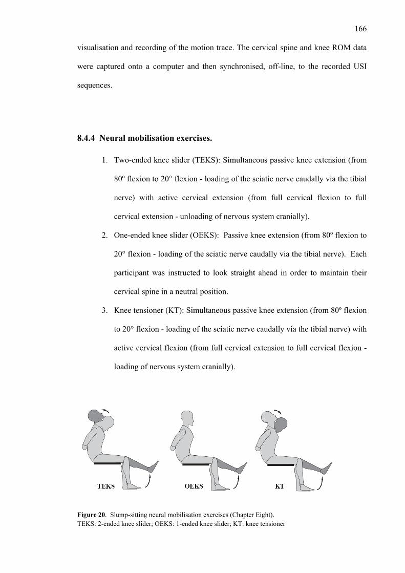
166
visualisation and recording of the motion trace. The cervical spine and knee ROM data
were captured onto a computer and then synchronised, off-line, to the recorded USI
sequences.
8.4.4 Neural mobilisation exercises.
1. Two-ended knee slider (TEKS): Simultaneous passive knee extension (from
80º flexion to 20° flexion - loading of the sciatic nerve caudally via the tibial
nerve) with active cervical extension (from full cervical flexion to full
cervical extension - unloading of nervous system cranially).
2. One-ended knee slider (OEKS): Passive knee extension (from 80º flexion to
20° flexion - loading of the sciatic nerve caudally via the tibial nerve). Each
participant was instructed to look straight ahead in order to maintain their
cervical spine in a neutral position.
3. Knee tensioner (KT): Simultaneous passive knee extension (from 80º flexion
to 20° flexion - loading of the sciatic nerve caudally via the tibial nerve) with
active cervical flexion (from full cervical extension to full cervical flexion -
loading of nervous system cranially).
Figure 20. Slump-sitting neural mobilisation exercises (Chapter Eight). TEKS: 2-ended knee slider; OEKS: 1-ended knee slider; KT: knee tensioner

167
To prevent movement at the ankle and foot a rigid thermoplastic ankle-foot
orthosis, set at neutral (0° dorsiflexion) was worn by each participant. The Biodex
performed the passive knee extension (60°) at a set angular velocity of 20°/second.
Taking into account acceleration and deceleration of the Biodex, when creating the
movement, constant velocity was maintained between 0.5 seconds and 2.7 seconds (see
the grey shaded area on Figures 21-24).
The order in which the exercises were completed was randomly assigned. Each
participant completed all three exercises. Two repetitions of each movement were
performed as practice/familiarisation trials. Then a further three repetitions of each
movement were performed for data collection. There was a one minute rest period
between each mobilisation exercise.
8.4.5 Ultrasound imaging and ultrasound video selection criteria.
Excursion of the sciatic nerve was assessed at the level of the PMT (half-way
between the gluteal crease and popliteal crease). High levels of reliability have been
reported for this measurement point (Ellis et al., 2008). Initial transverse imaging at the
PMT allowed localisation of the sciatic nerve. Once identified, the ultrasound
transducer was rotated into the longitudinal plane. This method of peripheral nerve
location is recommended (Coppieters et al., 2009; Echigo et al., 2008). A sonographer
with five years experience performed all ultrasound scans. The sonographer was
blinded to all ultrasound measurements taken.
B-mode real time ultrasound scanning was performed using a Philips iU22
(Philips Medical Systems Company, Eindhoven, The Netherlands) ultrasound machine

168
with a 12-5 MHz, 50mm, linear array transducer. An ultrasound sequence of the nerve
in longitudinal plane was recorded for each exercise trial.
Each ultrasound video sequence was converted to digital format (bitmaps). The
image size for each of the frames was 800x600 pixels. ImageJ (Version 1.42, National
Institute of Health, Maryland, USA) digital image analysis software was used to
calculate the image resolution and also the scale conversion for pixels to millimetres.
Image resolution varied between 7.3 – 10.4 pixels/millimetre depending on the depth of
ultrasound penetration required to capture the sciatic nerve.
Each ultrasound video sequence was reviewed several times. To be selected for
analysis, the video sequence must have had clear pixilation and clear identification of
the sciatic nerve throughout the three second duration. Of the video sequences that met
these criteria, two were randomly chosen for each of the three neural mobilisations, per
participant, for cross-correlation analysis.
During the USI sequence selection and cross-correlation analysis the researcher
was blinded to the participant (including their relevant demographic data), the recording
session and the neural mobilisation exercise that was tested.
8.4.6 Frame-by-frame cross-correlation algorithm and calculation software.
Each video sequence was then analysed off-line using a method of frame-by-
frame cross-correlation analysis that was developed in Matlab (Mathworks, USA) by
Dilley et al. (Dilley et al., 2001). This method employs a cross-correlation algorithm to
determine relative movement between successive frames in a sequence of ultrasound
images (Dilley et al., 2001). During the analysis, the program compares the gray scale
values of speckle features from the regions of interest (ROI’s) within the nerve between

169
adjacent frames of the image sequence. In the compared frame, the coordinates of the
ROI are offset along the horizontal and vertical image planes a pixel at a time within a
predetermined range. A correlation coefficient is calculated for each individual pixel
shift. The peak of a quadratic equation fitted to the maximum three correlation
coefficients is equivalent to the pixel shift/movement between adjacent frames (Dilley
et al., 2001). Pixel shift measurements for the nerve were offset against (subtracted
from) pixel shifts measurements within the same USI field, from stationary structures
(i.e. subcutaneous layers, bone, etc.). This method allows for any slight movement of
the ultrasound transducer to be eliminated from the analysis. This method has proved to
be a highly reliable method of assessment of nerve motion (Coppieters et al., 2009;
Dilley et al., 2001; Ellis et al., 2008).
8.4.7 Synchronisation of data collection systems.
As data of the variables were able to occur at a collection rate divisible by three,
synchronisation of data across the three second testing period was simplified (e.g. over
the three second period 60° passive knee movement, 90 samples for cervical/knee ROM
recording and 90 ultrasound frames). Although the cervical movement occurred over a
three second period, this was an active movement which was performed by each
individual participant. Therefore a set velocity of movement could not be utilised.
However, each participant was instructed to complete the cervical movement over the
three second period, and all participants had familiarisation trials to learn the timing
required. The recording equipment (Fastrak and USI) were simultaneously initiated at
the same time the Biodex initiated the joint movements. All data were captured onto a
computer and then synchronised, off-line.

170
8.4.8 Statistical analysis.
To ensure standardisation between the three neural mobilisations, mean and
range calculations were performed for cervical and knee ROM, recorded from the
Fastrak system. Paired t-tests were used to calculate whether any differences were
present for the total cervical and knee ROM recordings between each of the neural
mobilisations exercises. For all tests, the alpha level was set was at 0.05.
The point of greatest sciatic nerve excursion was calculated by finding the
maximum difference in excursion between each successive frame during the USI
sequence and relating this to the corresponding time point across the three second
testing period. Paired t-tests were used to calculate whether any differences were
present for the time point where maximum sciatic nerve excursion occurred, between
each of the neural mobilisations exercises. For all tests, the alpha level was set was at
0.05.
8.5 RESULTS
Descriptive statistics in respect to the amount of sciatic nerve excursion recorded
with each neural mobilisation exercise are presented in Table 9. Relevant cervical, knee
ROM and sciatic nerve excursion data were also determined at each quarter during the
three second exercise performance (Table 10).

171
Table 9. Descriptive results for longitudinal sciatic nerve excursion across the neural mobilisation exercises
Neural Mean nerve Standard Standard 95% Confidence interval for mean
Mobilisation Excursion (mm) deviation (SD) error (SE) Lower bound Upper bound
TEKS 3.21 1.97 0.36 2.47 3.95
OEKS 2.63 1.44 0.26 2.09 3.17
KT 2.56 1.49 0.27 2.00 3.12
Abbreviations: TEKS, 2-ended knee slider; OEKS, 1-ended knee slider; KT: knee tensioner
Graphical representations of all data sets for each of the three neural
mobilisation exercises are presented in Figures 21, 22 and 23. For each graph the
dashed line represents the time where the maximum rate of sciatic nerve excursion
occurred.
The TEKS and KT depicted a sigmoidal sequence of nerve excursion (Figures
21 and 23). The OEKS did not as there was no shoulder region evident (Figure 22).
For the TEKS the maximum sciatic nerve excursion was 0.21mm after 73% of the
exercise was completed (Figure 21), for the OEKS 0.17mm after 80% (Figure 22) and
for the KT 0.17mm after 77% (Figure 23). Combined data sets for all three neural
mobilisation exercises are presented in Figure 24.
The first point where a significant difference in nerve excursion between the
TEKS and OEKS (P<0.05) occurred after 67% of the exercise was completed. After
90% of the exercise was completed, a significant difference in sciatic nerve excursion
existed between the TEKS and KT (P<0.05). No significant difference in sciatic nerve
excursion was seen between the OEKS and KT (P>0.05).

172
Table 10. Cervical, knee ROM and sciatic nerve excursion at each quarter during the three second exercise performance
Quarters during neural mobilisation
performance (% of movement time)
Neural Mobilisation Data 25% 50% 75% 100%
Cervical ROM (°) 43.0 -11.0 -40.6 -53.8
Knee ROM (°) 63.4 43.3 24.5 19.6 TEKS
Sciatic nerve excursion (mm) 0.12 0.80 2.09 3.21
Cervical ROM (°) 27.2 27.2 26.5 28.0
Knee ROM (°) 65.8 45.7 26.5 20.0 OEKS
Sciatic nerve excursion (mm) 0.06 0.62 1.67 2.63
Cervical ROM (°) 4.3 56.5 87.7 93.1
Knee ROM (°) 63.7 43.3 24.2 20.6 KT
Sciatic nerve excursion (mm) 0.09 0.74 1.71 2.56
Abbreviations: TEKS, 2-ended knee slider; OEKS, 1-ended knee slider; KT: knee tensioner; ROM: range of movement
In respect to the knee ROM utilised across the three neural mobilisations the
mean±SD ROM was 58.0°±1.3° knee extension (from 78.5°±1.8° to 20.5°±0.1°
flexion). There were no significant difference seen between the start and end positions
for knee ROM across all three neural mobilisation (P>0.3). For the TEKS the mean
range of cervical extension was 125.3°±17.5° extension (81.4°±21.7° flexion through to
43.9°±17.9° extension). For the KT the mean range of cervical flexion was
126.3°±23.3° (29.5°±21.3° extension through to 96.8°±21.7° flexion). There was no
significant difference seen between the TEKS and KT for the full cervical ROM utilised
(P>0.8). For the OEKS (where the cervical spine was stationary) the mean neutral
cervical position was 27.3°±17.9° flexion.

173
Figure 21. Relationship of sciatic nerve excursion, knee and cervical movement during TEKS. Positive value for cervical ROM = flexion, negative value = extension. Grey box represents period of constant Biodex velocity (knee joint). Dashed line represents time of peak nerve excursion (0.21mm at 2.2 seconds, 73% of the exercise was completed).

174
Figure 22. Relationship of sciatic nerve excursion, knee and cervical movement during OEKS. Positive value for cervical ROM = flexion, negative value = extension. Grey box represents period of constant Biodex velocity (knee joint). Dashed line represents time of peak nerve excursion (0.17mm at 2.4 seconds, 80% of the exercise was completed).
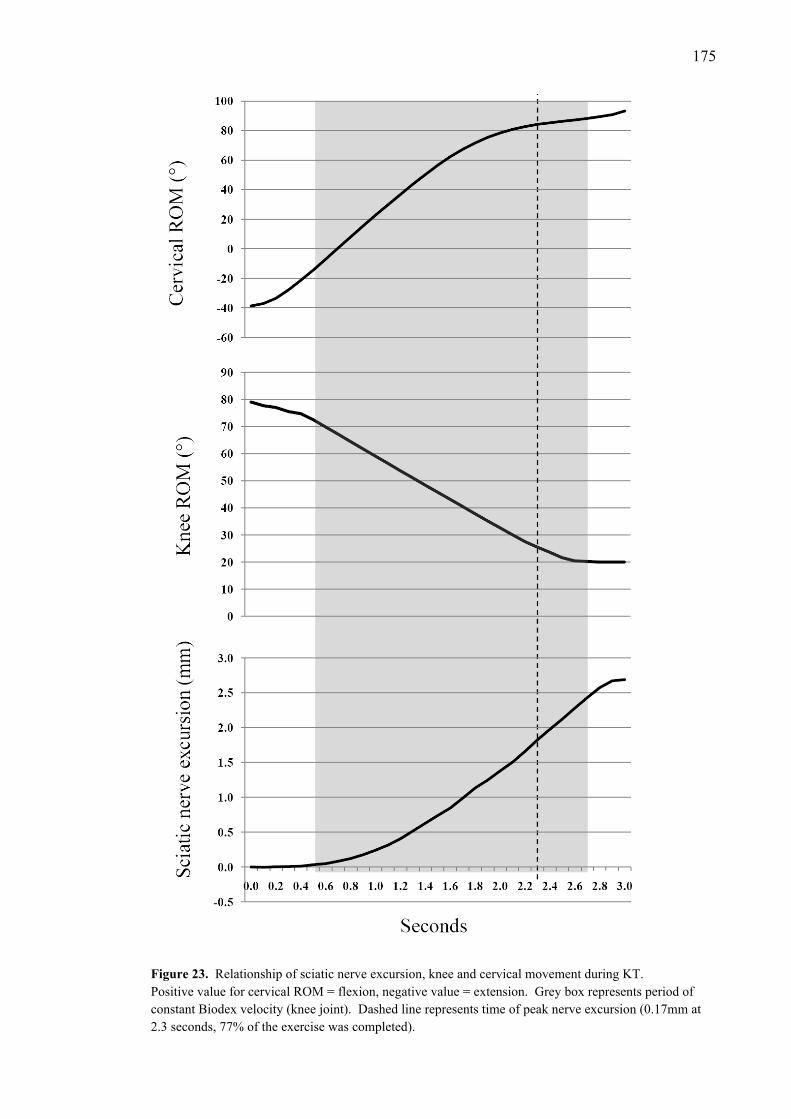
175
Figure 23. Relationship of sciatic nerve excursion, knee and cervical movement during KT. Positive value for cervical ROM = flexion, negative value = extension. Grey box represents period of constant Biodex velocity (knee joint). Dashed line represents time of peak nerve excursion (0.17mm at 2.3 seconds, 77% of the exercise was completed).

176
Figure 24. Sciatic nerve excursion, knee and cervical ROM data for all neural mobilisation exercises Positive value for cervical ROM = flexion, negative value = extension. Grey box represents period of constant Biodex velocity (knee joint). Vertical lines demarcate the percentage of the exercise complete (i.e. 25%, 50%, 75%, 100%). * = significant difference in sciatic nerve excursion (P<0.05). TEKS: 2-ended knee slider; OEKS: 1-ended knee slider; KT: knee tensioner
177
8.6 DISCUSSION
Although joint movement will result in the excursion and stretch of many
different soft tissues, this study specifically examined excursion of the sciatic nerve.
This was achieved by ensuring that the frame-by-frame cross-correlation software
assessed pixel movement confined to the sciatic nerve and not that for the adjacent soft
tissues. Subsequently, the results of this study demonstrate that there is a defined
sequence of sciatic nerve excursion during neural mobilisation exercises. The primary
hypothesis predicted that the plot of sciatic nerve excursion data would follow a
sigmoidal pattern. A sigmoid curve is an S shaped curve that is distinguished by a
gradual toe region followed by a steeper linear region and finishes with a flatter
shoulder region. Theories suggest that nerves will exhibit a sequence of movement, in
response to limb movement, with unfolding first followed by a period of excursion,
excursion will then taper off and elongation will occur (Topp & Boyd, 2006). Two of
the three exercises that were tested (TEKS and KT) demonstrated a sigmoidal sequence
for excursion as predicted in the study hypothesis. Whilst the OEKS did demonstrate
toe and linear regions no shoulder region was evident.
All three neural mobilisation exercises demonstrated a clearly defined toe
region. The toe region represents a period of nerve unfolding (Julius et al., 2004).
Upon completion of the first 25% of the exercise (at 0.75 seconds) sciatic nerve
excursion was minimal (<0.13mm) with no statistically significant differences seen
across the three exercises.
In using the Biodex dynamometer, a constant velocity of passive knee extension
was maintained between 0.5 – 2.7 seconds for all three neural mobilisation exercises
(see grey shaded area in Figures 21-24). As the toe regions for all three exercises

178
extended beyond 0.5 seconds it is apparent that this toe region was not solely due to
acceleration of the Biodex.
The linear region represents the most significant period of nerve excursion. In
this study the linear region extended into the last quarter of each of the exercises
performance (between 75% - 100% completion). The first point where a significant
difference in nerve excursion occurred was between the TEKS and OEKS at 2.0
seconds (upon completion of 67% of the exercise). Of note is the fact that the
maximum rate of nerve excursion for each of the exercises occurred just after this point
(TEKS 2.2 seconds, OEKS 2.4 seconds, KT 2.3 seconds).
The secondary hypothesis of this study was that the greatest rate of sciatic nerve
excursion would occur during the middle half of the exercise performance (between
25% and 75% of the exercise being completed, 0.75 – 2.25 seconds). This hypothesis
was only confirmed with the TEKS. However the greatest rate of nerve excursion for
the OEKS and KT was very close to the mid-range of each exercise, just after 75% of
the exercise had been completed.
The shoulder region of a sigmoid curve would suggest that nerve excursion is
starting to reduce, as the movement progresses towards nerve elongation. A key factor
was that during all three exercises passive knee movement began to decelerate as the
Biodex reached the end of the set knee ROM. This deceleration could also give the
nerve excursion curve an appearance of reduced excursion which may be mistaken for a
shoulder region. However the OEKS did not demonstrate a shoulder region (Figure 22).
This was the only exercise that relied solely on passive knee movement to generate
nerve excursion. Had the deceleration of the passive knee movement been significant at
the end of the period of constant knee velocity (2.7 seconds) then all three exercises
would have exhibited a reduction of nerve excursion (i.e. a shoulder region). Therefore

179
the shoulder region exhibited during the TEKS and KT is due to nerve elongation rather
than deceleration of the Biodex.
Effectively for the OEKS, upon completion of the exercise, nerve excursion
remained within the linear region. Unlike the OEKS, the TEKS and KT demonstrated a
shoulder region. However both the TEKS and KT utilised cervical movement as part of
the neural mobilisation sequence whereas the cervical spine remained stationary during
the OEKS. The direction of cervical movement may have influenced the excursion
sequence directly. During the TEKS, cervical extension, in theory, would have lent
more available length to the nerve tract which resulted in greater nerve excursion
towards an elongation phase. Opposite to this, cervical flexion has the potential to
increase general neural load, via the spinal cord and lumbosacral nerve roots, which
would potentially result in a faster transition from a linear region into the shoulder
region as elongation occurs.
Identification of a sequence of nerve excursion, during neural mobilisation
exercises, may provide insight for future exercise design. As has previously been
suggested, sliders allow more excursion with less tension, whereas tensioners offer less
excursion with more tension (Coppieters & Alshami, 2007; Coppieters & Butler, 2008;
Coppieters et al., 2009). Furthermore, an understanding of where or at what point
during the range, of a particular exercise, excursion or elongation can be maximised
could be useful for use clinical disorders where nerve excursion may be impaired.
The implication from this research is that maximal sciatic nerve excursion
occurs during the mid-late range during a specific exercise. For example, during the
TEKS sciatic nerve excursion was greatest as the knee extends to approximately (≈) 25°
flexion and the cervical spine extends to ≈41° extension. Performance of this type of
neural mobilisation will not maximise sciatic nerve excursion at the PMT unless these

180
ranges of joint movement are met. Likewise if sciatic nerve elongation was desired then
the choice of a KT would be logical and ideally would be performed with knee
extension beyond ≈21° flexion and cervical flexion beyond ≈88°.
A limitation of this study is that passive knee movement did not continue
towards terminal knee extension (0°). The deliberate intention of this study was to
avoid the limits of knee extension in order to reduce any possible adverse effects of
stretching the sciatic nerve and associated nerve tracts. Therefore any discussion
regarding the sequence of nerve excursion can only be presented in respect to the three
second testing period and the limited knee joint range that were used.
Although it was possible to create a standardised range and velocity of knee
joint movement, the same could not be created for active cervical movement. The only
standardising factor was the performance of cervical movement over the three second
testing period, which presents another limiting factor.
8.7 CONCLUSION
It is apparent during neural mobilisation exercises that a specific sequence of
nerve excursion occurs when the exercises utilised simultaneous joint movements at
either end of the nerve bed. This sequence can be associated with specific
biomechanical features of nerve movement such as unfolding, sliding and elongation.
Sciatic nerve excursion is greatest towards the mid-late range of a neural mobilisation
exercise performance. This information will be crucial in the specific design and
prescription of neural mobilisation exercises to suit certain neural pathologies.

181
REFERENCES
The references for this paper have been included in the full reference list of this
thesis.

182
Chapter Nine. Discussion and conclusions
Neural mobilisation is utilised to directly influence peripheral nerves in order to
optimise nerve mechanics and nerve physiology. However the central issue remains
that much of the discussion regarding neural mobilisation is based on theory. There is
currently a lack of scientific evidence to support the clinical use of neural mobilisation
and to validate the theoretical benefits that it offers (Di Fabio, 2001; Medina McKeon &
Yancosek, 2008).
Furthermore, there is limited research evidence, certainly within in-vivo human
populations, that documents the mechanical effects of neural mobilisation. The research
that is available has focused upon the upper limb. There are no studies that have
specifically assessed in-vivo nerve mechanics, during neural mobilisation, for the lower
limb. Consequently this thesis has sought to address these issues, particularly in
response to enhancing future design and prescription of neural mobilisation and to allow
more accurate interpretation of their clinical value.
9.1 TOWARDS A BETTER UNDERSTANDING OF NEURAL
MOBILISATION
Contemporary views regarding the use of neural mobilisation have tended to
focus upon theoretical concepts of directly influencing nerve mechanics. Clinical
importance is given to neural mobilisation as there is a belief that impaired peripheral
nerve movement is a feature of a number of different peripheral neuropathies (Dilley et
al., 2008; Erel et al., 2003; Greening et al., 2005; Greening et al., 2001; Greening et al.,

183
1999; Hough et al., 2007a; Nakamichi & Tachibana, 1995; Rozmaryn et al., 1998;
Szabo et al., 1994; Wilgis & Murphy, 1986).
The difficulty exists in the lack of clinical trials that have directly assessed nerve
mechanics during neural mobilisation. The research to date, that has examined neural
mobilisation from a therapeutic perspective, has used measures of clinical improvement
(i.e. pain scales, joint ROM, functional outcomes measures, neurodynamic testing, etc.)
rather than outcome measures which directly assess nerve mechanics. This is
problematic considering one of the primary aims of neural mobilisation is to enhance
nerve movement.
The systematic review conducted as part of this thesis collated all RCTs that
have examined the therapeutic benefit of neural mobilisation. One of the key findings
of this review was the heterogeneity among outcomes measures and the lack of
methodological rigour, which supported the views of a similar systematic review
(Medina McKeon & Yancosek, 2008). Neural mobilisation exercises, symptom
duration, lack of control group standardisation and differences in control group
treatment have been identified as methodological inconsistencies in RCTs that were
examined from the systematic review for this thesis.
Furthermore, none of the trials utilised an outcome measure that directly
assessed nerve mechanics, of which the future assessment of neural mobilisation must
include. However, in order to provide context for populations with nerve disorders, the
assessment of nerve movement in healthy participants needs to be established first.
This doctoral research has sought to achieve this by examining the influence of neural
mobilisation on movement of the sciatic nerve.
184
9.2 ULTRASOUND ASSESSMENT OF SCIATIC NERVE EXCURSION
USI provides a significant opportunity to assess nerve motion. Its true
advantage is that it provides the ability to assess nerve mechanics in real-time and in-
vivo which most other imaging techniques do not (Chiou et al., 2003; Echigo et al.,
2008; Hashimoto et al., 1999; Jeffery, 2003; Martinoli et al., 2000). Already there are
many studies which have utilised USI to assess peripheral nerve motion (Coppieters et
al., 2009; Dilley et al., 2003; Dilley et al., 2008; Dilley et al., 2007; Echigo et al., 2008;
Ellis et al., 2008; Erel et al., 2003; Erel et al., 2010; Greening et al., 2005; Greening et
al., 2001; Greening et al., 1999; Hough et al., 2000a, 2000b, 2007a; Julius et al., 2004).
However, there are only a few published studies, to date, which have used USI to assess
nerve movement (median nerve) during neural mobilisation (Coppieters et al., 2009;
Echigo et al., 2008).
From a research perspective, it is vital that USI is a reliable and a valid tool for
assessing nerve motion. Already several studies have concluded excellent reliability in
the assessment of median nerve movement using USI utilising either speckle tracking
(Coppieters et al., 2009; Dilley et al., 2001) or Doppler ultrasound (Hough et al., 2000b)
to calculate movement.
The initial two studies of this thesis (Chapters Four and Five) utilised USI and
frame-by-frame cross-correlation software to assess sciatic nerve excursion, during
neural mobilisation exercises, and concluded excellent reliability at the PMT. These
findings were consistent with the findings of previous research which has used similar
methods (Coppieters et al., 2009; Dilley et al., 2001). This doctoral research is the first
to assess the reliability of measuring sciatic nerve movement in-vivo during neural
mobilisation. It is also one of the first to attempt to quantify in-vivo movement of any
of the lower limb peripheral nerves.

185
From the findings of this thesis, in agreement with Hough et al. (2000b), USI
analysis provides a reliable research tool to quantify nerve movement during therapeutic
techniques which are designed and utilised to deliberately manipulate and exploit nerve
movement. As further research, using USI, examines in-vivo nerve mechanics, the
underlying premise that neural mobilisation can influence nerve movement can be
scrutinised. Furthermore, an initial focus within healthy populations can be taken
forwards into clinical populations.
9.3 SCIATIC NERVE EXCURSION IN RESPECT TO DIFFERENT
TYPES OF NEURAL MOBILISATION
The contemporary use of neural mobilisation has seen the development of
different types of exercises. Several studies have been conducted which have assessed
the difference in nerve mechanics between different exercises: sliding techniques and
tensioning techniques (Coppieters & Alshami, 2007; Coppieters & Butler, 2008;
Coppieters et al., 2009). The general conclusions from these studies were that sliders
resulted in greater amounts of nerve excursion compared to tensioners. Sliders initiated
simultaneously at two ends (i.e. two-ended sliders) generally resulted in greater nerve
excursion compared to those initiated at one end only (i.e. one-ended sliders).
Cadaveric studies (Coppieters & Alshami, 2007; Coppieters & Butler, 2008) have also
concluded a relationship between nerve excursion and tension, with tensioners resulting
in greater nerve tension but less nerve excursion compared to sliders.
The study that examined sciatic nerve excursion between different neural
mobilisation exercises, conducted as part of this thesis, revealed findings in agreement
with those of Coppieters et al. (2009). For example, the two-ended slider, involving
simultaneous knee extension and cervical extension performed in slump-sitting (TEKS)

186
resulted in significantly more sciatic nerve excursion (3.21mm) compared to the
tensioner (KT) (2.56mm) (P<0.02). Furthermore, the two-ended slider resulted in
significantly more sciatic nerve excursion (3.21mm) compared to the one-ended slider
(2.63mm, involving only knee extension) (P<0.02).
From the studies mentioned above, there is a consistent pattern that a two-ended
slider will result in the greatest amount of nerve excursion. It has been postulated that a
cumulative effect is created during a two-ended slider. As tension is taken up in one
end of the nerve bed, tension is being released at the other resulting in greater nerve
excursion (Coppieters et al., 2009).
The joint movements utilised in this doctoral research were generated at the
knee and cervical spine. It is interesting to note that cervical extension, in combination
with knee extension, resulted in a cumulative effect as seen with significantly greater
nerve excursion from the two-ended compared to the one-ended technique. However,
cervical flexion (CF) on its own did not result in significant sciatic nerve excursion (-
0.09 ± 0.13mm, a negative value indicating a proximal movement). Furthermore no
significant difference was seen between the one-ended knee slider (OEKS) and the
tensioner (KT) (the only difference between the two exercises from adding cervical
flexion to become the tensioner) (P=0.70).
Cervical flexion has been shown in research involving cadavers to cause cranial
movement of lumbar and thoracic spinal nerve roots (Breig, 1978; Breig & Marions,
1963), up to 3 - 4mm of movement at the thoracic nerve roots (Breig & Marions, 1963).
Although theories suggest that the entire nervous system is continuous, it is generally
accepted that, in spite of nerve movement being greatest closer to the axis of joint
rotation, nerve excursion becomes less the further away movement is occurring from the
187
joint (Coppieters et al., 2006; Echigo et al., 2008; Ellis et al., 2008; Hall et al., 1998;
Shacklock, 2005a).
It is likely that the reason behind minimal sciatic nerve excursion seen on
cervical flexion alone, and the non-significant difference observed between the one-
ended slider and the tensioner, is that the cervical spine is distant to the scanning
location (PMT). Neural compliance throughout the nervous system between these two
locations was enough to accommodate neural tissue unfolding only without a great
degree of excursion witnessed.
It is worth considering, therefore, whether the use of cervical movement is the
best choice for maximising sciatic nerve excursion, at the level of the PMT, during
slump-sitting based neural mobilisation exercises. From this research it would be
worthwhile examining the effect of using joint motion that is closer to the region of
interest, for example movement at the lumbar spine or hips.
9.4 THE BIOMECHANICS OF SCIATIC NERVE EXCURSION DURING
NEURAL MOBILISATION
An understanding that different neural mobilisation exercises will result in
different amounts of nerve excursion is certainly useful, from a clinical perspective, in
order to choose the framework for the choice of a particular mobilisation to utilise.
However, there is more information that must be incorporated into a deeper construction
of neural mobilisation.

188
9.4.1 The relevance of the continual system.
The fact that the nervous system is a continuum is a crucial fact (Breig, 1978;
Butler, 2000; Gifford, 1998; Lew et al., 1994; Shacklock, 2005a, 2007; Walsh, 2005).
Essentially tension that is imposed upon the nervous system will have a local effect on
excursion and physiology, but it will also have distant effects upon the continual
system, albeit less in magnitude (Dilley et al., 2003; Dilley et al., 2008; Echigo et al.,
2008; Ellis et al., 2008; Julius et al., 2004; Reid, 1960; Wright et al., 2001).
As has been shown in this thesis, significantly greater sciatic nerve excursion, in
all directions assessed (transverse and longitudinal), was observed at the popliteal
crease compared to the PMT from excursion being induced by passive ankle
dorsiflexion during a two-ended slider in sitting (see Chapter Four). Evidence of the
same phenomenon occurred when using a fixed scanning location, the PMT, which
resulted in significantly greater sciatic nerve excursion seen from extending the knee
compared to passive dorsiflexion of the ankle, once again with a two-ended slider in
sitting (see Chapter Six). Sciatic nerve excursion also reduced significantly when
greater tension was added, during a side-lying neural mobilisation, from progressively
extending the knee (see Chapter Five).
In order to decrease the possible summation of excessive mechanical force in
response to elongation of the nerve bed (Butler, 1989, 2000; Shacklock, 2005a),
peripheral nerves dissipate load by moving along a pressure gradient towards the axis of
movement (Phillips et al., 2004; Shacklock, 1995b, 2005a; Topp & Boyd, 2006). The
findings of this thesis certainly support this premise with sciatic nerve movement
consistently towards the moving joint. This is in line with previous research, conducted
within cadaver models (Coppieters et al., 2006; Goddard & Reid, 1965; Reid, 1960;

189
Smith, 1956) and in-vivo in the upper limb (Coppieters et al., 2006; Goddard & Reid,
1965; Reid, 1960; Smith, 1956).
9.4.2 The effect of increased neural tension.
There is an inversely proportional relationship between nerve strain and
excursion in that increased nerve tension will result in decreased nerve excursion
(Coppieters & Alshami, 2007; Coppieters & Butler, 2008). Following this doctoral
research this aforementioned phenomenon was evident. For example, a two-ended
slider (which has been shown in cadaver research to cause significantly less nerve strain
than other neural mobilisation exercises) (Coppieters & Alshami, 2007; Coppieters &
Butler, 2008) resulted in significantly less sciatic nerve excursion compared to a
tensioner (3.21mm compared to 2.56mm) (Chapter Six).
However upon closer inspection of the components used in the slump-sitting
neural mobilisations, it seems that the use of cervical flexion, to add further load to the
system, did not consistently result in a decrease of sciatic nerve excursion. The addition
of cervical flexion to a one-ended slider, induced with ankle dorsiflexion, resulted in a
significant decrease in sciatic nerve excursion (at the level of the PMT). However, the
decrease in sciatic nerve excursion seen from adding cervical flexion, to a one-ended
slider induced with knee extension, was not significant (Chapter Seven).
There is evidence to suggest that cervical flexion can add tension generally to
the spinal cord and associated nerve roots as it is considered an important aspect of the
slump test. There are reports in cadaver research that cervical flexion can result in
cranial excursion of the lumbo-sacral nerve roots (Breig, 1978; Breig & Marions, 1963;
Breig & Troup, 1979b). In-vivo studies have recorded a reduction in knee extension

190
when cervical flexion is added to a slump test (Fidel et al., 1996; Herrington et al.,
2008; Johnson & Chiarello, 1997; Tucker et al., 2007; Yeung et al., 1997).
However, certainly for movement induced at the knee in slump-sitting, it was
not seen in this research that the addition of cervical flexion resulted in a significant
decrease in sciatic nerve excursion. It is possible that the knee ROM that was utilised
(60° of knee extension) was not sufficient to cause elongation of the sciatic nerve tract
thereby reducing nerve excursion. It also needs to be considered that for more proximal
aspects of the sciatic nerve tract, excursion is likely to be more evident closer to the
sacral plexus and spinal nerve roots. It is a limitation of this research that nerve
excursion measurement, using USI, was not extended to this level.
In order to more clearly understand the relevance of general neural tension
imposed during slump-sitting neural mobilisation exercises, similar research needs to be
conducted comparing the amount of sciatic nerve excursion during exercises performed
in slump-sitting compared to upright sitting. From the pilot results of seven participants
(unpublished data from this doctoral research), who were assessed for sciatic nerve
excursion at the PMT, there was greater sciatic nerve excursion during both a two-
ended knee slider and one-ended knee slider performed in upright sitting as opposed to
slump-sitting (two-ended slider P=0.37, one-ended slider P=0.01; paired t-tests,
α<0.05). The mixed results of this pilot trial are encouraging enough to expand this
analysis across a wider population to further examine the influence of slump and
general neural tension.

191
9.4.3 Exploiting the sequence of nerve excursion.
The analysis of sciatic nerve excursion at the PMT, in response to different
neural mobilisation exercises, demonstrated a sigmoidal pattern for two of the three
neural mobilisations that were examined (the two-ended knee slider (TEKS) and knee
tensioner (KT)). A defined toe-region (indicative of unfolding) followed by a linear
region (indicative of excursion) followed by a shoulder region (indicative of elongation)
was evident following the analysis of nerve excursion. The one-ended knee slider
(OEKS) exhibited a toe-region and a liner region, but did not exhibit a shoulder region
(see Chapter Eight). Although there has been clinical commentary in regard to a
sequence of nerve excursion (Topp & Boyd, 2006), this doctoral research is the first to
identify a sequence of nerve excursion during neural mobilisation. What has been seen
following this research supports the theories that have already been presented by Topp
and Boyd (2006).
From a clinical perspective, understanding this sequence of nerve excursion is
crucial for the optimal design of neural mobilisation exercises. For example, if nerve
excursion is to be targeted for a condition where excursion is perceived to be impaired,
then the range of joint movement that is used must include the linear region to maximise
nerve excursion.
From the assessment of the two-ended knee slider and knee tensioner, the linear
region occurred from 0.75 – 2.7 seconds over a 3 second testing period. In other words
the maximum sciatic nerve excursion occurred during the mid-outer range of motion
during these two neural mobilisations (at approximately 25° knee flexion, 0° is
equivalent to terminal knee extension). Furthermore, to exploit the shoulder-region,
neural mobilisation exercises need to use outer range joint movements to lengthen the

192
nerve bed for genuine elongation or stretching to occur (beyond 21° of knee flexion
towards terminal knee extension) (see Chapter Eight).
9.5 CLINICAL IMPLICATIONS
The identification of the underlying pathogenesis of peripheral nerve disorders
may also improve the selection of neural mobilisation techniques (Medina McKeon &
Yancosek, 2008; Muller et al., 2004). For example, if impaired nerve excursion is
suspected, then selection of neural mobilisation exercises, such as sliders, to maximise
nerve excursion would be justified (Coppieters et al., 2009; Medina McKeon &
Yancosek, 2008). In order to assess the potential changes in nerve excursion that may
occur from using neural mobilisation, it is imperative that reliable tools for of assessing
nerve excursion (i.e. USI) are utilised as primary outcome measures. However the key
problem exists that there is still a lack of concrete evidence that a loss of nerve
excursion is a primary causative factor in many peripheral nerve disorders.
Concern has been raised in regard to the design of neural mobilisation exercises
and protocols (particularly those used post-operatively) when there is still a lack of clear
understanding regarding nerve biomechanics (Szabo et al., 1994). The relationship
between nerve strain and nerve excursion must be carefully and judiciously balanced
when designing and prescribing neural mobilisation exercises.
The findings of this doctoral research suggest that sliders will result in greater
nerve sciatic nerve excursion compared to tensioners. This supports similar research
conducted in the upper limb (Coppieters et al., 2009). This finding needs to be kept in
context that sliders have also been shown to result in a reduction in nerve strain and vice
versa for tensioners (Coppieters & Alshami, 2007; Coppieters & Butler, 2008). This

193
doctoral research also concluded that a two-ended slider resulted in greater nerve
excursion compared to one-ended sliders, nerve excursion was greatest closest to the
axis of joint movement and that nerve excursion exhibited a sigmoidal sequence.
Therefore to maximise nerve excursion, large amplitude movements in the mid-
outer ranges of joint movement will be useful. Alternatively, to maximise nerve
elongation, large to short amplitude movements towards the outer ranges will be
required. Based on these findings, several recommendations for the design and use of
neural mobilisation are offered:
It must also be noted, however, that currently there is controvesy regarding the
presence of impaired nerve movement in many pathological conditions. Although it is
appropriate to discuss clinical implications within this thesis, this discussion must be
taken within the context that this research was conducted in normal, healthy populations
and may change when more evidence is available regarding abnormal nerve mechanics.
9.5.1 Recommendations for the prescription of sliders.
Based upon previous research and the findings of the studies conducted within
this thesis, the recommendations for designing a neural mobilisation to maximise nerve
excursion at a local site, include:
• Choose a two-ended slider.
• Utilise joint movements that will lengthen the nerve bed, closest to the local site
of dysfunction (as guided by clinical irritability).
• Utilise large amplitude joint movements within mid-outer range.

194
• Reduce excessive pre-load on the nervous system from the adopted exercise
position, for example perform seated sliders in upright sitting, as opposed to
slump-sitting.
9.5.2 Recommendations for the prescription of tensioners.
Based upon previous research and the findings of the studies conducted within
this thesis, the recommendations for designing a neural mobilisation to encourage nerve
elongation and stretch, include:
• Choose a tensioner.
• Utilise joint movements that will lengthen the nerve bed, closest to the local site
of dysfunction (as guided by clinical irritability).
• Utilise large amplitude joint movements towards the outer range.
• Add pre-load on the nervous system from the adopted exercise position, for
example perform seated sliders in slump-sitting, as opposed to upright sitting.
9.6 STUDY LIMITATIONS
• A limitation of all imaging studies that utilise USI occurs when the target tissue,
in this study the sciatic nerve, moves beyond the viewing field of the ultrasound
transducer (Hough et al., 2000a, 2007a). On the occasions where this occurred,
either images were unable to be used or participants excluded. However this
research limitation is by no means isolated to this study, in fact to most studies
utilising USI to assess tissue motion.
• This research is limited to the findings of nerve excursion at the level of the
posterior mid-thigh and popliteal fossa. Excursion of more proximal aspects of
195
the sciatic nerve and related nerve tract (including the sacral plexus and spinal
nerve roots) was not conducted.
• For the final studies which utilised the Biodex to perform passive lower limb
movement, a deliberate decision was made to limit both ankle and knee passive
movement. As slump-sitting positions increase general load on the nervous
system (Fidel et al., 1996; Slater et al., 1994), to avoid potential adverse effects
of excessive increases in neural tension, the Biodex dynamometer was set to
ranges below that of maximum knee and ankle joint movement. Clinically it
would be common practice to use a range of joint movement, during a neural
mobilisation exercise, up to or just prior to a level which would induce clinical
symptoms (Shacklock, 2005a). The exercises used in this study did not attempt
to do this and so must be viewed in context.
9.7 FUTURE RESEARCH FOR THE DEVELOPMENT OF NEURAL
MOBILISATION
• The systematic review of RCTs which have assessed neural mobilisation,
conducted as part of this thesis, highlighted the following methodological
weaknesses: lack of (or lack of reporting) random allocation, concealed
allocation, intention to treat analysis for participant drop-outs and inadequate
blinding. Future RCTs designed to assess the therapeutic efficacy of neural
mobilisation should strive to address these weaknesses.
• This doctoral research represents the first studies to examine the effect of neural
mobilisation upon nerves of the lower limb. The systematic review conducted
for this thesis also highlighted a lack of RCTs which have assessed neural

196
mobilisation in the treatment of peripheral nerve disorders of the lower quadrant
and lower limb. Future research must explore these gaps.
• To date there is limited evidence that many peripheral nerve disorders truly
involve a loss or impairment of nerve movement (i.e. loss of glide and slide, or
inability to be able to elongate). There are only a few studies which have
examined this issue in regard to CTS (Erel et al., 2003; Erel et al., 2010;
Greening et al., 1999; Hough et al., 2007a), whiplash (Greening et al., 2005),
NSAP (Dilley et al., 2008; Greening et al., 2005; Greening et al., 2001) and
lumbar IVD herniation (Kobayashi et al., 2003). The use of dynamic imaging,
like USI, should be a research tool that is utilised to establish whether loss or
impairment of nerve movement is indeed a feature, particularly in light of the
underlying premise of neural mobilisation to mechanically influence peripheral
nerves and the relevant interface.
• The outcome measures which are used to measure treatment effect need to
reflect as many of the perceived benefits that neural mobilisation may have. As
has been discussed, neural mobilisation may have biomechanical,
neurophysiological, and morphological benefits for the peripheral nervous
system. Therefore standard clinical outcome measures (i.e. pain scales,
functional outcome measures and neurodynamic tests) need to be matched with
measures of nerve mechanics (i.e. nerve excursion measurement with USI),
neurophysiological measures (i.e. oedema, blood flow, nerve conductance, etc.)
and central effects (i.e. pain-pressure threshold testing, thermal sensitivity, etc.).
In doing so neural mobilisation can be judged in accordance with the potential
clinical, mechanical, neurophysiological, and central effects which they may
possess.

197
• Two-dimensional (2D) USI was utilised in this research. Like all studies using
2D USI, the limitation exists that this method does not allow analysis of
simultaneous movement within multiple planes. The movement of peripheral
nerves and their surrounding interface occurs in multiple planes and it would be
ideal to be able to assess nerve mechanics in multiple planes. Deng et al. (2000)
suggests that the use of three (3D) and four-dimensional (4D) USI may provide
the opportunity to measure real-time soft-tissue movement, including nerves, in
multiple planes (Deng et al., 2000). More sophisticated USI technologies, such
as i.e. 3D/4D imaging and sonoelastography, have been utilised successfully to
determine in-vivo measures for volumetric changes in muscle (Barber, Barrett,
& Lichtwark, 2009; Weller et al., 2007) and stress/strain changes in muscle,
tendon and vessel walls (Duenwald, Kobayashi, Frisch, Lakes, & Vanderby,
2011; Klauser & Peetrons, 2010). Cadaver studies have established a
relationship between nerve strain and excursion during neural mobilisation
(Coppieters & Alshami, 2007; Coppieters & Butler, 2008). It would be
significant progress to be able to determine whether the same relationship exists
in-vivo by utilising new USI technologies (i.e. 3D/4D imaging and
sonoelastography).
198
References Abe, Y., Doi, K., & Kawai, S. (2005). An experimental model of peripheral nerve
adhesion in rabbits. British Journal of Plastic Surgery, 58(4), 533-540.
doi:10.1016/j.bjps.2004.05.012
Ackerman, I. N., & Bennell, K. L. (2004). Does pre-operative physiotherapy improve
outcomes from lower limb joint replacement surgery? A systematic review.
Australian Journal of Physiotherapy, 50(1), 25-30.
Akalin, E., El, O., Peker, O., Senocak, O., Tamci, S., Gulbahar, S., ... Oncel, S. (2002).
Treatment of carpal tunnel syndrome with nerve and tendon gliding exercises.
American Journal of Physical Medicine and Rehabilitation, 81(2), 108-113.
doi:10.1097/00002060-200202000-00006
Allison, G. T., Nagy, B. M., & Hall, T. (2002). A randomized clinical trial of manual
therapy for cervico-brachial pain syndrome - a pilot study. Manual Therapy,
7(2), 95-102. doi:10.1054/math.2002.0453
Allmann, K. H., Horch, R., Uhl, M., Gufler, H., Altehoefer, C., Stark, G. B., & Langer,
M. (1997). MR imaging of the carpal tunnel. European Journal of Radiology,
25(2), 141-145. doi:10.1016/S0720-048X(96)01038-8
Alshami, A. M., Babri, A. S., Souvlis, T., & Coppieters, M. W. (2008). Strain in the
tibial and plantar nerves with foot and ankle movements and the influence of
adjacent joint positions. Journal of Applied Biomechanics, 24(4), 368-376.
Amiri, M., Jull, G., & Bullock-Saxton, J. (2003). Measuring range of active cervical
rotation in a position of full head flexion using the 3D Fastrak measurement
199
system: an intra-tester reliability study. Manual Therapy, 8(3), 176-179.
doi:10.1016/S1356-689X(03)00009-2
Anderson, T., & McDicken, W. N. (1999). Measurement of tissue motion. Proceedings
of the Institution of Mechanical Engineers. Part H, Journal of Engineering in
Medicine, 213, 181-191.
Apfelberg, D. B., & Larson, S. J. (1973). Dynamic anatomy of the ulnar nerve at the
elbow. Plastic and Reconstructive Surgery, 51(1), 76-81.
Armstrong, T. J., Castelli, W. A., Evans, G., & Diaz-Perez, R. (1984). Some
histological changes in carpal tunnel contents and their biomechanical
implications. Journal of Occupational Medicine, 26(3), 197-201.
Baker, P., Ladds, M., & Rubinson, K. (1977). Measurement of the flow properties of
isolated axoplasm in a defined chemical environment. The Journal of
Physiology, 269, 10P-11P.
Barber, L., Barrett, R., & Lichtwark, G. (2009). Validation of a freehand 3D ultrasound
system for morphological measures of the medial gastrocnemius muscle.
Journal of Biomechanics, 42(9), 1313-1319.
doi:10.1016/j.jbiomech.2009.03.005
Bardak, A. N., Alp, M., Erhan, B., Paker, N., Kaya, B., & Onal, A. E. (2009).
Evaluation of the clinical efficacy of conservative treatment in the management
of carpal tunnel syndrome. Advances in Therapy, 26(1), 107-116.
doi:10.1007/s12325-008-0134-7
200
Bay, B. K., Sharkey, N. A., & Szabo, R. M. (1997). Displacement and strain of the
median nerve at the wrist. Journal of Hand Surgery, 22(4), 621-627.
doi:10.1016/S0363-5023(97)80118-9
Baysal, O., Altay, Z., Ozcan, C., Ertem, K., Yologlu, S., & Kayhan, A. (2006).
Comparison of three conservative treatment protocols in carpal tunnel syndrome.
International Journal of Clinical Practice, 60(7), 820-828. doi:10.1111/j.1368-
5031.2006.00867.x
Beaton, D. E., Boers, M., & Wells, G. A. (2002). Many faces of the minimal clinical
important difference (MCID): a literature review and directions for future
research. Current Opinion in Rheumatology, 14, 109-114.
Beekman, R., van der Plas, J. P. L., Uitdehaag, B. M. J., Schellens, R. L. L. A., &
Visser, L. H. (2004). Clinical, electrodiagnostic, and sonographic studies in
ulnar neuropathy at the elbow. Muscle and Nerve, 30, 202-208.
doi:10.1002/mus.20093
Beekman, R., & Visser, L. H. (2003). Sonography in the diagnosis of carpal tunnel
syndrome: a critical review of the literature. Muscle and Nerve, 27(1), 26-33.
doi:10.1002/mus.10227
Beekman, R., & Visser, L. H. (2004). High-resolution sonography of the peripheral
nervous system - a review of the literature. European Journal of Neurology,
11(5), 305-314. doi:10.1111/j.1468-1331.2004.00773.x
Beel, J. A., Groswald, D. E., & Luttges, M. W. (1984). Alterations in the mechanical
properties of peripheral nerve following crush injury. Journal of Biomechanics,
17(3), 185-193.

201
Beith, I. D., Richards, P. R., & Robins, E. J. (1994, 6 April, 1994). Changes in length of
peripheral nerve beds of the lower limb during movements of adjacent joints.
Journal of Anatomy. Symposium conducted at the meeting of the Anatomical
Society of Great Britain and Ireland and the Nederlandse Anatomen Vereniging,
University of Utrecht, Utrecht, The Netherlands.
Beith, I. D., Robins, E. J., & Richards, P. R. (1995). An assessment of the adaptive
mechanisms within and surrounding the peripheral nervous system, during
changes in nerve bed length resulting from underlying joint movement. In M. O.
Shacklock (Ed.), Moving in on pain (pp. 194-203). Chatswood: Butterworth-
Heinemann.
Beneciuk, J. M., Bishop, M. D., & George, S. Z. (2009). Effects of upper extremity
neural mobilization on thermal pain sensitivity: a sham-controlled study in
asymptomatic participants. Journal of Orthopaedic and Sports Physical
Therapy, 39(6), 428-438. doi:10.2519/jospt.2009.2954
Bertelli, J. A., Tumilasci, O., Mira, J. C., & Loda, G. (1993). Longitudinal excursion of
rat sciatic nerve: an experimental study [L'EXCURSION LONGITUDINALE
DU NERF SCIATIQUE: ETUDE EXPERIMENTALE CHEZ LE RAT
ADULTE]. Annales de Chirurgie de la Main, 12(1), 73-79. doi:10.1016/S0753-
9053(05)80264-9
Bertolini, G. R. F., Silva, T. S., Trindade, D. L., Ciena, A. P., & Carvalho, A. R. (2009).
Neural mobilization and static stretching in an experimental sciatica model - an
experimental study. Revista Brasileira de Fisioterapia, 13(6), 493-498.
Bialosky, J. E., Bishop, M. D., Price, D. D., Robinson, M. E., & George, S. Z. (2009a).
The mechanisms of manual therapy in the treatment of musculoskeletal pain: a

202
comprehensive model. Manual Therapy, 14(5), 531-538.
doi:10.1016/j.math.2008.09.001
Bialosky, J. E., Bishop, M. D., Price, D. D., Robinson, M. E., Vincent, K. R., & George,
S. Z. (2009b). A randomized sham-controlled trial of a neurodynamic technique
in the treatment of carpal tunnel syndrome. Journal of Orthopaedic and Sports
Physical Therapy, 39(10), 709-723. doi:10.2519/jospt.2009.0411
Bleakley, C., McDonough, S., & MacAuley, D. (2004). The use of ice in the treatment
of acute soft-tissue injury: a systematic review of randomized controlled trials.
American Journal of Sports Medicine, 32(1), 251-261.
Bohs, L. N., & Trahey, G. E. (1991). A novel method for angle independent ultrasonic
imaging of blood flow and tissue motion. IEEE Transactions on Biomedical
Engineering, 38(3), 280-286. doi:Cited By (since 1996) 181
Export Date 28 November 2011
Boland, R. A., & Adams, R. D. (2000). Effects of ankle dorsiflexion on range and
reliability of straight leg raising. Australian Journal of Physiotherapy, 46(3),
191-200.
Bove, G. (2008). Epi-perineurial anatomy, innervation, and axonal nociceptive
mechanisms. Journal of Bodywork and Movement Therapies, 12, 185-190.
Bove, G., & Light, A. (1995). Unmyelinated nociceptors in rat paraspinal tissues.
Journal of Neurophysiology, 73(5), 1752-1762.
Bove, G., & Light, A. (1997). The nervi nervorum: missing link for neuropathic pain?
Pain Forum, 6(3), 181-190.

203
Boyd, B. S., Puttlitz, C., Gan, J., & Topp, K. S. (2005). Strain and excursion in the rat
sciatic nerve during a modified straight leg raise are altered after traumatic nerve
injury. Journal of Orthopaedic Research, 23(4), 764-770.
doi:10.1016/j.orthres.2004.11.008
Boyd, B. S., Wanek, L., Gray, A. T., & Topp, K. S. (2009). Mechanosensitivity of the
lower extremity nervous system during straight-leg raise neurodynamic testing
in healthy individuals. The Journal of Orthopaedic and Sports Physical Therapy,
39(11), 780-790. doi:10.2519/jospt.2009.3002
Breig, A. (1978). Adverse mechanical tension in the central nervous system. Stockholm:
Almqvist and Wiksell.
Breig, A., & Marions, O. (1963). Biomechanics of the lumbosacral nerve roots. Acta
Radiologica, 1, 1141-1160.
Breig, A., & Troup, J. D. G. (1979a). Biomechanical considerations in the straight-leg-
raising test: cadaveric and clinical studies of the effects of medial hip rotation.
Spine, 4(3), 242-250.
Breig, A., & Troup, J. D. G. (1979b). Biomechanical considerations in the straight-leg-
raising test. Cadaveric and clinical studies of the effects of medial hip rotation.
Spine, 4(3), 242-250.
Brown, C. L., Gilbert, K. K., Brismée, J.-M., Sizer, P. S., James, C. R., & Smith, M. P.
(2011). The effects of neurodynamic mobilization on fluid dispersion within the
tibial nerve at the ankle: an unembalmed cadaveric study. Journal of Manual &
Manipulative Therapy, 19(1), 34. doi:10.1179/204261810Y.0000000003
204
Bruhn, J., Van Geffen, G. J., Gielen, M. J., & Scheffer, G. J. (2008). Visualization of
the course of the sciatic nerve in adult volunteers by ultrasonography. Acta
Anaesthesiologica Scandinavica, 52(9), 1298-1302. doi:10.1111/j.1399-
6576.2008.01695.x
Bullock-Saxton, J. E., Wong, W. J., & Hogan, N. (2001). The influence of age on
weight-bearing joint reposition sense of the knee. Experimental Brain Research,
136(3), 400-406. doi:10.1007/s002210000595
Burke, F. D., Ellis, J., McKenna, H., & Bradley, M. J. (2003). Primary care
management of carpal tunnel syndrome. Postgraduate Medical Journal,
79(934), 433.437. doi:10.1136/pmj.79.934.433
Bushong, S. C. (1999). Diagnostic ultrasound. New York: McGraw-Hill.
Butler, D. S. (1989). Adverse mechanical tension in the nervous system: a model for
assessment and treatment. Australian Journal of Physiotherapy, 35(4), 227-238.
Butler, D. S. (1991). Mobilisation of the nervous system. Melbourne: Churchill
Livingstone.
Butler, D. S. (2000). The sensitive nervous system. Adelaide: Noigroup Publications.
Butler, D. S. (2005). The neurodynamic techniques. Adelaide: Noigroup Publications.
Butler, D. S., & Gifford, L. (1989a). The concept of adverse mechanical tension in the
nervous system. Part 2: examination and treatment. Physiotherapy, 75(11), 629-
637.
Butler, D. S., & Gifford, L. (1989b). The concept of adverse mechanical tension in the
nervous system. Part 1: testing for 'dural tension'. Physiotherapy, 75(11), 622-
628.

205
Butler, D. S., Shacklock, M. O., & Slater, H. (1994). Treatment of altered nervous
system mechanics. In J. Boyling & N. Palastanga (Eds.), Grieve's modern
manual therapy: the vertebral column (2nd ed., pp. 693-703). Edinburgh:
Livingston Churchill.
Butler, D. S., & Slater, H. (1994). Neural injury in the thoracic spine: a conceptual basis
for manual therapy. In R. Grant (Ed.), Physical therapy of the cervical and
thoracic spines (2nd ed., pp. 313-338). New York: Churchill Livingstone.
Byl, C., Puttlitz, C., Byl, N., Lotz, J., & Topp, K. (2002). Strain in the median and ulnar
nerves during upper-extremity positioning. Journal of Hand Surgery, 27(6),
1032-1040. doi:10.1053/jhsu.2002.35886
CEBP. (2006, 6th August, 2006). PEDro Scale. Retrieved from
http://www.pedro.fhs.usyd.edu.au/test/scale_item.html
Chan, V. W. S., Nova, H., Abbas, S., McCartney, C. J. L., Perlas, A., & Xu, D. O.
(2006). Ultrasound examination and localization of the sciatic nerve: a volunteer
study. Anesthesiology, 104(2), 309-314.
Charnley, J. (1951). Orthopaedic signs in the diagnosis of disc protrusion with special
reference to the straight-leg-raising test. The Lancet, 1(6648), 186-192.
Chen, M., Tsubota, S., Aoki, M., Echigo, A., & Han, M. (2009). Gliding distance of the
extensor pollicis longus tendon with respect to wrist positioning: observation in
the hands of healthy volunteers using high-resolution ultrasonography. Journal
of Hand Therapy, 22(1), 44-48. doi:10.1016/j.jht.2008.08.002

206
Chiou, H.-J., Chou, Y.-H., Chiou, S.-Y., Liu, J.-B., & Chang, C.-Y. (2003). Peripheral
nerve lesions: role of high-resolution US. Retrieved from
http://radiographics.rsnajnls.org/cgi/content/full/23/6/E15
Clark, B. D., Al-Shatti, T. A., Barr, A. E., Amin, M., & Barbe, M. F. (2004).
Performance of a high-repetition, high-force task induces carpal tunnel
syndrome in rats. Journal of Orthopaedic and Sports Physical Therapy, 34(5),
244-253. doi:10.2519/jospt.2004.1520
Clark, B. D., Barr, A. E., Safadi, F. F., Beitman, L., Al-Shatti, T., Amin, M., ... Barbe,
M. F. (2003). Median nerve trauma in a rat model of work-related
musculoskeletal disorder. Journal of Neurotrauma, 20(7), 681-695.
Clark, H. D., Wells, G. A., Huet, C., McAlister, F. A., Salmi, L. R., Fergusson, D., &
Laupacis, A. (1999). Assessing the quality of randomized trials: Reliability of
the Jadad scale. Controlled Clinical Trials, 20(5), 448-452. doi:10.1016/S0197-
2456(99)00026-4
Cleland, J. A., Childs, J. D., Palmer, J. A., & Eberhart, S. (2007). Slump stretching in
the management of non-radicular low back pain: a pilot clinical trial. Manual
Therapy, 11(4), 279-286. doi:10.1016/j.math.2005.07.002
Cleland, J. A., Hunt, G. C., & Palmer, J. (2004). Effectiveness of neural mobilization in
the treatment of a patient with lower extremity neurogenic pain: a single-case
design. Journal of Manual and Manipulative Therapy, 12(3), 143-152.
Coppieters, M. W., & Alshami, A. M. (2007). Longitudinal excursion and strain in the
median nerve during novel nerve gliding exercises for carpal tunnel syndrome.
Journal of Orthopaedic Research, 25(7), 972-980. doi:10.1002/jor.20310
207
Coppieters, M. W., Alshami, A. M., Babri, A. S., Souvlis, T., Kippers, V., & Hodges, P.
W. (2006). Strain and excursion of the sciatic, tibial, and plantar nerves during a
modified straight leg raising test. Journal of Orthopaedic Research, 24(9),
1883–1889. doi:10.1002/jar.20210
Coppieters, M. W., Bartholomeeusen, K., & Stappaerts, K. H. (2004). Incorporating
nerve-gliding techniques in the conservative treatment of cubital tunnel
syndrome. Journal of Manipulative and Physiological Therapeutics, 27(9), 560-
568. doi:10.1016/j.jmpt.2004.10.006
Coppieters, M. W., & Butler, D. S. (2008). Do 'sliders' slide and 'tensioners' tension? An
analysis of neurodynamic techniques and considerations regarding their
application. Manual Therapy, 13(3), 213-221. doi:10.1016/j.math.2006.12.008
Coppieters, M. W., Hough, A. D., & Dilley, A. (2009). Different nerve-gliding
exercises induce different magnitudes of median nerve longitudinal excursion:
an in vivo study using dynamic ultrasound imaging. Journal of Orthopaedic and
Sports Physical Therapy, 39(3), 164-171. doi:10.2519/jospt.2009.2913
Coppieters, M. W., Stappaerts, K. H., Wouters, L. L., & Janssens, K. (2003a). Aberrant
protective force generation during neural provocation testing and the effect of
treatment in patients with neurogenic cervicobrachial pain. Journal of
Manipulative and Physiological Therapeutics, 26(2), 99-106.
doi:10.1067/mmt.2003.16
Coppieters, M. W., Stappaerts, K. H., Wouters, L. L., & Janssens, K. (2003b). The
immediate effects of a cervical lateral glide treatment technique in patients with
neurogenic cervicobrachial pain. Journal of Orthopaedic and Sports Physical
Therapy, 33(7), 369-378.

208
Cowell, I. M., & Phillips, D. R. (2002). Effectiveness of manipulative physiotherapy for
the treatment of a neurogenic cervicobrachial pain syndrome: a single case study
- experimental design. Manual Therapy, 7(1), 31-38.
doi:10.1054/math.2001.0429
Dahlin, L. B., & McLean, W. G. (1986). Effects of graded experimental compression on
slow and fast axonal transport in rabbit valgus nerve. Journal of Neurological
Sciences, 72(1), 19-30.
Dall'Alba, P. T., Sterling, M. M., Treleaven, J. M., Edwards, S. L., & Jull, G. A. (2001).
Cervical range of motion discriminates between asymptomatic persons and those
with whiplash. Spine, 26(19), 2090-2094. doi:10.1097/00007632-200110010-
00009
Del Pinal, F., & Taylor, G. I. (1990). The venous drainage of nerves; anatomical study
and clinical implications. British Journal of Plastic Surgery, 43(5), 511-520.
Deng, J., Newton, N. M., Hall-Craggs, M. A., Shirley, R. A., Linney, A. D., Lees, W.
R., ... McGrouther, D. A. (2000). Novel technique for three-dimensional
visualisation and quantification of deformable, moving soft-tissue body parts.
Lancet, 356(9224), 127-131.
Devor, M. (1991). Neuropathic pain and injured nerve: peripheral mechanisms. British
Medical Bulletin, 47(3), 619-630.
Di Fabio, R. P. (2001). Neural mobilization: the impossible. Journal of Orthopaedic
and Sports Physical Therapy, 31(5), 224-225.
Dilley, A., Greening, J., Lynn, B., Leary, R., & Morris, V. (2001). The use of cross-
correlation analysis between high-frequency ultrasound images of longitudinal

209
median nerve movement. Ultrasound in Medicine and Biology, 27(9), 1211-
1218. doi:10.1016/S0301-5629(01)00413-6
Dilley, A., Lynn, B., Greening, J., & DeLeon, N. (2003). Quantitative in vivo studies of
median nerve sliding in response to wrist, elbow, shoulder and neck movements.
Clinical Biomechanics, 18(10), 899-907. doi:10.1016/S0268-0033(03)00176-1
Dilley, A., Lynn, B., & Pang, S. J. (2005). Pressure and stretch mechanosensitivity of
peripheral nerve fibres following local inflammation of the nerve trunk. Pain,
117(3), 462-472. doi:10.1016/j.pain.2005.08.018
Dilley, A., Odeyinde, S., Greening, J., & Lynn, B. (2008). Longitudinal sliding of the
median nerve in patients with non-specific arm pain. Manual Therapy, 13(6),
536-543. doi:10.1016/j.math2007.07.004
Dilley, A., Summerhayes, C., & Lynn, B. (2007). An in vivo investigation of ulnar
nerve sliding during upper limb movements. Clinical Biomechanics, 22(7), 774-
779. doi:10.1016/j.clinbiomech.2007.04.004
Drechsler, W. I., Knarr, J. F., & Snyder-Mackler, L. (1997). A comparison of two
treatment regimens for lateral epicondylitis: a randomized trial of clinical
interventions. Journal of Sport Rehabilitation, 6(3), 226-234.
Duenwald, S., Kobayashi, H., Frisch, K., Lakes, R., & Vanderby, R. (2011). Ultrasound
echo is related to stress and strain in tendon. Journal of Biomechanics, 44(3),
424-429. doi:10.1016/j.jbiomech.2010.09.033
Dyck, P. (1984). Lumbar nerve root: the enigmatic eponyms. Spine, 9(1), 3-6.
Echigo, A., Aoki, M., Ishiai, S., Yamaguchi, M., Nakamura, M., & Sawada, Y. (2008).
The excursion of the median nerve during nerve gliding exercises: an

210
observation with high-resolution ultrasonography. Journal of Hand Therapy, 21,
221-228. doi:10.1197/j.jht.2007.11.001
El Miedany, Y. M., Aty, S. A., & Ashour, S. (2004). Ultrasonography versus nerve
conduction study in patients with carpal tunnel syndrome: substantive or
complementary tests? Rheumatology, 43(7), 887-895.
doi:10.1093/rheumatology/keh190
Ellis, R. F. (2007). Diagnostic assessment of sciatic nerve movement during neural
mobilisation: Quantitative assessment and reliability. . Thesis. Auckland
University of Technology.
Ellis, R. F., & Hing, W. A. (2008). Neural mobilization: a systematic review of
randomized controlled trials with an analysis of therapeutic efficacy. Journal of
Manual & Manipulative Therapy, 16(1), 8-22.
doi:10.1179/106698108790818594
Ellis, R. F., Hing, W. A., Dilley, A., & McNair, P. J. (2008). Reliability of measuring
sciatic and tibial nerve movement with diagnostic ultrasound during a neural
mobilisation technique. Ultrasound in Medicine and Biology, 34(8), 1209-1216.
doi:10.1016/j.ultrasmedbio.2008.01.003
Elvey, R. L., & Hall, T. (2004). Neural tissue evaluation and treatment. In R. A.
Donatelli (Ed.), Physical therapy of the shoulder (4th ed., pp. 187-204). St.
Louis: Churchill Livingstone.
Erdfelder, E., Faul, F., & Buchner, A. (1996). GPOWER: a general power analysis
program. Behavior Research Methods, Instruments, and Computers, 28, 1-11.

211
Erel, E., Dilley, A., Greening, J., Morris, V., Cohen, B., & Lynn, B. (2003).
Longitudinal sliding of the median nerve in patients with carpal tunnel
syndrome. Journal of Hand Surgery, 28B(5), 439-443. doi:10.1016/S0266-
7681(03)00107-4
Erel, E., Dilley, A., Turner, S., Kumar, P., Bhatti, W. A., & Lees, V. C. (2010).
Sonographic measurements of longitudinal median nerve sliding in patients
following nerve repair. Muscle and Nerve, 41(3), 350-354.
doi:10.1002/mus.21501
Esselinckx, W., Trigaux, J. P., Maldague, B., Malghem, J., Knoops, P., & Thauvoy, C.
(1986). Sciatic neuropathy at the popliteal fossa: clinical, ultrasonographic and
computed tomographic diagnosis. Clinical Rheumatology, 5(1), 107-111.
Ettema, A. M., Amadio, P. C., Zhao, C., Wold, L. E., & An, K. N. (2004). A
histological and immunohistochemical study of the subsynovial connective
tissue in idiopathic carpal tunnel syndrome. Journal of Bone and Joint Surgery -
Series A, 86(7), 1458-1466.
Fahrni, W. H. (1966). Observations on straight-leg-raising with special reference to
nerve root adhesions. Canadian Journal of Surgery, 9, 44-48.
Falconer, M. A., McGeorge, M., & Begg, A. C. (1948). Observations on the cause and
mechanism of symptom-production in sciatica and low-back pain. Journal of
Neurology, Neurosurgery and Psychiatry, 11, 12-26.
Faul, F., Erdfelder, E., Lang, A.-G., & Buchner, A. (2007). G*Power 3: a flexible
statistical power analysis for the social, behavioural and biomedical sciences.
Behavior Research Methods, 39, 175-191.
212
Fernández-de-las-Peñas, C., Cleland, J. A., Ortega-Santiago, R., de-la-Llave-Rincon, A.
I., Martinez-Perez, A., & Pareja, J. A. (2010). Central sensitization does not
identify patients with carpal tunnel syndrome who are likely to achieve short-
term success with physical therapy. Experimental Brain Research, 207(1-2), 85-
94. doi:10.1007/s00221-010-2436-7
Fernández-de-las-Peñas, C., de la Llave-Rincon, A. I., Fernandez-Carnero, J.,
Cuadrado, M. L., Arendt-Nielsen, L., & Pareja, J. A. (2009). Bilateral
widespread mechanical pain sensitivity in carpal tunnel syndrome: Evidence of
central processing in unilateral neuropathy. Brain, 132(6), 1472-1479.
doi:10.1093/brain/awp050
Fidel, C., Martin, E., Dankaerts, W., Allison, G., & Hall, T. (1996). Cervical spine
sensitizing maneuvers during the slump test. Journal of Manual & Manipulative
Therapy, 4(1), 16-21.
Fleiss, J. L. (1986). The design and analysis of clinical experiments. New York: John
Wiley and Sons.
Fleming, P., Lenehan, B., O’Rourke, S., McHugh, P., Kaar, K., & McCabe, J. P. (2003).
Strain on the human sciatic nerve in vivo during movement of the hip and knee.
The Journal of Bone and Joint Surgery, 85(3), 363-365. doi:10.1302/0301-
620X.85B3.13220
Fornage, B. D. (1988). Peripheral nerves of the extremities: imaging with US.
Radiology, 167, 179-182.
Fornage, B. D. (1993). Sonography of peripheral nerves of the extremities. La
Radiologia Medica, 85, Suppl 1(5), 162-167.

213
Gajdosik, R. L., LeVeau, B. F., & Bohannon, R. W. (1985). Effects of ankle
dorsiflexion on active and passive unilateral straight leg raising. Physical
Therapy, 65(10), 1478-1482.
Gelberman, R. H., Hergenroeder, P. T., & Hargens, A. R. (1981). The carpal tunnel
syndrome: a study of canal pressures. Journal of Bone and Joint Surgery, 63A,
380-383.
Gelberman, R. H., Szabo, R. M., Hargens, A. R., Yaru, N. C., & Minteer-Convery, M.
A. (1983). Tissue pressure threshold for peripheral nerve viability. Clinical
Orthopaedics, 178, 285-291.
Gelberman, R. H., Yamaguchi, K., Hollstein, S. B., Winn, S. S., Heidenreich Jr., F. P.,
Bindra, R. R., ... Silva, M. J. (1998). Changes in interstitial pressure and cross-
sectional area of the cubital tunnel and of the ulnar nerve with flexion of the
elbow: an experimental study in human cadavera. Journal of Bone and Joint
Surgery, 80A(4), 492-501.
George, S. Z. (2002). Characteristics of patients with lower extremity symptoms treated
with slump stretching: a case series. Journal of Orthopaedic and Sports Physical
Therapy, 32(8), 391-398.
George, V., & Smith, A. G. (1996). Anatomic considerations of the peripheral nerve in
compressive neuropathies of the upper extremity. Orthopedic Clinics of North
America, 27(2), 211-218.
Gibbon, W. W. (1998). Diagnostic ultrasound in sports medicine. British Journal of
Sports Medicine, 32(1), 3.

214
Gifford, L. (1998). Neurodynamics. In J. Pitt-Brooke, H. Reid, J. Lockwood, & K. Kerr
(Eds.), Rehabilitation of movement (pp. 159-195). London: WB Saunders
Company Ltd.
Gilbert, K. K., Brismée, J.-M., Collins, D. L., James, C. R., Shah, R. V., Sawyer, S. F.,
& Sizer, P. S. (2007a). Lumbosacral nerve root displacement and strain: Part 1.
A novel measurement technique during straight leg raise in unembalmed
cadavers. Spine, 32(14), 1513-1520. doi:10.1097/BRS.0b013e318067dd55
Gilbert, K. K., Brismée, J.-M., Collins, D. L., James, C. R., Shah, R. V., Sawyer, S. F.,
& Sizer, P. S. (2007b). Lumbosacral nerve root displacement and strain: Part 2.
A comparison of 2 straight leg raise conditions in unembalmed cadavers. Spine,
32(14), 1521-1525.
Goddard, M. D., & Reid, J. D. (1965). Movements induced by straight leg raising in the
lumbo-sacral roots, nerves and plexus, and in the intrapelvic section of the
sciatic nerve. Journal of Neurology, Neurosurgery and Psychiatry, 28, 12-18.
González-Iglesias, J., Huijbregts, P., Fernández-de-las-Peñas, C., & Cleland, J. A.
(2010). Differential diagnosis and physical therapy management of a patient
with radial wrist pain of 6 months' duration: a case report. Journal of
Orthopaedic and Sports Physical Therapy, 40(6), 361-368.
doi:10.2519/jospt.2010.3210
Graif, M., Seton, A., Nerubai, J., Horoszowski, H., & Itzchak, Y. (1991). Sciatic nerve:
sonographic evaluation and anatomic-pathologic considerations. Radiology,
181(2), 405-408.
215
Greening, J. (2006). Workshop: Clinical implications for clinicians treating patients
with non-specific arm pain, whiplash and carpal tunnel syndrome. Manual
Therapy, 11, 171-172. doi:10.1016/j.math.2006.03.001
Greening, J., Dilley, A., & Lynn, B. (2005). In vivo study of nerve movement and
mechanosensitivity of the median nerve in whiplash and non-specific arm pain
patients. Pain, 115(3), 248-253. doi:10.1016/j.pain.2005.02.023
Greening, J., Lynn, B., Leary, R., Warren, L., O'Higgins, P., & Hall-Craggs, M. (2001).
The use of ultrasound imaging to demonstrate reduced movement of the median
nerve during wrist flexion in patients with non-specific arm pain. Journal of
Hand Surgery, 26B(5), 401-406. doi:10.1054/jhsb.2001.0582
Greening, J., Smart, S., Leary, R., Hall-Craggs, M., O'Higgins, P., & Lynn, B. (1999).
Reduced movement of median nerve in carpal tunnel during wrist flexion in
patients with non-specific arm pain. Lancet, 354(9174), 217-218.
doi:10.1016/S0140-6736(99)02958-X
Grewal, R., Varitimidis, S. E., Vardakas, D. G., Fu, F. H., & Sotereanos, D. G. (2000).
Ulnar nerve elongation and excursion in the cubital tunnel after decompression
and anterior transposition. Journal of Hand Surgery (European Volume), 25B(5),
457-460. doi:10.1054/jhsb.2000.0404
Hakkennes, S., & Keating, J. L. (2005). Constraint-induced movement therapy
following stroke: a systematic review of randomised controlled trials. Australian
Journal of Physiotherapy, 51(4), 221-231.
Hall, T., Cacho, A., McNee, C., Riches, J., & Walsh, J. (2001). Effects of the mulligan
traction straight leg raise technique on range of movement. Journal of Manual
and Manipulative Therapy, 9(3), 128-133.
216
Hall, T., & Elvey, R. L. (1999). Nerve trunk pain: physical diagnosis and treatment.
Manual Therapy, 4(2), 63-73. doi:10.1054/math.1999.0172
Hall, T., & Elvey, R. L. (2004). Management of mechanosensitivity of the nervous
system in spinal pain syndromes. In J. Boyling & G. Jull (Eds.), Grieve's modern
manual therapy: The vertebral column (3rd ed., pp. 413–432). Edinburgh:
Churchill Livingstone.
Hall, T., Zusman, M., & Elvey, R. L. (1998). Adverse mechanical tension in the
nervous system? Analysis of straight leg raise. Manual Therapy, 3(3), 140-146.
Hammer, H. B., Hovden, I. A. H., Haavardsholm, E. A., & Kvien, T. K. (2006).
Ultrasonography shows increased cross-sectional area of the median nerve in
patients with carpal tunnel syndrome. Rheumatology, 45, 584-588.
doi:10.1093/rheumatology/kei218
Harvey, C. J., Pilcher, J. M., Eckersley, R. J., Blomley, M. J. K., & Cosgrove, D. O.
(2002a). Advances in ultrasound. Clinical Radiology, 57, 157-177.
doi:10.1053/crad.2001.0918
Harvey, L., Herbert, R., & Crosbie, J. (2002b). Does stretching induce lasting increases
in joint ROM? A systematic review. Physiotherapy Research International, 7(1),
1-13.
Hashimoto, B. E., Kramer, D. J., & Wiitala, L. (1999). Applications of musculoskeletal
sonography. Journal of Clinical Ultrasound, 27(6), 293-318.
doi:10.1002/(SICI)1097-0096(199907/08)27:6<293::AID-JCU1>3.0.CO;2-C

217
Heebner, M. L., & Roddey, T. S. (2008). The effects of neural mobilization in addition
to standard care in persons with carpal tunnel syndrome from a community
hospital. Journal of Hand Therapy, 21, 229-241. doi:10.1197/j.jht.2007.12.001
Hein, I. A., & O'Brien, W. D. (1993). Current time-domain methods for assessing tissue
motion by analysis from reflected ultrasound echoes. A review. IEEE
Transactions on Ultrasonics, Ferroelectrics, and Frequency Control, 40(2), 84-
102. doi:Cited By (since 1996) 135
Export Date 28 November 2011
Heinemeyer, O., & Reimers, C. D. (1999). Ultrasound of radial, ulnar, median and
sciatic nerves in healthy subjects and patients with hereditary motor and sensory
neuropathies. Ultrasound in Medicine and Biology, 25(3), 481-485.
doi:10.1016/S0301-5629(98)00187-2
Hemmerich, A., Brown, H., Smith, S., Marthandam, S. S. K., & Wyss, U. P. (2006).
Hip, knee, and ankle kinematics of high range of motion activities of daily
living. Journal of Orthopaedic Research, 24(4), 770-781.
Herrington, L. (2006). Effect of different neurodynamic mobilization techniques on
knee extension range of motion in the slump position. The Journal of Manual
and Manipulative Therapy, 14(2), 101-107.
Herrington, L., Bendix, K., Cornwell, C., Fielden, N., & Hankey, K. (2008). What is the
normal response to structural differentiation within the slump and straight leg
raise tests? Manual Therapy, 13(4), 289-294. doi:10.1016/j.math.2007.01.013

218
Hoddick, W. K., Callen, P. W., Filly, R. A., Mahony, B. S., & Edwards, M. B. (1984).
Ultrasound evaluation of benign sciatic nerve sheath tumors. Journal of
Ultrasound in Medicine, 3, 505-507.
Hough, A. D., Moore, A. P., & Jones, M. P. (2000a). Measuring longitudinal nerve
motion using ultrasonography. Manual Therapy, 5(3), 173-180.
doi:10.1054/math.2000.0362
Hough, A. D., Moore, A. P., & Jones, M. P. (2000b). Peripheral nerve motion
measurement with spectral doppler sonography: a reliability study. Journal of
Hand Surgery, 25B(6), 585-589. doi:10.1054/jhsb.2001.0582
Hough, A. D., Moore, A. P., & Jones, M. P. (2007a). Reduced longitudinal excursion of
the median nerve in carpal tunnel syndrome. Archives of Physical Medicine and
Rehabilitation, 88(5), 569-576. doi:10.1016.j.apmr.2007.02.015
Hough, A. D., Moore, A. P., & Jones, M. P. (2007b). Reduced longitudinal excursion of
the median nerve in carpal tunnel syndrome. Archives of Physical Medicine and
Rehabilitation, 88(5), 569-576.
Hsu, H. T., Lu, I. C., Chang, Y. L., Wang, F. Y., Kuo, Y. W., Chiu, S. L., & Chu, K. S.
(2007). Lateral rotation of the lower extremity increases the distance between
the femoral nerve and femoral artery: an ultrasonographic study. Kaohsiung
Journal of Medical Sciences, 23(12), 618-623.
Hunter, J. M. (1991). Recurrent carpal tunnel syndrome, epineural fibrous fixation, and
traction neuropathy. Hand Clinics, 7(3), 491-504.

219
Hunter, J. M. (1996). Reconstruction of the transverse carpal ligament to restore median
nerve gliding: the rationale of a new technique for revision of recurrent median
nerve neuropathy. Hand Clinics, 12(2), 365-378.
Inman, V. T., & Saunders, J. (1941). The clinico-anatomical aspects of the lumbosacral
region. Radiology, 38, 669-678.
Jasiewicz, J. M., Treleaven, J., Condie, P., & Jull, G. (2007). Wireless orientation
sensors: Their suitability to measure head movement for neck pain assessment.
Manual Therapy, 12(4), 380-385. doi:10.1016/j.math.2006.07.005
Jeffery, R. (2003). Diagnostic ultrasound imaging and its role within musculoskeletal
medicine. Auckland University of Technology, Auckland.
Johnson, E. K., & Chiarello, C. M. (1997). The slump test: the effects of head and lower
extremity position on knee extension. Journal of Orthopaedic and Sports
Physical Therapy, 26(6), 310-317.
Julius, A., Lees, R., Dilley, A., & Lynn, B. (2004). Shoulder posture and median nerve
sliding. BMC Musculoskeletal Disorders, 5. doi:10.1186/1471-2474-5-23
Karjalainen, K., Malmivaara, A., van Tulder, M., Roine, R., Jauhiainen, M., Hurri, H.,
& Koes, B. (2001). Multidisciplinary biopsychosocial rehabilitation for subacute
low back pain in working-age adults: a systematic review within the framework
of the Cochrane Collaboration Back Review Group. Spine, 26(3), 262-269.
Keir, P. J., & Rempel, D. (2005). Pathomechanics of peripheral nerve loading. Evidence
in carpal tunnel syndrome. Journal of Hand Therapy, 18(2), 259-269.
doi:10.1197/j.jht.2005.02.001

220
Kerns, J. M. (2008). The microstructure of peripheral nerves. Techniques in Regional
Anesthesia and Pain Management, 12(3), 127-133.
doi:10.1053/j.trap.2008.03.001
Kitteringham, C. (1996). The effect of straight leg raise exercises after lumbar
decompression surgery - a pilot study. Physiotherapy, 82(2), 115-123.
doi:10.1016/S0031-9406(05)66966-0
Klauser, A. S., Halpern, E. J., De Zordo, T., Feuchtner, G. M., Arora, R., Gruber, J., ...
Loscher, W. N. (2009). Carpal tunnel syndrome assessment with US: value of
additional cross-sectional area measurements of the median nerve in patients
versus healthy volunteers. Radiology, 250(1), 171-177.
Klauser, A. S., & Peetrons, P. (2010). Developments in musculoskeletal ultrasound and
clinical applications. Skeletal Radiology, 39, 1061-1071. doi:10.1007/s00256-
009-0782-y
Kleinrensink, G. J., Stoeckart, R., Vleeming, A., Snijders, C. J., & Mulder, P. G. H.
(1995). Mechanical tension in the median nerve: the effects of joint positions.
Clinical Biomechanics, 10(5), 240-244. doi:10.1016/0268-0033(95)99801-8
Ko, H. Y., Park, B. K., Park, J. H., Shin, Y. B., Shon, H. J., & Lee, H. C. (2006).
Intrathecal movement and tension of the lumbosacral roots induced by straight-
leg raising. American Journal of Physical Medicine and Rehabilitation, 85(3),
222-227. doi:10.1097/01.phm.0000200386.28819.6a
Kobayashi, S., Hayakawa, K., Nakane, T., Meir, A., Mwaka, E. S., Yayama, T., ...
Baba, H. (2009). Visualization of intraneural edema using gadolinium-enhanced
magnetic resonance imaging of carpal tunnel syndrome. Journal of Orthopaedic
Science, 14(1), 24-34.
221
Kobayashi, S., Shizu, N., Suzuki, Y., Asai, T., & Yoshizawa, H. (2003). Changes in
nerve root motion and intraradicular blood flow during an intraoperative
straight-leg-raising test. Spine, 28(13), 1427-1434. doi:10.1097/00007632-
200307010-00014
Kobayashi, S., Takeno, K., Yayama, T., Awara, K., Miyazaki, T., Guerrero, A., &
Baba, H. (2010). Pathomechanisms of sciatica in lumbar disc herniation: effect
of periradicular adhesive tissue on electrophysiological values by an
intraoperative straight leg raising test. Spine, 35(22), 2004-2014.
doi:10.1097/BRS.0b013e3181d4164d
Kornberg, C., & Lew, P. C. (1989). The effect of stretching neural structures on grade
one hamstring injuries. Journal of Orthopaedic and Sports Physical Therapy,
10(2), 481-487.
Korstanje, J. W. H., Schreuders, T. R., van der Sijde, J., Hovius, S. E. R., Bosch, J. G.,
& Selles, R. W. (2010a). Ultrasonographic assessment of long finger tendon
excursion in Zone V during passive and active tendon gliding exercises. Journal
of Hand Surgery, 35(4), 559-565. doi:10.1016/j.jhsa.2010.01.010
Korstanje, J. W. H., Selles, R. W., Henk, J. S., Hovius, S. E. R., & Bosch, J. G. (2009).
Dedicated ultrasound speckle tracking to study tendon displacement Symposium
conducted at the meeting of the Progress in Biomedical Optics and Imaging -
Proceedings of SPIE, Lake Buena Vista, FL. Retrieved from
http://www.scopus.com/inward/record.url?eid=2-s2.0-
67149091086&partnerID=40&md5=7397ff2ac425ae6ecdeedb37b9538ac7
Korstanje, J. W. H., Selles, R. W., Stam, H. J., Hovius, S. E. R., & Bosch, J. G. (2010b).
Development and validation of ultrasound speckle tracking to quantify tendon

222
displacement. Journal of Biomechanics, 43(7), 1373-1379.
doi:10.1016/j.jbiomech.2010.01.001
Kossoff, G. (2000). Basic physics and imaging characteristics of ultrasound. World
Journal of Surgery, 24(2), 134-142. doi:10.1007/s002689910026
Kostopoulos, D. (2004). Treatment of carpal tunnel syndrome: a review of the non-
surgical approaches with emphasis in neural mobilization. Journal of Bodywork
and Movement Therapies, 8(1), 2-8. doi:10.1016/S1360-9592(03)0068-8
Koury, M. J., & Scarpelli, E. (1994). A manual therapy approach to evaluation and
treatment of a patient with a chronic lumbar nerve root irritation. Physical
Therapy, 74(6), 548-560.
Kwan, M. K., Wall, E. J., Massie, J., & Garfin, S. R. (1992). Strain, stress and stretch of
peripheral nerve. Acta Orthopaedica Scandinavia, 63(3), 267-272.
doi:10.3109/17453679209154780
LaBan, M. M., Friedman, N. A., & Zemenick, G. A. (1986). 'Tethered' median nerve
stress test in chronic carpal tunnel syndrome. Archives of Physical Medicine and
Rehabilitation, 67(11), 803-804.
LaBan, M. M., MacKenzie, J., & Zemenick, G. (1989). Anatomic observations in carpal
tunnel syndrome as they relate to the Tethered Median Nerve Stress Test.
Archives of Physical Medicine and Rehabilitation, 70, 44-46.
Laprade, J., & Lee, R. (2005). Real-time measurement of patellofemoral kinematics in
asymptomatic subjects. Knee, 12(1), 63-72. doi:10.1016/j.knee.2004.02.004

223
Lew, P. C., Morrow, C. J., Lew, A. M., & Fairbank, J. C. T. (1994). The effect of neck
and leg flexion and their sequence on the lumbar spinal cord: Implications in low
back pain and sciatica. Spine, 19(21), 2421-2425.
Li, J., & Shi, R. (2007). Stretch-induced nerve conduction deficits in guinea pig ex vivo
nerve. Journal of Biomechanics, 40(3), 569-578.
doi:10.1016/j.jbiomech.2006.02.009
Linton, S. J., & van Tulder, M. W. (2001). Preventive interventions for back and neck
pain problems: what is the Evidence? Spine, 26(7), 778-787.
doi:10.1097/00007632-200104010-00019
Louis, R. (1981). Vertebroradicular and vertebromedullar dynamics. Anatomica Clinica,
3(1), 1-11.
Lundborg, G. (1975). Structure and function of the intraneural microvessels as related to
trauma, edema formation, and nerve function. Journal of Bone and Joint Surgery
- Series A, 57(7), 938-948.
Lundborg, G., & Dahlin, L. B. (1996). Anatomy, function, and pathophysiology of
peripheral nerves and nerve compression. Hand Clinics, 12, 185-193.
Lundborg, G., Myers, R., & Powell, H. (1983). Nerve compression injury and increased
endoneurial fluid pressure: a 'miniature compartment syndrome'. Journal of
Neurology, Neurosurgery and Psychiatry, 46, 1119-1124.
Lundborg, G., & Rydevik, B. (1973). Effects of stretching the tibial nerve of the rabbit.
A preliminary study of the intraneural circulation and the barrier function of the
perineurium. Journal of Bone and Joint Surgery, 55-B(1), 390-401.
224
Lundy-Ekman, L. (2002). Neuroscience: fundamentals for rehabilitation (2nd ed.).
Philadelphia: Elsevier (USA).
Mackinnon, S. E. (2002). Pathophysiology of nerve compression. Hand Clinics, 18(2),
231-241. doi:10.1016/S0749-0712(01)00012-9
Maher, C. G., Sherrington, C., Herbert, R. D., Moseley, A. M., & Elkins, M. (2003).
Reliability of the PEDro scale for rating randomised controlled trials. Physical
Therapy, 83(3), 713-721.
Maitland, G. D. (1985). The slump test: examination and treatment. Australian Journal
of Physiotherapy, 31(6), 215-219.
Marshall, J. (1883). Bradshaw lecture on nerve-stretching for the relief or cure of pain.
British Medical Journal, 2(1198), 1173-1179.
Martinoli, C., Bianchi, S., Dahmane, M., Pugliese, F., Bianchi-Zamorani, M., & Valle,
M. (2002). Ultrasound of tendons and nerves. European Radiology, 12(1), 44-
55. doi:10.1007/s00330-001-1161-9
Martinoli, C., Bianchi, S., & Derchi, L. (2000). Ultrasonography of peripheral nerves.
Seminars in Ultrasound, CT and MR, 21(3), 205-213.
doi:10.1053/sult.2000.8562
McCartney, C. J. L., Brauner, I., & Chan, V. W. S. (2004). Ultrasound guidance for a
lateral approach to the sciatic nerve in the popliteal fossa. Anaesthesia, 59(10),
1023-1025. doi:10.1111/j.1365-2044.2004.03872.x
McLellan, D. L. (1975). Longitudinal sliding of median nerve during hand movements:
a contributory factor in entrapment neuropathy? Lancet, 1(7907), 633-634.

225
McLellan, D. L., & Swash, M. (1976). Longitudinal sliding of the median nerve during
movements of the upper limb. Journal of Neurology, Neurosurgery and
Psychiatry, 39, 566-570.
Medina McKeon, J. M., & Yancosek, K. E. (2008). Neural gliding techniques for the
treatment of carpal tunnel syndrome: a systematic review. Journal of Sport
Rehabilitation, 17(3), 324-341.
Méndez-Sánchez, R., Alburquerque-Sendín, F., Fernández-de-las-Peñas, C., Barbero-
Iglesias, F. J., Sánchez-Sánchez, C., Calvo-Arenillas, J. I., & Huijbregts, P.
(2010). Immediate effects of adding a sciatic nerve slider technique on lumbar
and lower quadrant mobility in soccer players: a pilot study. Journal of
Alternative and Complementary Medicine, 16(6), 669-675.
doi:10.1089/acm.2009.0403
Millesi, H. (1986). The nerve gap. Theory and clinical practice. Hand Clinics, 2(4),
651-663.
Millesi, H., Zoch, G., & Rath, T. (1990). The gliding apparatus of peripheral nerve and
its clinical significance. Annales de Chirurgie de la Main et du Membre
Superieur, 9(2), 87-97.
Millesi, H., Zoch, G., & Reihsner, R. (1995). Mechanical properties of peripheral
nerves. Clinical Orthopaedics and Related Research, 314, 76-83.
Mondelli, M., Filippou, G., Aretini, A., Frediani, B., & Reale, F. (2008).
Ultrasonography before and after surgery in carpal tunnel syndrome and
relationship with clinical and electrophysiological findings. A new outcome
predictor? Scandinavian Journal of Rheumatology, 37(3), 219-224.
doi:10.1080/03009740801914850
226
Muller, M., Tsui, D., Schnurr, R., Biddulph-Deisroth, L., Hard, J., & MacDermid, J. C.
(2004). Effectiveness of hand therapy interventions in primary management of
carpal tunnel syndrome: a systematic review. Journal of Hand Therapy, 17(2),
210-228. doi:10.1197/j.jht.2004.02.009
Nakamichi, K.-I., & Tachibana, S. (1995). Restricted motion of the median nerve in
carpal tunnel syndrome. Journal of Hand Surgery, 20B(4), 460-464.
doi:10.1016/S0266-7681(05)80153-6
Nee, R. J., Yang, C. H., Liang, C. C., Tseng, G. F., & Coppieters, M. W. (2010). Impact
of order of movement on nerve strain and longitudinal excursion: a
biomechanical study with implications for neurodynamic test sequencing.
Manual Therapy, 15(4), 376-381. doi:10.1016/j.math.2010.03.001
O'Connell, J. E. A. (1946). Clinical diagnosis of lumbar intervertebral disk protrusions
with indications for their operative removal. British Medical Journal, 1, 122-
124.
O'Shea, S. D., Taylor, N. F., & Paratz, J. (2004). Peripheral muscle strength training in
COPD: a systematic review. Chest, 126(3), 903-914.
Ogata, K., & Naito, M. (1986). Blood flow of peripheral nerve effects of dissection,
stretching and compression. Journal of Hand Surgery, 11B(1), 10-14.
Olmarker, K., Holm, S., & Rydevik, B. (1990). Importance of compression onset rate
for the degree of impairment of impulse propagation in experimental
compression injury of the porcine cauda equina. Spine, 15(5), 416-419.
Olmarker, K., Rydevik, B., & Holm, S. (1989). Edema formation in spinal nerve roots
induced by experimental, graded compression: an experimental study on the pig

227
cauda equina with special reference to differences in effects between rapid and
slow onset of compression. Spine, 14(6), 569-573.
Oskay, D., Meric, A., Kirdi, N., Firat, T., Ayhan, C., & Leblebicioglu, G. (2010).
Neurodynamic mobilization in the conservative treatment of cubital tunnel
syndrome: long-term follow-up of 7 cases. Journal of Manipulative and
Physiological Therapeutics, 33(2), 156-163. doi:10.1016/j.jmpt.2009.12.001
Ottenbacher, K. J. (1991). Statistical conclusion validity. Multiple Inferences in
rehabilitation research. American Journal of Physical Medicine and
Rehabilitation, 70(6), 317-322.
Overington, M., Goddard, D., & Hing, W. (2006). A critical appraisal and literature
critique on the effect of patellar taping - is patellar taping effective in the
treatment of patellofemoral pain syndrome. The New Zealand Journal of
Physiotherapy, 34(2), 66-80.
Paungmali, A., Vicenzino, B., & Smith, M. (2003). Hypoalgesia induced by elbow
manipulation in lateral epicondylalgia does not exhibit tolerance. The Journal of
Pain, 4(8), 448-454. doi:10.1067/S1526-5900(03)00731-4
Pearcy, M. J., & Hindle, R. J. (1989). New method for the non-invasive three-
dimensional measurement of human back movement. Clinical Biomechanics,
4(2), 73-79.
Peer, S., Kovacs, P., Harpf, C., & Bodner, G. (2002). High-resolution sonography of
lower extremity peripheral nerves. Journal of Ultrasound in Medicine, 21(3),
315-322.

228
Penning, L., & Wilmink, J. T. (1981). Biomechanics of lumbosacral dural sac. A study
of flexion-extension myelography. Spine, 6(4), 398-408.
Phillips, J. B., Smit, X., De Zoysa, N., Afoke, A., & Brown, R. A. (2004). Peripheral
nerves in the rat exhibit localized heterogeneity of tensile properties during limb
movement. Journal of Physiology, 557(3), 879-887.
doi:10.1113/jphysiol.2004.061804
Piña-Oviedo, S., & Ortiz-Hidalgo, C. (2008). The normal and neoplastic perineurium: a
review. Advances in Anatomic Pathology, 15(3), 147-164.
doi:10.1097/PAP.0b013e31816f8519
Pinar, L., Enhos, A., Ada, S., & Gungor, N. (2005). Can we use nerve gliding exercises
in women with carpal tunnel syndrome? Advances in Therapy, 22(5), 467-475.
Powell, H. C., & Myers, R. R. (1986). Pathology of experimental nerve compression.
Laboratory Investigation, 55(1), 91-100.
Ranger, M. R. B., Irwin, G. J., Bunbury, K. M., & Peutrell, J. M. (2008). Changing
body position alters the location of the spinal cord within the vertebral canal: a
magnetic resonance imaging study. British Journal of Anaesthesia, 101(6), 804-
809. doi:10.1093/bja/aen295
Rath, T., & Millesi, H. (1990). The gliding tissue of the median nerve in the carpal
tunnel. Handchirurgie Mikrochirurgie Plastische Chirurgie, 22(4), 203-205.
Reid, J. D. (1960). Effects of flexion-extension movements of the head and spine upon
the spinal cord and nerve roots. Journal of Neurology, Neurosurgery, and
Psychiatry, 23, 214-221.
229
Reid, S. A., & Rivett, D. A. (2005). Manual therapy treatment of cervicogenic
dizziness: a systematic review. Manual Therapy, 10, 4-13.
Rempel, D., Dahlin, L., & Lundborg, G. (1999). Pathophysiology of nerve compression
syndromes: response of peripheral nerves to loading. Journal of Bone and Joint
Surgery, 81(11), 1600-1610.
Rempel, D., & Diao, E. (2004). Entrapment neuropathies: pathophysiology and
pathogenesis. Journal of Electromyography and Kinesiology, 14(1), 71-75.
doi:10.1016/j.jelekin.2003.09.009
Ricci, S. (2005). Ultrasound observation of the sciatic nerve and its branches at the
popliteal fossa: always visible, never seen. European Journal of Vascular and
Endovascular Surgery, 30(6), 659-663. doi:10.1016/j.ejvs.2005.06.004
Rozmaryn, L. M., Dovelle, S., Rothman, E. R., Gorman, K., Olvey, K. M., & Bartko, J.
J. (1998). Nerve and tendon gliding exercises and the conservative management
of carpal tunnel syndrome. Journal of Hand Therapy, 11(3), 171-179.
Rydevik, B., Brown, M. D., & Lundborg, G. (1984). Pathoanatomy and
pathophysiology of nerve root compression. Spine, 9(1), 7-15.
Rydevik, B., Kwan, M. K., Myers, R. R., Brown, R. A., Triggs, K. J., Woo, S. L. Y., &
Garfin, S. R. (1990). An in vitro mechanical and histological study of acute
stretching on rabbit tibial nerve. Journal of Orthopaedic Research, 8(5), 694-
701.
Rydevik, B., & Lundborg, G. (1977). Permeability of intraneural microvessels and
perineurium following acute, graded experimental nerve compression.
Scandinavian Journal of Plastic and Reconstructive Surgery, 11, 179-187.

230
Rydevik, B., Lundborg, G., & Bagge, U. (1981). Effects of graded compression on
intraneural blood flow. The Journal of Hand Surgery, 6(1), 3-13.
Rydevik, B., Lundborg, G., Olmarker, K., & Myers, R. R. (2001). Biomechanics of
peripheral nerves and spinal nerve roots. In M. Nordin & V. H. Frankel (Eds.),
Basic biomechanics of the musculoskeletal system (3rd ed., pp. 126-147).
Philadelphia, PA: Lippincott Williams and Wilkins.
Rydevik, B., Pedowitz, R. A., Hargens, A. R., Swenson, M. R., Myers, R. R., & Garfin,
A. R. (1991). Effects of acute, graded compression on spinal nerve root function
and structure: an experimental study of the pig cauda equina. Spine, 16(5), 487-
493.
Salaffi, F., Stancati, A., Silvestri, C. A., Ciapetti, A., & Grassi, W. (2004). Minimal
clinically important changes in chronic musculoskeletal pain intensity measured
on a numerical rating scale. European Journal of Pain, 8(4), 283-291.
doi:10.1016/j.ejpain.2003.09.004
Sauer, S. K., Bove, G. M., Averbeck, B., & Reeh, P. W. (1999). Rat peripheral nerve
components release calcitonin gene-related peptide and prostaglandin E2 in
response to noxious stimuli: evidence that nervi nervorum are nociceptors.
Neuroscience, 92(1), 319-325. doi:10.1016/S0306-4522(98)00731-3
Schafer, A., Hall, T., Muller, G., & Briffa, K. (2011). Outcomes differ between
subgroups of patients with low back and leg pain following neural manual
therapy: a prospective cohort study. European Spine Journal, 20(3), 482-490.
doi:10.1007/s00586-010-1632-2
231
Schafhalter-Zoppoth, I., Younger, S. J., Collins, A. B., & Gray, A. T. (2004). The
"Seesaw" Sign: improved sonographic identification of the sciatic nerve.
Anesthesiology, 101(3), 808-809. doi:10.1097/00000542-200409000-00045
Schmid, A. B., Brunner, F., Luomajoki, H., Held, U., Bachmann, L. M., Kunzer, S., &
Coppieters, M. W. (2009). Reliability of clinical tests to evaluate nerve function
and mechanosensitivity of the upper limb peripheral nervous system. BMC
Musculoskeletal Disorders, 10.
Schonstrom, N., Lindahl, S., Willen, J., & Hansson, T. (1989). Dynamic changes in the
dimensions of the lumbar spinal canal: an experimental study in vitro. Journal of
Orthopaedic Research, 7, 115-121.
Schwemmer, U., Markus, C. K., Greim, C. A., Brederlau, J., Kredel, M., & Roewer, N.
(2005). Sonographic imaging of the sciatic nerve division in the popliteal fossa.
Ultraschall in der Medizin, 26(6), 496-500. doi:10.1055/s-2005-858531
Schwemmer, U., Markus, C. K., Greim, C. A., Brederlau, J., Trautner, H., & Roewer,
N. (2004). Sonographic imaging of the sciatic nerve and its division in the
popliteal fossa in children. Paediatric Anaesthesia, 14(12), 1005-1008.
doi:10.1111/j.1460-9592.2004.01372.x
Scrimshaw, S., & Maher, C. (2001). Randomized controlled trial of neural mobilization
after spinal surgery. Spine, 26(24), 2647-2652. doi:10.1097/00007632-
200112150-00002
Seradge, H., Parker, W., Baer, C., Mayfield, K., & Schall, L. (2002). Conservative
treatment of carpal tunnel syndrome: an outcome study of adjunct exercises.
Journal of the Oklahoma State Medical Association, 95(1), 7-14.

232
Shacklock, M. O. (1995a). Clinical applications of neurodynamics. In M. O. Shacklock
(Ed.), Moving in on pain (pp. 123-131). Chatswood: Butterworth-Heinemann.
Shacklock, M. O. (1995b). Neurodynamics. Physiotherapy, 81(1), 9-16.
doi:10.1016/S0031-9406(05)67024-1
Shacklock, M. O. (2005a). Clinical neurodynamics: a new system of
neuromusculoskeletal treatment. Oxford: Butterworth Heinemann.
Shacklock, M. O. (2005b). Improving application of neurodynamic (neural tension)
testing and treatments: a message to researchers and clinicians. Manual Therapy,
10, 175-179. doi:10.1016/j.math.2005.03.001
Shacklock, M. O. (Ed.). (2007). Biomechanics of the nervous system: Breig revisited.
Adelaide: Neurodynamic Solutions.
Shacklock, M. O., Butler, D. S., & Slater, H. (1994). The dynamic central nervous
system: structure and clinical neurobiomechanics. In J. Boyling & N. Palastanga
(Eds.), Grieve's modern manual therapy: the vertebral column (2nd ed., pp. 21-
38). Edinburgh: Churchill Livingstone.
Singh, A., Kallakuri, S., Chen, C., & Cavanaugh, J. M. (2009). Structural and functional
changes in nerve roots due to tension at various strains and strain rates: An in-
vivo study. Journal of Neurotrauma, 26(4), 627-640.
doi:10.1089/neu.2008.0621
Sinha, A., & Chan, V. (2004). Ultrasound imaging for popliteal sciatic nerve block.
Regional Anesthesia and Pain in Medicine, 29(2), 130-134. doi:10.1111/j.1460-
9592.2004.01372.x

233
Sladjana, U. Z., Ivan, J. D., & Bratislav, S. D. (2008). Microanatomical structure of the
human sciatic nerve. Surgical and Radiologic Anatomy, 30(8), 619-626.
doi:10.1007/s00276-008-0386-6
Slater, H., Butler, D. S., & Shacklock, M. O. (1994). The dynamic central nervous
system: examination and assessment using tension tests. In J. Boyling & N.
Palastanga (Eds.), Grieve's modern manual therapy: the vertebral column (2nd
ed., pp. 587-606). Edinburgh: Churchill Livingstone.
Smith, C. G. (1956). Changes in length and posture of the segments of the spinal cord
with changes in posture in the monkey. Radiology, 66(2), 259-265.
Smith, S. A., Massie, J. B., Chesnut, R., & Garfin, A. R. (1993). Straight leg raising:
anatomical effects on the spinal nerve root without and with fusion. Spine, 18(8),
992-999.
Sterling, M., Jull, G., Carlsson, Y., & Crommert, L. (2002). Are cervical physical
outcome measures influenced by the presence of symptomatology?
Physiotherapy Research International, 7(3), 113-121.
Sterling, M., Pedler, A., Chan, C., Puglisi, M., Vuvan, V., & Vicenzino, B. (2010).
Cervical lateral glide increases nociceptive flexion reflex threshold but not
pressure or thermal pain thresholds in chronic whiplash associated disorders:
A pilot randomised controlled trial. Manual Therapy, 15(2), 149-153.
doi:10.1016/j.math.2009.09.004
Sunderland, S. (1965). The connective tissues of peripheral nerves. Brain, 88(4), 841-
854.

234
Sunderland, S. (1978). Nerves and nerve injuries (2nd ed.). Edinburgh: Churchill
Livingstone.
Sunderland, S. (1990). The anatomy and physiology of nerve injury. Muscle and Nerve,
13(9), 771-784.
Sunderland, S., & Bradley, K. C. (1949). The cross-sectional area of peripheral nerve
trunks devoted to nerve fibres. Brain, 72(3), 428-439.
doi:10.1093/brain/72.3.428
Sunderland, S., & Bradley, K. C. (1952). The perineurium of peripheral nerves. The
Anatomical Record, 113(2), 125-141.
Sunderland, S., & Bradley, K. C. (1961a). Stress-strain phenomena in human peripheral
nerve trunks. Brain, 84(1), 102-119.
Sunderland, S., & Bradley, K. C. (1961b). Stress-strain phenomena in human spinal
nerve roots. Brain, 84(1), 120-124.
Szabo, R. M., Bay, B. K., Sharkey, N. A., & Gaut, C. (1994). Median nerve
displacement through the carpal tunnel. The Journal of Hand Surgery, 19(6),
901-906.
Takiguchi, T., Yamaguchi, K., Hashizume, Y., & Kitajima, T. (2004a). Movement of
the cauda equina during the lateral decubitus positions with fully flexed leg.
Anesthesiology, 101(5), 1250. doi:10.1097/00000542-200411000-00044
Takiguchi, T., Yamaguchi, K., Okuda, Y., & Kitajima, T. (2004b). Deviation of the
cauda equina by changing positions. Anesthesiology, 100(3), 754-755.

235
Takiguchi, T., Yamaguchi, K., Usui, Y., Kitajima, T., & Matsuno, K. (2006).
Morphological findings of the cauda equina in the lateral decubitus position in
cadavers. Anesthesia and Analgesia, 103, 501-502.
Takiguchi, T., Yamaguchi, S., Tezuka, M., & Kitajima, T. (2009). Measurement of shift
of the cauda equina in the subarachnoid space by changing position. Regional
Anesthesia and Pain Medicine, 34(4), 326-329.
doi:10.1097/AAP.0b013e3181ac7bff
Tal-Akabi, A., & Rushton, A. (2000). An investigation to compare the effectiveness of
carpal bone mobilisation and neurodynamic mobilisation as methods of
treatment for carpal tunnel syndrome. Manual Therapy, 5(4), 214-222.
doi:10.1054/math.2000.0355
Talebi, G. A., Taghipour-Darzi, M., & Norouzi-Fashkhami, A. (2010). Treatment of
chronic radiculopathy of the first sacral nerve root using neuromobilization
techniques: a case study. Journal of Back and Musculoskeletal Rehabilitation,
23(3), 151-159. doi:10.3233/BMR-2010-0260
Thelander, U., Fagerlund, M., Fribery, S., & Larsson, A. S. (1992). Straight leg raising
test versus radiologic size, shape, and position of lumbar disc hernias. Spine,
17(4), 395-399.
Topp, K. S., & Boyd, B. S. (2006). Structure and biomechanics of peripheral nerves:
nerve responses to physical stresses and implications for physical therapist
practice. Physical Therapy, 86(1), 92-109.
Totten, P. A., & Hunter, J. M. (1991). Therapeutic techniques to enhance nerve gliding
in thoracic outlet syndrome and carpal tunnel syndrome. Hand Clinics, 7(3),
505-520.

236
Trott, P. H., Pearcy, M. J., Ruston, S. A., Fulton, I., & Brien, C. (1996). Three-
dimensional analysis of active cervical motion: the effect of age and gender.
Clinical Biomechanics, 11(4), 201-206. doi:10.1016/0268-0033(95)00072-0
Troup, J. D. G. (1986). Biomechanics of the lumbar spinal canal. Clinical
Biomechanics, 1(1), 31-43.
Tucker, N., Reid, D., & McNair, P. (2007). Reliability and measurement error of active
knee extension range of motion in a modified slump test position: a pilot study.
Journal of Manual & Manipulative Therapy, 15(4), E85-E91.
Tuzuner, S., Ozkaynak, S., Acikbas, C., Yildirim, A., Huang, J. H., Zager, E. L., ...
Gruen, J. P. (2004). Median nerve excursion during endoscopic carpal tunnel
release. Neurosurgery, 54(5), 1155-1161.
doi:10.1227/01.NEU.0000119232.57668.98
Valls-Sole, J., Alvarez, R., & Nunez, M. (1995). Limited longitudinal sliding of the
median nerve in patients with carpal tunnel syndrome. Muscle and Nerve, 18,
761-767. doi:10.1002/mus.880180713
van der Heide, B., Bourgoin, C., Eils, G., Garnevall, B., & Blackmore, M. (2006). Test-
retest reliability and face validity of a modified neural tissue provocation test in
patients with cervicobrachial pain syndrome. Journal of Manual & Manipulative
Therapy, 14(1), 30-36.
van Tulder, M. W., Furlan, A., Bombardier, C., & Bouter, L. (2003). Updated method
guidelines for systematic reviews in the Cochrane Collaboration Back Review
Group. Spine, 28(12), 1290-1299. doi:10.1097/00007632-200306150-00014

237
Vicenzino, B., Collins, D., & Wright, A. (1996). The initial effects of a cervical spine
manipulative physiotherapy treatment on the pain and dysfunction of lateral
epicondylalgia. Pain, 68(1), 69-74. doi:10.1016/S0304-3959(96)03221-6
Vloka, J. D., Hadžić, A., April, E., & Thys, D. M. (2001). The division of the sciatic
nerve in the popliteal fossa: anatomical implications for popliteal nerve blockade
Anesthesia and Analgesia, 92(1), 215-217.
Walker, F. O., & Cartwright, M. S. (2011). Neuromuscular ultrasound: Emerging from
the twilight. Muscle and Nerve, 43(6), 777-779. doi:10.1002/mus.22062
Walker, F. O., Cartwright, M. S., Wiesler, E. R., & Caress, J. (2004). Ultrasound of
nerve and muscle. Clinical Neurophysiology, 115, 495-507.
doi:10.1016/j.clinph.2003.10.022
Wall, E. J., Kwan, M. K., Rydevik, B. L., Woo, S. L.-Y., & Garfin, S. R. (1991). Stress
relaxation of a peripheral nerve. The Journal of Hand Surgery, 16(5), 859-863.
Wall, E. J., Massie, J. B., Kwan, M. K., Rydevik, B. L., Myers, R. R., & Garfin, S. R.
(1992). Experimental stretch neuropathy. Changes in nerve conduction under
tension Journal of Bone and Joint Surgery, 74-B(1), 126-129.
Walsh, J., & Hall, T. (2009a). Agreement and correlation between the straight leg raise
and slump tests in subjects with leg pain. Journal of Manipulative and
Physiological Therapeutics, 32(3), 184-192. doi:10.1016/j.jmpt.2009.02.006
Walsh, J., & Hall, T. (2009b). Reliability, validity and diagnostic accuracy of palpation
of the sciatic tibial and common peroneal nerves in the examination of low back
related leg pain. Manual Therapy, 14, 623-629. doi:10.1016/j.math.2008.12.007

238
Walsh, M. T. (2005). Upper limb neural tension testing and mobilization: fact, fiction,
and a practical approach. Journal of Hand Therapy, 18(2), 241-258.
doi:10.1197/j.jht.2005.02.010
Webright, W., Randolph, B., & Perrin, D. (1997). Comparison of non-ballistic active
knee extension in neural slump position and static stretch techniques on
hamstring flexibility. Journal of Orthopaedic and Sports Physical Therapy,
26(1), 8-13.
Weller, R., Pfau, T., Ferrari, M., Griffith, R., Bradford, T., & Wilson, A. (2007). The
determination of muscle volume with a freehand 3D ultrasonography system.
Ultrasound in Medicine and Biology, 33(3), 402-407.
doi:10.1016/j.ultrasmedbio.2006.08.007
Wilgis, E. F. S., & Murphy, R. (1986). The significance of longitudinal excursion in
peripheral nerves. Hand Clinics, 2(4), 761-766.
Wilkinson, M., Grimmer, K., & Massy-Westropp, N. (2001). Ultrasound of the carpal
tunnel and median nerve: a reproducibility study. Journal of Diagnostic Medical
Sonography, 17(6), 323-328.
Woodhall, B., & Hayes, G. J. (1950). The Well-leg-raising test of Fajersztajn in the
diagnosis of ruptured lumbar intervertebral disc. Journal of Bone and Joint
Surgery, 32, 786-792.
Wright, A. (1995). Hypoalgesia post-manipulative therapy: a review of a potential
neurophysiological mechanism. Manual Therapy, 1(1), 11-16.
doi:10.1054/math.1995.0244

239
Wright, A., & Vicenzino, B. (1995). Cervical mobilisation techniques, sympathetic
nervous system effects and their relationship to analgesia. In M. O. Shacklock
(Ed.), Moving in on pain. Sydney: Butterworth-Heinemann.
Wright, T. W., Glowczewskie, F., Cowin, D., & Wheeler, D. L. (2001). Ulnar nerve
excursion and strain at the elbow and wrist associated with upper extremity
motion. The Journal of Hand Surgery, 26A, 655-662.
doi:10.1053/jhsu.2001.26140
Wright, T. W., Glowczewskie, F., Cowin, D., & Wheeler, D. L. (2005). Radial nerve
excursion and strain at the elbow and wrist associated with upper-extremity
motion. Journal of Hand Surgery (American), 30(5), 990-996.
doi:10.1016/j.jhsa.2005.06.008
Wright, T. W., Glowczewskie, F., Wheeler, D., Miller, G., & Cowin, D. (1996).
Excursion and strain of the median nerve. The Journal of Bone and Joint
Surgery, 78-A(12), 1897-1903.
Yeung, E., Jones, M., & Hall, B. (1997). The response to the slump test in a group of
female whiplash patients. Australian Journal of Physiotherapy, 43, 245-252.
Yoshii, Y., Zhao, C., Henderson, J., Zhao, K. D., Zobitz, M. E., An, K. N., & Amadio,
P. C. (2008). Effects of carpal tunnel release on the relative motion of tendon,
nerve, and subsynovial connective tissue in a human cadaver model. Clinical
Biomechanics, 23(9), 1121-1127. doi:10.1016/j.clinbiomech.2008.06.006
Yuan, Q., Dougherty, L., & Marguiles, S. S. (1998). In vivo human cervical spinal cord
deformation and displacement in flexion. Spine, 23(15), 1677-1683.
doi:10.1097/00007632-199808010-00012

240
Zochodne, D., & Ho, L. (1991). Stimulation-induced peripheral nerve hyperemia:
mediation by fibers innervating vasa nervorum? Brain Research, 546(1), 113-
118.
Zoech, G., Reihsner, R., Beer, R., & Millesi, H. (1991). Stress and strain in peripheral
nerves. Neuro-orthopedics, 10, 73-82.
Zusman, M. (2004). Mechanisms of musculoskeletal physiotherapy. Physical Therapy
Reviews, 9, 39-49.

241
Appendices
APPENDIX 1
Participant Information Sheet
Date Information Sheet Produced:
1st September, 2005
Project Title The reliability of diagnostic ultrasound assessment of sciatic nerve movement during neural mobilisation. Invitation You are invited to take part in a research study performed as part of the requirements for completion of a Masters thesis. Information from this research will be presented within a written thesis (as per the requirements of a Masters of Health Science) and may also be presented within academic publications or verbal presentations. Participation is completely voluntary and you may withdraw from the study at anytime without giving a reason or being disadvantaged. What is the purpose of this research? To establish the accuracy of measuring sciatic nerve movement during therapeutic nerve exercises using Diagnostic Ultrasound. How are people chosen to be asked to be part of this research? People with ‘normal’ sciatic nerve function will be asked to volunteer to take part in the study. If you have a history of lumbar spine or pelvic problems or upper leg or spinal surgery, please do not volunteer. What happens in this research? You will be asked to sit on a seat with your leg comfortably supported on a frame. An image of your sciatic nerve (through the hamstring muscles) using Diagnostic Ultrasound is taken. Images will be taken during a sequence of gentle leg and neck movements. The probe of the machine will be applied to the skin surface with transmission gel and moved around until a clear image is achieved. The image will show on a monitor from which recordings and calculations can be made. Prior to imaging, a mark will be drawn onto your skin, on the back of your thigh and your knee. These will be used as a reference marker.

242
What are the discomforts and risks? There are no risks or discomfort from the ultrasound scanning. The transmission gel is water-based thus precluding an allergic reaction. The researcher will assist you with the gentle leg and neck movements and these will be performed without discomfort. What are the benefits? The benefit of performing this research is validation of a valuable tool which will provide the basis for future research. Diagnostic Ultrasound can be used over a broad range of pathologies in a number of clinical settings. Ultimately this will lead to an improvement in the quality of care delivered to the public through better diagnosis, recovery monitoring, biofeedback and improved treatments. What compensation is available for injury or negligence? Compensation is available through the Accident Compensation Corporation (ACC) within its normal limitations. How will my privacy be protected? Your privacy will be protected by identifying you only by a number. Access to the data is restricted to the researchers. What are the costs of participating in this research? There is no monetary cost. It will however cost approximately 30 minutes of your time in total. What opportunity do I have to consider this invitation? Before volunteering, please consider carefully whether you are prepared to be part of the study. There will some flexibility around the appointment times for the data collection. Please communicate clearly with us so convenience is optimised for all concerned, and appointments run smoothly and are on time. How do I agree to participate in this research? You will need to read and sign a Consent Form in order to participate in this study. A consent form can be obtained from either of the researchers (see contact details below). Please contact the researchers if you wish to join this study. You will be contacted prior to the start of data collection which is scheduled for October, 2006. This may be slightly earlier or later depending on how the set up of the study progresses. Will I receive feedback on the results of this research? Results will be made available to you at the completion of the study, and will be in the form of a written summary. If you wish to receive this, please indicate on the relevant section of the consent form. Any papers that may be published arising from the research can be accessed on request. What do I do if I have concerns about this research? If you have any concerns regarding the nature of this project then you should contact the Project Supervisor, Dr Wayne Hing, 921-9999 ext 7800. Any concerns regarding the conduct of the research should be made to the Executive Secretary, AUTEC, Madeline Banda, [email protected] , 921 9999 ext 8044.

243
Who do I contact for further information about this research? Researcher contact details: Richard Ellis, work phone: 921-9999 ext 7090, [email protected] Project Supervisor Contact Details Dr Wayne Hing, work phone: 921-9999 ext 7800
Approved by the Auckland University of Technology Ethics Committee on 10th October, 2005. AUTEC Reference number 05/186.

244
APPENDIX 2
Consent to Participation in Research Title of Project: The reliability of diagnostic ultrasound assessment of sciatic nerve movement during neural mobilisation. Project Supervisor: Dr. Wayne Hing Researcher: Richard Ellis I have read and understood the information provided about this research project (Information Sheet dated 1st September, 2005.). I verify that I do not meet any of the Exclusion Criteria detailed in the Information Sheet (namely if you have a history of lumbar spine or pelvic problems or upper leg or spinal surgery, please do not volunteer). I have had an opportunity to ask questions and to have them answered. I understand that I may withdraw myself or any information that I have provided for this project at any time prior to completion of data collection, without being disadvantaged in any way. I agree to take part in this research. I wish to receive a copy of the report from the research: tick one: Yes О No О Participant signature: .....................................................…………………….. Participant name: ……………………………………………………………. Participant Contact Details (if appropriate): ……………………………………………………………………………………….. ……………………………………………………………………………………….. ……………………………………………………………………………………….. ……………………………………………………………………………………….. Date: Approved by the Auckland University of Technology Ethics Committee on 17th October, 2005. AUTEC Reference number 05/186 Note: The Participant should retain a copy of this form.

245
APPENDIX 3
M E M O R A N D U M
To: Wayne Hing From: Madeline Banda Executive Secretary, AUTEC Date: 2 November 2005 Subject: Ethics Application Number 05/186 The reliability of diagnostic
ultrasound assessment of longitudinal sciatic nerve movement during neural mobilisation.
Dear Wayne
Thank you for providing written evidence as requested. I am pleased to advise that it satisfies the points raised by the Auckland University of Technology Ethics Committee (AUTEC) at their meeting on 10 October 2005. Your ethics application is now approved for a period of three years until 2 November 2008.
I advise that as part of the ethics approval process, you are required to submit to AUTEC the following:
• A brief annual progress report indicating compliance with the ethical approval given using form EA2, which is available online through http://www.aut.ac.nz/research/ethics, including a request for extension of the approval if the project will not be completed by the above expiry date;
• A brief report on the status of the project using form EA3, which is available online through http://www.aut.ac.nz/research/ethics. This report is to be submitted either when the approval expires on 2 November 2008 or on completion of the project, whichever comes sooner;
You are reminded that, as applicant, you are responsible for ensuring that any research undertaken under this approval is carried out within the parameters approved for your application. Any change to the research outside the parameters of this approval must be submitted to AUTEC for approval before that change is implemented.
Please note that AUTEC grants ethical approval only. If you require management approval from an institution or organisation for your research, then you will need to make the arrangements necessary to obtain this.
To enable us to provide you with efficient service, we ask that you use the application number and study title in all written and verbal correspondence with us. Should you

246
have any further enquiries regarding this matter, you are welcome to contact Charles Grinter, Ethics Coordinator, by email at [email protected] or by telephone on 921 9999 at extension 8860.
On behalf of the Committee and myself, I wish you success with your research and look forward to reading about it in your reports.
Yours sincerely
Madeline Banda Executive Secretary Auckland University of Technology Ethics Committee
Cc: Richard Francis Ellis [email protected]
247
APPENDIX 4
Participant Information Sheet
Date Information Sheet Produced: 21st November, 2007 Project Title Can measurement of peripheral nerve movement strengthen and support the clinical findings of neurodynamic tests? Invitation You are invited to take part in a research study performed as part of the requirements for completion of a Doctoral (PhD) thesis. Information from this research will be presented within a written thesis (as per the requirements of a Doctor of Philosophy programme) and may also be presented within academic publications or verbal presentations. Participation is completely voluntary and you may withdraw from the study at anytime without giving a reason or being disadvantaged. What is the purpose of this research? To establish the accuracy of measuring sciatic nerve movement during therapeutic nerve exercises using Diagnostic Ultrasound. How are people chosen to be asked to be part of this research? People with sciatic nerve (the main nerve in the upper leg) dysfunction will be asked to volunteer to take part in the study. Sciatic nerve dysfunction refers to any symptoms which may arise from irritation of the sciatic nerve (i.e. low back, buttock, thigh pain; pins-and-needles; numbness etc). If you have a history of major trauma or surgery to the lumbar spine, pelvic or upper leg, please do not volunteer. It is intended to recruit between 20-25 participants. What happens in this research? You will be asked to sit on a seat with your leg comfortably supported on a frame. An image of your sciatic nerve (through the hamstring muscles) using Diagnostic Ultrasound is taken. Images will be taken during a sequence of gentle leg and neck movements. The probe of the machine will be applied to the skin surface with transmission gel and moved around until a clear image is achieved. The image will show on a monitor from which recordings and calculations can be made. Prior to imaging, a mark will be drawn onto your skin, on the back of your thigh and your calf. These will be used as a reference marker. You will be required to attend 1 visit which will last approximately 1 hour. Where will the research take place? Health and Rehabilitation Research Centre, AUT University, North Shore Campus, Akoranga Drive, Northcote.

248
What are the discomforts and risks? The leg and neck movements are designed to reproduce the symptoms that you are experiencing. To limit your discomfort, you will only be asked to perform these movements until and stop at the very first symptom onset. Also, you will not be asked to hold any uncomfortable positions. There are no risks or discomfort from the ultrasound scanning. The transmission gel is water-based thus precluding an allergic reaction. The researcher will assist you with the gentle leg and neck movements and these will be performed without discomfort. What are the benefits? The benefit of performing this research is to confirm a potential link and correlation between clinical tests and ultrasound imaging designed to highlight sciatic nerve dysfunction. Ultimately this will lead to an improvement in the quality of care delivered to the public through better diagnosis, recovery monitoring, biofeedback and improved treatments. What compensation is available for injury or negligence? Any harm caused to you by participation in this study may be covered by the Accident Rehabilitation and Compensation Insurance Act 1992. If it is determined that the reaction or event is likely to have been caused by participation in the research study, your GP will initiate the claims process, and assist you to complete the necessary paperwork to submit an ACC claim should you choose to do so. As with all claims to ACC, acceptance of your claim is not guaranteed and is subject to normal ACC claims assessment processes. How will my privacy be protected? Your privacy will be protected by identifying you only by a number. Access to the data is restricted to the researchers. No material which could personally identify you will be used in any reports on this study. What are the costs of participating in this research? There is no monetary cost. It will however cost approximately 60 minutes of your time in total. What opportunity do I have to consider this invitation? Before volunteering, please consider carefully whether you are prepared to be part of the study. There will some flexibility around the appointment times for the data collection. Please communicate clearly with us so convenience is optimised for all concerned, and appointments run smoothly and are on time. Your participation is entirely voluntary. You do not have to take part in this study, and if you choose not to take part this will not effect any future care or treatment. If you decide to take part in this study, you will be free to withdraw at any stage. How do I agree to participate in this research? You will need to read and sign a Consent Form in order to participate in this study. A consent form can be obtained from either of the researchers (see contact details below). Please contact the researchers if you wish to join this study. You will be contacted prior to the start of data collection which will take place soon after you have indicated your availability.

249
Will I receive feedback on the results of this research? Results will be made available to you at the completion of the study, and will be in the form of a written summary. If you wish to receive this, please indicate on the relevant section of the consent form. Any papers that may be published arising from the research can be accessed on request. What do I do if I have concerns about this research? If you have any concerns regarding the nature of this project then you should contact the Lead Researcher, Richard Ellis, 921-9999 ext 7612. If you have any questions or concerns about your rights as a participant in this research study you can contact an independent health and disability advocate. This is a free service provided under the Health and Disability Commissioner Act. Telephone: (NZ wide): 0800 555 050 Free Fax (NZ wide): 0800 2787 7678 (0800 2 SUPPORT) Email (NZ wide): [email protected] Who do I contact for further information about this research? Researcher contact details: Richard Ellis, work phone: 921-9999 ext 7612, [email protected] Project Supervisor contact details: Dr Wayne Hing, work phone: 921-9999 ext 7800
This study has received ethical approval from the Health and Disability Ethics Committee (Northern Y Regional Ethics
Committee) on 22 January, 2008. Ethics reference no.: NTY/07/12/131

250
APPENDIX 5
Consent to Participation in Research Title of Project: Can measurement of peripheral nerve movement strengthen and support the clinical findings of neurodynamic tests? Project Supervisor: Dr. Wayne Hing Researcher: Richard Ellis I have read and understood the information provided about this research project (Information Sheet dated 21st November, 2007.). I verify that I do not meet any of the Exclusion Criteria detailed in the Information Sheet (namely if you have a history of major trauma or surgery of the lumbar spine, pelvis or upper leg, please do not volunteer). I have had an opportunity to ask questions and to have them answered. I understand that I may withdraw myself or any information that I have provided for this project at any time prior to completion of data collection, without being disadvantaged in any way. I agree to take part in this research. I consent to have the results of this study disseminated with my General Practitioner (GP) I wish to receive a copy of the report from the research: tick one: Yes О No О Participant signature: .....................................................…………………….. Participant name: ……………………………………………………………. Participant Contact Details (if appropriate): ……………………………………………………………………………………….. ……………………………………………………………………………………….. ……………………………………………………………………………………….. ……………………………………………………………………………………….. Date: ……………………………………………………

251
APPENDIX 6
Participant Information Sheet
Date Information Sheet Produced: 07 April, 2010 Project Title Neurodynamic evaluation of the sciatic nerve using ultrasound imaging: Examination of the clinical implications of sciatic nerve movement during neurodynamic sliders and tensioners Invitation You are invited to take part in a research study performed as part of the requirements for completion of a PhD thesis, conducted by Richard Ellis and Dr. Wayne Hing (Primary Supervisor). Information from this research will be presented within a written thesis (as per the requirements of a PhD) and may also be presented within academic publications or verbal presentations. Participation is completely voluntary and you may withdraw from the study at anytime without giving a reason or being disadvantaged. What is the purpose of this research? To determine and compare the longitudinal excursion of the sciatic nerve during different neural mobilisation exercises (exercises that encourage nerve movement). How are people chosen to be asked to be part of this research? People with ‘normal’ sciatic nerve function will be asked to volunteer to take part in the study. If you have a history of lumbar spine or pelvic problems or upper leg or spinal surgery, please do not volunteer. What happens in this research? You will be asked to be seated in a Biodex dynamometer (a laboratory sitting platform) with your leg comfortably supported. An image of your sciatic nerve (through the hamstring muscles) using Diagnostic Ultrasound is taken. Images will be taken during a sequence of gentle leg and neck movements. The probe of the machine will be applied to the skin surface with water-based, transmission gel and moved around until a clear image is achieved. The image will show on a monitor from which recordings and calculations can be made. You will be required to wear shorts so that the ultrasound probe can be applied to the back of your thigh. What are the discomforts and risks? There are no risks or discomfort from the ultrasound scanning. The transmission gel is water-based thus precluding an allergic reaction. The researcher and Biodex 3 dynamometer will assist you with the gentle leg movements and these will be performed without discomfort. What are the benefits? The benefit of performing this research is to provide evidence to support the use of neural mobilisation exercises in the clinical setting and to establish which exercises allows for the greatest amount of nerve movement therefore providing the best therapeutic treatment for nerve pathologies.

252
What compensation is available for injury or negligence? In the unlikely event of a physical injury as a result of your participation in this study, rehabilitation and compensation for injury by accident may be available from the Accident Compensation Corporation, providing the incident details satisfy the requirements of the law and the Corporation's regulations. How will my privacy be protected? Your privacy will be protected by identifying you only by a number. Access to the data is restricted to the researchers. What are the costs of participating in this research? There is no monetary cost. It will however cost approximately 60 minutes of your time in total. What opportunity do I have to consider this invitation? Before volunteering, please consider carefully whether you are prepared to be part of the study. There will be some flexibility around the appointment times for the data collection. Please communicate clearly with us so convenience is optimised for all concerned, and appointments run smoothly and are on time. How do I agree to participate in this research? You will need to read and sign a Consent Form in order to participate in this study. A consent form can be obtained from the researcher (see contact details below). Please contact the researcher if you wish to join this study. You will be contacted prior to the start of data collection which is scheduled for between July and December, 2010. This may be slightly earlier or later depending on how the set up of the study progresses. Will I receive feedback on the results of this research? Results will be made available to you at the completion of the study, and will be in the form of a written summary. If you wish to receive this, please indicate on the relevant section of the consent form. Any papers that may be published arising from the research can be accessed on request. What do I do if I have concerns about this research? If you have any concerns regarding the nature of this project then you should contact the Project Supervisor, Dr Wayne Hing, 921-9999 ext 7800. Any concerns regarding the conduct of the research should be made to the Executive Secretary, AUTEC, Madeline Banda, [email protected] , 921 9999 ext 8044. Who do I contact for further information about this research? Who do I contact for further information about this research? Researcher contact details: Richard Ellis, work phone: 921-9999 ext 7612, [email protected] Project Supervisor contact details: Dr Wayne Hing, work phone: 921-9999 ext 7800

253
APPENDIX 7
Consent to Participation in Research Title of Project: Neurodynamic evaluation of the sciatic nerve using ultrasound imaging: Examination of the clinical implications of sciatic nerve movement during neurodynamic sliders and tensioners Project Supervisor: Dr. Wayne Hing Researcher: Richard Ellis I have read and understood the information provided about this research project (Information Sheet dated 7th April, 2010). I verify that I do not meet any of the Exclusion Criteria detailed in the Information Sheet (namely if you have a history of lumbar spine or pelvic problems or upper leg or spinal surgery, please do not volunteer). I have had an opportunity to ask questions and to have them answered. I understand that I may withdraw myself or any information that I have provided for this project at any time prior to completion of data collection, without being disadvantaged in any way. I agree to take part in this research. I wish to receive a copy of the report from the research: tick one: Yes О No О Participant signature: .....................................................…………………….. Participant name: ……………………………………………………………. Participant Contact Details (if appropriate): ……………………………………………………………………………………….. ……………………………………………………………………………………….. Date: Approved by the Auckland University of Technology Ethics Committee on 28 May 2010 AUTEC Reference number 10/70 Note: The Participant should retain a copy of this form.

254
APPENDIX 8
M E M O R A N D U M Auckland University of Technology Ethics Committee (AUTEC)
To: Wayne Hing From: Madeline Banda Executive Secretary, AUTEC Date: 28 May 2010 Subject: Ethics Application Number 10/70 Neurodynamic evaluation of the
sciatic nerve using ultrasound imaging: Examination of the clinical implications of sciatic nerve movement during neurodynamic sliders and tensioners.
Dear Wayne
Thank you for providing written evidence as requested. I am pleased to advise that it satisfies the points raised by the Auckland University of Technology Ethics Committee (AUTEC) at their meeting on 10 May 2010 and that I have approved your ethics application. This delegated approval is made in accordance with section 5.3.2.3 of AUTEC’s Applying for Ethics Approval: Guidelines and Procedures and is subject to endorsement at AUTEC’s meeting on 14 June 2010.
Your ethics application is approved for a period of three years until 28 May 2013.
I advise that as part of the ethics approval process, you are required to submit the following to AUTEC:
• A brief annual progress report using form EA2, which is available online through http://www.aut.ac.nz/research/research-ethics. When necessary this form may also be used to request an extension of the approval at least one month prior to its expiry on 28 May 2013;
• A brief report on the status of the project using form EA3, which is available online through http://www.aut.ac.nz/research/research-ethics. This report is to be submitted either when the approval expires on 28 May 2013 or on completion of the project, whichever comes sooner;
It is a condition of approval that AUTEC is notified of any adverse events or if the research does not commence. AUTEC approval needs to be sought for any alteration to the research, including any alteration of or addition to any documents that are provided

255
to participants. You are reminded that, as applicant, you are responsible for ensuring that research undertaken under this approval occurs within the parameters outlined in the approved application.
Please note that AUTEC grants ethical approval only. If you require management approval from an institution or organisation for your research, then you will need to make the arrangements necessary to obtain this. Also, if your research is undertaken within a jurisdiction outside New Zealand, you will need to make the arrangements necessary to meet the legal and ethical requirements that apply within that jurisdiction.
When communicating with us about this application, we ask that you use the application number and study title to enable us to provide you with prompt service. Should you have any further enquiries regarding this matter, you are welcome to contact Charles Grinter, Ethics Coordinator, by email at [email protected] or by telephone on 921 9999 at extension 8860.
On behalf of the AUTEC and myself, I wish you success with your research and look forward to reading about it in your reports.
Yours sincerely
Madeline Banda Executive Secretary Auckland University of Technology Ethics Committee
Cc: Richard Ellis [email protected]