neurobiologyofdisease stabilizationofnontoxica … · c.l.m., p.a.a., and k.j.b. are advisors to...
TRANSCRIPT
Neurobiology of Disease
Stabilization of Nontoxic A�-Oligomers: Insights into theMechanism of Action of Hydroxyquinolines in Alzheimer’sDisease
Timothy M. Ryan,1* Blaine R. Roberts,1* Gawain McColl,1* X Dominic J. Hare,1,2 Philip A. Doble,2 Qiao-Xin Li,1
Monica Lind,1 Anne M. Roberts,1 X Haydyn D. T. Mertens,3 Nigel Kirby,3 Chi L. L. Pham,4 Mark G. Hinds,5,6
Paul A. Adlard,1 Kevin J. Barnham,1,4,5 Cyril C. Curtain,1 and Colin L. Masters1
1Florey Institute of Neuroscience and Mental Health, University of Melbourne, Parkville 3052, Victoria, Australia, 2Elemental Bio-imaging Facility,University of Technology Sydney, Broadway 2007, New South Wales, Australia, 3Australian Synchrotron, Clayton 3168, Victoria, Australia, 4Department ofPharmacology, University of Melbourne, Parkville 3052, Victoria, Australia, 5Bio21 Molecular Science and Technology Institute, University of Melbourne,Parkville 3052, Victoria, Australia, and 6School of Chemistry, University of Melbourne, Parkville 3052, Victoria, Australia
The extracellular accumulation of amyloid � (A�) peptides is characteristic of Alzheimer’s disease (AD). However, formation of diffus-ible, oligomeric forms of A�, both on and off pathways to amyloid fibrils, is thought to include neurotoxic species responsible for synapticloss and neurodegeneration, rather than polymeric amyloid aggregates. The 8-hydroxyquinolines (8-HQ) clioquinol (CQ) and PBT2 weredeveloped for their ability to inhibit metal-mediated generation of reactive oxygen species from A�:Cu complexes and have both under-gone preclinical and Phase II clinical development for the treatment of AD. Their respective modes of action are not fully understood andmay include both inhibition of A� fibrillar polymerization and direct depolymerization of existing A� fibrils. In the present study, we findthat CQ and PBT2 can interact directly with A� and affect its propensity to aggregate. Using a combination of biophysical techniques, wedemonstrate that, in the presence of these 8-HQs and in the absence of metal ions, A� associates with two 8-HQ molecules and forms adimer. Furthermore, 8-HQ bind A� with an affinity of 1–10 �M and suppress the formation of large (�30 kDa) oligomers. The stabilizedlow molecular weight species are nontoxic. Treatment with 8-HQs also reduces the levels of in vivo soluble oligomers in a Caenorhabditiselegans model of A� toxicity. We propose that 8-HQs possess an additional mechanism of action that neutralizes neurotoxic A� oligomerformation through stabilization of small (dimeric) nontoxic A� conformers.
Key words: Alzheimer’s disease; amyloid beta peptide; hydroxyquinoline; oligomer; PBT2; toxicity
IntroductionAlzheimer’s disease (AD) is characterized by the accumulation ofthe amyloid-� (A�) peptide within the extracellular space of thebrain (Masters et al., 1985; Kang et al., 1987). Although thesedeposits of A� are the pathognomonic feature of AD, the degree
of synaptic loss correlates more with the soluble/diffusible speciesof A� oligomers (Lambert et al., 1998), which in turn are inequilibrium with insoluble A� aggregates (Mawuenyega et al.,2010). This has led to the soluble oligomer theory of AD, where aspecific small, diffusible A� oligomer causes synaptic damagebeyond the brain’s repair capacity. This theory has been sup-ported by the finding of SDS-stable dimers in human brain tissue,which affect LTP (Shankar et al., 2008), and the observation thatlevels of a 56 kDa oligomer (labeled A� 56*) correlate with theseverity of cognitive dysfunction (Lesne et al., 2006). There aremany different methods for generating toxic oligomers in vitro(for review, see Benilova et al., 2012), which lead to a correspond-ing array of numerous oligomeric forms of A�, with little evi-dence of which in vitro configurations actually exist in vivo, or arespecific to disease pathogenesis (for reviews, see Hayden andTeplow, 2013; Ryan et al., 2013b). Despite this uncertainty, assaysof these oligomers and development of antibodies targeting theA� peptide have been a major theme in the recent efforts todevelop a therapy for AD (Imbimbo et al., 2012; Tayeb et al.,2013).
Clioquinol (CQ; 5-chloro-7-iodo-quinolin-8-ol; Fig. 1A, in-set), a first-generation metal-protein attenuating compound and
Received July 16, 2014; revised Dec. 7, 2014; accepted Dec. 22, 2014.Author contributions: T.M.R., B.R.R., G.M., D.J.H., H.D.T.M., C.L.L.P., M.G.H., P.A.A., K.J.B., C.C.C., and C.L.M.
designed research; T.M.R., B.R.R., G.M., D.J.H., Q.-X.L., M.L., A.M.R., H.D.T.M., N.K., C.L.L.P., M.G.H., P.A.A., and C.C.C.performed research; T.M.R. and P.A.D. contributed unpublished reagents/analytic tools; T.M.R., G.M., D.J.H., Q.-X.L.,H.D.T.M., N.K., M.G.H., P.A.A., C.C.C., and C.L.M. analyzed data; T.M.R., B.R.R., G.M., D.J.H., and C.L.M. wrote thepaper.
The work was supported by the Australian National Health and Medical Research Council Program Grant 628946and Project Grant 1050751 (G.M.), the Knott Family Equipment Grant, and the Pierce Armstrong Trust Grant. T.M.R.was supported by the Australian Alzheimer’s Dementia Research Foundation full fellowship. The Florey Institute ofNeuroscience and Mental Health is supported by Operational Infrastructure Support funding from the Victorian StateGovernment. The authors thank Dr. Robert Cherny and Prana Biotechnology for their comments on our data.
C.L.M., P.A.A., and K.J.B. are advisors to and have interests in Prana Biotechnology. The remaining authorsdeclare no competing financial interests.
*T.M.R., B.R.R., and G.M. contributed equally to this work.Correspondence should be addressed to Dr. Timothy M. Ryan, Florey Institute of Neuroscience and Mental Health,
Kenneth Myer Building, Genetics Lane, University of Melbourne, Parkville 3052, Victoria, Australia. E-mail:[email protected].
DOI:10.1523/JNEUROSCI.2912-14.2015Copyright © 2015 the authors 0270-6474/15/352871-14$15.00/0
The Journal of Neuroscience, February 18, 2015 • 35(7):2871–2884 • 2871
2-(dimethylamino)methyl-5,7-dichloro-8-hydroxyquinoline (PBT2;Figure 1A, inset) (Telpoukhovskaia et al., 2014), a second-generation metal-protein attenuating compound that crosses theblood– brain barrier (Padmanabhan et al., 1989; Adlard et al.,2008), are examples of alternative approaches to targeting A� inthe brain. This approach uses small molecules directed towardinhibiting aberrant interactions of metals (e.g., Cu and Zn) andA�. PBT2 has successfully completed two Phase II clinical trials inAD, the first of which has shown PBT2 to have significant positiveeffects on cognition with concomitant reduction in CSF A� levels(Lannfelt et al., 2008; Faux et al., 2010). The results of the secondPhase II trial are currently being evaluated.
The initial proposed mechanisms of action for PBT2 and CQinclude a metal chaperone activity, inhibiting reactive oxygenspecies generated from A�:Cu complexes, inhibition of dity-rosine cross-links, disaggregation of A� amyloid fibrils, and pro-moting uptake of metals by cells (Cherny et al., 2001; White et al.,2006; Adlard et al., 2008; Bush, 2008; Crouch et al., 2011). Twocurrent studies have implicated the formation of ternary com-plexes between 8-hydroxyquinoline (8-HQ), A�, and a divalentmetal cation in the proposed mechanism of action (Kenche et al.,2013; Matlack et al., 2014). It has also been observed that 8-HQsaffect the aggregation of a range of proteins, including the A�peptide (LeVine et al., 2009; Cheng et al., 2012), Huntingtin poly-glutamine, TDP-43, and �-synuclein (Tardiff et al., 2012) with noaddition of exogenous metal ions. The direct, metal-independentinteraction of 8-HQs with A� may provide an additional mech-anism of action (LeVine et al., 2009). Here we investigated theeffect of these compounds on oligomerization of A� undermetal-free conditions. The in vivo effects of CQ and PBT2 werealso explored on a Caenorhabditis elegans model of A� toxicity,showing that hydroxyquinoline treatment results in a change inthe in vivo oligomeric status of A�.
Materials and MethodsHuman and mouse A�1– 42 was synthesized by the W. M. Keck Labora-tory (Yale University, New Haven, CT) and its purity confirmed by highperformance liquid chromatography (HPLC) and mass spectrometry,which indicated no significant oxidation or modification. N-terminallyfluorescein-tagged A� was synthesized in-house using standard Fmocchemistry. Peptide identity and purity were confirmed by reverse-phaseHPLC and mass spectrometry. PBT2 was kindly donated by Prana Bio-technology. CQ was purchased from Sigma (catalog #33931-100MG-R).Solutions of both 8-HQs, PBS, Tris-HCl buffered saline, and water usedfor buffer preparation were confirmed to be metal free (��0.03 �M), withinductively coupled plasma mass spectrometry (ICPMS). All other re-agents were of analytical grade.
A�1– 42 preparation. A�1– 42 was resupended using a sodium hydro-xide-based protocol as described previously (Ryan et al., 2013a). Briefly,1 mg of peptide was resuspended in 200 �l of 60 mM NaOH and incu-bated for 5 min at room temperature. This solution was diluted with 700�l of distilled water and bath sonicated at room temperature for a further5 min. The sonicated solution was neutralized with 100 �l of 10� PBS,pH 7 (50 mM sodium phosphate, 150 mM sodium chloride) and centri-fuged at 14,000 � g. The optical density at 214 nm of the supernatant,containing the resolubilized A�1– 42, was determined with a quartz mi-croplate and a Flexstation 3 plate reader (Molecular Devices) equippedwith absorbance optics. The concentration was calculated from the ab-sorbance value at 214 nm using an extinction coefficient for A�1– 42 of95,452 M �1 cm �1 (McColl et al., 2009). Peptide purity was determinedusing matrix-assisted laser desorption/ionization-time of flight-massspectrometry (MALDI-TOF MS; Bruker). Recovery of A�1– 42 was typ-ically 60%– 80% based on dry weight of the peptide.
Thioflavin T (ThT) assays. The effect of PBT2 on the aggregation ofA�1– 42 was measured using a continuous ThT fluorescence assay de-
scribed previously (McColl et al., 2009; Ryan et al., 2012, 2013a).PBT2/CQ was prepared as a 1 mM stock in DMSO, and diluted into PBScontaining ThT (10 �M) to a final concentration of 10 �M. A�1– 42 wasadded to a final concentration of 5 �M, and the plate was incubated at37°C, with shaking every 7 min, for 3 s, before the measurement of theThT fluorescence intensity (444 nm excitation and 485 nm emission),using a Flexstation 3 Plate reader. All measurements were conducted intriplicate and averaged, and the experiment was repeated 4 times inde-pendently, with different reagent batches. Controls where DMSO alonewas added were also conducted.
In addition, point-based-ThT measures were acquired to determine50% inhibitory concentrations (IC50 values) and the effect of the 8-HQon preformed fibrils. To determine IC50 values, a series of solutions (intriplicate) were prepared in 1.5 ml microcentrifuge tubes comprising 5�M A� in PBS, pH 7.4, supplemented with 0, 0.8, 1.6, 3.25, 7.5, 10, or 15�M of PBT2/CQ, and 10 �M ThT. These solutions were incubated at 37°Cwith shaking. After 24 h, 100 �l aliquots were transferred to 96 well platesand the ThT fluorescence intensity (444 nm excitation, 485 nm emission)was measured in triplicate with the Flexstation 3 plate reader.
To investigate the effect of 8-HQs on preformed fibrils, A� peptides (5�M) were incubated in 1 ml of PBS, 10 �M ThT for 24 h at 37°C withshaking. Increased ThT fluorescence, suggesting the presence of fibrils,was confirmed using the above point-based assay. PBT2 and CQ wereadded to a final concentration of 10 �M, by adding 1 �l of a 1 mM stocksolution per 100 �l of sample. Controls, where the equivalent amount ofDMSO was added, were also prepared. The solutions were incubated at37°C with no shaking and periodically measured by the above point-based assay.
Further analysis was conducted by centrifuging 100 �l of A�1– 42solutions incubated with the 8-HQs (after 24 h) or solutions wherehydroxyquinolines were added to preformed fibrils at 100,000 � g for30 min, and then quantifying the protein levels of the supernatant andpellet with a BCA assay (Pierce). All measurements were performed intriplicate.
Electron microscopy. Solutions from the continuous aggregation assayswere vortexed to suspend particulate matter, and an aliquot was spottedonto carbon-coated copper grids (ProSciTech). The grids were washedseveral times with distilled water to remove excess phosphate and thenallowed to air dry. The fibrils were negatively stained with 0.5% uranylacetate for 3 min and then washed several times with water. The sampleswere analyzed on a Siemens ELMIS-KOP 102 electron microscope. Thefilms were scanned using a flatbed scanner and the resulting image savedas a tiff file. These experiments were conducted in triplicate, from threedifferent experiments. Twenty images were taken across all grids to en-sure that the selected images were representative of the sample.
Analytical ultracentrifugation experiments. Sedimentation experimentswere conducted at a concentration of 15 �M A�1– 42 using an XL-Ianalytical ultracentrifuge (Beckman Coulter). Sedimentation velocitymeasurements were performed at 50,000 rpm and 20°C, using centrifugecells equipped with quartz windows and charcoal epon dual-sector cen-terpieces and a sample volume of 300 �l. The sedimentation velocity ofthe 8-HQ compounds at various ratios with A�1– 42, solubilized andprepared as described above using PBS as the buffering solution, wasinvestigated by acquiring a radial 350 nm absorbance scan every 7 min.
Sedimentation velocity analysis of A�1– 42 was measured specificallyby using fluorescein-labeled A�, which was detected using a Fluores-cence Detection System (Aviv Biomedicals), which constitutes a 488 nmexcitation laser and detection of fluorescence emission via a photomul-tiplier tube equipped with a 505 nm long-pass filter. With this system, thefluorescence intensity of 15 �M A�1– 42 peptide spiked with 1 nM
N-terminally fluorescein-labeled A�1– 42 in the presence and absence of15 �M 8-HQ was acquired as a function of radial position every 2 min forthe duration of the experiment. All velocity data were analyzed using acontinuous sedimentation coefficient distribution model [c(S)] and theprogram SEDFIT version 9.4 (Schuck, 2000, 2003). The amount of sedi-menting compound was assessed after baseline correction by taking theaverage of the first 10 data points of the first scan of the run (total opticaldensity) and subtracting the average of the first 10 points of the final scan(change in optical density). This was expressed as a proportion by divid-
2872 • J. Neurosci., February 18, 2015 • 35(7):2871–2884 Ryan et al. • Stabilization of Nontoxic A�-Oligomers by 8-HQ

ing the by total optical density and was plotted as function of 8-HQconcentration. The weight average sedimentation coefficient (Sw) wasassessed by integrating the c(S) distributions provided by SEDFIT, andthis value was also plotted as a function of PBT2 concentration.
Sedimentation equilibrium analysis was also performed. In contrast tothe sedimentation velocity, only 100 �l of sample was loaded into thecentrifuge cells. Samples were spun at 26,000, 36,000, and 46,000 rpm,and the concentration gradient formed over time at these speeds wasmonitored using the absorbance at 230 nm. When the samples hadreached equilibrium (i.e., there was no change in the concentration gra-dient over a period of 4 h), a high-quality scan was acquired, using a stepsize of 10 �m and averaging of 10 separate scans at 230 nm. With thiswavelength, there was a significant contribution of the compound absor-bance. Data were analyzed using SedpHAT (Vistica et al., 2004) with amodel involving two molecular weight (MW) populations, using MW ofthe hydroxyquinoline (305.5 for CQ, 271.2 for PBT2) as a fixed param-eter. This experiment was repeated twice.
Size exclusion chromatography (SEC). SEC was conducted using anAkta basic FPLC system and a 10 � 300 mm Superdex S75 size exclusioncolumn (GE Healthcare). Samples containing 15 �M A� alone, 15 �M
PBT2/CQ alone, or mixtures of A� and compound were loaded in 1 ml ofPBS supplemented with 0.05 mM EDTA. The column was developed at aflow rate of 1 ml per minute using 1� PBS, pH 7.4, 0.05 mM EDTA, andthe chromatogram was acquired using a flow absorbance detector set at280 nm. The column was calibrated using the low MW calibration kitfrom GE Healthcare containing aprotinin (MW 6.5 kDa), ribonuclease A(MW 13.7 kDa), carbonic anhydrase (MW 29 kDA), ovalbumin (MW 43kDa), conalbumin (MW 75 kDa), and blue dextran (MW � 2000 kDa).
To measure the stability of the high MW species present in the absenceof hydroxyquinolines and the A�-dimeric species induced by the pres-ence of 8-HQs, corresponding fractions were collected and reapplied tothe SEC column after intervals of 1 and 2 h.
Small angle x-ray scattering (SAXS). SAXS measurements were ac-quired at the SAXS/WAXS beamline of the Australian Synchrotron(Kirby et al., 2013). Solutions of A�1– 42 (1 mg/ml, 221 �M, in PBS) inthe presence and absence of 15 �M 8-HQ were analyzed at a cameralength of 3.3 m corresponding to a range of momentum transfer 0.005 �q � 0.35 Å�1 (where q � 4�sin�/�, 2� is the scattering angle, and � is thex-ray wavelength: 1.03 Å at 12 KeV), using a Pilatus 1M detector (Dec-tris). Data were normalized using an integrated beamstop and intensitiesput on an absolute scale using distilled water as a standard. To limitradiation damage, 10 frames of 1 s exposure were collected as 50 �lsamples/buffers were flowed through a 1.5 mm quartz capillary. Theindividual frames were compared for agreement before being averagedwith the scatterBrain IDL program (http://www.synchrotron.org.au/).
SAXS data were analyzed using the ATSAS software suite (Petoukhovet al., 2007; Petoukhov et al., 2012), including the packages Primus(Konarev et al., 2003) and GNOM, and ab initio models generated usingDammif (Franke and Svergun, 2009). Ensemble optimization modelingwas conducted using ensemble optimization method (Bernado et al.,2007) specifying a dimer of A�1– 42 for the ensemble generation.
Slot blot retention assay. A slot blot apparatus (Bio-Rad) was used asper the manufacturer’s instructions. A nitrocellulose membrane (0.22�m) wetted with PBS was used as a filter. A�1– 42 at a concentrationof 5 �M in 100 �l of PBS was mixed with CQ ranging in concentrationfrom 0 to 25 �M and applied to the slot blot apparatus. Each slot blotwell was washed thrice with PBS, and then the apparatus was disman-tled and the membrane dried at room temperature. The retentionassay was performed in triplicate for each sample, and a total of threereplicate assays were conducted. The membranes were then imagedusing laser ablation (LA)-ICPMS (detailed methods below) or theindividual bands of retained material were excised and eluted off ofthe membrane using 1% nitric acid. The resulting solution was ana-lyzed by bulk ICPMS (detailed methods below). The ability of PBT2to compete for the CQ binding site was assayed by supplementing theA� solution with PBT2 at a constant concentration of 10 �M. Theaffinity of PBT2 was then assayed by supplementing the A� solutionwith CQ at 10 �M and titrating PBT2 from 0 to 50 �M. All measure-ments were conducted in triplicate.
LA-ICPMS. LA-ICPMS analysis and imaging were performed using aNewWave Research UP213 laser ablation unit (Kennelec Scientific),emitting a 213 nm laser pulse in the fifth harmonic. The ablated materialwas directed to an Agilent 7500ce ICPMS (Mulgrave), fitted with “cs”lenses for enhanced sensitivity. Imaging methods were adapted fromthose previously described (Austin et al., 2009; Lear et al., 2012). Briefly,a 100 �m laser beam diameter was rastered along the slot blot membraneat 300 �m/s. Iodine (m/z � 127) was monitored in time-resolved analysismode. Images were produced using purpose-designed ISIDAS imagingsoftware (Hare et al., 2010, 2012).
Bulk ICPMS measurements. The elemental content of the retainedbands was also analyzed by bulk ICPMS measurements using an Agi-lent 7700x ICPMS (Mulgrave). For typical instrument parameters,refer to Hare et al. (2013) or Rembach et al. (2014). Briefly, themembrane-containing retained material from the slot blot was care-fully excised using surgical scissors and covered with 300 �l of 1%nitric acid. The samples were incubated at room temperature for 2 d,and the solution containing the eluted material was carefully aspi-rated into 1.5 ml Eppendorf tubes. These samples were introduced theICPMS using the integrated peristaltic pump sample introductionsystem. Signals from iodine, copper, zinc, and iron were monitored(m/z � 126.9, 63.5, 65.4, and 55.8, respectively).
Binding analysis. The proportion of 8-HQ that is free and the propor-tion that is bound to A� can be determined from either the retentiondata, as described above, or from the sedimentation velocity data, also asdescribed above. These data can be relatively easily analyzed by Equation1, where the proportion of bound hydroxyquinoline per A� ( R) is givenby the maximal proportion of bound hydroxyquinoline (Bmax), theamount of free hydroxyquinoline (Cf), and the dissociation constant(Kd) as follows:
R Bmax Cf
Cf � Kd(1)
This equation was fitted to the titration data using the nonlinear regres-sion tools of Sigmaplot version 12.4, as described previously (Ryan et al.,2011), and converted back to the change in the proportion of sediment-ing PBT2 using mass action assumptions.
Analysis of weight average sedimentation coefficient. Analysis of thechange in weight average sedimentation coefficient was conducted in asimilar fashion to the methods of Schuck (2003). Briefly, the weightaverage sedimentation coefficient (Sw) is proportion to the fractionalamount of each species that is present in the sedimenting population.The UV absorbance is arising from the hydroxyquinoline alone; thus, thefree A�, which has no absorbance at 350 nm, is not affecting the weightaverage sedimentation coefficient we observe. Assuming that the weightaverage sedimentation coefficient at the highest concentration of PBT2 isequivalent to that of the complex, we can use simple mass action laws todetermine the proportion of complex (AC) and free ligand (Cf). Thereby,we can determine the amount of free A� (Af) and the dissociation con-stant (Kd). The two pertinent equations (Eqs. 2 and 3) are as follows:
Sw� fAC � SAC (2)
where fAC is the mol fraction of the A�/PBT2 complex and SAC is thesedimentation coefficient of the A�/PBT2 complex (1.25 S). This equa-tion can be used in conjunction with Equation 3, as follows:
Kd �AC�
�Af � �Cf �(3)
to determine the dissociation constant for the complex, as describedpreviously (Schuck, 2003; Bailey et al., 2009; Mok et al., 2011).
NMR analysis. NMR spectra were acquired at 25°C on a Bruker-BioSpin Avance 700 MHz spectrometer equipped with a cryogenicallycooled triple-resonance pulsed field gradient probe. Spectra were ob-tained on samples that were typically 215 �M A�1– 42 in 150 mM NaCl,PBS at pH 7.0 in 1H2O: 2H2O (95:5). Spectra were referenced to 4,4-dimethyl-4-silapentane-1-sulfonic acid at 0 ppm and processed withTopSpin (Bruker-BioSpin).
Ryan et al. • Stabilization of Nontoxic A�-Oligomers by 8-HQ J. Neurosci., February 18, 2015 • 35(7):2871–2884 • 2873

Multielectrode array (MEA) measurements ofA�1– 42 toxicity. For all MEA experiments, weused an MEA2100 system (MultiChannel Sys-tems), with ground plate heating set at 37°Cand 60MEA200/30iR-Ti chips (electrode grid:8 � 8, 60 TiN electrodes; electrode spacing: 200�M; electrode diameter: 30 �M; MultiChannelSystems). Dissociated primary mouse corticalneurons (E14) were plated (200,000 cells perchip) and grown on poly-L-lysine-coated (1mg/ml, prepared in borate buffer) MEA chipsusing standard culture procedures for 2.5weeks before experimentation. During thistime, the cultures were kept in a humidifiedatmosphere (95% air, 5% CO2, 37°C), wherethey formed stable neuronal networks. Cellswere removed from the incubator and baselinelevels of activity recorded for each individualexperiment (3 min; using MultiChannel Sys-tems MC Rack version 4.5.13 software). Cellswere then replaced in the incubator for 30min before being removed and treated with ei-ther A� alone (3 �M final concentration on thechip, n � 6), or in combination with CQ (10�M final concentration on the chip, n � 4).Vehicle controls were performed separately(n � 2) and had no significant effect on activ-ity. Cells were then returned to the incubatorfor 24 h before a repeated measurement of lev-els of activity (3 min). For each individualexperiment, the number of spikes and burstswere recorded and values post-A� andpost-A� CQ normalized to the baseline ac-tivity for each experiment. Data were then an-alyzed with GraphPad Prism 6 for Mac OS X(version 6.0b).
Preparation of soluble fraction C. elegans ho-mogenate. C. elegans strains GMC101, dvIs100[pCL354(unc-54. DA-A�1– 42) pCL26(mtl-2:GFP)], and CL2122; dvIs15(mtl-2:GFP) werecultured on 8P media at 20°C as previously re-ported (McColl et al., 2012). Embryos weredevelopmentally synchronized via alkaline hy-pochlorite treatment of gravid adults and over-night hatching of L1 larva in S-basal, 0.1 M
NaCl and 0.05 M KxPO3 buffer, pH 6.0(Brenner, 1974). Approximately 50,000 L1 lar-vae were transferred to 8P media for 24 h andthen an additional 24 h on 8P media � PBT2 orCQ, with DMSO as the vehicle (final concen-tration 0.5% v/v). Populations were shifted to25°C for 24 h to allow the A�-induced paralysisphenotype to manifest (McColl et al., 2012).Samples were washed from media with S-basal,and eggs, larvae, and debris were removed viafiltration through 40 �m nylon filters (BD Sci-ences). Approximately 0.5 ml (wet pellet) ofadults per replicate was frozen in liquid N2 forlysis. Samples were disrupted in chilled PBS(Invitrogen ), with an ice-chilled bath sonica-tor (Bioruptor, Diagenode), using 5 on– off cy-cles of 30 s durations on high power (320 W),then clarified by ultracentrifugation (100,000 � g for 30 min at 4°C).Supernatant was removed and kept on ice or immediately used.
A� oligomer ELISA assay. A� oligomer levels were determined usingthe sandwich ELISA with the same antibody used for both capture anddetection. The procedures and buffers are similar to those describedpreviously for the monomeric A� ELISA (White et al., 2006; Lim et al.,2011). The assay was conducted in 384 well, high-binding polystyrene
plates (Greiner). The plate was precoated with 25 �l of 10 �g/ml W02monoclonal antibody (epitope A�5– 8) (Ida et al., 1996) in 50 mM so-dium carbonate, pH 9.6, at 4°C overnight. After washing with PBST (PBScontaining 0.05% Tween), the plate was blocked with 0.5% (w/v) caseinin PBS buffer, pH 7.4, to minimize nonspecific binding, and washed withPBS before addition of samples and WO2-biotin (final concentration 0.3�g/ml, made from a 1 mg/ml stock) and incubated overnight at 4°C. The
Figure 1. The effect of CQ and PBT2 on A�1– 42 aggregation. A, Aggregation of 5 �M A�1– 42 was monitored by ThTfluorescence in the absence (blue) and presence of 10 �M CQ (black) or PBT2 (red). Inset, I, Structure of PBT2. II, Structure of CQ. B,Titration of the effective concentration for inhibition of ThT signal for CQ (black) and PBT2 (red) A�1– 42 alone is also plotted,showing no significant change over the time frame (blue). C, D, E, Representative images of the grids for A�1– 42 alone (C) and inthe presence of CQ (D) and PBT2 (E). Images are representative20 images acquired over the EM grids. F, The effect of 10 �M PBT2(red) and CQ (black) on the ThT signal of preformed fibrils (blue represents 5 �M monomer equivalent, alone; t � 0; open bars)after 5 min of incubation (average of 3 measurements). G, The effect of 10 �M PBT2 (red) and CQ (black) on the ThT signal ofpreformed fibrils (blue represents 5 �M monomer equivalent, alone) over time (average of 3 measurements). H, I, The proportionof protein in the supernatant and pellet fractions of freshly refolded (H ) or fibrillar A� (I ) incubated alone (blue) and in thepresence of CQ (black) or PBT2 (red) for 24 h.
2874 • J. Neurosci., February 18, 2015 • 35(7):2871–2884 Ryan et al. • Stabilization of Nontoxic A�-Oligomers by 8-HQ

plate was washed with PBS and europium-labeled streptavidin (finalconcentration 1 nM, made from a 10 �M stock) added for 1 h at roomtemperature. After a final wash, the plate was developed with a commer-cial enhancement solution, which allows the europium to become fluo-rescent. Time-resolved analysis was performed using a Wallac Victor 2
1420 Multilabel Plate Reader (PerkinElmer) with excitation at 340 nmand emission at 613 nm, a delay of 400 �s, and a measurement window of400 �s. Relative fluorescence levels minus the background were used foreach sample as an indication of the A� oligomeric levels. Each measure-ment was performed in triplicate.
Size exclusion analysis of C. elegans homogenates. The supernatant of C.elegans homogenate (100 �l) was applied to a 10 � 300 mm Superdex S75size exclusion column (GE Healthcare) using an Agilent 1200 seriesHPLC. The column was developed at a flow rate of 1 ml per minute using1� PBS, pH 7.4, 0.05 mM EDTA, and the chromatogram was acquiredusing a flow absorbance detector set at 280 nm. Fractions (750 �l) were
collected across the entire elution, and theamount of monomeric A� peptide in eachfraction was determined by sandwich ELISA,following the protocol for the oligomer ELISAdescribed above, except that the detection an-tibody was substituted for 1E8-biotin (epitope17–22), which, as it has a different detectionepitope from the capture antibody (WO2), willdetect monomeric, oligomeric, and fibrillarA�1– 42 peptides.
ResultsThe effect of PBT2 onA� self-associationHydroxyquinoline effects on A�1– 42 fi-bril formation were determined using acontinuous ThT assay. Measurements,conducted in triplicate and averaged,indicate that A�1– 42 alone followed a sig-moidal increase in ThT fluorescence in-tensity over a 20 –24 h time period,whereas A� in the presence of CQ orPBT2 did not produce a significant in-crease in ThT fluorescence (Fig. 1A). Thiswas consistent with our previous studieson inhibitory compounds (Ryan et al.,2012). Inhibition was concentration de-pendent, with an IC50 of 4.7 �M and 5.1�M for CQ and PBT2, respectively (Fig.1B). One possibility for this phenomenonis that the hydroxyquinoline competes forThT-binding sites on A� amyloid fibrils.We tested this hypothesis by adding CQand PBT2 to preformed fibrils simultane-ously with an equimolar amount of ThTand monitored the change in ThT fluores-cence over time (Fig. 1C,D). Over shorttime frames (5 min), there was no signifi-cant change in ThT fluorescence; how-ever, over longer time frames (24 – 48 h),there was an 50% reduction in the fluo-rescence intensity of the samples contain-ing CQ or PBT2 compared with that ofA� alone, which was not significantlychanged over the course of the experi-ment. This was suggestive of longer timeprocesses, such as a disturbance of theequilibria associated with fibril formationtoward nonfibrillar species, resulting ina slow redistribution of A�1– 42 from
fibrillar to monomeric nonfibrillar species, rather than competi-tion for binding sites, which would be expected to occur overshort time frames. Furthermore, electron micrographs of thetime course samples from Figure 1A (representative of three sep-arate time course experiments, Fig. 1E–G) show the presence offibrillar aggregates only in the A�1– 42 alone samples; in theA�/CQ and A�/PBT2 cases, there were no aggregates of any typeto be observed. These results are strongly indicative that these8-HQ do not interfere with ThT binding directly, which is con-sistent with previous results obtained with a radiolabeled form ofCQ that showed that ThT does not compete with 8-HQs for fibrilbinding sites (Opazo et al., 2006). Further evidence for a loss ofamyloid, rather than competition with ThT, was provided by apelleting assay. The level of protein in the pellet fraction was
Figure 2. The effect of hydroxyquinolines on A�1– 42 self-association. A, The distribution of A�1– 42 species present insamples of A�1– 42 alone (10 �M; blue line) or A�1– 42 (10 �M) mixed with CQ (10 �M; black line) or PBT2 (10 �M; red line) wasanalyzed by SEC using a 10 � 300 Superdex 75 column (flow rate 0.75 ml/min, equilibrated in PBS). Inset, Full-scale plot. Arrowsindicate the migration of MW standards. Purple arrow indicates ribonuclease A (MW 13.7 kDa). Blue arrow indicates carbonicanhydrase (MW 29 kDA). Red arrow indicates ovalbumin (MW 43 kDa). Orange arrow indicates conalbumin (MW 75 kDa). Grayarrow indicates blue dextran (MW � 2000 kDa). Black arrow indicates the migration of A�1– 42 monomer. B, Same as for A,except that the samples are PBT2 (10 �M; red line) and CQ (10 �M; black line) alone. C, D, The stability of oligomers wasinvestigated by fractionating and reapplying the high MW oligomers formed by A�1– 42 alone (C) and the low MW oligomerformed by A�1– 42 in the presence of 8-HQ (D) after 1 h (blue line) and 2 h incubation (red line). The original trace for both samplesfrom A is provided for comparison (black line). E, The distribution of A�1– 42 species present in samples of A�1– 42 alone (10 �M;blue line) or A�1– 42 (10 �M) mixed with equimolar CQ (black line) or equimolar PBT2 (red dashed line) was reinvestigated usinga sedimentation velocity experiment. F, G, Sedimentation equilibrium analysis of equimolar A�1– 42 in the presence of CQ (F ) orPBT2 (G). Analysis of these data gives a MW of 10 kDa (calculated from fitting to a noninteracting species model in SedPhat12).Solid lines indicate fit. Symbols represent every fourth data point.
Ryan et al. • Stabilization of Nontoxic A�-Oligomers by 8-HQ J. Neurosci., February 18, 2015 • 35(7):2871–2884 • 2875

reduced 50% upon treatment with hy-droxyquinoline for 24 h, consistent withthe changes in the amount of ThT fluores-cence (Fig. 1H), indicating a loss of aggre-gated material as well as a loss of cross �structure.
To further investigate the effect ofthese 8-HQ on A�1– 42 self-association,we used SEC under native conditions.Freshly resuspended A� was confirmed toexist as an equilibrium mixture of large(�30 kDa) and small (�20 kDa) oligom-ers (Fig. 2A; 60% high MW, 40% lowMW). Application of 1:1 A�/8-HQ mix-tures to the column resulted in the elutionof two major peaks with retention vol-umes of 18.8 and 21 ml, with no evi-dence of the high MW species. The peak at21 ml was replicated by conducting theexperiment in the presence of PBT2/CQonly (Fig. 2B), indicating that this peakrepresented the free ligand, whereas thepeak at 18.8 ml most likely represented asmall, low MW A� oligomer. MonomericA�1– 42 peptides eluted from this columnat 19.5 ml.
By fractionating the elution and reap-plying the fractionated sample to thecolumn at various time intervals, we in-vestigated the stability of the major oli-gomers present in the samples. As themajor oligomer for A�1– 42 was centeredat 10 ml, a 2 ml fraction from 9 to 11 mlwas manually collected, and half was rein-jected at 1 h, and after 2 h (Fig. 2C). Overthis time frame, the peak at 10 min slowlydisappeared, and the distribution shiftedto resemble the original SEC chromato-gram of A�1– 42 alone. This indicatedthat all of the oligomers in this samplewere in a slow equilibrium and that thehigh MW species dissociated in the ab-sence of low MW species. Similarly, the8-HQ-stabilized species at 18 ml was col-lected manually in a 2 ml fraction from 17to 19 ml and reapplied to the SEC column(Fig. 2D). Over this time frame, there wasa decrease in the peak at 18 ml, and theappearance of both higher and lower MWpeaks, consistent with dissociation of thecomplex resulting in free CQ and A� ca-pable of self-associating into higher MWpeaks. This dissociation of the stabilizedspecies indicated that the dimer was notstable in the absence of excess hydroxyquinolines, suggesting alow-affinity interaction.
The effects of the 8-HQ on the aggregation state of A� werefurther explored using analytical ultracentrifugation (Fig. 2E–G).Our previous work has shown that low MW A� species areformed upon addition of small amphipathic detergent moleculesto freshly prepared A�, which is itself a heterogeneous mix ofsmall and large oligomers (Ryan et al., 2012). As PBT2 and CQabsorb strongly in the UV range and could potentially bias the
analysis, we monitored the oligomeric status of A� in solutionusing trace amounts of fluorescently labeled A� peptide. A�alone, as has previously been observed (Ryan et al., 2012), dis-plays a main (60% of the total) peak at 0.6 S (MW 5 kDa) anda distribution of populations with sedimentation coefficientsranging from 5 to 50 S (Fig. 2E; MW 21–1000 kDa). Addition ofPBT2 or CQ suppressed the formation of large aggregates (sedi-mentation coefficients 5–50 S) and shifted the low S peak from0.6 S to 1.2 S (approximate MW of 10 –15 kDa) (Fig. 2E). Sedi-
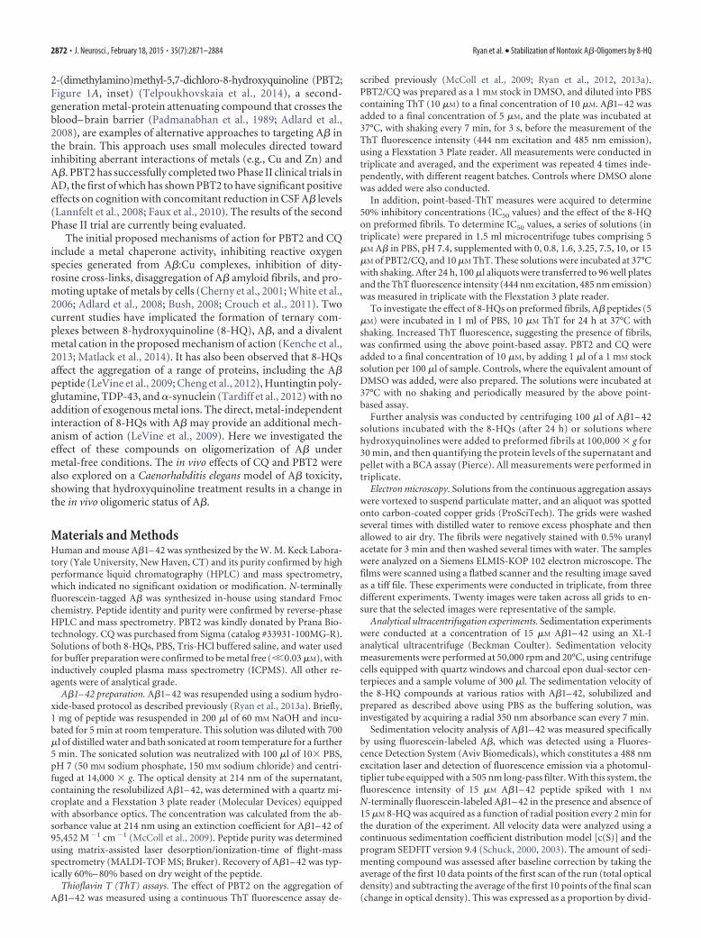
Figure 3. SAXS analysis of the A�/CQ complex. A, SAXS data for A�1– 42 (0.2 mg/ml, 20 �M) in the presence of 20 �M CQwas acquired at the Australian synchrotron. B, Guinier analysis of the CQ data in A. C, Kratky analysis of the CQ data in A. D, De novoreconstruction of the scattering entity induced by CQ using Dammif. Line indicates overall dimension (Dmax). E, F, Ensembleoptimization modeling of the CQ-induced A�1– 42 dimers provides information on the distribution in the radius of gyration (Rg,E) and maximal dimension (Dmax, F ) in relation to a pool of randomly generated structures: blue represents pool; red representsselected structure parameters. G, SAXS data for A�1– 42 (0.2 mg/ml, 20 �M) in the presence of 20 �M PBT2 (red squares) werealso acquired at the Australian synchrotron but had too high a ratio of signal-to-noise and thus were not of sufficient quality forfurther analysis.
2876 • J. Neurosci., February 18, 2015 • 35(7):2871–2884 Ryan et al. • Stabilization of Nontoxic A�-Oligomers by 8-HQ
mentation velocity measurements are affected by moleculargeometry so a sedimentation equilibrium experiment was con-ducted to accurately determine the mass of the species. A� in thepresence of PBT2 (Fig. 2F) or CQ (Fig. 2G) was determined tohave a molecular mass of 10.2 � 0.3 kDa and 9.95 � 0.26 kDa,respectively. It is not possible to accurately assess the mass of A�alone using this method, as equilibrium between sedimentationand diffusion is never reached, presumably because of an equi-librium distribution between aggregated and nonaggregatedforms of A� that changes over time.
The above results indicate that the presence of equimolar orgreater hydroxyquinolines induces the formation of a relativelystable population of low MW species of A�. We chose to investi-gate these species further using Synchrotron SAXS. A� in thepresence of CQ provided high-quality data for analysis (Fig. 3A),but A� in the presence of PBT2 did not provide data of sufficientquality (Fig. 3G) and, thus, was not used in this analysis. Guinieranalysis of the CQ:A� SAXS data demonstrates that the complexwas clearly oligomeric, with an average radius of gyration (Rg) of35.6 Å (Fig. 3B) [expected Rg for unfolded monomeric A�1– 42based on sequence is 18.3 Å (Kohn et al., 2004)], and a Kratky plotrepresentation of the data is consistent with that expected forpartially unfolded A� (Fig. 3C). The MW of the CQ:A� complexestimated from the forward scattering intensity (I0 � 0.010) was11–12 kDa, consistent with the results from analytical ultracen-trifugation (AUC) and SEC. The particle distance distributionfunction, P(r), was calculated via indirect Fourier transformationof the scattering data with Gnom (Svergun, 1992) giving a max-imum diameter (Dmax) of 122.45 Å and an average real space Rg of37.8 Å. De novo reconstruction of the shape of the CQ/A� com-
plex was performed using the bead modeling program Dammif(Franke et al., 2009), yielding extended structures with dimen-sions consistent with dimeric A� (Fig. 3D). The orientation of theA�1– 42 molecules in the dimer cannot be determined by SAXSanalysis because of the nonsequence dependence of x-rayscattering.
As the Kratky plot representation and the significantly ex-tended ab initio model suggest a flexible system, an ensembleapproach to modeling was subsequently conducted using the en-semble optimization method (Bernado et al., 2007). This ap-proach generates a pool of conformations (typically 10,000structures) based on the input protein sequence, and specifiedoligomerization state (in this case a dimer), and then uses a ge-netic algorithm to select a subset of structures that best fit theexperimental SAXS data. This analysis provides distance distri-butions (Rg and Dmax) that describe the predominant structuralfeatures of the sample in solution. These distributions are shownin Figure 3E, F, where the distributions for the selected structuresthat fit the SAXS data (fit shown in Fig. 3A, red line) providerelatively narrow distributions relative to the initial pool, withmodal values of 34.5 Å (Rg) and 128.6 Å (Dmax). These values arelarger than the modal value for the pool distribution indicatingthat the CQ/A� complex is not simply a distribution of randomcoil dimeric structures, but a more specific extended dimericstructure, and the narrow Rg/Dmax distributions also suggest thatthe complex is at least partially folded.
Analysis of the interaction of 8-HQ with A�The ThT, pelleting, AUC, SEC, and SAXS results in combinationindicate that A� in the presence of hydroxyquinoline is most
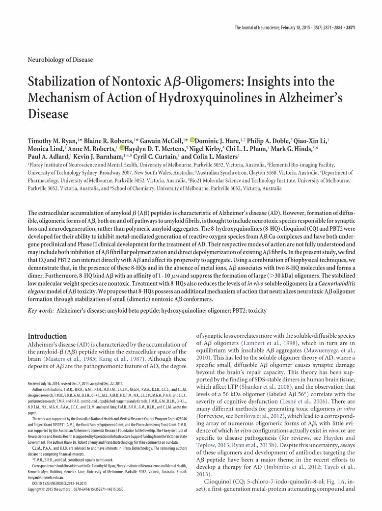
Figure 4. Investigation of the association of PBT2 and CQ with A�1– 42. A–C, Sedimentation velocity profiles acquired using 350 nm absorbance for PBT2 alone, PBT2 0.5 equivalents of A�,and PBT2 1.5 equivalents of A�1– 42, respectively. Data are normalized to provide better representation of the proportion of sedimenting material. D, c(S) distributions for PBT2 in the absenceor in the presence of increasing proportions of A� (15 �M PBT2, 15 �M A�, black line; 7.5 �M PBT2, 15 �M A�, red line; 5 �M PBT2, 15 �M A�, orange line; 2.5 �M PBT2, 15 �M A�, green line;0.1 �M PBT2, 15 �M A�, blue line). E, Proportion of sedimenting optical density as a function of CQ (dark green symbols) or PBT2 (dark red symbols) concentration. F, Weight average sedimentationcoefficient plotted as a function of CQ (dark green symbols) or PBT2 (dark red symbols) concentration. E, F, Lines through the data indicate fits of the equations described in the text and indicate aKd of 1–10 �M.
Ryan et al. • Stabilization of Nontoxic A�-Oligomers by 8-HQ J. Neurosci., February 18, 2015 • 35(7):2871–2884 • 2877

likely a low MW complex, of approximately two 8-HQ (700Da) and two A� molecules (9024 Da), giving a stoichiometry of 1hydroxyquinoline:A� molecule. These data, however, do not in-dicate the affinity of A� for 8-HQ. To address this, we initiallytook advantage of inherent UV absorbance of the 8-HQ mole-cules, which provide an excellent tool to monitor the associationof these compounds with A�. Analyzing the absorbance uniqueto a species in solution, such as PBT2 or CQ, is a powerful tool tomeasure direct interactions, as exemplified by studies investigat-ing the interaction of 7-nitrobenz-2-oxa-1,3-diazol-4-yl amino-labeled phospholipids with apoC-II (Mok et al., 2011; Ryan et al.,2011), or the interaction of Alexa-488 labeled DNA with Klenowfragment (Bailey et al., 2009). PBT2 and CQ are particularlysuited to this approach because they have a low molecular massand do not sediment effectively in the aqueous-based buffer sys-tems of the experiment. Figure 4 shows the absorbance of 15 �M
PBT2 as a function of radial position in the absence of A� (Fig.4A), and PBT2 at 7.5 �M (Fig. 4B) and 22.5 �M in the presence of15 �M A� (Fig. 4C). The addition of A� induces the formation ofboundaries in the absorbance data, which are dependent on theconcentration of PBT2. Similar data for CQ were also acquiredand showed a similar trend. This indicates that there is a specificsaturable interaction between these two molecules. From the ab-sorbance of PBT2 (or CQ), we can estimate the amount of hy-droxyquinoline required to saturate the sedimentation analysis(15 �M), which gives a stoichiometry of 1:1 (Fig. 4D). Furtheranalysis of the data at the saturating concentration with the c(S)model in SEDFIT9.4 showed that PBT2 and CQ were associatedwith an A� species having a sedimentation coefficient of 1.25 �0.1 S (11–14 kDa) (Fig. 4E). Thus, the observed complex has anapproximate MW of 10 kDa and consists of two A� peptides andtwo hydroxyquinoline molecules, giving a 2:2, or apparent 1:1,
Figure 5. Elemental analysis of the CQ interaction with A�1– 42. A, LA-ICPMS detection of iodine was used to generate an image of a slot blot assay where A�1– 42 was mixed with increasingconcentrations of CQ (as marked). Retained iodine is an integral part of CQ and is quantitative of the amount bound to A�. B, The concentration dependence of iodine retention in the slot blot assayobtained through quantitation of the image in A, corrected for signal from CQ only samples. C, Quantitation of retained iodine, obtained through excision of bands on the nitrocellulose and extractionusing nitric acid and measurement using standard bulk ICPMS analysis. D, The retained CQ, determined by each method (red represents bulk ICPMS analysis; blue represents laser ablation ICPMSanalysis), was converted to proportion CQ bound per A�, which was then plotted as a function of free CQ. This was analyzed to obtain a Kd of 1 �M. E, Retained CQ as a function of concentrationin the absence of A�, LA-ICMPS. F, Retained CQ as a function of concentration in the absence of A�, bulk ICMPS. G, Quantitation of retained iron, copper, and zinc, using the excision and extractionICPMS method.
2878 • J. Neurosci., February 18, 2015 • 35(7):2871–2884 Ryan et al. • Stabilization of Nontoxic A�-Oligomers by 8-HQ

stoichiometry. Analysis of the concentration titration data forboth the proportion of sedimenting PBT2 and CQ, and theweight average sedimentation coefficient (obtained by integrat-ing the c(S) distributions, representative data for PBT2 shown inFig. 4D), indicated an apparent dissociation constant in the lowmicromolar range (Fig. 4E and Fig. 4F, respectively), which is arelatively weak interaction that appears to have a very significanteffect on the self-association of A�.
There is a body of research using cell culture and electronparamagetic resonance spectroscopy that suggests A� is capableof forming a metal ion-mediated ternary complex with PBT2/CQ(Kenche et al., 2013). The AUC and SEC results were performedwith buffers where the metal content is below the detection limit
of the methods used and in the presence ofan excess of EDTA. Under these condi-tions, the formation of such a metal ionternary complex is unlikely; however, cat-egorical, conclusive evidence for the ab-sence of metal ion involvement in theinteraction is difficult to address usingthese assays. Thus, we used ICPMS to de-termine whether the A�/hydroxyquino-line was a ternary complex involvingmetal ion or a metal independent binaryinteraction. ICPMS analysis also providesdetection that is significantly more sensi-tive and specific for the compound thanthe other methods we have used. The ap-proach involved a slot blot membrane re-tention assay, similar to those used forradioisotope measurements of protein-CQinteractions (Opazo et al., 2006), wherethe protein (A� peptide) is mixed with anincreasing concentration of ligand (CQ)and is applied to a nitrocellulose mem-brane. Instead of using radiation countingto detect the proportion of bound ligand,we used ICPMS to measure the presenceof various elements, including iodine (asthe unique heteroatom component ofCQ), copper, zinc, and iron. For confir-mation of our results, we did this analysisvia two different sample introductionmethods, in two separate locations. First,we used LA-ICPMS to directly measurethe elements retained on the nitrocellu-lose membrane, providing the image inFigure 5A. Second, we used a digestionprotocol to solubilize the retained ele-ments, and introduced this solubilizedmaterial into the ICPMS with standardliquid handling approaches (solutionnebulization ICPMS). The image in Fig-ure 5A shows a clear trend where increas-ing the concentration of CQ in the A�/CQmixture results in increased retention ofiodine, consistent with the formation of aCQ/A� complex. This is quantified forLA-ICPMS in Figure 5B, and for bulkICPMS in Figure 5C, corrected for reten-tion of CQ alone by subtraction of the val-ues shown in Figure 5E, F. Metal ions,including iron, copper, and zinc, showed
no significant retention (Fig. 5D). As this is a retention assay andwe did not observe any metal present in our hydroxyquinoline,A�, or buffer reagents, this indicates that there is no metal re-tained with the complex. Thus, these metal ions are not signifi-cantly associated with the A�/CQ complex and, under theconditions used in this study, there is no evidence for the forma-tion of a ternary complex between A�, CQ, and a metal ion.Retention of CQ in the absence of A� was not significant at theconcentrations where CQ is soluble; however, at 50 �M, whereCQ is insoluble, we observed appreciable iodine signal (shown inFig. 5E).
The retained iodine signal is directly proportional to the con-centration of the A�/CQ complex and consequently provides a
Figure 6. Elemental analysis of the PBT2 interaction with A�1– 42. A, Titration of PBT2 against a set concentration of CQ (10�M) in the presence of A�1– 42 (10 �M) resulted in a loss of iodine signal. Analysis of these data indicates a Kd of 1.1 �M for theinteraction of PBT2 and A�. B, Quantitation of retained iron, copper, and zinc, measured using the bulk ICPMS method.
Ryan et al. • Stabilization of Nontoxic A�-Oligomers by 8-HQ J. Neurosci., February 18, 2015 • 35(7):2871–2884 • 2879

measure of the free CQ. These data can beplotted to produce a classical bindingcurve (Fig. 5D). Nonlinear regression,with a single site binding model, of thesedata provides a stoichiometry of 1 CQ:1A� and a dissociation constant (Kd) of 1�M (R 2 � 0.95 for LA-ICPMS, 0.91 forsolution nebulization ICPMS).
Relative affinity of PBT2 and CQ for A�As PBT2 does not contain iodine, we can-not use the above retention assay to di-rectly interrogate the affinity of PBT2 forA�. However, we can use competition be-tween the CQ and PBT2 to determine therelative affinity of these compounds forA�. Titration of PBT2 against a set con-centration of 10 �M CQ in the presence ofA� resulted in a concentration-dependentdecrease in the retained iodine signal,consistent with a classic “cold” competi-tion assay (Fig. 6A). Analysis of the datawith a standard one-site competitive bindingmodel provided an approximate equilibriuminhibitor dissociation constant (Ki) of 1.1 �M
for the interaction of PBT2 to A�. Consistentwith the CQ results, there was also no changeinthestatusofanyofthemetalionsupontitra-tion of PBT2 (Fig. 6B).
Molecular analysis of theA�/PBT2 interactionThe observation of apparent hydroxy-quinoline:A�1– 42 complexes with AUC,SEC, and SAXS and an apparent dissocia-tion constant of �10 �M led us to attemptnuclear magnetic resonance analysis ofthe samples. Using natural isotopic abun-dance in the peptide sample, concentra-tions of 1–2 mg/ml (214 – 428 �M) wererequired for sufficient sensitivity, and thisprecludes the use of CQ in these experi-ments, as its solubility is 30 �M in ourbuffers. PBT2 has no such constraint;thus, these experiments were conductedsolely with this 8-HQ. The majority of theA�1– 42 resonances are unperturbed inthe presence of the drug PBT2 (Fig. 7). Inthe presence of one equivalent of PBT2 to A�1– 42, resonancesassociated with PBT2 are significantly broadened with a smallchemical shift perturbation (0.04 ppm for the methyl resonances)from the positions of PBT2 in the absence of A� and reflect a verylow affinity interaction that is in intermediate exchange on theNMR time scale under these conditions (Fig. 7).
Cellular toxicity of the hydroxyquinoline stabilizedA�1– 42 oligomerTo investigate the effect of these A�/hydroxyquinoline com-plexes on neuronal function and toxicity, we conducted an MEAexperiment (Fig. 8). This provides information on the effect of acompound or peptide on a neuronal network. We specificallyinvestigated the spikes (Fig. 8A) and bursts (Fig. 8B) of activity incultured mouse primary cortical neurons after treatment with A�
alone or A� mixed with approximately twofold concentrations ofCQ. A� alone significantly decreased the activity of the neuronalculture, consistent with previous measures of A� toxicity to neu-ronal cultures. Interestingly, addition of A� and CQ not onlystopped the reduction in activity but also resulted in a significantincrease in activity, consistent with the stabilized A� dimer beingnontoxic, and CQ enhancing the activity of neurons.
In vivo effects of PBT2 on A�1– 42 oligomerization inC. elegansIn cell culture, we have shown that the stabilized complex is non-toxic. To investigate the effect of 8-HQs in whole animal modelsof A� toxicity, we used a transgenic C. elegans model that displaysparalysis upon expression of A�1– 42. We previously reportedthat PBT2 can reduce the toxicity of A�1– 42 in transgenic C.
Figure 7. 1D 1H NMR spectra of A�1– 42 in the presence and in the absence of PBT2. A, PBT2 alone. B, A�1– 42 with 1equivalent PBT2. C, A�1– 42 alone. Spectra were acquired at 700 MHz in 150 mM PBS, pH 7.0, and 25°C.
2880 • J. Neurosci., February 18, 2015 • 35(7):2871–2884 Ryan et al. • Stabilization of Nontoxic A�-Oligomers by 8-HQ

elegans (McColl et al., 2012). Consistent with the protective ef-fects of 8-HQ, we determined that CQ was also able to signifi-cantly reduce toxicity of human A�1– 42 expressed in the bodywall muscle cells (Fig. 9A). Using a range of concentrations, weobserved that 24 h exposure to 30 �M PBT2/CQ provided a ro-bust level of protection against A�-induced paralysis. This treat-ment significantly delays the onset of the toxic phenotype butdoes not completely prevent it. This indicates that these com-pounds can only partially rescue A� toxicity in this model. Ini-tially, we investigated the effect of 8-HQ compounds on A�oligomerization using an ELISA, which uses one antibody to bothcapture and detect A� (W02, epitope: A�5– 8) (Ida et al., 1996),based on the assumption that only oligomeric forms of A� willdisplay a second epitope for W0 –2 to bind. The validity of thisassumption was determined by comparing the effects of equimo-lar amounts of preaggregated A� (which showed a high responsein the assay) against freshly prepared and denatured A�, both ofwhich showed no response. Soluble lysates of C. elegans treatedwith PBT2 or CQ had a significant (p � 0.0001), but moderate,20% reduction in the ELISA signal compared with untreated con-trols (Fig. 9B). Although this was suggestive of an altered oli-
gomerization state upon treatment, thisassay is not quantitative, nor does it pro-vide any indication of the molecular massof the oligomeric species. Thus, we alsoexamined soluble extracts of C. eleganstransgenics using size exclusion fraction-ation on the Superdex 75 column (Fig.9C). This analysis shows a clear and signif-icant decrease in high MW A� and a smallincrease in low MW A� species (quanti-fied by measuring the area under each re-spective peak; Fig. 9D), consistent withthe AUC and SEC observations describedabove. Interestingly, the biologically de-rived A� oligomers appear larger than thesynthetically derived species. This may bedue to increased time to aggregate, a vari-ety of post-translational modifications, orany number of macromolecular interac-tions that may occur in the complex bio-logical milieu of a whole animal model.Importantly, although treatment with8-HQ altered the levels of soluble A� oli-gomers in vivo as predicted from the invitro studies, the treatment did not com-pletely abolish the presence of A� oligom-ers in C. elegans. This result is consistentwith both partially reduced phenotyperather than complete prevention of A�-induced toxicity (McColl et al., 2012) andwith the idea of 8-HQ having multiplemodes of action.
DiscussionWe have investigated the 8-HQ (CQ) andthe novel 8-HQ compound PBT2 foractivity in modulating A�1– 42 self-association and aggregation through a di-rect interaction. Our aggregation assayresults show that these compounds are ca-pable of inhibiting amyloid fibril forma-tion (Fig. 1), and the AUC, SEC, and SAXSresults show that both PBT2 and CQ sup-
press large aggregates (Figs. 2 and 3) and appear to induce a lowMW species of A� at 1:1 stoichiometry (Figs. 2 and 3). Theformation of a stable 8-HQ/A� complex, however, was not de-tected by the NMR experiments (Fig. 7). These results parallelother studies on hydroxyquinoline interactions with A�, whichhave shown that hydroxyquinoline-based compounds can in-hibit aggregation (LeVine et al., 2009). In contrast to other stud-ies on PBT2 and CQ interacting with A� (Kenche et al., 2013), weshow that the interaction can be independent of divalent transi-tion elements. Analysis of our buffers and reagents with ICPMSindicates that divalent metal levels are insignificant (��0.03 �M)and are not retained with the complex on nitrocellulose (Fig. 5).Further support of this conclusion is provided by the size exclu-sion results, in which the buffers contained 50 �M EDTA andwhere the same inhibition of aggregation and stabilization of lowMW species was observed as in experiments where EDTA was notpresent.
There are several toxicity studies, including LTP and mousecognition studies (Cherny et al., 2001; Adlard et al., 2008), celland yeast culture studies (Abramov et al., 2003; Tardiff et al.,
Figure 8. Assessment of the toxicity of CQ-induced A� dimers by multielectrode array analysis. Recording of neuronal activityin primary mouse neuronal cells treated with either A� or A� CQ. A, The total spike activity, normalized to baseline recordings(set to 100%). B, The total number of bursts, normalized to baseline recordings (set to 100%). **p � 0.01.
Ryan et al. • Stabilization of Nontoxic A�-Oligomers by 8-HQ J. Neurosci., February 18, 2015 • 35(7):2871–2884 • 2881

2012; Matlack et al., 2014), and our previ-ous C. elegans experiments (McColl et al.,2012), indicating that A� toxicity can beattenuated in the presence of PBT2 or CQ.We have reinvestigated the toxicity ofA�1– 42 in the presence of 8-HQ underour experimental conditions (Fig. 8) andhave confirmed that the stabilized dimeris nontoxic. Furthermore, our currentC. elegans results confirm a significantchange in the toxic phenotype observedwith A� expression. The previous C. el-egans experiments also indicated no sig-nificant change in amyloid deposits(McColl et al., 2012), although our cur-rent results indicate a significant reduc-tion in oligomeric species. These resultsare consistent with the current A� oli-gomer hypothesis that oligomeric speciesof A� are toxic and while the amyloidplaques/fibrils are not inherently toxic buta consequence of the aberrant aggregationof A� (for review, see Hayden et al., 2013).In particular, our results suggest that highMW (i.e., �20 –30 kDa, minimum stoi-chiometry of a tetramer) oligomers repre-sent the pool of toxic species, whereas lowMW oligomers (i.e., dimers) represent arelatively nontoxic form of the peptide.This is consistent with the observationthat tetramers may constitute the mini-mum toxic species (Streltsov et al., 2011).Furthermore, our data support the mech-anism that 8-HQ can directly engage A� oligomers and mono-mers to ameliorate the activity of toxic high stoichiometry andMW (�30 kDa) soluble oligomers of A�. The previous toxicity(Abramov et al., 2003; Adlard et al., 2008; McColl et al., 2012;Tardiff et al., 2012; Matlack et al., 2014) data in conjunction withour toxicity data (Fig. 8) and characterization of A� in the pres-ence of 8-HQ, suggests that these stabilized low MW oligomersare nontoxic. This further indicates that PBT2 and CQ not onlystabilize A� and suppress aggregation but that these compoundsalso neutralize large toxic A� peptides and oligomers.
In addition to the currently explored mechanisms of action ofPBT2 and CQ, we demonstrated that hydroxyquinolines have thecapability to directly influence the self-association and in vivotoxicity of the A� peptide. This activity is complimentary to theother mechanisms of action described for PBT2, and particularlycorrelates with the disaggregation of amyloid fibrils, inhibition ofreactive oxygen species generated by A� copper complexes, andinhibition of dityrosine crosslinks. The stabilization of nontoxicA� oligomers does not preclude a metal chaperone activity thatcan occur in the complex biological milieu (Adlard et al., 2008;Crouch et al., 2011) or an ionophoric activity resulting in promo-tion of intracellular transition metal uptake by neurons. How-ever, these results do indicate that 8-HQ have an additionalbeneficial mode of action that relates to the formation and deg-radation of toxic oligomeric species.
These results also raise some interesting questions in regardsto what the toxic oligomeric species may be. Our results suggestthat 8-HQ-stabilized low MW (dimeric) A� oligomers are nottoxic, and the observations in the wider literature of a range ofoligomers (for review of A� oligomerization, see Hayden et al.,
2013) having toxic properties suggest that synaptic loss in AD ismediated by not a single oligomeric species, but a range of differ-ent oligomers having varying degrees of toxicity, oligomeric sta-tus, and solubility. This complexity may preclude identifying atherapy targeted to a specific oligomer; as such, a targeted ap-proach could miss a multitude of other toxic oligomeric species.An alternative to targeting a specific oligomer for neutralizationor depletion is to stabilize a native, nontoxic, or nonpathogenicform of the protein/peptide (for review, see Johnson et al., 2012).This approach has been applied with great success to transthyre-tin (TTR) systemic amyloidosis, where the natively folded TTRtetramer slowly dissociates, generally under the influence of mu-tation, to misfolded monomeric proteins that rapidly aggregatevia an energetically favorable “downhill” polymerization processinto amyloid fibrils (Colon and Kelly, 1992; Hurshman et al.,2004; Foss et al., 2005; Johnson et al., 2005). In this pathway, therate-limiting step is the dissociation of the native tetramer (Fosset al., 2005), and this has led to a significant effort to identifymethods to stabilize the native TTR conformation. This researcheffort identified Tamafidis [2-(3,5-dichloro-phenyl)-benzoxa-zole-6-carboxylic acid] for the treatment of TTR systemic amy-loidosis in familial amyloid neuropathy (Johnson et al., 2012).This compound acts by binding with high specificity and affinity(50 nM) to one of the thyroxine binding sites on TTR andstabilizing the dimer– dimer interface of the tetramer throughhydrophobic and electrostatic interactions (Bulawa et al., 2012).This prevents the dissociation of the tetramer and thus formationof misfolded monomer and amyloid fibrils (Bulawa et al., 2012).Tafamidis was shown to significantly slow progression of the dis-ease process, including autonomic and peripheral neuropathies,
Figure 9. 8-HQs affect A�1– 42 toxicity in model animals. A, The effect of CQ on the paralysis phenotype observed in transgenicC. elegans, where the number of animals trialed: n � 105 (Ctl) and n � 108 (CQ). ***p � 0.001. B, ELISA quantitation of theoligomeric status of A� expressed in soluble lysates derived from C. elegans cultured in the absence or presence of 30 �M CQ orPBT2. C, Size exclusion (Superdex S75, PBS, 0.75 ml/min) analysis of the oligomeric status of A�1– 42 in the soluble fraction oflysates from transgenic C. elegans cultured in the absence (blue) or presence of 30 �M CQ (black) or PBT2 (red). D, Area under curveanalysis of the high MW (elution volume 5–10 ml) and low MW (elution volume 15–20 ml) A�1– 42 oligomers. Blue representstransgenic C. elegans alone. Black represents CQ-treated transgenic C. elegans. Red represents PBT2-treated transgenic C. elegans.*p � 0.05, ***p � 0.001.
2882 • J. Neurosci., February 18, 2015 • 35(7):2871–2884 Ryan et al. • Stabilization of Nontoxic A�-Oligomers by 8-HQ

and improved autonomic nervous system function and cachexia(Coelho et al., 2012, 2013). This efficacy proves the concept thatstabilization of a native, stable conformation can have significantoutcomes in protein misfolding diseases. Although TTR follows adifferent pathway from the typical nucleated polymerization(NP) models applied to amyloidogenesis, the activation energyfor monomer dissociation is akin to the energy required to forma nucleus in the NP model. This suggests that similar strategies,where a nontoxic conformation is stabilized by the binding ofparticular compounds, may be applicable to the wider range ofamyloid related diseases. In support of this theory, a recent studyon the effect of gallic acid interacting with �-synuclein showedthat the binding of this compound prevented the collapse of theprotein into an aggregation prone form (Liu et al., 2014). Inter-estingly, this interaction, while having large and significant effectson the biophysics of the protein, also only displayed extremelysubtle effects when investigated by NMR (Liu et al., 2014). Ourresults lend further support to these theories, suggesting thatPBT2/CQ variants have an additional mechanism of actionwhereby A� is stabilized in a nontoxic low MW form.
ReferencesAbramov AY, Canevari L, Duchen MR (2003) Changes in intracellular cal-
cium and glutathione in astrocytes as the primary mechanism of amyloidneurotoxicity. J Neurosci 23:5088 –5095. Medline
Adlard PA, Cherny RA, Finkelstein DI, Gautier E, Robb E, Cortes M, VolitakisI, Liu X, Smith JP, Perez K, Laughton K, Li QX, Charman SA, NicolazzoJA, Wilkins S, Deleva K, Lynch T, Kok G, Ritchie CW, Tanzi RE, Cappai R,Masters CL, Barnham KJ, Bush AI (2008) Rapid restoration of cognitionin Alzheimer’s transgenic mice with 8-hydroxy quinoline analogs is asso-ciated with decreased interstitial Abeta. Neuron 59:43–55. CrossRefMedline
Austin C, Hare D, Rozelle AL, Robinson WH, Grimm R, Doble P (2009)Elemental bio-imaging of calcium phosphate crystal deposits in kneesamples from arthritic patients. Metallomics 1:142–147. CrossRefMedline
Bailey MF, Angley LM, Perugini MA (2009) Methods for sample labelingand meniscus determination in the fluorescence-detected analytical ultra-centrifuge. Anal Biochem 390:218 –220. CrossRef Medline
Benilova I, Karran E, De Strooper B (2012) The toxic Abeta oligomer andAlzheimer’s disease: an emperor in need of clothes. Nat Neurosci 15:349 –357. CrossRef Medline
Bernado P, Mylonas E, Petoukhov MV, Blackledge M, Svergun DI (2007)Structural characterization of flexible proteins using small-angle X-rayscattering. J Am Chem Soc 129:5656 –5664. CrossRef Medline
Brenner S (1974) The genetics of Caenorhabditis elegans. Genetics 77:71–94.Medline
Bulawa CE, Connelly S, Devit M, Wang L, Weigel C, Fleming JA, Packman J,Powers ET, Wiseman RL, Foss TR, Wilson IA, Kelly JW, Labaudiniere R(2012) Tafamidis, a potent and selective transthyretin kinetic stabilizerthat inhibits the amyloid cascade. Proc Natl Acad Sci U S A 109:9629 –9634. CrossRef Medline
Bush AI (2008) Drug development based on the metals hypothesis of Alz-heimer’s disease. J Alzheimers Dis 15:223–240. Medline
Cheng XR, Sze Hung VW, Scarano S, Mascini M, Minunni M, Kerman K(2012) Label-free methods for probing the interaction of clioquinol withamyloid-[small beta]. Anal Methods 4:2228 –2232. CrossRef
Cherny RA, Atwood CS, Xilinas ME, Gray DN, Jones WD, McLean CA,Barnham KJ, Volitakis I, Fraser FW, Kim Y, Huang X, Goldstein LE, MoirRD, Lim JT, Beyreuther K, Zheng H, Tanzi RE, Masters CL, Bush AI(2001) Treatment with a copper-zinc chelator markedly and rapidly in-hibits beta-amyloid accumulation in Alzheimer’s disease transgenic mice.Neuron 30:665– 676. CrossRef Medline
Coelho T, Maia LF, Martins da Silva A, Waddington Cruz M, Plante-Bordeneuve V, Lozeron P, Suhr OB, Campistol JM, Conceicao IM,Schmidt HH, Trigo P, Kelly JW, Labaudiniere R, Chan J, Packman J,Wilson A, Grogan DR (2012) Tafamidis for transthyretin familial amy-loid polyneuropathy: a randomized, controlled trial. Neurology 79:785–792. CrossRef Medline
Coelho T, Maia LF, da Silva AM, Cruz MW, Plante-Bordeneuve V, Suhr OB,Conceicao I, Schmidt HH, Trigo P, Kelly JW, Labaudiniere R, Chan J,Packman J, Grogan DR (2013) Long-term effects of tafamidis for thetreatment of transthyretin familial amyloid polyneuropathy. J Neurol260:2802–2814. CrossRef Medline
Colon W, Kelly JW (1992) Partial denaturation of transthyretin is sufficientfor amyloid fibril formation in vitro. Biochemistry 31:8654 – 8660.CrossRef Medline
Crouch PJ, Savva MS, Hung LW, Donnelly PS, Mot AI, Parker SJ, GreenoughMA, Volitakis I, Adlard PA, Cherny RA, Masters CL, Bush AI, BarnhamKJ, White AR (2011) The Alzheimer’s therapeutic PBT2 promotesamyloid-beta degradation and GSK3 phosphorylation via a metal chap-erone activity. J Neurochem 119:220 –230. CrossRef Medline
Faux NG, Ritchie CW, Gunn A, Rembach A, Tsatsanis A, Bedo J, Harrison J,Lannfelt L, Blennow K, Zetterberg H, Ingelsson M, Masters CL, Tanzi RE,Cummings JL, Herd CM, Bush AI (2010) PBT2 rapidly improves cog-nition in Alzheimer’s disease: additional phase II analyses. J AlzheimersDis 20:509 –516. CrossRef Medline
Foss TR, Wiseman RL, Kelly JW (2005) The pathway by which the tetra-meric protein transthyretin dissociates. Biochemistry 44:15525–15533.CrossRef Medline
Franke D, Svergun DI (2009) DAMMIF, a program for rapid ab-initio shapedetermination in small-angle scattering. J Appl Crystallogr 42:342–346.CrossRef
Hare DJ, George JL, Grimm R, Wilkins S, Adlard PA, Cherny RA, Bush AI,Finkelstein DI, Doble P (2010) Three-dimensional elemental bio-imaging of Fe, Zn, Cu, Mn and P in a 6-hydroxydopamine lesioned mousebrain. Metallomics 2:745–753. CrossRef Medline
Hare DJ, Grubman A, Ryan TM, Lothian A, Liddell JR, Grimm R, Matsuda T,Doble PA, Cherny RA, Bush AI, White AR, Masters CL, Roberts BR(2013) Profiling the iron, copper and zinc content in primary neuron andastrocyte cultures by rapid online quantitative size exclusion chromato-graphy-inductively coupled plasma-mass spectrometry. Metallomics5:1656 –1662. CrossRef Medline
Hare DJ, Lee JK, Beavis AD, van Gramberg A, George J, Adlard PA, Finkel-stein DI, Doble PA (2012) Three-dimensional atlas of iron, copper, andzinc in the mouse cerebrum and brainstem. Anal Chem 84:3990 –3997.CrossRef Medline
Hayden EY, Teplow DB (2013) Amyloid beta-protein oligomers and Alz-heimer’s disease. Alzheimers Res Ther 5:60. CrossRef Medline
Hurshman AR, White JT, Powers ET, Kelly JW (2004) Transthyretin aggre-gation under partially denaturing conditions is a downhill polymeriza-tion. Biochemistry 43:7365–7381. CrossRef Medline
Ida N, Hartmann T, Pantel J, Schroder J, Zerfass R, Forstl H, Sandbrink R,Masters CL, Beyreuther K (1996) Analysis of heterogeneous A4 peptidesin human cerebrospinal fluid and blood by a newly developed sensitiveWestern blot assay. J Biol Chem 271:22908 –22914. CrossRef Medline
Imbimbo BP, Ottonello S, Frisardi V, Solfrizzi V, Greco A, Seripa D, Pilotto A,Panza F (2012) Solanezumab for the treatment of mild-to-moderateAlzheimer’s disease. Expert Rev Clin Immunol 8:135–149. CrossRefMedline
Johnson SM, Connelly S, Fearns C, Powers ET, Kelly JW (2012) The tran-sthyretin amyloidoses: from delineating the molecular mechanism of ag-gregation linked to pathology to a regulatory-agency-approved drug.J Mol Biol 421:185–203. CrossRef Medline
Johnson SM, Wiseman RL, Sekijima Y, Green NS, Adamski-Werner SL, KellyJW (2005) Native state kinetic stabilization as a strategy to ameliorateprotein misfolding diseases: a focus on the transthyretin amyloidoses. AccChem Res 38:911–921. CrossRef Medline
Kang J, Lemaire HG, Unterbeck A, Salbaum JM, Masters CL, Grzeschik KH,Multhaup G, Beyreuther K, Muller-Hill B (1987) The precursor of Alz-heimer’s disease amyloid A4 protein resembles a cell-surface receptor.Nature 325:733–736. CrossRef Medline
Kenche VB, Zawisza I, Masters CL, Bal W, Barnham KJ, Drew SC (2013)Mixed ligand Cu 2 complexes of a model therapeutic with Alzheimer’samyloid-beta peptide and monoamine neurotransmitters. Inorg Chem52:4303– 4318. CrossRef Medline
Kirby NM, Mudie ST, Hawley AM, Cookson DJ, Mertens HDT, Cowieson N,Samardzic-Boban V (2013) A low-background-intensity focusingsmall-angle X-ray scattering undulator beamline. J Appl Crystallogr 46:1670 –1680. CrossRef
Kohn JE, Millett IS, Jacob J, Zagrovic B, Dillon TM, Cingel N, Dothager RS,
Ryan et al. • Stabilization of Nontoxic A�-Oligomers by 8-HQ J. Neurosci., February 18, 2015 • 35(7):2871–2884 • 2883

Seifert S, Thiyagarajan P, Sosnick TR, Hasan MZ, Pande VS, Ruczinski I,Doniach S, Plaxco KW (2004) Random-coil behavior and the dimen-sions of chemically unfolded proteins. Proc Natl Acad Sci U S A 101:12491–12496. CrossRef Medline
Konarev PV, Volkov VV, Sokolova AV, Koch MHJ, Svergun DI (2003)PRIMUS: a Windows PC-based system for small-angle scattering dataanalysis. J Appl Crystallogr 36:1277–1282. CrossRef
Lambert MP, Barlow AK, Chromy BA, Edwards C, Freed R, Liosatos M,Morgan TE, Rozovsky I, Trommer B, Viola KL, Wals P, Zhang C, FinchCE, Krafft GA, Klein WL (1998) Diffusible, nonfibrillar ligands derivedfrom Abeta1– 42 are potent central nervous system neurotoxins. ProcNatl Acad Sci U S A 95:6448 – 6453. CrossRef Medline
Lannfelt L, Blennow K, Zetterberg H, Batsman S, Ames D, Harrison J, MastersCL, Targum S, Bush AI, Murdoch R, Wilson J, Ritchie CW (2008)Safety, efficacy, and biomarker findings of PBT2 in targeting Abeta as amodifying therapy for Alzheimer’s disease: a phase IIa, double-blind, ran-domised, placebo-controlled trial. Lancet Neurol 7:779 –786. CrossRefMedline
Lear J, Hare DJ, Fryer F, Adlard PA, Finkelstein DI, Doble PA (2012) High-resolution elemental bioimaging of Ca, Mn, Fe, Co, Cu, and Zn employ-ing LA-ICPMS and hydrogen reaction gas. Anal Chem 84:6707– 6714.CrossRef Medline
Lesne S, Koh MT, Kotilinek L, Kayed R, Glabe CG, Yang A, Gallagher M, AsheKH (2006) A specific amyloid-beta protein assembly in the brain im-pairs memory. Nature 440:352–357. CrossRef Medline
LeVine H 3rd, Ding Q, Walker JA, Voss RS, Augelli-Szafran CE (2009) Clio-quinol and other hydroxyquinoline derivatives inhibit Abeta(1– 42) oli-gomer assembly. Neurosci Lett 465:99 –103. CrossRef Medline
Lim NK, Villemagne VL, Soon CP, Laughton KM, Rowe CC, McLean CA,Masters CL, Evin G, Li QX (2011) Investigation of matrix metallopro-teinases, MMP-2 and MMP-9, in plasma reveals a decrease of MMP-2 inAlzheimer’s disease. J Alzheimers Dis 26:779 –786. CrossRef Medline
Liu Y, Carver JA, Calabrese AN, Pukala TL (2014) Gallic acid interacts withalpha-synuclein to prevent the structural collapse necessary for its aggre-gation. Biochim Biophys Acta 1844:1481–1485. CrossRef Medline
Masters CL, Simms G, Weinman NA, Multhaup G, McDonald BL,Beyreuther K (1985) Amyloid plaque core protein in Alzheimer diseaseand Down syndrome. Proc Natl Acad Sci U S A 82:4245– 4249. CrossRefMedline
Matlack KE, Tardiff DF, Narayan P, Hamamichi S, Caldwell KA, CaldwellGA, Lindquist S (2014) Clioquinol promotes the degradation of metal-dependent amyloid-beta (Abeta) oligomers to restore endocytosis andameliorate Abeta toxicity. Proc Natl Acad Sci U S A 111:4013– 4018.CrossRef Medline
Mawuenyega KG, Sigurdson W, Ovod V, Munsell L, Kasten T, Morris JC,Yarasheski KE, Bateman RJ (2010) Decreased clearance of CNS beta-amyloid in Alzheimer’s disease. Science 330:1774. CrossRef Medline
McColl G, Roberts BR, Gunn AP, Perez KA, Tew DJ, Masters CL, BarnhamKJ, Cherny RA, Bush AI (2009) The Caenorhabditis elegans A beta 1– 42model of Alzheimer disease predominantly expresses A beta 3– 42. J BiolChem 284:22697–22702. CrossRef Medline
McColl G, Roberts BR, Pukala TL, Kenche VB, Roberts CM, Link CD, RyanTM, Masters CL, Barnham KJ, Bush AI, Cherny RA (2012) Utility of animproved model of amyloid-beta (Abeta1– 42) toxicity in Caenorhabditiselegans for drug screening for Alzheimer’s disease. Mol Neurodegener7:57. CrossRef Medline
Mok YF, Ryan TM, Yang S, Hatters DM, Howlett GJ, Griffin MD (2011)Sedimentation velocity analysis of amyloid oligomers and fibrils usingfluorescence detection. Methods 54:67–75. CrossRef Medline
Opazo C, Luza S, Villemagne VL, Volitakis I, Rowe C, Barnham KJ, Strozyk D,Masters CL, Cherny RA, Bush AI (2006) Radioiodinated clioquinol as abiomarker for beta-amyloid: Zn complexes in Alzheimer’s disease. AgingCell 5:69 –79. CrossRef Medline
Padmanabhan G, Becue I, Smith J (1989) Clioquinol. In: Analytical profilesof drug substances (Klauss E, Florey E, eds). San Diego: Academic.
Petoukhov MV, Franke D, Shkumatov AV, Tria G, Kikhney AG, Gajda M,Gorba C, Mertens HD, Konarev PV, Svergun DI (2012) New develop-ments in the ATSAS program package for small-angle scattering dataanalysis. J Appl Crystallogr 45:342–350. CrossRef Medline
Petoukhov MV, Konarev PV, Kikhney AG, Svergun DI (2007) ATSAS 2.1:towards automated and web-supported small-angle scattering data anal-ysis. J Appl Crystallogr 40 [Suppl]:S223–S228.
Rembach A, Hare DJ, Doecke JD, Burnham SC, Volitakis I, Fowler CJ, ChernyRA, McLean C, Grimm R, Martins R, Ames D, Masters CL, Bush AI,Roberts BR (2014) Decreased serum zinc is an effect of ageing and notAlzheimer’s disease. Metallomics 6:1216 –1219. CrossRef Medline
Ryan TM, Caine J, Mertens HD, Kirby N, Nigro J, Breheney K, WaddingtonLJ, Streltsov VA, Curtain C, Masters CL, Roberts BR (2013a) Ammo-nium hydroxide treatment of Abeta produces an aggregate free solutionsuitable for biophysical and cell culture characterization. PeerJ 1:e73.CrossRef Medline
Ryan TM, Friedhuber A, Lind M, Howlett GJ, Masters C, Roberts BR (2012)Small amphipathic molecules modulate secondary structure and amyloidfibril-forming kinetics of Alzheimer disease peptide Abeta(1– 42). J BiolChem 287:16947–16954. CrossRef Medline
Ryan TM, Griffin MD, Bailey MF, Schuck P, Howlett GJ (2011) NBD-labeled phospholipid accelerates apolipoprotein C-II amyloid fibril for-mation but is not incorporated into mature fibrils. Biochemistry 50:9579 –9586. CrossRef Medline
Ryan TM, Roberts BR, Streltsov VA, Nuttall SD, Masters CL (2013b) Therole of A� in Alzheimer’s disease: amyloid fibrils and prefibrillar aggre-gates. New York: Wiley-VCH Verlag.
Schuck P (2000) Size-distribution analysis of macromolecules by sedi-mentation velocity ultracentrifugation and lamm equation modeling.Biophys J 78:1606 –1619. CrossRef Medline
Schuck P (2003) On the analysis of protein self-association by sedimenta-tion velocity analytical ultracentrifugation. Anal Biochem 320:104 –124.CrossRef Medline
Shankar GM, Li S, Mehta TH, Garcia-Munoz A, Shepardson NE, Smith I,Brett FM, Farrell MA, Rowan MJ, Lemere CA, Regan CM, Walsh DM,Sabatini BL, Selkoe DJ (2008) Amyloid-beta protein dimers isolated di-rectly from Alzheimer’s brains impair synaptic plasticity and memory.Nat Med 14:837– 842. CrossRef Medline
Streltsov VA, Varghese JN, Masters CL, Nuttall SD (2011) Crystal structureof the amyloid-beta p3 fragment provides a model for oligomer formationin Alzheimer’s disease. J Neurosci 31:1419 –1426. CrossRef Medline
Svergun ND (1992) Determination of the regularization parameter inindirect-transform methods using perceptual criteria. J Appl Crystallogr25:495–503. CrossRef
Tardiff DF, Tucci ML, Caldwell KA, Caldwell GA, Lindquist S (2012)Different 8-hydroxyquinolines protect models of TDP-43 protein, alpha-synuclein, and polyglutamine proteotoxicity through distinct mecha-nisms. J Biol Chem 287:4107– 4120. CrossRef Medline
Tayeb HO, Murray ED, Price BH, Tarazi FI (2013) Bapineuzumab and so-lanezumab for Alzheimer’s disease: is the ‘amyloid cascade hypothesis’still alive? Expert Opin Biol Ther 13:1075–1084. CrossRef Medline
Telpoukhovskaia MA, Rodríguez-Rodríguez C, Cawthray JF, Scott LE, PageBD, Alí-Torres J, Sodupe M, Bailey GA, Patrick BO, Orvig C (2014)3-Hydroxy-4-pyridinone derivatives as metal ion and amyloid bindingagents. Metallomics 6:249 –262. CrossRef Medline
Vistica J, Dam J, Balbo A, Yikilmaz E, Mariuzza RA, Rouault TA, Schuck P(2004) Sedimentation equilibrium analysis of protein interactions withglobal implicit mass conservation constraints and systematic noise de-composition. Anal Biochem 326:234 –256. CrossRef Medline
White AR, Du T, Laughton KM, Volitakis I, Sharples RA, Xilinas ME, HokeDE, Holsinger RM, Evin G, Cherny RA, Hill AF, Barnham KJ, Li QX, BushAI, Masters CL (2006) Degradation of the Alzheimer disease amyloidbeta-peptide by metal-dependent up-regulation of metalloprotease activ-ity. J Biol Chem 281:17670 –17680. CrossRef Medline
2884 • J. Neurosci., February 18, 2015 • 35(7):2871–2884 Ryan et al. • Stabilization of Nontoxic A�-Oligomers by 8-HQ