neurobiologyofdisease lmo4isanessentialcofactorinthesnail2 ... · sk-n-lp neuroblastoma cells were...
TRANSCRIPT

Neurobiology of Disease
LMO4 is an Essential Cofactor in the Snail2-MediatedEpithelial-to-Mesenchymal Transition of Neuroblastomaand Neural Crest Cells
Tiago Ferronha,1* M. Angeles Rabadan,1* Estel Gil-Guinon,2* Gwenvael Le Dreau,1 Carmen de Torres,2
and Elisa Martí1
1Instituto de Biología Molecular de Barcelona, Consejo Superior de Investigaciones Científicas, Parc Científic de Barcelona, Barcelona 08028, Spain, and2Developmental Tumor Biology Laboratory, Hospital Sant Joan de Deu, Fundacio Sant Joan de Deu, 08950 Barcelona, Spain
Neuroblastoma is an embryonic tumor derived from cells of the neural crest. Taking advantage of a newly developed neural crest lineagetracer and based on the hypothesis that the molecular mechanisms that mediate neural crest delamination are also likely to be involvedin the spread of neuroblastoma, we were able to identify genes that are active both in neural crest development and neuroblastoma tumorformation. A subsequent search of the neuroblastoma gene server for human orthologues of genes differentially expressed in the chickembryo neural crest screen retrieved the LIM domain only protein 4 (LMO4), which was expressed in both cell types analyzed. Functionalexperiments in these two model systems revealed that LMO4 activity is required for neuroblastoma cell invasion and neural crestdelamination. Moreover, we identified LMO4 as an essential cofactor in Snail2-mediated cadherin repression and in the epithelial-to-mesenchymal transition of both neural crest and neuroblastoma cells. Together, our results suggest that the association of high levels ofLMO4 with aggressive neuroblastomas is dependent on LMO4 regulation of cadherin expression and hence, tumor invasiveness.
IntroductionNeuroblastoma (NB) is a neurological tumor that arises fromneural crest (NC) cells and it is the most common extracranialtumor in children (Brodeur, 2003; Maris et al., 2007). NB repre-sents a very heterogeneous group of tumors in terms of theirbiological, genetic, and morphological characteristics. Clinically,these tumors may develop distinctly, ranging from spontaneousremission by differentiation and/or apoptosis (resembling nor-mal NC behavior), to aggressive metastatic disease with low over-all survival rates. The neuroectodermal origin of NB suggests thatthese tumors can spread from their primary site using mecha-nisms similar to those involved in the delamination and disper-sion of the embryonic NC, and that gene(s) implicated in NC cellmigration may also be involved in the acquisition of the invasive
NB phenotype (De Preter et al., 2006; Jiang et al., 2011). Thus,better understanding the biology of NC development may shedlight on the basic mechanisms involved in NB progression andaid the search for directed therapies.
NC cells delaminate from the dorsal neural tube (NT) andmigrate toward their destinations (Thiery et al., 2009). Beforedelamination, cells in the dorsal NT are specified as NC progen-itors (Betancur et al., 2010; Theveneau and Mayor, 2012). Snailtranscription factors are expressed in the premigratory NC of allvertebrate species analyzed, and they are particularly crucial forNC development. Moreover, their activity is required for theepithelial-to-mesenchymal transition (EMT) and for delamina-tion of these cells from the NT. EMT converts epithelial cells intomigratory and invasive mesenchymal cells, and it has also beenimplicated in the metastatic cascade of tumors (Thiery et al.,2009).
Given the similarities in the behavior of NC cells and NB, wesought to identify genes involved in both NC development andNB tumor formation. Taking advantage of a newly developed NClineage tracer, we performed a genome-wide screen of the genesexpressed in the chick embryo. Searching the NeuroblastomaGene Server for the human orthologues of genes differentiallyexpressed in the NC screen, we retrieved the LIM domain only(LMO) protein 4. LMO4 encodes a transcriptional regulator thatcontains two LIM zinc-binding domains for protein–protein in-teractions, but lacks the DNA-binding or catalytic domains.LMO4 belongs to a protein subfamily encoded by four genes(LMO1– 4), and its paralogues LMO1 and LMO3 are NB onco-genes (Aoyama et al., 2005; Isogai et al., 2011; Wang et al., 2011).In the developing mammalian spinal cord, LMO4 contributes to
Received Sept. 20, 2012; revised Oct. 27, 2012; accepted Dec. 4, 2012.Author contributions: T.F., M.A.R., E.G.-G., G.L.D., and E.M. designed research; T.F., M.A.R., E.G.-G., G.L.D., and
C.D.T. performed research; T.F., M.A.R., E.G.-G., G.L.D., and E.M. contributed unpublished reagents/analytic tools;T.F., M.A.R., E.G.-G., G.L.D., C.d.T., and E.M. analyzed data; E.M. wrote the paper.
Work in E.M.’s laboratory was supported by Grants BFU2010-18959 and CSD2007-00008. T.F. is a recipient ofPredoctoral Fellowship Grant BES-2008-003977. Work in C.d.T.’s laboratory was supported by Cellex Foundation,Caja Navarra, Ass P. Ugarte, and J. M. Buesa (Spanish Group for Sarcoma Research). We are grateful to the membersof the Martí lab and to Dr Angela Nieto for helpful discussions, as well as to Susana Usieto for invaluable technicalassistance. For DNAs and antibodies, we thank Drs B. Andersen, A. Nieto, E. Battle, C. Birchmeier, M. Goulding, andS. Pons.
The authors declare no competing financial interests.*T.F., M.A.R., and E.G.-G. contributed equally to this work.Correspondence should be addressed to either Carmen de Torres, Developmental Tumour Biology Laboratory,
Hospital Sant Joan de Deu, Fundacio Sant Joan de Deu, Barcelona, Spain, E-mail: [email protected]; or ElisaMartí, Instituto de Biología Molecular de Barcelona, CSIC, Parc Científic de Barcelona, C/Baldiri i Reixac 20, Barcelona08028, Spain, E-mail: [email protected].
DOI:10.1523/JNEUROSCI.4511-12.2013Copyright © 2013 the authors 0270-6474/13/332773-11$15.00/0
The Journal of Neuroscience, February 13, 2013 • 33(7):2773–2783 • 2773

the segregation of neuronal subtypes (Lee et al., 2005; Joshi et al.,2009) and, in the Xenopus embryo, participates in the acquisitionof NC identity (Ochoa et al., 2012). Here, we show that LMO4 isexpressed in human NB cells and chick NC cells. Functional ex-periments in both models revealed that LMO4 activity is requiredfor the invasiveness of NB cells and NC delamination. Further-more, we show that LMO4 is an essential cofactor in Snail2-mediated cadherin repression and EMT in NC and NB cells.
Materials and MethodsDNA constructs. The BMP reporter construct (BRE-tk-GFP) used herehas been previously described (Le Dreau et al., 2012), as has theE-cadherin-luciferase reporter construct (Batlle et al., 2000). HumanLMO4 (Lu et al., 2006) and chicken Snail2 (Morales et al., 2007) weresubcloned into pCAGGS-ires-GFP or pCAGGS-ires-H2B-RFP.
To knockdown LMO4 in chick embryos, short RNA hairpin (shRNA)-based expression vectors were generated (Tables 1). The knockdown ofhuman LMO4 in neuroblastoma cell lines was achieved using shRNAplasmids from the RNAi Consortium (Sigma-Aldrich), (Table 2).
FACS and microarray analysis. DNA was microinjected into the em-bryonic NT and the embryos were then electroporated using an IntracelDual Pulse TSS-100 electroporator. The NT was recovered 24 h aftercoelectroporation of the pBRE-tk-GFP reporter and the pCAGGS-ires-H2B-RFP construct, and a single-cell suspension was obtained followinga 10 –15 min Trypsin-EDTA digestion. GFP and RFP fluorescence wasdetermined, cells were sorted by flow cytometry using a MoFlo flowcytometer (Dako), and total RNA was extracted using the standard Trizol(Promega) protocol.
cDNAs were synthesized and hybridized to the GeneChip ChickenGenome Array (Affymetrix). Bioconductor software was used to analyzethe data, which were normalized using the “rma” algorithm to identifydifferentially expressed genes. The results were filtered using thresholdsof [log2FC] � 0.5849 and a p � 0.05.
RT-PCR and evaluation of RNAi efficiency by quantitative real-time-PCR. The primers used for qPCR amplification of target genes werepurchased from QuantiTec Primer Assays (Qiagen). PCR amplificationswere assessed from pools of electroporated NT (15/pool) using two tothree independent pools per experimental condition, GFP � cells wereFACS sorted, and total RNA was extracted. The data represent the meanvalues � SEM.
Total RNA was isolated from neuroblastoma cell lines using theTRI Reagent (Sigma-Aldrich) and qPCR was performed on an ABIPrism 7000 Sequence Detection System using Q-PCR Taqman GeneExpression Assays (Applied Biosystems). The quantification of LMO4(Hs00232488_m1) and E-cadherin mRNA (Hs 01023894_m1) tran-scripts was normalized to TATA box binding protein (Hs00427620_ms1)expression and to the cell line with the lowest amplification level(LAN1).
Immunohistochemistry and in situ hybridization. Embryos of either sexwere fixed for 2–4 h at 4°C in 4% paraformaldehyde (PFA) in PBS andsections were immunostained following standard procedures with antibod-ies against: Islet1, Lmx1b, and Pax7 (Developmental Studies HybridomaBank); Id4 and ZO1 (Invitrogen); aPKC (Santa Cruz Biotechnology);HNK-1 and Laminin (Sigma); N-cadherin (Zymed); phospho-Histone-3(Millipore); and cleaved caspase 3 (BD Biosciences). Cells were counted inimages obtained from at least six different chick embryos for each experi-mental condition. The data are represented as the mean � SEM.
For in situ hybridization, embryos were fixed overnight at 4°C in 4%PFA in PB and then processed for whole-mount RNA in situ hybridiza-tion following standard procedures. The embryos were hybridized withprobes against chick LMO4, FoxD3, Snail2, Sox9, and Sox10 (from thechicken EST project, UK-HGMP RC) and were postfixed in 4% PFA.Vibratome sections were photographed on a Leica DMR microscope.
Luciferase reporter assay. Transcriptional activity assays of LMO4 andSnail2 were performed using the E-cadherin-luciferase reporter (Batlle etal., 2000). Embryos were electroporated with the indicated DNAs to-gether with the firefly-luciferase vector and a renilla-luciferase reporterconstruct (Promega) for normalization, harvested at 24 h postelectropo-ration (hpe) and homogenized in passive lysis buffer. Firefly and renillaluciferase activity was measured using the Dual Luciferase Reporter As-say System (Promega). Data are represented as the mean � SEM from8 –12 embryos per experimental condition.
SH-SY5Y cells were transfected using X-treme 9 transfection reagent(Roche) with indicated DNAs, harvested 24 h after transfection, and thefirefly and renilla luciferase activities were measured using the Dual Lu-ciferase Reporter Assay System.
Neural crest explant culture and time-lapse analysis. Chick embryoswere electroporated with the indicated DNAs and the NT was dissectedout at 3 hpe. NT explants were cultured in DMEM-F12 (D6421; Sigma),0.01% penicillin-streptomycin (Invitrogen), and 0.001% Myto � (BDBioscience). Time lapse analysis of NC cell migration was performed aspreviously described (Duband et al., 2009) using a Leica SP5 confocalmicroscope.
The data from the cell tracking assay was processed using ImageJ,whereby migratory cells were treated as particles centered on their ownnuclei and tracked using the ImageJ Manual Tracking plug-in.
Cell lines. The LAN-1, SK-N-LP, and SK-N-JD cell lines were providedby Dr N. K. V. Cheung (Memorial Sloan Kettering Cancer Center, NewYork, NY), the LA1–5S line by Dr B. Spengler (Fordham University, NewYork, NY), and the SH-SY5Y cells by Dr V. J. Yuste (Institut de Neuro-ciencies, Medicina, UAB, Spain). The SK-N-AS cell line was purchasedfrom the European Collection of Cell Cultures (Wiltshire, UK).
Lentivirus production and lentivirus-mediated shRNA knockdown. Len-tiviral virions expressing shLMO4 and control shRNA were produced bytransient cotransfection of the HEK 293T packaging cell line withpCMV-VSV-G and the vectors containing the LMO4 shRNA (pLKO.1-
Table 1. Sequences of primers used for the generation of chick sh-LMO4 shRNA
Ref. Oligo Sequence
sh-LMO4-a Fw Oligo 5�-gatcCCCGCATGATCCTCTGCAGAAATTCAAGAGATTTCTGCAGAGGATCATGCttttta�3�Re Oligo 5�-agcttAAAAAGCATGATCCTCTGCAGAAATCTCTTGAATTTCTGCAGAGGATCATGCggg�3�
sh-LMO4-b Fw Oligo 5�-gatcCCCGGAGATCGGTTTCACTACATTCAAGAGATGTAGTGAAACCGATCTCCttttta�3�Re Oligo 5�-agcttAAAAAGGAGATCGGTTTCACTACATCTCTTGAATGTAGTGAAACCGATCTCCggg�3�
sh-LMO4-c Fw Oligo 5�-gatcCCCTGTCTATCATCTGAAGTGTTTCAAGAGAACACTTCAGATGATAGACAtttttggaaa�3�Re Oligo 5�-agcttttccaaaaaTGTCTATCATCTGAAGTGTTCTCTTGAAACACTTCAGATGATAGACAggg�3�
Fw, Forward; Re, reverse.
Table 2. Sequences of primers used for the generation of human sh-LMO4 shRNA
Ref. Clone ID Clone name Match position Sequence
sh-A TRCN0000013240 NM_006769.2-1156s1c1 1156 5�-CCGG-GATCGGTTTCACTACATCAAT-CTCGAG-ATTGATGTAGTGAAACCGATC-TTTTT-3�sh-B TRCN0000013241 NM_006769.2-1165s1c1 1165 5�-CCGG-CACTACATCAATGGCAGTTTA-CTCGAG-TAAACTGCCATTGATGTAGTG-TTTTT-3�sh-C TRCN0000013242 NM_006769.2-999s1c1 999 5�-CCGG-CAGAAATGACTACATTAGGTT-CTCGAG-AACCTAATGTAGTCATTTCTG-TTTTT-3�
2774 • J. Neurosci., February 13, 2013 • 33(7):2773–2783 Ferronha et al. • LMO4 and Snail in EMT

LMO4; Sigma-Aldrich). Control lentivirus was produced using thepLKO.1 vector containing a scrambled nontargeting short-hairpin RNAsequence (Addgene).
SK-N-LP neuroblastoma cells were transduced with lentiviral-condictioned media and selected for 2 weeks in medium containingpuromycin (Sigma-Aldrich) and puromycin-resistant cells were sub-sequently pooled.
SDS-PAGE and Western blotting. Electroporated chick NTs were re-covered at 24 hpe, cleaned, and frozen. Protein was extracted using RIPAbuffer and quantified by the Bradford method. These samples were ex-amined in Western blots probed with antibodies against N-cadherin(Invitrogen) and b-tubulin (Millipore). Densitometric analysis was per-formed using Quantity One software (Bio-Rad).
Total cell lysates from shRNA-LMO4 NB (SK-N-LP) cell lines wereharvested in hot lysis buffer, quantified by the Bio-Rad Protein Assay,and analyzed by Western blots using antibodies against cleaved caspase-3(Cell Signaling Technology) and GAPDH (Millipore). SH-SY5Y cellstreated for 6 h with 1 �M Staurosporine (Sigma) were used as a positivecontrol.
Thymidine proliferation assay. NB SK-N-LP shLMO4 cells were cul-tured in complete medium for 48 h and then incubated for 4 h with3H-thymidine (Amershan-Pharmacia Biotech). Cells were lysed with0.04% SDS before processing the lysates for liquid scintillation counting(Optiphase 2; PerkinElmer Life Sciences) and measurements were per-formed with a WinSpectral 1414 liquid scintillation counter (Wallac).
In vitro wound-healing migration assay. SK-N-LP cells expressing sh-LMO4 were seeded into 60 cm plates (Corning) and grown to conflu-ence. A single wound was then created in the cell monolayer andmigration of the cells from the edge of the wound was analyzed in imagestaken every 3 h. The area between the wound edges was measured at eachtime point using ImageJ software (as described previously by Dr KeesStraatman, Advanced Imaging Facilities, University of Leicester, Leices-ter, UK). The area immediately after creation of the wound was set as100%, and the relative change was calculated as a percentage of the initialarea.
In vitro invasion assay. The membranes of the Transwell invasion assayinserts (Corning Costar) were coated with Matrigel (BD Biosciences) andhyaluronic acid. shLMO4-SK-N-LP cells were plated onto the upperchamber in RPMI 1640 medium supplemented with 0.1% fetal bovineserum and complete medium was added to the lower chamber. After 24 hincubation, cells on the upper side of the membrane were removed andthose that had migrated into the receiver well were trypsinized andstained with propidium iodide (Sigma). Analyses were performed withan Epics XL flow cytometer (Coulter) using Flowcheck (BeckmanCoulter) as an internal standard for cell counting. Experiments wereperformed in triplicate and the data is represented as an index of invasionrelative to the pLKO control.
Statistics. Statistical analysis was performed using Statview software,analyzing the data using a Student’s t test, except for the increasing con-centrations of Snail2, the effects of which were analyzed by ANOVAfollowed by a Student–Newman–Keuls test. A heatmap graph was gen-erated using R software (The R Project for Statistical Computing). Quan-titative data are presented as the mean � SEM.
ResultsLMO4 is common to neural crest and neuroblastoma cellsTo identify elements of the NC cell transcriptome, we used theBMP-Responsive Element (BRE-tk-GFP) to drive stable GFP ex-pression in the dorsal spinal cord (Fig. 1A) (Le Dreau et al., 2012).Chick embryos [Hamilton–Hamburger (HH) stage 10] were co-electroporated with BRE-tk-GFP and pCAGGS-ires-H2B-RFP,which drives RFP expression along the entire dorsal-ventral axisof the NT. The embryos were allowed to develop for 24 h andtransfected cells expressing GFP or RFP were purified by FACS.Total RNA was extracted from these cells and gene expressionwas assayed in the two cell populations using the Affymetrix Ge-neChip. Several genes known to be expressed in the dorsal NTwere enriched in the GFP-expressing cell population. Accord-
ingly, we identified a total of 413 genes that were differentiallyexpressed by the two cell populations (Fig. 1B).
We next searched the Neuroblastoma Gene Server to identify thehuman orthologues of these 413 genes, retrieving a total of 80 genescommon to NC cells and NB tumor cells (Fig. 1C). Among them,LMO4 was of particular interest as high levels of LMO4 have beenreported in aggressive stage IV NB (Yamanaka et al., 2002). Further-more, two different NB cell lines strongly express LMO4 in tran-scriptome profiles associated with an aggressive phenotype(Schramm et al., 2005; Schulte et al., 2005). Since the role of thisprotein in NB remains unknown, we analyzed the expression ofLMO4 in a panel of human NB cell lines. Gene expression analysis ofthese NB cell lines serve to classify them as neuroblastic N-type(LAN-1 and SH-SY5Y), intermediate I-type (SK-N-JD and SK-N-LP), and Schwannian/glial S-type (SK-N-AS and LA1–5S) (Fig. 1D).LMO4 was expressed more strongly in the aggressive I-type cell lines(Fig. 1E). Thus, we selected the SK-N-LP cell line for further func-tional analysis of LMO4.
Neuroblastoma cell invasiveness is dependent on LMO4To investigate the function of LMO4 in NB, we knocked downLMO4 expression using a shRNA containing lentivirus (Fig. 2A).In contrast to breast cancer cells in which low levels of LMO4expression is associated with reduced proliferation (Sum et al.,2005), reduced LMO4 expression had no effect on the prolifera-tion or apoptosis of NB cells (Fig. 2B,C). Hence, the activity ofLMO4 does not appear to be essential for NB tumor cell viability.
When we next analyzed the migratory properties of NB cells ina wound-healing assay, the impaired capacity of NB cells to closethe wound reflected the extent of LMO4 expression, linkingLMO4 activity with cell motility (Fig. 2D–F). On the basis ofthese results, we examined the invasive capacity of NB cells in anextracellular matrix (Matrigel), an assay that provides a correlateof in vivo metastatic potential. These confirmed that the invasivecapacity of NB cells is dependent on the levels of LMO4 expres-sion (Fig. 2G). Together, these findings strongly suggest thatLMO4 regulates the migratory and invasive behavior of NB cells,without affecting their viability. As such, and in the search for acommon mechanism controlling NB and NC cell behavior, wenext investigated the role of LMO4 in neural development in vivo.
LMO4 is expressed by the neural crestLMO4 is necessary for the development of the mammalian ner-vous system as mutant mouse embryos lacking LMO4 exhibitexencephaly and die (Hahm et al., 2004; Tse et al., 2004; Lee et al.,2005). LMO4 is expressed in the neural plate and in NC cells ofthe Xenopus embryo (Ochoa et al., 2012) and, since we identifiedLMO4 when attempting to study the chick NC transcriptome, itappears likely that LMO4 function is conserved in early neuraldevelopment across vertebrates.
To validate the results of our screening, we analyzed the ex-pression of LMO4 in chick embryos, demonstrating that it isexpressed in the cranial NC at the 10-somite stage (Fig. 3A). Inthe anterior NT, LMO4 expression is restricted to the dorsal-most NT, containing premigratory NC (Fig. 3A), whereas cau-dally, it is widely expressed in the open neural plate (Fig.3Aiv,Av). After NT closure and the onset of NC migration, LMO4expression is restricted to the dorsal NT, the migratory NC, andthe trunk NC derivatives (Fig. 3B and data not shown).
The early expression of LMO4 in the chick embryo suggests itfulfils a role in the specification and/or maintenance of NC iden-tity. To test this possibility, we overexpressed LMO4 in the NT ofchick embryos and analyzed the expression of components of the
Ferronha et al. • LMO4 and Snail in EMT J. Neurosci., February 13, 2013 • 33(7):2773–2783 • 2775

NC gene regulatory network (del Barrio and Nieto, 2002; Cheunget al., 2005; Betancur et al., 2010). In contrast to the findings inXenopus embryos (Ochoa et al., 2012), LMO4 activity was insuf-ficient to ectopically activate NC-specific genes at this develop-mental stage in the chick (Fig. 3C).
LMO4 controls the delamination of neural crest cells from thedorsal NTBased on the control exerted by LMO4 on the migratory andinvasive behavior of NB cells, we investigated whether gain or lossof LMO4 function in chick embryos in vivo affected NC migra-tion. Knockdown of endogenous LMO4 was achieved in the de-veloping NT by electroporating short-hairpin RNA. Thisapproach produced a significant reduction in RNA levels �36hpe, but it did not affect cell proliferation or survival in the NT(Fig. 4A–C).
LMO4 knockdown blocked NC migration in a cell autono-mous manner. Indeed, when compared with the controls, the lossof LMO4 led to the retention of cells within the NT at 24 hpe (Fig.4D), and an �80% loss of migratory NC cells at 48 hpe (Fig.5A,B). Moreover, the number of GFP-labeled cells was reduced
in the trunk NC to a similar extent in all derivatives [dorsal rootganglia (DRG), sympathetic ganglia, and melanocytes; Fig. 5)].Impaired delamination was rescued by the overexpression of hu-man LMO4, which reverted the effect of the shRNA, demonstrat-ing the specificity of this phenotype (Fig. 5C–E). Furthermore,LMO4 knockdown resulted in a �20% reduction of total DRGcells (data not shown).
Overexpressing LMO4 had no effect on proliferation, eitherwithin the NT or in migratory cells (Fig. 4C and data not shown)but increased NC migration 24 hpe (Fig. 4D). After 48 hpe, itproduced a �30% increase in the number of GFP-labeled migra-tory NC cells when compared with the control embryos (Fig. 5B).LMO4 expressing cells were not preferentially located to distinctNC derivatives, indicating that LMO4 promotes the migration ofall trunk NC and suggesting a role for LMO4 in the early stages ofNC migration.
Since LMO4 appears to influence NC migration, we analyzedNC cell movement in an explant assay. When cell movement wasanalyzed by time-lapse microscopy, a similar number of NC leftNT explants obtained from control and LMO4-GFP electropo-rated embryos (Fig. 6), without exhibiting directional migration
Figure 1. Identification of LMO4 as a gene expressed by both neural crest and neuroblastoma cells. A, Representative image showing coelectroporation with BRE-tk-GFP (green) and a controlplasmid expressing pCAGGS-H2B-RFP (red). GFP-expressing cells were restricted to the dorsal NT and NC cells, which also express the NC marker HNK1 (blue). B, Schematic representation of the FACSsorting and transcriptome screening for NC genes. C, Schematic representation of the analysis of the BRE-array data, which was compared with Neuroblastoma Gene Server data to retrieve LMO4 asa common hit. D, Molecular characterization of neuroblastoma cell lines. While the expression of Phox2b identifies these cells as neuroblastomas, the relative expression of glial markers (GFAP,PMP22, and S100�) and neural markers (GAP43 and neurofilaments) allows for the subgroup association as neural (N-type), glial (S-type), or intermediate (I-type). E, LMO4 expression measuredby RT-qPCR in neuroblastoma cell lines; results are expressed relative to those of LAN1 cells.
2776 • J. Neurosci., February 13, 2013 • 33(7):2773–2783 Ferronha et al. • LMO4 and Snail in EMT

and showing similar speed of movement (Fig. 6). However, cellsoverexpressing LMO4 covered the first 50 �m in �70 min, whilecontrol cells required 100 min to cover a similar distance (Fig. 6),suggesting precocious delamination from the explants.
Together, these data indicate that LMO4 is not sufficient totrigger the genetic program underlying NC production, nor doesit regulate proliferation or survival of NB or NC cells. However,LMO4 does appear to play a conserved role in NB invasivenessand the delamination of NC cells, prompting us to investigate themechanisms underlying such effects.
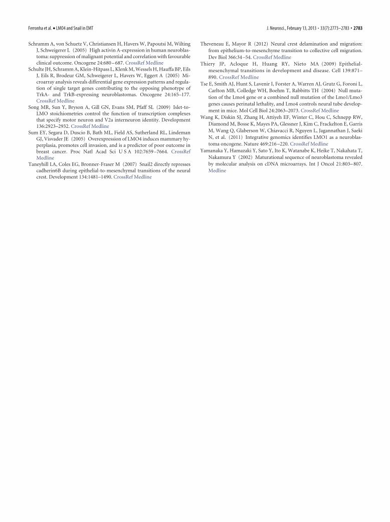
LMO4 is a necessary cofactor of Snail2 when mediating EMTin neural crest cellsDelamination of the NC from the NT is a bona fide example of fullEMT, and transcription factors from the Snail family are sufficient totrigger a full EMT program in the NT (Nieto, 2011). However, in ourin vivo knockdown experiments, endogenous Snail2 was insufficientto trigger NC delamination in the absence of LMO4 (Fig. 5A). Inaddition, the increase of NC migration produced by overexpressionof Snail2 is significantly reduced in the absence of LMO4 (Fig. 7A,B).Interestingly, we showed that increased dosage of Snail2 can over-come impaired NC migration (Fig. 7C,D). Hence, a functional in-teraction appears to exist between LMO4 and Snail2, which isconsistent with the direct protein interaction between the LIM-domains of LMO4 and the N terminus domain of Snail1 and Snail2recently described (Ochoa et al., 2012).
To further study the role of LMO4 in delamination, we ana-lyzed the integrity of the basal lamina. Staining with laminin
revealed that the ectopic disruptions of the basal lamina causedby Snail2 overexpression were absent following LMO4 knock-down (Fig. 7B). However, overexpression of LMO4 alone was notsufficient to disrupt the basal lamina (data not shown), indicatingthat disruption of the basal lamina is not directly dependent onLMO4 activity.
We also analyzed components of the apical complex, in-cluding the tight junction proteins ZO1 and aPKC that line thelumen of neuroepithelial cells. Overexpression of LMO4 wasinsufficient to either downregulate or alter the subcellular dis-tribution of ZO1 or aPKC (data not shown). N-cadherin ishighly localized to the apical junctional region at the lumen ofthe NT (Fig. 8A). Delamination of NC cells involves the loss ofN-cadherin (Thiery et al., 2009; Park and Gumbiner, 2010,2012) and overexpression of LMO4 was insufficient to alterthe subcellular distribution of N-cadherin, although the totalN-cadherin protein was reduced in a manner similar to thatobserved following Snail2 overexpression (Fig. 8 B, C). Inter-estingly, LMO4 and Snail2 exhibited an additive capacity todownregulate N-cadherin expression (Fig. 8 B, C), suggestingthere is a functional interaction between these proteins thatenhances the downregulation of cadherin expression.
LMO4 and Snail2 cooperate in repressing E-cadherin expressionTo study the LMO4/Snail2 functional interaction in a conservedrepressor context, we focused on the well characterized humanE-cadherin promoter, in which Snail2 binds directly to the E2 box
Figure 2. Neuroblastoma cell invasiveness is dependent on LMO4. A, Efficiency of LMO4 gene silencing using shRNA in SK-N-LP cells, as measured by RT-qPCR. LMO4 mRNA expression wascalculated relative to that of scrambled shRNA (pLKO control; error bars indicate the SD). B, LMO4 expression is not required for SK-N-LP cell proliferation. DNA synthesis was determined bymeasuring 3H-thymidine incorporation in cells expressing the shRNAs indicated during the last 4 h of a 48 h culture. Data represent the means (�SEM) of four independent cultures. C,Downregulation of LMO4 in SK-N-LP cells does not significantly increase apoptotic caspase activation. Total cell extracts from SK-N-LP cells expressing the indicated shRNAs were analyzed in Westernblots probed with cleaved caspase-3 antibody. GAPDH was used as a loading control. D–F, LMO4 activity is required for NB cell migration. D, Schematic representation of the wound healing assayand its quantification. Measurement of the wounded areas was performed using ImageJ by enhancing the contrast between cells and the wound, thereby permitting automatic measurement of thearea. E, Representative images of cell migration in a wound-healing assay using SK-N-LP cells expressing the indicated shRNAs. F, Changes in wound area were calculated as a percentage (mean �SEM) of the initial wound area (***p � 0.001). G, NB cell invasiveness is dependent on LMO4 activity. Schematic representation of the insert containing the Matrigel-coated porous membrane onwhich NB cells were seeded. After 24 hours, the fraction of cells that migrated through the matrix was determined by flow cytometry. The data represent the index of invasion relative to pLKO control(***p � 0.001). Cell lines with lower levels of LMO4 exhibited reduced invasive properties.
Ferronha et al. • LMO4 and Snail in EMT J. Neurosci., February 13, 2013 • 33(7):2773–2783 • 2777

Figure 3. LMO4 is expressed in neural crest cells. In situ hybridization analysis shows LMO4 expression in chick embryos. A, At HH stage 10, LMO4 was expressed in the early NT along ananterior–posterior gradient. Anterior LMO4 was expressed in the dorsal NT and migratory NC in the cranial and trunk. Posterior LMO4 was expressed in the open NT. B, At HH stage 14, LMO4 wasexpressed in the migratory NC. C, LMO4 is insufficient to regulate the expression of NC genes. Expression of the indicated NC markers was analyzed in chick embryos electroporated at HH stage 1024 hpe, which revealed no obvious change in the expression of any NC-specific genes.
Figure 4. LMO4 controls the delamination of NC cells from the dorsal NT. A–C, Generation and characterization of chick shLMO4. A, Embryos were electroporated at HH stage 12 with controlshRNA or with various shLMO4 constructs. Cells were harvested 36 h later, GFP� cells were purified by FACS, and endogenous expression of cLMO4 transcripts was analyzed by RT-qPCR. On the basisof the �70% reduction in expression produced by shLMO4a, this was used in all subsequent experiments. B, Knockdown of LMO4 did not compromise cell survival or cell proliferation, as determinedby cleaved caspase-3 staining or phospho-H3 staining in control and shLMO4 electroporated embryos. C, Quantitative data show the mean�SEM ratios of pH3-expressing cells in the electroporatedversus control nonelectroporated side of the embryos. D, Embryos electroporated at HH stage 10 with the DNAs indicated were analyzed 24 hpe for migration of GFP-expressing cells in awhole-mount preparation. Lateral view shows NC streams across three somites.
2778 • J. Neurosci., February 13, 2013 • 33(7):2773–2783 Ferronha et al. • LMO4 and Snail in EMT

to repress expression (Batlle et al., 2000). Electroporation of thehuman E-cadherin-Luc reporter in chick embryos revealed thatalthough E-cadherin was not endogenously expressed in the NT,neuroepithelial cells contained sufficient endogenous transcrip-tional regulators to activate this reporter (Fig. 8D,E). WhileLMO4 or low concentrations of Snail2 alone failed to repressE-cadherin expression, coelectroporation of LMO4 and an equalconcentration of Snail2 strongly repressed E-cadherin expres-sion, supporting a role for LMO4 as a Snail2 corepressor. Inter-estingly, similar results were obtained when we assayed the
activity of the E-cadherin reporter in NBcells overexpressing LMO4, Snail2, orboth (Fig. 8D,F).
Finally we investigated the impact ofendogenous LMO4 activity on the expres-sion of endogenous E-cadherin in NBcells. E-cadherin expression in NB cellswas inversely correlated with that ofLMO4 (Fig. 8G), demonstrating a clearassociation with the invasive capacity ofthese NB cells.
DiscussionIn the present study, we identify LMO4 asa gene that is expressed by both neuro-blastoma and NC cells. LMO4 belongs to aprotein subfamily encoded by four genes(LMO1– 4 ), and its LMO1 and LMO3paralogues are known NB oncogenes(Aoyama et al., 2005; Isogai et al., 2011;Wang et al., 2011). We describe a novelrole of LMO4 in NB, mediating tumor cellinvasiveness, which is independent ofany influence on the control of prolifer-ation and/or apoptosis. Together, ourresults indicate that the link betweenhigh LMO4 levels and unfavorable out-comes in aggressive stage IV NBs(Yamanaka et al., 2002) is based on theregulation of cadherin expression byLMO4 and hence, on the invasiveness ofthese tumors. These findings mightserve to design directed therapies totreat invasive neuroblastoma by reduc-ing the level of LMO4 expression,and/or the ability of LMO4 to bindSnail2, and thus to maintain cadherinexpression.
LMO4 is a transcriptional modulatorThe LMO proteins (LMO1– 4) containtwo LIM zinc-binding domains for pro-tein–protein interactions but they lackDNA-binding or catalytic domains. Al-though LMO proteins have no DNA-binding activity, a strong interactionbetween LMO and the nuclear LIM inter-actor (NLI/LDB1/CLIM2) was predictedto be responsible for negatively regulatingtranscription by inhibiting NLI fromforming complexes with LIM-HD factors.This prediction was supported by studiesin Drosophila and the vertebrate spinalcord (Milan et al., 1998; Lee et al., 2008;
Joshi et al., 2009; Song et al., 2009). However, LMO4 and itsbinding partner NLI have been shown to interact with the basic-helix-loop-helix protein Neurogenin 2 (NGN2) to form a multi-protein transcription complex, which is recruited to the E-boxcontaining enhancers of NGN2-target genes to activate transcrip-tion (Asprer et al., 2011). These observations suggest dual roles ofLMO4 in controlling the assembly of transcription factor com-plexes through competition with LIM factors or the recruitmentof non-LIM-domain-based transcription factors.
Figure 5. LMO4 controls the delamination of all trunk NC derivatives. A, Transverse sections (48 hpe) were analyzed for theexpression of Islet1 and Id4, markers of sensory neurons. GFP-expressing cells were located in the NT and NC derivatives, includingthe DRG. B, Quantitative data represent the mean total � SEM of GFP-expressing cells in control, LMO4, and shLMO4 electropo-rated embryos, and in NC derivatives: DRG, sympathetic ganglia (SG), and melanocytes. C, D, The specificity of the shLMO4 wastested by coelectroporation of human LMO4 (resistant to the shRNA), which reversed the impaired NC migration. D, Quantitativedata represent mean � SEM ratios of GFP-expressing migratory cells following shLMO4 and shLMO4 � human LMO4 coelectro-poration. E, Quantitative data (mean ratios or mean cell numbers � SEM) for indicated experiments. *p � 0.05; **p � 0.01;***p � 0.001.
Ferronha et al. • LMO4 and Snail in EMT J. Neurosci., February 13, 2013 • 33(7):2773–2783 • 2779
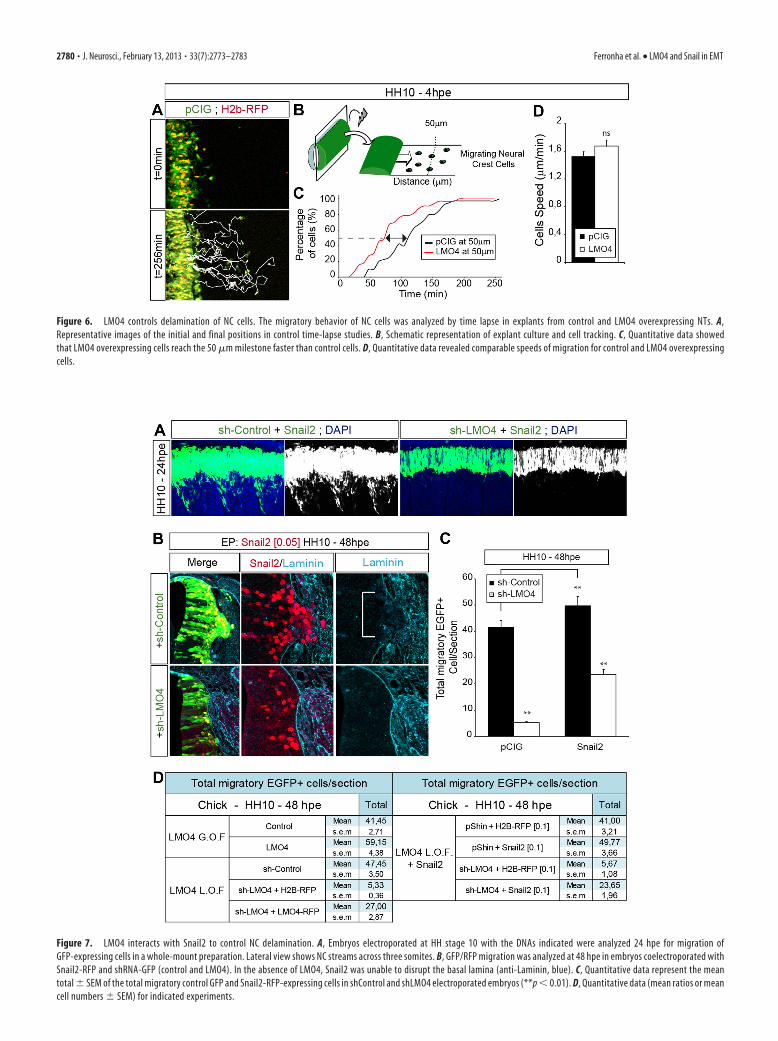
Figure 6. LMO4 controls delamination of NC cells. The migratory behavior of NC cells was analyzed by time lapse in explants from control and LMO4 overexpressing NTs. A,Representative images of the initial and final positions in control time-lapse studies. B, Schematic representation of explant culture and cell tracking. C, Quantitative data showedthat LMO4 overexpressing cells reach the 50 �m milestone faster than control cells. D, Quantitative data revealed comparable speeds of migration for control and LMO4 overexpressingcells.
Figure 7. LMO4 interacts with Snail2 to control NC delamination. A, Embryos electroporated at HH stage 10 with the DNAs indicated were analyzed 24 hpe for migration ofGFP-expressing cells in a whole-mount preparation. Lateral view shows NC streams across three somites. B, GFP/RFP migration was analyzed at 48 hpe in embryos coelectroporated withSnail2-RFP and shRNA-GFP (control and LMO4). In the absence of LMO4, Snail2 was unable to disrupt the basal lamina (anti-Laminin, blue). C, Quantitative data represent the meantotal � SEM of the total migratory control GFP and Snail2-RFP-expressing cells in shControl and shLMO4 electroporated embryos (**p � 0.01). D, Quantitative data (mean ratios or meancell numbers � SEM) for indicated experiments.
2780 • J. Neurosci., February 13, 2013 • 33(7):2773–2783 Ferronha et al. • LMO4 and Snail in EMT

Acting as a transcriptional coactivator, LMO4 can interactwith the MH1 and linker domains of receptor-regulated Smadproteins to coactivate the TGF-� response in epithelial cells(Lu et al., 2006). We identified LMO4 in a genome-widescreening based on a BMP-reporter and found that LMO4 wassufficient to activate the BRE-tk-GFP reporter in the NT (datanot shown). As Smad1/5 (Le Dreau et al., 2012) and Smad3(García-Campmany and Marti, 2007) are expressed in the de-veloping NT, we propose a model in which LMO4 enhancesthe transcriptional activity of endogenous Smad1/5 proteinsto activate BMP-dependent responses.
LMO4 is a necessary cofactor for Snail-mediated EMTIn addition to nervous tissue, epithelial cells express high levels ofLMO4, often at locations where active epithelial-to-mesenchymalinteractions occur, such as the mammary gland (Sum et al., 2005; Luet al., 2006). Furthermore, while downregulation of LMO4 expres-sion in breast cancer cells reduces their capacity to migrate and in-vade the extracellular matrix, LMO4 overexpression in noninvasive,immortalized human MCF10A cells promotes their motility andinvasion (Sum et al., 2005). These phenomena closely resemble theeffects of LMO4 in neural cells, suggesting that the mechanisms un-derlying the activity of this protein are conserved in several tissues.
Figure 8. LMO4 and Snail2 cooperate in repressing cadherin expression. A–C, N-cadherin expression (N-Cad) was analyzed 24 hpe in embryos coelectroporated with Snail2-RFP andLMO4-GFP, and it was reduced on the electroporated side. B, C, In Western blots, there was a reduction in total protein levels after electroporation of Snail2 or LMO4, and the additiveeffect of LMO4 � Snail2 electroporation was evident. D–F, The transcriptional activity of Snail2 and LMO4 was analyzed using the human E-cadherin-Luc reporter, comparing it to thatof the control pCIG vector. The data demonstrate the cooperative activity of LMO4 and Snail2 in repressing E-cadherin. E, Activity was tested by in ovo coelectroporation at HH stage 10,quantifying luciferase activity at 24 hpe. F, Activity was analyzed by transfection of NB cells and quantification of luciferase activity 24 h posttransfection. The data represent the mean �SEM of the relative luciferase activity expressed in arbitrary units (the mean of the control pCIG � 1.0). G, Expression of E-cadherin in NB cells reveals an inverse correlation with LMO4expression. Efficient LMO4 downregulation is accompanied by an increase in E-cadherin levels. RT-qPCR results expressed as the mean � SD relative to the nontargeting pLKO controlshRNA. *p � 0.05.
Ferronha et al. • LMO4 and Snail in EMT J. Neurosci., February 13, 2013 • 33(7):2773–2783 • 2781

Many transcription factors contribute to EMT by directly re-pressing surface adhesive components of epithelial cells, such asE-cadherin, claudins, and occludins. The Snail family of tran-scriptional repressors are central regulators of EMT in tissuemorphogenesis and human pathology (Nieto, 2011). Here wedemonstrate the absolute requirement of LMO4 in two models ofEMT: NC and NB tumor cells. LMO4 depletion phenocopied theloss of Snail activity in the chick embryo, resulting in impairedNC delamination. Moreover, we observed clear, dose-dependent,cooperative activity of LMO4 and Snail in repressing E-cadherinexpression, which may be conserved in neural cells under normaland pathological conditions. The N-terminal SNAG domain ofSnail is required for the repression of transcription, and the LIM-domains of LMO4 bind directly to the SNAG-repressor domainof Snail (Ochoa et al., 2012). Moreover, a distant member of theLIM family of transcriptional regulators, the Ajuba-LIM protein,also binds directly to the SNAG-repressor domain of Snail andregulates E-cadherin expression (Langer et al., 2008).
However, despite the numerous studies demonstrating theessential role of the cadherin switch in NC delamination andimplicating Snail in this EMT, the direct targets of Snail-mediatedEMT in NC delamination remain unknown (Nakagawa andTakeichi, 1995, 1998; Taneyhill et al., 2007; Park and Gumbiner,2010). Snail2 binding sites (E boxes) have been identified withinthe cadherin 6B regulatory region (Taneyhill et al., 2007), how-ever, cadherin 6B activity is required for deepithelialization anddisruption of the apical junction complex containing N-cadherinin the dorsal NT (Park and Gumbiner, 2010). While Snail genesare thought to function as transcriptional repressors from Dro-sophila to humans (Nieto, 2002), their promotion of EMTthrough the direct regulation of cadherin 6B would suggest anadditional and novel role as transcriptional activators.
Here, we propose a model in which LMO4 acts as a requiredcofactor in Snail-dependent cadherin repression and EMT. Itwould be interesting to determine whether LMO4 proteins arealso implicated in the mesenchymal-to-epithelial transition(MET). MET occurs during the morphogenesis of various organsunder normal physiological and pathological conditions, such aswhen metastatic cells reform epithelial-like tumors at metastaticsites (Thiery et al., 2009).
ReferencesAoyama M, Ozaki T, Inuzuka H, Tomotsune D, Hirato J, Okamoto Y, Tokita
H, Ohira M, Nakagawara A (2005) LMO3 interacts with neuronal tran-scription factor, HEN2, and acts as an oncogene in neuroblastoma. Can-cer Res 65:4587– 4597. CrossRef Medline
Asprer JS, Lee B, Wu CS, Vadakkan T, Dickinson ME, Lu HC, Lee SK (2011)LMO4 functions as a co-activator of neurogenin 2 in the developing cor-tex. Development 138:2823–2832. CrossRef Medline
Batlle E, Sancho E, Francí C, Domínguez D, Monfar M, Baulida J, GarcíaDe Herreros A (2000) The transcription factor snail is a repressor ofE-cadherin gene expression in epithelial tumour cells. Nat Cell Biol2:84 – 89. CrossRef Medline
Betancur P, Bronner-Fraser M, Sauka-Spengler T (2010) Assembling neuralcrest regulatory circuits into a gene regulatory network. Annu Rev CellDev Biol 26:581– 603. CrossRef Medline
Brodeur GM (2003) Neuroblastoma: biological insights into a clinicalenigma. Nat Rev Cancer 3:203–216. CrossRef Medline
Cheung M, Chaboissier MC, Mynett A, Hirst E, Schedl A, Briscoe J (2005)The transcriptional control of trunk neural crest induction, survival, anddelamination. Dev Cell 8:179 –192. CrossRef Medline
del Barrio MG, Nieto MA (2002) Overexpression of Snail family membershighlights their ability to promote chick neural crest formation. Develop-ment 129:1583–1593. Medline
De Preter K, Vandesompele J, Heimann P, Yigit N, Beckman S, Schramm A,Eggert A, Stallings RL, Benoit Y, Renard M, De Paepe A, Laureys G,
Påhlman S, Speleman F (2006) Human fetal neuroblast and neuroblas-toma transcriptome analysis confirms neuroblast origin and highlightsneuroblastoma candidate genes. Genome Biol 7:R84. CrossRef Medline
Duband JL, Blavet C, Jarov A, Fournier-Thibault C (2009) Spatio-temporalcontrol of neural epithelial cell migration and epithelium-to-mesenchyme transition during avian neural tube development. DevGrowth Differ 51:25– 44. CrossRef Medline
García-Campmany L, Martí E (2007) The TGFbeta intracellular effectorSmad3 regulates neuronal differentiation and cell fate specification in thedeveloping spinal cord. Development 134:65–75. CrossRef Medline
Hahm K, Sum EY, Fujiwara Y, Lindeman GJ, Visvader JE, Orkin SH (2004)Defective neural tube closure and anteroposterior patterning in micelacking the LIM protein LMO4 or its interacting partner Deaf-1. Mol CellBiol 24:2074 –2082. CrossRef Medline
Isogai E, Ohira M, Ozaki T, Oba S, Nakamura Y, Nakagawara A (2011)Oncogenic LMO3 collaborates with HEN2 to enhance neuroblastoma cellgrowth through transactivation of Mash1. PLoS One 6:e19297. CrossRefMedline
Jiang M, Stanke J, Lahti JM (2011) The connections between neural crestdevelopment and neuroblastoma. Curr Top Dev Biol 94:77–127.CrossRef Medline
Joshi K, Lee S, Lee B, Lee JW, Lee SK (2009) LMO4 controls the balancebetween excitatory and inhibitory spinal V2 interneurons. Neuron 61:839 – 851. CrossRef Medline
Langer EM, Feng Y, Zhaoyuan H, Rauscher FJ 3rd, Kroll KL, Longmore GD(2008) Ajuba LIM proteins are snail/slug corepressors required for neu-ral crest development in Xenopus. Dev Cell 14:424 – 436. CrossRefMedline
Le Dreau G, Garcia-Campmany L, Rabadan MA, Ferronha T, Tozer S, BriscoeJ, Martí E (2012) Canonical BMP7 activity is required for the generationof discrete neuronal populations in the dorsal spinal cord. Development139:259 –268. CrossRef Medline
Lee SK, Jurata LW, Nowak R, Lettieri K, Kenny DA, Pfaff SL, Gill GN (2005)The LIM domain-only protein LMO4 is required for neural tube closure.Mol Cell Neurosci 28:205–214. CrossRef Medline
Lee S, Lee B, Joshi K, Pfaff SL, Lee JW, Lee SK (2008) A regulatory networkto segregate the identity of neuronal subtypes. Dev Cell 14:877– 889.CrossRef Medline
Lu Z, Lam KS, Wang N, Xu X, Cortes M, Andersen B (2006) LMO4 caninteract with Smad proteins and modulate transforming growth factor-beta signaling in epithelial cells. Oncogene 25:2920 –2930. CrossRefMedline
Maris JM, Hogarty MD, Bagatell R, Cohn SL (2007) Neuroblastoma. Lancet369:2106 –2120. CrossRef Medline
Milan M, Diaz-Benjumea FJ, Cohen SM (1998) Beadex encodes an LMOprotein that regulates Apterous LIM-homeodomain activity in Drosophilawing development: a model for LMO oncogene function. Genes Dev12:2912–2920. CrossRef Medline
Morales AV, Acloque H, Ocana OH, de Frutos CA, Gold V, Nieto MA (2007)Snail genes at the crossroads of symmetric and asymmetric processes inthe developing mesoderm. EMBO Rep 8:104 –109. CrossRef Medline
Nakagawa S, Takeichi M (1995) Neural crest cell– cell adhesion con-trolled by sequential and subpopulation-specific expression of novelcadherins. Development 121:1321–1332. Medline
Nakagawa S, Takeichi M (1998) Neural crest emigration from the neuraltube depends on regulated cadherin expression. Development 125:2963–2971. Medline
Nieto MA (2002) The Snail superfamily of zinc-finger transcription factors.Nat Rev Mol Cell Biol 3:155–166. CrossRef Medline
Nieto MA (2011) The ins and outs of the epithelial to mesenchymal transi-tion in health and disease. Annu Rev Cell Dev Biol 27:347–376. CrossRefMedline
Ochoa SD, Salvador S, LaBonne C (2012) The LIM adaptor protein LMO4 isan essential regulator of neural crest development. Dev Biol 361:313–325.CrossRef Medline
Park KS, Gumbiner BM (2010) Cadherin 6B induces BMP signaling andde-epithelialization during the epithelial mesenchymal transition of theneural crest. Development 137:2691–2701. CrossRef Medline
Park KS, Gumbiner BM (2012) Cadherin-6B stimulates an epithelial mes-enchymal transition and the delamination of cells from the neural ecto-derm via LIMK/cofilin mediated non-canonical BMP receptor signaling.Dev Biol 366:232–243. CrossRef Medline
2782 • J. Neurosci., February 13, 2013 • 33(7):2773–2783 Ferronha et al. • LMO4 and Snail in EMT
Schramm A, von Schuetz V, Christiansen H, Havers W, Papoutsi M, WiltingJ, Schweigerer L (2005) High activin A-expression in human neuroblas-toma: suppression of malignant potential and correlation with favourableclinical outcome. Oncogene 24:680 – 687. CrossRef Medline
Schulte JH, Schramm A, Klein-Hitpass L, Klenk M, Wessels H, Hauffa BP, EilsJ, Eils R, Brodeur GM, Schweigerer L, Havers W, Eggert A (2005) Mi-croarray analysis reveals differential gene expression patterns and regula-tion of single target genes contributing to the opposing phenotype ofTrkA- and TrkB-expressing neuroblastomas. Oncogene 24:165–177.CrossRef Medline
Song MR, Sun Y, Bryson A, Gill GN, Evans SM, Pfaff SL (2009) Islet-to-LMO stoichiometries control the function of transcription complexesthat specify motor neuron and V2a interneuron identity. Development136:2923–2932. CrossRef Medline
Sum EY, Segara D, Duscio B, Bath ML, Field AS, Sutherland RL, LindemanGJ, Visvader JE (2005) Overexpression of LMO4 induces mammary hy-perplasia, promotes cell invasion, and is a predictor of poor outcome inbreast cancer. Proc Natl Acad Sci U S A 102:7659 –7664. CrossRefMedline
Taneyhill LA, Coles EG, Bronner-Fraser M (2007) Snail2 directly repressescadherin6B during epithelial-to-mesenchymal transitions of the neuralcrest. Development 134:1481–1490. CrossRef Medline
Theveneau E, Mayor R (2012) Neural crest delamination and migration:from epithelium-to-mesenchyme transition to collective cell migration.Dev Biol 366:34 –54. CrossRef Medline
Thiery JP, Acloque H, Huang RY, Nieto MA (2009) Epithelial-mesenchymal transitions in development and disease. Cell 139:871–890. CrossRef Medline
Tse E, Smith AJ, Hunt S, Lavenir I, Forster A, Warren AJ, Grutz G, Foroni L,Carlton MB, Colledge WH, Boehm T, Rabbitts TH (2004) Null muta-tion of the Lmo4 gene or a combined null mutation of the Lmo1/Lmo3genes causes perinatal lethality, and Lmo4 controls neural tube develop-ment in mice. Mol Cell Biol 24:2063–2073. CrossRef Medline
Wang K, Diskin SJ, Zhang H, Attiyeh EF, Winter C, Hou C, Schnepp RW,Diamond M, Bosse K, Mayes PA, Glessner J, Kim C, Frackelton E, GarrisM, Wang Q, Glaberson W, Chiavacci R, Nguyen L, Jagannathan J, SaekiN, et al. (2011) Integrative genomics identifies LMO1 as a neuroblas-toma oncogene. Nature 469:216 –220. CrossRef Medline
Yamanaka Y, Hamazaki Y, Sato Y, Ito K, Watanabe K, Heike T, Nakahata T,Nakamura Y (2002) Maturational sequence of neuroblastoma revealedby molecular analysis on cDNA microarrays. Int J Oncol 21:803– 807.Medline
Ferronha et al. • LMO4 and Snail in EMT J. Neurosci., February 13, 2013 • 33(7):2773–2783 • 2783