neurobiology research tools for biologically relevant …€¦ · neurobiology research tools for...
TRANSCRIPT

Bio Research
Os nos ad tat. Igna faccum eroses iriusti onulla feuisisit adio con veliquamcore magna alis nonulla non er irilla am, quiscil dolobor eu faccum quisisim quis nullaor rem iril er summy nulpute tinos ipisci te facilit nulla facilis molore.
BioResearch
NeurobiologyResearch Tools for Biologically Relevant Results
Neurobiology Research Tools

BioResearchNeurobiology Research Tools
Avoid Research Roadblocks and Take a Direct Path to Results
Primary Cells and Media
Transfection
" Non-viral transfection doesn’t give me the transfer efficiency I’m looking for."
" Sourcing cells from specific species and brain regions requires a lot of my time and effort."
SOLUTION: Select from Lonza’s range of well-characterized and ready-to-use primary cells. Simply thaw and culture. Page 5
SOLUTION: Our non-viral Nucleofector™ Technology delivers 70% gene transfer efficiency and high expression levels. Page 9
2

Cell Analysis
" I can’t find reliable, ready-to-use assay tools for mRNA, protein expression, cell proliferation, or cytotoxicity."
SOLUTION: Choose from our variety of neurology-specific assay tools, including qPCR arrays, ready-to-use protein gels, and bioassay kits to measure cell proliferation andcytotoxicity. Page 14
Drug Discovery
" I need human model systems to avoid species extrapolation in my drug discovery process."
SOLUTION: Now you can access assay-ready embryonic stem cell-derived pure human motor neurons. Page 13
3

BioResearchNeurobiology Research Tools
Your Complete Neurobiology Tool Kit
Streamline your neurobiology workflow by choosing from convenient, innovative research tools that have been designed and tested to work together.
Lonza solutions provide biologically relevant results, from high-quality primary cells, through efficient transfection technology, to expert analy-sis tools.
Primary Cells and Media Transfection Cell Analysis Drug Discovery
Ready-to-use cryopreserved Clonetics™ or Poietics™ Primary Neural Cells:– Astrocytes
(mouse, rat, or human)– Cerebellar granule cells (rat)– Cortical neurons (mouse or rat)– Dorsal root ganglia
(rat; embryonic or neonatal)– Hippocampal neurons
(rat or mouse)– Hypothalamus neurons (rat)– Neural progenitor cells (human)– Retinal neurons (rat)– Striatum neurons (mouse or rat)
Optimized Clonetics™ Primary Neural Cell Growth Media:– PNGM™ or PNGM™-A BulletKit™
Media for embryonal or adult primary animal neurons
– AGM™ BulletKit™ for human and animal astrocytes
– NPMM™ or NPDM™ BulletKit™ Media for neural progenitor cell growth or differentiation
Amaxa™ Nucleofector™ Technology with optimized protocols for primary cells, including:– Hippocampal neurons– Cortical neurons– Dorsal root ganglia– Cerebellar granule cells– Astrocytes– Oligodendrocytes– Neural stem cells
Also optimized for numerous cell lines, including:– PC-12– SH-SY5Y– Neuro2A
StellARray™ qPCR Arrays for mRNA expression profiling:– Alzheimer's (mouse or human)– Autism (mouse or human)– Huntington's (mouse or human)– Mood disorder
(mouse or human)– Neurodegeneration
(mouse or human)– Parkinson's (mouse or human)
Protein Expression Analysis:– PAGEr™ Precast Protein Gels for
Western blotting
Assay-ready human embryonic stem cell (ESC) derived neurons: – NeuroPlate™ Human ESC
Neuronal Progenitors– MotorPlate™ Standard Human
ESC Motor Neuron Progenitors– MotorPlate™ Mature Human ESC
Motor Neuron Progenitors
4

Primary Neural Cells and Growth Media
Rat Mouse(CD1 or C57)
Recommended Media
Hippocampal neurons n n PNGM™
Cortical neurons n n PNGM™
Striatum neurons n n PNGM™
Dorsal root ganglia (embryonic or neonatal)
n
–
PNGM™
Cerebellar granule cells n – PNGM™-A
Hypothalamus neurons n – PNGM™
Retinal neurons n – PNGM™
Rat cortical neuronal cells were thawed, cultured 14 days, and immunofluores-cently stained with anti-PGP 9.5 and anti ß-tubulin.
Access neural cells when you need them. Choose from Lonza’s extensive range of isolated primary neural cells and culture them in media that we have already optimized and tested to give you the best results. You will never have to put research on hold for animal pregnancies again. Simply store Lonza cells in your freezer and thaw them as needed.
Cryopreserved Clonetics™ Primary Neurons
Choose from our broad selection of high-quality, high-yield cultures of dissociated primary animal neurons that are:
– Tested for neuron-specific markers – Guaranteed to perform in culture – Guaranteed free of mycoplasma and bacteria – Tested to ensure they will perform after shipping – Lots can be reserved to repeat experiments using cells from the
same batch
Clonetics™ PNGM™ Primary Neuron Growth Media
Clonetics™ PNGM™ gives you a proven alternative to Neurobasal™ and B27® for long-term culture of embryonic or adult rat and mouse neurons:
– Serum-free media in BulletKit™ Format – Guaranteed to perform with all Clonetics™ Primary Animal Neurons – Batch tested with primary neurons and a pre-screened B27®-
equivalent supplement
Applications – Culture embryonic primary neurons in PNGM™ BulletKit™ Media. – Culture adult primary neurons and cerebellar granule cells in
PNGM™-A BulletKit™ Media
Clonetics™ Rat Cortical Neurons were seeded at 40% recommended density and cultured in PNGM™ BulletKit™ Media and a competitor media. After 6 DIV, viability was determined by ViaLight™ Plus Assay. Values were normalized to “no cell” control.
0
10
20
30
40
50
60
70
80
90
No Cells Competitor PNGM
Rela
tive
RLU
(nor
mal
ized
to c
ontr
ol)
Growth of Rat Cortical Neurons in Different Media
Applications – Transfection – Evaluating electrophysiological properties, neurotransmitters,
and receptor function – Drug screening – Research into ion channels, intracellular transport,
and neurotoxicity
" Sourcing cells from specific species and brain regions requires a lot of my time and effort."
SOLUTION: Select from Lonza’s range of well-characterized and ready-to-use primary cells. Simply thaw and culture.
5

Cryopreserved Clonetics™ Primary Astrocytes
Improve your workflow with cryopreserved, high-quality human, and animal glial cells. There’s no need for animal care or glial cell isolation with these products, which are:
– Passaged once after isolation – Batch tested for growth characteristics and morphology (GFAP) – Guaranteed free of mycoplasma – Able to be reserved by lot so you can repeat experiments using cells
from the same batch
Rat Mouse(CD1 or C57)
Human
Hippocampal astrocytes n – –
Cortical astrocytes n – –
Striatum astrocytes n – –
Cx-Hi-Cp mixed Astrocytes n – –
Cx-Hi-Striatum mixed Astrocytes – n –
Astrocytes – – n
Indirect immunofluorescence staining of Clonetics™ Normal Human Astrocytes for glial fibrillary acidic protein (GFAP).
Applications – Transfection – Pharmacology – Drug screening – Research into neurogenesis, cell physiology, Alzheimer’s disease,
Parkinson’s disease, spinal cord injury, astrocyte-mediated neuro-toxicity, ion channels, electrophysiology, and patch clamping
BioResearchNeurobiology Research Tools
Clonetics™ AGM™ Primary Astrocyte Growth Media
Culture human and animal astrocytes with serum-free media, in BulletKit™ format. This media has been tested and guaranteed to perform with all Clonetics™ Primary Astrocytes.
6
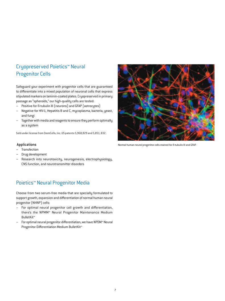
Normal human neural progenitor cells stained for ß-tubulin III and GFAP.
Cryopreserved Poietics™ Neural Progenitor Cells
Safeguard your experiment with progenitor cells that are guaranteed to differentiate into a mixed population of neuronal cells that express stipulated markers on laminin-coated plates. Cryopreserved in primary passage as “spheroids,” our high-quality cells are tested:
– Positive for ß-tubulin III (neurons) and GFAP (astrocytes) – Negative for HIV-1, Hepatitis B and C, mycoplasma, bacteria, yeast,
and fungi – Together with media and reagents to ensure they perform optimally
as a system
Sold under license from StemCells, Inc. US patents 5,968,829 and 5,851, 832.
Applications – Transfection – Drug development – Research into neurotoxicity, neurogenesis, electrophysiology,
CNS function, and neurotransmitter disorders
Poietics™ Neural Progenitor Media
Choose from two serum-free media that are specially formulated to support growth, expansion and differentiation of normal human neural progenitor (NHNP) cells:
– For optimal neural progenitor cell growth and differentiation, there’s the NPMM™ Neural Progenitor Maintenance Medium BulletKit™
– For optimal neural progenitor differentiation, we have NPDM™ Neural Progenitor Differentiation Medium BulletKit™
7

Primary Cell Case Study: Co-cultures for Neuropathology Research
Co-culture of cryopreserved rat primary cortical and striatal neurons.B. Tinner-Staines, et al. 2003. Society for Neuroscience; Abstract No. 427.15
Background
Most primary neuronal cell cultures use neurons from a single brain region but this does not provide an adequate model to study neuropathologies such as Alzheimer’s or Parkinson’s diseases. De-afferented neurons demonstrate biochemical and physiological abnormalities that limit scientific study and preclude drug screening. To model the circuitry associated with different neuropathologies, relevant neuronal cell types must be co-cultured in vivo.
It is difficult to time and stage co-culture of freshly isolated neurons according to the normal developmental staging of cell connectivity. How-ever, this study shows that cortical and striatal cryopreserved neurons in co-culture could provide viable models for examining questions of basal ganglia function and toxicology.
Method
Cryopreserved primary Clonetics™ Rat Cortical and Striatal Neurons were thawed, plated in tandem, and incubated for 7 – 35 days in vitro. Cultures were characterized using synaptic marker antibodies, receptor and second messenger proteins, and structural marker proteins.
Results
Cortical and striatal co-cultures displayed characteristics of each indi-vidual neuron type. Furthermore, co-cultures showed nerve terminals expressing vesicular GABA transporter or glutamate transporter (latter not shown here), which is evidence of neuronal crosstalk, thus reflect-ing the in vivo situation.
Plating a combination of cryopreserved rat striatal and cortical neurons yields cultures characteristic of both cell types cultured in-dividually, as shown by staining of cell soma (PGP 9.5) and neurofilaments (NF-160).
Thawed cortical and striatal rat neurons were co-cultured for 21 days and stained with antibodies against vesicular GABA transporter (vGAT; green) and dopamine receptor protein (DARPP-32, red). The pres-ence of GABAergic innervation of DARPP-positive neurons gives evidence of cross-innervation between neurons.
Conclusion
Results demonstrate that in vitro modeling of brain circuits can be easily accomplished by using a co-culture of cryopreserved primary neural cells. These facilitate co-cultures according to the normal developmental staging of cell connectivity because researchers do not need to coordinate isolations from different brain regions or embryonic developmental stages.
BioResearchNeurobiology Research Tools
8

Nucleofection™ was the first efficient non-viral transfection technology for primary neurons. Its unique combination of electrical parameters with cell type-specific solutions and protocols gives you up to 70% gene transfer and excellent cell viability.
Now this innovative technology also allows you to transfect primary neurons or glial cells in adherence at later developmental stages.
Amaxa™ Nucleofector™ Technology
Nucleofection™ gives you: – High transfection efficiencies and cell viability – Preservation of cell functionality – A variety of device platforms for different cell numbers and
throughput – Nucleofection™ of primary neurons or glial cells in adherence, even
after several days of culture – Optimized protocols to save time and effort
Transfection of Primary Neural Cells or Cell Lines
Choose the Nucleofection™ Platform that Suits Your Research Needs
Advanced Platform 96-well Add-on High-throughput Platform Basic Device
Device 4D-Nucleofector™ System 96-well Shuttle™ System 384-well Nucleofector™ System Nucleofector™ 2b Device
Unit
Throughput (samples per run) Low to medium (1 – 16) Low to high (1 – 96) High (384) Low (1)
Reaction volume 20 µl + 100 µl 20 µl 20 µl 100 µl
Electrode material Conductive polymer Conductive polymer Conductive polymer Aluminum
Low cell numbers (20 µl) 5 × 10⁴ to 5 × 10⁵ 5 × 10⁴ to 5 × 10⁵ 5 × 10⁴ to 5 × 10⁵ –
High cell numbers (100 µl) 4 – 5 × 10⁶ – – 4 – 5 × 10⁶
DNA Vector amount/sample
0.2 – 1 μg (20 µl) 1 – 5 μg (100 µl)
0.2 – 1 μg
0.2 – 1 μg
1 – 5 μg
siRNA amount/sample (concentration 2 nM – 2 µM)
0.04 – 40 pmol (20 µl) 0.2 – 200 pmol (100 µl)
0.04 – 40 pmol
0.04 – 40 pmol
0.2 – 200 pmol
Adherent Nucleofection™ n n – –
Compatibility with 96-well Shuttle™ System
n – – –
www.lonza.com/celldatabase – To find transfection data for your cell type of interest.
" Non-viral transfection doesn’t give me the transfer efficiency I’m looking for."
SOLUTION: Our non-viral Nucleofector™ Technology delivers 70% gene transfer efficiency and high expression levels.
9

Efficiency*
Viability*
Kit for 4D-Nucleofector™ and 96-well Shuttle™ Systems (name of specific protocol)
Kit for Nucleofector™ II/2b Device
Primary Neural Cells Astrocytes, mouse 55-65% 60-70% P3 Primary Cell (Neuron, Basic) Glial Cell, Basic
Astrocytes, rat 60-70% 70-80% P3 Primary Cell (Neuron, Basic) Glial Cell, Basic
Dorsal root ganglia (DRG), chicken 25-35% P3 Primary Cell (Neuron, Basic) Chicken Neuron
Dorsal root ganglia (DRG), rat 35-45% P3 Primary Cell (Neuron, Basic) Rat Neuron
Neuron, cortical, rat 30-70% 45-60% P3 Primary Cell (Neuron, rat) Rat Neuron
Neuron, hippocampal, chicken 40-50% P3 Primary Cell (Neuron, Basic) Chicken Neuron
Neuron, hippocampal, mouse 50-60% P3 Primary Cell (Neuron, Basic) Mouse Neuron
Neuron, hippocampal, rat 30-70% 45-60% P3 Primary Cell (Neuron, rat) Rat Neuron
Neuron, hippocampal, rat – adherent 30-50% 50-80% Basic Neuron AD (Neuron, Basic, AD) Not Applicable
Neuron, cortical, rat – adherent 30-60% 70-90% Basic Neuron AD (Neuron, Basic, AD) Not Applicable
Neuron, cortical, mouse – adherent 20-70% 70-90% Basic Neuron AD (Neuron, Basic, AD Not Applicable
Oligodendrocyte, rat 40-50% 55-65% P3 Primary Cell (Neuron, Basic) Glial Cell, Basic
Stem CellsNeural stem cell (NSC), mouse 75-85% Primary Cell Optimization Mouse NSC
Neural stem cell (NSC), rat 40-50% Primary Cell Optimization Rat NSC
Neuronal Cell Lines**
AGN2a 60-95% Cell Line Optimization Cell Line R
BV2 55-70% 70-95% Cell Line Optimization Cell Line T
C6 85-95% 70-80% Cell Line Optimization Cell Line V
H4 70-80% 80-90% Cell Line Optimization Cell Line V
IMR-32 75-85% 55-65% Cell Line Optimization Cell Line L
Neuro2A 47-85% 80-95% Cell Line SF Cell Line V
NG108-15 60-70% 75-85% Cell Line Optimization Cell Line V
PC-12 85-95% 75-85% Cell Line Optimization Cell Line V
SH-SY5Y 60-85% 40-80% Cell Line SF Cell Line V
SK-N-MC 60-95% 30-60% Cell Line Optimization Cell Line V
SK-N-SH 80-90% 65-75% Cell Line Optimization Cell Line V
U-87MG 35-45% 85-95% Cell Line Optimization Cell Line T
Primary cells marked blue have Lonza-validated optimized protocols. Cell lines marked blue also have optimized protocols with ATCC® clones.
*Approximate ranges extrapolated from larger result collections, including Lonza and customer data** This is only a selection of cell lines. www.lonza.com/celldatabase – For further neuronal cell lines and protocol guidance.
Transfection Efficiency and Cell Viability with Nucleofector™
BioResearchNeurobiology Research Tools
10

Adherent Nucleofection™ of Primary Neurons at Later Developmental Stages
Transfect primary neural cells at any stage of culture, including late development, without putting them in suspension. Forget about the traditional limitations of electroporation-based methods with our new 4D-Nucleofector™ System that also enables adherent Nucleofection™:
– Achieves transfection efficiencies of up to 70% – Basic Kits help pinpoint optimal conditions for primary neurons, glial
cells or neural stem cells – Has been tested for cryopreserved Clonetics™ Primary Neurons
Applications
4D-Nucleofector™ × Unit or 96-well Shuttle™ Add-on: – Culture cells for up to 14 days in specialized Nucleocuvette™
AD Strips (20 μl) – Transfect them at any time during this culture period – Suited for analysis by light and fluorescence microscopy; absorp-
tion, luminescence, or fluorescence assays; or mRNA expression
4D-Nucleofector™ Y Unit: – Culture cells in standard 24-well plates – For Nucleofection™, simply insert a disposable conductive polymer
24-well Dipping Electrode Array for Nucleofection™ – Ideal for post-transfection analysis by confocal microscopy or
patch clamping
Preserved functionality after adherent Nucleofection™ of neurons. Mouse cortical neurons were cultured in 24-well plates and transfected after 6 DIV with 17.5 μg pmaxFP™-Yellow-C Vector using the Basic Neuron 4D-Nucleofector™ Y AD Kit. Four days post Nucleofection™, cells were immuno-stained for maxFP™-Yellow expres-sion (green) and synapses (red), stained for nuclei (DAPI, blue), and analyzed by fluorescence microscopy. Transfected neurons show normal synapse formation.
Adherent Nucleofection™ of cryopreserved Clonetics™ Rat Hippocampal Neurons. Frozen rat hippocampal neurons were thawed and seeded into 24-well plates. After 2 days, cells were transfected with 17.5 μg pmaxGFP™ Vector using the Basic Neuron 4D-Nucleofector™ Y AD Kit. 1 day post Nucleofection™, cells were analyzed by fluorescence microscopy.
Nucleocuvette™ AD Strips (20 µl)
Dipping Electrode Arrays
Required functional 4D-Nucleofector™ Unit X Unit or 96-well Shuttle™
Y Unit
Pre- and post Nucleofection™ Culture
Nucleocuvette™ Wells
24-well culture plate
Nucleofection™ of cells plated on glass cover slips
–
n
High transfection efficiencies and viabilities n n
Cell analysis by: – transmission light or fluorescence microscopy – absorption, luminescence or fluroescence assays – confocal microscopy – patch clamping
n
n
––
n
n
n
n
Nucleofection™ of cryopreserved Clonetics™ Primary Animal Neurons
n
n
Adherent Nucleofection™ using Nucleocuvette™ AD Strips or 24-well Dipping Electrode Arrays. After isolation, primary neurons are directly plated into poly-D- or poly-L-lysine coated Nucleocuvette™ Strips (when using the × Unit) or 24-well culture plates (when using the Y Unit). Cells can be cultured up to 14 days and transfected by Nucleofection™ at any time during this period.
Adherent Nucleofection™ – All-in-one Process
Up to 14 days
Pre-culture AnalysisNucleofection™
or
11
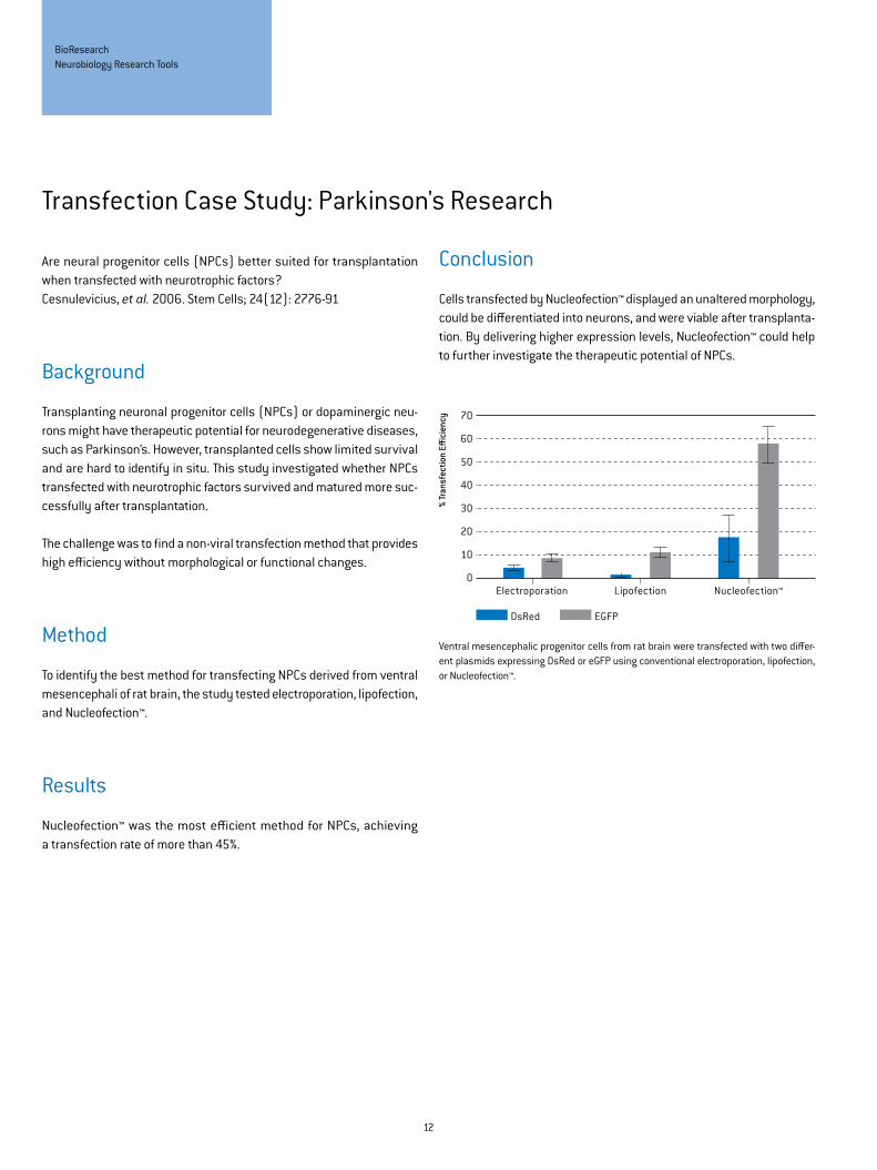
Are neural progenitor cells (NPCs) better suited for transplantation when transfected with neurotrophic factors?Cesnulevicius, et al. 2006. Stem Cells; 24(12): 2776-91
Background
Transplanting neuronal progenitor cells (NPCs) or dopaminergic neu-rons might have therapeutic potential for neurodegenerative diseases, such as Parkinson’s. However, transplanted cells show limited survival and are hard to identify in situ. This study investigated whether NPCs transfected with neurotrophic factors survived and matured more suc-cessfully after transplantation.
The challenge was to find a non-viral transfection method that provides high efficiency without morphological or functional changes.
Method
To identify the best method for transfecting NPCs derived from ventral mesencephali of rat brain, the study tested electroporation, lipofection, and Nucleofection™.
Results
Nucleofection™ was the most efficient method for NPCs, achieving a transfection rate of more than 45%.
Transfection Case Study: Parkinson's Research
Ventral mesencephalic progenitor cells from rat brain were transfected with two differ-ent plasmids expressing DsRed or eGFP using conventional electroporation, lipofection, or Nucleofection™.
0
10
20
30
40
50
60
70
Electroporation
DsRed EGFP
Lipofection Nucleofection™
% Tr
ansf
ectio
n Effi
cien
cy
Conclusion
Cells transfected by Nucleofection™ displayed an unaltered morphology, could be differentiated into neurons, and were viable after transplanta-tion. By delivering higher expression levels, Nucleofection™ could help to further investigate the therapeutic potential of NPCs.
BioResearchNeurobiology Research Tools
12
Drug Discovery
Have greater confidence in your data by using assay-ready human neurons to screen your drug candidates.
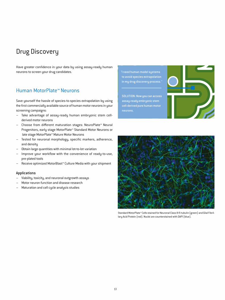
Human MotorPlate™ Neurons
Save yourself the hassle of species-to-species extrapolation by using the first commercially available source of human motor neurons in your screening campaigns:
– Take advantage of assay-ready human embryonic stem cell- derived motor neurons
– Choose from different maturation stages: NeuroPlate™ Neural Progenitors, early stage MotorPlate™ Standard Motor Neurons or late stage MotorPlate™ Mature Motor Neurons
– Tested for neuronal morphology, specific markers, adherence, and density
– Obtain large quantities with minimal lot-to-lot variation – Improve your workflow with the convenience of ready-to-use,
pre-plated tools – Receive optimized MotorBlast™ Culture Media with your shipment
Applications – Viability, toxicity, and neuronal outgrowth assays – Motor neuron function and disease research – Maturation and cell cycle analysis studies
Standard MotorPlate™ Cells stained for Neuronal Class III ß-tubulin (green) and Glial Fibril-lary Acid Protein (red). Nuclei are counterstained with DAPI (blue).
" I need human model systems to avoid species extrapolation in my drug discovery process."
SOLUTION: Now you can access assay-ready embryonic stem cell-derived pure human motor neurons.
13
BioResearchNeurobiology Research Tools
Human Mouse
Alzheimer n n
Autism n n
Huntington n n
Mood disorder n n
Neurodegeneration n n
Parkinson n n
Free your gene expression experiments from biases and interference with integrated research tools for every step of your workflow. Lonza solutions provide biologically relevant results – from cell proliferation and cytotoxicity assays, to sample preparation and qPCR, all the way through protein confirmation.
StellARray™ qPCR Arrays
StellARray™ qPCR Arrays contain 96 or 384 targeted, pre-validated qPCR assays:
– Choose from more than 150 pre-validated, research area-specific qPCR arrays or configure custom arrays online
– Use with standard qPCR equipment – Perform a comprehensive range of neurobiology experiments – Utilize ΔΔCt analysis, or achieve reliable normalization with
StellARray™ Proprietary GPR™ Software, which automatically identifies the best normalizer amplicons based on lowest variance
Cell Analysis
www.array.lonza.com – For more information on the StellARray™ System and its supporting GeneSieve™ Search Tool or Global Pattern Recognition™ (GPR) Analysis Tool.
Applications – Gene expression profiling – Pathway analysis – Microarray data validation – siRNA knock-down analysis – Biomarker discovery
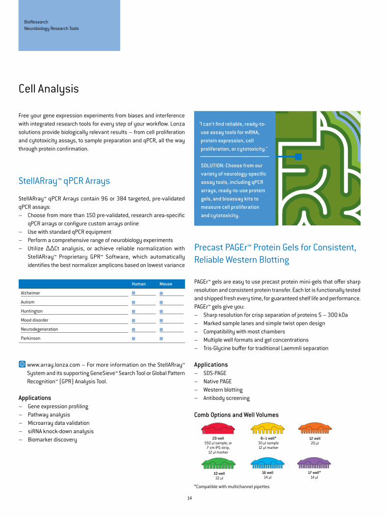
2D well550 μl sample, or
7 cm IPG strip, 12 μl marker
8+1 well*30 μl sample 12 μl marker
10 well32 μl
12 well20 μl
17 well*14 μl
16 well14 μl
* 8+1-well and 17-well gels are multichannel pipette compatible
2D well550 μl sample, or
7 cm IPG strip, 12 μl marker
8+1 well*30 μl sample 12 μl marker
10 well32 μl
12 well20 μl
17 well*14 μl
16 well14 μl
* 8+1-well and 17-well gels are multichannel pipette compatible
2D well550 μl sample, or
7 cm IPG strip, 12 μl marker
8+1 well*30 μl sample 12 μl marker
10 well32 μl
12 well20 μl
17 well*14 μl
16 well14 μl
* 8+1-well and 17-well gels are multichannel pipette compatible
2D well550 μl sample, or
7 cm IPG strip, 12 μl marker
8+1 well*30 μl sample 12 μl marker
10 well32 μl
12 well20 μl
17 well*14 μl
16 well14 μl
* 8+1-well and 17-well gels are multichannel pipette compatible
2D well550 μl sample, or
7 cm IPG strip, 12 μl marker
8+1 well*30 μl sample 12 μl marker
10 well32 μl
12 well20 μl
17 well*14 μl
16 well14 μl
* 8+1-well and 17-well gels are multichannel pipette compatible
Comb Options and Well Volumes
*Compatible with multichannel pipettes
Precast PAGEr™ Protein Gels for Consistent, Reliable Western Blotting
PAGEr™ gels are easy to use precast protein mini-gels that offer sharp resolution and consistent protein transfer. Each lot is functionally tested and shipped fresh every time, for guaranteed shelf life and performance. PAGEr™ gels give you:
– Sharp resolution for crisp separation of proteins 5 – 300 kDa – Marked sample lanes and simple twist open design – Compatibility with most chambers – Multiple well formats and gel concentrations – Tris-Glycine buffer for traditional Laemmli separation
Applications – SDS-PAGE – Native PAGE – Western blotting – Antibody screening
" I can’t find reliable, ready-to-use assay tools for mRNA, protein expression, cell proliferation, or cytotoxicity."
SOLUTION: Choose from our variety of neurology-specific assay tools, including qPCR arrays, ready-to-use protein gels, and bioassay kits to measure cell proliferation and cytotoxicity.
14

ViaLight™ Plus Kits
Measure cell proliferation and cytotoxicity by using stable biolumines-cence to detect adenosine triphosphate (ATP):
– Process and analyze a 96-well plate in less than 15 minutes – Detect as few as ten cells; allowing for lower seeding densities
and more assays – Easily perform scalable automation with our simple,
no-shake protocol – Add two reagents directly to your culture and read luminescence – Expand your dynamic range to five decades, in adherent or
suspension cultures – Run this test on a variety of luminometers or
scintillation counters – Avoid radioactive or toxic materials
www.lonza.com /vialight
ToxiLight™ Kits
Check a compound’s cytotoxic effects non-destructively by measuring adenylate kinase (AK) leakage from damaged cells:
– Generate results from as few as 10 cells – Eliminate the need to lyse by monitoring cytotoxicity from
supernatant – Simply add a single reagent directly to cells, or to
a supernatant aliquot – Process and analyze a 96-well plate in less than 10 minutes – Freeze supernatants and return to your work later without losing
AK activity
www.lonza.com/toxilight
Identity Dose-dependent Activities in Cells
Comparison of ViaLight™ Plus and ToxiLight™ Kits using HUVECs dosed with camptothecin. The ATP levels indicated by the ViaLight™ Plus RLUs reduce steadily in a dose-dependent manner. At the lower drug doses, the AK released from the cells is relatively low compared with that of the control, only increasing dramatically at the highest drug doses.
EC 50 Data Generated Using ViaLight Plus Shows Consistency Over Time
HepG2 cells were incubated with the alkylating agent Mitomycin C for 48 hours and the assayed using ViaLight™ Plus. The experimental values are the mean of eight replicant samples read every hour over a 5 hour period. The EC values remain consistent over the 5 hour read period.
15
BioResearchNeurobiology Research Tools
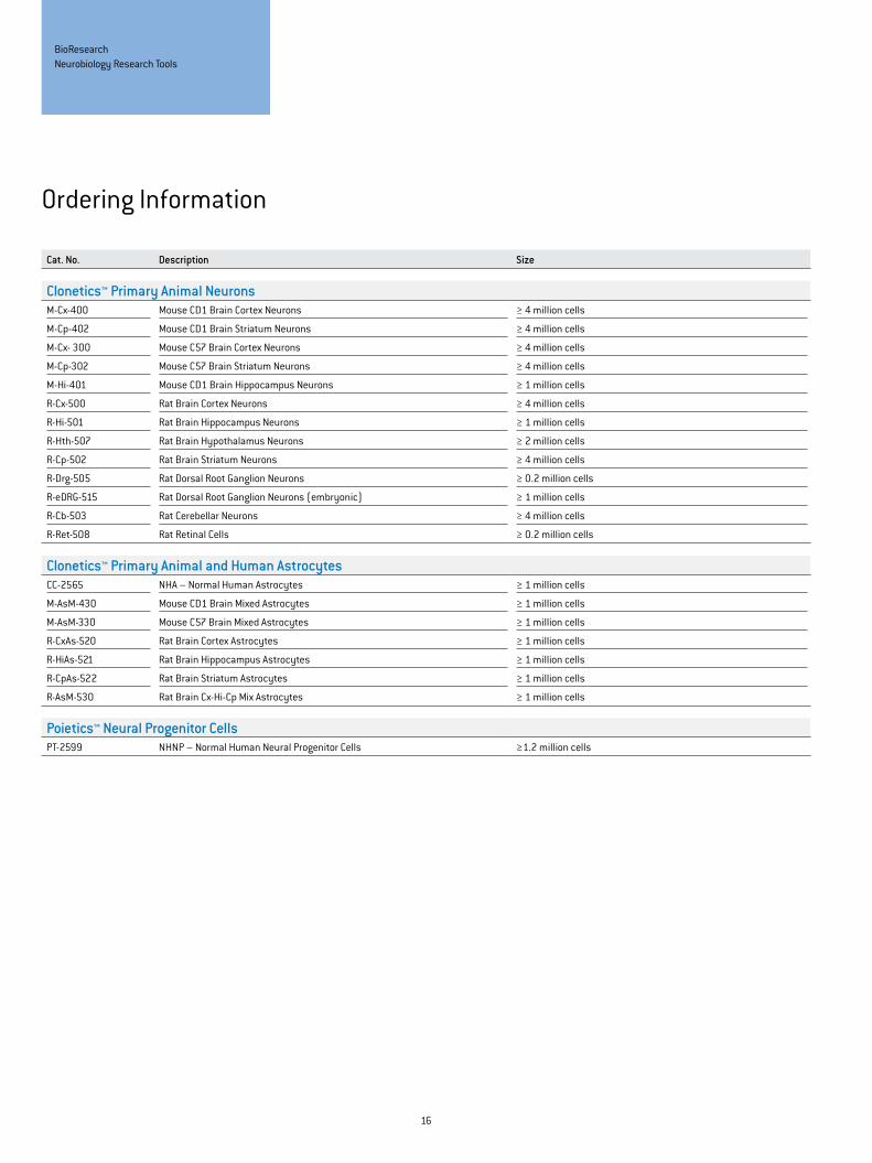
Ordering Information
Cat. No. Description Size
Clonetics™ Primary Animal NeuronsM-Cx-400 Mouse CD1 Brain Cortex Neurons ≥ 4 million cells
M-Cp-402 Mouse CD1 Brain Striatum Neurons ≥ 4 million cells
M-Cx- 300 Mouse C57 Brain Cortex Neurons ≥ 4 million cells
M-Cp-302 Mouse C57 Brain Striatum Neurons ≥ 4 million cells
M-Hi-401 Mouse CD1 Brain Hippocampus Neurons ≥ 1 million cells
R-Cx-500 Rat Brain Cortex Neurons ≥ 4 million cells
R-Hi-501 Rat Brain Hippocampus Neurons ≥ 1 million cells
R-Hth-507 Rat Brain Hypothalamus Neurons ≥ 2 million cells
R-Cp-502 Rat Brain Striatum Neurons ≥ 4 million cells
R-Drg-505 Rat Dorsal Root Ganglion Neurons ≥ 0.2 million cells
R-eDRG-515 Rat Dorsal Root Ganglion Neurons (embryonic) ≥ 1 million cells
R-Cb-503 Rat Cerebellar Neurons ≥ 4 million cells
R-Ret-508 Rat Retinal Cells ≥ 0.2 million cells
Clonetics™ Primary Animal and Human AstrocytesCC-2565 NHA – Normal Human Astrocytes ≥ 1 million cells
M-AsM-430 Mouse CD1 Brain Mixed Astrocytes ≥ 1 million cells
M-AsM-330 Mouse C57 Brain Mixed Astrocytes ≥ 1 million cells
R-CxAs-520 Rat Brain Cortex Astrocytes ≥ 1 million cells
R-HiAs-521 Rat Brain Hippocampus Astrocytes ≥ 1 million cells
R-CpAs-522 Rat Brain Striatum Astrocytes ≥ 1 million cells
R-AsM-530 Rat Brain Cx-Hi-Cp Mix Astrocytes ≥ 1 million cells
Poietics™ Neural Progenitor CellsPT-2599 NHNP – Normal Human Neural Progenitor Cells ≥1.2 million cells
16

Ordering Information
Cat. No. Description Size
Clonetics™/Poietics™ Primary Neural Cell Growth MediaCC-4461 PNGM™ Primary Neuron Growth Medium BulletKit™
Includes Basal Medium and SingleQuots™ KitKit
CC-4512 PNGM™-A Primary Neuron Growth Medium – Adult BulletKit™ Includes Basal Medium and SingleQuots™ Kit
Kit
CC-3256 PNBM™ Basal Medium 500 ml
CC-4462 PNGM™ SingleQuots™ Supplement and Growth Factors –
CC-4511 PNGM™-A Primary Neuron Growth Medium – Adult SingleQuots™ Kit Kit
CC-3186 AGM™ BulletKit™ Kit – Includes Basal Medium and SingleQuots™ Kit Kit
CC-3187l ABM™ Basal Medium 500 ml
CC-4123 AGM™ SingleQuots™ Supplements and Growth Factors –
CC-3209 NPMM™ Neural Progenitor Maintenance Medium BulletKit™ Kit
CC-3229 NPDM™ Neural Progenitor Differentiation Medium BulletKit™ Kit
CC-3210 NPBM™ Neural Progenitor Basal Medium 200 ml
CC-4241 Neural Progenitor Maintenance Medium SingleQuots™ Kit (contains hEGF and hFGF)
Kit
CC-4242 Neural Progenitor Supplement SingleQuots™ Kit (contains NSF-1 and GA) Kit
Pluripotent Stem Cell-derived Neuronal CellsFP-6020 NeuroPlate™ Human ESC Neuronal Progenitors 96-well plate
FP-6041 NeuroPlate™ Human ESC Neuronal Progenitors 384-well plate
FP-6011 MotorPlate™ Standard Human ESC Motor Neuron Progenitors 96-well plate
FP-6040 MotorPlate™ Standard Human ESC Motor Neuron Progenitor 384-well plate
FP-6046 MotorPlate™ Mature Human ESC Motor Neuron Progenitors 96-well plate
FP-6049 MotorPlate™ Mature Human ESC Motor Neuron Progenitors 384-well plate
Nucleofector™ DevicesAAB-1001 Nucleofector™ 2b Device –
AAF-1001B 4D-Nucleofector™ Core Unit –
AAF-1001X 4D-Nucleofector™ × Unit –
AAM-1001S 96-well Shuttle™ Device –
Kits for 4D-Nucleofector™ DeviceV4XP-3024 P3 Primary Cell 4D-Nucleofector™ × Kit L (for neurons and glial cells) 24 rxn (100 μl Nucleocuvette™)
V4XP-3032 P3 Primary Cell 4D-Nucleofector™ × Kit S (for neurons and glial cells) 32 rxn (20 μl Nucleocuvette™; 16-well)
V4XP-1A32 Basic Neuron 4D-Nucleofector™ × AD Kit (adherent) 32 rxn (20 μl Nucleocuvette™ AD; 16-well)
V4YP-1A24 Basic Neuron 4D-Nucleofector™ Y AD Kit (adherent) 24 rxn (Dipping Electrode)
V4XP-9096 Primary Cell Optimization 4D-Nucleofector™ × Kit (for neural stem cells) 96 rxn (20 μl Nucleocuvette™; 16-well)
Continued on Next Page
17
BioResearchNeurobiology Research Tools
Ordering Information
Cat. No. Description Size
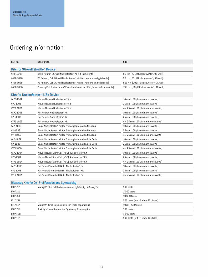
Kits for 96-well Shuttle™ DeviceVIPI-10003 Basic Neuron 96-well Nucleofector™ AD Kit (adherent) 96 rxn (20 μl Nucleocuvette™; 96-well)
V4SP-3096 P3 Primary Cell 96-well Nucleofector™ Kit (for neurons and glial cells) 96 rxn (20 μl Nucleocuvette™; 96-well)
V4SP-3960 P3 Primary Cell 96-well Nucleofector™ Kit (for neurons and glial cells) 960 rxn (20 μl Nucleocuvette™; 96-well)
V4SP-9096 Primary Cell Optimization 96-well Nucleofector™ Kit (for neural stem cells) 192 rxn (20 μl Nucleocuvette™; 96-well)
Kits for Nucleofector™ II/2b DeviceVAPG-1001 Mouse Neuron Nucleofector™ Kit 10 rxn (100 μl aluminum cuvette)
VPG-1001 Mouse Neuron Nucleofector™ Kit 25 rxn (100 μl aluminum cuvette)
VVPG-1001 Mouse Neuron Nucleofector™ Kit 4 × 25 rxn (100 μl aluminum cuvette)
VAPG-1003 Rat Neuron Nucleofector™ Kit 10 rxn (100 μl aluminum cuvette)
VPG-1003 Rat Neuron Nucleofector™ Kit 25 rxn (100 μl aluminum cuvette)
VVPG-1003 Rat Neuron Nucleofector™ Kit 4 × 25 rxn (100 μl aluminum cuvette)
VAPI-1003 Basic Nucleofector™ Kit for Primary Mammalian Neurons 10 rxn (100 μl aluminum cuvette)
VPI-1003 Basic Nucleofector™ Kit for Primary Mammalian Neurons 25 rxn (100 μl aluminum cuvette)
VVPI-1003 Basic Nucleofector™ Kit for Primary Mammalian Neurons 4 × 25 rxn (100 μl aluminum cuvette)
VAPI-1006 Basic Nucleofector™ Kit for Primary Mammalian Glial Cells 10 rxn (100 μl aluminum cuvette)
VPI-1006 Basic Nucleofector™ Kit for Primary Mammalian Glial Cells 25 rxn (100 μl aluminum cuvette)
VVPI-1006 Basic Nucleofector™ Kit for Primary Mammalian Glial Cells 4 × 25 rxn (100 μl aluminum cuvette)
VAPG-1004 Mouse Neural Stem Cell (NSC) Nucleofector™ Kit 10 rxn (100 μl aluminum cuvette)
VPG-1004 Mouse Neural Stem Cell (NSC) Nucleofector™ Kit 25 rxn (100 μl aluminum cuvette)
VVPG-1004 Mouse Neural Stem Cell (NSC) Nucleofector™ Kit 4 × 25 rxn (100 μl aluminum cuvette)
VAPG-1005 Rat Neural Stem Cell (NSC) Nucleofector™ Kit 10 rxn (100 μl aluminum cuvette)
VPG-1005 Rat Neural Stem Cell (NSC) Nucleofector™ Kit 25 rxn (100 μl aluminum cuvette)
VVPG-1005 Rat Neural Stem Cell (NSC) Nucleofector™ Kit 4 × 25 rxn (100 μl aluminum cuvette)
BioAssay Kits for Cell Proliferation and CytotoxicityLT07-221 ViaLight™ Plus Cell Proliferation and Cytotoxity BioAssay Kit 500 tests
LT07-121 1,000 tests
LT07-321 10,000 tests
LT17-221 500 tests (with 5 white TC plates)
LT17-517 ViaLight™ 100% Lysis Control Set (sold separately) 10 ml (200 tests)
LT07-217 ToxiLight™ Non-destructive Cytotoxity BioAssay Kit 500 tests
LT07-1117 1,000 tests
LT07-117 500 tests (with 5 white TC plates)
18

Ordering Information
Cat. No. Cat. No. Cat. No. Cat. No. Cat. No. Cat. No.
Gel Concentration/Separation Range Cassette Size (cm) 2D well 10 well 12 well 16 well 17 well 8 + 1 well
4 – 12% gradient 25 – 250 kDa
9 × 10 10 × 10
– –
58520 59520
58522 59522
58524 59524
– –
– –
4 – 20% gradient 5 – 200 kDa
9 × 10 10 × 10
– 59557
58511 59511
58505 59505
58517 59517
58545 59545
58551 59551
8 – 16% gradient 15 – 200 kDa
9 × 10 10 × 10
– 59564
58519 59519
58521 59521
58523 59523
58560 59560
58562 59562
10 – 20% gradient 5 – 150 kDa
9 × 10 10 × 10
– –
58512 59512
58506 59506
58518 59518
– –
– –
7.5% 50 – 200 kDa
9 × 10 10 × 10
– –
58507 59507
58501 59501
58513 59513
58540 –
– –
10% 25 – 200 kDa
9 × 10 10 × 10
– 59554
58508 59508
58502 59502
58514 59514
58542 59542
58548 59548
12% 20 – 200 kDa
9 × 10 10 × 10
– 59571
58509 59509
58503 59503
58515 59515
58543 59543
– –
15% 10 – 50 kDa
9 × 10 10 × 10
– 59556
58510 59510
58504 59504
58516 59516
58544 59544
58550 59550
PAGEr™ Gels and Accessories
Cat. No. Description Size
CNS StellARray™ qPCR AssaysPlease Inquire Human Alzheimer’s Disease StellARray™ Various formats
Please Inquire Human Autism StellARray™ Various formats
Please Inquire Human Huntington’s Disease StellARray™ Various formats
Please Inquire Human Mood Disorder StellARray™ Various formats
Please Inquire Human Neurodegeneration StellARray™ Various formats
Please Inquire Human Parkinson StellARray™ Various formats
Please Inquire Mouse Alzheimer’s Disease StellARray™ Various formats
Please Inquire Mouse Autism StellARray™ Various formats
Please Inquire Mouse Huntington’s Disease StellARray™ Various formats
Please Inquire Mouse Mood Disorder StellARray™ Various formats
Please Inquire Mouse Neurodegeneration StellARray™ Various formats
http://array.lonza.com – For StellARray™ qPCR Assay formats.
www.lonza.com/protein – For PAGEr™ Gel formats and accessories.
19
www.lonza.com/researchwww.lonza.com/neurobiology
Lonza Cologne GmbH 50829 Cologne, Germany For research use only. Not for use in diagnostic procedures.StellARray™, GeneSieve™, Global Pattern Recognition™ and GPR™ are trademarks of Bar Harbor Biotechnology. Neurobasal™ and B27® Serum Supplement are registered trademarks of Life Technologies®. ATCC® is a trademark of ATCC™ used under license. All other trademarks herein are marks of the Lonza Group or its affiliates.The information contained herein is believed to be correct and corresponds to the latest state of scien-tific and technical knowledge. However, no warranty is made, either expressed or implied, regarding its accuracy or the results to be obtained from the use of such information and no warranty is expressed or implied concerning the use of these products. The buyer assumes all risks of use and/or handling. No statement is intended or should be construed as a recommenda-tion to infringe any existing patent.
© 2011 Lonza Cologne GmbH. All rights reserved.BR-DseCmpgn 03/11 CD-BR014
Contact Information
North AmericaCustomer Service: 800 638 8174 (toll free)Scientific Support: 800 521 0390 (toll free)[email protected]
EuropeCustomer Service: + 32 87 321 [email protected] Support: + 49 221 99199 [email protected]
Online Orderinghttps://shop.lonza.com
InternationalContact your local Lonza DistributorCustomer Service: + 1 301 898 7025, ext. [email protected]
International OfficesAustralia +61 3 9550 0883Austria 0800 201 538 (toll free)Belgium +32 87 321 611Brazil +55 11 5641 3325Denmark 808 83 159 (toll free)France 0800 91 19 81 (toll free)Germany 0800 182 52 87 (toll free)India +91 22 4342 4000Ireland 1 800 654 253 (toll free)Italy 800 789 888Japan +81 3 5566 0622Luxemburg +32 87 321 611Poland +48 781 120 300Singapore +65 6521 4379Spain 900 963 298Sweden 020 790 220 (toll free)Switzerland 0800 83 86 20 (toll free)The Netherlands 0800 022 4525 (toll free)United Kingdom 0808 234 97 88 (toll free)