neuro-ophthalmology lectures 1, 2 & 3 supporting information · meningioma 80 optic...
TRANSCRIPT

1
Neuro-ophthalmology
Lectures 1, 2 & 3
Supporting information
Published: 14/10/13

2
Neuro-ophthalmology
Index
OPTIC NEUROPATHIES
Evaluation of optic nerve disease 5
Optic disc changes 5
Primary optic atrophy 5
Secondary optic atrophy 6
Classification of optic neuritis 7
Demyelinating optic neuritis 7
Parainfectious optic neuritis 13
Infectious optic neuritis 13
Non-arteritic anterior ischaemic optic neuropathy 14
Arteritic anterior ischaemic optic neuropathy 15
Leber hereditary optic neuropathy 19
Hereditary optic atrophy 20
Nutritional optic neuropathy 21
Papilloedema 22
CONGENITAL OPTIC NERVE ANOMALIES
Without neurological association 26
With neurological association 28
PUPILLARY REACTIONS
Anatomy 33
Afferent pupillary defect 34
Oculosympathetic palsy (Horner syndrome) 37
Adie pupil 41
Other abnormal reactions 41

3
MIGRAINE 43
CLUSTER HEADACHE45
OCULAR MOTOR NERVES 49
3rd nerve 49
4th nerve 53
6th nerve 56
NYSTAGMUS 60
Introduction 60
Physiological nystagmus 61
Vestibular nystagmus 61
Motor imbalance nystagmus 62
Sensory deprivation nystagmus 66
Surgery for nystagmus 66
Nystagmoid movements 66
SUPRANUCLEAR DISORDERS OF OCULAR MOTILITY 67
Conjugate eye movements 67
Horizontal gaze palsy 68
Vertical gaze palsy 70
CHIASM 72
Anatomy 72
Physiology 73
Pituitary adenomas 75
Craniopharyngioma 79
Meningioma 80
RETROCHIASMAL PATHWAYS 80
Optic tract 80
Optic radiations 82

4
Striate cortex 84
OCULAR MYOPATHIES 85
Myasthenia gravis 86
Myotonic dystrophy 88
Chronic progressive external ophthalmoplegia 89

5
Neuro-ophthalmology
Lecture 1 – Supporting information
Optic Neuropathies
A) Evaluation of Optic Nerve Disease
Clinical features of Optic Nerve Dysfunction:
1. Reduced VA
2. Afferent papillary conduction defect
3. Dyschromatopsia (CV impairment mainly affecting R & G) – uniocular defect
apparent when comparing a red object monocularly
4. Diminished light brightness sensitivity (may be present even if VA normal).
Often seen after attack of optic neuritis.
How to Demonstrate: shine a light into the normal eye and then into the
eye with the suspected lesion. The light appears less bright in the
affected eye.
5. Diminished contrast sensitivity – test using gratings. Sensitive to subtle visual
loss but not specific to optic nerve disease
6. Visual Field Defects – central scotoma, centrocaecal scotoma, altitudinal
defect & nerve fibre bundle defects.
Optic Disc changes
Four main appearances in acquired ON disorders are:
1. Normal disc – retrobulbar neuritis (and initially in Leber optic
neuropathy/compressive lesions)
2. Disc swelling –papilloedema, Arteritic AION, Non-arteritic AION, papillitis,
acute stage of Leber & with compressive lesions before devpt of optic atrophy
3. Optico-ciliary shunts – ON sheath meningioma and occasionally ON glioma
4. Optic atrophy – important sign of advanced ON disease
a. Primary optic atrophy – caused by lesions affecting the visual
pathway from the retrolaminar part of the ON to the Lateral geniculate
body. Lesions affecting the ON will result in unilateral optic atrophy,
those affecting the chiasm/optic tract will cause bilateral optic atrophy
i. Causes:
Retrobulbar neuritis
Compressive lesions (tumours/aneurysms)

6
Hereditary optic neuropathies
Toxic/nutritional optic neuropathies
ii. Disc appearance:
White, flat disc with clearly defined margins
Reduction in number of BV’s crossing disc
Attenuation of peripapillary BV’s and thinning of RNFL
Atrophy may be diffuse or sectorial depending on cause and
level of lesion; e.g. chiasmal lesions may involve nasal and
temporal portions, but spare superior and inferior
b. Secondary Optic atrophy – preceded by swelling of the ONH
i. Causes:
Papilloedema
AION
Papillitis
ii. Disc appearance:
Variable depending on cause
White slightly raised disc with poorly defined margins
Reduction in number of BVs crossing disc
Special Investigations:
1. Automated perimetry
2. MRI – method of choice for imaging ON
3. Visually Evoked potential – assessment of electrical activity of the visual
cortex created by stimulation of the retina. Stimulus can be a flash of light
or a reversing checkboard pattern on a screen. Amplitude and latency are
the 2 components assessed. In ON disease we see a decrease in
amplitude and an increase in latency.
4. FA – occasionally helpful in differentiation of papilloedema from OD
drusen. With papilloedema there is disc leakage, with drusen leakage is
absent.
B) Classification of Optic Neuritis
Acute or subacute inflammatory or demyelination process affecting the ON.
Classifications:
a) Ophthalmoscopic classification
i. Retrobulbar neuritis – OD appears normal because
pathological process doesn’t involve the ONH. Most frequent
type of ON in adults and is frequently associated with MS

7
ii. Papillitis – Process affects the ONH. Variable disc
hyperaemia and oedema. May rarely be associated with
peripapillary flame shaped haemorrhages. Cells in the
posterior vitreous may also be present. Most common type of
Optic neuritis in kids although it can also affect adults.
iii. Neuroretinitis – characterised by papillitis in association with
a macular star figure composed of hard exudates. Macular
lesion may not be present initially but appears within a few
days or weeks and becomes more prominent when the OD
swelling is resolving. Occasionally associated with
peripapillary retinal oedema and serous elevation of the
macula. Least common type of optic neuritis. Associated with
viral infections, cat-scratch fever, syphilis and Lyme disease.
NEVER MS. Usually self-limiting: 6-12 months.
b) Aetiological classification
i. Demyelinating – most common cause
ii. Parainfectious – following a viral infection or immunisation
iii. Infectious – sinus related, cat scratch fever, syphilis, Lyme
disease or crytococcal meningitis in px with AIDS.
C) Optic Neuritis and demyelination
Demyelination is a pathological process whereby normally myelinated nerve
fibres lose their insulating myelin layer. Demyelinating diseases disrupt nervous
conduction within the white matter tracts in the brain, brain stem and spinal cord.
1. Ocular features of demyelination
a. Visual pathway lesions – most frequently involve the optic nerve and
cause optic neuritis. Occasionally demyelination may involve the optic
chiasm. Rarely does it affect the optic tracts or radiations.
b. Brain stem lesions – may result in inter-nuclear ophthalmoplegia and
less frequently ocular motor cranial nerve palsy and oscillopsia
(unsteadiness of the visual world).
Internuclear ophthalmoplegia - a disorder of conjugate lateral gaze in which the
affected eye shows impairment of adduction. When an attempt is made to gaze
contralaterally (relative to the affected eye), the affected eye adducts minimally, if
at all. The contralateral eye abducts, however with nystagmus. Additionally, the
divergence of the eyes leads to horizontal diplopia. That is, if the right eye is
affected the patient will "see double" when looking to the left, seeing two images
side-by-side. Convergence is generally preserved.

8
2. Demyelination diseases that cause ocular problems are
a. Isolated optic neuritis – px has no clinical evidence of systemic
demyelination although often it subsequently develops
b. Multiple Sclerosis (see below)
c. Devic disease - very rare. Optic neuritis which is often bilateral
d. Schilder disease – very rare. Onset prior to age 10, death within 1-2
yrs. Bilateral optic neuritis may occur

9
Multiple Sclerosis
Common, idiopathic (uncertain/unknown cause), remitting neurological disease.
Typically affects young adults – women more often than men. Characterised by
demyelination of the CNS, but not the peripheral nervous system.
Clinical features:
1. Spinal cord lesions – weakness, stiffness and sphincter/sexual function
disturbance. Muscle spasms and sensory disturbances with a ‘trouser-like’
distribution.
2. Brain stem lesions – may produce diplopia, nystagmus, ataxia (lack of
voluntary coordination of muscle movements), dysarthria (motor speech
disorder – difficulty pronouncing words) and dysphagia (difficulty
swallowing).
3. Hemisphere lesions – may produce hemiparesis (weakness on one side
of the body), hemianopia (defective vision or blindness in half of the visual
field of one or both eyes), dysphasia (full or partial loss of verbal
communication skills). Also intellectual decline, depression, euphoria and
even dementia.
4. Transient phenomena – Lhermitte sign (electrical sensation on neck
flexion) transient dysarthria, disequilibrium (loss/lack of stability), diplopia
syndrome and tonic spasms (a continuous involuntary muscular
contraction). Trigeminal neuralgia (disorder of the fifth cranial nerve) that
causes episodes of sharp, stabbing pain in the cheek, lips, gums, or chin
on one side of the face in a young px should raise suspicion of possible
demyelination. Another feature is Uhthoff phenomenon – sudden
temporary worsening of visual or other symptoms brought on by physical
exercise or increase in body temperature.
Special Investigations:
1. Lumbar puncture
2. Visually Evoked Potentials – in px with acute optic neuritis we see
decreased amplitude with a more profound increase in latency. Following
an attack the amplitude recovers but the increase in latency remains
therefore useful in px with suspected MS even in the absence of a
definitive prior attack of optic neuritis
3. MRI – shows plaques

10
MS and Optic neuritis
1. Incidence:
a. Approx 70% of women and 35% of men with optic neuritis will
ultimately develop other neurological dysfunction and be classified as
having MS
b. Evidence of optic neuritis is found in 70% of established cases
c. Up to 70% of px with clinically isolated optic neuritis have abnormal
MRI similar to that seen in MS
d. In a px with optic neuritis, the subsequent risk of MS is increased with
winter onset, HLA-DR2 positivity (a broad antigen serotype) and
Uhthoff phenomenon.
2. Presentation
a. Sudden onset of monocular visual loss is the norm. Rarely both eyes
are involved simultaneously.
b. Discomfort in/around the eye is very common and is frequently
exacerbated by ocular movements. Discomfort may precede or occur
simultaneously with visual loss and usually lasts for only a few days.
c. Frontal headache and tenderness of the globe are present in some px
3. Signs
a. Disc is normal in 2/3rds of cases (retrobulbar neuritis) and the
remainder show papillitis. Temporal disc pallor may be seen in the
other eye, indicative of a previous attack.
b. Diminished VA – may be very mild to very severe.
c. Impairment of CV and contrast sensitivity is almost universal and
frequently worse than would be expected for the VA level.
d. Other features of optic nerve dysfunction are present, as previously
described.
4. Visual field defect
a. Typically a central scotoma, although other defects may be seen.
5. Clinical course
a. VA impairment becomes maximal after 1-2 weeks. Usually between
6/18 and 6/60 (rarely no LP).
b. Recovery takes 4-6 weeks (may be slower in some px)
6. Prognosis
a. Excellent for 75% of px with recovery of VA to 6/9 or better. 85%
recover to 6/9 or better even if initial acuity was reduced to no light
perception during the attack. However, other parameters of visual
function, e.g. CV, contrast sensitivity and light brightness appreciation
often remain abnormal. A mild afferent pupillary conduction defect may

11
persist and optic atrophy may ensue, particularly in px with recurrent
attacks.
7. Treatment
a. If presenting visual loss is mild, treatment is probably unnecessary
b. When VA within the first week of symptom onset is worse than 6/12,
treatment may speed up recovery (IV methylprednisolone for 3 days
followed by oral prednisolone for 11 days)
Optic Neuritis Treatment Trial The Optic Neuritis Treatment Trial (ONTT) was designed to compare the speed and level of visual recovery between patients treated with oral prednisone, intravenous methylprednisolone (IVMP), or placebo. Patients were randomized into 1 of 3 groups within 8 days of symptom onset. Those treated with oral prednisone (1mg/kg/day for 14 days) demonstrated an increased incidence of recurrent ON compared with those treated with IVMP (250mg every 6h for 3 days, followed by an oral taper) or placebo.[2]
The ONTT results suggest that IV steroids, oral steroids, and placebo all result in recovery of visual function over time. IV steroids hasten the rate of recovery but do not change the final visual outcome. In the ONTT, IV steroids seemed to decrease the incidence of the development of MS over a 2-year period, but this effect was not sustained after year 3.
At entry into the ONTT, 35% of patients had a visual acuity of 20/40 or better, 30% of patients had a visual acuity of between 20/50 and 20/200, and 35% of patients had a visual acuity of 20/200 or worse. Only 3% of patients had no light perception (NLP). Therefore, NLP should be considered a red flag for a diagnosis of ON; in such cases, other potential etiologies for vision loss (e.g. inflammatory, infiltrative, neoplastic) may need to be considered.
Nearly 100% of patients in the trial whose visual acuity was 20/50 or worse had a defect in their color sensitivity, and in those patients with a visual acuity of 20/20 or better, 51-70% had altered color vision.
Seventy-four percent of patients in the ONTT recovered visual acuity of 20/60 or better by 8 weeks, and most patients had a visual acuity of better than 20/40 by 6 months.
The most common visual field defects were as follows, in decreasing order of frequency:
Altitudinal - 28.8% 3 quadrant - 14.0% 1 quadrant - 11.8% Centrocecal - 8.7% Hemianopic - 8.3% Peripheral rim - 7.0% Arcuate - 7.4% Central - 7.0% Enlarged blind spot - 2.6% Nasal step - 1.3%
Mild to severe pain was present in 92.2% of patients.[2] Pain was constant in 7.3% of patients, constant and worse on extraocular motility in 51.3% of patients, and noted only with eye movement in 35.8% of patients.
Although all 3 treatment arms of the study had equal visual outcomes, oral prednisone in conventional doses increased the likelihood for a recurrent episode of ON and is not recommended. Higher doses of oral methylprednisolone have not produced similar increased recurrence rates of ON, but the number of patients in these studies was small.

12 Laboratory studies were not deemed helpful in establishing a typical demyelinating ON diagnosis (i.e. acute, unilateral optic neuropathy in a young patient with pain on extraocular movement and improvement over time). A lumbar puncture was optional and showed evidence of demyelinating disease only in patients with ON
MS References
1. Roodhooft JM. Ocular problems in early stages of multiple sclerosis. Bull Soc Belge Ophtalmol. 2009;65-8.[Medline].
2. Optic Neuritis Study Group. The clinical profile of optic neuritis. Experience of the Optic Neuritis Treatment Trial. Arch Ophthalmol. Dec 1991;109(12):1673-8. [Medline].
3. Optic Neuritis Study Group. The 5-year risk of MS after optic neuritis. Experience of the optic neuritis treatment trial. Neurology. Nov 1997;49(5):1404-13. [Medline].
4. Multiple sclerosis risk after optic neuritis: final optic neuritis treatment trial follow-up. Arch Neurol. Jun 2008;65(6):727-32. [Medline]. [Full Text].
5. Polman CH, Reingold SC, Edan G, Filippi M, Hartung HP, Kappos L, et al. Diagnostic criteria for multiple sclerosis: 2005 revisions to the "McDonald Criteria". Ann Neurol. Dec 2005;58(6):840-6. [Medline].
6. Balashov KE, Pal G, Rosenberg ML. Optic neuritis incidence is increased in spring months in patients with asymptomatic demyelinating lesions. Mult Scler. Feb 2010;16(2):252-4. [Medline]. [Full Text].
7. Villoslada P, Cuneo A, Gelfand J, et al. Color vision is strongly associated with retinal thinning in multiple sclerosis. Mult Scler. Jan 30 2012;[Medline].
8. Wang J, Cheng H, Hu YS, et al. The photopic negative response of the flash electroretinogram in multiple sclerosis. Invest Ophthalmol Vis Sci. Jan 24 2012;[Medline].
9. Lennon VA, Wingerchuk DM, Kryzer TJ, Pittock SJ, Lucchinetti CF, Fujihara K, et al. A serum autoantibody marker of neuromyelitis optica: distinction from multiple sclerosis. Lancet. Dec 11-17 2004;364(9451):2106-12. [Medline].
10. Coyle PK. Early treatment of multiple sclerosis to prevent neurologic damage. Neurology. Dec 9 2008;71(24 Suppl 3):S3-7. [Medline].
11. Beck RW, Chandler DL, Cole SR, Simon JH, Jacobs LD, Kinkel RP, et al. Interferon beta-1a for early multiple sclerosis: CHAMPS trial subgroup analyses. Ann Neurol. Apr 2002;51(4):481-90. [Medline].
12. Jacobs LD, Beck RW, Simon JH, Kinkel RP, Brownscheidle CM, Murray TJ, et al. Intramuscular interferon beta-1a therapy initiated during a first demyelinating event in multiple sclerosis. CHAMPS Study Group. N Engl J Med. Sep 28 2000;343(13):898-904. [Medline].
13. Filippi M, Rovaris M, Inglese M, Barkhof F, De Stefano N, Smith S, et al. Interferon beta-1a for brain tissue loss in patients at presentation with syndromes suggestive of multiple sclerosis: a randomised, double-blind, placebo-controlled trial. Lancet. Oct 23-29 2004;364(9444):1489-96. [Medline].
14. Miller NR. Optic neuritis. In: Walsh & Hoyt's Clinical Neuro-Ophthalmology. 4th ed. Lippincott Williams & Wilkins; 1982:227-48.

13
Other causes of optic neuritis
1. Parainfectious optic neuritis Optic neuritis may be associated with viral infections such as measles, mumps, chickenpox, whooping cough and glandular fever. It may also occur after immunisation. Children are affected more frequently than adults in this category.
a) Presentation - usually 1 to 3 weeks after a viral infection with an acute severe visual loss which may involve both eyes. May be associated with other neurological deficits e.g. headaches, seizures or ataxia
b) Signs – OD typically shows bilateral papillitis, although occasionally there may be a neuroretinitis, or the discs may be normal.
c) Treatment – unnecessary in the vast majority of px because the prognosis for spontaneous visual recovery is good. However, where visual loss is severe and bilateral, or when it involves an only seeing eye, IV steroids should be considered.
2. Infectious optic neuritis
a) Sinus-related – recurrent attacks of unilateral visual loss associated
with severe headache and acute ethmoidal sinusitis. b) Cat scratch fever – self limiting systemic infection. Characterised by
regional lymphadenopathy (tenderness, pain and swelling of regional lymph glands) preceded by a cat scratch. Organism involved is Bartonella henselae, a small gram negative rod. Neuroretinitis (unilateral or bilateral) may occur in some px. Responds to antibiotics e.g. ciprofloxacin. Visual prognosis is excellent, with recovery of vision within 1-4 weeks of starting therapy.
c) Syphilis – may cause acute optic papillitis or neuroretinitis during the primary or secondary stage. Involvement may be unilateral or bilateral and is frequently associated with mild vitritis.
d) Lyme disease – a spirochetal infection transmitted by a tick bite which may cause a neuroretinitis. In some cases it causes acute retrobulbar neuritis.
e) Cryptococcal meningitis – px with AIDS may be affected. May be associated with optic nerve involvement and acute visual loss which may be bilateral.

14
3. Non-arteritic anterior ischaemic optic neuropathy
1 Pathogenesis. Non-arteritic anterior ischaemic optic neuropathy (NAION) is
caused by occlusion of the short posterior ciliary arteries resulting in partial or
total infarction of the optic nerve head.
2
Predispositions include structural crowding of the optic nerve head so that the
physiological cup is either very small or absent, hypertension, diabetes mellitus,
hyperlipidaemia, collagen vascular disease, antiphospholipid antibody
syndrome, hyperhomocysteinaemia, sudden hypotensive events, cataract
surgery, sleep apnoea syndrome and erectile dysfunction.
3 Presentation is in the 6th–7th decades with sudden, painless, monocular
visual loss which is not associated with premonitory visual obscurations. Visual
loss is frequently discovered on awakening suggesting that nocturnal
hypotension may play an important role.
4 Signs
• VA, in about 30% of patients, is normal or only slightly reduced. The
remainder has moderate-to-severe impairment.
• Visual field defects are typically inferior altitudinal but central, paracentral,
quadrantic and arcuate defects may also be seen.
• Dyschromatopsia is usually proportional to the level of visual impairment,
in contrast with optic neuritis in which colour vision may be severely
impaired when visual acuity is reasonably good.
• Diffuse or sectoral hyperaemic disc swelling, often associated with a few
peripapillary splinter haemorrhages
• The swelling gradually resolves and pallor ensues 3–6 weeks after onset.
5 Special investigations include blood pressure, fasting lipid profile and blood
glucose. It is also very important to exclude occult giant cell arteritis (see
below).

15
6 Treatment. There is no definitive treatment although any underlying systemic
predispositions should be treated. Although aspirin is effective in reducing
systemic vascular events and is frequently prescribed in patients with NAION, it
does not appear to reduce the risk of involvement of the fellow eye.
7 Prognosis. In most patients there is no further loss of vision although
recurrence occurs in about 6%. Involvement of the fellow eye occurs in about
10% of patients after 2 years and 15% after 5 years. When the second eye
becomes involved, optic atrophy in one eye and disc oedema in the other gives
rise to the ‘pseudo-Foster Kennedy syndrome’. Two important risk factors for
fellow eye involvement are poor visual acuity in the first eye and diabetes
mellitus.
4. Arteritic anterior ischaemic optic neuropathy
Arteritic anterior ischaemic optic neuropathy (AAION) is caused by giant cell arteritis.
Diagnosis of giant cell arteritis
Giant cell arteritis (GCA) is a granulomatous necrotizing arteritis with a predilection
for large and medium-size arteries, particularly the superficial temporal, ophthalmic,
posterior ciliary and proximal vertebral. The severity and extent of involvement are
associated with the quantity of elastic tissue in the media and adventitia. Intracranial
arteries, which possess little elastic tissue, are usually spared.
1 Presentation is in old age with the following:
• Scalp tenderness, first noticed when combing the hair, is common.
• Headache that may be localized to the frontal, occipital or temporal areas
or be more generalized.
• Jaw claudication (pain on speaking and chewing) caused by ischaemia of
the masseter muscles is virtually pathognomonic.
• Polymyalgia rheumatica is characterized by pain and stiffness in proximal
muscle groups (typically the shoulders). The symptoms are usually worse
in the morning and after exertion, and may precede cranial symptoms by
many months.
• Non-specific symptoms such as neck pain, weight loss, fever, night
16
sweats, malaise and depression are common.
• Blindness of sudden onset with minimal systemic upset (occult arteritis) is
uncommon.
2 Other features
• Superficial temporal arteritis is characterized by thickened, tender,
inflamed and nodular arteries, which cannot be flattened against the skull.
• Pulsation is initially present, but later ceases, a sign strongly suggestive of
GCA, since a non-pulsatile superficial temporal artery is highly unusual in
a normal individual. The best location to examine pulsation is directly in
front of the pinna.
• In very severe cases, scalp gangrene may ensue.
• Rare complications include dissecting aneurysms, aortic incompetence,
myocardial infarction, renal failure and brainstem stroke.
3 Erythrocyte sedimentation rate (ESR) is often very high, with levels of
>60 mm/hr, although in approximately 20% of patients it is normal.
4 Blood platelet levels may be elevated.
5 C-reactive protein (CRP) is invariably raised and may be helpful when ESR is
equivocal.
6 Temporal artery biopsy (TAB) should be performed if GCA is suspected.
• Steroids should never be withheld pending biopsy, which should ideally
be performed within 3 days of commencing steroids.
• Systemic steroids for more than 7–10 days may suppress histological
evidence of active arteritis although this is not invariable.
• In patients with ocular involvement it is advisable to take the biopsy from
the ipsilateral side. The ideal location is the temple because it lessens the

17
risk of major nerve damage.
• At least 2.5 cm of the artery should be taken and serial sections examined
because of the phenomenon of ‘skip’ lesions in which segments of
histologically normal arterial wall may alternate with granulomatous
inflammation.
Treatment of giant cell arteritis
Treatment involves systemic steroids, the duration of which is governed by
symptoms and the level of the ESR or CRP. Symptoms may, however, recur without
a corresponding rise in ESR or CRP and vice versa. Most patients need treatment
for 1–2 years, although some may require indefinite maintenance therapy. CRP may
play an important role in monitoring disease activity, as the level seems to fall more
rapidly than the ESR in response to treatment.
Ophthalmic manifestations of giant cell arteritis
Arteritic anterior ischaemic optic neuropathy
AAION affects 30–50% of untreated patients of which one-third develop involvement
of the fellow eye, usually within 1 week of the first. Posterior ischaemic optic
neuropathy is much less common (see below).
1 Presentation is with sudden, profound unilateral visual loss which may be
accompanied by periocular pain and preceded by transient visual obscurations
and flashing lights. Bilateral simultaneous involvement is rare. Most cases
occur within a few weeks of the onset of GCA although at presentation about
20% of patients do not have systemic symptoms (i.e. occult GCA).
2 Signs
• Severe visual loss is the rule, commonly to LP or worse.
• A strikingly pale (‘chalky white’) oedematous disc is particularly suggestive
of GCA
• Occasionally AAION may be combined with occlusion of the cilioretinal
artery.

18
• Over 1–2 months, the swelling gradually resolves and severe optic
atrophy ensues.
3 Treatment is aimed at preventing blindness of the fellow eye, although the
second eye may still become involved in 25% of cases despite early and
adequate steroid administration, usually within 6 days of starting treatment.
Visual loss is usually profound and is unlikely to improve even with immediate
treatment. The regimen is as follows:
a Intravenous methylprednisolone, 1g/day for 3 days then oral
prednisolone 1–2 mg/kg/day. After 3 days the oral dose is reduced to
60 mg and then 50 mg each, for one week. The daily dose is then
reduced by 5 mg weekly until 10 mg is reached.
b Oral prednisolone alone may be administered as an alternative in some
circumstances (e.g. late presentation or systemic contraindications to
intravenous therapy).
c Antiplatelet therapy (e.g. aspirin 150 mg/day) should be commenced.
d Immunosuppressives may be used as adjuncts in steroid-resistant
cases or as steroid-sparing agents when extended treatment is required.
4 Prognosis is very poor because visual loss is usually permanent, although very
rarely, prompt administration of systemic steroids may be associated with
partial visual recovery.
Other manifestations
1 Transient ischaemic attacks (amaurosis fugax) may precede infarction of the
optic nerve head.
2 Cilioretinal artery occlusion may be combined with AAION.
3 Central retinal artery occlusion is usually combined with occlusion of a
posterior ciliary artery. This is because the central retinal artery often arises
from the ophthalmic artery by a common trunk with one or more of the posterior
ciliary arteries. However, ophthalmoscopy shows occlusion of only the central

19
retinal artery; the associated ciliary occlusion can be detected only on FA.
4 Ocular ischaemic syndrome clinical picture due to involvement of the
ophthalmic artery is rare.
5 Diplopia, transient or constant, may be caused by ischaemia of the ocular
motor nerves or extraocular muscles
5. Leber Hereditary Optic Neuropathy
Leber hereditary optic neuropathy (LHON) is a rare disease associated with
maternally-inherited mitochondrial DNA mutations, most notably 11778. The
condition typically affects males between the ages of 15 and 35 years, although
in atypical cases the condition may affect females and present at any age
between 10 and 60 years. The diagnosis of LHON should therefore be
considered in any patient with bilateral optic neuropathy, irrespective of age.
Unaffected carriers show thickening of temporal retinal fibres on OCT.
1 Presentation is typically with acute or subacute, severe painless unilateral loss
of central vision. The fellow eye becomes similarly affected within weeks or
months of the first.
2 Signs during the acute stage are often subtle and easily overlooked, and in
some patients the disc may be entirely normal.
• In typical cases there is disc hyperaemia with obscuration of the disc
margins.
• Dilated capillaries on the disc surface that may extend onto adjacent
retina (telangiectatic microangiopathy), swelling of the peripapillary nerve
fibre layer (pseudo-oedema) and dilatation and tortuosity of posterior pole
vasculature.
• Subsequently, the vessels regress and pseudo-oedema resolves.
• Severe optic atrophy supervenes, with nerve fibre layer dropout most
pronounced in the papillomacular bundle.
• Telangiectatic microangiopathy may be present in asymptomatic female
relatives.
• Surprisingly, the pupillary light reactions may remain fairly brisk.
3 FA shows absence of dye leakage.

20
4 Visual field defects usually consist of central or centrocaecal scotomas.
5 Treatment is generally ineffective although many modalities, including steroids,
hydroxocobalamin and surgical intervention have been tried. Smoking and
excessive consumption of alcohol should be discouraged, to minimize potential
stress on mitochondrial energy production.
6 Prognosis is poor, although some visual recovery may occur in a minority of
cases even years later. Most patients suffer severe, bilateral and permanent
visual loss with a final visual acuity of 6/60 or less. The 11778 mutation carries
the worst prognosis.
6. Hereditary optic atrophies
The hereditary optic atrophies (neuropathies) are a very rare heterogeneous group of
disorders that are primarily characterized by bilateral optic atrophy.
Kjer-type optic atrophy
1 Inheritance is AD.
2 Presentation is typically in the 1st–2nd decade with insidious visual loss.
3 Optic atrophy may be subtle and temporal or diffuse.
4 Prognosis is variable (final VA 6/12–6/60) with considerable differences within
and between families. Very slow progression over decades is typical.
5 Systemic abnormalities are absent in the majority of cases although some
develop sensorineural hearing loss.
Behr syndrome
1 Inheritance is AR.
2 Presentation is in the 1st decade with visual loss which stabilizes after a
variable period of progression.
3 Optic atrophy is diffuse.
4 Prognosis is variable with moderate to severe visual loss and nystagmus.
5 Systemic abnormalities include spastic gait, ataxia and mental handicap.

21
Wolfram syndrome
Wolfram syndrome is also referred to as DIDMOAD (Diabetes Insipidus,
Diabetes Mellitus, Optic Atrophy and Deafness).
1 Inheritance is AR.
2 Presentation is between the ages of 5 and 21 years.
3 Optic atrophy is diffuse and severe and may be associated with disc cupping.
4 Prognosis is very poor (final VA is <6/60).
5 Systemic abnormalities (apart from DIDMOAD) include anosmia, ataxia,
seizures, mental handicap, short stature, endocrine abnormalities and elevated
CSF protein.
7. Nutritional optic neuropathy
Nutritional optic neuropathy (tobacco-alcohol amblyopia) typically affects heavy
drinkers and smokers who are deficient in protein and the B vitamins. Most
patients have neglected their diet, obtaining their calories from alcohol instead.
Some of those affected also have defective vitamin B12 absorption and may
develop pernicious anaemia.
1 Presentation is with the insidious onset of progressive, usually symmetrical
bilateral visual impairment associated with dyschromatopsia.
2 Signs. The discs at presentation are normal in most cases. Some patients
show subtle temporal pallor, splinter-shaped haemorrhages on or around the
disc, or minimal disc oedema.
3 Visual field defects are bilateral, relatively symmetrical, centrocaecal
scotomas. The margins of the defects are difficult to define with a white target
but are easier to plot and larger when using a red target.
4 Treatment involves weekly injections of 1000 units of hydroxocobalamin for 10
weeks. Multivitamins including thiamine (100 mg b.d.) and folate (1 mg daily)
are also administered and patients should be advised to eat a well-balanced
diet and abstain from drinking and smoking.
5 Prognosis is good in early cases provided patients comply with treatment
although visual recovery may be slow. In advanced and unresponsive cases
there is permanent visual loss as a result of optic atrophy.

22
Neuro-ophthalmology
Lecture 2 – Supporting information
Papilloedema
Pathogenesis
Papilloedema is swelling of the optic nerve head secondary to raised intracranial
pressure. It is nearly always bilateral, although it may be asymmetrical. All other
causes of disc oedema in the absence of raised intracranial pressure are referred to
as ‘disc swelling’ and are usually associated with persistent visual impairment. All
patients with papilloedema should be suspected of having an intracranial mass
unless proved otherwise. However, not all patients with raised intracranial pressure
will necessarily develop papilloedema. Tumours of the cerebral hemispheres tend to
produce papilloedema later than those in the posterior fossa. Patients with a history
of previous papilloedema may develop a substantial increase in intracranial pressure
but fail to re-develop papilloedema because of glial scarring of the optic nerve head.
Cerebrospinal fluid and causes of raised intracranial pressure
1 Circulation
• Cerebrospinal fluid (CSF) is formed by the choroid plexus in the ventricles
of the brain.
• It leaves the lateral ventricles to enter the 3rd ventricle through the
foramina of Munro.
• From the 3rd ventricle, it flows through the Sylvian aqueduct to the 4th
ventricle.
• From the 4th ventricle, the CSF passes through the foramina of Luschka
and Magendie to enter the sub-arachnoid space, some flowing around the
spinal cord and the rest bathing the cerebral hemispheres.
• Absorption is into the cerebral venous drainage system through the
arachnoid villi.
2 Normal opening pressure of CSF on lumbar puncture is <80 mmH2O in infants,
<90 mm in children and <210 mm in adults.
3 Causes of raised intracranial pressure.

23
• Idiopathic intracranial hypertension (pseudotumour cerebri).
• Obstruction of the ventricular system by congenital or acquired lesions.
• Space-occupying intracranial lesions, including haemorrhage.
• Impairment of CSF absorption via arachnoid villi, which may be damaged
by meningitis, subarachnoid haemorrhage or cerebral trauma.
• Cerebral venous sinus thrombosis.
• Diffuse cerebral oedema from blunt head trauma.
• Severe systemic hypertension.
• Hypersecretion of CSF by a choroid plexus tumour, (very rare).
Diagnosis of raised intracranial pressure
1 Headaches may be of onset at any time of day but characteristically occur
early in the morning and may wake the patient from sleep. They tend to get
progressively worse and patients usually present to hospital within 6 weeks.
The headaches may be generalized or localized, and may intensify with head
movement, bending or coughing. Patients with lifelong headaches often report
a change in character of the headache. Very rarely, headache may be absent.
2 Sudden nausea and vomiting, often projectile, may partially relieve the
headache. Vomiting may occur as an isolated feature or may precede the onset
of headaches by months, particularly in patients with fourth ventricular tumours.
3 Deterioration of consciousness may be slight, with drowsiness and
somnolence. Dramatic deterioration of consciousness is indicative of brainstem
distortion with tentorial or tonsillar herniation and requires prompt attention.
4 Visual symptoms
a Transient obscurations lasting a few seconds are frequent in patients with
papilloedema.
b Horizontal diplopia due to 6th nerve palsy caused by stretching of one or
both 6th nerves over the petrous tip; this is therefore a false localizing
sign.
c Visual failure occurs late with secondary optic atrophy due to long-
standing papilloedema (see below).
5 Investigations. MR, CT and B-scan ultrasonography show an enlarged optic
nerve diameter in most cases.

24
Stages of papilloedema
1 Early
• Visual symptoms are absent and visual acuity normal.
• Mild disc hyperaemia with preservation of the optic cup.
• Indistinct peripapillary retinal nerve striations and disc margins (initially
nasal, later superior, inferior and temporal).
• There is loss of previous spontaneous venous pulsation – this may not be
significant because it is also absent in about 20% of normal individuals.
However, preserved venous pulsation renders the diagnosis of
papilloedema highly unlikely.
2 Established
• Transient visual obscurations lasting a few seconds may occur in one or
both eyes, often on standing or bending forwards.
• VA is normal or reduced.
• Severe disc hyperaemia, moderate disc elevation with indistinct margins
and absence of the physiological cup.
• Venous engorgement, peripapillary flame haemorrhages and frequently
cotton wool spots.
• As the swelling increases, the optic nerve head appears enlarged.
• Circumferential retinal folds (Paton lines) may develop temporally.
• Hard exudates may radiate from the centre of the fovea in the form of a
‘macular fan’ – an incomplete star with the temporal part missing.
• The blind spot is enlarged.
3 Chronic
• VA is variable and the visual fields begin to constrict.
• Severe disc elevation without cotton wool spots and haemorrhages.
• Optociliary shunts and drusen-like crystalline deposits (corpora amylacea)
may be present on the disc surface.
4 Atrophic (secondary optic atrophy)
• VA is severely impaired.

25
• The optic discs are a dirty grey colour, slightly elevated, with few crossing
blood vessels and indistinct margins.
Causes of optic disc elevation
1 Papilloedema
2 Accelerated hypertension
3 Anterior optic neuropathy
• Ischaemic
• Inflammatory
• Infiltrative
• Compressive
4 Pseudo-papilloedema
• Optic disc drusen
• Tilted optic disc
• Peripapillary myelinated nerve fibres
• Crowded disc in hypermetropia
• Leber hereditary optic neuropathy
• Methanol poisoning
5 Intraocular disease
• Central retinal vein occlusion
• Posterior uveitis
• Posterior scleritis
• Hypotony
26
Congenital ON anomalies
Without neurological associations:
Tilted disc
A tilted optic disc is a common, usually bilateral, anomaly caused by an oblique entry
of the optic nerve into the globe. This results in pseudorotation of the superior pole of
the disc, angulation of the optic cup axis and elevation of the neuroretinal rim.
1 Signs
• Small, oval or D-shaped disc in which the axis is most frequently directed
inferonasally but may be horizontally or nearly vertically
• The disc margin is indistinct where the retinal nerve fibres are elevated.
• Situs inversus in which the temporal vessels deviate nasally before
turning temporally.
• Associated findings include inferonasal chorioretinal thinning and myopic
astigmatic refractive error.
2 Perimetry may show superotemporal defects that do not respect the vertical
midline.
3 Complications, which are uncommon, include CNV and sensory macular
detachment.
Optic disc drusen
1 Pathogenesis. Optic disc drusen (hyaline bodies) are composed of hyaline-like
calcific material within the substance of the optic nerve head. Clinically, they are
present in about 0.3% of the population and are often bilateral. Although only a
minority of relatives manifest disc drusen nearly half have anomalous disc
vessels and absence of the optic cup.
2 Buried drusen. In early childhood drusen may be difficult to detect because
they lie deep beneath the surface of the disc. In this setting the appearance
may mimic papilloedema. Signs suggestive of disc drusen are:
• Elevated disc with a scalloped margin and no physiological cup.
• Hyperaemia is absent and the surface vessels are not obscured, despite
the disc elevation.
• Anomalous vascular patterns including early branching, increased number
of major retinal vessels and vascular tortuosity.

27
3 Exposed drusen. During the early teens drusen usually emerge at the surface
of the disc as waxy pearl-like irregularities that transilluminate by oblique
ophthalmoscopic illumination or with the slit-lamp beam and exhibit
autofluorescence.
4 Associations include retinitis pigmentosa, angioid streaks and Alagille
syndrome.
5 Complications, which are rare, include juxtapapillary choroidal
neovascularisation, disc neovascularisation, central retinal arterial and venous
occlusion, and progressive but limited loss of visual field with a nerve fibre
bundle pattern.
6 Imaging
a FA shows progressive hyperfluorescence due to staining but absence of
leakage.
b US is the most reliable method because of its ability to detect calcific
deposits that show high acoustic reflectivity.
c CT shows disc calcification but is less sensitive than ultrasonography.
Drusen may be detected incidentally on CT, when performed in the
course of investigation of other pathology.
Optic disc pit
1 Signs
• VA is normal in the absence of complications.
• The disc is larger than normal and contains a round or oval pit of variable
size that is usually located in the temporal aspect of the disc but may
occasionally be central.
• Visual field defects are common and may mimic those due to glaucoma.
2 Serous macular detachment develops in about half of eyes with non-central
disc pits (median age 30 years). The subretinal fluid is thought to be derived
from the vitreous; less likely sources are the subarachnoid space and leakage
from abnormal vessels within the base of the pit.
• Initially, there is a schisis-like separation of the inner layers of the retina
which communicates with the pit.
• This is followed by serous detachment of the outer retinal layers which
may be associated with subretinal deposits. Because this appearance
may be mistaken for central serous retinopathy it is important to examine
the optic disc carefully in all patients with suspected central serous

28
Myelinated nerve fibres
In normal eyes, optic nerve myelination stops at the cribriform plate. In eyes with
myelinated nerve fibres the ganglion cells retain a myelin sheath.
1 Signs. White feathery streaks running within the retinal nerve fibre layer
towards the disc. Only one eye is affected in the vast majority of cases.
2 Ocular associations of extensive nerve fibre myelination include high myopia,
anisometropia and amblyopia.
3 Systemic associations include NF1 and Gorlin (basal cell naevus) syndrome.
With neurological associations:
Optic disc coloboma
1 Pathogenesis. In the fully developed eye the embryonic fissure is inferior and
slightly nasal, and extends from the optic nerve to the margin of the pupil
(anterior part of the optic cup). A coloboma is the absence of part of an ocular
structure as a result of incomplete closure of the embryonic fissure. They may
involve the entire length of the fissure (complete coloboma) or only part (i.e. iris,
ciliary body, retina and choroid or the optic disc). A coloboma may be unilateral
or bilateral and usually occurs sporadically in otherwise normal individuals.
2 Signs
• VA is often decreased.
• The disc shows a discrete, focal, glistening, white, bowl-shaped
excavation, decentred inferiorly so that the inferior neuroretinal rim is thin
or absent and normal disc tissue is confined to a small superior wedge.
chorioretinopathy.
3 Treatment options
a Observation at 3-monthly intervals for evidence of spontaneous resolution
of the detachment, which occurs in up to 25% of cases.
b Laser photocoagulation may be considered if visual acuity is deteriorating.
The burns are applied along the temporal aspect of the disc. The success
rate is 25–35%.
c Vitrectomy with air-fluid exchange and postoperative prone positioning
may be considered if laser alone is unsuccessful; the success rate is high.

29
• The optic disc itself may be enlarged but the retinal vasculature is normal.
• The disc may also be involved by a large choroidal coloboma.
• A large choroidal coloboma may give rise to leukocoria.
3 FA shows hypofluorescence of the coloboma as compared with the superior
disc remnant.
4 Perimetry shows a superior defect which, in conjunction with the disc
appearance, may be mistaken for normal-pressure glaucoma.
5 Ocular associations include microphthalmos and colobomas of iris.
6 Complications
• Serous retinal detachment may occur either due to a break within the
choroidal coloboma or outside and unrelated to the lesion.
• Progressive enlargement of the excavation and neural rim thinning
despite normal intraocular pressure has been described.
• Peripapillary choroidal neovascularization is rare.
7 Systemic associations are numerous; the most notable are:
a Chromosomal anomalies include Patau syndrome (trisomy 13), Edwards
syndrome (trisomy 18) and cat-eye syndrome (trisomy 22).
b CHARGE syndrome comprises Coloboma, Heart defects, choanal Atresia,
Retarded growth and development, Genital and Ear anomalies.
c Other syndromes include Meckel–Gruber, Goltz, Walker–Warburg,
Goldenhar, Dandy–Walker and linear sebaceous naevus.
d Central nervous system anomalies.
Morning Glory anomaly
Morning glory anomaly is a very rare, usually unilateral sporadic condition that has a
spectrum of severity. Bilateral cases, which are rarer still, may be hereditary.
1 Signs
• VA may be normal or impaired to a variable extent.
• A large disc with a funnel-shaped excavation surrounded by an annulus of
chorioretinal disturbance.
• A white tuft of glial tissue overlies the central portion and represents

30
persistent hyaloid remnants.
• The blood vessels emerge from the rim of the excavation in a radial
pattern like the spokes of a wheel. They are increased in number and it is
difficult to distinguish arteries from veins.
2 Complications
• Serous retinal detachment develops in about 30% of cases.
• Choroidal neovascularization is less common and may develop adjacent
to the lesion.
3 Systemic associations, which are uncommon, include the following:
a Frontonasal dysplasia, the most important, is characterized by:
• Mid-facial anomalies consisting of hypertelorism, flat nasal bridge
(Fig. 19.28B) and occasionally a midline notch in the upper lip and a
midline cleft in the soft palate.
• Basal encephalocele resulting from a defect in the base of the skull.
• Midline brain malformations such as absent corpus callosum,
hypoplastic cerebellar vermis, small optic chiasm, malformed
occipital lobe, pituitary deficiency.
b NF2 is much less common.
c PHACE syndrome characterized by posterior fossa brain malformations,
large facial haemangiomas and cardiovascular anomalies. It almost
exclusively affects females.
Optic disc hypoplasia
The hypoplastic optic nerve, unilateral or bilateral, is characterized by a diminished
number of nerve fibres. It may occur as an isolated anomaly in an otherwise normal
eye, in a grossly malformed eye or in association with a heterogeneous group of
disorders most commonly involving the midline structures of the brain.
1 Predispositions include specific agents taken by the mother during gestation
including excess alcohol, LSD, quinine, protamine zinc insulin, steroids,
diuretics, cold remedies and anticonvulsants. Superior segmental hypoplasia
may be associated with maternal diabetes.
2 Presentation
• Severe bilateral cases present with blindness in early infancy with roving
eye movements and sluggish or absent pupillary light responses. Less
severe bilateral involvement may cause minor visual defects or squint at
31
any time in childhood.
• Unilateral cases usually present with squint, a relative afferent pupillary
conduction defect and unsteady fixation in the effected eye. In mild cases
visual acuity may improve with patching of the normal eye.
3 Signs
• VA may be normal or impaired to a variable degree, even to no light
perception.
• Small grey disc surrounded by a yellow halo of hypopigmentation caused
by concentric chorioretinal atrophy (double-ring sign); the outer ring
represents what would have been the normal disc margin.
• The distance from the fovea to the temporal border of the optic disc often
equals or exceeds three times the disc diameter – this strongly suggests
disc hypoplasia.
• Despite the small size of the disc, the retinal blood vessels are of normal
calibre, although they may be tortuous.
• Occasionally the disc may show hyperpigmentation.
4 Other features vary considerably, depending on severity. They include
astigmatism, field defects, dyschromatopsia, afferent pupillary defect, foveal
hypoplasia, aniridia, microphthalmos, strabismus and nystagmus. Mild cases
can easily be overlooked.
5 Systemic associations. Optic disc hypoplasia is associated with a wide variety
of midline developmental brain defects. The most common is de Morsier
syndrome (septo-optic dysplasia) which is present in about 10% of cases. In
addition to bilateral optic nerve hypoplasia, it is characterized by the following:
• Absence of the septum pellucidum, and thinning or agenesis of the corpus
callosum.
• Hypopituitarism with low growth hormone levels is common and if
recognized early, the hormone deficiency can be corrected and normal
growth resumed. It has been suggested that retinal venous tortuosity in
patients with bilateral optic nerve hypoplasia may be a marker for potential
endocrine dysfunction.
Aicardi syndrome
1 Inheritance is XLD; the condition is lethal in utero for males.
2 Signs. Bilateral multiple depigmented ‘chorioretinal lacunae’ clustered around
the disc that may be hypoplastic, colobomatous or pigmented.

32
3 Associated features include microphthalmos, iris colobomas, persistent
pupillary membranes and cataract.
4 Systemic features include infantile spasms, agenesis of the corpus callosum,
skeletal malformations and psychomotor retardation. Other serious CNS
malformations may also be present and death usually occurs within the first few
years of life.
Miscellaneous anomalies
1 Peripapillary staphyloma is a non-hereditary, usually unilateral condition in
which a relatively normal disc sits at the base of a deep excavation whose
walls, as well as the surrounding choroid and RPE, show atrophic changes.
Visual acuity is markedly reduced and local retinal detachment may be present.
Unlike other excavated optic disc anomalies it is rarely associated with other
congenital defects or systemic diseases.
2 Papillorenal (renal-coloboma) syndrome is an AD condition characterized by
renal hypoplasia. The discs are normal in size and may be surrounded by
variable pigmentary disturbance. Unlike colobomatous discs the excavation is
central, and the disc appears ‘vacant’, with replacement of the central retinal
vasculature by vessels of cilioretinal origin.
3 Optic disc dysplasia is a descriptive term for a markedly deformed disc that
does not conform to any recognizable category described above.
4 Megalopapilla is usually a bilateral condition in which the horizontal and vertical
disc diameters are 2.1 mm or more. Although the cup-to-disc ratio is greater
than normal, the cup retains its normal configuration with no evidence of
notching.
5 Optic nerve aplasia is extremely rare condition in which the optic disc is absent
or rudimentary and retinal vessels are absent or few in number and abnormal.
There may be a retinal pigmentary disturbance, especially at the site where the
optic disc might have been. Other ocular and systemic developmental defects
may be present.
6 Optic disc pigmentation may occur in isolation or in association with optic disc
hypoplasia.

33
Pupillary Abnormalities
Anatomy
Light reflex
The light reflex is mediated by the retinal photoreceptors and subserved by
four neurones
1 First (sensory) connects each retina with both pretectal nuclei in the midbrain at
the level of the superior colliculi. Impulses originating from the nasal retina are
conducted by fibres which decussate in the chiasm and pass up the opposite
optic tract to terminate in the contralateral pretectal nucleus. Impulses
originating in the temporal retina are conducted by uncrossed fibres (ipsilateral
optic tract) which terminate in the ipsilateral pretectal nucleus.
2 Second (internuncial) connects each pretectal nucleus to both Edinger–
Westphal nuclei. Thus a uniocular light stimulus evokes bilateral and
symmetrical pupillary constriction. Damage to internuncial neurones is
responsible for light-near dissociation in neurosyphilis and pinealomas.
3 Third (pre-gang ionic motor) connects the Edinger–Westphal nucleus to the
ciliary ganglion. The parasympathetic fibres pass through the oculomotor nerve,
enter its inferior division and reach the ciliary ganglion via the nerve to the
inferior oblique muscle.
4 Fourth (post-ganglionic motor) leaves the ciliary ganglion and passes in the
short ciliary nerves to innervate the sphincter pupillae. The ciliary ganglion is
located within the muscle cone, just behind the globe. It should be noted that,
although the ciliary ganglion serves as a conduit for other nerve fibres, only the
parasympathetic fibres synapse there.

34
Near reflex
The near reflex, a synkinesis rather than a true reflex, is activated when gaze
is changed from a distant to a near target. It comprises accommodation,
convergence and miosis. Vision is not a prerequisite and there is no clinical
condition in which the light reflex is present but the near response absent.
Although the final pathways for the near and light reflexes are identical (i.e.
3rd nerve, ciliary ganglion, short ciliary nerves), the centre for the near reflex
is ill-defined. There are probably two supranuclear influences: the frontal and
occipital lobes. The midbrain centre for the near reflex is probably located
more ventrally than the pretectal nucleus and this may explain why
compressive lesions such as pinealomas, preferentially involving the dorsal
internuncial neurones involved in the light reflex, spare the near reflex fibres
until later.
Afferent pupillary defect
Absolute afferent pupillary defect
An absolute afferent pupillary defect (amaurotic pupil) is caused by a
complete optic nerve lesion and is characterized by the following:

35
• The involved eye is completely blind (i.e. no light perception).
• Both pupils are equal in size.
• When the affected eye is stimulated by light neither pupil
reacts.
• When the normal eye is stimulated both pupils react normally.
• The near reflex is normal in both eyes.
Relative afferent pupillary defect
A relative pupillary defect (Marcus Gunn pupil) is caused by an incomplete
optic nerve lesion or severe retinal disease, but never by a dense cataract.
The clinical features are those of an amaurotic pupil but more subtle. Thus the
pupils respond weakly to stimulation of the diseased eye and briskly to that of
the normal eye. The difference between the pupillary reactions of the two
eyes is highlighted by the ‘swinging flashlight test’ in which a light source is
alternatively switched from one eye to the other and back, thus stimulating
each eye in rapid succession
a When the normal left eye is stimulated both pupils constrict.
b When the light is swung to the diseased right eye, both pupils dilate instead of
constricting.
c When the normal left eye is again stimulated, both pupils constrict once more.
d When the diseased right eye is stimulated both pupils dilate.

36
RIGHT LEFT
‘DISEASED’ ‘NORMAL’
EYE EYE

37
This paradoxical dilatation of the pupils in response to light occurs because
the dilatation produced by withdrawing the light from the normal eye
outweighs the constriction produced by stimulating the abnormal eye.
It should be emphasized that in afferent (sensory) lesions, the pupils are
equal in size. Anisocoria (inequality of pupillary size) implies disease of the
efferent (motor) nerve, iris or muscles of the pupil.
Oculosympathetic palsy (Horner syndrome)
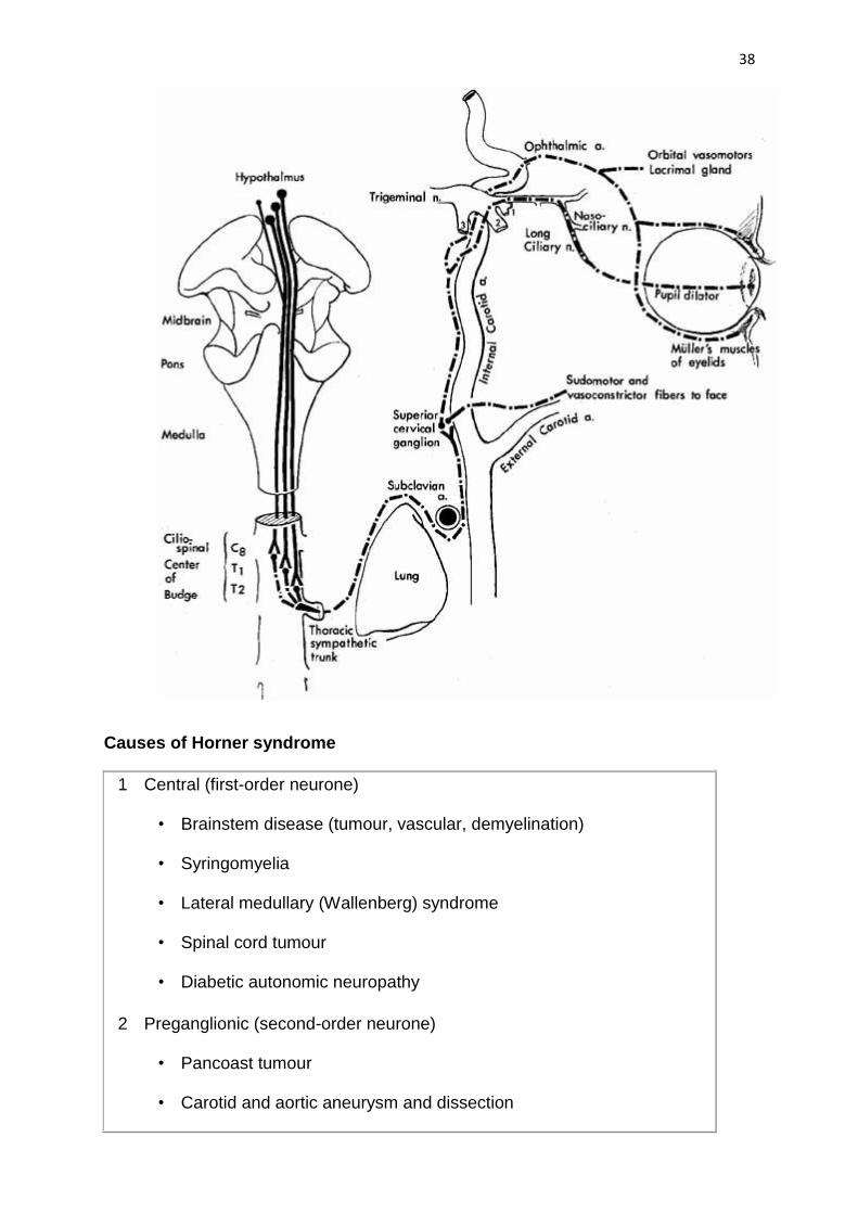
Anatomy
The sympathetic supply involves three neurones
1 First (central) starts in the posterior hypothalamus and descends, uncrossed,
down the brainstem to terminate in the ciliospinal centre of Budge, in the
intermediolateral horn of the spinal cord, located between C8 and T2.
2 Second (preganglionic) passes from the ciliospinal centre to the superior
cervical ganglion in the neck. During its long course, it is closely related to the
apical pleura where it may be damaged by bronchogenic carcinoma (Pancoast
tumour) or during surgery on the neck.
3 Third (postganglionic) ascends along the internal carotid artery to enter the
cavernous sinus where it joins the ophthalmic division of the trigeminal nerve.
The sympathetic fibres reach the ciliary body and the dilator pupillae muscle via
the nasociliary nerve and the long ciliary nerves.
38
Causes of Horner syndrome
1 Central (first-order neurone)
• Brainstem disease (tumour, vascular, demyelination)
• Syringomyelia
• Lateral medullary (Wallenberg) syndrome
• Spinal cord tumour
• Diabetic autonomic neuropathy
2 Preganglionic (second-order neurone)
• Pancoast tumour
• Carotid and aortic aneurysm and dissection

39
• Neck lesions (glands, trauma, postsurgical)
3 Postganglionic (third-order neurone)
• Cluster headaches (migrainous neuralgia)
• Internal carotid artery dissection
• Nasopharyngeal tumour
• Otitis media
• Cavernous sinus mass
Signs
The vast majority of cases are unilateral. Causes of bilateral involvement include
cervical spine injuries and as part of systemic autonomic diabetic neuropathy.
• Mild ptosis (usually 1–2 mm) as a result of weakness of Müller muscle, and
miosis due to the unopposed action of the sphincter pupillae with resultant
anisocoria.
• Miosis is accentuated in dim light since the Horner pupil will not dilate, unlike its
fellow.
• Normal pupillary reactions to light and near.
• Hypochromic heterochromia (irides of different colour – Horner is lighter) may
be seen if congenital or long-standing.
• Slight elevation of the inferior eyelid as a result of weakness of the inferior
tarsal muscle.
• Reduced ipsilateral sweating, but only if the lesion is below the superior cervical
ganglion, because the sudomotor fibres supplying the skin of the face run along
the external carotid artery.
Pharmacological tests
Cocaine confirms the diagnosis. Hydroxyamphetamine (Paredrine) may be used to
differentiate a preganglionic from a postganglionic lesion. Adrenaline may also be
used to assess denervation supersensitivity.
1 Cocaine 4% is instilled into both eyes.
a Result: the normal pupil will dilate but the Horner pupil will not. A post-
cocaine anisocoria of >0.8 mm in a dimly lit room is significant.

40
b Rationale: noradrenaline (NA) released at the post-ganglionic sympathetic
nerve endings is re-uptaken by the nerve endings, thus terminating its
action. Cocaine blocks this uptake. NA therefore accumulates and causes
pupillary dilatation. In Horner syndrome, there is no NA being secreted in
the first place – therefore cocaine has no effect. Cocaine thus confirms
the diagnosis of Horner syndrome by continued constriction of the
affected pupil.
2 Hydroxyamphetamine 1% is instilled into both eyes the next day, after the
effects of cocaine have worn off.
a Result:
• In a preganglionic lesion both pupils will dilate.
• In a postganglionic lesion the Horner pupil will not dilate.
b Rationale: hydroxyamphetamine potentiates the release of NA from
postganglionic nerve endings. If this neurone is intact (a lesion of the first
or second order neurone, and also the normal eye) NA will be released
and the pupil will dilate. In a lesion of the third order neurone
(postganglionic) there can be no dilatation since the neurone is destroyed.
3 Adrenaline 0.1% is instilled into both eyes.
a Result:
• In a preganglionic lesion neither pupil will dilate because adrenaline
is rapidly destroyed by monoamine oxidase
• In a postganglionic lesion, the Horner pupil will dilate and ptosis may
be temporarily relieved because adrenaline is not broken down due
to the absence of monoamine oxidase.
b Rationale: a muscle deprived of its motor supply manifests heightened
sensitivity to the excitatory neurotransmitter secreted by its motor nerve.
In Horner syndrome the dilator pupillae muscle similarly manifests
‘denervation hypersensitivity’ to adrenergic neurotransmitters. Therefore
adrenaline, even in minute concentration produces marked dilatation of
the Horner pupil.
4 Apraclonidine 0.5% or 1.0% is instilled into both eyes in order to confirm the
diagnosis, similar to the cocaine test.
a Result: a Horner pupil will dilate but a normal pupil is unaffected.
b Rationale: alpha-1 receptors are upregulated in the denervated dilator
pupillae.

41
Adie pupil
Adie (tonic) pupil is caused by denervation of the postganglionic supply to the
sphincter pupillae and the ciliary muscle, which may follow a viral illness. It typically
affects young adults and presents as a unilateral condition in 80% of cases although
involvement of the second eye typically develops within months or years.
1 Signs
• Large and regular pupil.
• Direct light reflex is absent or sluggish and is associated with vermiform
movements of the pupillary border.
• Consensual light reflex is absent or sluggish.
• The pupil responds slowly to near, following which re-dilatation is also
slow.
• Accommodation may manifest similar tonicity, in that once a near object
has been fixated, the time taken to re-focus in the distance (relax the
ciliary muscle) is prolonged.
• In long-standing cases the pupil may become small (‘little old Adie’).
2 Associations, in some cases, are diminished deep tendon reflexes (Holmes–
Adie syndrome) and wider autonomic nerve dysfunction.
3 Pharmacological testing. If 2.5% methacholine or 0.125% pilocarpine is instilled
into both eyes, the normal pupil will not constrict, but the abnormal pupil will
because of denervation hypersensitivity. Some diabetic patients may also show
this response and wider very occasionally both pupils constrict in normal
individuals.
Other abnormal reactions
1 Right physiological anisocoria
a In dim light right pupil is larger than the left.
b In bright light both pupils constrict normally.
c After instillation of cocaine 4% to both eyes, both pupils dilate.
2 Right pharmacological mydriasis
a Right mydriasis in dim illumination.
b In bright light the right pupil does not constrict.

42
c On accommodation the right pupil does not constrict.
d After instillation of pilocarpine 0.1% into both eyes neither pupil constricts.
e After instillation of pilocarpine 1% into both eyes, the right pupil does not
constrict but the left does.
3 Argyll Robertson pupils are caused by neurosyphilis and are characterized
by:
a In dim light both pupils are small and may be irregular.
b In bright light neither pupil constricts.
c On accommodation both pupils constrict (light-near dissociation).
d After instillation of pilocarpine 0.1% into both eyes, neither pupil
constricts.
The pupils do not dilate well in the dark but atropine or cocaine induces
mydriasis unless extensive iris atrophy is present.
4 Tectal (dorsal midbrain) pupils
a In dim light there is bilateral mydriasis which may be asymmetrical.
b In bright light neither pupil constricts.
c On accommodation both pupils constrict normally.
d After instillation of pilocarpine 0.1% to both eyes, neither pupil constricts.
5 Right episodic mydriasis
a In dim light the right pupil is larger than the left.
b In bright light the right pupil does not constrict.
c On accommodation the right pupil does not constrict.
d Instillation of pilocarpine 0.1% to both eyes fails to constrict either pupil.
e Instillation of pilocarpine 1% to both eyes induces bilateral miosis.
f After 24 hours both pupils are equal.
Light-near dissociation
In this condition the light reflex is absent or sluggish but the near response is
normal. The causes are shown in the table below

43
Causes of light-near dissociation
1 Unilateral
• Afferent conduction defect
• Adie pupil
• Herpes zoster ophthalmicus
• Aberrant regeneration of the 3rd nerve
2 Bilateral
• Neurosyphilis
• Type 1 diabetes
• Myotonic dystrophy
• Parinaud (dorsal midbrain) syndrome
• Familial amyloidosis
• Encephalitis
• Chronic alcoholism
Migraine
Clinical features
Migraine is often a familial disorder, more prevalent in females, characterized by
recurrent attacks of headache widely variable in intensity, duration and frequency.
The headache is commonly unilateral, associated with nausea and vomiting and may
be preceded by, or associated with, neurological and mood disturbances. However,
all these characteristics are not necessarily present during each attack or in every
patient. The main types of migraine are discussed below.
Common migraine
Common migraine (migraine without aura) is characterized by headache with
autonomic nervous system dysfunction (e.g. pallor and nausea), but without
stereotypical neurological or ophthalmic features as in classical migraine (see later).
• Premonitory features include changes in mood, frequent yawning or other non-
specific prodromal symptoms such as poor concentration.

44
• The headache starts anywhere on the cranium and is pounding or throbbing. It
usually spreads to involve one half or even the whole head. If retro-orbital, the
pain may be mistaken for ocular or sinus disease.
• During the attack, which lasts from hours to a day or more, the patient is
frequently photophobic and phonophobic and seeks relief in a quiet dark
environment or through sleep.
• Because of the absence of the well-known migrainous visual distortions, severe
nausea and vomiting, common migraine often goes unrecognized.
Classical migraine
Classical migraine (migraine with aura) is less common but better recognized.
• The attack is heralded by a visual aura which lasts about 20 minutes. This may
consist of bright or dark spots, zig-zags (‘fortification spectra’), ‘heat haze’
distortions, jigsaw puzzle effects, scintillating scotomas, tunnel vision, which
may progress to homonymous hemianopia.
• A small bright positive paracentral scotoma develops, lined on one side with
luminous zig-zag lines
• After several minutes the fortification spectrum gradually enlarges with the open
end pointing centrally
• It is often lined on the inner edge by an absent area of vision (negative
scotoma)
• As the scotoma expands, it may drift towards the temporal periphery before
breaking up
• Full visual recovery within 30 minutes is the rule and symptoms persisting
longer than an hour should lead to consideration of an alternative diagnosis.
• These visual features, more or less pathognomonic of migraine, may rarely be
caused by degenerative arterial disease or arteriovenous malformation in the
occipital poles.

45
• The headache follows the aura by about 30 minutes and is usually hemicranial,
opposite the hemianopia and is accompanied by nausea and photophobia. It
may, however, be absent, trivial or very severe, with considerable variation
between attacks even in the same individual.
• Visual aura without headache (migraine sine migraine) is not uncommon in the
over 40s but there should virtually always be a history of common or classical
migraine in the patient's early 20s.
• A visual field defect may occasionally be permanent but migraine should be a
diagnosis of exclusion in these circumstances.
Cluster headache
Cluster headache (migrainous neuralgia) is a migraine variant which typically affects
men during the 4th–5th decades. It is of particular interest to ophthalmologists
because it is associated with ocular features and may initially be misdiagnosed as a
local ocular problem. The condition is characterized by a stereotyped headache
accompanied by various autonomic phenomena occurring almost every day for a
period of some weeks
• The headache is unilateral, oculotemporal, excruciating, sharp and deep.
• It begins relatively abruptly, lasts between 10 minutes and 2 hours, and then
clears quickly.
• The patient cannot keep still and is very agitated, in contrast to a patient with
migraine who would rather lie quietly in a dark room.
• It may occur several times in a 24-hour period often at particular times, not
infrequently at around 2 a.m.
• Once the ‘cluster’ is over, there may be a long headache-free interval of several
years.
• Associated autonomic phenomena include lacrimation, conjunctival injection
and rhinorrhoea.
• Cluster headaches are also a common cause of a transient or permanent

46
postganglionic Horner syndrome.
Other types of migraine
1 Focal migraine is characterized by transient dysphasia, hemisensory
symptoms or even focal weakness in addition to other symptoms of migraine.
2 Retinal migraine is characterized by acute, transient unilateral visual loss.
Since this may occur in middle-aged patients without a past history of migraine,
it is prudent to investigate such individuals as undergoing attacks of retinal
embolisation until proved otherwise.
3 Ophthalmoplegic migraine is rare and typically starts before the age of 10
years. It is characterized by a recurrent transient 3rd nerve palsy which begins
after the headache.
4 Familial hemiplegic migraine is characterized by a failure of full recovery of
focal neurological features after an attack of migraine subsides.
5 Basilar migraine occurs in children. It is characterized by a typical migrainous
aura associated with numbness and tingling of the lips and extremities which is
often bilateral. Ataxia of gait and speech also occur, with occasional impairment
of consciousness.
Treatment
1 General measures include the elimination of conditions and agents that may
precipitate an attack of migraine, such as coffee, chocolate, alcohol, cheese,
oral contraceptives, stress, lack of sleep and long intervals without food.
2 Prophylaxis is indicated if the frequency and/or severity of the attacks are
beyond the patient's tolerance. This may involve beta-adrenergic blockers,
calcium channel blockers, amitriptyline, clonidine, pizotifen and low-dose
aspirin.
3 Treatment of an acute attack may be with simple analgesics (aspirin, codeine
analogues, paracetamol or NSAIDs) and, if appropriate, an anti-emetic such as
metoclopramide. Other drugs, usually reserved for patients who are refractory
to analgesics, include sumatriptan and ergotamine tartrate.

47
Differential diagnosis
Visual phenomena
The visual phenomena of migraine are typically binocular, zig-zag, scintillating and
migrate within the visual field. This is often associated with a scotoma and/or
homonymous visual loss. A patient may often report loss of vision only in the eye
ipsilateral to the hemianopic symptoms. The following conditions should be
considered in the differential diagnosis:
1 Acute posterior vitreous detachment is characterized by photopsia,
usually associated with the sudden onset of floaters. The flashing lights are
usually projected into the temporal visual field and may be precipitated by
movements of the head or eyes.
2 Transient ischaemic attacks due to retinal microembolisation are
unilateral and not scintillating. The patient often describes a ‘shade’ or
‘cloud’ which typically starts in the upper or lower parts of the visual field
and spreads centrally. It lasts several minutes and clears from the centre to
the periphery.
3 Transient visual obscurations last only a few seconds and are
characterized by a ‘greying out’ or ‘darkening’ of vision in one or both eyes.
They classically occur in patients with papilloedema and are often
precipitated by changes in posture. They may also precede anterior
ischaemic optic neuropathy in patients with giant cell arteritis.
4 Occipital epilepsy is very rare; the patient typically sees coloured circles
during an attack.
Neuralgias
The following conditions should be considered in the differential diagnosis of ocular
or periocular pain in the absence of apparent physical disease:
1 Herpes zoster ophthalmicus frequently presents with pain 2–3 days before
the onset of the characteristic vesicular rash.
2 Trigeminal neuralgia is characterized by brief attacks of severe pain that start
in the distribution of one of the divisions of the trigeminal nerve. The pain is
paroxysmal and sharp, like an electric shock, usually occurring in multiple
bursts lasting a few seconds, in rapid succession. Attacks can be triggered
either by cutaneous stimulation such as touching the face whilst shaving or by

48
motor activity such as chewing; sleep is usually undisturbed by pain. Facial
sensation is normal. Treatment involves antiepileptic drugs such as
carbamazepine, phenytoin and sodium valproate. Trigeminal neuralgia of
compressive aetiology may necessitate intracranial surgical decompression of
the trigeminal nerve.
3 Raeder paratrigeminal syndrome typically affects middle-aged men. It is
characterized by severe unilateral headache with periocular pain in the
distribution of the first division of the trigeminal nerve associated with an
ipsilateral Horner syndrome. The pain may last from hours to weeks before it
resolves spontaneously. Carotid dissection needs to be excluded before
making the diagnosis.
4 Greater occipital neuralgia is characterized by attacks of pain that begin in
the occipital region and then spread to the eye, temple and face. The attacks
frequently occur at night and are associated with flushing of the face, dizziness
and sometimes ipsilateral nasal obstruction. Examination during an attack may
reveal extreme tenderness between the mastoid process and occipital
protuberance.
5 Ophthalmodynia periodica is characterized by short, sharp stabbing ocular
pain which often causes the patient to place the hand over the involved eye. A
second series of episodes may immediately follow the initial attack.
6 Ice-pick syndrome is characterized by attacks of momentary, multifocal,
sharp, pain around the skull, face and eyes. Unlike trigeminal neuralgia there
are no specific trigger points; the pain also does not conform to the anatomical
distribution of the trigeminal nerve.

49
Neuro-ophthalmology 3
Ocular Motility Defects: Recognition of the most commonly encountered motility
disturbances - 3rd, 4th & 6th Nerve palsies
Ocular motor nerves
3rd nerve
Nuclear complex
The nuclear complex of the 3rd (oculomotor) nerve is situated in the midbrain at the
level of the superior colliculus, ventral to the Sylvian aqueduct. It is composed of
the following paired and unpaired subnuclei.
1 Levator subnucleus is an unpaired caudal midline structure which innervates
both levator muscles. Lesions confined to this area will therefore give rise to
bilateral ptosis.
2 Superior rectus subnuclei are paired: each innervates the respective
contralateral superior rectus. A nuclear 3rd nerve palsy will spare the ipsilateral,
and affect the contralateral, superior rectus.
3 Medial rectus, inferior rectus and inferior oblique subnuclei are paired and
innervate their corresponding ipsilateral muscles. Lesions confined to the
nuclear complex are relatively uncommon. The most frequent causes are
vascular disease, primary tumours and metastases. Involvement of the paired
medial rectus subnuclei cause a wall-eyed bilateral internuclear
ophthalmoplegia (WEBINO), characterized by exotropia, defective convergence
and adduction. Lesions involving the entire nucleus are often associated with
involvement of the adjacent and caudal 4th nerve nucleus.
Fasciculus
The fasciculus consists of efferent fibres which pass from the 3rd nerve nucleus
through the red nucleus and the medial aspect of the cerebral peduncle. They then
emerge from the midbrain and pass into the interpeduncular space. The causes of
nuclear and fascicular lesions are similar, except that demyelination may affect the
fasciculus.
1 Benedikt syndrome involves the fasciculus as it passes through the red nucleus
and is characterized by ipsilateral 3rd nerve palsy and contralateral
extrapyramidal signs such as hemitremor.
2 Weber syndrome involves the fasciculus as it passes through the cerebral
peduncle and is characterized by ipsilateral 3rd nerve palsy and a contralateral

50
hemiparesis.
3 Nothnagel syndrome involves the fasciculus and the superior cerebellar
peduncle and is characterized by ipsilateral 3rd nerve palsy and cerebellar
ataxia.
4 Claude syndrome is a combination of Benedikt and Nothnagel syndromes.
Basilar
The basilar part starts as a series of ‘rootlets’ which leave the midbrain on the
medial aspect of the cerebral peduncle, before coalescing to form the main trunk.
The nerve then passes between the posterior cerebral and superior cerebellar
arteries, running lateral to and parallel with the posterior communicating artery. As
the nerve traverses the base of the skull along its subarachnoid course
unaccompanied by any other cranial nerve, isolated 3rd nerve palsies are
commonly basilar. The following two are important causes:
1 Aneurysm of the posterior communicating artery, at its junction with the internal
carotid artery, typically presents as an acute, painful 3rd nerve palsy with
involvement of the pupil.
2 Head trauma, resulting in extradural or subdural haematoma, may cause a
tentorial pressure cone with downward herniation of the temporal lobe. This
compresses the 3rd nerve as it passes over the tentorial edge, initially causing
irritative miosis followed by mydriasis and total 3rd nerve palsy.
Intracavernous
The 3rd nerve then enters the cavernous sinus by piercing the dura just lateral to
the posterior clinoid process. Within the cavernous sinus, the 3rd nerve runs in the
lateral wall above the 4th nerve. In the anterior part of the cavernous sinus, the
nerve divides into superior and inferior branches which enter the orbit through the
superior orbital fissure within the annulus of Zinn. The following are important
causes of intracavernous 3rd nerve palsies:
1 Diabetes may cause a vascular palsy, which usually spares the pupil.
2 Pituitary apoplexy (haemorrhagic infarction) may cause a 3rd nerve palsy (e.g.
after childbirth) if the gland swells laterally and impinges on the cavernous
sinus.
3 Intracavernous pathology such as aneurysm, meningioma, carotid-cavernous
fistula and granulomatous inflammation (Tolosa–Hunt syndrome) may all cause
3rd nerve palsy. Because of its close proximity to other cranial nerves,
intracavernous 3rd nerve palsies are usually associated with involvement of the
4th and 6th nerves and the first division of the trigeminal nerve.

51
Intraorbital
1 Superior division innervates the levator and superior rectus muscles.
2 Inferior division innervates the medial rectus, the inferior rectus and the inferior
oblique muscles. The branch to the inferior oblique also contains pre-ganglionic
parasympathetic fibres from the Edinger–Westphal subnucleus, which innervate
the sphincter pupillae and the ciliary muscle. Lesions of the inferior division are
characterized by limited adduction and depression, and a dilated pupil. Both
superior and inferior division palsies are commonly traumatic or vascular.
Pupillomotor fibres
Between the brainstem and the cavernous sinus, the pupillomotor parasympathetic
fibres are located superficially in the superomedial part of the 3rd nerve. They
derive their blood supply from the pial blood vessels, whereas the main trunk of the
3rd nerve is supplied by the vasa nervorum. Involvement or otherwise of the pupil is
of great importance because it frequently differentiates a ‘surgical’ from a ‘medical’
lesion. Pupillary involvement, like other features of 3rd nerve palsy, may be
complete or partial, and may demonstrate features of recovery. Mild mydriasis and
non-reactivity may therefore be clinically significant.
1 'Surgical’ lesions such as aneurysms, trauma and uncal herniation
characteristically involve the pupil by compressing the pial blood vessels and
the superficially located pupillary fibres.
2 ‘Medical' lesions such as hypertension and diabetes usually spare the pupil.
This is because the microangiopathy associated with medical lesions involves
the vasa nervorum, causing ischaemia of the main trunk of the nerve, sparing
the superficial pupillary fibres.
These principles are, however, not infallible because pupillary involvement may be
seen in some diabetic-associated 3rd nerve palsies, while pupillary sparing does not
invariably exclude aneurysm or some other compressive lesion. Pupillary
involvement may develop a few days after onset of diplopia as an aneurysm
expands. Sometimes pupillary involvement may be the only sign of 3rd nerve palsy
(basal meningitis, uncal herniation).
Signs of 3rd nerve palsy
Right 3rd nerve palsy is characterized by the following:
a Weakness of the levator causing profound ptosis, due to which there is often no
diplopia.
b Unopposed action of the lateral rectus causing the eye to be abducted in the
primary position. The intact superior oblique muscle causes intorsion of the eye
at rest which increases on attempted downgaze.

52
c Normal abduction because the lateral rectus is intact.
d Weakness of the medial rectus limiting adduction.
e Weakness of superior rectus and inferior oblique, limiting elevation.
f Weakness of inferior rectus limiting depression.
g Parasympathetic palsy causing a dilated pupil associated with defective
accommodation.
h Partial involvement will produce milder degrees of ophthalmoplegia.
Aberrant regeneration
Aberrant regeneration may follow acute traumatic and compressive, but not
vascular, 3rd nerve palsies. This is because the endoneural nerve sheaths, which
may be breached in traumatic and compressive lesions, remain intact in vascular
pathology. Bizarre defects in ocular motility such as elevation of the upper eyelid on
attempted adduction or depression (the pseudo-Graefe phenomenon), are caused
by misdirection of regenerating axons which reinnervate the wrong extraocular
muscle. The pupil may also be involved.
Causes of isolated 3rd nerve palsy
1 Idiopathic: about 25% have no identifiable cause.
2 Vascular disease, such as that associated with hypertension and diabetes, is
the most common cause of pupil-sparing 3rd nerve palsy. In most cases
spontaneous recovery occurs within 3 months. Diabetic 3rd nerve palsy is often
associated with periorbital pain and may occasionally be the presenting feature
of diabetes. The presence of pain is therefore not helpful in differentiating
aneurysmal and diabetic 3rd nerve palsy.
3 Aneurysm of the posterior communicating artery at its junction with the internal
carotid is a very important cause of isolated painful 3rd nerve palsy with
involvement of the pupil.
4 Trauma, both direct and secondary to subdural haematoma with uncal
herniation, is also a common cause. However, the development of 3rd nerve
palsy following relatively trivial head trauma, not associated with loss of
consciousness, should alert the clinician to the possibility of an associated
basal intracranial tumour which has caused the nerve trunk to be stretched and
tethered.
5 Miscellaneous uncommon causes include tumours, syphilis, giant cell arteritis
and other types of vasculitis associated with collagen vascular disorders. Brief
episodes of 3rd nerve dysfunction with spontaneous recovery may be idiopathic
or occur with migraine, compression, ischaemia and alterations in intracranial
pressure. Myasthenia may also mimic intermittent pupil-sparing 3rd nerve

53
palsy.
Treatment
1 Non-surgical treatment options include the use of Fresnel prisms if the angle of
deviation is small, uniocular occlusion to avoid diplopia (if ptosis is partial or
recovering) and botulinum toxin injection into the uninvolved lateral rectus
muscle to prevent its contracture before the deviation improves or stabilizes.
2 Surgical treatment, as with other ocular motor nerve palsies, should be
contemplated only after all spontaneous improvement has ceased. This is
usually not earlier than 6 months from the date of onset.
4th nerve
Anatomy
1 Important features of the 4th (trochlear) nerve are as follows:
• It is the only cranial nerve to emerge from the dorsal aspect of the brain.
• It is a crossed cranial nerve; this means that the 4th nerve nucleus
innervates the contralateral superior oblique muscle.
• It is a very long and slender nerve.
2 The nucleus is located at the level of the inferior colliculi ventral to the Sylvian
aqueduct. It is caudal to, and continuous with, the 3rd nerve nuclear complex.
3 The fasciculus consists of axons which curve posteriorly around the aqueduct
and decussate completely in the anterior medullary velum.
4 The trunk leaves the brainstem on the dorsal surface, just caudal to the inferior
colliculus. It then curves laterally around the brainstem, runs forwards beneath
the free edge of the tentorium, and (like the 3rd nerve) passes between the
posterior cerebral artery and the superior cerebellar artery. It then pierces the
dura and enters the cavernous sinus.
5 The intracavernous part runs in the lateral wall of the sinus, inferiorly to the 3rd
nerve and above the first division of the 5th. In the anterior part of the
cavernous sinus it rises and passes through the superior orbital fissure above
and lateral to the annulus of Zinn.
6 The intraorbital part innervates the superior oblique muscle.

54
Signs
Acute onset of vertical diplopia in the absence of ptosis, combined with a
characteristic head posture, strongly suggests 4th nerve disease. The features of
nuclear, fascicular and peripheral 4th nerve palsies are clinically identical, except
that nuclear palsies produce contralateral superior oblique weakness.
Left 4th nerve palsy is characterized by the following:
a Left hypertropia (‘left-over-right’) in the primary position.
b Increase in left hypertropia on right gaze due to left inferior oblique
overaction.
c Limitation of left depression on adduction.
d Normal left abduction.
e Normal left depression.
f Normal left elevation.
Abnormal head posture avoids diplopia which is vertical, torsional and worse on
looking down.
• To intort the eye (alleviate excyclotorsion) there is contralateral head tilt to the
right.
• To alleviate the inability to depress the eye is adduction, the face is turned to
the right and the chin is slightly depressed.
• The left eye cannot look down and to the right or intort – the head therefore
does this and thus compensates.
Compensatory head posture in left 4th nerve palsy; head tilt to right, face turn to the
right and chin depressed
Bilateral involvement
Bilateral involvement should always be suspected until proved otherwise. It is
characterized by the following:
• Right hypertropia in left gaze, left hypertropia in right gaze.
• Greater than 10° of cyclodeviation on double Maddox rod test (see below).
• ‘V’ pattern esotropia.
• Bilaterally positive Bielschowsky test (see below).
Special tests

55
1 Parks three step test is very useful in the diagnosis of 4th nerve palsy and is
performed as follows:
a First step
• Assess which eye is hypertropic in the primary position.
• Left hypertropia may be caused by weakness of one of the following
four muscles: one of the depressors of the left eye (superior oblique
or inferior rectus) or one of the elevators of the right eye (superior
rectus or inferior oblique).
• In a 4th nerve palsy, the involved eye is higher.
b Step two
• Determine whether the left hypertropia is greater in right gaze or left
gaze. Increase on right gaze implicates either the right superior
rectus or left superior oblique.
• Increase on left gaze implicates either the right inferior oblique or left
inferior rectus. (In 4th nerve palsy the deviation is Worse On
Opposite Gaze – WOOG.)
c Step three
• The Bielschowsky head tilt test is performed with the patient fixating
a straight ahead target at 3 metres.
• The head is tilted to the right and then to the left.
• Increase of left hypertropia on left head tilt implicates the left
superior oblique and increase of right hypertropia on left head tilt
implicates the right inferior rectus. (In 4th nerve palsy the deviation is
Better On Opposite Tilt – BOOT.)
2 Double Maddox rod test
• Red and green Maddox rods, with the cylinders vertical, are placed one in
front of either eye.
• Each eye will therefore perceive a more or less horizontal line of light.
• In the presence of cyclodeviation, the line perceived by the paretic eye will
be tilted and therefore distinct from that of the other eye.
• One Maddox rod is then rotated till fusion (superimposition) of the lines is
achieved.

56
• The amount of rotation can be measured in degrees and indicates the
extent of cyclodeviation.
• Unilateral 4th nerve palsy is characterized by less than 10° of
cyclodeviation whilst bilateral cases may have greater than 20° of
cyclodeviation. This can also be measured with a synoptophore.
• Positive Bielschowsky test in left 4th nerve palsy. (A) No hypertropia on
right head tilt; (B) marked hypertropia on left head tilt
Causes of isolated 4th nerve palsy
1 Congenital lesions are common, although symptoms may not develop until
decompensation occurs in adult life. Unlike acquired lesions patients are not
usually aware of the torsional aspect. Examination of old photographs for the
presence of an abnormal head posture may be helpful, as is the presence of an
increased vertical prism fusional range.
2 Trauma frequently causes bilateral 4th nerve palsy. The long and slender
nerves are vulnerable as they decussate in the anterior medullary velum,
through impact with the tentorial edge. Bilateral lesions are often thought to be
unilateral until squint surgery is performed following which the contralateral 4th
nerve palsy is often revealed.
3 Vascular lesions are common but aneurysms and tumours are extremely rare.
Routine neuroimaging for isolated trochlear palsy is not required.
6th nerve
Nucleus
The nucleus of the 6th (abducens) nerve lies at the mid-level of the pons, ventral to
the floor of the 4th ventricle, where it is closely related to the horizontal gaze centre.
The fasciculus of the 7th nerve curves around the abducent nucleus and produces
an elevation in the floor of the 4th ventricle. Isolated 6th nerve palsy is therefore
never nuclear in origin. A lesion in and around the 6th nerve nucleus causes the
following signs:
• Ipsilateral weakness of abduction as a result of involvement of the 6th nerve.
• Failure of horizontal gaze towards the side of the lesion resulting from
involvement of the horizontal gaze centre in the PPRF.
• Ipsilateral lower motor neurone facial nerve palsy caused by concomitant
involvement of the facial fasciculus is also common.

57
Fasciculus
The fasciculus passes ventrally to leave the brainstem at the pontomedullary
junction, just lateral to the pyramidal prominence.
1 Foville syndrome involves the fasciculus as it passes through the PPRF and is
most frequently caused by vascular disease or tumours involving the dorsal
pons. It is characterized by ipsilateral involvement of the 5th to 8th cranial
nerves and central sympathetic fibres.
• 5th nerve – facial anaesthesia.
• 6th nerve palsy combined with gaze palsy (PPRF).
• 7th nerve (nuclear or fascicular damage) – facial weakness.
• 8th nerve – deafness.
• Central Horner syndrome.
2 Millard–Gubler syndrome involves the fasciculus as it passes through the
pyramidal tract and is most frequently caused by vascular disease, tumours or
demyelination. It is characterized by the following:
• Ipsilateral 6th nerve palsy.
• Contralateral hemiplegia (since the pyramidal tracts decussate further
inferiorly, in the medulla, to control contralateral voluntary movement).
• Variable number of signs of a dorsal pontine lesion.
Basilar
The basilar part enters the prepontine basilar cistern. It then passes upwards close
to the base of the skull and is crossed by the anterior inferior cerebellar artery. It
pierces the dura below the posterior clinoids and angles forwards over the tip of the
petrous bone, passing through or around the inferior petrosal sinus, through the
Dorello canal (under the petroclinoid ligament), to enter the cavernous sinus. The
following are important causes of damage to the basilar part of the nerve:
1 Acoustic neuroma may damage the 6th nerve at the pontomedullary junction. It
should be emphasized that the first symptom of an acoustic neuroma is hearing
loss and the first sign diminished corneal sensitivity. It is therefore very
important to test hearing and corneal sensation in all patients with 6th nerve
palsy.
2 Nasopharyngeal tumours may invade the skull and its foramina and damage
the nerve during its basilar course.
3 Raised intracranial pressure may cause a downward displacement of the

58
brainstem. This may stretch the 6th nerve over the petrous tip between its point
of emergence from the brainstem and its point of entry into the cavernous
sinus. In this situation 6th nerve palsy, which may be bilateral, is a false
localizing sign.
4 Basal skull fracture may cause both unilateral and bilateral palsies.
5 Gradenigo syndrome, most frequently caused by mastoiditis or acute petrositis,
may result in damage of the 6th nerve at the petrous tip. The latter is frequently
accompanied by facial weakness and pain, and hearing difficulties.
Intracavernous and intraorbital
1 The intracavernous part runs forwards below the 3rd and 4th nerves, as well as
the 1st division of the 5th. Although the other nerves are protected within the
wall of the sinus, the 6th is most medially situated and runs through the middle
of the sinus in close relation to the internal carotid artery. It is therefore more
prone to damage than the other nerves. Occasionally, an intracavernous 6th
nerve palsy is accompanied by a postganglionic Horner syndrome (Parkinson
sign) because in its intracavernous course the 6th nerve is joined by
sympathetic branches from the paracarotid plexus. The causes of
intracavernous 6th nerve and 3rd nerve lesions are similar.
2 The intraorbital part enters the orbit through the superior orbital fissure within
the annulus of Zinn to innervate the lateral rectus muscle.
Signs
1 Acute left 6th nerve palsy.
a Left esotropia in the primary position.
b Marked limitation of left abduction.
2 Long-standing left 6th nerve palsy.
a Left esotropia in the primary position due to unopposed action of the left
medial rectus. The deviation is characteristically worse for a distant target
and less or absent for near fixation.
b Marked limitation of left abduction due to weakness of the left lateral
rectus.
c Normal left adduction.
Compensatory face turn is into the field of action of the paralyzed muscle (i.e. to the
left) to minimize diplopia, so that the eyes do not need to look towards the field of
action of the paralyzed muscle (i.e. to the left).

59
Differential diagnosis
The following conditions may mimic 6th nerve palsy.
1 Myasthenia gravis can mimic virtually any ocular motility defect. Distinguishing
features include variability of diplopia and other signs such as lid fatigue and
the Cogan twitch sign (see later).
2 Restrictive thyroid myopathy involving the medial rectus may give rise to
limitation of abduction. Associated features include orbital and eyelid signs and
a positive forced duction test.
3 Medial orbital wall blowout fracture with entrapment of the medial rectus, giving
rise to limitation of abduction.
4 Orbital myositis involving the lateral rectus is characterized by weakness of
abduction and pain when this is attempted.
5 Duane syndrome is a congenital condition characterized by defective abduction
and narrowing of the palpebral fissure on adduction.
6 Convergence spasm typically affects young adults and is characterized by
convergence with miosis and increased accommodation.
7 Divergence paralysis is a rare condition which may be difficult to distinguish
from unilateral or bilateral 6th nerve palsy. However, unlike 6th nerve palsy the
esotropia may remain the same or diminish on lateral gaze.
8 Early-onset esotropia.

60
Nystagmus
Physiological principles
• Nystagmus is a repetitive, involuntary, to-and-fro oscillation of the eyes, which
may be physiological or pathological. Thus, nystagmus that occurs in response
to rotation of an optokinetic drum or of the body in space is normal and acts to
preserve clear vision.
• Ocular movements that bring about fixation on an object of interest are called
foveating and those that move the fovea away from the object are defoveating.
• In pathological nystagmus, each cycle of movement is usually initiated by an
involuntary, defoveating drift of the eye away from the object of interest,
followed by a returning refixation saccadic movement.
• The plane of nystagmus may be horizontal, vertical, torsional or non-specific.
The amplitude of nystagmus refers to how far the eyes move, while the
frequency refers to how rapidly the eyes oscillate. On the basis of amplitude,
nystagmus may be fine or coarse, while the frequency may be high, moderate
or low.
Classification
1 Jerk nystagmus is saccadic with a slow defoveating ‘drift’ movement and a fast
corrective refoveating saccadic movement. The direction of nystagmus is
described in terms of the direction of the fast component, so that jerk
nystagmus may be right, left, up, down or rotatory. Jerk nystagmus can be
divided into gaze-evoked and gaze-paretic; the latter is slow and usually
indicates brainstem damage.
2 Pendular nystagmus is non-saccadic in that both the foveating and defoveating
movements are slow (i.e. the velocity of nystagmus is equal in both directions).
• Congenital pendular nystagmus is horizontal, conjugate and may convert
to jerk on lateral gaze.
• Acquired pendular nystagmus has horizontal, vertical and torsional
components.
• If the horizontal and vertical components of pendular nystagmus are in

61
phase (i.e. occur simultaneously), the perceived direction becomes
oblique.
• If the horizontal and vertical components are out of phase, the direction
becomes elliptical or rotary.
3 Mixed nystagmus involves pendular nystagmus in the primary position and jerk
nystagmus on lateral gaze.
Physiological nystagmus
1 End-point nystagmus is a fine jerk nystagmus of moderate frequency found
when the eyes are in extreme positions of gaze. The fast phase is in the
direction of gaze.
2 OKN is a jerk nystagmus induced by moving repetitive targets across the visual
field.
• The slow phase is a pursuit movement in which the eyes follow the target;
the fast phase is a saccadic movement in the opposite direction as the
eyes fixate on the next target.
• If the OKN tape or drum is moved from right to left, the left parieto-
occipito-temporal region controls the slow (pursuit) phase to the left, and
the left frontal lobe controls the rapid (saccadic) phase to the right.
• OKN nystagmus is useful for detecting malingerers who feign blindness
and for testing visual acuity in the very young. It may also be helpful in
determining the cause of an isolated homonymous hemianopia (see
below).
Vestibular nystagmus
1 Physiological vestibular nystagmus is a jerk nystagmus caused by altered
input from the vestibular nuclei to the horizontal gaze centres. The slow phase
is initiated by the vestibular nuclei and the fast phase by the brainstem and
frontomesencephalic pathway. Vestibular nystagmus may be elicited by caloric
stimulation as follows:
• When cold water is poured into the right ear the patient will develop left

62
jerk nystagmus (i.e. fast phase to the left).
• When warm water is poured into the right ear the patient will develop right
jerk nystagmus (i.e. fast phase to the right). A useful mnemonic is ‘COWS’
(cold-opposite, warm-same) indicating the direction of the nystagmus.
• When cold water is poured into both ears simultaneously, a jerk
nystagmus with the fast phase upwards develops; warm water in both
ears elicits nystagmus with the fast phase downwards (cold ‘slows things
down’).
2 Pathological peripheral vestibular nystagmus is caused by disease affecting
the ear such as labrynthitis, Ménière's disease and middle or inner ear
infections.
Motor imbalance nystagmus
Motor imbalance nystagmus is the result of primary defects in the efferent mechanisms.
Primary congenital nystagmus
1 Inheritance is XLR or AD, and rarely AR.
2 Presentation is about 2–3 months after birth and persists throughout life.
3 Signs
• In the primary position there is low-amplitude pendular nystagmus that
converts to jerk nystagmus on side gaze.
• In upgaze and downgaze the nystagmus remains in the horizontal plane.
• The nystagmus may be dampened by convergence and is not present
during sleep.
• There is usually a null point – a position of gaze in which nystagmus is
minimal.

63
• In order to move the eyes into the null point, an abnormal head posture
may be adopted.
Adults with congenital forms of nystagmus do not notice oscillopsia but it is noticed
by adults with acquired nystagmus.
Spasmus nutans
1 Presentation of this rare condition is between 3 and 18 months.
2 Signs
• Unilateral or bilateral small-amplitude high-frequency horizontal
nystagmus associated with head nodding.
• It is frequently asymmetrical with increased amplitude in abduction.
• Vertical and torsional components may be present.
3 Causes
• Idiopathic which spontaneously resolves by age 3 years.
• Glioma of anterior visual pathway, empty sella syndrome and
porencephalic cyst.
Latent nystagmus
Latent nystagmus is associated with infantile esotropia and dissociated
vertical deviation. It is characterized by the following:
• With both eyes open there is no nystagmus.
• Horizontal nystagmus becomes apparent on covering one eye or reducing the
amount of light reaching the eye.
• Fast phase is in the direction of the uncovered fixating eye.

64
• Occasionally, an element of latency may be superimposed on a manifest
nystagmus so that when one eye is covered the amplitude of nystagmus
increases (manifest-latent nystagmus).
Periodic alternating nystagmus
1 Signs
• Conjugate horizontal jerk nystagmus that periodically reverses its
direction.
• Each cycle may be divided into active and quiescent phases as follows.
• During the active phase, the amplitude, frequency and slow-phase
velocity of nystagmus first progressively increase then decrease.
• This is followed by a short, quiet interlude, lasting 4–20 sec, during which
time the eyes are steady and show low-intensity, often pendular
movements.
• A similar sequence in the opposite direction occurs thereafter, the whole
cycle lasting between 1 and 3 minutes.
2 Causes include isolated congenital, cerebellar disease, ataxia telangiectasia
(Louis–Bar syndrome) and drugs such as phenytoin.
Convergence-retraction nystagmus
Convergence-retraction nystagmus is the result of co-contraction of the extraocular
muscles, particularly the medial recti.
1 Signs
• Jerk nystagmus is induced by passing an OKN tape or rotating a drum
downwards.
• The upward refixation saccade brings the two eyes towards each other in
a convergence movement.

65
• Associated retraction of the globe into the orbit.
2 Causes include lesions of the pretectal area such as pinealoma and vascular
accidents (Parinaud dorsal midbrain syndrome).
Downbeat nystagmus
1 Signs. Vertical nystagmus with the fast phase beating downwards, which is
more easily elicited in lateral gaze and downgaze.
2 Causes
• Lesions of the craniocervical junction at the foramen magnum such as
Arnold–Chiari malformation and syringobulbia.
• Drugs such as lithium, phenytoin, carbamazepine and barbiturates.
• Wernicke encephalopathy, demyelination and hydrocephalus.
Upbeat nystagmus
1 Signs. Vertical nystagmus with the fast phase beating upwards.
2 Causes include posterior fossa lesions, drugs and Wernicke encephalopathy.
See-saw nystagmus
1 Signs. Pendular nystagmus, in which one eye elevates and intorts while the
other depresses and extorts; the eyes then reverse direction.
2 Causes include parasellar tumours often associated with bitemporal
hemianopia, syringobulbia and brainstem stroke.
Ataxic nystagmus
Ataxic nystagmus is a horizontal jerk nystagmus which occurs in the
abducting eye of a patient with an INO (see above).

66
Bruns nystagmus
1 Signs. Coarse cerebellar horizontal jerk nystagmus in one eye and fine high
frequency vestibular nystagmus in the other.
2 Causes. Cerebellopontine angle tumours such as acoustic neuroma. The
lesion is ipsilateral to the side with coarse cerebellar nystagmus.
Sensory deprivation nystagmus
Sensory deprivation (ocular) nystagmus is caused by defective vision. Horizontal and
pendular, it can often be dampened by convergence. The severity depends on the
degree of visual loss. An abnormal head posture may be adopted to decrease the
amplitude of the nystagmus. It is caused by severe impairment of central vision in
early life (e.g. congenital cataract, macular hypoplasia). In general, children who
sustain bilateral loss of central vision before the age of 2 years develop nystagmus.
Surgery for nystagmus
Surgery for nystagmus may be considered if there is an abnormal head posture with
a null position or for congenital motor/sensory nystagmus without a null point.
1 Surgery for nystagmus with a null point is aimed at moving muscles in order
to mimic muscle tension while the eyes and face are straight. For example, in a
patient with a null point in left gaze with a right head turn it is necessary to
weaken (recess) the right medial rectus and strengthen (resect) the right lateral
rectus, and weaken the left lateral rectus and strengthen the left medial rectus.
2 Surgery for congenital motor/sensory nystagmus should be contemplated
only when the wave form and characteristic of nystagmus have been
determined by eye movement studies. Surgery involves large recessions on the
four horizontal recti to reduce the amplitude of nystagmus and increase
recognition time.
Nystagmoid movements
Nystagmoid movements resemble nystagmus but differ in that the initial pathological
defoveating movement is a saccadic intrusion.
Ocular flutter and opsoclonus
1 Signs

67
• Saccadic oscillations with no intersaccadic interval.
• In ocular flutter oscillations are purely horizontal and in opsoclonus they
are multiplanar.
2 Causes include viral encephalitis, myoclonic encephalopathy in infants
('dancing eyes and dancing feet’), transient (idiopathic) in healthy neonates and
drug-induced (lithium, amitriptyline, and phenytoin).
Ocular bobbing
1 Signs. Rapid, conjugate, downward eye movements with a slow drift up to the
primary position.
2 Causes include pontine lesions (usually haemorrhage), cerebellar lesions
compressing the pons and metabolic encephalopathy.
Supranuclear disorders of eye movement
Supranuclear disorders of ocular motility
Conjugate eye movements
Conjugate eye movements or ‘versions’ are binocular movements in which the eyes
move synchronously and symmetrically in the same direction. The three main types
are: (a) saccadic, (b) smooth pursuit and (c) non-optical reflex. Saccadic and pursuit
movements are controlled at both cerebral and brainstem levels. Supranuclear
disturbances produce gaze palsies, characterized by absence of diplopia and normal
vestibulo-ocular reflexes (e.g. oculocephalic movements and caloric stimulation).
Saccadic movements
1 Function of saccadic (fixating) movements is to place the object of interest on
to the fovea rapidly or to move the eyes from one object to another. This can be
done voluntarily or as a reflex, triggered by the presence of an object in the
peripheral visual field. Voluntary saccades are similar to the gunnery system of
rapidly locating a moving target.
2 Pathway for horizontal saccades originates in the premotor cortex (the frontal
eye fields). From there, fibres pass to the contralateral horizontal gaze centre in
the paramedian pontine reticular formation (PPRF). Each frontal lobe therefore
initiates contralateral saccades. Irritative lesions may therefore cause ocular

68
deviation to the opposite side.
Smooth pursuit movements
1 Function of pursuit movements is to maintain fixation on a target once it has
been located by the saccadic system. The stimulus is movement of the image
near the fovea. The movements are slow and smooth.
2 Pathway is extremely complex involving several regions of cortex as well as
the PPRF, the superior colliculi, cerebellum and other structures. The pathways
are ipsilateral, the cortex on one side controlling pursuit to the same side.
Non-optical reflexes
1 Function of non-optical (vestibular) reflexes is to maintain eye position with
respect to any change of head and body position without conscious input.
2 Pathway originates in the labyrinths and proprioceptors in the neck muscles
which mediate information concerning head and neck movements. Afferent
fibres synapse in the vestibular nuclei and pass to the horizontal gaze centre in
the PPRF.
Horizontal gaze palsy
Anatomy
• After initiation horizontal eye movements are generated in a common pathway
from the horizontal gaze centre in the PPRF. From here motor neurones
connect to the ipsilateral 6th nerve nucleus which innervates the lateral rectus.
• From the 6th nerve nucleus internuclear neurones cross the midline at the level
of the pons and pass up the contralateral medial longitudinal fasciculus (MLF)
to synapse with motor neurones in the medial rectus subnucleus in the 3rd
nerve complex which innervates the medial rectus.
• Stimulation of the PPRF on one side therefore causes a conjugate movement
of the eyes to the same side.
• Loss of normal horizontal eye movements occurs when these pathways are
disrupted. The causes are shown in Table 19.6 below.

69
Table 19.6 -- Causes of internuclear ophthalmoplegia
• Demyelination
• Vascular disease
• Tumours of the brainstem and 4th ventricle
• Trauma
• Encephalitis
• Hydrocephalus
• Progressive supranuclear palsy
• Drug-induced
• Remote effects of carcinoma
Signs
1 PPRF lesion gives rise to ipsilateral horizontal gaze palsy with inability to look in
the direction of the lesion.
2 MLF lesion is responsible for the clinical syndrome of internuclear
ophthalmoplegia (INO). A left INO is characterized by:
a Straight eyes in the primary position.
b Defective left adduction and ataxic nystagmus of the right eye on right
gaze.
c Left gaze is normal.

70
Convergence is intact if the lesion is discrete; this may help to differentiate INO
from myasthenia. Vertical nystagmus occurs on attempted upgaze.
3 Bilateral INO is characterized by:
a Limitation of left adduction and ataxic nystagmus of the right eye on right
gaze.
b Limitation of right adduction and ataxic nystagmus of the left eye on left
gaze.
c Convergence is intact if the lesion is discrete but may be absent if the
lesion is extensive.
4 PPRF and MLF combined lesions on the same side give rise to the ‘one-and-a-
half syndrome’ which is characterized by a combination of ipsilateral gaze palsy
and INO so that the only residual movement is abduction of the contralateral
eye which also exhibits ataxic nystagmus.
Vertical gaze palsy
Anatomy
Vertical eye movements are generated from the vertical gaze centre (rostral
interstitial nucleus of the MLF) which lies in the midbrain just dorsal to the red
nucleus. From the vertical gaze centre, impulses pass to the subnuclei of the eye
muscles controlling vertical gaze in both eyes. Cells mediating upward and
downward eye movements are intermingled in the vertical gaze centre, although
selective paralysis of upgaze and downgaze may occur in spite of this.
Parinaud (dorsal midbrain) syndrome
1 Signs
• Straight eyes in the primary position.
• Supranuclear upgaze palsy.

71
• Defective convergence.
• Large pupils with light-near dissociation.
• Lid retraction (Collier sign).
• Convergence-retraction nystagmus.
2 Causes
a In children: aqueduct stenosis, meningitis and pinealoma.
b In young adults: demyelination, trauma and arteriovenous
malformations.
c In the elderly: midbrain vascular accidents, mass lesions involving the
periaqueductal grey matter and posterior fossa aneurysms.
Progressive supranuclear palsy
Progressive supranuclear palsy (Steele–Richardson–Olszewski syndrome) is a
severe degenerative disease which presents in old age and is characterized by:
• Supranuclear gaze palsy, which initially primarily affects downgaze.
• As the disease progresses, upgaze is also affected.
• Horizontal movements subsequently become impaired and eventually global
gaze palsy develops.
• Pseudobulbar palsy.
• Extrapyramidal rigidity, gait ataxia and dementia.
• Paralysis of convergence.

72
Disorders of the chiasm
Pituitary adenoma, Craniopharyngioma and Meningioma
Chiasm
Anatomy
Pituitary gland
The sella turcica (Turkish saddle) is a deep saddle-shaped depression in the
superior surface of the body of the sphenoid bone in which the pituitary gland lies.
The roof of the sella is formed by a fold of dura mater which stretches from the
anterior to the posterior clinoids (diaphragma sellae). The optic nerves and chiasm
lie above the diaphragma sellae; a visual field defect in a patient with a pituitary
tumour therefore indicates suprasellar extension. Tumours less than 10 mm in
diameter (microadenomas) often remain intrasellar, whereas those larger than
10 mm (macroadenomas) tend to manifest extrasellar extension. Posteriorly the
chiasm is continuous with the optic tracts and forms the anterior wall of the 3rd
ventricle.
Chiasmal neural pathways
Optic nerve fibres passing through the chiasm are arranged as follows:
1 Lower nasal fibres traverse the chiasm inferiorly and anteriorly. They are
therefore most vulnerable to damage from expanding pituitary lesions, so the
upper temporal quadrants of the visual fields are involved first. The inferonasal
fibres loop forwards into the contralateral optic nerve, before passing posteriorly
into the optic tract (anterior knee of von Willebrand) and may therefore be
damaged by lesions affecting the posterior part of the optic nerve.
2 Upper nasal fibres traverse the chiasm high and posteriorly and therefore are
involved first by lesions coming from above the chiasm (e.g.
craniopharyngiomas). If the lower temporal quadrants of the visual field are
affected more than the upper, a pituitary adenoma is unlikely.
3 Macular fibres decussate throughout the chiasm.
Anatomical variants
The following anatomical variations in the location of the chiasm may have important
clinical significance:
1 Central chiasm, which is present in about 80% of normals, lies directly above
the sella so that expanding pituitary tumours will involve the chiasm first.

73
2 Prefixed chiasm, which is present in about 10% of normals, is located more
anteriorly, over the tuberculum sellae, so that pituitary tumours involve the optic
tracts first.
3 Postfixed chiasm, which is present in the remaining 10% of normals, is located
more posteriorly, over the dorsum sellae, so that pituitary tumours are apt to
damage the optic nerves first.
Parachiasmal vascular structures
1 Cavernous sinuses lie lateral to the sella so that laterally expanding pituitary
tumours affect the cavernous sinus and may damage the intracavernous parts
of the 3rd, 4th and 6th cranial nerves. Conversely, aneurysms arising from the
intracavernous part of the internal carotid artery may erode into the sella and
mimic pituitary tumours.
2 Internal carotid arteries curve posteriorly and upwards from the cavernous sinus
and lie immediately below the optic nerves. They then ascend vertically
alongside the lateral aspect of the chiasm. The precommunicating portion of the
anterior cerebral artery is closely related to the superior surface of the chiasm
and optic nerves. An aneurysm in this region can therefore compress the optic
nerve or chiasm.
Physiology
Pituitary hormones
The lobules of the anterior part of the pituitary gland are composed of six cell types.
Five of these secrete hormones and the sixth (follicular cell) has no secretory
function. The hormones secreted by the anterior pituitary gland are follicle
stimulating hormone (FSH), luteinizing hormone (LH), adrenocorticotrophic hormone
(ACTH), thyroid stimulating hormone (TSH), and beta-lipotrophin (C-terminal part of
the ACTH precursor molecule). The posterior pituitary releases antidiuretic hormone
(ADH) and oxytocin.
The anterior pituitary is itself under the control of the various inhibiting and releasing
factors which are synthesized in the hypothalamus and which pass to the anterior
pituitary through the hypothalamo-hypophyseal portal system.
Although pituitary adenomas are classified as basophil, acidophil and chromophobe,
tumours of mixed-cell type are common; any of the six cell types may proliferate to
produce an adenoma.
Causes of hypopituitarism
1 Direct pressure on the secreting cells in the anterior pituitary by a mass.
Secondary deposits are common in the pituitary but do not normally affect
hormone secretion.

74
2 Vascular damage to the pituitary (e.g. pituitary apoplexy after childbirth –
Sheehan syndrome).
3 Iatrogenic causes such as pituitary surgery and/or radiotherapy.
4 Interference with the synthesis of inhibiting and releasing factors in the
hypothalamus by gliomas or impediment of their transport in the portal system.
The clinical features are dictated by both the pattern of hormone deficiency and the
stage of growth and development of the patient at the time. Usually gonadotrophin
secretion is impaired first, followed by that of growth hormone; deficiencies in other
hormones occur later. Table 19.5 shows the causes of chiasmal disease.
Table 19.5 -- Causes of chiasmal disease
1 Tumours
• Pituitary adenomas
• Craniopharyngioma
• Meningioma
• Glioma
• Chordoma
• Dysgerminoma
• Nasopharyngeal tumours
• Metastases
2 Non-neoplastic masses
• Aneurysm
• Rathke pouch cysts
• Fibrous dysplasia
• Sphenoidal sinus mucoceles
• Arachnoid cysts
3 Miscellaneous
• Demyelination
• Inflammation

75
• Trauma
• Radiation-induced necrosis
• Vasculitis
Pituitary adenomas
Basophil adenoma
Basophil tumours secrete ACTH and cause Cushing disease (Cushing syndrome
refers to the clinical picture due to any cause of increased blood cortisol).
1 Signs
a Obesity may be generalized or classically involve the trunk, abdomen and
neck (‘buffalo hump’).
b The face is swollen (‘moon face’) and the complexion plethoric; females
may be hirsute.
c The skin is thin and susceptible to bruising. Purple striae may be seen.
Hyperpigmentation may develop with ACTH-dependent Cushing
syndrome.
d Other features include depression/psychosis, osteoporosis, slow wound
healing and proximal myopathy.
2 Complications include hypertension, diabetes, pathological fractures and acute
necrosis of the femoral head.
3 Investigations in Cushing syndrome are targeted at first establishing the
presence of elevated cortisol levels and then identifying the underlying cause
(unless iatrogenic); they are best undertaken by an endocrinologist.
4 Treatment
a Surgical removal of pituitary adenoma or adrenal secreting tumour.
Ectopic foci of ACTH secretion may also be amenable to excision.
b Medical suppression of cortisol secretion with metyrapone or
aminoglutethimide.
5 Ophthalmic features. Bitemporal hemianopia is uncommon with secreting
pituitary adenomas, which tend to present with systemic features of
hypersecretion, as opposed to non-secreting pituitary tumours, which tend to
present with chiasmal compression.

76
Acidophil adenoma
Acidophil tumours cause gigantism in children and acromegaly in adults. Acromegaly
is caused by excessive growth hormone (GH) occurring during adult life, after
epiphyseal closure and is almost invariably due to a secreting pituitary acidophil
adenoma (hypersecretion of growth hormone in childhood, prior to epiphyseal
closure, results in gigantism).
1 Presentation is in the 4th–5th decades.
2 Signs
a Skin. Hyperhidrosis, seborrhoea, acne and hirsutism in females.
b Face
• Coarseness of features with thick lips, exaggerated nasolabial folds,
prominent supraorbital ridges.
• Enlargement of the jaw with dental malocclusion.
c Enlargement of the head, hands, feet, tongue and internal organs.
3 Complications include osteoarthritis, carpal tunnel syndrome, cardiomyopathy,
hypertension, respiratory disease, diabetes mellitus, gonadal dysfunction and
neuropathy.
4 Investigations. The diagnosis may be confirmed by measuring GH levels in
response to an oral glucose tolerance test. Normal individuals manifest
suppression of GH levels to below 2mU/L. However, in acromegaly, GH levels
do not fall, and may paradoxically rise.
5 Treatment options include bromocriptine (a long-acting dopamine agonist),
radiotherapy (external beam or by implantation of yttrium rods in the pituitary)
and trans-sphenoidal hypophysectomy.
6 Ophthalmic features
a Common. Bitemporal hemianopia and optic atrophy.
b Rare. Angioid streaks and see-saw nystagmus of Maddox.
Chromophobe adenoma
Chromophobe adenomas may secrete prolactin and are referred to as
prolactinomas. In women excessive levels of prolactin lead to the infertility-
amenorrhoea-galactorrhoea syndrome, and in men they cause hypogonadism,
impotence, sterility, decreased libido, and occasionally gynaecomastia and
galactorrhoea. Some chromophobe adenomas are non-secreting. Chromophobe
adenoma is the most common primary intracranial tumour to produce neuro-

77
ophthalmological features. Although generally detected by endocrinologists, non-
secreting tumours may first present to ophthalmologists.
1 Presentation is typically during early adult life or middle age with the following:
a Headache may be prominent due to involvement of pain-sensitive fibres in
the diaphragma sellae. As the tumour expands upwards and breaks
through the diaphragma the headaches may stop. The headache is non-
specific and does not have the usual features associated with raised
intracranial pressure. Diagnostic delay is therefore common in the
absence of obvious endocrine disturbances.
b Visual symptoms usually have a very gradual onset and may not be
noticed by the patient until well-established. It is therefore essential to
examine visual function in all patients with non-specific headaches or
endocrine disturbance.
2 Visual field defects depend on the anatomical relationship between the pituitary
and chiasm.
• If the chiasm is central, both superotemporal fields are affected first, as
the tumour grows upwards and splays the anterior chiasmal notch,
compressing the crossing inferonasal fibres.
• The defects then progress into the lower temporal fields. As tumour
growth is often asymmetrical, the degree of visual field loss is usually
different on the two sides.
• Patients may not present until central vision is affected from pressure on
the macular fibres. The eye with the greater field loss usually also has
more marked impairment of visual acuity.
3 Differential diagnosis of bitemporal defects includes dermatochalasis of the
upper eyelids, tilted discs, optic nerve colobomas, nasal retinoschisis, nasal
retinitis pigmentosa and functional visual loss.
4 Colour desaturation across the vertical midline of the uniocular visual field is an
early sign of chiasmal compression which can be detected very simply with a
red pin or pen top.
• Each eye is tested separately.
• The patient is asked to compare the colour and intensity of the target as it
is brought from the nasal to the temporal visual field.
• Another technique is to simultaneously present red targets in precisely
symmetrical parts of the temporal and nasal visual fields, and to ask if the
colours appear the same.

78
• The patient may also miss the temporal number on Ishihara testing.
5 Optic atrophy is present in approximately 50% of cases with field defects
caused by pituitary lesions. Patients are invariably more aware of difficulties
with central vision (e.g. when reading) than with peripheral vision. It is therefore
important to perform very careful visual field examinations on both eyes in
patients with unexplained unilateral deterioration of central vision. When optic
atrophy is present the prognosis for visual recovery after treatment is guarded.
When nerve fibre loss is confined to fibres originating in the nasal retina (i.e.
nasal to the fovea), only the nasal and temporal aspects of the disc will be
involved resulting in a band or ‘bow tie’-shaped atrophy.
6 Extensive loss of the temporal visual field in both eyes can disrupt sensory
fusion, decompensate a phoria or cause problems with near vision.
‘Postfixation blindness’ refers to the presence of a blind area distal to the
fixation point.
7 Miscellaneous features include diplopia as a result of lateral expansion into the
cavernous sinus and involvement of ocular motor nerves and, rarely, see-saw
nystagmus.
Special investigations of pituitary adenomas
1 MR demonstrates the relationship between a mass lesion and the chiasm. The
optimal study consists of coronal, axial and sagittal thin sections through the
chiasm and optic nerves before and after gadolinium injection. The coronal
plane is optimal for demonstrating sellar contents. Pituitary adenomas are
typically hypointense on T1 images, hyperintense on T2 images and enhance
strongly with gadolinium in a heterogeneous fashion.
2 CT will demonstrate enlargement or erosion of the sella.
3 Endocrinological evaluation should be tailored to the individual patient. All
patients suspected of having a pituitary adenoma should have assays of serum
prolactin, FSH, TSH and growth hormone. An insulin stress test may also be
required in selected cases. Patients with large adenomas with visual field
defects are at some risk of pituitary apoplexy if the hypoglycaemic response is
profound.
Treatment of pituitary adenomas
Not all tumours require treatment; observation may be appropriate for incidentally
discovered and clinically silent tumours.
1 Medical therapy to shrink a prolactin-secreting tumour involves dopamine
agonists such as cabergoline or bromocriptine. Patients with significant visual
field defects should have an urgent prolactin level assay and, if elevated,
treatment should be started as soon as possible. Visual function may improve

79
within hours. Endocrine function also often improves with cessation of
galactorrhoea, improvement of libido and return of menstruation.
2 Surgery
a Indications are mass effects causing severe compressive problems, or
failure to respond to medical therapy or radiotherapy.
b Technique. Hypophysectomy is most frequently performed through a
trans-sphenoidal approach through the upper gum under the lips or via
the nasal mucosa. Occasionally both trans-sphenoidal hypophysectomy
and a craniotomy are required to remove tissue above the diaphragma
sellae.
c Visual recovery is tri-phasic.
• An early fast phase in the first week may lead to normalization of
visual fields in some patients.
• A subsequent slow phase between 1–4 months is the period of most
notable improvement.
• A late phase (6 months–3 years) of mild improvement follows.
3 Radiotherapy is often used as an adjunct following incomplete removal of
tumour. It can also be used as primary treatment in selected cases.
4 Gamma knife stereotactic radiotherapy is a relatively new method of delivering
a concentrated dose of radiation to the tumour with little radiation to
surrounding tissues. It is therefore of particular value in treating adenomas in
close proximity to the optic nerve or when the cavernous sinus has been
invaded.
Craniopharyngioma
Craniopharyngioma is a slow-growing tumour arising from vestigial remnants of the
Rathke pouch along the pituitary stalk.
1 Presentation depends on the age of the patient:
a Children frequently present with dwarfism, delayed sexual development
and obesity due to interference with hypothalamic function.
b Adults usually present with visual impairment and visual field defects.
2 Visual field defects are complex and may be due to involvement of the optic
nerves, chiasm or tracts.
• The initial defect frequently involves both inferotemporal fields because
the tumour compresses the chiasm from above and behind, damaging the

80
upper nasal fibres.
• The defects then spread to involve the upper temporal fields.
3 MR shows a solid tumour that appears isointense on T1 images. Cystic
components appear hyperintense on T1 images.
4 Treatment is mainly surgical, although intimacy to the chiasm may preclude
complete removal. Postoperative radiotherapy may be helpful, but recurrences
are common, necessitating lifelong follow-up.
Meningioma
Intracranial meningiomas typically affect middle-aged women. Visual field defects
and clinical signs depend on the location of the tumour.
1 Tuberculum sellae meningiomas typically compress the junction of the chiasm
with the optic nerve. This gives rise to an ipsilateral central scotoma caused by
optic nerve compression and a contralateral upper temporal defect (‘junctional’
scotoma) due to damage to the anterior knee of von Willebrand.
2 Sphenoidal ridge tumours compress the optic nerve early if the tumour is
located medially and late if the lateral aspect of the sphenoid bone and middle
cranial fossa are involved. A classic finding in the latter is fullness in the
temporal fossa as a result of hyperostosis.
3 Olfactory groove meningioma may cause loss of the sense of smell, as well as
optic nerve compression.
4 Treatment involves surgery but postoperative radiotherapy is frequently used in
the event of incomplete excision.
Disorders of the retrochiasmal visual pathways and visual
cortex
Clinical features of optic tract lesions, lesions of optic radiations and lesions of the striate
calcarine cortex
Retrochiasmal pathways
Optic tract
Overview
Retrochiasmal pathology results in binocular visual field defects involving
contralateral visual space. Both eyes therefore manifest partial or total visual
hemifield loss opposite the side of a retrochiasmal lesion. Such a ‘hemianopia’

81
involving the same side of visual space in both eyes is homonymous, in
contradistinction to that seen in chiasmal compression, which produces
heteronymous (bitemporal) hemianopia, in which opposite sides of the visual field
are affected in each eye.
Incongruity
A homonymous hemianopia may be incomplete or complete. In the context of
incomplete hemianopia, congruity refers to how closely the extent and pattern of field
loss in one eye matches that of the other. Almost identical field defects in either eye
are therefore highly congruous, while mismatching right and left visual field defects
are incongruous. Hemianopia secondary to pathology in the anterior retrochiasmal
visual pathways is characteristically incongruous, while that due to pathology further
back (i.e. the posterior optic radiations) manifests a higher degree of congruity.
Clinical features
1 Homonymous hemianopia
• The optic tracts arise at the posterior aspect of the chiasm, diverge and
extend posteriorly around the cerebral peduncles, to terminate in the
lateral geniculate bodies.
• Each optic tract contains crossed fibres from the contralateral nasal
hemiretina, and uncrossed fibres from the ipsilateral temporal hemiretina.
• Nerve fibres originating from corresponding retinal elements are, however,
not closely aligned.
• Homonymous hemianopia caused by optic tract lesions is therefore
characteristically incongruous.
• Lesions of the lateral geniculate body also produce asymmetrical
hemianopic defects.
• The causes of optic tract disease are similar to those affecting the chiasm
but the tract is particularly vulnerable when the chiasm is pre-fixed.

82
2 Wernicke hemianopic pupil
• The optic tracts contain both visual and pupillomotor fibres. The visual
fibres terminate in the lateral geniculate body but the pupillary fibres leave
the optic tract anterior to the lateral geniculate body, projecting through
the brachium of the superior colliculus to terminate in the pretectal nuclei.
• An optic tract lesion may therefore give rise to an afferent pupillary
conduction defect.
• Characteristically, the pupillary light reflex will be normal when the
unaffected hemiretina is stimulated, and absent when the involved
hemiretina is stimulated (i.e. light is shone from the hemianopic side).
• In practice, this Wernicke hemianopic pupillary reaction is difficult to elicit
because of scatter of light within the eye – hence the need for a very fine
beam of light.
3 Optic atrophy may occur when the optic tracts are damaged because the
fibres in the optic tract are the axons of the retinal ganglion cells. The ipsilateral
disc manifests atrophy of the superior and inferior aspects of the neuroretinal
rim (fibres from the temporal retina), while the contralateral disc manifests a
‘bow tie’ pattern of atrophy (nasal retinal fibres).
4 Contralateral pyramidal signs may occur when an optic tract lesion also
damages the ipsilateral cerebral peduncle.
Optic radiations
Anatomy
The optic radiations extend from the lateral geniculate body to the striate cortex,
which is located on the medial aspect of the occipital lobe, above and below the
calcarine fissure. As the radiations pass posteriorly, fibres from corresponding retinal
elements lie progressively closer together. For this reason, incomplete hemianopia
caused by lesions of the posterior radiations are more congruous than those
involving the anterior radiations. Because these fibres are third order neurones that
originate in the lateral geniculate body, lesions of the optic radiations do not produce
optic atrophy. The optic radiations and visual cortex have a dual blood supply from
the middle and posterior cerebral arteries.

83
Temporal radiations
1 Visual field defect consists of a contralateral, homonymous, superior
quadrantanopia (‘pie in the sky’), because the inferior fibres of the optic
radiations, which subserve the upper visual fields, first sweep anteroinferiorly
into the temporal lobe (Meyer loop) around the anterior tip of the temporal horn
of the lateral ventricle.
2 Associated features include contralateral hemisensory disturbance and mild
hemiparesis, because the temporal radiations pass very close to the sensory
and motor fibres of the internal capsule before passing posteriorly and rejoining
the superior fibres. Other features of temporal lobe disease include paroxysmal
olfactory and gustatory hallucinations (uncinate fits), formed visual
hallucinations, and seizures, with receptive dysphasia if the dominant
hemisphere is involved.
Anterior parietal radiations
1 Visual field defect consists of a contralateral, homonymous, inferior
quadrantanopia (‘pie on the floor’) because the superior fibres of the radiations,
which subserve the inferior visual fields, proceed directly posteriorly through the
parietal lobe to the occipital cortex. A lesion involving only the anterior parietal
part of the radiations is, however, very rare. In general, hemianopia resulting
from parietal lobe lesions tends to be relatively congruous.
2 Associated features of dominant parietal lobe disease include acalculia,
agraphia, left-right disorientation and finger agnosia. Non-dominant lobe lesions
may cause dressing and constitutional apraxia and spatial neglect.
Main radiations
• Deep in the parietal lobe, the optic radiations lie just external to the trigone and
the occipital horn of the lateral ventricle. Lesions in this area usually cause a
complete homonymous hemianopia.
• Optokinetic nystagmus (OKN) may be useful in localizing a lesion causing an
isolated homonymous hemianopia that does not conform to any set pattern in a
patient without associated neurological deficits. Normally OKN involves smooth
pursuit of a target, followed by a saccade in the opposite direction to fixate on
the next target.
• If a homonymous hemianopia is due to a lesion in the parietal lobe, the smooth
pursuit pathways towards the side of the lesion are likely to be affected, making
this component of OKN defective. OKN will therefore be asymmetrical: erratic

84
when the target is moved towards the side of the lesion, but regular when the
target is moved away from the side of the lesion. If the lesion is in the occipital
lobe, the smooth pursuit pathways are intact and OKN will be symmetrical
(Cogan dictum, which also states that the parietal lobe lesion is more likely to
be a tumour).
Striate cortex
Clinical features
1 Visual field defects
• In the striate cortex the peripheral visual fields are represented anteriorly.
This part of the occipital lobe is supplied by a branch of the posterior
cerebral artery.
• Central macular vision is represented posteriorly just lateral to the tip of
the calcarine cortex, an area supplied mainly by a branch of the middle
cerebral artery. Occlusion of the posterior cerebral artery will therefore
tend to produce a macular-sparing congruous homonymous hemianopia.
• Damage to the tip of the occipital cortex, as might occur from a head
injury, tends to give rise to congruous, homonymous, macular defects,
although asymmetrical macular sparing may sometimes occur with
vascular lesions of the occipital lobe.
• The anteriormost part of the calcarine cortex subserves the temporal
extremity of the visual field of the contralateral eye, the area of visual
space that extends beyond the field of binocular single vision and is
perceived monocularly. A lesion in this area may therefore give rise to a
monocular temporal field defect in the contralateral eye, known as a
temporal crescent, or conversely to sparing of this area.
2 Associated features of visual cortex disease (cortical blindness) are:
• Formed visual hallucinations, particularly involving the hemianopic field.
• Denial of blindness (Anton syndrome).

85
• Riddoch phenomenon which is characterized by the ability to perceive
kinetic, but not static visual targets.
Causes
1 Stroke in the territory of the posterior cerebral artery is responsible for over
90% of isolated homonymous hemianopia with no other neurological deficits.
2 Other causes, which are less common, include migraine, trauma and primary
or metastatic tumours.
Ocular myopathies and related disorders
Ocular myopathies
Myasthenia gravis
Myasthenia gravis is an autoimmune disease in which antibodies mediate damage
and destruction of acetylcholine receptors in striated muscle. The resultant
impairment of neuromuscular conduction causes weakness and fatigability of
skeletal musculature, but not of cardiac and involuntary muscles. The disease affects
females twice as commonly as males. Myasthenia may be (a) ocular, (b) bulbar or
(c) generalized.
Systemic myasthenia
1 Presentation is usually in the 3rd decade, but may be at any time after the first
year of life, most frequently with ptosis or diplopia. Patients with generalized
involvement may develop painless fatigue, often brought on by exercise, which
may be worse towards the end of the day, and provoked by infection or stress.
2 Signs. The most important feature is fatigability, affecting musculature of the
limbs and that involved in facial expression, ocular movements, mastication and
speech.
a Peripheral
• Weakness, particularly of the arms and proximal muscles of the
legs.
• Permanent myopathic wasting may occur in long-standing cases.

86
b Facial. Lack of expression and ptosis (myopathic facies).
c Bulbar. Difficulties with swallowing (dysphagia), speaking (dysarthria) and
chewing.
d Respiratory. Difficulty with breathing is rare but serious.
3 Investigations include the following:
• Edrophonium test (see below).
• Raised serum acetylcholine receptor antibody levels.
• Thoracic CT or MR to detect thymoma, which is present in 10% of
patients. Patients under the age of 40 years without thymoma generally
have a hyperplastic thymus; in older patients the thymus is usually normal
(atrophic).
4 Treatment options include anticholinesterase drugs (pyridostigmine,
neostigmine), steroids, immunosuppressive drugs (azathioprine, ciclosporin),
plasma exchange, intravenous immunoglobulins and thymectomy. Patients with
pure ocular myasthenia are usually not helped by thymectomy.
Ocular myasthenia
• Ocular involvement occurs in 90% of cases and is the presenting feature in
60%. Two-thirds of patients have both ptosis and diplopia. Less than 10% of
patients have ptosis alone and less than 30% have diplopia alone.
1 Ptosis is insidious, bilateral and frequently asymmetrical.
• It is worse at the end of the day and least on awakening.
• Ptosis is worse on prolonged upgaze due to fatigue.
• If one eyelid is elevated manually as the patient looks up, the fellow eyelid
will show fine oscillatory movements.

87
• Cogan twitch sign is a brief upshoot of the eyelid as the eyes saccade
from depression to the primary position.
• Positive ice test demonstrates an improvement in the severity of ptosis
improves after an ice pack is placed on the eyelid for 2 minutes as cold
improves neuromuscular transmission. The test is negative in non-
myasthenic ptosis.
2 Diplopia is frequently vertical, although any or all of the extraocular muscles
may be affected. A pseudo-internuclear ophthalmoplegia may be seen. Patients
with stable deviations may benefit from muscle surgery, botulinum toxin
injection or a combination of both.
3 Nystagmoid movements may be present on extremes of gaze. Bizarre defects
of ocular motility may also occur so that myasthenia should be considered in
the differential diagnosis of any ocular motility disorder that does not fit with a
recognized pattern.
Edrophonium test
Edrophonium is a short-acting anticholinesterase agent which increases the amount
of acetylcholine available at the neuromuscular junction. In myasthenia this results in
transient improvement of symptoms and signs. The estimated sensitivity is 85% in
ocular and 95% in systemic myasthenia. Potential but uncommon complications
include bradycardia, loss of consciousness and even death. The test should
therefore never be performed without an assistant, and a resuscitation trolley should
also be close at hand in case of sudden cardiorespiratory arrest. The test is
performed as follows:
a Objective baseline measurements are made of the ptosis, or of the diplopia with
a Hess test.
b Intravenous injection of atropine 0.3 mg is given to minimize muscarinic side-
effects.
c Intravenous test dose of 0.2 mL (2 mg) edrophonium hydrochloride is given. If
definite symptomatic improvement is noted, the test is terminated forthwith.
d The remaining 0.8 mL (8 mg) is given after 60 seconds, provided there is no

88
hypersensitivity.
e Final measurements/repeat Hess testing are made and the results compared,
remembering that the effect lasts only 5 minutes.
Positive edrophonium test in myasthenia gravis is shown by: Asymmetrical
ptosis in the primary position and defective upgaze. Following injection of
edrophonium there is marked bilateral improvement of ptosis and modest
improvement of left upgaze
Myotonic dystrophy
Myotonic dystrophy (dystrophia myotonica) is characterized by delayed muscular
relaxation after cessation of voluntary effort (myotonia). There are two forms: the
classic form, dystrophia myotonica 1 (DM1), is caused by a mutation in the
dystrophia myotonica protein kinase gene DMPK on chromosome 19q13. DM2
(proximal muscle myopathy, PROMM) involves the gene ZNF9 on 3q; DM2 has
fewer systemic features (although cataract is common) and a better long-term
prognosis. The following refers to DM1:
1 Inheritance is AD.
2 Presentation is in the 3rd–6th decades with weakness of the hands and
difficulty in walking. Successive generations exhibit progressively earlier onset
and greater severity of disease, a phenomenon termed ‘anticipation’.
3 Signs
a Peripheral. Difficulty in releasing grip, muscle wasting and weakness.
b Central. Mournful facial expression caused by bilateral facial wasting with
hollow cheeks, and slurred speech from involvement of the tongue and
pharyngeal muscles.
c Other. Frontal baldness in males, hypogonadism, mild endocrine
abnormalities, cardiomyopathy, pulmonary disease, intellectual
deterioration and bone changes.
4 Investigations. Electromyography shows myotonic and myopathic potentials;
serum creatine kinase is elevated.

89
5 Treatment involves exercise and prevention of contractures.
6 Ophthalmic features
a Common. Early-onset cataract and ptosis.
b Uncommon. External ophthalmoplegia, pupillary light-near dissociation,
mild pigmentary retinopathy, bilateral optic atrophy and low intraocular
pressure.
Chronic progressive external ophthalmoplegia
Chronic progressive external ophthalmoplegia (CPEO) refers to a group of disorders
characterized by ptosis and slowly progressive bilateral ocular immobility. The
condition may occur in isolation or in association with the Kearns–Sayre syndrome or
oculopharyngeal dystrophy.
Signs
1 Ptosis, usually the first sign, is bilateral and may be asymmetrical. Surgical
correction may improve compensatory head posture but does not restore
normal movements and is associated with a risk of corneal exposure. Pupils are
usually not involved.
2 External ophthalmoplegia begins in young adulthood and typically is
symmetrical. It is characterized by a progressive course without remission or
exacerbation. Initially upgaze is involved; subsequently lateral gaze is affected
so that the eyes may become virtually fixed. Because of this symmetrical loss of
eye movements diplopia is rare although reading may be a problem due to
inadequate convergence. A minority of patients with diplopia may benefit from
surgery.