neuro chemistry international - ibplibrary.ibp.ac.cn/html/slwj/000319089000006.pdf · (nf-jb), a...
TRANSCRIPT

Neurochemistry International 62 (2013) 940–947
Contents lists available at SciVerse ScienceDi rect
Neuro chemistry International
journal homepage: www.elsevier .com/locate /nc i
Neuroprotective effects of aqueous extracts of Uncaria tomentosa: Insights from 6-OHDA induced cell damage and transgenic Caenorhabditis elegans model
Zhenhua Shi a,1, Zhongbing Lu b,1, Yashuo Zhao a, Yueqi Wang a, Xi Zhao-Wilson c, Peng Guan a,Xianglin Duan a, Yan-Zhong Chang a,⇑, Baolu Zhao d,e,⇑a The Institute of Molecular Neurobiology and Neuropharmacology, Hebei Normal University, Shijiazhuang 050016, China b College of Life Sciences, University of Chinese Academy of Science, Beijing 100049, China c BioMarker Pharmaceuticals, Inc., 5941 Optical Court, San Jose, CA 95138, USA d Pony Testing International Group Co. Ltd, Beijing 100080, China e State Key Laboratory of Brain and Cognitive Science, Institute of Biophysics, Chinese Academy of Science, Beijing 100101, China
a r t i c l e i n f o
Article history:Received 5 December 2012 Received in revised form 26 February 2013 Accepted 3 March 2013 Available online 14 March 2013
Keywords:AC11Oxidative stress Parkinson’s disease a-Synuclein aggregation
0197-0186/$ - see front matter � 2013 Elsevier Ltd. Ahttp://dx.doi.org/10.1016/j.neuint.2013.03.001
⇑ Corresponding authors. Address: Laboratory ofCollege of Life Science, Hebei Normal University,Province, China. Tel.: +86 311 86267215; fax: +86Address: State Key Laboratory of Brain and CogBiophysics, Chinese Academy of Sciences, Beijing 164888569; fax: +86 10 64871293 (B. Zhao).
E-mail addresses: [email protected] (Yp.ac.cn (B. Zhao).
1 These authors contributed equally to this work.
a b s t r a c t
Previous pharmacological studies have indicated that AC11 (a standardized aqueous extract of Uncariatomentos a) has beneficial effects on DNA repair and immune function. However, its benefits go beyond this. The present study utilized electron spin resonance (ESR) and spin trapping technique, as well asthe 6-OHDA-induced cell damage and transgenic Caenorhabd itis elegans models, towards exploring the antiox idant and neuroprotective ability of AC11. Our results showed that AC11 could scavenge several types of free radicals, especially hydroxyl radicals (60% of hydroxyl radicals were scavenged by 30 lg/ml of AC11). In SH-SY5Y cells, we found that AC11 could dose dependently protect 6-OHDA induced cell dama ge by increase cell viability and mitochondrial membrane potential. AC11 pretreatment also signif- icantly decreased the level of lipid peroxidation, intracellular reactive oxygen species and nitric oxide in6-OHDA treated cells. In NL5901 C. elegans , 10 lg/ml AC11 could reduce the aggregation of a-synucleinby 40%. These findings encourage further investigation on AC11 and its active constituent compounds, aspossibl e therapeutic intervention against Parkinson’s disease.
� 2013 Elsevier Ltd. All rights reserved.
1. Introduction
Parkinson’s disease (PD) is a neurodegen erative disorder char- acterized by the progressive loss of dopaminergic neurons in pars compacta of the substanti a nigra and aggregation of protein a-syn-uclein (Schapira et al., 1998 ). Although the underlying biochemical and molecular mechanisms leading to neuronal degenerati on in PDremain unclear, the oxidation of dopamine is known to generate so-called reactive oxygen species (ROS), and an unbalanced over- production of ROS inducing neuronal damage, ultimately leading to neuronal death via apoptosis or necrosis, has been implicated in the pathological process of PD (Fahn and Cohen, 1992; Beal,1995). As shown in our previous studies (Guo et al., 2005; Guo et al., 2007 ), nitric oxide (NO) was also shown to be involved in this
ll rights reserved.
Molecular Iron Metabolism,Shijiazhuang 050016, Hebei 311 87888526 (Y. Chang).nitive Science, Institute of00101, China. Tel.: +86 10
.-Z. Chang), [email protected]
pathologi cal mechanis m by reacting with superoxi de to form more reactive peroxynitrite. The excessive formation of ROS and reactive nitrogen species (RNS) may damage key cellular components such as lipids, proteins and DNA and impair cell viability in PD. There- fore, antioxidant s may hold the key to preventative measure sagainst neurodegen erative diseases.
Uncaria tomentosa , popularly known as Cat’s claw, is widely used in traditional Peruvian medicine for the treatment of several diseases, particularly as a potent anti-inflammatory agent (Mam-mone et al., 2006 ). It has been shown that the aqueous extract ofU. tomentosa can protect against oxidant-induced stress in human erythrocyte s (Bors et al., 2011 ) and attenuate indometh acin-in- duced chronic intestinal inflammation in rats (Sandoval-Chac onet al., 1998; Sandoval et al., 2002 ). U. tomentosa extract was also found to prevent the activation of the nuclear factor kappa beta (NF-jB), a potential mechanis m for the anti-inflammatory activity of AC11 (Allen-Hall et al., 2010 ). As a commercially available , stan- dardized aqueous extract of U. tomentosa , AC11 is relatively alka- loid free (<0.05%), unlike many other commerc ial preparations from this species. Animal and human studies have demonstrat edthe beneficial effect of AC11 on enhancing DNA repair and immune function (Sheng et al., 2000, 2001 ). However , the protective action of AC11 in neurodegen erative disease is still unknown. Here we
Z. Shi et al. / Neurochemistry International 62 (2013) 940–947 941
used a classical PD cell model, 6-hydroxydop amine (6-OHDA)-in-duced apoptosis in SH-SY5Y cells, and a transgenic Caenorhabditiselegans model NL5901 which has human a-synuclei n–YFP expres- sion in the muscles (Punc-54::a-synuclein::Y FP + unc-119) to eval- uate the neuropro tective properties of AC11. To our knowledge, the data presente d here revealing the new beneficial effects of AC11 has not been reported elsewhere.
2. Materials and methods
2.1. Materials
AC11 (Optigenex, Inc., New York, NY) was dissolved in distilled water before use. Dulbecco’s modified Eagle’s medium (DMEM), fe- tal calf serum and 3-(4,5-dimethylthiazol-2-yl )-2,5-diph-enyltetrazol iumbromide (MTT) were purchased from Gibco BRL (Grand Island, NY, USA). Quinic acid (QA), 6-OHDA, 20,70-dichloro-fluorescein diacetate (DCF-DA), 4,5-diaminofluorescein diacetate (DAF-2DA), 5,5-dimethyl-py rroline-oxide (DMPO), 2,2-diphenyl- 1-picrylhyd razyl (DPPH), 2, 2, 6, 6-tetramethy l-4-piperidone hydrochlori de (TEMP), Hoechst 33258 and 2,2-azino-bi sc3-ethyl- benzothiazol ine-6-sulfonic acid (ABTS) were purchase d from Sig- ma (St. Louis, MO, USA). 2-butylamino- 2-demethoxy-hy pocrellin B (2-BA-2-DMHB) was synthesized as previously reported (Yanget al., 2001 ). Rabbit polyclonal antibodies for NF- jB, iNOS, TNF- aand b-actin were purchased from Santa Cruz Biotechnology (SantaCruz, CA, USA). All other chemical s made in China were of analyt- ical grade.
2.2. Assay for free radical scavenging activity in vitro
ABTS and DPPH reducing activity were determined by measur- ing the changes in absorbance changes at 734 nm and 517 nmrespectively as described previously (Parihar et al., 2007 ). The AC11 kinetic analysis with ABTS and DPPH was carried out using the IC50 concentr ation.
Electron spin resonance (ESR) and spin trapping technique were used to determine the scavenging effect of AC-11 on singlet oxy- gen, hydroxyl radicals and superoxide anion. Singlet oxygen,superoxide anion and hydroxyl radicals were produced by 2-BA- 2-DMHB (Lu et al., 2006a, 2006b ), riboflavin and H2O2 + Fe(SO4)2,respectively (Noda et al., 1997 ). For the singlet oxygen assay, the final reaction volume (45 ll) contained 30 ll PBS (pH 7.4), 5 llTEMP (30 mM), 5 ll 2-BA-2-DMHB and 5 ll of AC11 at different concentratio ns, and the ESR spectrum was recorded after 5 min of illumination . For the superoxide assay, the final reaction volume (30 ll) containe d 10 ll DMPO (150 mM), 5 ll EDTA (10 mM), 5 lldiethylenet riaminepentaa cetic acid (DETAPAC) (3 mM), 5 ll ovo- flavin (0.5 mM) and 5 ll AC11 at different concentrations , and the ESR spectrum was recorded after 2 min of illumination . For the hydroxyl radical assay, the final reaction volume (50 ll) con- tained 25 ll PBS (pH 7.4), 10 ll DMPO, 5 ll Fe(SO4)2, 5 ll H2O2
and 5 ll AC11 at different concentratio ns, and the ESR spectrum was recorded after 2 min of illumination . ESR spectra was recorded at room temperat ure in a quartz tube with an ER-200 spectromete r(Bruker, Karlsruhe, Germany) operating at X-band with 100 kHz modulation , modulation amplitude 1 G, microwave power 20 mW, scan width 200 G, time constant 0.2 s.
2.3. Cell culture and treatment
SH-SY5Y cells were grown in DMEM supplemented with 10% fe- tal bovine serum, 100 U/ml penicillin and 100 lg/ml streptomy cin.Cells were maintained at 37 �C in a humidified 5% CO2/95% air incubator. The cells were pre-treated with 50,100 or 200 lg/ml
AC11 for 1 h, and then 6-OHDA was added to a final concentratio nof 100 lM. All of the determination s were performed 24 h later.Selection of AC11 concentrations was based on a previous report (Allen-Hal l et al., 2010 ). Cell viability, nuclear morphology, lipid peroxida tion, mitochond rial membrane potential and intracellular ROS/NO were measured as previous reported (Guo et al., 2005 ).TUNEL staining was performed with a kit from Roche Group.
2.4. Western blotting
The cells were grown in 75 mm2 sterile culture flasks and trea- ted with different concentrations of AC11 and 100 lM 6-OHDA for 24 h. After incubation, the medium was removed , and the cells were washed with PBS and lysed with 200 ll RIPA lysis buffer con- taining 100 lg/ml PMSF, 1 lg/ml aprotinin. The lysate was col- lected, kept on ice for 15 min and centrifuged at 12,000 �g for10 min at 4 �C. The pellet (containing nuclei) was used to detect NF-jB.
For the detection of iNOS, the cells were lysed on ice for 30 min with lysis buffer (50 mM Tris–Cl, 150 mM NaCl, 100 lg/ml PMSF,1 lg/ml aprotinin and 1% Triton X-100). The lysates were then cen- trifuged at 12,000 �g for 20 min at 4 �C. The supernatant was used for sodium dodecyl sulfate–polyacrylamide gel electrophor esis (SDS–PAGE), and the protein concentratio n was determined using a BCA kit (Pierce Inc., USA). Proteins were separated on gels and transferred to a nitrocellul ose membrane. The membran e was incubate d in TBST-M (20 mM Tris pH 7.5, 150 mM NaCl, 0.1%Tween-2 0, 5% BSA) overnight at 4 �C. Thereafter, the membrane was incubate d for 2 h with antibodies against iNOS and NF-kB at1:200 dilution and an anti- b-actin antibody in 1:400 dilution.The samples were then incubate d with a peroxidase-con jugated secondar y antibody for 1 h with constant agitation. After incuba- tion with the secondary antibody, the samples were washed, re- acted with the Supersignal chemilumines cent substrate (Pierce,Rockford, IL, USA) and exposed to Kodak-X AR film. The film was digitized and analyzed using NIH imaging software.
2.5. C. elegans culture and AC-11 treatment
NL5901 strain was obtained from the Caenorhabd itis Genetics Center (University of Minnesota). Worms were raised on the OP50 seeded standard Nematode Growth Medium (NGM) and grown at 22 �C. AC11 was diluted in OP50 before seeding onto NGM plates. The plates were incubated overnight for optimum growth of bacteria OP50 following which, age synchronized worms were grown on the plates, for further studies.
2.6. Assay for analysis of a-synuclein protein aggregation
Aggregati on of a-synuclein protein was observed in control and AC-11 treated (10 lg/ml) NL5901 strain of C. elegans as previous report (Jadiya et al., 2011; Van Ham et al., 2008 ). After 48 h oftreatment, worms were washed thrice with M-9 buffer to remove adhering bacteria and transferred to agar padded slides (2% aga- rose) and sealed with a cover slip. Worms were immobilized with 100 mM sodium azide. Imaging of live (immobilised) worms (15–20 worms in each group) using confocal microscopy (OlympusFV500, Tokyo, Japan) was carried out to monitor the a-synuc-lein–YFP protein with excitatio n/emission filter (500/545 nm).The aggregation was quantified by measuring fluorescence inten- sity in each worm with image J software (Image National Institutes of Health, Bethesda, MD).
942 Z. Shi et al. / Neurochemistry International 62 (2013) 940–947
2.7. Statistical analysis
All values are expresse d as mean ± standard error. Statistical significance was defined as p < 0.05. One-way analysis of variance (ANOVA) was used to test each variable for differenc es among the treatment groups with StatView (SAS Institute Inc.). If ANOVA demonstrat ed a significant effect, pair wise post hoc comparis ons were made with Fisher’s least significant difference test.
3. Results
3.1. Free radical scavenging capacity of AC11 in solution
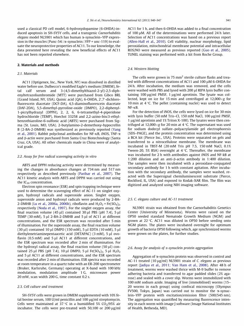
As shown in Fig. 1A and B, AC11 scavenged ABTS and DPPH rad- icals in a concentratio n-dependent manner, and the maximum scavenging rates of 74.8% and 86.1% were observed at 30 lg/mland 64 lg/ml, respectively. IC50 values for ABTS and DPPH radicals were determined to be 20.1 lg/ml and 32.2 lg/ml, respectively .With ESR and spin trapping techniqu e, we have measure d the scav- enging capacity of singlet oxygen, hydroxyl radicals and superox- ide anions. As shown in Fig. 1C and Table 1, AC11 could scavenge all those free radicals and the capacity of AC11 at 25 lg/ml was 55.71% on hydroxyl radicals, 21.06% on superoxi de anion, 14.21%on singlet oxygen and 28.89% on lipid peroxidation. Although QAhas been identified as one of the active ingredient of AC11, itshowed no scavenging effect on free radicals at 25 lg/ml, and the scavenging rate on hydroxyl radicals was only about 15% at5 mg/ml.
3.2. AC11 attenuated 6-OHDA induced cell viability loss and apoptosis
Cell viability was expresse d as an MTT conversion rate. No sig- nificant difference in cell viability was seen for 24 h among the different concentr ations of AC11 and QA treatment (Fig. 2A and
Table 1Comparing the effect of AC11 with QA and positive control Epigallocatechin gallate (EGCG
Scavenging capacity (% of control)
Hydroxyl radical Superoxide
AC-11 (25 lg/ml) 55.71 ± 6.2 * 21.06 ± 2.1QA (5 mg/ml) 15.47 ± 1.2 * –EGCG (25 lg/ml) 76.65 ± 4.2 * 67.983.7*
* p < 0.01 compare with blank control.
Fig. 1. Effects of AC11 on scavenging ABTS (A), DPPH (B) and hydroxyl radicals (Ci, contrwere expressed as a percentage of the untreated control, n = 3, ⁄p < 0.01 compared with
C). However, cell viability was decrease d by about 55% in the presence of 100 lM 6-OHDA alone for 24 h. When the SH-SY5Y cells were pre-treated with different concentrations of AC11 and QA for 1 h, followed by 24 h of incubation with 100 lM 6-OHDA,the cell viability increased with increasing concentratio ns ofAC11 compared with cells treated with 6-OHDA alone (Fig. 2B).No significant change of cell viability was observed with increasing concentr ations of QA compare d with cells treated with 6-OHDA alone (Fig. 2D). These results indicate that AC11 could dose-dep en- dently enhance the viability of SH-SY5Y cells in the presence of 6-OHDA. While QA showed no protective effect on SH-SY5Y cells against6-OHDA- induced cell damage. Another representative antioxidant ,a-tocopherol (Vitamin E, Ve), at the same concentratio n (200 lg/ml) showed much weaker protective effect on cell viability as com- pared with AC11 (Fig 2).
To further study the neuroprotective effects of AC11, its influ-ence on cell apoptosis was determined by detecting morphologi cal changes induced by 6-OHDA treatment. As shown in Fig. 3, the majority of cells in the control group showed a normal nuclear staining pattern (Fig. 3A), while exposure to 100 lM 6-OHDA for 24 h led to typical apoptotic morphology (condensed chromatin and bright staining) in SH-SY5Y cells (Fig. 3B). Such nuclear mor- phology changes were attenuated after AC11 treatment (Fig. 3C–E).
The cell apoptosis rate was also confirmed by TUNEL staining.As shown in Fig 3F–J, 6-OHDA treatment dramatically increased the number of TUNEL-posi tive cells (compare G to F). When cells were pretreated with different dose of AC11, the 6-OHDA- induced apoptosis was greatly inhibited (compare I/J to G).
3.3. AC11 suppressed 6-OHDA induced intracellular ROS
As shown in Fig. 4A, intracellul ar ROS levels were examine dusing DCF-DA. SH-SY5Y cells treated with 100 lM 6-OHDA for
) on free radical scavenging and lipid peroxidation inhibiting in solutio n.
anion Singlet oxygen Lipid peroxidation
4 * 14.21 ± 1.28 * 28.89 ± 4.2 *
– –79.37 ± 4.8 * 46.04 ± 3.5 *
ol; Cii, AC11 treated) in vitro by absorption spectrometry and ESR. For A and B, Data control.

Fig. 2. Effects of AC11, QA and 6-OHDA on SH-SY5Y cell viability. Cells were incubated in drug-free medium or medium containing different concentrations of AC11 (A) and QA (C) for 24 h or pre-incubated with various concentrations of AC11 or 200 lg/ml Vitamin E as positive control (B) and QA (D) for 1 h, and then 6-OHDA (100 lM) was added for an additional 24 h. Data are expressed as a percentage of the untreated control ± SE, n = 3. ⁄p < 0.01 compared with control cells; #p < 0.05 compared with cells treated with 6-OHDA alone.
Fig. 3. AC11 decreased cell apoptosis induced by 6-OHDA. Cells were cultured with 100 lM 6-OHDA in the presence or absence of different concentrations of AC11 for 24 h.Nuclear morphological changes were detected as described in Section 2. (A) Control, (B) 6-OHDA, (C) 6-OHDA plus 50 lg/ml AC11, (D) 6-OHDA plus 100 lg/ml AC11, (E) 6-OHDA plus 200 lg/ml AC11. Cell apoptosis were also detected by TUNEL staining: (F) Control, (G) 6-OHDA, (H) 6-OHDA plus 50 lg/ml AC11, (I) 6-OHDA plus 100 lg/ml AC11,(J) 6-OHDA plus 200 lg/ml AC11.
Z. Shi et al. / Neurochemistry International 62 (2013) 940–947 943
24 h exhibited a significant increase in the DCF signal relative tothe control (p < 0.01). However , this effect was significantly atten- uated by different concentratio ns of AC11. The results indicate that AC11 could dose-dependentl y inhibit the increase of intracellular ROS induced by 6-OHDA in SH-SY5Y cells.
3.4. AC11 attenuated 6-OHDA induced mitochondrial membrane potential loss
The levels of mitochondr ial membrane potential correspond tothe viability of cells. Exposure of SH-SY5Y cells to 6-OHDA for 24 h

Fig. 4. AC11 attenuated 6-OHDA induced accumulation of ROS (A), decrease of mitochondrial membrane potential (B), increase of TBARS (C) and intracellular NO (D). Cells were cultured with 100 lM 6-OHDA in the presence or absence of AC11 for 24 h. Cell viability was measured as described in Materials and Methods. Data are expressed as apercentage of the untreated control ± SE, n = 3. ⁄p < 0.01 compared with control cells, #p < 0.05 compared with cells treated with 6-OHDA alone.
944 Z. Shi et al. / Neurochemistry International 62 (2013) 940–947
decreased the fluorescent intensity of Rhodamine 123 staining,representing a fall in the mitochondr ial membrane potential.AC11 attenuated the decrease of mitochondr ial membrane poten- tial caused by 6-OHDA at 50,100 and 200 lg in a concentration- dependent manner (Fig. 4B).
3.5. AC11 attenuated lipid peroxidation in 6-OHDA treated cells
TBARS, an end product of lipid peroxida tion, were measure d inSH-SY5Y cells under various condition s as indicated in Fig. 4C. The results demonstrat e that the lipid peroxide level increased byabout 40% with 6-OHDA treatment compare d with the control group. Co-admini stration of 100 lg/ml and 200 lg/ml AC11 with 6-OHDA decreased the level of TBARS by about 8% and 20%, respec- tively, which were statistically significant.
3.6. Effects of AC11 on intracellular NO level and expression of iNOS,NF-jB and TNF- a
As shown in Fig. 4D, intracellular NO levels increased signifi-cantly after treatment with 6-OHDA alone compare d with the con- trol group, and this effect was reduced in a concentratio n-dependent manner by pre-treatment with AC11 at 50, 100 and 200 lg/ml. 6-OHDA exposure up-regulate d iNOS, NF- jB and TNF- a as revealed by Western blot analysis (Fig. 5A–D), and these in- creases were dose-dependentl y attenuated by co-treatment ofAC11.
3.7. AC11 reduced a-synuclein protein aggregation
Worms of untreated and AC11 treated groups were observed under confocal microscope for assaying their a-synuclein aggrega-
tion pattern. Phenotypical ly, the worms appeared normal and opti- mally fed. A marked and significant reduction in the aggregat ion ofa-synuclein in case of NL5901 worms treated with AC11 was ob- served as compared to that of control group (Fig. 6A). The images for fluorescence intensity of a-synuclei n aggregation were quanti- fied using Image J software. Treatment of worms with AC11 showed significantly reduced fluorescence intensity of aggregat ion as compared to that of untreated worms. The mean fluorescence(GFP) intensity was 5.808 ± 0.728 arbitrary units in control worms and 3.582 ± 0.515 arbitrary units in AC11 treated subjects (Fig. 6),indicating 40% reduction (p < 0.05) in a-synuclein protein aggrega- tion after AC11 treated. Treatment of same concentratio n of a-tocopherol caused 25% reduction (p < 0.05 compare with control group).
4. Discussion
To the best of our knowledge, this is the first report assessing the antioxidant and neuroprotecti ve effect of AC11. The major new finding is that AC11 significantly attenuated 6-OHDA induced cell death, oxidative stress in SH-SY5Y cells. AC11 also significantlyreduced the a-synuclein aggregat ion in NL5901 C. elegans . The antioxidant activity, especially the hydroxyl radicals scavenging capacity, may be responsible for the neuroprotecti ve effects ofAC11.
Many advances in pharmacolo gical therapies for PD have been made over the past few years; however, there is still no drug cur- rently available to cure or significantly prevent the advancemen t ofthis disease. Although the detailed mechanism causing degenera- tion in dopaminer gic (DA) neurons in PD is not well understo od,oxidative stress has been regarded as one of the intermediar y risk factors that can initiate and/or promote degeneration of DA neu-

Fig. 5. Effects of AC11 and 6-OHDA on the expression of NF- jB, TNF- a and iNOS. Cells were exposed to 6-OHDA (100 lM) with various concentrations of AC11 for 24 h. NF- kB, TNF- a and iNOS were detected in the cell lysates by Western blot (A, C), and quantified (B, D). The data were calculated as the ratio of the band intensity of NF-kB, TNF- a oriNOS over that of actin and expressed as the mean ± SE, n = 3. ⁄p < 0.05 compared with control cells, #p < 0.05 compared with cells treated with 6-OHDA alone.
Fig. 6. a-Ynuclein aggregation in NL5901 strain of C. elegans fed on OP50 (A–C), OP50 plus 10 lg/ml AC11 (D–F) and OP50 plus 10 lg/ml a-tocopherol (G–I). Images A, D and G are fluorescent images; B, E and H are images grabbed using Differential Interference Contrast (DIC) optics; C, F and I are overlap images. J is graphical representation for fluorescence intensity of the nematodes as quantified using Image J software from 20 worms each group. ⁄p < 0.05 compare with control group.
Z. Shi et al. / Neurochemistry International 62 (2013) 940–947 945
rons (Fahn and Cohen, 1992; Beal, 1995 ). Therefore, supplementa- tion with antioxidant s may prevent or reduce the rate of progres- sion of this disease (Prasad et al., 1999; Zhao, 2009 ). For example,
our previous studies have demonstrat ed the protective effect ofgreen tea polyphenol s on neurons against apoptosis of cellular and animal PD models (Guo et al., 2005, 2007 ). Currently,

946 Z. Shi et al. / Neurochemistry International 62 (2013) 940–947
U. tomentosa is one of the most popular herbal remedies in the Uni- ted States due to its beneficial effects on DNA repair and immune function (Sheng et al., 2000, 2001 ). There is growing evidence tosupport its use in treating cancer, inflammation, viral infection and vascular conditions, as well as for its use as an immunostimu- lant, antioxidant and antibacterial agent (Heitzman et al., 2005 ).Here we found that AC11 significantly attenuated the cell death in- duced by 6-OHDA (Figs. 2 and 3), suggesting AC11 may act as a po- tential neuroprotective agent in PD. To clarify the protective mechanism of AC11 against 6-OHDA- induced cytotoxicity, the lev- els of intercellular ROS and lipid peroxidatio n were studied. Inagreement with a number of previous reports (Lu et al., 2006a,2006b), we have shown that 6-OHDA does indeed induce oxidative stress, as demonstrat ed by the increase of intracellul ar ROS levels and TBARS content (Fig. 4A and C), whereas AC11 pretreatmen tsignificantly attenuated these increases in a dose-dependent man- ner, suggesting that the protectiv e effect of AC11 is at least par- tially through its antioxidant property .
The neuroprotecti on by the selective iNOS inhibitor GW274150 in a model of PD demonst rated the important role of activation ofiNOS in the pathogenesis of PD (Broom et al., 2011 ). The present study, as well as our previous study (Guo et al., 2005 ), showed that 6-OHDA treatment caused increases of iNOS expression and intra- cellular NO levels. AC11 pretreatmen t significantly decreased iNOS induction, thereby decreasing the production of NO (Figs. 4D–5C).As the production of ONOO - in vivo is highly dependent on the met- abolic pathways of NO and superoxide, our results indicated that AC11 may exert its protectio n against 6-OHDA cytotoxicity via modulation of intracellul ar ROS and NO levels, which is consisten twith our previous studies (Guo et al., 2005, 2007 ).
It has been well described that mitochondrial alteration s are associated with the cytotoxic effect of 6-OHDA, especiall y the loss of mitochondr ial membrane potential (Tirmenstein et al., 2005 ). Inthe present study, 100 lM 6-OHDA treatments caused a significantdecrease in the fluorescent intensity of Rhodamine 123. However ,AC11 treatment significantly attenuated the mitochondrial mem- brane potential loss in a dose-depend ent manner.
NF-jB is a family of inducible transcrip tion factors that are acti- vated in response to inflammatory stimulation. There is evidence showing that there is a marked increase in NF- jB activation within the midbrain of animals undergoing neurodegenera tion as a result of 1-methy l-4-phenyl-1,2 ,3,6-tetrahydro pyridine administrat ion,as well as in the substantia nigra pars compacta of PD patients (Hu-not et al., 1997; Ghosh et al., 2007 ), suggesting that targeting the classical pathway of NF- jB may serve as a useful therapeutic ap- proach to the treatment of PD (Flood et al., 2011 ). Our results showed that 6-OHDA increased the expression of NF- jB and TNF-a in SH-SY5Y cells, and those induction s were significantlyattenuated by AC11 treatment (Fig. 4). The modulatory effect ofAC11 on NF- jB and TNF- a regulator y pathway is in agreement with previous reports demonstrating that U. tomentosa acts as apotent TNF- a inhibitor through NF- jB.
PD is also characterized by the aggregation of several proteins involved in vesicle recycling and protein degradation such as a-synuclein, parkin, and synphilin-1. It has been suggested that aggregation of these proteins lead to oxidative stress, suggesting that compounds that can inhibit protein aggregation can be useful in the treatment of PD. In NL5901 C. elegans , 10 lg/ml AC11 could reduce the aggregat ion of a-synuclein by 40%, indicating the neu- roprotective effect of AC11 is also associated with reducing protein aggregation . It has been suggested that therapeuti c drugs for PD re- quire aromatic elements for binding to the a-synuclei n monomer /oligomer and vicinal hydroxyl groups on a single phenyl ring (Caruana et al., 2011 ). It has also been indicating that decrease oxi- dative stress by antioxidant could reduce a-synuclein aggregation (Shastry, 2003 ). Using infrared spectrum analysis (figures not
shown), we found that polyphenols was one of major components in AC11. Meanwhile, we also found AC11 could scavenge several kinds of free radical (Fig 1 and Table 1) and decrease 6-OHDA in- duced oxidative stress in SH-SY5Y cells (Fig 4). So there may betwo possibilities which explain the anti a-synuclein aggregat ion effect of AC11: one is through direct interaction between the poly- phenols ingredient in AC11 with a-synuclei n; the other is through decreasing oxidative stress by free radical scavenging.
5. Conclusion
Overall, this study demonstrat es that AC11 is a potential neuro- protectiv e antioxidant for the mitigation of PD and a promising therapeuti c candidate for neurodegenera tive disorders .
Acknowled gements
This work was funded by National Natural Science Foundation of China (31271146) and the Applied Research Project of Hebei Province (10966120D). Zhongbing Lu gratefully acknowledges the support of State Key Laboratory of Biomacrom olecules (2012kf03) and Hundred Talents Program of the Chinese Academy of Sciences.
References
Allen-Hall, L., Arnason, J.T., Cano, P., Lafrenie, R.M., 2010. Uncaria tomentosa acts as apotent TNF-alpha inhibitor through NF-kappaB. J. Ethnopharmacol. 127, 685–693.
Beal, M.F., 1995. Aging, energy, and oxidative stress in neurodegenerative diseases.Ann. Neurol. 38, 357–366.
Bors, M., Bukowska, B., Pilarski, R., Gulewicz, K., Oszmianski, J., Michalowicz, J.,Koter-Michalak, M., 2011. Protective activity of the Uncaria tomentosa extractson human erythrocytes in oxidative stress induced by 2,4-dichlorophenol (2,4-DCP) and catechol. Food Chem. Toxicol. 49, 2202–2211.
Broom, L., Marinova-Mutafchieva, L., Sadeghian, M., Davis, J.B., Medhurst, A.D.,Dexter, D.T., 2011. Neuroprotection by the selective iNOS inhibitor GW274150 in a model of Parkinson disease. Free Radic. Biol. Med. 50, 633–640.
Caruana, M., Hogen, T., Levin, J., Hillmer, A., Giese, A., Vassallo, N., 2011. Inhibition and disaggregation of alpha-synuclein oligomers by natural polyphenolic compounds. FEBS Lett. 585, 1113–1120.
Fahn, S., Cohen, G., 1992. The oxidant stress hypothesis in Parkinson’s disease:evidence supporting it. Ann. Neurol. 32, 804–812.
Flood, P.M., Qian, L., Peterson, L.J., Zhang, F., Shi, J.S., Gao, H.M., Hong, J.S., 2011.Transcriptional Factor NF-kappaB as a Target for Therapy in Parkinson’s Disease.Parkinsons Dis. 2011, 216298.
Ghosh, A., Roy, A., Liu, X., Kordower, J.H., Mufson, E.J., Hartley, D.M., Ghosh, S.,Mosley, R.L., Gendelman, H.E., Pahan, K., 2007. Selective inhibition of NF-kappaB activation prevents dopaminergic neuronal loss in a mouse model ofParkinson’s disease. Proc. Natl. Acad. Sci. USA 104, 18754–18759.
Guo, S., Bezard, E., Zhao, B., 2005. Protective effect of green tea polyphenols on the SH-SY5Y cells against 6-OHDA induced apoptosis through ROS-NO pathway.Free Radic. Biol. Med. 39, 682–695.
Guo, S., Yan, J., Yang, T., Yang, X., Bezard, E., Zhao, B., 2007. Protective effects of green tea polyphenols in the 6-OHDA rat model of Parkinson’s disease through inhibition of ROS-NO pathway. Biol. Psychiatry 62, 1353–1362.
Heitzman, M.E., Neto, C.C., Winiarz, E., Vaisberg, A.J., Hammond, G.B., 2005.Ethnobotany, phytochemistry and pharmacology of Uncaria (Rubiaceae).Phytochemistry 66, 5–29.
Hunot, S., Brugg, B., Ricard, D., Michel, P.P., Muriel, M.P., Ruberg, M., Faucheux, B.A.,Agid, Y., Hirsch, E.C., 1997. Nuclear translocation of NF-kappaB is increased indopaminergic neurons of patients with parkinson disease. Proc. Natl. Acad. Sci.USA 94, 7531–7536.
Jadiya, P., Khan, A., Sammi, S.R., Kaur, S., Mir, S.S., Nazir, A., 2011. Anti-Parkinsonian effects of Bacopa monnieri : insights from transgenic and pharmacological Caenorhabditis elegans models of Parkinson’s disease. Biochem. Biophys. Res.Commun. 413, 605–610.
Lu, Z., Nie, G., Belton, P.S., Tang, H., Zhao, B., 2006a. Structure–activity relationship analysis of antioxidant ability and neuroprotective effect of gallic acid derivatives. Neurochem. Int. 48, 263–274.
Lu, Z., Tao, Y., Zhou, Z., Zhang, J., Li, C., Ou, L., Zhao, B., 2006b. Mitochondrial reactive oxygen species and nitric oxide-mediated cancer cell apoptosis in 2-butylamino-2-demethoxyhypocrellin B photodynamic treatment. Free Radic.Biol. Med. 41, 1590–1605.
Mammone, T., Akesson, C., Gan, D., Giampapa, V., Pero, R.W., 2006. A water soluble extract from Uncaria tomentosa (Cat’s Claw) is a potent enhancer of DNA repair in primary organ cultures of human skin. Phytother. Res. 20, 178–183.

Z. Shi et al. / Neurochemistry International 62 (2013) 940–947 947
Noda, Y., Anzai, K., Mori, A., Kohno, M., Shinmei, M., Packer, L., 1997. Hydroxyl and superoxide anion radical scavenging activities of natural source antioxidants using the computerized JES-FR30 ESR spectrometer system. Biochem. Mol. Biol.Int. 42, 35–44.
Parihar, V.K., Dhawan, J., Kumar, S., Manjula, S.N., Subramanian, G., Unnikrishnan,M.K., Rao, C.M., 2007. Free radical scavenging and radioprotective activity ofdehydrozingerone against whole body gamma irradiation in Swiss albino mice.Chem. Biol. Interact. 170, 49–58.
Prasad, K.N., Cole, W.C., Kumar, B., 1999. Multiple antioxidants in the prevention and treatment of Parkinson’s disease. J. Am. Coll. Nutr. 18, 413–423.
Sandoval, M., Okuhama, N.N., Zhang, X.J., Condezo, L.A., Lao, J., Angeles’, F.M., Musah,R.A., Bobrowski, P., Miller, M.J., 2002. Anti-inflammatory and antioxidant activities of cat’s claw (Uncaria tomentosa and Uncaria guianensis ) are independent of their alkaloid content. Phytomedicine 9, 325–337.
Sandoval-Chacon, M., Thompson, J.H., Zhang, X.J., Liu, X., Mannick, E.E., Sadowska- Krowicka, H., Charbonnet, R.M., Clark, D.A., Miller, M.J., 1998. Antiinflammatoryactions of cat’s claw: the role of NF-kappaB. Aliment Pharmacol. Ther. 12, 1279–1289.
Schapira, A.H., Gu, M., Taanman, J.W., Tabrizi, S.J., Seaton, T., Cleeter, M., Cooper,J.M., 1998. Mitochondria in the etiology and pathogenesis of Parkinson’s disease. Ann. Neurol. 44, S89–S98.
Shastry, B.S., 2003. Neurodegenerative disorders of protein aggregation.Neurochem. Int. 43, 1–7.
Sheng, Y., Bryngelsson, C., Pero, R.W., 2000. Enhanced DNA repair, immune function and reduced toxicity of C-MED-100, a novel aqueous extract from Uncariatomentosa. J. Ethnopharmacol. 69, 115–126.
Sheng, Y., Li, L., Holmgren, K., Pero, R.W., 2001. DNA repair enhancement of aqueous extracts of Uncaria tomentosa in a human volunteer study. Phytomedicine 8,275–282.
Tirmenstein, M.A., Hu, C.X., Scicchitano, M.S., Narayanan, P.K., McFarland, D.C.,Thomas, H.C., Schwartz, L.W., 2005. Effects of 6-hydroxydopamine onmitochondrial function and glutathione status in SH-SY5Y human neuroblastoma cells. Toxicol. In Vitro 19, 471–479.
Van Ham, T.J., Thijssen, K.L., Breitling, R., Hofstra, R.M.W., Plasterk, R.H.A., et al.,2008. C. elegans model identifies genetic modifiers of a-synuclein inclusion formation during aging. PLoS Genet. 4 (3), e1000027. http://dx.doi.org/10.1371/journal.pgen.1000027.
Yang, H., Wu, T., Zhang, M., Zhang, Z., 2001. A novel photosensitizer, 2-butylamino- 2-demethoxy-hypocrellin B (2-BA-2-DMHB) – its photodynamic effects on HeLa cells: efficacy and apoptosis. Biochim. Biophys. Acta 1540, 22–31.
Zhao, B., 2009. Natural antioxidants protect neurons in Alzheimer’s disease and Parkinson’s disease. Neurochem. Res. 34, 630–638.