neural mechanisms in williams syndrome: a unique window to
TRANSCRIPT

*Unit for Systems Neuroscience in Psychiatry, ‡ Neuroimaging Core Facility,
§Genes, Cognition and Psychosis Program, ||Section on Integrative Neuroimaging, National Institute of Mental Health, NIH, DHHS, 9000 Rockville Pike, Bethesda, Maryland 20892-1365, USA. ¶Neurodevelopmental Sciences Laboratory, Department of Psychological and Brain Sciences, University of Louisville, Louisville, Kentucky 40292, USA. Correspondence to A.M.L. e-mail: [email protected]:10.1038/nrn1906
Haploinsufficiency Presence of only a single functional copy of a gene that does not provide sufficient transcript or protein production to assure normal function.
Hypercalcaemia Abnormally high calcium concentration in the blood.
Neural mechanisms in Williams syndrome: a unique window to genetic influences on cognition and behaviourAndreas Meyer-Lindenberg*‡§, Carolyn B. Mervis¶ and Karen Faith Berman§||
Abstract | Williams syndrome, a rare disorder caused by hemizygous microdeletion of about 28 genes on chromosome 7q11.23, has long intrigued neuroscientists with its unique combination of striking behavioural abnormalities, such as hypersociability, and characteristic neurocognitive profile. Williams syndrome, therefore, raises fundamental questions about the neural mechanisms of social behaviour, the modularity of mind and brain development, and provides a privileged setting to understand genetic influences on complex brain functions in a ‘bottom-up’ way. We review recent advances in uncovering the functional and structural neural substrates of Williams syndrome that provide an emerging understanding of how these are related to dissociable genetic contributions characterized both in special participant populations and animal models.
Williams syndrome (WS) is a neurodevelopmental disorder caused by a hemizygous deletion of ~1.6 megabases, containing ~28 genes, on chromosome 7q11.23 (FIG. 1a). The incidence is usually given as 1 in 20,000 live births, but new prevalence estimates are as high as 1 in 7,500 (REF. 1), which means that WS could account for 6% of all cases of mental retardation of genetic origin1.
WS was first described as a combination of a distinct facial appearance with growth retardation and cardio-vascular abnormalities2,3, which are present in ~80% of individuals with WS4. The cardiovascular and some of the facial features have been linked to haploinsufficiency for elastin (ELN)5. Other common somatic symptoms are endocrine (for example, transient hypercalcaemia and impaired glucose tolerance), gastrointestinal (con-stipation, prolapse and diverticula) and orthopaedic (scoliosis or joint contractures) problems6. Neurological problems include coordination difficulties (for exam-ple, trouble walking down a staircase), hyperreflexia, strabismus, nystagmus6,7, hypersensitivity to sound4,6, and sensorineural hearing loss8,9.
WS is associated with mild to moderate mental retardation or learning difficulties. Of central interest to research is a distinctive cognitive profile with peaks and valleys10,11. In particular, a severe visuospatial construc-tion deficit is a fundamental stable phenotype in WS11, contrasting with a relative strength in verbal short-term
memory and language11,12. Emotionally, a particularly striking feature of children with WS is their high socia-bility13,14 and empathy for others13. Typically, individuals with WS are socially fearless, engaging eagerly in social interaction even with strangers13,14. Intriguingly, this remarkable hypersociability is coupled with a strong undercurrent of anxiety that relates to non-social objects13,15,16. Attention deficit hyperactivity disorder (ADHD, predominantly inattentive type or combined type) is common (>50%) in children and adolescents with WS16.
Unfortunately, the intellectual impairment character-istic of WS limits comparison of typical participants with WS to a normal-intelligence control group and reduces the ability of individuals with WS to perform consist-ently during testing. This can be avoided by selecting normal-intelligence participants with WS17 (BOX 1), who can cooperate with extensive cognitive and imaging procedures and be appropriately compared with normal controls. It is likely that abnormalities found even in this high-performing group would be characteristic of the syndrome as a whole, and close to the genetic substrate of the disorder.
Because the genes involved in WS are known, and the dosage of at least some of these genes is clearly abnor-mal, the study of neural mechanisms in WS affords a privileged setting to understand genetic influences on complex brain functions in a ‘bottom-up’ way. This
R E V I E W S
380 | MAY 2006 | VOLUME 7 www.nature.com/reviews/neuro

c
Centromere
Mus musculus
Homo sapiens
CEN
Pom121
Nsun5
Trim50a
Fkbp6Fzd9Baz1bBcl7bTbl2M
lxiplW
bscr18W
bscr22V
ps37dStx1aA
bhd11
Cldn4
Cldn3
Wbscr27
Wbscr28
ElnLim
k1W
bscr1/E1f4hLat2Rfc2C
yln2G
tf2ird1G
tf2IN
cf1G
tf2ird2
TEL
Typical WSRef. 140Ref. 140
Refs 149,150Ref. 151
Refs 5,57Ref. 5Ref. 5Ref. 5
Ref. 5Ref. 154
Ref. 154Ref. 154
POM
121
NSU
N5
TRIM
50A
FKBP
6FZ
D9
BAZ1
BBC
L7B
TBL2
MLX
IPL
WBS
CR1
8W
BSC
R22
VPS
37D
STX1
AA
BHD
11
WBS
CR2
7W
BSC
R28
CLD
N3
CLD
N4
ELN
LIM
K1W
BSC
R1/E
1F4H
LAT2
RFC
2
CYL
N2
GTF
2IRD
1
GTF
2I
NC
F1
GTF
2IRD
2
a b
Hyperreflexia Exaggerated deep tendon reflexes.
Strabismus Eye misalignment; also known as ‘crossed eyes’.
Nystagmus Involuntary and often rapid and repetitive oscillatory movements of the eyeballs.
Homologous recombination Exchange of DNA segments of similar sequence. Occurs by breakage and reunion in paired chromosomes during meiosis.
(Arnold-)Chiari malformations A group of disorders characterized by protrusion of the cerebellum through the large opening in the base of the skull into the spinal canal.
extends and complements efforts to understand genetic mechanisms of behaviour in the general population, in which contributions of individual genes are small, gene–gene and gene–environment interactions are the rule, and unambiguously functional variations are uncommon and difficult to characterize.
The goal of this article is to review recent advances in defining the neural substrates of the unique neuro-psychiatric features of WS and to begin to define separable neural subsystems in this syndrome, speci-fying mechanisms for visuospatial cognition, social behaviour and memory under genetic control. We hope that these emerging convergent results form a point of departure not only for a deeper understand-ing of WS, but also for the investigation of dissociable genetic contributions to complex behaviour in humans in general.
Genetics of Williams syndromeWS is a genomic disorder in which the clinical pheno-type is a consequence of abnormal gene dosage due to a hemizygous deletion that results from unequal homologous recombination during meiosis18. The common 1.6-Mb deletion involving 7q11.23 (FIG. 1a) is mediated by flanking groups of low copy repeat sequences causing
misalignment because of high sequence homology19–21 (FIG. 1b). Inversion of the same segment has been found as a polymorphic variant in ~7% of the general popula-tion22 and 27–35% of transmitting parents19,22, and has been reported in some individuals with mental retar-dation and features associated with WS23. However, these individuals did not fit either the cognitive profile or the personality profile associated with WS (C.B.M., C. A. Morris and L. R. Osborne, unpublished observa-tions). The first case of duplication of the WS region was recently reported24. The phenotype, which includes severe speech and expressive language delay in the context of visuospatial construction skills at the level of other family members, contrasts strongly with the WS phenotype.
Structural abnormalities in WSAt post-mortem, reduced brain size25, Chiari malforma-tions26,27, corpus callosum shape changes28,29 and altered cell size and density in primary visual cortex171 have been described in WS. Using structural MRI, reductions were localized to the parietal lobule30 and occipital31 grey mat-ter. Cerebellar size is preserved32,33, although the neuro-nal integrity marker N-acetyl aspartate (NAA)34 may be reduced there.
Figure 1 | Genetics of Williams syndrome. a | Chromosomal location of the hemideleted region. b | Map140 of the region in humans (centre) and the homologous region in mice (top). Location of low copy repeat regions marked by bars. c | Extent of typical Williams syndrome deletion and examples of small (atypical) deletions. Dashed lines indicate uncertain extent of the deletion in that portion. Adapted, with permission, from REF. 140 © (2005) American Academy for the Advancement of Science.
R E V I E W S
NATURE REVIEWS | NEUROSCIENCE VOLUME 7 | MAY 2006 | 381

Differential Ability Scales-School Age(DAS-School Age). A standardized assessment of general intellectual functioning designed to provide specific information about an individual’s strengths and weaknesses across a wide range of intellectual abilities. It is particularly appropriate for assessing individuals with WS because it yields separate standard scores for verbal, nonverbal reasoning and spatial abilities, as well as an overall standard score (general conceptual ability (GCA), which is similar to IQ).
Voxel-based morphometry (VBM). A widely used method for the analysis of imaging data that enables a statistically principled voxel-wise between-groups comparison of local grey matter volume, unconstrained by anatomical landmarks.
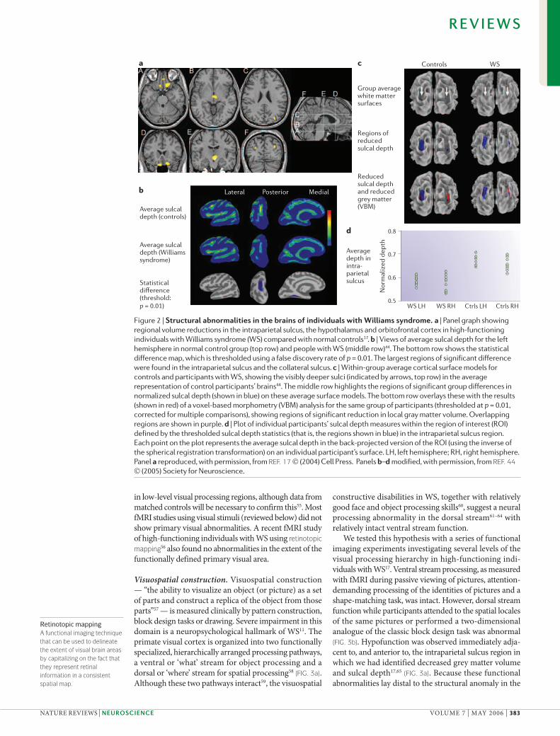
Significant advances in our understanding of the structural basis of WS have come from the application of voxel-based morphometry (VBM), which allows the study of genetic variation without restriction to ana-tomical boundaries. In our cohort of high-functioning participants with WS, this approach identified circum-scribed symmetrical grey matter volume reductions in WS in three regions (FIG. 2a): the intraparietal sulcus, around the third ventricle, and the orbitofrontal cortex (OFC)17. The intraparietal sulcus finding was recently confirmed in children with WS and mental retardation35. Another study of typically functioning individuals with WS also found the most pronounced reduction in the intra parietal sulcus36, but, in addition, identified relative regional density increases in the orbital and medial pre-frontal cortices, the anterior cingulate, insular cortex and superior temporal gyrus36, whereas no volume increases were found in our cohort. Recent work from these two groups suggests that methodological differences contrib-ute to these discrepancies, such as the choice of template used to match images and the method used to map indi-vidual images into a common anatomical space.
Although the intraparietal sulcus has been implicated in the visuospatial construction deficit of WS17, and the OFC has been linked to hypersociability37, clear func-tional correlates of the structural abnormality around the third ventricle have yet to be defined. We have specu-lated17 that hypothalamic abnormalities might contribute to the many poorly understood hormonal disturbances found in WS4,38. Thalamic abnormalities, if confirmed, could affect several sites of cortical processing.
Insights from analysis of regional volume are supple-mented and extended by new analyses of cortical shape. Overall curvature of the brain is reduced in WS39, and abnormally increased gyrification has been noted in the parietal and occipital lobes40, and the temporo parietal zone41. Length reductions in the central gyrus42,43 might be related to overall brain size reduction in WS. Convergent
evidence demonstrates reductions in sulcal depth in the intraparietal sulcus (FIG. 2b–d) both in participants with WS of normal IQ44 and those with mental retardation45, clarifying the macroanatomical correlate of this regional volume reduction seen in VBM.
Although findings in sulcal depth and regional vol-ume largely agree in directionality and location, a recent study on cortical thickness found increases of 5–10% in the right perisylvian and inferior temporal zone in WS41. This supports theoretical predictions that regional vol-ume and cortical thickness should be subject to different maturational mechanisms46. It has been proposed47 that tension along axons might explain how and why the cor-tex folds in a characteristic pattern. WS results would be consistent with this theory if a reduced population of neurons in the intraparietal sulcus results in a reduction in white matter connections of this area48. Further inves-tigation, particularly based on diffusion tensor imaging, would be helpful. In preliminary data from a subgroup of five high-functioning participants with WS, we found abnormal white matter integrity immediately underlying the intraparietal sulcus49.
Functional abnormalitiesSensory function. There are few reliable data to suggest abnormal primary sensory processing in WS. The results of one functional MRI (fMRI) study using noise and music stimuli in a small group of participants with WS and mental retardation suggested reduced activation in auditory cortex in WS at a lenient statistical threshold50. Visually, individuals with WS have a high incidence of strabismus51,52 and reduced visual acuity53, especially stereo acuity54. These primary sensory problems do not correlate with the visuospatial constructive deficit53, which indicates that the latter has an independent neural basis. Changes in evoked potential recordings during illusory contour completion were found in participants with WS who had mental retardation, indicating possible changes
Box 1 | Williams syndrome and intelligence
Contrary to some initial reports80, the results of recent research10 indicate that severe mental impairment is rare in Williams syndrome (WS). Rather, the mean full scale IQ for WS is typically in the mild mental impairment range. For example, 112 8–17 year olds who had classic WS deletions and did not have autism spectrum disorder (ASD) completed the Differential Ability Scales-School Age version (DAS-School Age155). Because of the unusual cognitive profile associated with WS, the DAS-School Age, which provides separate standard scores for Verbal, Nonverbal Reasoning, and Spatial abilities (clusters) is a particularly useful full-scale measure of intelligence. For the general population, mean general conceptual ability (GCA, similar to IQ) and mean standard score on each of the clusters is 100, with a standard deviation of 15. For this sample of individuals with WS, mean GCA was 59.19 with a standard deviation of 11.59 (C.B.M., unpublished observations); 19% scored in the normal range (≥70). Mean cluster standard scores were 70.59 for Verbal, 67.88 for Nonverbal Reasoning, and 55.00 for Spatial. For 83% of participants, the Verbal and/or Nonverbal Reasoning standard score was significantly higher than expected for GCA, which indicates that the use of the overall standard score from an omnibus IQ measure as the determinant of intelligence level is not appropriate for most people with WS.
Most behavioural researchers studying WS report IQs from the Kaufman Brief Intelligence Test (KBIT156). For a sample of 374 individuals aged 5–55 years with WS who have classic deletions and do not have ASD (C.B.M., unpublished observations), mean IQ (68.46) on this test, which includes only Verbal (mean = 71.52) and Nonverbal Reasoning (mean = 70.83) subtests, is about two standard deviations below the general population mean (100); the standard deviation (14.84) is similar to that for the general population (15). Therefore, the shape of the IQ distribution for the KBIT, which does not assess spatial ability, is similar to that for the general population, but negatively displaced by about two standard deviations. The higher mean IQ for the KBIT than the DAS-School Age is primarily due to the inclusion of Spatial subtests on the DAS but not the KBIT; mean performance on the Verbal subtests of the two measures was similar, as was mean performance on the Nonverbal Reasoning subtests.
R E V I E W S
382 | MAY 2006 | VOLUME 7 www.nature.com/reviews/neuro
a
b
Controls
Statistical difference(threshold:p = 0.01)
Average sulcaldepth (controls)
Average sulcal depth (Williamssyndrome)
WS
0.8
0.7
0.6
0.5
Nor
mal
ized
dep
th
WS LH WS RH Ctrls LH Ctrls RH
Lateral Posterior Medial
c
d
Group averagewhite mattersurfaces
Reducedsulcal depthand reducedgrey matter (VBM)
Average depth in intra-parietal sulcus
Regions ofreducedsulcal depth
Retinotopic mapping A functional imaging technique that can be used to delineate the extent of visual brain areas by capitalizing on the fact that they represent retinal information in a consistent spatial map.
in low-level visual processing regions, although data from matched controls will be necessary to confirm this55. Most fMRI studies using visual stimuli (reviewed below) did not show primary visual abnormalities. A recent fMRI study of high-functioning individuals with WS using retinotopic mapping56 also found no abnormalities in the extent of the functionally defined primary visual area.
Visuospatial construction. Visuospatial construction — “the ability to visualize an object (or picture) as a set of parts and construct a replica of the object from those parts”57 — is measured clinically by pattern construction, block design tasks or drawing. Severe impairment in this domain is a neuropsychological hallmark of WS11. The primate visual cortex is organized into two functionally specialized, hierarchically arranged processing pathways, a ventral or ‘what’ stream for object processing and a dorsal or ‘where’ stream for spatial processing58 (FIG. 3a). Although these two pathways interact59, the visuospatial
constructive disabilities in WS, together with relatively good face and object processing skills60, suggest a neural processing abnormality in the dorsal stream61–64 with relatively intact ventral stream function.
We tested this hypothesis with a series of functional imaging experiments investigating several levels of the visual processing hierarchy in high-functioning indi-viduals with WS17. Ventral stream processing, as measured with fMRI during passive viewing of pictures, attention-demanding processing of the identities of pictures and a shape-matching task, was intact. However, dorsal stream function while participants attended to the spatial locales of the same pictures or performed a two-dimensional analogue of the classic block design task was abnormal (FIG. 3b). Hypofunction was observed immediately adja-cent to, and anterior to, the intraparietal sulcus region in which we had identified decreased grey matter volume and sulcal depth17,65 (FIG. 3a). Because these functional abnormalities lay distal to the structural anomaly in the
Figure 2 | Structural abnormalities in the brains of individuals with Williams syndrome. a | Panel graph showing regional volume reductions in the intraparietal sulcus, the hypothalamus and orbitofrontal cortex in high-functioning individuals with Williams syndrome (WS) compared with normal controls17. b | Views of average sulcal depth for the left hemisphere in normal control group (top row) and people with WS (middle row)44. The bottom row shows the statistical difference map, which is thresholded using a false discovery rate of p = 0.01. The largest regions of significant difference were found in the intraparietal sulcus and the collateral sulcus. c | Within-group average cortical surface models for controls and participants with WS, showing the visibly deeper sulci (indicated by arrows, top row) in the average representation of control participants’ brains44. The middle row highlights the regions of significant group differences in normalized sulcal depth (shown in blue) on these average surface models. The bottom row overlays these with the results (shown in red) of a voxel-based morphometry (VBM) analysis for the same group of participants (thresholded at p = 0.01, corrected for multiple comparisons), showing regions of significant reduction in local gray matter volume. Overlapping regions are shown in purple. d | Plot of individual participants’ sulcal depth measures within the region of interest (ROI) defined by the thresholded sulcal depth statistics (that is, the regions shown in blue) in the intraparietal sulcus region. Each point on the plot represents the average sulcal depth in the back-projected version of the ROI (using the inverse of the spherical registration transformation) on an individual participant’s surface. LH, left hemisphere; RH, right hemisphere. Panel a reproduced, with permission, from REF. 17 © (2004) Cell Press. Panels b–d modified, with permission, from REF. 44 © (2005) Society for Neuroscience.
R E V I E W S
NATURE REVIEWS | NEUROSCIENCE VOLUME 7 | MAY 2006 | 383

Dorsal stream:spatial visionpathway
Intraparietal sulcus
Ventral stream:object recognition
a Lateral b
c
V1
Posterior
HandlesSeat
Spokes
Wheel
Wheel withspokesPedals
dorsal stream, it seemed that the latter was serving as a roadblock to the hierarchically organized dorsal stream information flow from earlier, more inferior-posterior, to later, superior-anterior visual processing areas. This was formally tested with path analysis, a method that allows statistical assessment of interactions among regional nodes in a predefined neural system model. This model was based on well-known anatomical constraints, similar to those of previous path analyses of the visual system66, and consisted of functional data from nodes in early visual areas, in the most activated ventral stream region, in the structurally changed intraparietal sulcus region and in a dorsal stream location of pronounced hypo function in WS. It fit well with the functional data from both partici-pants with WS and matched controls. When each inter-regional path was tested for significance of contribution to the overall fit, the only difference between groups was that the path from the intraparietal sulcus to the later dorsal stream region was significant in controls but not in individuals with WS17.
These data suggest candidate genes that might influ-ence visuospatial constructive cognition in the general population through an impact on intraparietal sulcus function. It would, therefore, be of great interest to
determine whether genetic variations in the general population have an impact on the anatomical integrity and functional activation of this region. Such findings would add a new biological dimension to the study of visuospatial construction.
Memory and hippocampal function. Although verbal short-term memory is a relative strength in WS11, several cognitive domains linked to the hippocampal formation are severely affected, including spatial navigation67,68, and especially long-term memory, both in the verbal69 and spatial70 domains.
We undertook a multimodal imaging study aimed at comprehensively characterizing the hippocampal forma-tion (HF) in our cohort of high-functioning individuals with WS71. Baseline neurofunctional status, measured during rest with oxygen-15 water positron emission tomography, was profoundly reduced bilaterally in the hippocampal formation, extending into the entorhinal
cortex (FIG. 4a). We also used proton magnetic resonance spectroscopy (MRS) for in vivo assay of NAA, a cellular integrity marker and measure of synaptic abundance72. Reduced NAA (as a ratio to creatine), which was more pronounced in the left hippocampal formation, was found
Figure 3 | Dorsal visual stream functional deficits in Williams syndrome. a | Spatial relationship between area found to be structurally abnormal with voxel-based morphometry (yellow), and two tasks tapping into the function of the dorsal visual stream (red and blue) in high-functioning participants with Williams syndrome (WS) compared with controls during functional MRI (fMRI)17. Overlapping regions in the parietal lobule are coloured purple, showing consistent functional abnormalities in the dorsal visual stream directly adjacent to the structural abnormality. Lower panel shows location of proposed functional–structural impairment in the dorsal stream at the intraparietal sulcus. V1, primary visual cortex. b | Left panels show examples of square completion stimuli, used for a task that tests visuoconstruction (participants check whether the two pieces can be assembled into a square). Right panels show significant hypoactivation in the parietal lobe of people with WS compared with controls in the square completion visuoconstruction task during fMRI, showing dorsal visual stream impairment. c | Drawing of a bicycle by a 9 year old with WS showing pronounced problems with visuospatial construction. It should be noted, however, that drawings like this are part of the sequence of learning to draw, even for typically developing children. Although people with WS usually show extreme delay in learning to draw, by adulthood most produce drawings that reflect the global aspects of the intended object10. Panels a and b reproduced, with permission, from REF. 17 © (2004) Cell Press. Panel c reproduced, with permission, from REF. 10 © (2000) John Wiley & Sons.
R E V I E W S
384 | MAY 2006 | VOLUME 7 www.nature.com/reviews/neuro

Para
met
er e
stim
ate
(% c
hang
e BO
LD) 0.4
0.2
0.0
–0.2
–0.4Face House
Control WSFace House
Left hippocampusRight hippocampus
Controls > WS Controls > WS
Time
Face
18 s
House
18 s
Scrambled
18 s
y = –6
x = 30 y = –82
z = –22
t score8.5
7.7
6.5
5.3
3.2
y = –16
z = –18 2.5
6.0t score
c
ba
P
A
L R
0.4
0.3
0.2
0.1
0.0
–0.1
–0.2Anterior
hippocampusPosteriorhippocampus
18 s
Rest
*
Fornix
Mammillary bodies
Long-term potentiation(LTP). Enduring increase in the amplitude of excitatory postsynaptic potentials as a result of high-frequency stimulation of afferent pathways; LTP has been most studied in the hippocampus.
in participants with WS71. As long-term potentiation (LTP) is highly dependent on intact oxidative metabolism73, and hippocampal NAA levels are indicative of tissue gluta-mate concentration74, these findings, together with the deficit in resting blood flow, indicate overall depression of hippo campal energy metabolism and synaptic activity in WS.
Functional reactivity of the hippocampal formation was assessed with fMRI during passive viewing of face and house stimuli, which differ in their relevance to spa-tial cognition75: faces preferentially activate the ventral stream, whereas houses are processed by both the ventral and dorsal streams76. If hippocampal formation activa-
tion deficits in WS were a consequence of deficient dorsal stream input (via the parahippocampal cortex, including regions of the parietal lobe77), hippocampal formation activation to houses should be predominantly impaired. By contrast, abnormal processing in the hippocampal formation itself should have an impact regardless of stimulus type. A group difference emerged in the bilat-eral anterior hippocampal formation that corresponded well with the resting blood flow reduction (FIG. 4b): the control group showed more activation for face than house stimuli, whereas no activation was seen to either stimulus in individuals with WS, suggesting primary hippocampal dysfunction rather than impaired dorsal stream input37.
Figure 4 | Hippocampal abnormalities in Williams syndrome. a | Marked reduction of regional cerebral blood flow (rCBF, measured using positron emission tomography) at rest in the anterior hippocampal formation bilaterally in high-functioning participants with Williams syndrome (WS) relative to normal controls (p <0.05, corrected for multiple comparisons). Bottom right panel shows reduction in rCBF in the intraparietal/occipitoparietal sulcus in WS (p <0.001, uncorrected). b | Results of functional MRI (fMRI) experiment during the presentation of face and house stimuli, which differentially activate the hippocampal formation in normal controls. Top left: average blood oxygen level dependent (BOLD) response in participants with WS compared with that of healthy controls shows reductions in the anterior hippocampal formation bilaterally (significant at p <0.05, corrected for multiple comparisons) shown superimposed on an average fMRI brain volume. Top right, parameter estimates of percentage change in BOLD response to face and house stimuli relative to baseline in both participant groups at the left and right voxels of maximum group difference, located in the hippocampus. Bottom: schema of stimuli and experimental paradigm. c | Map of shape change, computed using deformation-based morphometry (a variant of voxel-based morphometry), rendered on average hippocampal template (posterior view). Negative values indicate relative local volume reduction in WS relative to controls; positive values indicate relative local volume expansion in WS relative to controls. Panels a–c modified, with permission, from REF. 71 © (2005) American Society for Clinical Investigation.
R E V I E W S
NATURE REVIEWS | NEUROSCIENCE VOLUME 7 | MAY 2006 | 385

Theory of mind The ability to interpret people’s behaviour in terms of their mental states. Includes both social–perceptual (capacity to distinguish between people and objects, and to infer mental disposition from facial, prosodic and body expressions) and social–cognitive (explicit representation of and reasoning about others’ beliefs and intentions) components.
In contrast to the marked functional changes, struc-tural changes were subtle (FIG. 4c), similar to observations in mouse models (see below). Manual delineation of hip-pocampal structure on high-resolution structural MRIs showed volume to be normal in individuals with WS. This is consistent with previous data17,36 and with the idea that the reduction in blood flow, NAA metabolism and functional responsivity of the hippocampal forma-tion is not due to volume loss but rather to functional impairment of neurons in this region.
The strong impact on the hippocampal system observed in WS, and the remarkable similarity between observations in humans and phenotypes in mouse knock-outs (discussed below) strongly suggests that the WS region contains genes that are important for hippocampal function. The study of functional variation of these genes in the general population, and their interaction with genes known to modulate function in this region in humans, such as brain-derived neurotrophic factor (BDNF)78, is therefore a promising line of future inquiry.
Verbal and musical abilities. In the early 1990s, the pre-vailing view was that individuals with WS had normal language abilities despite severe mental retardation79–81. Recent research has provided a more nuanced picture, suggesting that the claim of normal language abilities was due to the specific comparison group initially chosen: individuals with Down syndrome (DS)10,82,83. Performance of individuals with WS on standardized assessments of language84 indicates clearly that their lan-guage is below age expectations83,84. Initial reports that individuals with WS use unusual vocabulary80,81,85 have not been replicated83,86,87. Grammatical abilities of chil-dren with WS, although superior to age- and IQ-matched children with DS80,81,85,88, are at the same level as age- and IQ-matched children with mental retardation other than DS, or typically developing (TD) children matched for mental age88–91. Studies of morphological processing show children with WS at or below the level of matched younger TD children92,93. Although the language pro-duced by older children and adults with WS is usually grammatical and fluent, pragmatic abilities such as turn-taking and topic maintenance are weak83,94.
In most ways, language acquisition by children with WS is best characterized as normal but delayed83. The relationship between language and verbal memory abil-ity in children with WS seems to be stronger than for TD children88,95,96, and has been interpreted to indicate that children with WS might need to depend more on verbal memory abilities to acquire language. Karmiloff-Smith and colleagues97 have raised the more general possibility that although language development in WS may mostly follow a normal but delayed path, the proc-esses by which abilities are acquired may not be the same as for TD children. Unfortunately, no imaging studies have examined language function in WS, with the exception of one electrophysiological experiment that suggested altered left temporal response to seman-tic anomalies98. Further work is needed to establish or refute the assumption of a neural deficit in the language domain in individuals with WS.
It is often stated in the media that WS is associated with unusual musical talent99, and it has been sug-gested100 that this perceived aptitude in people with WS indicates that musical ability may even reflect an inde-pendent module of mind. However, although as a group individuals with WS are highly interested in music101,102, most individuals with WS are not musically gifted103 and their performance on standardized tests of musical abil-ity104,105 is the same106 as that of a mental age-matched TD group, and below that of an age-matched TD group107.
Social and emotional processing. The gregariousness of individuals with WS is the most immediately strik-ing aspect of this condition. Increased interest in social interaction is evident from infancy onwards108–110, and increased empathy10,13, positive interpersonal bias14, social disinhibition — even towards people they objectively do not consider approachable111 — and overfriendliness extend into adulthood112,113 (see BOX 2 for a comparison of WS and autism). Positive face stimuli (but not other emotions) are highly salient and rated more approach-able by participants with WS114. Tager-Flusberg and col-leagues have distinguished between social–perceptual and social–cognitive components of theory of mind115. Although initial studies suggested that the social–perceptual component might be relatively spared in WS, more recent studies have indicated that both the social–perceptual116 and the social–cognitive115 components of theory of mind are at or below the level of age- and IQ-matched controls with other forms of developmental disability, and well below the level of age-matched TD controls. Therefore, despite their social gregariousness, people with WS encounter problems in everyday interac-tions because of an inability to detect and respect social danger signals, and overall social adaptation and suc-cess is low109,111,113,117,118. Although people with WS often appear outwardly happy119, closer observation indicates that there is considerable anxiety in this population. Individuals with WS show high rates of symptoms of generalized and anticipatory anxiety8,15,16,117, and ~50% meet DSM-IV criteria for specific phobia8,16.
Early on, it was proposed14 that the social abnor-mality in WS could be related to abnormal amygdala function because the amygdala monitors environmental events such as danger120. Lesions of the amygdala and linked cortical regions, such as the OFC, impair social functioning and can lead to disinhibition121. However, the presence of increased non-social anxiety in WS suggests additional neural mechanisms. To investigate this question, we studied the differential response of the amygdala and its neural regulation using tasks requiring perceptual processing of threatening visual stimuli previ-ously shown to reliably engage the amygdala122. The tasks involved presenting either threatening and fearful scenes, which are rarely encountered and socially less relevant, or angry and fearful facial expressions, which are commonly encountered and socially highly relevant. Amygdala reactivity in individuals with WS to threatening, socially relevant stimuli was significantly diminished (FIG. 5b). As such signalling is crucial for appropriate avoidance behaviour120, this abnormal response might contribute
R E V I E W S
386 | MAY 2006 | VOLUME 7 www.nature.com/reviews/neuro

to diminished fear of strangers and consequent social disinhibition14. Conversely, and again in excellent agree-ment with the clinical profile of WS, amygdala reactivity to socially irrelevant stimuli was abnormally increased (FIG. 5b). As specific phobia has been associated with increased amygdala reactivity123, this observation offers a potential mechanism for the high rate of non-social anxiety seen in individuals with WS15. Clearly, however, the proximate cause of the social behavioural abnormality in WS was not a deficit of amygdala activation, which was actually increased for non-social stimuli, pointing towards abnormal regulation of this structure.
In the same study, we observed that IQ-matched nor-mal controls differentially activated regulatory regions in the dorsolateral prefrontal cortex (DLPFC), medial prefrontal cortex (MPFC) and OFC in response to task difficulty, whereas high-functioning participants with WS did not (FIG. 5a). In particular, the OFC was not differentially activated in the WS group at all. Together with the structural abnormalities observed in the OFC,
this provided convergent evidence for a deficiency of this region in the context of social processing. In both human and animal models, OFC activity has been asso-ciated with representation of the relative reward value of primary and secondary (learned) reinforcers124. In par-ticular, OFC and OFC–amygdala interactions are crucial for stimulus-reinforcement association learning125. Meta-analyses of the functional imaging and lesion literature have defined a medial–lateral distinction within the OFC, with the lateral areas, where abnormalities were found, involved in evaluating the valence of reinforcers, which may change ongoing behaviour124. Specifically with regard to social cognition, the role of OFC–amygdala interactions has been proposed to link sensory repre-sentations of stimuli with social judgements made about them on the basis of their motivational value126. Lesions of the OFC are associated with social disinhibition127 and impaired ability to detect faux pas128.
In this context, a functionally abnormal OFC is in good agreement with the social disinhibition and impairments in adjusting behaviour according to social cues that are found in individuals with WS14,111. This was further substantiated by an analysis of functional inter-actions between the prefrontal cortex and amygdala, which showed that the OFC does not interact with the amygdala in WS, whereas a significant negative correla-tion was found in controls. This suggests a primary OFC deficiency, which would be predicted to contribute to social disinhibition, reduced reactivity to social cues and increased tendency to approach strangers, as is typical for individuals with WS. During maturation, aversive consequences of these dysfunctional social interactions would become increasingly apparent but, with a deficient OFC, could not be translated into appropriate emotional valence adjustments, rendering the amygdala signal useless to guide behaviour.
Apart from the implication of genes in the WS region in the emergence of orbitofrontal regulatory function in social cognition, these data have relevance for social neu-roscience in general. They provide human data suggest-ing a possible dissociation between the neural substrate of social and non-social fear, supported by data from non-human primates with neonatal amygdala lesions129, which show increased social but decreased non-social fear, the inverse of the situation in WS. In addition, these data identify a core network of prefrontal–amygdala interaction for social cognition under genetic control that can, and has, been used to investigate the impact of genetic variation on socially relevant brain function in the general human population130.
Modularity of mindThe concept of modularity of mind has been widely dis-cussed in the psychological, neuroscientific and philo-sophical literature. In the initial definition by Fodor131, several criteria for cognitive modular systems were delineated, most importantly domain specificity (mod-ules process only certain types of input), informational encapsulation (modules operate independently) and fixed neural architecture. The article80 and related conference presentations of Bellugi and colleagues catapulted WS to
Box 2 | Williams syndrome and autism
The neurobehavioural profile of Williams syndrome (WS), particularly in the social domain, invites comparison with another neurodevelopmental disorder, autism. In contrast to the hypersociability, increased empathy and fascination with faces seen in WS, autism is characterized by qualitative disability in social interaction, including failure to develop age-appropriate peer relationships and lack of social or emotional reciprocity157. Unlike the relative language strengths of WS, language in people with autism is usually relatively weak; many individuals do not develop spoken language, others have marked impairment in initiating conversation, and/or stereotyped, repetitive or idiosyncratic use of language157.
Autism and WS have, therefore, been seen as clinical opposites. However, several cognitive domains are impaired in both: the use of non-verbal behaviours, including eye-to-eye gaze, facial expression, body postures, and gestures to regulate social interaction, and judging facial expressions111,158. In addition, the two disorders coexist in some individuals. In a series of 128 4–16-year-olds with WS, 9 cases of autism spectrum disorder (ASD) were identified16; 6 other cases were previously reported159,160. Rather than being neuromechanistically related, such dual diagnoses might represent instances of coincident separate ‘hits’.
Nevertheless, there is considerable interest in comparing the neurobiological substrates, particularly of social cognition, in these disorders. Although no formal, direct cross-diagnostic assessment of brain structure or function has been published, some inferences can be drawn. In autism, as in WS, attention has been focused on the amygdala161, with findings of increased cell-packing density and both increased and decreased amygdala volume162. Amygdalar hypofunction has been reported in autism163–165 as well as hypofunction in other key regions for face processing and social function158, including the fusiform gyrus164,166,167. However, increased amygdala activity during face processing has recently been reported along with a positive correlation between duration of gaze fixation and amygdala and fusiform activation, suggesting both heightened emotional response and an explanation for previous findings of hypoactivation in autism168. Therefore, there is no consensus about the direction of amygdala activation to faces in autism, and attentional and cognitive bias concerns have not been adequately addressed. These concerns are mitigated in individuals with WS, who have increased interest in faces, making it behaviourally unlikely that reduced visual processing of faces accounts for reduced amygdala activation that we reported in response to threatening faces. Moreover, we observed no group difference in fusiform activation or in differential response to threatening faces versus scenes37. Also, in a detailed group and single-subject analysis of ventral visual stream activation to neutral expression faces17, fusiform face area activation was normal in individuals with WS, again mitigating concerns about abnormal bottom-up control of amygdala function. Although not yet explicitly studied, it is likely that, as in WS, the neurobiology of social cognition in autism involves system-level dysregulation, is context dependent, and is best understood when viewed developmentally.
R E V I E W S
NATURE REVIEWS | NEUROSCIENCE VOLUME 7 | MAY 2006 | 387

Face
sSc
enes
Normal controls
L R
L R
WS Difference
0.3
0.2
0.1
0.0Faces Scenes
Stimuli
Rig
ht a
myg
dala
act
ivat
ion
(%)
0.3
0.2
0.0
–0.1
0.1
–0.2Faces Scenes
Stimuli
Rig
ht O
FC a
ctiv
atio
n (%
)
0.3
0.2
0.1
0.0Faces Scenes
Stimuli
Rig
ht D
LPFC
act
ivat
ion
(%)
Stimuli
0.3
0.2
0.1
0.0Faces Scenes
Rig
ht M
PFC
act
ivat
ion
(%)
a Faces versus scenes
b c
DLPFC
OFC
Cingulate cortex
Amygdala
the centre of the modularity debate85,132 with the argument that WS represents a clear dissociation between intact language abilities and severe cognitive deficits, suggest-ing the existence of a ‘language module’, and secondary sources often take WS as evidence for a strong modular-ity position implied in the initial statements of Bellugi et al.133,134. As reviewed here, however, subsequent work has largely challenged the research base supporting these claims: in particular, language abilities in WS, although a relative strength compared with visuospatial construc-tion abilities, are not intact, and the cognitive impairment in WS is not severe10. Emphasizing interdependence, and not modularity, in people with WS, both vocabulary and
grammatical abilities are strongly correlated with verbal working memory, nonverbal reasoning ability and visuo-spatial constructive ability86 to an even greater degree than for the general population95. The modularity view has also been comprehensively criticized for lacking a developmental perspective135.
The characterization of neural mechanisms offers a fresh view of the question of modularity. In particular, the evidence relating to the visual system suggests that the pronounced impairment in visuospatial construc-tive function in WS, sparing object perception and primary visual function, is a consequence of a local-ized structural–functional abnormality in a system that
Figure 5 | Neural mechanisms of hypersociability in Williams syndrome. a | Regions of significant difference between normal controls (NC) and people with Williams syndrome (WS) (increases or decreases) in reactivity to a task in which a pair of fearful faces (socially relevant stimuli) or fearful scenes (socially less relevant) are matched against a third picture. Activation during functional MRI, rendered in red on standard brain surface. Statistical threshold is p <0.05, corrected for multiple comparisons. Changes in blood oxygen level dependent (BOLD) response were seen in the right amygdala, dorsolateral prefrontal cortex (DLPFC), orbitofrontal cortex (OFC) and medial prefrontal cortex (MPFC). Mean + standard error shown for each group and stimulus condition (right). b | Amygdala activation (p <0.05, corrected for multiple comparisons) for face and scene stimuli, rendered on normal coronal MRI at +1 mm to the anterior commissure. First column, normal controls; second column, high-functioning participants with Williams syndrome; third column, significant differences between groups (blue NC>WS, red WS>NC) in the amygdala. c | Schema depicting key regions for social cognition and emotional regulation affected in WS169: amygdala, OFC, DLPFC and cingulate cortex. Panels a and b reproduced, with permission, from REF. 37 © (2005) Macmillan Publishers Ltd. Anatomical image adapted, with permission, from REF. 170 © (1996) Appleton & Lange.
R E V I E W S
388 | MAY 2006 | VOLUME 7 www.nature.com/reviews/neuro
Endophenotype A quantitative biological trait associated with a complex genetic disorder that is hoped to more directly index the underlying pathophysiology, facilitating efforts to find or characterize contributing genes.
is itself highly modular and hierarchical in design17. Conversely, the evidence for social cognition showed an abnormal activation profile in the amygdala that mirrored the clinical phenotype. However, the data did not suggest localized dysfunction, but rather abnormal regulation by the prefrontal cortex that was consistent with compensatory, and possibly adaptive, reorganiza-tion between cortical subregions that is consistent with a developmental phenomenon37.
One conclusion that could be drawn from these data for the modularity debate in general is that even if Fodor’s criterion of “fixed neural architecture” is accepted, there is no easy correspondence between cognitive domain specificity and independence, and the properties of neural systems in which these functions are instantiated. Whether or not a given genetic or neurodevelopmental functional impairment — even if it affects a circum-scribed area of the brain — will have consequences that appear modular at the cognitive level depends on the details of neural connectivity of the systems in which this region participates.
Isolation of genetic mechanismsWith the neural endophenotype of WS coming into focus, the next major advances will come from delin-eating the individual and combined contribution of the ~28 genes in the WS region to these neural abnor-malities. Several complementary strategies are being pursued. Mouse knockout models allow the investi-gation of single gene effects in animals. The detec-tion and study of individuals with atypical (smaller) deletions — which, although rare, sometimes occur as a result of the complex genetic structure of the WS region — allows us to make inferences about contribu-tions of single genes or groups of genes not deleted in such individuals if they show relevant differences to the common WS phenotype. At present, candidate genes for neurobehavioural abnormalities include LIM domain kinase 1 (LIMK1), implicated by linkage in the genesis of the visuospatial constructive deficit57, and cytoplasmic linker 2 (CYLN2)136. Knockout mouse models for both of these genes have recently been described136,137, as have knockouts for two other genes in the WS region, frizzled 9 (Fz9) (REF. 138) and GTF2IRD1 (general transcription factor II i repeat domain-containing 1)139,140.
Gene knockout models. In mice, the WS region is found, with considerable synteny, on chromosome 5 (REFS 141,142). Knockout methodology has opened up the possibility of studying both single and multi-ple gene haploinsufficiency in animal models. These studies have revealed intriguing similarities between neural systems found to be impaired in neuroimaging studies of individuals with WS, and neuropathological, neurophysiological and behavioural abnormalities in mice. Although fear conditioning abnormalities have been investigated, and tap into some of the same neural circuits, social interactions have unfortunately not been studied; this would be of great interest given the leading role of social symptoms in the WS phenotype.
The first of these knockout models concerned Limk1, a regulator of cofilin phosphorylation and actin dynamics that has been strongly linked to the visuospatial deficit of WS by the study of small deletion kindreds (see below). Limk1 encodes a cytoplasmic protein kinase, which is prominently expressed in the developing brain143 and has been implicated in the control of growth cone motility in cultured neurons144. As actin and its regulation are essential for cellular motility, dendrite outgrowth and synaptic regulation at mature synapses, including hippocampal LTP, hemi-insufficiency of the gene encod-ing LIMK1 is an attractive candidate for many of the observed structural and functional abnormalities of WS. Unfortunately, data on hemizygous knockout mice have not been reported. However, null mutants showed abnormal dendritic morphology (elongated spines) in the neocortex and hippocampus in the setting of grossly normal hippocampal volume137. High-frequency hippocampal LTP was also enhanced. Behaviourally, mice showed increased locomotor activity, enhanced fear conditioning to simple sound stimuli, and impair-ment in the Morris water maze task — although they acquired the spatial positions of the maze normally, they were impaired in altering the response when the location of the spatial cues was changed. It is intriguing to specu-late that behavioural disinhibition and enhanced fear conditioning in a non-social context might be related to the abnormal hyper-reactivity of the amygdala to non-social fear-inducing stimuli observed in humans with WS; further studies focusing on amygdala function in this animal model, ideally in hemizygous mice, would be highly desirable.
The second report described mice hemi-insufficient for Cyln2. Cyln2 encodes a cytoplasmic linker protein of 115 kDa (CLIP-115) that has been implicated in the local regulation of microtubule dynamics, especially in response to positional cues136. Brain anatomy of hemizygous knockout mice was macroscopically nor-mal136. Behaviourally, mice showed no change in amount of locomotor activity, but some impairment in coordi-nation. However, they did show decreased contextual, but not cue-conditioned, fear response, arguing for a hippocampal, but not amygdalar, abnormality in fear processing associated with this gene. This was supported by decreased hippocampal LTP in hemizygous mice.
Both Cyln2 and Limk1 encode proteins that regulate dynamic aspects of the cytoskeleton of the cell, either via the actin filament system (LIMK1), or through the micro-tubule network (CLIP-115)144. These alterations might lead to defects in neuronal structure and synaptic plastic-ity in adulthood and/or to defects during brain develop-ment, and suggest a possible convergent mechanism for structural–functional abnormalities in WS.
WNTs regulate the development of dorsal structures (including the hippocampus) in the developing mamma-lian CNS. Recent studies have examined the mutants of one class of WNT receptor, the frizzled genes, and found them to be involved in various phenotypes in the devel-oping brain, including regional neurotrophic functions and regulation of major fibre tracts. Fzd9 is selectively expressed in the hippocampus. Fzd9-hemizygous mice
R E V I E W S
NATURE REVIEWS | NEUROSCIENCE VOLUME 7 | MAY 2006 | 389

had generally normal gross anatomical hippocampal organization, but showed large increases in apoptotic cell death in the developing dentate gyrus, which was partly compensated by an increase in precursor cells. In a test of memory with a visuospatial component, homozygous mice lacking Fzd9 were somewhat (though not signifi-cantly) impaired, indicating a possible functional link of the observed hippocampal abnormalities to memory and visuospatial cognition. Intriguingly, the seizure thresh-old in mice hemi- or homozygous for the deletion was reduced. Epilepsy has been described in individuals with WS, but is not usually of a psychomotor type.
General transcription factor II, i (GTF2I) and GTF2IRD1 emerge as candidate genes from the study of individuals with WS who have small deletions. Both are ubiquitously expressed and belong to a family of transcription factors that interact promiscuously with multiple proteins and DNA, linking signal transduction to transcription, and could, therefore, influence a broad range of neural physiological and developmental proc-esses145,146. Gtf2ird1 has been knocked out in mice, but a characterization of the neural–behavioural phenotype is lacking, although no obvious behavioural problems or lifespan reductions have been observed139,140.
Involvement of other genes in the WS region, such as WBSCR14 (REF. 147) and syntaxin 1A (STX1A), which has been implicated in synaptic vesicle docking148, has been proposed on the basis of their expression in the brain, but has not been established further. The evi-dence from individuals with small deletions indicates that neither of these genes is likely to have a major role in the neural–behavioural phenotype. See online Supplementary information S1 (table) for a summary of information about brain expression from public data sources for genes in the WS region not discussed further in this review.
Small deletions in the WS region. The typical WS dele-tion contains ~28 genes, but the complex genetic insta-bility of the region leads to only partial deletions in some individuals. If ELN is affected, many come to medical attention as a result of cardiovascular abnormalities. A study of these cases is of high interest, because genes not deleted can be related to aspects of the WS pheno-type not present, which helps to narrow down the list of candidate genes for neural and behavioural phenotypes of WS. Conversely, (a subset of) genes deleted in small-deletion individuals can be associated with aspects of the phenotype that are present. The extent of these deletions is indicated in FIG. 1c.
Three individuals have been described with the cogni-tive phenotype and mental retardation, but with atypical centromeric breakpoints resulting in smaller deletions. STX1A and the genes proximal to it were not deleted, but the telomeric breakpoint was consistent with the com-mon deletion at or just beyond GTF2I (REFS. 149,150). These cases therefore argue against a major role of genes centromeric to STX1A in the neural abnormalities of WS. This is supported by data from a highly intel-ligent individual, lacking cognitive symptoms, with an atypical 850-kb deletion that included these genes, but
not genes telomeric to replication factor C2 (RFC2)151. Unfortunately, the literature is less consistent regarding the involvement of genes telomeric to ELN, especially LIMK1. A typical cognitive profile was seen in 11 of 13 individuals from families with atypical deletions encom-passing only ELN and LIMK1 (REF. 57). As ELN mutations do not affect cognitive function and elastin is not strongly expressed in the brain, this led to the proposal that LIMK1 hemizygosity contributes prominently to impairment in visuospatial constructive cognition. In a follow-up study of five such families, none had mental retardation, but affected family members fit the WS cognitive profile5. Again, all families shared a deletion of LIMK1. However, three individuals with similar deletions who do not fit the cognitive profile have also been described152. Reasons for this discrepancy have been discussed5,151,153 and include: variable penetrance of this phenotype; gene dosage; the possibility that higher IQ in individuals with small dele-tions might allow them to use alternative strategies to circumvent visuospatial constructive impairment; as well as gene–gene interaction effects, such as those discussed above for LIMK1 and CYLN2.
With regard to other neuropsychiatric aspects of WS, deletions in five families with the WS cognitive phenotype but no mental retardation5 did not include FK506 binding protein 6 (FKBP6) or GTF2I, suggesting that the mental retardation typically seen in WS might be associated with deletion of either the centromeric and/or telomeric por-tions of the WS region. Comparison of these five families with the evidence excluding genes centromeric to STX1A from the neural phenotype most strongly suggests the necessity for GTF2I hemideletion in the mental retarda-tion of WS. Whether GTF2I haploinsufficiency alone is also sufficient for the emergence of mental retardation is an open question; it is possible that deletion of additional gene(s) telomeric to STX1A is also required.
In these cases, as well as in three other individuals in whom GTF2IRD1 and GTF2I were spared, visuospa-tial constructive function was less impaired compared with that seen in typical WS, but was not normal154. A recent case of a patient in whom GTF2IRD1 was partially deleted, but GTF2I spared, also showed milder visuospa-tial construction difficulties140. At this point, although more work is clearly necessary, LIMK1, CYLN2 and GTF2I emerge as the most promising candidate genes for the cognitive/behavioural/neural phenotype in WS, with only LIMK1 heterozygosity being sufficient for the gen-eration of the cognitive phenotype in at least some cases. It has been noted151 that, apart from the cardiovascular aspects and ELN, no other part of the WS phenotype has been recognized as an isolated Mendelian dominant character in families with a point mutation in one of the crucial genes. This could be because the phenotype is subtle and complex, and/or because gene effects in the deleted region are additive or otherwise interacting. The convergent cellular mechanisms affected by LIMK1 and CYLN2 suggest such a model. In addition, the presence of a large deletion could itself contribute to the pheno-type, either by a dosage effect or by interactions with adjacent (for example, silencing) elements. Delineation of the neural phenotype should enable a search for
R E V I E W S
390 | MAY 2006 | VOLUME 7 www.nature.com/reviews/neuro

1. Strømme, P., Bjørnstad, P. G. & Ramstad, K. Prevalence estimation of Williams syndrome. J. Child Neurol. 17, 269–271 (2002).
2. Beuren, A. J., Apitz, J. & Harmjanz, D. Supravalvular aortic stenosis in association with mental retardation and a certain facial appearance. Circulation 26, 1235–1240 (1962).
3. Williams, J. C., Barratt-Boyes, B. G. & Lowe, J. B. Supravalvular aortic stenosis. Circulation 24, 1311–1318 (1961).The initial description of WS.
4. American Academy of Pediatrics: Health care supervision for children with Williams syndrome. Pediatrics 107, 1192–1204 (2001).
5. Morris, C. A. et al. GTF2I hemizygosity implicated in mental retardation in Williams syndrome: genotype–phenotype analysis of five families with deletions in the Williams syndrome region. Am. J. Med. Genet. 123A, 45–59 (2003).Provides evidence that GTF2I is important for normal intellectual ability.
6. Morris, C. A. in Williams–Beuren syndrome: Research, Evaluation, and Treatment (eds Morris, C. A., Lenhoff, H. M. & Wang, P. P.) 3–17 (Johns Hopkins Univ. Press, Baltimore, 2006).
7. Chapman, C. A., du Plessis, A. & Pober, B. R. Neurologic findings in children and adults with Williams syndrome. J. Child Neurol. 11, 63–65 (1996).
8. Cherniske, E. M. et al. Multisystem study of 20 older adults with Williams syndrome. Am. J. Med. Genet. 131A, 255–264 (2004).
9. Marler, J. A., Elfenbein, J. L., Ryals, B. M., Urban, Z. & Netzloff, M. L. Sensorineural hearing loss in children and adults with Williams syndrome. Am. J. Med. Genet. A 138, 318–327 (2005).
10. Mervis, C. B. & Klein-Tasman, B. P. Williams syndrome: cognition, personality, and adaptive behaviour. Ment. Retard. Dev. Disabil. Res. Rev. 6, 148–158 (2000).Overview of cognition, language and personality in WS.
11. Mervis, C. B. et al. The Williams syndrome cognitive profile. Brain Cogn. 44, 604–628 (2000).
12. Farran, E. K. & Jarrold, C. Visuospatial cognition in Williams syndrome: reviewing and accounting for the strengths and weaknesses in performance. Dev. Neuropsychol. 23, 173–200 (2003).
13. Klein-Tasman, B. P. & Mervis, C. B. Distinctive personality characteristics of 8-, 9-, and 10-year-olds with Williams syndrome. Dev. Neuropsychol. 23, 269–290 (2003).
14. Bellugi, U., Adolphs, R., Cassady, C. & Chiles, M. Towards the neural basis for hypersociability in a genetic syndrome. Neuroreport 10, 1653–1657 (1999).A classic paper proposing amygdala involvement in the genesis of the social symptoms of WS.
15. Dykens, E. M. Anxiety, fears, and phobias in persons with Williams syndrome. Dev. Neuropsychol. 23, 291–316 (2003).
16. Leyfer, O. T., Woodruff-Borden, J., Klein-Tasman, B. P., Fricke, J. S. & Mervis, C. B. Prevalence of psychiatric disorders in 4–16-year-olds with Williams syndrome. Am. J. Med. Genet. B (in the press).
17. Meyer-Lindenberg, A. et al. Neural basis of genetically determined visuospatial construction deficit in Williams syndrome. Neuron 43, 623–631 (2004).Identification of structural–functional abnormalities in intraparietal sulcus and adjacent parietal areas underlying the visuoconstructive deficit in WS.
18. Urban, Z. et al. 7q11.23 deletions in Williams syndrome arise as a consequence of unequal meiotic crossover. Am. J. Hum. Genet. 59, 958–962 (1996).
19. Bayes, M., Magano, L. F., Rivera, N., Flores, R. & Perez Jurado, L. A. Mutational mechanisms of Williams–Beuren syndrome deletions. Am. J. Hum. Genet. 73, 131–151 (2003).
20. Korenberg, J. R. et al. VI. Genome structure and cognitive map of Williams syndrome. J. Cogn. Neurosci. 12 (Suppl. 1), 89–107 (2000).
21. Hillier, L. W. et al. The DNA sequence of human chromosome 7. Nature 424, 157–164 (2003).
22. Hobart, H. H. et al. Heterozygotes for the microinversion of the Williams–Beuren region have an increased risk for affected offspring. Presented at the Annual Meeting of The American Society of Human Genetics, October 2004, Toronto, Ontario, Canada <http://genetics.faseb.org/genetics/ashg/annmeet/2004/menu-annmeet-2004.shtml>
23. Osborne, L. R. et al. A 1.5 million-base pair inversion polymorphism in families with Williams–Beuren syndrome. Nature Genet. 29, 321–325 (2001).
24. Somerville, M. J. et al. Severe expressive-language delay related to duplication of the Williams–Beuren locus. N. Engl. J. Med. 353, 1694–1701 (2005).Describes the first reported case of duplication of the WS region. Documents that the phenotype for duplication of 7q11.23 is very different from the phenotype for hemideletion (WS).
25. Jernigan, T. L. & Bellugi, U. Anomalous brain morphology on magnetic resonance images in Williams syndrome and Down syndrome. Arch. Neurol. 47, 529–533 (1990).
26. Mercuri, E. et al. Chiari I malformation in asymptomatic young children with Williams syndrome: clinical and MRI study. Eur. J. Paediatr. Neurol. 1, 177–181 (1997).
27. Pober, B. R. & Filiano, J. J. Association of Chiari I malformation and Williams syndrome. Pediatr. Neurol. 12, 84–88 (1995).
28. Schmitt, J. E., Eliez, S., Warsofsky, I. S., Bellugi, U. & Reiss, A. L. Corpus callosum morphology of Williams syndrome: relation to genetics and behavior. Dev. Med. Child Neurol. 43, 155–159 (2001).
29. Tomaiuolo, F. et al. Morphology and morphometry of the corpus callosum in Williams syndrome: a T1-weighted MRI study. Neuroreport 13, 2281–2284 (2002).
30. Eckert, M. A. et al. Evidence for superior parietal impairment in Williams syndrome. Neurology 64, 152–153 (2005).
31. Reiss, A. L. et al. IV. Neuroanatomy of Williams syndrome: a high-resolution MRI study. J. Cogn. Neurosci. 12 (Suppl. 1), 65–73 (2000).
32. Wang, P. P., Hesselink, J. R., Jernigan, T. L., Doherty, S. & Bellugi, U. Specific neurobehavioral profile of Williams’ syndrome is associated with neocerebellar hemispheric preservation. Neurology 42, 1999–2002 (1992).
33. Jones, W. et al. Cerebellar abnormalities in infants and toddlers with Williams syndrome. Dev. Med. Child Neurol. 44, 688–694 (2002).
34. Rae, C. et al. Brain biochemistry in Williams syndrome: evidence for a role of the cerebellum in cognition? Neurology 51, 33–40 (1998).
35. Boddaert, N. et al. Parieto-occipital grey matter abnormalities in children with Williams syndrome. Neuroimage (in the press).
36. Reiss, A. L. et al. An experiment of nature: brain anatomy parallels cognition and behaviour in Williams syndrome. J. Neurosci. 24, 5009–5015 (2004).A study of structural abnormalities in participants with WS and mental retardation using VBM.
37. Meyer-Lindenberg, A. et al. Neural correlates of genetically abnormal social cognition in Williams syndrome. Nature Neurosci. 8, 991–993 (2005).Identification of abnormal function in the amygdala and the prefrontal cortical regions that regulate it, suggesting mechanisms for hypersociability and increased non-social fear in WS.
38. Cammareri, V., Vignati, G., Nocera, G., Beck-Peccoz, P. & Persani, L. Thyroid hemiagenesis and elevated thyrotropin levels in a child with Williams syndrome. Am. J. Med. Genet. 85, 491–494 (1999).
39. Schmitt, J. E., Eliez, S., Bellugi, U. & Reiss, A. L. Analysis of cerebral shape in Williams syndrome. Arch. Neurol. 58, 283–287 (2001).
40. Schmitt, J. E. et al. Increased gyrification in Williams syndrome: evidence using 3D MRI methods. Dev. Med. Child Neurol. 44, 292–295 (2002).
41. Thompson, P. M. et al. Abnormal cortical complexity and thickness profiles mapped in Williams syndrome. J. Neurosci. 25, 4146–4158 (2005).Innovative paper showing regionally increased gyrification and cortical thickness in participants with WS.
42. Jackowski, A. P. & Schultz, R. T. Foreshortened dorsal extension of the central sulcus in Williams syndrome. Cortex 41, 282–290 (2005).
43. Galaburda, A. M. et al. Dorsal forebrain anomaly in Williams syndrome. Arch. Neurol. 58, 1865–1869 (2001).
44. Kippenhan, J. S. et al. Genetic contributions to human gyrification: sulcal morphometry in Williams syndrome. J. Neurosci. 25, 7840–7846 (2005).
subtle, clinically subthreshold effects of genetic varia-tion in these candidate genes in the general population, which, if found, would greatly strengthen the implication of any such gene in the WS neural phenotype.
Conclusions and future directionsIn recent years, there has been rapid progress in defin-ing neural systems that are specifically altered in WS. Visuospatial processing associated with abnormal function and structure of parietal regions, hippocampal dysfunction, and dysregulated amygdala in the context of orbitofrontal disturbances have emerged as neural mechanisms plausibly linked to the unique behavioural phenotype of this condition. Both neuroscientific data and recent psychological evidence show that WS emerges from a complex interplay of these altered systems that needs to be viewed from a developmental perspective and is not generally consistent with cognitive or neural modularity.
These results form a point of departure for future research, which needs to move towards a detailed and mechanistic inquiry into specific genetic mechanisms for executive and social cognition. In particular, we hope that the question of whether and how individual genes, or their interactions, in the WS region contribute to the emergence of separable neural systems abnor-malities will be pursued, using animal models, study of atypical participants and, potentially, the examination of variation in WS region genes in the general population. Another important requirement is for developmental studies to investigate the time course of emergence and modification of altered neural circuitry in WS. Because it relates to genetic mechanisms underlying complex behaviour in general, the potential research and clinical relevance of such work promises to reach far beyond the population of people with WS, truly making this rare condition a unique window to genetic contributions to neural function.
R E V I E W S
NATURE REVIEWS | NEUROSCIENCE VOLUME 7 | MAY 2006 | 391

An analysis of cortical geometry showing abnormal sulcal depth in the OFC and intraparietal sulcus in WS.
45. Van Essen, D. C. Towards a quantitative, probabilistic neuroanatomy of cerebral cortex. Cortex 40, 211–212 (2004).
46. Rakic, P. Genetic control of cortical convolutions. Science 303, 1983–1984 (2004).
47. Van Essen, D. C. A tension-based theory of morphogenesis and compact wiring in the central nervous system. Nature 385, 313–318 (1997).
48. Mima, T. & Mikawa, T. Folding of the tectal cortex by local remodeling of neural differentiation. Dev. Dyn. 229, 475–479 (2004).
49. Marenco, S. et al. Preliminary diffusion tensor imaging (DTI) observations in 5 individuals with Williams syndrome (WS). Neuroimage 22 (Suppl. 1), M0363 (2004).
50. Levitin, D. J. et al. Neural correlates of auditory perception in Williams syndrome: an fMRI study. Neuroimage 18, 74–82 (2003).
51. Winter, M., Pankau, R., Amm, M., Gosch, A. & Wessel, A. The spectrum of ocular features in the Williams–Beuren syndrome. Clin. Genet. 49, 28–31 (1996).
52. Kapp, M. E., von Noorden, G. K. & Jenkins, R. Strabismus in Williams syndrome. Am. J. Ophthalmol. 119, 355–360 (1995).
53. Atkinson, J. et al. Visual and visuospatial development in young children with Williams syndrome. Dev. Med. Child Neurol. 43, 330–337 (2001).
54. Sadler, L. S., Olitsky, S. E. & Reynolds, J. D. Reduced stereoacuity in Williams syndrome. Am. J. Med. Genet. 66, 287–288 (1996).
55. Grice, S. J. et al. ERP abnormalities of illusory contour perception in Williams syndrome. Neuroreport 14, 1773–1777 (2003).
56. Olsen, R. K. et al. Retinotopic mapping of early visual areas in Williams syndrome. Presented at the Human Brain Mapping Meeting 2003 (New York, USA). Neuroimage 19 (Suppl. 1), 1550 (2003).
57. Frangiskakis, J. M. et al. LIM-kinase1 hemizygosity implicated in impaired visuospatial constructive cognition. Cell 86, 59–69 (1996).The first study implicating a specific gene in a cognitive characteristic of WS.
58. Ungerleider, L. G. & Mishkin, M. in Analysis of Visual Behavior (eds Ingle, D. J., Goodale, D. J. & Mansfield, R. J. W.) 549–586 (MIT Press, Cambridge, Massachusetts, 1982).
59. Van Essen, D. C., Anderson, C. H. & Felleman, D. J. Information processing in the primate visual system: an integrated systems perspective. Science 255, 419–423 (1992).
60. Landau, B., Hoffman, J. E. & Kurz, N. Object recognition with severe spatial deficits in Williams syndrome: sparing and breakdown. Cognition (in the press).
61. Atkinson, J. et al. Neurobiological models of visuospatial cognition in children with Williams syndrome: measures of dorsal-stream and frontal function. Dev. Neuropsychol. 23, 139–172 (2003).
62. Galaburda, A. M., Holinger, D. P., Bellugi, U. & Sherman, G. F. Williams syndrome: neuronal size and neuronal-packing density in primary visual cortex. Arch. Neurol. 59, 1461–1467 (2002).
63. Nakamura, M. et al. Williams syndrome and deficiency in visuospatial recognition. Dev. Med. Child Neurol. 43, 617–621 (2001).
64. Paul, B. M., Stiles, J., Passarotti, A., Bavar, N. & Bellugi, U. Face and place processing in Williams syndrome: evidence for a dorsal-ventral dissociation. Neuroreport 13, 1115–1119 (2002).
65. Glabus, M. F. et al. Interindividual differences in functional interactions among prefrontal, parietal and parahippocampal regions during working memory. Cereb. Cortex 13, 1352–1361 (2003).
66. McIntosh, A. R. et al. Network analysis of cortical visual pathways mapped with PET. J. Neurosci. 14, 655–666 (1994).
67. Nardini, M., Breckenridge, K. E., Eastwood, R. L., Atkinson, J. & Braddick, O. J. Distinct developmental trajectories in three systems for spatial encoding between the ages of 3 and 6 years. Perception S33, 28b (2004).
68. O’Hearn, K., Landau, B. & Hoffman, J. E. Multiple object tracking in people with Williams syndrome and in normally developing children. Psychol. Sci. 16, 905–912 (2005).
69. Nichols, S. et al. Mechanisms of verbal memory impairment in four neurodevelopmental disorders. Brain Lang. 88, 180–189 (2004).
70. Vicari, S., Bellucci, S. & Carlesimo, G. A. Visual and spatial long-term memory: differential pattern of impairments in Williams and Down syndromes. Dev. Med. Child Neurol. 47, 305–311 (2005).
71. Meyer-Lindenberg, A. et al. Functional, structural, and metabolic abnormalities of the hippocampal formation in Williams syndrome. J. Clin. Invest. 115, 1888–1895 (2005).Pronounced functional and subtle structural abnormalities in the hippocampal formation in WS.
72. Pan, J. W. & Takahashi, K. Interdependence of N-acetyl aspartate and high-energy phosphates in healthy human brain. Ann. Neurol. 57, 92–97 (2005).
73. Izumi, Y. & Zorumski, C. F. Involvement of nitric oxide in low glucose-mediated inhibition of hippocampal long-term potentiation. Synapse 25, 258–262 (1997).
74. Petroff, O. A., Errante, L. D., Rothman, D. L., Kim, J. H. & Spencer, D. D. Neuronal and glial metabolite content of the epileptogenic human hippocampus. Ann. Neurol. 52, 635–642 (2002).
75. Preston, A. R., Shrager, Y., Dudukovic, N. M. & Gabrieli, J. D. Hippocampal contribution to the novel use of relational information in declarative memory. Hippocampus 14, 148–152 (2004).
76. Grill-Spector, K. The neural basis of object perception. Curr. Opin. Neurobiol. 13, 159–166 (2003).
77. Suzuki, W. A. & Amaral, D. G. Perirhinal and parahippocampal cortices of the macaque monkey: cortical afferents. J. Comp. Neurol. 350, 497–533 (1994).
78. Egan, M. F. et al. The BDNF val66met polymorphism affects activity-dependent secretion of BDNF and human memory and hippocampal function. Cell 112, 257–269 (2003).
79. Bellugi, U., Bihrle, A., Jernigan, T., Trauner, D. & Doherty, S. Neuropsychological, neurological, and neuroanatomical profile of Williams syndrome. Am. J. Med. Genet. Suppl. 6, 115–125 (1990).
80. Bellugi, U., Sabo, H. & Vaid, J. in Language Development in Exceptional Circumstances (eds Bishop, D. V. M. & Mogford-Bevan, K.) 177–189 (Churchill Livingstone, Edinburgh; New York, 1988).A classic paper that sparked broad interest in WS in the cognitive neurosciences.
81. Bellugi, U., Wang, P. P. & Jernigan, T. L. in Atypical Cognitive Deficits in Developmental Disorders: Implications for Brain Function (eds Broman, S. H. & Grafman, J.) 23–56 (Erlbaum, Hillsdale, New Jersey, 1994).
82. Bates, E., Tager-Flusberg, H., Vicari, S. & Volterra, V. Debate over language’s link with intelligence. Nature 413, 565–566 (2001).
83. Mervis, C. B. in Williams–Beuren Syndrome: Research, Evaluation, and Treatment (eds Morris, C. A., Lenhoff, H. M. & Wang, P. P.) 159–206 (Johns Hopkins Univ. Press, Baltimore, 2006).
84. Dunn, L. M. & Dunn, L. M. Peabody Picture Vocabulary Test (American Guidance Service, 1997).
85. Bellugi, U., Lichtenberger, L., Jones, W., Lai, Z. & St George, M. I. The neurocognitive profile of Williams Syndrome: a complex pattern of strengths and weaknesses. J. Cogn. Neurosci. 12 (Suppl. 1), 7–29 (2000).
86. Mervis, C. B., Morris, C. A., Bertrand, J. & Robinson, B. F. in Neurodevelopmental Disorders (ed. Tager-Flusberg, H.) 65–110 (MIT Press, Cambridge, Massacusetts, 1999).
87. Temple, C. M., Almazan, M. & Sherwood, S. Lexical skills in Williams syndrome: a cognitive neuropsychological analysis. J. Neurolinguistics 15, 463–495 (2002).
88. Mervis, C. B., Robinson, B. F., Rowe, M. L., Becerra, A. M. & Klein-Tasman, B. P. Language abilities of individuals who have Williams syndrome. Int. Rev. Res. Ment. Retard. 27, 35–81 (2003).
89. Grant, J., Valian, V. & Karmiloff-Smith, A. A study of relative clauses in Williams syndrome. J. Child Lang. 29, 403–416 (2002).
90. Udwin, O. & Yule, W. Expressive language of children with Williams syndrome. Am. J. Med. Genet. Suppl. 6, 108–114 (1990).
91. Zukowski, A. Knowledge of constraints on compounding in children and adolescents with Williams syndrome. J. Speech Lang. Hear. Res. 48, 79–92 (2005).
92. Karmiloff-Smith, A. et al. Language and Williams syndrome: how intact is ‘intact’? Child Dev. 68, 246–262 (1997).
93. Levy, Y. & Hermon, S. Morphological abilities of Hebrew-speaking adolescents with Williams syndrome. Dev. Neuropsychol. 23, 59–83(2003).
94. Laws, G. & Bishop, D. Pragmatic language impairment and social deficits in Williams syndrome: a comparison with Down’s syndrome and specific language impairment. Int. J. Lang. Commun. Disord. 39, 45–64 (2004).
95. Robinson, B. F., Mervis, C. B. & Robinson, B. W. The roles of verbal short-term memory and working memory in the acquisition of grammar by children with Williams syndrome. Dev. Neuropsychol. 23, 13–31 (2003).
96. Lukács, A., Racsmány, M. & Pléh, C. Vocabulary and morphological patterns in Hungarian children with Williams syndrome: a preliminary report. Acta Linguistica Hungarica 48, 243–269 (2001).
97. Karmiloff-Smith, A., Brown, J. H., Grice, S. & Paterson, S. Dethroning the myth: cognitive dissociations and innate modularity in Williams syndrome. Dev. Neuropsychol. 23, 227–242 (2003).
98. Bellugi, U., Lichtenberger, L., Mills, D., Galaburda, A. & Korenberg, J. R. Bridging cognition, the brain and molecular genetics: evidence from Williams syndrome. Trends Neurosci. 22, 197–207 (1999).
99. Mons, A. W., Wilmowski, W. A. & Detweiler, C. Williams Syndrome: a Highly Musical Species (EO Productions International, 1996).
100. Levitin, D. J. & Bellugi, U. Musical abilities in individuals with Williams syndrome. Music Percept. 15, 357–389 (1998).
101. Levitin, D. J. et al. Characterizing the musical phenotype in individuals with Williams Syndrome. Child Neuropsychol. 10, 223–247 (2004).
102. Dykens, E. M., Rosner, B. A., Ly, T. & Sagun, J. Music and anxiety in Williams syndrome: a harmonious or discordant relationship? Am. J. Ment. Retard. 110, 346–358 (2005).
103. Carrasco, X., Castillo, S., Aravena, T., Rothhammer, P. & Aboitiz, F. Williams syndrome: pediatric, neurologic, and cognitive development. Pediatr. Neurol. 32, 166–172 (2005).
104. Gordon, F. E. in Musical Aptitude Profile (Houghton Mifflin, Boston, 1995).
105. Gordon, F. E. Primary Measures of Music Audiation (G. I. A. Publications, Chicago, 1980).
106. Don, A. J., Schellenberg, G. & Rourke, B. P. Music and language skills of children with Williams syndrome. Child Neuropsychol. 5, 154–170 (1999).
107. Hopyan, T., Dennis, M., Weksberg, R. & Cytrynbaum, C. Music skills and the expressive interpretation of music in children with Williams–Beuren syndrome: pitch, rhythm, melodic imagery, phrasing, and musical affect. Child Neuropsychol. 7, 42–53 (2001).
108. Jones, W. et al. II. Hypersociability in Williams syndrome. J. Cogn. Neurosci. 12 (Suppl. 1), 30–46 (2000).
109. Gosch, A. & Pankau, R. Social–emotional and behavioral adjustment in children with Williams–Beuren syndrome. Am. J. Med. Genet. 53, 335–339 (1994).
110. Mervis, C. B. Williams syndrome: 15 years of psychological research. Dev. Neuropsychol. 23, 1–12 (2003).
111. Frigerio, E. et al. Is everybody always my friend? Perception of approachability in Williams syndrome. Neuropsychologia 44, 254–259 (2006).
112. Davies, M., Udwin, O. & Howlin, P. Adults with Williams syndrome. Preliminary study of social, emotional and behavioural difficulties. Br. J. Psychiatry 172, 273–276 (1998).
113. Greer, M. K., Brown, F. R., Pai, G. S., Choudry, S. H. & Klein, A. J. Cognitive, adaptive, and behavioral characteristics of Williams syndrome. Am. J. Med. Genet. 74, 521–525 (1997).
114. Gagliardi, C. et al. Facial expression recognition in Williams syndrome. Neuropsychologia 41, 733–738 (2003).
115. Tager-Flusberg, H. & Sullivan, K. A componential view of theory of mind: evidence from Williams syndrome. Cognition 76, 59–90 (2000).Relates cognitive subcomponents of theory of mind to social function in WS.
116. Plesa Skwerer, D., Faja, S., Schofield, C., Verbalis, A. & Tager-Flusberg, H. Perceiving facial and vocal expressions of emotion in Williams syndrome. Am. J. Ment. Retard. (in the press).
R E V I E W S
392 | MAY 2006 | VOLUME 7 www.nature.com/reviews/neuro

117. Udwin, O. & Yule, W. A cognitive and behavioural phenotype in Williams syndrome. J. Clin. Exp. Neuropsychol. 13, 232–244 (1991).
118. Davies, M., Howlin, P. & Udwin, O. Independence and adaptive behavior in adults with Williams syndrome. Am. J. Med. Genet. 70, 188–195 (1997).
119. Levine, K. & Wharton, R. Williams syndrome and happiness. Am. J. Ment. Retard. 105, 363–371 (2000).
120. LeDoux, J. The emotional brain, fear, and the amygdala. Cell. Mol. Neurobiol. 23, 727–738 (2003).
121. Amaral, D. G. The primate amygdala and the neurobiology of social behavior: implications for understanding social anxiety. Biol. Psychiatry 51, 11–17 (2002).
122. Hariri, A. R., Tessitore, A., Mattay, V. S., Fera, F. & Weinberger, D. R. The amygdala response to emotional stimuli: a comparison of faces and scenes. Neuroimage 17, 317–323 (2002).
123. Dilger, S. et al. Brain activation to phobia-related pictures in spider phobic humans: an event-related functional magnetic resonance imaging study. Neurosci. Lett. 348, 29–32 (2003).
124. Kringelbach, M. L. & Rolls, E. T. The functional neuroanatomy of the human orbitofrontal cortex: evidence from neuroimaging and neuropsychology. Prog. Neurobiol. 72, 341–372 (2004).
125. Pears, A., Parkinson, J. A., Hopewell, L., Everitt, B. J. & Roberts, A. C. Lesions of the orbitofrontal but not medial prefrontal cortex disrupt conditioned reinforcement in primates. J. Neurosci. 23, 11189–11201 (2003).
126. Adolphs, R. Cognitive neuroscience of human social behaviour. Nature Rev. Neurosci. 4, 165–178 (2003).An excellent and inspiring synthesis of social cognitive neuroscience.
127. Rolls, E. T., Hornak, J., Wade, D. & McGrath, J. Emotion-related learning in patients with social and emotional changes associated with frontal lobe damage. J. Neurol. Neurosurg. Psychiatry 57, 1518–1524 (1994).
128. Stone, V. E., Baron-Cohen, S. & Knight, R. T. Frontal lobe contributions to theory of mind. J. Cogn. Neurosci. 10, 640–656 (1998).
129. Bauman, M. D., Lavenex, P., Mason, W. A., Capitanio, J. P. & Amaral, D. G. The development of social behaviour following neonatal amygdala lesions in rhesus monkeys. J. Cogn. Neurosci. 16, 1388–1411 (2004).Dissociation of social/non-social fears in the primate after neonatal lesions.
130. Meyer-Lindenberg, A. et al. Neural mechanisms for genetic risk for violence and impulsivity in humans. Proc. Natl Acad. Sci. USA (in the press).
131. Fodor, J. A. The Modularity of Mind: an Essay on Faculty Psychology (MIT Press, Cambridge, Massachusetts, 1983).
132. Paterson, S. J., Brown, J. H., Gsödl, M. K., Johnson, M. H. & Karmiloff-Smith, A. Cognitive modularity and genetic disorders. Science 286, 2355–2358 (1999).
133. Pinker, S. Words and Rules: The Ingredients of Language (Basic Books, New York, 1999).
134. Piattelli-Palmarini, M. Speaking of learning: how do we acquire our marvellous facility for expressing ourselves in words? Nature 411, 887–888 (2001).
135. Karmiloff-Smith, A. Beyond Modularity: a Developmental Perspective on Cognitive Science (MIT Press, Cambridge, Massachusetts, 1992).
136. Hoogenraad, C. C. et al. Targeted mutation of Cyln2 in the Williams syndrome critical region links CLIP-115 haploinsufficiency to neurodevelopmental abnormalities in mice. Nature Genet. 32, 116–127 (2002).A key paper that shows the effects of Cyln2 hemi-insufficiency on hippocampal function in mice.
137. Meng, Y. et al. Abnormal spine morphology and enhanced LTP in LIMK-1 knockout mice. Neuron 35, 121–133 (2002).Abnormal microstructure and hippocampal function in Limk1 null knockouts.
138. Zhao, C. et al. Hippocampal and visuospatial learning defects in mice with a deletion of frizzled 9, a gene in the Williams syndrome deletion interval. Development 132, 2917–2927 (2005).
139. Durkin, M. E., Keck-Waggoner, C. L., Popescu, N. C. & Thorgeirsson, S. S. Integration of a c-myc transgene results in disruption of the mouse Gtf2ird1 gene, the homologue of the human GTF2IRD1 gene hemizygously deleted in Williams–Beuren syndrome. Genomics 73, 20–27 (2001).
140. Tassabehji, M. et al. GTF2IRD1 in craniofacial development of humans and mice. Science 310, 1184–1187 (2005).
141. Bayarsaihan, D. et al. Genomic organization of the genes Gtf2ird1, Gtf2i, and Ncf1 at the mouse chromosome 5 region syntenic to the human chromosome 7q11.23 Williams syndrome critical region. Genomics 79, 137–143 (2002).
142. DeSilva, U. et al. Generation and comparative analysis of approximately 3.3 Mb of mouse genomic sequence orthologous to the region of human chromosome 7q11.23 implicated in Williams syndrome. Genome Res. 12, 3–15 (2002).
143. Proschel, C., Blouin, M. J., Gutowski, N. J., Ludwig, R. & Noble, M. Limk1 is predominantly expressed in neural tissues and phosphorylates serine, threonine and tyrosine residues in vitro. Oncogene 11, 1271–1281 (1995).
144. Hoogenraad, C. C., Akhmanova, A., Galjart, N. & De Zeeuw, C. I. LIMK1 and CLIP-115: linking cytoskeletal defects to Williams syndrome. Bioessays 26, 41–150 (2004).
145. Franke, Y., Peoples, R. J. & Francke, U. Identification of GTF2IRD1, a putative transcription factor within the Williams–Beuren syndrome deletion at 7q11.23. Cytogenet. Cell Genet. 86, 296–304 (1999).
146. Wang, Y. K., Perez-Jurado, L. A. & Francke, U. A mouse single-copy gene, Gtf2i, the homolog of human GTF2I, that is duplicated in the Williams–Beuren syndrome deletion region. Genomics 48, 163–170 (1998).
147. Cairo, S., Merla, G., Urbinati, F., Ballabio, A. & Reymond, A. WBSCR14, a gene mapping to the Williams–Beuren syndrome deleted region, is a new member of the Mlx transcription factor network. Hum. Mol. Genet. 10, 617–627 (2001).
148. Botta, A. et al. Expression analysis and protein localization of the human HPC-1/syntaxin 1A, a gene deleted in Williams syndrome. Genomics 62, 525–528 (1999).
149. Botta, A. et al. Detection of an atypical 7q11.23 deletion in Williams syndrome patients which does not include the STX1A and FZD3 genes. J. Med. Genet. 36, 478–480 (1999).
150. Heller, R., Rauch, A., Luttgen, S., Schroder, B. & Winterpacht, A. Partial deletion of the critical 1.5 Mb interval in Williams–Beuren syndrome. J. Med. Genet. 40, E99 (2003).
151. Karmiloff-Smith, A. et al. Using case study comparisons to explore genotype–phenotype correlations in Williams–Beuren syndrome. J. Med. Genet. 40, 136–140 (2003).
152. Tassabehji, M. et al. Williams syndrome: use of chromosomal microdeletions as a tool to dissect cognitive and physical phenotypes. Am. J. Hum. Genet. 64, 118–125 (1999).
153. Donnai, D. & Karmiloff-Smith, A. Williams syndrome: from genotype through to the cognitive phenotype. Am. J. Med. Genet. 97, 164–171 (2000).
154. Hirota, H. et al. Williams syndrome deficits in visual spatial processing linked to GTF2IRD1 and GTF2I on chromosome 7q11.23. Genet. Med. 5, 311–321 (2003).
155. Elliott, C. D. Differential Ability Scales (Psychological Corporation, San Antonio, Texas, 1990).
156. Kaufman, A. S. & Kaufman, N. L. Kaufman Brief Intelligence Test (American Guidance Services, Pines, Minnesota, 1990).
157. American Psychiatric Association Task Force on DSM-IV. Diagnostic and statistical manual of mental disorders: DSM-IV (American Psychiatric Association, Washington, DC, 1994).
158. Pelphrey, K., Adolphs, R. & Morris, J. P. Neuroanatomical substrates of social cognition dysfunction in autism. Ment. Retard. Dev. Disabil. Res. Rev. 10, 259–271 (2004).
159. Reiss, A. L., Feinstein, C., Rosenbaum, K. N., Borengasser-Caruso, M. A. & Goldsmith, B. M. Autism associated with Williams syndrome. J. Pediatr. 106, 247–249 (1985).
160. Gillberg, C. & Rasmussen, P. Brief report: four case histories and a literature review of Williams syndrome and autistic behavior. J. Autism Dev. Disord. 24, 381–393 (1994).
161. Baron-Cohen, S. & Belmonte, M. K. Autism: a window onto the development of the social and the analytic brain. Annu. Rev. Neurosci. 28, 109–126 (2005).
162. Bauman, M. L. & Kemper, T. L. Neuroanatomic observations of the brain in autism: a review and future directions. Int. J. Dev. Neurosci. 23, 183–187 (2005).
163. Baron-Cohen, S. et al. Social intelligence in the normal and autistic brain: an fMRI study. Eur. J. Neurosci. 11, 1891–1898 (1999).
164. Critchley, H. D. et al. The functional neuroanatomy of social behaviour: changes in cerebral blood flow when people with autistic disorder process facial expressions. Brain 123, 2203–2212 (2000).
165. Wang, A. T., Dapretto, M., Hariri, A. R., Sigman, M. & Bookheimer, S. Y. Neural correlates of facial affect processing in children and adolescents with autism spectrum disorder. J. Am. Acad. Child Adolesc. Psychiatry 43, 481–490 (2004).
166. Schultz, R. T. et al. Abnormal ventral temporal cortical activity during face discrimination among individuals with autism and Asperger syndrome. Arch. Gen. Psychiatry 57, 331–340 (2000).
167. Pierce, K., Muller, R. A., Ambrose, J., Allen, G. & Courchesne, E. Face processing occurs outside the fusiform ‘face area’ in autism: evidence from functional MRI. Brain 124, 2059–2073 (2001).
168. Dalton, K. M. et al. Gaze fixation and the neural circuitry of face processing in autism. Nature Neurosci. 8, 519–526 (2005).
169. Adolphs, R. The neurobiology of social cognition. Curr. Opin. Neurobiol. 11, 231–239 (2001).
170. Martin, J. H. Neuroanatomy: Text and Atlas 2nd edn (Appleton & Lange, Stamford, Connecticut, 1996).
171. Galaburda, A. M., Holinger, D. P., Bellugi, U. & Sherman, G. F. Williams syndrome: neuronal size and neuronal-packing density in primary visual cortex. Arch. Neurol. 59, 1461–1467 (2002).
AcknowledgementsThis work was supported by the National Institute of Mental Health (NIMH) Intramural Research Program (IRP), a grant from the National Institute of Neurological Disorders and Stroke (C.B.M.) and a grant from the National Institute of Child Health and Human Development (C.B.M.). We thank C. Rainey for help with figure preparation, L. Kempf for preparation of the supplementary table and S. Kippenhan, P. Kohn and C. Morris for their ongoing collaboration on our neuroimaging work.
Competing interests statementThe authors declare no competing financial interests.
DATABASESThe following terms in this article are linked online to:Entrez Gene: http://www.ncbi.nlm.nih.gov/entrez/query.fcgi?db=geneBDNF | CYLN2 | ELN | FKBP6 | frizzled 9 | GTF2IRD1 | LIMK1 | STX1A OMIM: http://www.ncbi.nlm.nih.gov/entrez/query.fcgi?db=OMIMDown syndrome | Williams syndrome
FURTHER INFORMATIONMeyer-Lindenberg’s laboratory: http://snp.nimh.nih.gov
SUPPLEMENTARY INFORMATIONSee online article: S1 (table)Access to this links box is available online.
R E V I E W S
NATURE REVIEWS | NEUROSCIENCE VOLUME 7 | MAY 2006 | 393