networks of task co-activations - brainmap.org | homebrainmap.org/pubs/lairdni13.pdf · ·...
TRANSCRIPT

NeuroImage 80 (2013) 505–514
Contents lists available at SciVerse ScienceDirect
NeuroImage
j ourna l homepage: www.e lsev ie r .com/ locate /yn img
Networks of task co-activations
Angela R. Laird a,⁎, Simon B. Eickhoff b,c, Claudia Rottschy b,d, Danilo Bzdok b,Kimberly L. Ray e, Peter T. Fox e,f
a Department of Physics, Florida International University, Miami, FL, USAb Institute of Neuroscience and Medicine (INM-1), Research Center Jülich, Jülich, Germanyc Institute of Clinical Neuroscience and Medical Psychology, Heinrich-Heine University, Düsseldorf, Germanyd Department of Psychiatry, Psychotherapy and Psychosomatic, RWTH Aachen University, Aachen, Germanye Research Imaging Institute, University of Texas Health Science Center, San Antonio, TX, USAf Research Service, South Texas Veterans Administration Medical Center, USA
⁎ Corresponding author at: Department of Physics, FlModesto Maidique Campus, CP 204, 11200 SW 8th SFax: +1 305 348 6700.
E-mail address: [email protected] (A.R. Laird).
1053-8119/$ – see front matter © 2013 Elsevier Inc. Allhttp://dx.doi.org/10.1016/j.neuroimage.2013.04.073
a b s t r a c t
a r t i c l e i n f oArticle history:Accepted 16 April 2013Available online 28 April 2013
Keywords:Meta-analysisCo-activationsTask co-occurrenceBrainMapIntrinsic connectivity networksFunctional brain networksFunctional connectivityResting state networksNeuroinformatics
Recent progress in neuroimaging informatics and meta-analytic techniques has enabled a novel domain ofhuman brain connectomics research that focuses on task-dependent co-activation patterns across behavioraltasks and cognitive domains. Here, we review studies utilizing the BrainMap database to investigate datatrends in the activation literature using methods such as meta-analytic connectivity modeling (MACM),connectivity-based parcellation (CPB), and independent component analysis (ICA). We give examples ofhow these methods are being applied to learn more about the functional connectivity of areas such as theamygdala, the default mode network, and visual area V5. Methods for analyzing the behavioral metadata cor-responding to regions of interest and to their intrinsically connected networks are described as a tool for localfunctional decoding. We finally discuss the relation of observed co-activation connectivity results to restingstate connectivity patterns, and provide implications for future work in this domain.
© 2013 Elsevier Inc. All rights reserved.
Introduction
The study of connectomics predominantly involves investigation ofthe functional and structural connectivity of the human brain throughthe use of functional magnetic resonance imaging (FMRI), diffusiontensor imaging (DTI), and structuralMRI. To this end, amassive amountof data is being acquired, analyzed, and published to provide a morecomplete understanding of the organization and interactions betweencortical and subcortical brain regions that enable human cognition.Prominent and valued databasing projects include BrainMap (http://brainmap.org; Fox and Lancaster, 2002; Laird et al., 2005a), Neurosynth(http://neurosynth.org; Yarkoni et al., 2011), OpenfMRI.org, 1000Functional Connectomes/INDI (Mennes et al., in press), the HumanConnectome Project (Van Essen et al., 2013), BIRN (Fennema-Notestine, 2009; Keator et al., 2008), OASIS (Marcus et al., 2007a), andXNAT Central (Marcus et al., 2007b). Two decades of progress inneuroinformatics research is now coming to fruition, as these databasesare being used to aggregate, synthesize, andmine the collective work of
orida International University,treet, Miami, FL 33199, USA.
rights reserved.
the neuroimaging community. Many of these projects archive restingstate FMRI (rs-FMRI), DTI, or structural MRI data, but others focus onneuroimaging results acquired during activation studies. While thesedata are less recognized in connectomics discussions, the repositoriesof the task-based FMRI and PET literature offer significant opportunityto expand our knowledge of task-dependent functional connectivity.
Here, we review studies describing task co-activation networks,which identify and examine networks of brain regions that are consis-tently observed to activate in coordination with each other across arange of experimental neuroimaging tasks and paradigms. Thesenetworks are derived from meta-analytic methods, in which sets ofactivation patterns are extracted across multiple published studies inthe form of three-dimensional stereotactic coordinates and assessedfor convergent spatial locations. We review a range of meta-analytictechniques for investigation of task co-activation networks, from meta-analytic connectivity modeling (MACM) that examines seed-based co-activations for a user-defined region of interest, to connectivity-basedparcellation (CBP) that computes these MACM patterns at the level ofvoxels and investigates their similarity using clustering techniques, toindependent component analysis (ICA) of large scale brain networks ar-chived in a task activation database. We also address how activation da-tabases may allow functional interpretation of regions or networks ofinterest via forward or reverse inference methods. While the methodol-ogy differs, each of thesemethods has been developed to provide amore

506 A.R. Laird et al. / NeuroImage 80 (2013) 505–514
complete understanding of functional connectivity, but in the context ofwhile active during a range of goal-directed tasks.
Many of these approaches have been implemented in conjunctionwith the BrainMap database, which has been a key resource for devel-oping and applying meta-analytic methods in both healthy and clinicalpopulations across a range of behavioral conditions examining action,cognition, emotion, perception, and interoception (Laird et al., 2009a).The BrainMap database was created in 1988 and has been in steadydevelopment since. In addition to published three-dimensional stereo-tactic coordinates in Talairach (Talairach and Tournoux, 1988) or MNI(Collins et al., 1994; Evans et al., 1993) space that both standardizeand summarize the spatial locations of brain activations, BrainMapalso archives extensive metadata that furnish formalized descriptionsof a study's experimental design, including subject population and be-havioral task conditions, as well as relevant details of imaging and anal-ysis parameters (Fox et al., 2005). Given the prior success of BrainMapto provide a schemaof annotations for functional neuroimaging studies,this metadata taxonomy was eventually extended into a cognitiveparadigm ontology (CogPO; Turner and Laird, 2012) for use by otherdatabasing projects.
The introduction of a coordinate-based meta-analysis method byTurkeltaub et al. (2002) led to dramatic increases in the utility and ap-plication of BrainMap. This algorithm allowed neuroimagers interest-ed in pooling brain activation patterns to assess groups of studies forconsistency and spatial convergence. Since then, the activation likeli-hood estimation (ALE) method has gone through several iterationsof improvements and extensions (Eickhoff et al., 2009, 2011, 2012;Laird et al., 2005b; Turkeltaub et al., 2012), and has been applied incurrently over 200 published meta-analyses across a wide range ofcognitive neuroscience topics. ALE meta-analyses can be performedusing the BrainMap GingerALE application (http://brainmap.org/ale).
Meta-analytic connectivity modeling
Themajority of published ALEmeta-analyses are carried out by do-main experts who limit their literature searches to an often-narrowset of inclusion criteria, which are focused on specific paradigms of in-terest. Instead of limiting such synthesis of studies to a particular do-main, other meta-analytic approaches have sought to examinefunctional co-activations of a given region of interest across adomain-arching pool of studies examining different mental tasksand functions (Koski and Paus, 2000; Postuma and Dagher, 2006).This general conceptwas extended across thewhole brain and appliedto a larger corpus of the literature when Toro et al. (2008) mined 3402experiments (published in a total of 825 scientific papers) in theBrainMap database and seeded regions to identify the correspondingwhole-brain co-activation profile of these seeds. Thus for every voxelin stereotactic space, they produced a meta-analytic co-activationimage that contained a complete three-dimensional volume of thatvoxel's individual co-activations with the whole brain, yielding nearly45,000 individual brain volumes (one for every 4 mm3 voxel in thebrain). The result was a query tool that allowed a user to graphicallyspecify a region of interest and be shown an image of which regionsco-activate with that seed location. Toro et al. noted that the individu-al meta-co-activationmapswere strongly similar to seed-based corre-lation maps derived from rs-FMRI data, and many of the volumespresented complex patterns of cortical and subcortical regions ofco-activation. Given that a group of regions consistently reported toconcurrently change activity across various experiments associatedwith coordinated execution of mental goals, the authors followedthat they therefore were functionally connected. Using this method,Toro et al. were able to recover canonical functional brain networks ofmany cognitive domains, such as the cortico-diencephalo-cerebellarmotor network, the default mode network, and the fronto-parietal at-tention network. Notably, these co-activation maps demonstratedsome of the brain's fundamental connectivity principles, such as a
higher degree of connectivity in the near-neighborhood of the seed re-gion, as well as symmetric interhemispheric connections (Fig. 1). Thisstudy was the first to propose that meta-analytic co-activations pro-vide a novel measure of functional connectivity, which reflects task-based activations, and is therefore complementary to resting state cor-relations. Source code for a co-activationmap graphic user interface hasbeen made available for this work, available at http://coactivationmap.sourceforge.net.
Once it was established that meta-analytic task co-occurrencesprovide an alternate means to examining functional connectivity, anew method was introduced for interrogation of whole brain co-activation patterns of user-defined seed regions, which was termedmeta-analytic connectivity modeling (MACM). The first step ofperforming MACM on a region of interest is to filter a task activationdatabase, such as BrainMap or NeuroSynth, for those experimentsthat feature at least one focus of activation within the seed region.Only studies reporting group analyses of functional mapping experi-ments of healthy subjects should be considered in the search, whilethose dealing with disease or drug effects or any other between-subject comparison should be excluded. For the analysis of significantco-activations and task-dependent functional connectivity, a meta-analysis, such as activation likelihood estimation, is performed overall foci of the retrieved experiments to quantify their convergence.As all of the experiments are identified in the database by virtue offeaturing at least one activation within the seed region, the highestdegree of convergence will inevitably be found in that region. Signifi-cant convergence outside the seed indicates the above-chance recruit-ment of additional areas whenever the seed was active, i.e., significantco-activation. MACMwas first applied to provide new insight into thetask-based functional connectivity of regions of the default mode net-work (Laird et al., 2009b), the amygdala (Robinson et al., 2010) andthe parietal operculum (Eickhoff et al., 2010). This approach is partof the growing field of investigations into the functional connectivityof specific pre-defined seed regions. Such studies usually start withan anatomically (by external knowledge of histological or macro-scopical brain architecture) or functionally (by activation informationfromprior neuroimaging experiments ormeta-analyses) defined regionof interest, often derived from a previous study (Jakobs et al., 2012). Theaim of seed-based connectivity mapping is to identify brain regions thatare significantly related to and presumably interact with the seed.
In Laird et al. (2009b), regions of the default mode network (DMN)were isolated from in the BrainMap database by performing an ALEmeta-analysis of all coordinates reported as task-related decreasesduring cognitive subtraction experiments using rest or fixation as acontrol condition. Once identified, these regions of convergence de-activation were then individually seeded and analyzed using MACMto identify their whole-brain co-activation patterns in the context oftask-related increases, which included both within-DMN and non-DMN connections. Fig. 2 illustrates how the network of task-relateddecreases showed substantial overlap with a seed region's co-activation network for some regions (e.g., posterior cingulate cortexand right middle temporal gyrus), while other seeds demonstratedminimal overlap (e.g., right inferior parietal cortex and ventral anteriorcingulate cortex). This suggested increased or reduced coherence, re-spectively, between a region's role in DMN functioning and their rolein task-based activity across a range of behavioral conditions (Lairdet al., 2009b).
The MACM approach can also be applied as a means to investigatethe functional interactions of histologically defined areas to provide alink between (micro-) structure, function, and connectivity (Bzdok etal., in press; Eickhoff et al., 2010). Alternatively, a key application is thecharacterization of morphometric findings, i.e., brain regions showingatrophy in a particular group of patients or a significant associationwith a particular behavioral trait (Reetz et al., 2012). Such findingsare as commonplace in the literature as they are difficult to interpret.The main challenge lies in the fact that the investigation used to

Fig. 1. Meta-analytic maps demonstrating symmetric interhemispheric co-activations. In one of the first applications that examined task-based functional connectivity patternsusing a database approach, a graphical user interface was created that allowed users to specify a seed location, which produced corresponding whole-brain meta-analyticco-activation profiles. Co-activation networks corresponding to seed location voxels are shown. Seed regions in a given hemisphere generally showed strong co-activation withsymmetric regions in the opposite hemisphere, as shown for volume reconstructions (A, B) as well as specific slices in the axial (C) and coronal planes (D). Seed locations are in-dicated by the white squares seen in the slice images. For more details, see http://coactivationmap.sourceforge.net and Toro et al. (2008).
507A.R. Laird et al. / NeuroImage 80 (2013) 505–514
provide the respective effect (usually some form of voxel-based mor-phometry or cortical thickness mapping) does inherently not containany information about the function or connectivity associated withthe respective findings, opening the door for subjective and hence po-tentially biased reverse inference (Poldrack, 2006). MACM analysescan be performed using the BrainMap Sleuth (http://brainmap.org/sleuth) and GingerALE applications (http://brainmap.org/ale); alter-natively, co-activation maps can be generated within the web inter-face of the NeuroSynth Project (http://neurosynth.org).
Connectivity-based parcellation
A new and still developing extension ofMACManalyses is its appli-cation to connectivity-based parcellation (CBP) as an approach toidentify functionally homogenous sub-clusters of voxels within a
seed region. The key idea behind CBP is to perform whole-brain con-nectivity analysis individually for each and every voxel within theseed region of interest. The connection strength of all other voxels inthe brain are then recorded and aggregated into an Ns × Nt connectiv-ity matrix, with Ns being the number of voxels in the seed region andNt the number of voxels in the rest of the brain serving as the target.The difference in these whole-brain connectivity profiles is then com-puted between any pair of voxels with the seed region, yielding a dis-tance matrix reflecting the dissimilarity between all different seedvoxels (cf. Johansen-Berg et al., 2004 for an early implementation ofthis idea). The next step in a CBP analysis is to cluster the seed voxelsinto distinct groups in such a manner that the voxels within a clusterfeature a similar whole brain connectivity pattern, whereas thepatterns of the different clusters are maximally different. The actualclustering method has, however, varied greatly over studies. Early
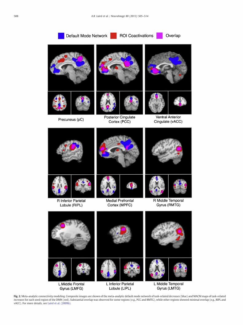
Fig. 2.Meta-analytic connectivitymodeling. Composite images are shown of themeta-analytic defaultmode network of task-related decreases (blue) andMACMmaps of task-relatedincreases for each seed region of the DMN (red). Substantial overlap was observed for some regions (e.g., PCC and RMTG), while other regions showedminimal overlap (e.g., RIPL andvACC). For more details, see Laird et al. (2009b).
508 A.R. Laird et al. / NeuroImage 80 (2013) 505–514

Fig. 3. Connectivity-based parcellation. Both cytoarchitectonic (left) and connectivity-based (right) parcellation analyses were performed, yielding a strong agreement in spa-tial continuity and localization for sub-regions corresponding to the laterobasal (blue),centromedial (red), and superficial (green) nuclei groups. For more details, see Bzdoket al. (in press).
509A.R. Laird et al. / NeuroImage 80 (2013) 505–514
applications have used a semi-automated approach based on spectralreordering of the distance matrix (Johansen-Berg et al., 2004; Kelly etal., 2010), while later applications have used k-means (Cauda et al.,2011; Kahnt et al., 2012; Kim et al., 2010; Nanetti et al., 2009) or hier-archical clustering analysis (Bellec et al., 2006; Bzdok et al., in press;Cordes et al., 2002).
It is important to note that the CBP approach described above iscompletely independent of the modality on which the whole-brainconnectivity profiles are based. While originally described for ana-tomical connectivity measures based on diffusion-weighted images(Johansen-Berg et al., 2004) and resting state functional connectivity(Cordes et al., 2002; Kelly et al., 2010; Kim et al., 2010; van denHeuvel et al., 2008), the very same concept may also be applied totask-based functional connectivity measures, i.e., MACM. Similar toprocedures employed in the other modalities mentioned above, thewhole-brain co-activation pattern is first computed for each voxelwithin the seed region. Subsequently, a distance matrix is computed,indicating the degree of dissimilarity between the co-activationprofiles of each voxel. Finally, the voxels are clustered into distinctsub-regions of the original seed based on this information. In orderto characterize the differences in whole-brain co-activation patternsbetween the ensuing clusters and hence the variations in connectivitythat drove theparcellation of the seed region, follow-upMACManalysesare usually performed using the derived clusters as seeds. MACM-CBPis a relatively new technique for identifying connectivity-based sub-regions of a seed volume, but has already provided new insight intothe functional segregation of the pre-SMA and SMA (Eickhoff et al.,2011), dorsolateral prefrontal cortex (Cieslik et al., 2013), and theamygdala (Bzdok et al., in press). Using this technique, Fig. 3 illustratesthe remarkable correspondence observed between cytoarchitectonic-(left) and connectivity- (right) based parcellations of the amygdalainto the laterobasal, centromedial, and superficial nuclei groups. Thestudy by Bzdok et al. (in press) is an excellent example of hownewly developed neuroimaging analysis methods are providing themeans to investigate concurrence across structural, connectional,and functional sub-specialization, which is critical for progress inconnectomics research.
Discovery of ICA-derived co-activation networks
The early correlational work of Biswal et al. (1995) provided a tan-talizing hint that functionally connected networks could be studied inresting state FMRI data. During the mid to late 2000's an increasinglylarge group of neuroimagers became intrigued by the investigationof resting state networks (RSNs) derived from independent compo-nent analysis (ICA) of rs-FMRI data. With the advent of ICA to the neu-roimaging community, the trend of studying individual seed-basednetworks was broadened to utilize resting state data to simultaneous-ly investigate many of the brain's functionally connected RSNs at once(Beckmann et al., 2005; Damoiseaux et al., 2006). Using ICA, FMRI dataare decomposed into sets of d networks, which typically range from alow model order (e.g., d = 20) to a high model order (e.g., d = 100).Generally speaking, low model ICA decompositions provide a broadassessment of large-scale resting state networks, while high modeldecompositions offer more finely-grained examination of thesenetworks, yet also provide a more complete understanding of thecomplexities associated with how the low model order networksfractionate into higher model order sub-networks as d is increased(Abou-Elseoud et al., 2010).
Given evidence that the brain's functional networks could beextracted from correlations of co-activation data (Toro et al., 2008),and the first set of results provided by MACM (Eickhoff et al., 2010;Laird et al., 2009b; Robinson et al., 2010), it was hypothesized thatmeaningful inferences could be made across a broad range of func-tional brain networks by direct comparison of co-activation networkswith resting state networks. Thus, in a seminal publication, Smith et al.
(2009) independently applied ICA to two types of data: first, from dataacquired during rs-FMRI in 36 individual subjects, and second, togroup results of task activation patterns from 7342 neuroimaging ex-periments (1687 publications) archived in the BrainMap database. Forthe BrainMap data, co-occurrence of different activation locations wasinvestigated across the range of tasks in the database by using ICA toestimate the low model order (i.e., d = 20) set of spatial maps andassociated time series of the major networks of covariance in thebrain. When ICA is applied to BrainMap data, which are 3D sets ofGaussian modeled activation images, the fourth dimension of thedata matrix analyzed refers “experiment ID” (rather than “time” inrs-FMRI data), such that each time point in one component's “timecourse” describes how strongly the observed spatial map relates tothat particular experiment's activation image in BrainMap. When theresults of these independent analyses were compared using spatialPearson cross correlation, Smith et al. demonstrated that over two-

510 A.R. Laird et al. / NeuroImage 80 (2013) 505–514
thirds of the non-artifactual networks at d = 20 matched across rest-ing state and task-based conditions (e.g., 10 of 20 components), andthat functional characterization of these networks was possibleusing BrainMap metadata. These networks have also been extractedfrom the NeuroSynth database (Yarkoni et al., 2011) using a topicmapping approach (Poldrack et al., 2012), adding to the evidencethat these networks represent fundamental components of the brain'sfunctional architecture. Moreover, when the BrainMap task networkswere closely examined by Laird et al. (2011) in comparison to theICA-derived resting state networks observed by Biswal et al. (2010),it was shown that the degree of correspondence across the sets ofresting state and task co-activation networks increased from the ini-tial estimate provided by Smith et al. (2009). Indeed, 12 of the non-artifactual components were an excellent match to those publishedby Biswal et al., whereas four components were a close partialmatch. This improved rate of agreement was attributed to the greatersample size studied, which was increased from 36 subjects in Smithet al. (2009) to 306 subjects in Biswal et al. (2010). Ongoing work isbeing carried out to assess the degree of agreement for higher modelorders (e.g., d = 70, 100). Regardless, the Smith et al. (2009) studydemonstrated that the major task-based functional networks in theactive brain show similar organization to those of the majority of thenetworks of spontaneous covariation in the resting brain. The impor-tant implication here is that the resting state can be shown to be inclu-sive of the brain's functional dynamics from a range of mental tasks.Moreover, there is an intrinsic organization to the brain's functionalnetworks, whose topography is invariable during rest and task.
Functional interpretation of co-activation networks
Across different imaging modalities, mapping the whole-brainconnectivity of large-scale brain networks or specific regions of in-terest generates quantitative and statistically testable informationconcerning connectivity and interactions across neural regions. Inrs-FMRI, relating functional brain networks to specific mental func-tions is difficult, since by definition the resting state lacks behavioralspecificity. Although recent work demonstrates that intrinsic connec-tivity networks can be related to specific behavioral measures (Meieret al., 2012; Mennes et al., 2011), more progress is needed to addressthis gap in our knowledge. The combination of meta-analytic investi-gations and databases such as BrainMap offer an opportunity for thisin that they also allow inference on the characteristics and propertiesof experiments that underlie the co-activations. A wealth of informa-tion concerning the experimental design and methodological detailsof each archived experiment is coded in BrainMap according to awell-defined taxonomy, which has been refined by experts in thefield for nearly two decades. Neuroinformatics tools have subsequent-ly been developed to exploit this valuable source of information toprovide associations with psychological constructs and thus potentialfunctional interpretations for a specific region or regions of interest.
As a follow-up to the Smith et al. (2009) publication, Laird et al.(2011) sought to provide a more complete functional characterizationof the 20 lowmodel order networks. In this study, a slightly larger vol-ume of the literature was available in BrainMap, and hence included8637 experiments from 1840 publications. The BrainMap metadatataxonomy includes the fields of “Behavioral Domain” and “ParadigmClass” that characterize experiments resulting in activation of thespecified region of interest. The behavioral domain (BD) of a particularexperiment identifies the mental process isolated by the statisticalcontrast of images, and includes the main categories of cognition, ac-tion, perception, emotion, and interoception, as well as their relatedsub-categories (Fox et al., 2005). Paradigm class (PC) categorizes thespecific task employed in the published study (Turner and Laird,2012). A metadata matrix that quantified the relationships betweenthe ICA component images and BrainMap experimental metadata forBDs and PCs was generated and analyzed with hierarchical clustering
to determine groupings of similar metadata classes as well as similarsets of networks. Fig. 4 provides a summary of the spatial topographyof the 20 low order BrainMap co-activation networks shown by Lairdet al. (2011), which match those originally presented by Smith et al.(2009) and demonstrate strong correspondence to resting state net-works. Clustering results revealed that the BrainMap co-activationnetworks could be classified into 4 groups relevant to their associatedmental processes: [1] emotional and interoceptive processes that in-cluded networks for limbic and medial temporal areas, subgenualACC and OFC, bilateral basal ganglia and thalamus, bilateral anteriorinsula and anterior cingulate cortex; [2] motor and visuospatial inte-gration, coordination, and execution that included premotor and sup-plementary motor cortices, DLPFC and posterior parietal cortices,hand areas of the primary sensorimotor cortices, and superior parietallobule; [3] visual perception, including visual association cortices, aswell as lateral and medial posterior occipital cortices; and [4] highercognitive processes that included the default mode network, cerebel-lar network, right-lateralized fronto-parietal cortices, auditory corti-ces, mouth areas of the primary sensorimotor cortices, and left-lateralized fronto-parietal cortices. Complete functional explicationof the BrainMap behavioral metadata associated with these networkswas provided by Laird et al. (2011), and the results of these analyseshave been shared with the community with the aim that they willbe useful for functional interpretations of observed resting state net-works in both healthy and clinical investigations (www.brainmap.org/icns).
While this prior work focuses on developing methods to examinethe functional or behavioral interpretation of large-scale brain net-works using BrainMap metadata, similar methods may be applied toindividual regions of interest. Subsequent to applying MACM, func-tional decoding may be performed on a given region of interest,again using the BD and PC metadata fields. The behavioral functionsconsistently associated with the particular part of the brain identifiedas a region of interest are quantitatively inferred by testing which ofthe different BDs and PCs are significantly over-represented amongthe experiments that featured activation in the seed. In other words,a BrainMap metadata analysis identifies which types of experimentsaremore likely than onewould expect by chance to result in activationof the seed region. Similarly, functional interpretation techniques arealso provided by the NeuroSynth Project (Yarkoni et al., 2011; http://neurosynth.org). In contrast to BrainMap, which relies onmanual anno-tation of neuroimaging experiments by its user community, NeuroSynthhas implemented automated harvesting of three-dimensional stereotac-tic and annotations that are tagged for each publication representingterms that occur at high frequency (i.e., 20 ormore studies). A list of sev-eral thousand text-mined terms has been generated that allows quanti-tative associations to be made between a specific term and a givenregion of interest. NeuroSynth's web interface provides access to toolscapable of quickly generating dynamicmeta-analysismaps representingterm-based maps or co-activation maps.
Using their different frameworks for manual and automated anno-tations, BrainMap and NeuroSynth provide tools for examining bothforward inference (i.e., how likely is the region activated given a par-ticular taxonomic label?) and reverse inference (i.e., how likely is aparticular taxonomic label given activation in this region?) (Yarkoniet al., 2011). In other words, forward inference on the functional char-acterization tests the probability of observing activity in a brain regiongiven knowledge of the psychological process, whereas reverse infer-ence tests the probability of a psychological process being presentgiven knowledge of activation of a particular brain region. Whileboth are usually considered in the functional interpretation of an ef-fect, reverse inference is somewhat closer to the colloquial meaningof the question “what is this region doing?”. However, the results ofthis reverse inference analyses are alsomore dependent on the a prioristructure of the database queried or the kind of experiments thatare routinely carried in functional neuroimaging studies. That is, if a
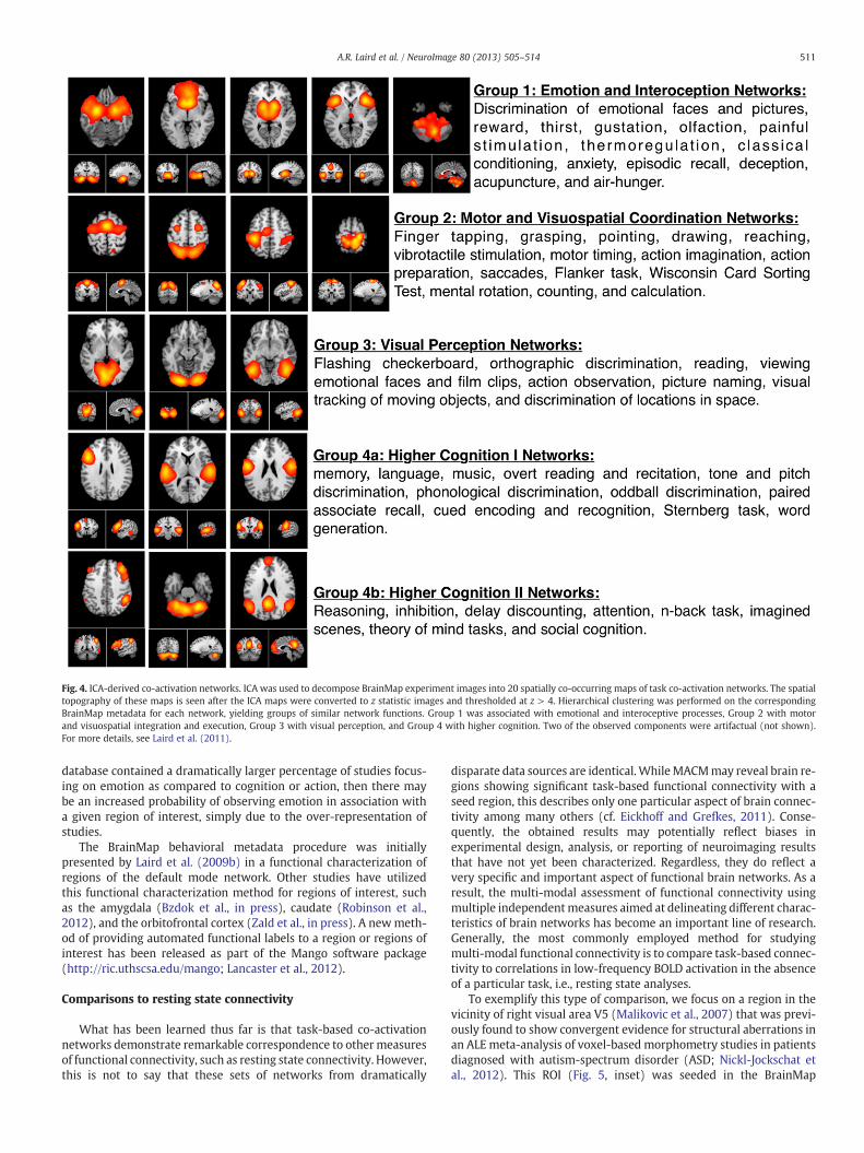
Fig. 4. ICA-derived co-activation networks. ICA was used to decompose BrainMap experiment images into 20 spatially co-occurring maps of task co-activation networks. The spatialtopography of these maps is seen after the ICA maps were converted to z statistic images and thresholded at z > 4. Hierarchical clustering was performed on the correspondingBrainMap metadata for each network, yielding groups of similar network functions. Group 1 was associated with emotional and interoceptive processes, Group 2 with motorand visuospatial integration and execution, Group 3 with visual perception, and Group 4 with higher cognition. Two of the observed components were artifactual (not shown).For more details, see Laird et al. (2011).
511A.R. Laird et al. / NeuroImage 80 (2013) 505–514
database contained a dramatically larger percentage of studies focus-ing on emotion as compared to cognition or action, then there maybe an increased probability of observing emotion in association witha given region of interest, simply due to the over-representation ofstudies.
The BrainMap behavioral metadata procedure was initiallypresented by Laird et al. (2009b) in a functional characterization ofregions of the default mode network. Other studies have utilizedthis functional characterization method for regions of interest, suchas the amygdala (Bzdok et al., in press), caudate (Robinson et al.,2012), and the orbitofrontal cortex (Zald et al., in press). A newmeth-od of providing automated functional labels to a region or regions ofinterest has been released as part of the Mango software package(http://ric.uthscsa.edu/mango; Lancaster et al., 2012).
Comparisons to resting state connectivity
What has been learned thus far is that task-based co-activationnetworks demonstrate remarkable correspondence to othermeasuresof functional connectivity, such as resting state connectivity. However,this is not to say that these sets of networks from dramatically
disparate data sources are identical.WhileMACMmay reveal brain re-gions showing significant task-based functional connectivity with aseed region, this describes only one particular aspect of brain connec-tivity among many others (cf. Eickhoff and Grefkes, 2011). Conse-quently, the obtained results may potentially reflect biases inexperimental design, analysis, or reporting of neuroimaging resultsthat have not yet been characterized. Regardless, they do reflect avery specific and important aspect of functional brain networks. As aresult, the multi-modal assessment of functional connectivity usingmultiple independentmeasures aimed at delineating different charac-teristics of brain networks has become an important line of research.Generally, the most commonly employed method for studyingmulti-modal functional connectivity is to compare task-based connec-tivity to correlations in low-frequency BOLD activation in the absenceof a particular task, i.e., resting state analyses.
To exemplify this type of comparison, we focus on a region in thevicinity of right visual area V5 (Malikovic et al., 2007) that was previ-ously found to show convergent evidence for structural aberrations inan ALE meta-analysis of voxel-based morphometry studies in patientsdiagnosed with autism-spectrum disorder (ASD; Nickl-Jockschat etal., 2012). This ROI (Fig. 5, inset) was seeded in the BrainMap

512 A.R. Laird et al. / NeuroImage 80 (2013) 505–514
database, yielding 198 experiments (performed on 2811 subjects andreporting a total of 3320 foci). As seen in Fig. 5A, MACM results acrossthese 198 experiments indicated significant co-activation betweenthe V5 region affected in ASD and the bilateral lateral occipital cortex,inferior parietal cortex, inferior frontal cortex including the ventralpremotor cortex and BA 44 (Broca's area), and anterior insula andfrontal operculum (P b 0.05, cluster-level corrected for multiple com-parisons). A separate analysis was then performed in which the sameright visual area V5 region was seeded in resting state FMRI data froma group of 132 healthy subjects derived from the NKI/Rocklandsample (Nooner et al., 2012), using standard preprocessing and anal-ysis methods. Fig. 5B reveals that the resting state approach yieldedmuch more extensive functional connectivity in comparison to theMACM approach with almost all posterior parts of the brain, includingthe ventral and dorsal occipital cortex, as well as the inferior andsuperior parietal lobe. Significant resting state connectivity with thepremotor cortex was also found, but with a stronger emphasis onits dorsal aspect than observed in the task-dependent data. Whencomputing the conjunction between both approaches, the occipitaland parietal regions that featured significant MACM and restingstate connectivity with the V5 seed closely resembled those foundin the MACM analysis (Fig. 5C). In turn, significant frontal connectiv-ity that was robust over both analyses was only observed in a smallcluster of the left precentral gyrus and a larger one in the right inferi-or frontal gyrus located within BA 44 and the adjacent ventralpremotor cortex.
In light of the as-yet limited number of studies combining MACMand resting state functional connectivity analyses (Cieslik et al., 2013;Eickhoff et al., 2011; Jakobs et al., 2012; Reetz et al., 2012; Rottschyet al., in press), the above results obtained for the V5 region thatwas shown to be structurally affected in ASD features several typicalaspects. First, all of these previous studies demonstrated the presenceof robust networks of functional interaction with a seed region thatare likewise present in both approaches for mapping whole-brainfunctional interactions. Given the independent nature of the data aswell as the conceptual and methodological differences betweenMACM and resting state analyses, this convergence holds importantimplications. In particular, the ensuing regions may be considered a“core” network that is robustly interacting with the seed independentof whether the subjects are in a self-referentially, endogenouslycontrolled mind-wandering state (cf. Schilbach et al., 2012 for a psy-chological interpretation of this mental state) or in an exogenouslydriven state imposed by a structured experimental paradigm. Thatis, these regions showing significant association across methods andmental states may be deemed the most robust and consistent interac-tions of the seed. Another rather frequent observation is the fact thatin addition to this core network there are usually also appreciable dif-ferences in the revealed interactions. These differences demonstratethat in spite of the same underlying concept of “functional connectiv-ity”, MACM and resting state analyses actually do reflect differentinformation about brain connectivity. In this respect, resting statecorrelations often tend to show a more extensive network whenthresholded at the same level of significance. In contrast, regionsexhibiting significant MACM co-activation but not resting state func-tional connectivity are much more sparse. Nevertheless, as shown bythe example focusing on right visual area V5, those regions neverthe-less commonly exist (e.g., the anterior insula/frontal operculum).There are multiple different factors that may contribute to this kindof pattern. The potentially most interesting one pertains to the differ-ence in mental states, i.e., endogenously vs. exogenously orientationof attention. Following this line of reasoning, the differences observedbetween MACM and resting state analyses could reflect differences infunctional interaction that are conditioned upon the current mentalstate of the subjects. However, technical and conceptual differencesbetween both approaches, as alluded to above, may not be discountedeither. Consequently, it must remain open at present, whether the
observed differences reflect primarily differential biases (or even arti-facts) of the twomethods or true differences in interaction patterns ofthe seed region dependent on the mental state of the subjects.
Limitations or considerations for the co-activation approach
Themeta-analytic approach for identifying functionally co-activatedbrain networks is an appealing area of connectomics research since it al-lows for insight into the brain's connectivity during the engagement ofgoal-directed behavior. However, this behavior is not limited to a single,specific task, but encompasses a diverse range of tasks. This is both anadvantage and a disadvantage, depending on one's research perspec-tive. Although we place much emphasis on the results of Smith et al.(2009), which revealed strong similarity between resting state andco-activation networks, it is important to consider that these networksare not explicitly identical. Recent work by Mennes et al. (2013) hasshown that the relationship between intrinsic and extrinsic connectivi-ty is quite complex, and that evoked interaction patterns show weakercorrespondence to intrinsic connectivity networks, particularly for sub-cortical and limbic regions, aswell as primary sensorimotor areas. Thereis no substitute for the precision, temporal resolution, and power of acarefully controlled task-based neuroimaging experiment, and we donot advocate abandonment of this domain of research. Rather, we pro-mote simultaneous and symbiotic exploration of what knowledge maybe gleaned from a meta-analytic co-activation perspective.
Forward progress in developing meta-analytic methods can bechallenging as this data is sometimes construed as being too highlyvariable and noisy. Clusters of functional brain activations have highlycomplex and rich shape in three-dimensional space. Extracting thecenters of masses of these clusters and analysis of the reported fociof these locations represents a dramatic loss in spatial sensitivityand specificity. Moreover, pooling foci across studies results in a lossof precision with respect to various experimental parameters, suchas scanner strength, imaging acquisition and analysis, subject samplesize and individual variability, and variations in behavioral condi-tions. However, it can be reasoned that observation of consistent re-sults across studies despite this variability represents an undeniablypowerful response of the brain to task and should therefore be exam-ined to the fullest. Furthermore, the fact that the brain's co-activationpatterns can be disentangled to extract functional brain networksnot only validates that meta-analysis is indeed an experimentallyvalid source of data, but it is also telling us something extraordinaryabout brain organization and how we conceptualize the associationsbetween behavioral tasks and the mental operations they elicit.
Conclusion
While one of the more novel aspects of human connectomics re-search, task co-activation networks are providing biologically mean-ingful insight into functional brain dynamics and interactions. Meta-analytic connectivity modeling, connectivity-based parcellation, andbehavioral metadata analyses have been successfully applied to re-gions of the default mode network, visual area V5, and the amygdala,among others. Resting state analyses are undeniably powerful, yetco-activation connectivity via the task activation literature offers aquantitative means to address the behavioral and experimental spec-ificity of a region of interest. Moreover, although there is much workyet to be done, preliminary studies have shown that the level of con-vergence and divergence in co-activation connectivity and restingstate connectivity is quite complex, and may offer increased insightinto the relationship between external and internal orienting of atten-tion. Future examination of task co-occurrence networks will focus onelucidating these patterns, and on applying the methods describedhere to AN even larger array OF cortical and subcortical brain regions.
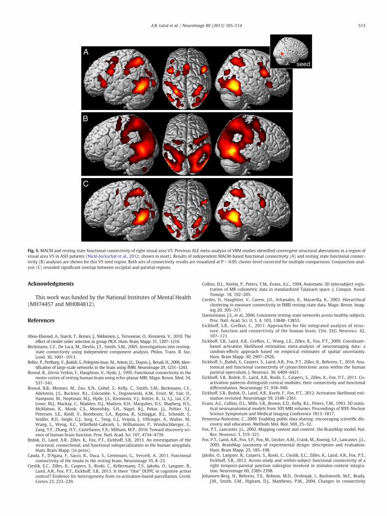
Fig. 5. MACM and resting state functional connectivity of right visual area V5. Previous ALE meta-analysis of VBM studies identified convergent structural aberrations in a region ofvisual area V5 in ASD patients (Nickl-Jockschat et al., 2012; shown in inset). Results of independent MACM-based functional connectivity (A) and resting state functional connec-tivity (B) analyses are shown for this V5 seed region. Both sets of connectivity results are visualized at P b 0.05, cluster-level corrected for multiple comparisons. Conjunction anal-ysis (C) revealed significant overlap between occipital and parietal regions.
513A.R. Laird et al. / NeuroImage 80 (2013) 505–514
Acknowledgments
This work was funded by the National Institutes of Mental Health(MH74457 and MH084812).
References
Abou-Elseoud, A., Starck, T., Remes, J., Nikkienen, J., Tervonene, O., Kiviniemi, V., 2010. Theeffect of model order selection in group PICA. Hum. Brain Mapp. 31, 1207–1216.
Beckmann, C.F., De Luca, M., Devlin, J.T., Smith, S.M., 2005. Investigations into resting-state connectivity using independent component analysis. Philos. Trans. R. Soc.Lond. 36, 1001–1013.
Bellec, P., Perlbarg, V., Jbabdi, S., Pelegrini-Issac, M., Anton, J.L., Doyon, J., Benali, H., 2006. Iden-tification of large-scale networks in the brain using fMRI. Neuroimage 29, 1231–1243.
Biswal, B., Zerrin Yetkin, F., Haughton, V., Hyde, J., 1995. Functional connectivity in themotor cortex of resting human brain using echo-planar MRI. Magn. Reson. Med. 34,537–541.
Biswal, B.B., Mennes, M., Zuo, X.N., Gohel, S., Kelly, C., Smith, S.M., Beckmann, C.F.,Adelstein, J.S., Buckner, R.L., Colcombe, S., Dogonowski, A.M., Ernst, M., Fair, D.,Hampson, M., Hoptman, M.J., Hyde, J.S., Kiviniemi, V.J., Kötter, R., Li, S.J., Lin, C.P.,Lowe, M.J., Mackay, C., Madden, D.J., Madsen, K.H., Margulies, D.S., Mayberg, H.S.,McMahon, K., Monk, C.S., Mostofsky, S.H., Nagel, B.J., Pekar, J.J., Peltier, S.J.,Petersen, S.E., Riedl, V., Rombouts, S.A., Rypma, B., Schlaggar, B.L., Schmidt, S.,Seidler, R.D., Siegle, G.J., Sorg, C., Teng, G.J., Veijola, J., Villringer, A., Walter, M.,Wang, L., Weng, X.C., Whitfield-Gabrieli, S., Williamson, P., Windischberger, C.,Zang, Y.F., Zhang, H.Y., Castellanos, F.X., Milham, M.P., 2010. Toward discovery sci-ence of human brain function. Proc. Natl. Acad. Sci. 107, 4734–4739.
Bzdok, D., Laird, A.R., Zilles, K., Fox, P.T., Eickhoff, S.B., 2013. An investigation of thestructural, connectional, and functional subspecialization in the human amygdala.Hum. Brain Mapp. (in press).
Cauda, F., D'Agata, F., Sacco, K., Duca, S., Geminiani, G., Vercelli, A., 2011. Functionalconnectivity of the insula in the resting brain. Neuroimage 55, 8–23.
Cieslik, E.C., Zilles, K., Caspers, S., Roski, C., Kellermann, T.S., Jakobs, O., Langner, R.,Laird, A.R., Fox, P.T., Eickhoff, S.B., 2013. Is there “One” DLPFC in cognitive actioncontrol? Evidence for heterogeneity from co-activation-based parcellation. Cereb.Cortex 23, 223–229.
Collins, D.L., Neelin, P., Peters, T.M., Evans, A.C., 1994. Automatic 3D intersubject regis-tration of MR columetric data in standardized Talairach space. J. Comput. Assist.Tomogr. 18, 192–205.
Cordes, D., Haughton, V., Carew, J.D., Arkanakis, K., Maravilla, K., 2002. Hierarchicalclustering to measure connectivity in fMRI resting-state data. Magn. Reson. Imag-ing 20, 305–317.
Damoiseaux, J.S., et al., 2006. Consistent resting-state networks across healthy subjects.Proc. Natl. Acad. Sci. U. S. A. 103, 13848–13853.
Eickhoff, S.B., Grefkes, C., 2011. Approaches for the integrated analysis of struc-ture, function and connectivity of the human brain. Clin. EEG Neurosci. 42,107–121.
Eickhoff, S.B., Laird, A.R., Grefkes, C., Wang, L.E., Zilles, K., Fox, P.T., 2009. Coordinate-based activation likelihood estimation meta-analysis of neuroimaging data: arandom-effects approach based on empirical estimates of spatial uncertainty.Hum. Brain Mapp. 30, 2907–2926.
Eickhoff, S., Jbabdi, S., Caspers, S., Laird, A.R., Fox, P.T., Zilles, K., Behrens, T., 2010. Ana-tomical and functional connectivity of cytoarchitectonic areas within the humanparietal operculum. J. Neurosci. 30, 6409–6421.
Eickhoff, S.B., Bzdok, D., Laird, A.R., Roski, C., Caspers, S., Zilles, K., Fox, P.T., 2011. Co-activation patterns distinguish cortical modules, their connectivity and functionaldifferentiation. Neuroimage 57, 938–949.
Eickhoff, S.B., Bzdok, D., Laird, A.R., Kurth, F., Fox, P.T., 2012. Activation likelihood esti-mation revisited. Neuroimage 59, 2349–2361.
Evans, A.C., Collins, D.L., Mills, S.R., Brown, E.D., Kelly, R.L., Peters, T.M., 1993. 3D statis-tical neuroanatomical models from 305 MRI volumes. Proceedings of IEEE-NuclearScience Symposium and Medical Imaging Conference 1813–1817.
Fennema-Notestine, C., 2009. Enabling public data sharing: encouraging scientific dis-covery and education. Methods Mol. Biol. 569, 25–32.
Fox, P.T., Lancaster, J.L., 2002. Mapping context and content: the BrainMap model. Nat.Rev. Neurosci. 3, 319–321.
Fox, P.T., Laird, A.R., Fox, S.P., Fox, M., Uecker, A.M., Crank, M., Koenig, S.F., Lancaster, J.L.,2005. BrainMap taxonomy of experimental design: description and evaluation.Hum. Brain Mapp. 25, 185–198.
Jakobs, O., Langner, R., Caspers, S., Roski, C., Cieslik, E.C., Zilles, K., Laird, A.R., Fox, P.T.,Eickhoff, S.B., 2012. Across-study and within-subject functional connectivity of aright temporo-parietal junction subregion involved in stimulus-context integra-tion. Neuroimage 60, 2389–2398.
Johansen-Berg, H., Behrens, T.E., Robson, M.D., Drobnjak, I., Rushworth, M.F., Brady,J.M., Smith, S.M., Higham, D.J., Matthews, P.M., 2004. Changes in connectivity

514 A.R. Laird et al. / NeuroImage 80 (2013) 505–514
profiles define functionally distinct regions in human medial frontal cortex. Proc.Natl. Acad. Sci. U. S. A. 101, 13335–13340.
Kahnt, T., Chang, L.J., Park, S.Q., Heinzle, J., Haynes, J.D., 2012. Connectivity-basedparcellation of the human orbitofrontal cortex. J. Neurosci. 32, 6240–6250.
Keator, D.B., Grethe, J.S., Marcus, D., Ozyurt, B., Gadde, S., Murphy, S., et al., 2008. A na-tional human neuroimaging collaboratory enabled by the Biomedical InformaticsResearch Network (BIRN). IEEE Trans. Inf. Technol. Biomed. 12, 162–172.
Kelly, C., Uddin, L.Q., Shehzad, Z., Margulies, D.S., Castellanos, F.X., Milham, M.P., Petrides,M., 2010. Broca's region: linking human brain functional connectivity data and non-human primate tracing anatomy studies. Eur. J. Neurosci. 32, 383–398.
Kim, J.H., Lee, J.M., Jo, H.J., Kim, S.H., Lee, J.H., Kim, S.T., Seo, S.W., Cox, R.W., Na, D.L.,Kim, S.I., Saad, Z.S., 2010. Defining functional SMA and pre-SMA subregions inhuman MFC using resting state fMRI: functional connectivity-based parcellationmethod. Neuroimage 49, 2375–2386.
Koski, L., Paus, T., 2000. Functional connectivity of the anterior cingulate cortex withinthe human frontal lobe: a brain-mapping meta-analysis. Exp. Brain Res. 133,55–65.
Laird, A.R., Lancaster, J.L., Fox, P.T., 2005a. BrainMap: the social evolution of a functionalneuroimaging database. Neuroinformatics 3, 65–78.
Laird, A.R., Fox, M., Price, C.J., Glahn, D.C., Uecker, A.M., Lancaster, J.L., Turkeltaub, P.E.,Kochunov, P., Fox, P.T., 2005b. ALE meta-analysis: controlling the false discoveryrate and performing statistical contrasts. Hum. Brain Mapp. 25, 155–164.
Laird, A.R., Eickhoff, S.B., Kurth, F., Fox, P.M., Uecker, A.M., Turner, J.A., Robinson, J.L.,Lancaster, J.L., Fox, P.T., 2009a. ALE meta-analysis workflows via the BrainMapdatabase: progress towards a probabilistic functional brain atlas. Front.Neuroinformatics 3, 23.
Laird, A.R., Eickhoff, S.B., Li, K., Robin, D.A., Glahn, D.C., Fox, P.T., 2009b. Investigatingthe functional heterogeneity of the default mode network using coordinate-based meta-analytic modeling. J. Neurosci. 29, 14496–14505.
Laird, A.R., Fox, P.M., Eickhoff, S.B., Turner, J.A., Ray, K.L., McKay, D.R., Glahn, D.C.,Beckmann, C.F., Smith, S.M., Fox, P.T., 2011. Behavioral interpretations of intrinsicconnectivity networks. J. Cogn. Neurosci. 23, 4022–4037.
Lancaster, J.L., Laird, A.R., Eickhoff, S.B., Martinez, M.J., Fox, P.M., Fox, P.T., 2012. Automatedregional behavioral analysis for human brain images. Front. Neuroinformatics 6, 23.
Malikovic, A., Amunts, K., Schleicher, A., Mohlberg, H., Eickhoff, S.B., Wilms, M.,Palomero-Gallagher, N., Armstrong, E., Zilles, K., 2007. Cytoarchitectonic analysisof the human extrastriate cortex in the region of V5/MT+: a probabilistic, stereo-taxic map of area hOc5. Cereb. Cortex 17, 562–574.
Marcus, D.S., Wang, T.H., Parker, J., Csernansky, J.G., Morris, J.C., Buckner, R.L., 2007a.Open Access Series of Imaging Studies (OASIS): cross-sectional MRI data inyoung, middle aged, nondemented, and demented older adults. J. Cogn. Neurosci.19, 1498–1507.
Marcus, D.S., Olsen, T.R., Ramaratnam, M., Buckner, R.L., 2007b. The Extensible Neuro-imaging Archive Toolkit: an informatics platform for managing, exploring, andsharing neuroimaging data. Neuroinformatics 5, 11–34.
Meier, T.B., Wildenberg, J.C., Liu, J., Chen, J., Calhoun, V.D., Biswal, B.B., Meyerand, M.E., Birn,R.M., Prabhakaran, V., 2012. Parallel ICA identifies sub-components of resting state net-works that covary with behavioral indices. Front. Hum. Neurosci. 6, 281.
Mennes, M., Zuo, X.N., Kelly, C., Di Martino, A., Zang, Y.F., Biswal, B., Castellanos, F.X.,Milham, M.P., 2011. Linking inter-individual differences in neural activation andbehavior to intrinsic brain dynamics. Neuroimage 54, 2950–2959.
Mennes, M., Kelly, C., Colcombe, S., Castellanos, F.X., Milham, M.P., 2013. The extrinsicand intrinsic functional architectures of the human brain are not equivalent. Cereb.Cortex 23, 223–229.
Mennes, M., Biswal, B.B., Castellanos, F.X., Milham, M.P., 2013. Making data shar-ing work: the FCP/INDI experience. Neuroimage. http://dx.doi.org/10.1016/j.neuroimage.2012.10.064 (in press).
Nanetti, L., Cerliani, L., Gazzola, V., Renken, R., Keysers, C., 2009. Group analyses ofconnectivity-based cortical parcellation using repeated k-means clustering.Neuroimage 47, 1666–1677.
Nickl-Jockschat, T., Habel, U., Michel, T.M., Manning, J., Laird, A.R., Fox, P.T., Schneider,F., Eickhoff, S.B., 2012. Brain structure anomalies in autism spectrum disorder—Ameta-analysis of VBM studies using anatomic likelihood estimation. Hum. BrainMapp. 33, 1470–1489.
Nooner, K.B., Colcombe, S.J., Tobe, R.H., Mennes, M., Benedict, M.M., Moreno, A.L.,Panek, L.J., Brown, S., Zavitz, S.T., Li, Q., Sikka, S., Gutman, D., Bangaru, S.,Schlachter, R.T., Kamiel, S.M., Anwar, A.R., Hinz, C.M., Kaplan, M.S., Rachlin, A.B.,Adelsberg, S., Cheung, B., Khanuja, R., Yan, C., Craddock, C.C., Calhoun, V.,Courtney, W., King, M., Wood, D., Cox, C.L., Kelly, A.M., Di Martino, A., Petkova, E.,Reiss, P.T., Duan, N., Thomsen, D., Biswal, B., Coffey, B., Hoptman, M.J., Javitt, D.C.,Pomara, N., Sidtis, J.J., Koplewicz, H.S., Castellanos, F.X., Leventhal, B.L., Milham,M.P., 2012. The NKI-Rockland sample: a model for accelerating the pace of discov-ery science in psychiatry. Front. Neurosci. 6, 152.
Poldrack, R.A., 2006. Can cognitive processes be inferred from neuroimaging data?Trends Cogn. Sci. 10, 59–63.
Poldrack, R.A., Mumford, J.A., Schonberg, T., Kalar, D., Barman, B., Yarkoni, T., 2012. Dis-covering relations between mind, brain, and mental disorders using topic map-ping. PLoS Comp. Biol. 8, e1002707.
Postuma, R.B., Dagher, A., 2006. Basal ganglia functional connectivity based on a meta-analysis of 126 positron emission tomography and functional magnetic resonanceimaging publications. Cereb. Cortex 16, 1508–1521.
Reetz, K., Dogan, I., Rolfs, A., Binkofski, F., Schulz, J.B., Laird, A.R., Fox, P.T., Eickhoff,S.B., 2012. Investigating function and connectivity of morphometric findings —Exemplified on cerebellar atrophy in spinocerebellar ataxia 17 (SCA17).Neuroimage 62, 1354–1366.
Robinson, J.L., Laird, A.R., Glahn, D.C., Lovallo, W.R., Fox, P.T., 2010. Meta-analytic con-nectivity modelling: delineating the functional connectivity of the human amygda-la. Hum. Brain Mapp. 31, 173–184.
Robinson, J.L., Laird, A.R., Glahn, D.C., Blangero, J., Sanghera, M.K., Pessoa, L., Fox, P.M.,Uecker, A., Friehs, G., Young, K.A., Griffin, J.L., Lovallo, W.R., Fox, P.T., 2012.The functional connectivity of the human caudate: an application of meta-analytic connectivity modeling with behavioral filtering. Neuroimage 60, 117–129.
Rottschy, C., Caspers, S., Roski, C., Reetz, K., Dogan, I., Schulz, J.B., Zilles, K., Laird, A.R.,Fox, P.T., Eickhoff, S.B., 2013. Differentiated parietal connectivity of frontal regionsfor “what” and “where” memory. Brain Struct. Funct. (in press).
Schilbach, L., Bzdok, D., Timmermans, B., Fox, P.T., Laird, A.R., Vogeley, K., Eickhoff, S.B.,2012. Introspective minds: using ALE meta-analyses to study commonalities in theneural correlates of emotional processing, social & unconstrained cognition. PLoSOne 7, e30920.
Smith, S.M., Fox, P.T., Miller, K.L., Glahn, D.C., Fox, P.M., Mackay, C.E., Filippini, N., Watkins,K.E., Toro, R., Laird, A.R., Beckmann, C.F., 2009. Correspondence of the brain's functionalarchitecture during activation and rest. Proc. Natl. Acad. Sci. U. S. A. 106, 13040–13045.
Talairach, J., Tournoux, P., 1988. Co-planar Stereotaxic Atlas of the Human Brain.Thieme, New York.
Toro, R., Fox, P.T., Paus, T., 2008. Functional coactivation map of the human brain. Cereb.Cortex 18, 2553–2559.
Turkeltaub, P.E., Eden, G.F., Jones, K.M., Zeffiro, T.A., 2002. Meta-analysis of the func-tional neuroanatomy of single-word reading: method and validation. Neuroimage16, 765–780.
Turkeltaub, P.E., Eickhoff, S.B., Laird, A.R., Fox, M., Wiener, M., Fox, P., 2012. Minimizingwithin-experiment and within-group effects in activation likelihood estimationmeta-analyses. Hum. Brain Mapp. 33, 1–13.
Turner, J.A., Laird, A.R., 2012. The cognitive paradigm ontology: design and application.Neuroinformatics 10, 57–66.
van den Heuvel, M., Mandl, R., Hulshoff, Pol, 2008. Normalized cut group clustering ofresting-state fMRI data. PLoS One 3, e2001.
Van Essen, D.C., Ugurbil, K., Auerbach, E., Barch, D., Behrens, T.E., Bucholz, R., Chang, A.,Chen, L., Corbetta, M., Curtiss, S.W., Della Penna, S., Feinberg, D., Glasser, M.F.,Harel, N., Heath, A.C., Larson-Prior, L., Marcus, D., Michalareas, G., Moeller, S.,Oostenveld, R., Petersen, S.E., Prior, F., Schlaggar, B.L., Smith, S.M., Snyder, A.Z.,Xu, J., Yacoub, E., WU-Minn HCP Consortium, 2012. The Human ConnectomeProject: a data acquisition perspective. Neuroimage 62, 2222–2231.
Yarkoni, T., Poldrack, R.A., Nichols, T.E., Van Essen, D.C., Wager, T.D., 2011. Large-scale automated synthesis of human functional neuroimaging data. Nat.Methods 8, 665–670.
Zald, D.H., McHugo, M., Ray, K.L., Glahn, D.C., Eickhoff, S.B., Laird, A.R., 2013. Meta-analytic connectivity modeling reveals differential functional connectivity of themedial and lateral orbitofrontal cortex. Cereb. Cortex (in press).