nervous tissue - butler community collegeinstructors.butlercc.edu/sforrest/apch12fa08.pdfproperties...
TRANSCRIPT

12-1
Nervous Tissue
• Overview of the nervous system
• Nerve cells (neurons)• Supportive cells
(neuroglia)• Electrophysiology of
neurons• Synapses• Neural integration

12-2
Overview of Nervous System
• Endocrine and nervous system maintain internal coordination– endocrine = chemical messengers
(hormones) delivered to the bloodstream
– nervous = three basic steps• sense organs receive information• brain and spinal cord determine responses• brain and spinal cord issue commands to
glands and muscles

12-3
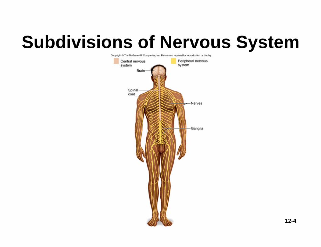
Subdivisions of Nervous System
Two major anatomical subdivisions• Central nervous system (CNS)
– brain and spinal cord enclosed in bony coverings
• Peripheral nervous system (PNS)– nerve = bundle of axons in connective
tissue– ganglion = swelling of cell bodies in a
nerve
12-4
Subdivisions of Nervous System
12-5
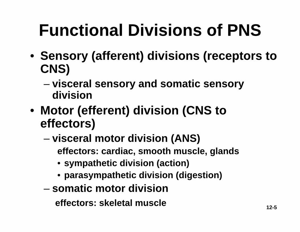
• Sensory (afferent) divisions (receptors to CNS)– visceral sensory and somatic sensory
division• Motor (efferent) division (CNS to
effectors)– visceral motor division (ANS)
effectors: cardiac, smooth muscle, glands• sympathetic division (action)• parasympathetic division (digestion)
– somatic motor divisioneffectors: skeletal muscle
Functional Divisions of PNS

12-6
Subdivisions of Nervous System

12-7
Fundamental Types of Neurons
• Sensory (afferent) neurons– detect changes in body and external environment– information transmitted into brain or spinal cord
• Interneurons (association neurons)– lie between sensory and motor pathways in CNS– 90% of our neurons are interneurons– process, store and retrieve information
• Motor (efferent) neuron– send signals out to muscles and gland cells– organs that carry out responses called effectors

12-8
Fundamental Types of Neurons
12-9
Properties of Neurons
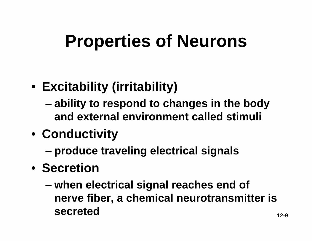
• Excitability (irritability)– ability to respond to changes in the body
and external environment called stimuli• Conductivity
– produce traveling electrical signals• Secretion
– when electrical signal reaches end of nerve fiber, a chemical neurotransmitter is secreted

12-10
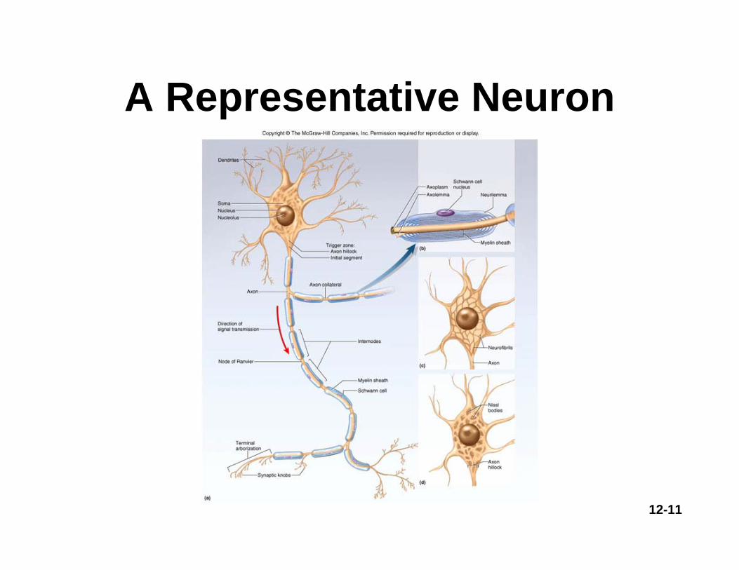
Structure of a Neuron• Cell body = perikaryon =
soma– single, central nucleus with
large nucleolus
– cytoskeleton of microtubules and neurofibrils (bundles of actin filaments)
• compartmentalizes RER into Nissl bodies
– lipofuscin product of breakdown of worn-out organelles -- more with age
• Vast number of short dendrites
– for receiving signals• Singe axon (nerve fiber)
arising from axon hillock for rapid conduction
– axoplasm and axolemma and synaptic vesicles
12-11
A Representative Neuron

12-12
Variation in Neural Structure• Multipolar neuron
– most common– many dendrites/one
axon• Bipolar neuron
– one dendrite/one axon– olfactory, retina, ear
• Unipolar neuron– sensory from skin and
organs to spinal cord• Anaxonic neuron
– many dendrites/no axon
– help in visual processes

12-13
Types of Neuroglial Cells 1
• Oligodendrocytes form myelin sheaths in CNS– each wraps around many nerve fibers
• Ependymal cells line cavities and produce CSF
• Microglia (macrophages) formed from monocytes– in areas of infection, trauma or stroke

12-14
Types of Neuroglial Cells 2
• Astrocytes– most abundant glial cells - form framework of CNS– contribute to BBB and regulate composition of brain tissue
fluid– convert glucose to lactate to feed neurons– secrete nerve growth factor promoting synapse formation– electrical influence on synaptic signaling– sclerosis – damaged neurons replace by hardened mass of
astrocytes• Schwann cells myelinate fibers of PNS• Satellite cells with uncertain function

12-15
Neuroglial Cells of CNS

12-16
Myelin 1• Insulating layer around a nerve fiber
– oligodendrocytes in CNS and schwann cells in PNS
– formed from wrappings of plasma membrane• 20% protein and 80 % lipid (looks white)
– all myelination completed by late adolescence• In PNS, hundreds of layers wrap axon
– the outermost coil is schwann cell (neurilemma)
– covered by basal lamina and endoneurium

12-17
Myelin 2• In CNS - no neurilemma or endoneurium• Oligodendrocytes myelinate several fibers
– Myelination spirals inward with new layers pushed under the older ones
• Gaps between myelin segments = nodes of Ranvier
• Initial segment (area before 1st schwann cell) and axon hillock form trigger zone where signals begin
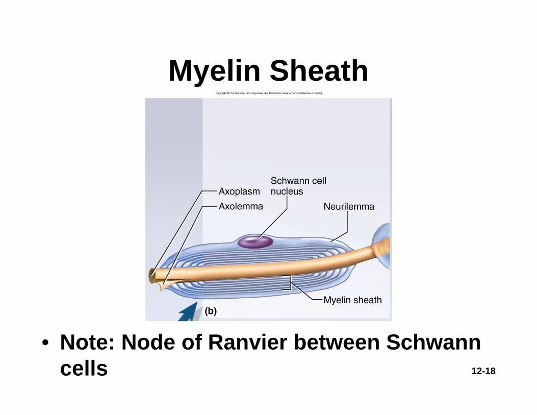
12-18
Myelin Sheath
• Note: Node of Ranvier between Schwann cells
12-19
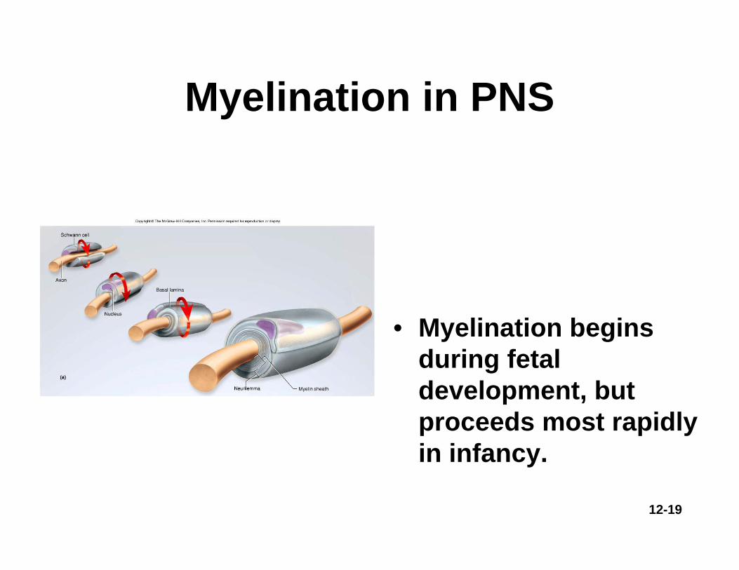
Myelination in PNS
• Myelination begins during fetal development, but proceeds most rapidly in infancy.

12-20
Unmyelinated Axons of PNS
• Schwann cells hold small nerve fibers in grooves on their surface with only one membrane wrapping

12-21
Myelination in CNS
12-22
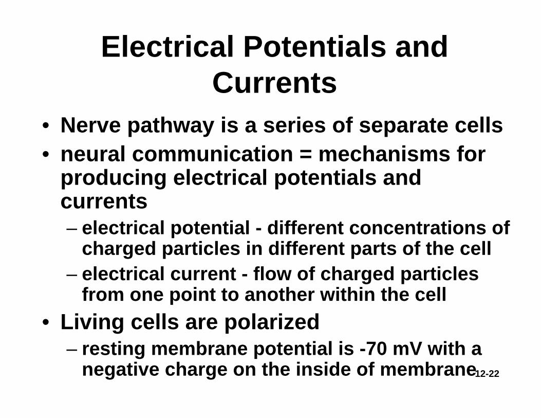
Electrical Potentials and Currents
• Nerve pathway is a series of separate cells• neural communication = mechanisms for
producing electrical potentials and currents– electrical potential - different concentrations of
charged particles in different parts of the cell– electrical current - flow of charged particles
from one point to another within the cell• Living cells are polarized
– resting membrane potential is -70 mV with a negative charge on the inside of membrane

12-23
Resting Membrane Potential
• Unequal electrolytes distribution between ECF/ICF
• Diffusion of ions down their concentration gradients
• Selective permeability of plasma membrane
• Electrical attraction of cations and anions
12-24
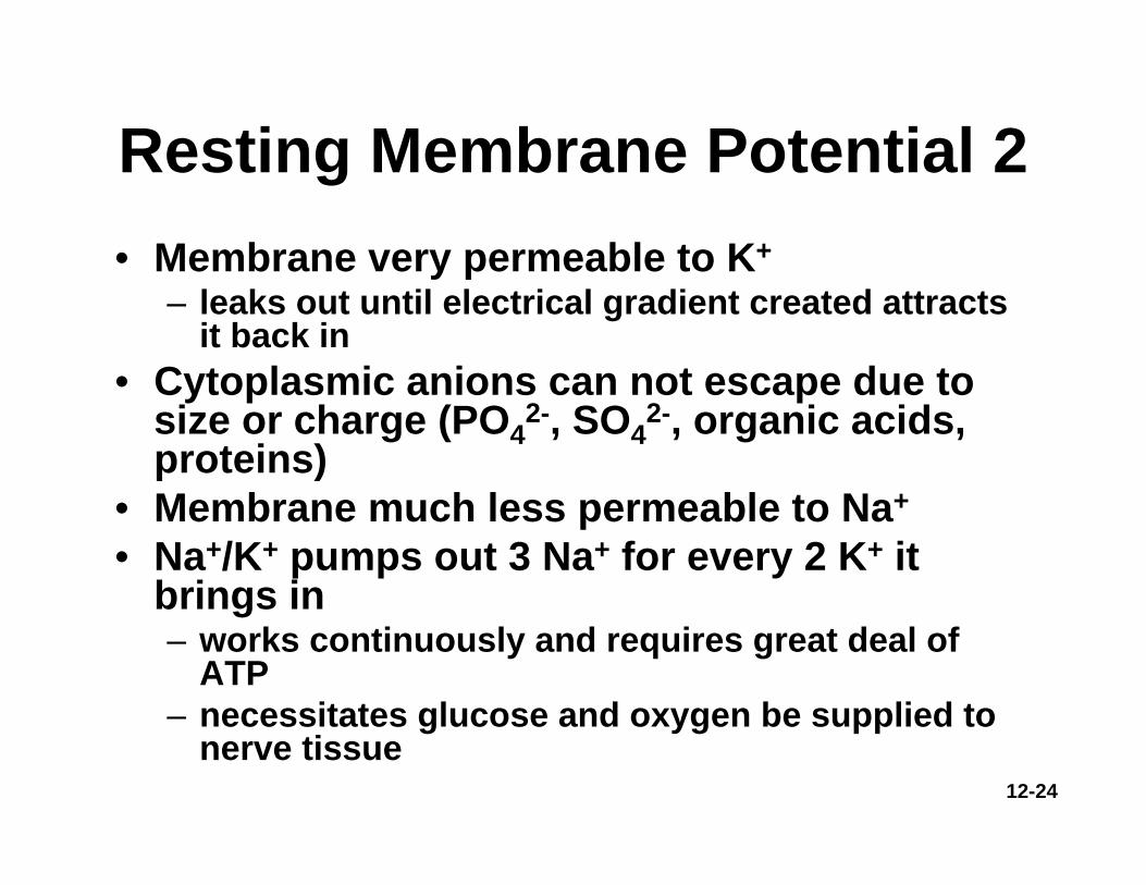
Resting Membrane Potential 2• Membrane very permeable to K+
– leaks out until electrical gradient created attracts it back in
• Cytoplasmic anions can not escape due to size or charge (PO4
2-, SO42-, organic acids,
proteins)• Membrane much less permeable to Na+
• Na+/K+ pumps out 3 Na+ for every 2 K+ it brings in– works continuously and requires great deal of
ATP– necessitates glucose and oxygen be supplied to
nerve tissue

12-25
Ionic Basis of Resting Membrane Potential
• Na+ concentrated outside of cell (ECF) • K+ concentrated inside cell (ICF)
12-26
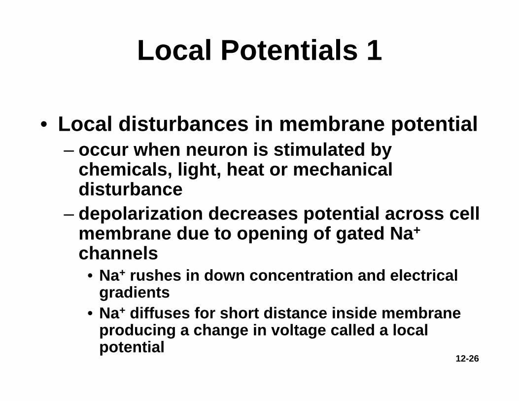
Local Potentials 1
• Local disturbances in membrane potential – occur when neuron is stimulated by
chemicals, light, heat or mechanical disturbance
– depolarization decreases potential across cell membrane due to opening of gated Na+
channels • Na+ rushes in down concentration and electrical
gradients• Na+ diffuses for short distance inside membrane
producing a change in voltage called a local potential

12-27
Local Potentials 2
• Differences from action potential– are graded (vary in magnitude with
stimulus strength)– are decremental (get weaker the farther
they spread)– are reversible as K+ diffuses out of cell– can be either excitatory or inhibitory
(hyperpolarize)

12-28
Chemical Excitation

12-29
Action Potentials• More dramatic change in membrane
produced where high density of voltage-gated channels occur– trigger zone up to 500 channels/μm2 (normal is
75)• If threshold potential (-55mV) is reached
voltage-gated Na+ channels open (Na+
enters causing depolarization)• Past 0 mV, Na+ channels close =
depolarization• Slow K+ gates fully open• K+ exits repolarizing the cell• Negative overshoot produces
hyperpolarization– excessive exiting of K+

12-30
Action Potentials• Called a spike• Characteristics of AP
– follows an all-or-none law • voltage gates either open or
don’t– nondecremental (do not get
weaker with distance)– irreversible (once started
goes to completion and can not be stopped)
12-31
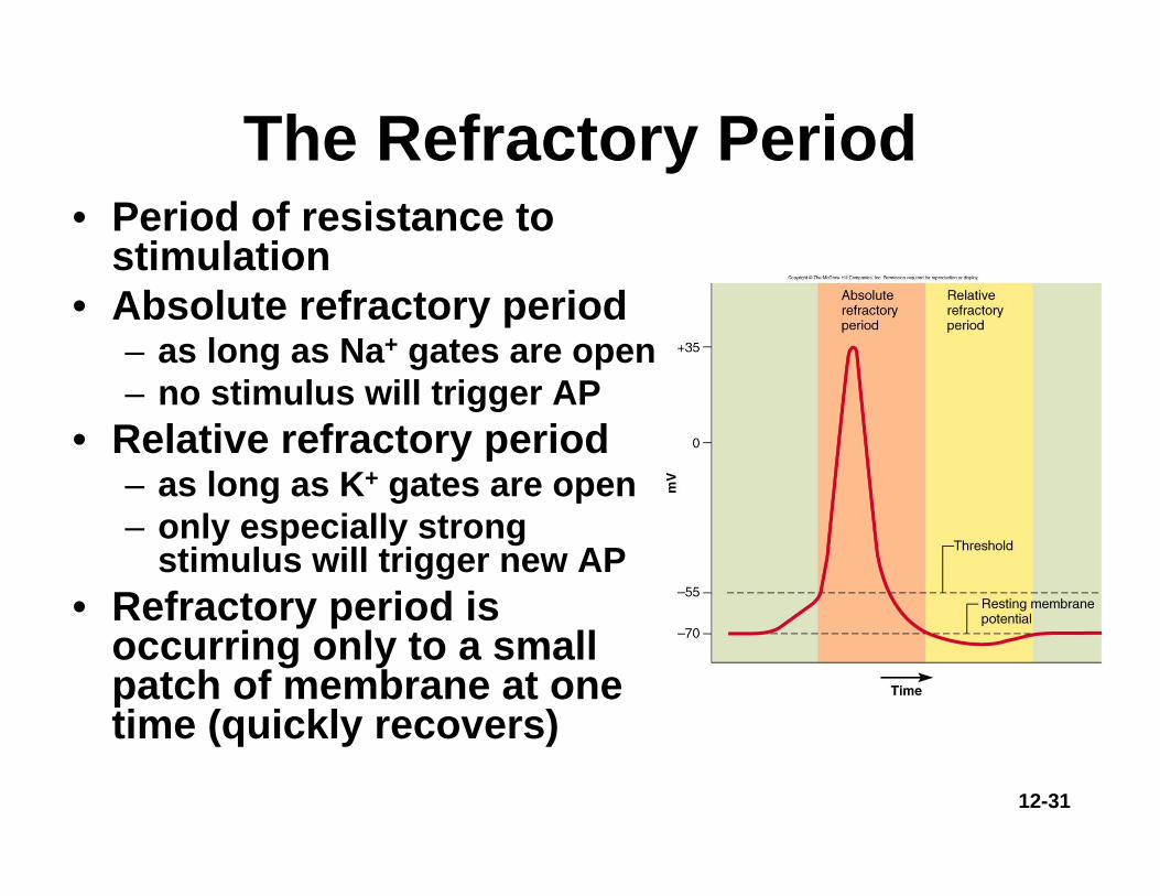
The Refractory Period• Period of resistance to
stimulation• Absolute refractory period
– as long as Na+ gates are open– no stimulus will trigger AP
• Relative refractory period– as long as K+ gates are open– only especially strong
stimulus will trigger new AP• Refractory period is
occurring only to a small patch of membrane at one time (quickly recovers)

12-32
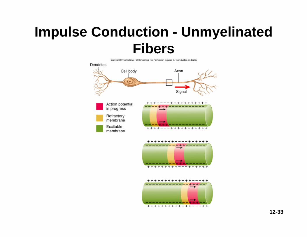
Impulse Conduction in Unmyelinated Fibers
• Threshold voltage in trigger zone begins impulse
• Nerve signal (impulse) - a chain reaction of sequential opening of voltage-gated Na+ channels down entire length of axon
• Nerve signal (nondecremental) travels at 2m/sec
12-33
Impulse Conduction - Unmyelinated Fibers

12-34
Saltatory Conduction - Myelinated Fibers
• Voltage-gated channels needed for APs– fewer than 25 per μm2 in myelin-covered regions – up to 12,000 per μm2 in nodes of Ranvier
• Fast Na+ diffusion occurs between nodes

12-35
Saltatory Conduction
• Notice how the action potentials jump from node of Ranvier to node of Ranvier.

12-36
Synapses between Neurons• First neuron releases neurotransmitter
onto second neuron that responds to it– 1st neuron is presynaptic neuron– 2nd neuron is postsynaptic neuron
• Synapse may be axodendritic, axosomatic or axoaxonic
• Number of synapses on postsynaptic cell variable– 8000 on spinal motor neuron– 100,000 on neuron in cerebellum

12-37
Synaptic Relationships between Neurons

12-38
Chemical Synapse Structure
• Presynaptic neurons have synaptic vesicles with neurotransmitter and postsynaptic have receptors
12-39
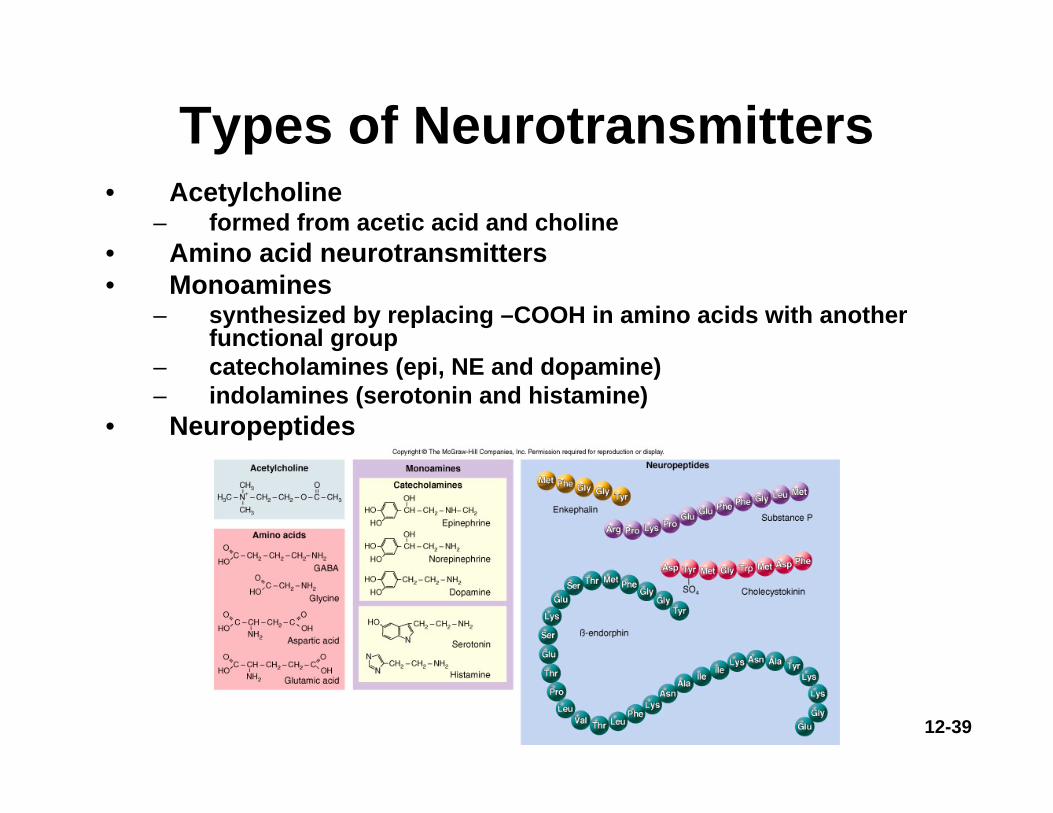
Types of Neurotransmitters• Acetylcholine
– formed from acetic acid and choline• Amino acid neurotransmitters• Monoamines
– synthesized by replacing –COOH in amino acids with another functional group
– catecholamines (epi, NE and dopamine)– indolamines (serotonin and histamine)
• Neuropeptides

12-40
Neuropeptides• Chains of 2 to 40 amino acids• Stored in axon terminal as larger secretory granules (called dense-
core vesicles)• Act at lower concentrations• Longer lasting effects• Some released from nonneural tissue
– gut-brain peptides cause food cravings• Some function as hormones
– modify actions of neurotransmitters

12-41
Synaptic Transmission
3 kinds of synapses with different modes of action
• Excitatory cholinergic synapse = ACh• Inhibitory GABA-ergic synapse = GABA• Excitatory adrenergic synapse = NE
Synaptic delay (.5 msec) – time from arrival of nerve signal at synapse to
start of AP in postsynaptic cell

12-42
Excitatory Cholinergic Synapse• Nerve signal opens voltage-
gated calcium channels in synaptic knob
• Triggers release of ACh which crosses synapse
• ACh receptors trigger opening of Na+ channels producing local potential (postsynaptic potential)
• When reaches -55mV, triggers APin postsynaptic neuron

12-43
Inhibitory GABA-ergic Synapse
• Nerve signal triggers release of GABA (γ-aminobutyric acid) which crosses synapse
• GABA receptors trigger opening of Cl-channels producing hyperpolarization
• Postsynaptic neuron now less likely to reach threshold

12-44
Excitatory Adrenergic Synapse• Neurotransmitter is NE (norepinephrine)• Acts through 2nd messenger systems (cAMP)
– receptor is an integral membrane protein associated with a G protein, which activates adenylate cyclase, which converts ATP to cAMP
• cAMP has multiple effects– binds to ion gate inside of membrane (depolarizing)– activates cytoplasmic enzymes– induces genetic transcription and production of new
enzymes• Its advantage is enzymatic amplification

12-45
Excitatory Adrenergic Synapse

12-46
Cessation and Modification of Signal
• Mechanisms to turn off stimulation– diffusion of neurotransmitter away into ECF
• astrocytes return it to neurons– synaptic knob reabsorbs amino acids and
monoamines by endocytosis – acetylcholinesterase degrades ACh
• choline reabsorbed and recycled
• Neuromodulators modify transmission– raise or lower number of receptors– alter neurotransmitter release, synthesis or
breakdown
12-47
Neural Integration• More synapses a neuron has the greater its
information-processing capability– cells in cerebral cortex with 40,000 synapses– cerebral cortex estimated to contain 100 trillion
synapses• Chemical synapses are decision-making
components of the nervous system– ability to process, store and recall information
is due to neural integration• Based on types of postsynaptic potentials
produced by neurotransmitters

12-48
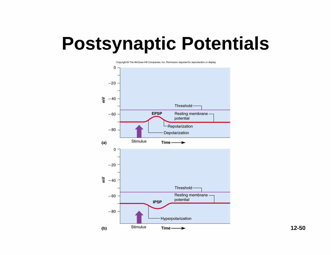
Postsynaptic Potentials- EPSP
• Excitatory postsynaptic potentials (EPSP)– a positive voltage change causing
postsynaptic cell to be more likely to fire• result from Na+ flowing into the cell
– glutamate and aspartate are excitatory neurotransmitters
• ACh and norepinephrine may excite or inhibit depending on cell

12-49
Postsynaptic Potentials- IPSP
• Inhibitory postsynaptic potentials (IPSP)– a negative voltage change causing
postsynaptic cell to be less likely to fire (hyperpolarize)
• result of Cl- flowing into the cell or K+ leaving the cell
– glycine and GABA are inhibitory neurotransmitters
• ACh and norepinephrine may excite or inhibit depending upon cell
12-50
Postsynaptic Potentials

12-51
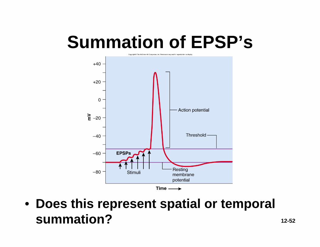
Summation - Postsynaptic Potentials• Net postsynaptic
potentials in trigger zone– firing depends on net input
of other cells• typical EPSP voltage = 0.5
mV and lasts 20 msec• 30 EPSPs needed to reach
threshold– temporal summation
• single synapse receives many EPSPs in short time
– spatial summation• single synapse receives
many EPSPs from many cells
12-52
Summation of EPSP’s
• Does this represent spatial or temporal summation?
12-53
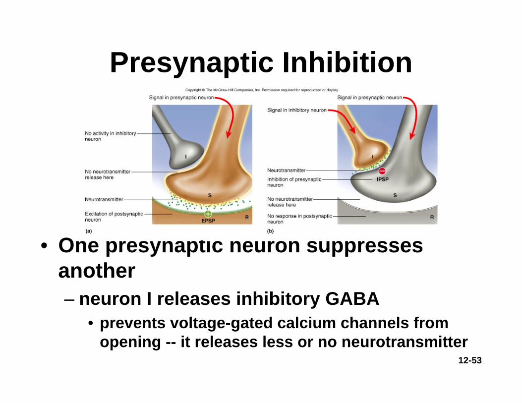
Presynaptic Inhibition
• One presynaptic neuron suppresses another– neuron I releases inhibitory GABA
• prevents voltage-gated calcium channels from opening -- it releases less or no neurotransmitter