nervous system sensory systems - napa valley college · middle ear consists of the: the tympanic...
TRANSCRIPT

Nervous System – Sensory Systems
Biol 105
Lecture 11
Chapter 9

Copyright © 2009 Pearson Education, Inc.
1. Depolarization is caused by ______ ions entering or
leaving (which one) the axon
1. The gap in between two neurons is called the
________.
2. What is the name for the chemicals that are held in
vesicles and released from one neuron, and bind to
receptors of the next neuron?
3. What part of the autonomic nervous system stimulates
digestion?
4. What is the thin outer layer of the cerebrum where most
of the higher thinking and processing takes place called
5. The part of the brain that processes sensory
information (except smell) is called the ______.
Concepts to Know:

Copyright © 2009 Pearson Education, Inc.
Outline
I. Senses
II. Sensory receptors
III. Touch
IV. Vision
V. Hearing and balance
VI. Smell

Copyright © 2009 Pearson Education, Inc.
Senses
Major senses – touch, hearing, smelling, taste,
and seeing.
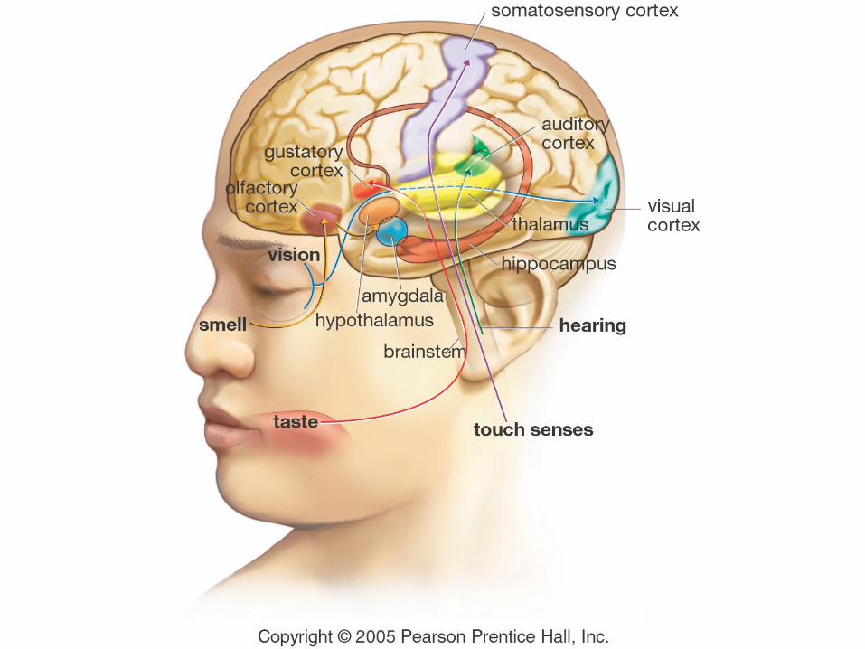
All the sensory nerves are routed through the
thalamus except the nerves for smell.
Copyright © 2009 Pearson Education, Inc.
Sensory receptor cells
Sensory receptors are specialized structures
that detect stimuli (stimulus)
Sensory receptor cells change the stimulation
into an electrical response that is transmitted
through the nerves
If a sensory receptor is continuously
stimulated, it will stop responding = sensory
adaptation
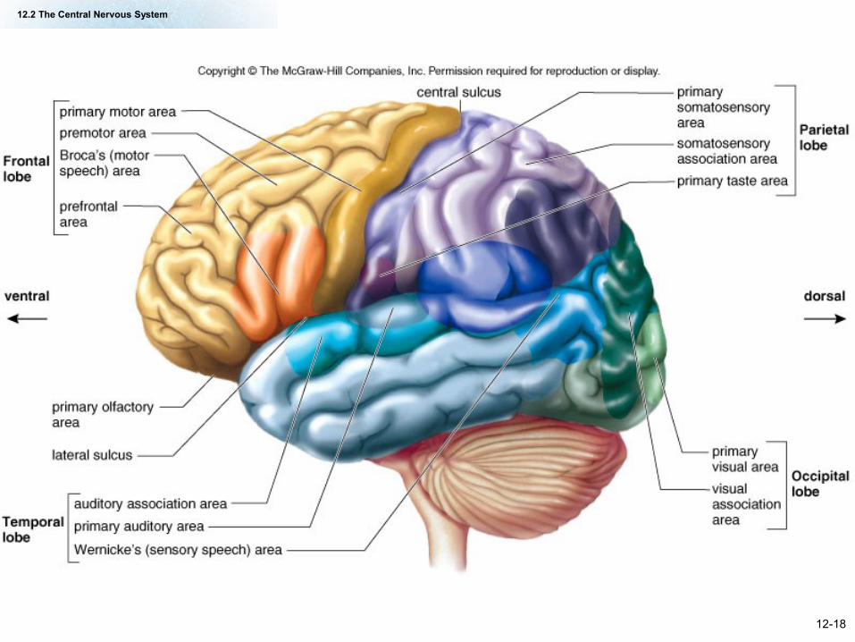
12.2 The Central Nervous System
12-18

12-28
Copyright © 2009 Pearson Education, Inc.
Touch
We can sense different things through
touch:
Thermal
Tactile
Pain
Vibration

Copyright © 2009 Pearson Education, Inc.
Figure 9.2 Sense receptors of the skin

Copyright © 2009 Pearson Education, Inc.
Figure 9.2 Sense receptors of the skin
Copyright © 2009 Pearson Education, Inc.
Types of receptors in the skin
Free nerve endings
Merkel disks
Meissner’s corpuscles
Pacinian corpuscles
Ruffini corpuscles
Thermoreceptors
Copyright © 2009 Pearson Education, Inc.
Free Nerve Endings
Free nerve endings – tips of dendrites of
sensory neurons (free nerve endings may be
wrapped around hair), detect touch and pain

Copyright © 2009 Pearson Education, Inc.
Figure 9.2 Free nerve endings

Copyright © 2009 Pearson Education, Inc.
Figure 9.2 Free nerve endings
Copyright © 2009 Pearson Education, Inc.
Merkel Disks
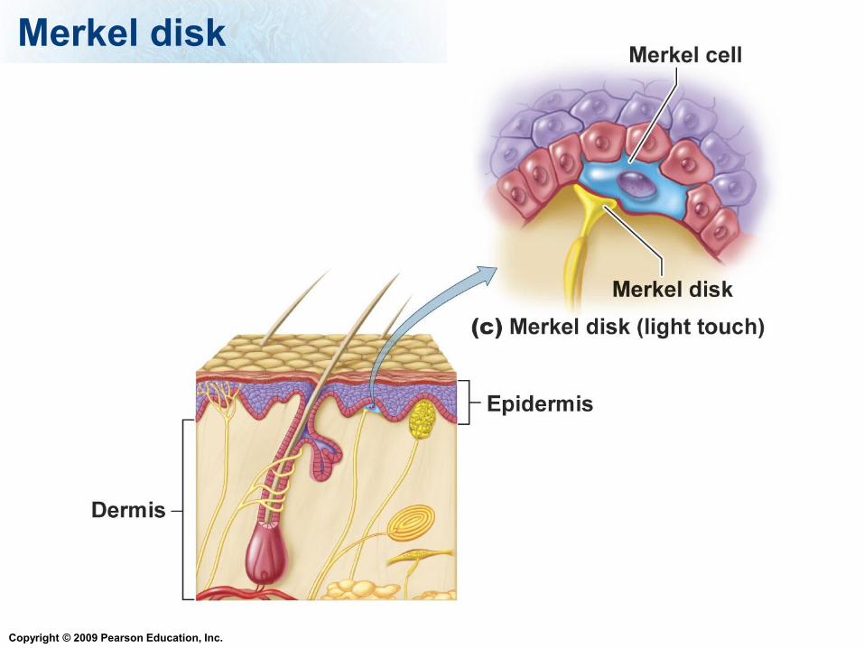
Merkel disks – comprised of free nerve endings
and Merkel cells, detect touch
Copyright © 2009 Pearson Education, Inc.
Merkel disk
Copyright © 2009 Pearson Education, Inc.
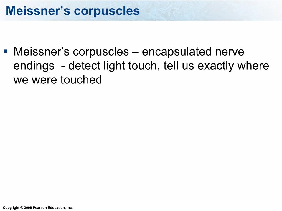
Meissner’s corpuscles
Meissner’s corpuscles – encapsulated nerve
endings - detect light touch, tell us exactly where
we were touched

Copyright © 2009 Pearson Education, Inc.
Meissners corpuscle

Copyright © 2009 Pearson Education, Inc.
Pacinian corpuscles
Pacinian corpuscles – layers of tissues surround
the nerve ending, detects pressure when first
applied, important in sensing vibration

Copyright © 2009 Pearson Education, Inc.
Pacinian corpuscle

Copyright © 2009 Pearson Education, Inc.
Ruffini corpuscles
Ruffini corpuscles – encapsulated nerve endings in
deep layers that respond to continuous pressure

Copyright © 2009 Pearson Education, Inc.
Ruffini corpuscle

Copyright © 2009 Pearson Education, Inc.
Thermoreceptors
Thermoreceptors – specialized nerve endings,
detects changes in temperature.

Copyright © 2009 Pearson Education, Inc.
Vision
Sight is complex:
Light enters the eye, it is focused, then the
light has to be transformed into it into an
electrical signal that then has to be
processed.

Copyright © 2009 Pearson Education, Inc.
Vision
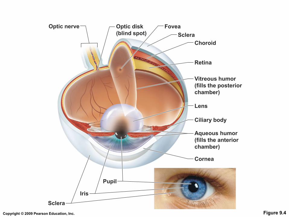
Light enters through the cornea
The lens focuses it to the back of the eye
The retina is a layer at the back of the eye
where light is transformed into electrical
signals
Copyright © 2009 Pearson Education, Inc. Figure 9.4
Retina
FoveaOptic disk
(blind spot)
Optic nerve
Choroid
Sclera
Vitreous humor
(fills the posterior
chamber)
Iris
Ciliary body
Pupil
Cornea
Aqueous humor
(fills the anterior
chamber)
Sclera
Lens

Copyright © 2009 Pearson Education, Inc.
Layers of the Eye – Outer layer
The sclera
Protects and shapes the eye
Provides attachment for muscles
The cornea
Allows light to enter

Copyright © 2009 Pearson Education, Inc.
Outer Layer of Eye
Table 9.1 (1 of 4)
Copyright © 2009 Pearson Education, Inc.
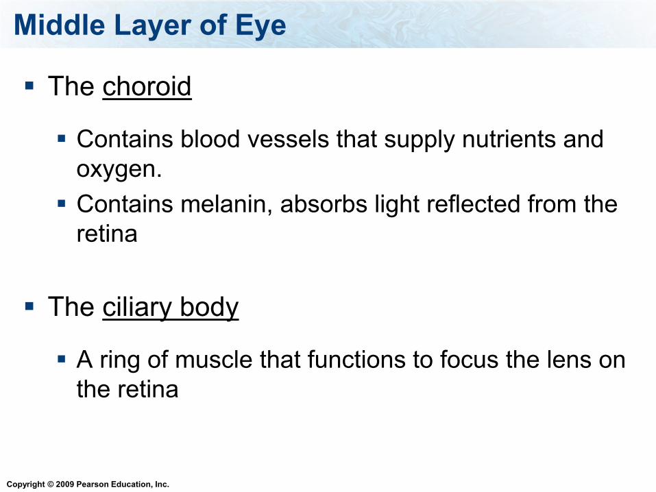
Middle Layer of Eye
The choroid
Contains blood vessels that supply nutrients and
oxygen.
Contains melanin, absorbs light reflected from the
retina
The ciliary body
A ring of muscle that functions to focus the lens on
the retina

Copyright © 2009 Pearson Education, Inc.
Middle Layer of Eye
The iris
The colored portion of the eye
Contains smooth muscle that dilates or constricts
to regulate the amount of light entering the eye
The pupil
The opening in the center of the iris that lets light
into the eye
Copyright © 2009 Pearson Education, Inc.
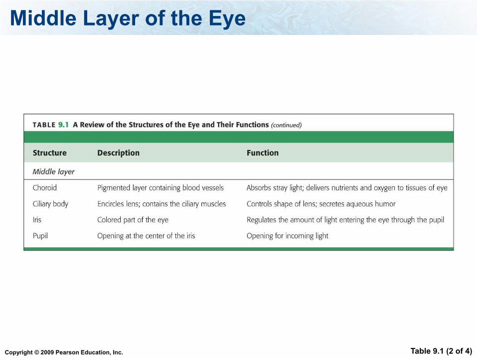
Middle Layer of the Eye
Table 9.1 (2 of 4)
Copyright © 2009 Pearson Education, Inc.
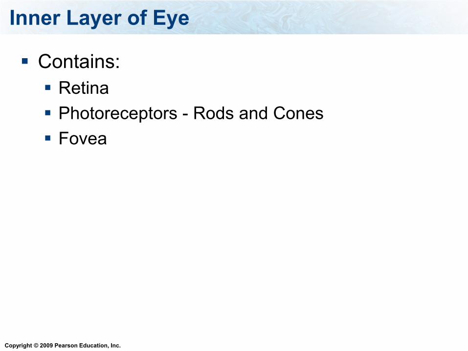
Inner Layer of Eye
Contains:
Retina
Photoreceptors - Rods and Cones
Fovea

Copyright © 2009 Pearson Education, Inc.
Inner Layer of Eye - Retina
The retina contains photoreceptors
Rods
Cones – detect color
The fovea is a pit in the retina with a high
concentration of cones

Copyright © 2009 Pearson Education, Inc.
Vision Depends on the Eye
Table 9.1 (3 of 4)

Copyright © 2009 Pearson Education, Inc.
Structures of the Eye
Optic Nerve
Fluid
Aqueous humor
Vitreous humor
Lens
Copyright © 2009 Pearson Education, Inc.
Optic Nerve
The optic nerve
Carries visual information to the brain
Forms a blind spot where it leaves the retina
Copyright © 2009 Pearson Education, Inc.
Fluid in the Eye
There are two fluid filled chambers in the eye
Vitreous humor – jelly like fluid in posterior
chamber. Holds retina against the wall of the
eye
Aqueous humor – clear fluid in anterior
chamber. Supplies nutrients and oxygen to
cornea and lens, removes the waste. Creates
pressure in eye to maintain shape of eye.

Copyright © 2009 Pearson Education, Inc.
Lens
The lens can change shape to focus on
near and far objects.
Focuses the light onto the retina
Ciliary muscles are attached to lens by
ligaments

Copyright © 2009 Pearson Education, Inc.
Vision Depends on the Eye
Table 9.1 (4 of 4)

Copyright © 2009 Pearson Education, Inc.
Photoreceptors
Cones and Rods have pigments that
absorb
Cones work best in bright light and provide
color vision
Rods work in low light situations but can only
provide black and white vision

Copyright © 2009 Pearson Education, Inc.
Photoreceptors
The photoreceptors (rods and cones) have
pigments that absorb light
When there is no light coming in, they are
releasing neurotransmitters (opposite of most
receptors)
When they absorb light they stop releasing
neurotransmitters

Copyright © 2009 Pearson Education, Inc.
Photoreceptors
The neurotransmitters are inhibitory
When the neurotransmitters diminish, cells
that process the information are stimulated
This information from these cells (bipolar and
ganglion cells) is transmitted to the optic
nerve to the thalamus to the visual cortex

Copyright © 2009 Pearson Education, Inc. Figure 9.8a
(a) Light enters the left eye
and strikes the retina.
Light
Retina
Choroid
Sclera
Blind spot

Copyright © 2009 Pearson Education, Inc. Figure 9.8b
Ganglion
cell layer
Bipolar
cell layer
Retina
Photoreceptor
cells
Pigment layer
Choroid
Sclera
Rod
Electrical
signals
Axons
Cone
Light
Vitreous
humor

Copyright © 2009 Pearson Education, Inc. Figure 9.8c
(c) The axons of the ganglion cells leave the eye at the blind spot,
carrying nerve impulses to the brain (viewed from below) by means
of the optic nerve.
Retina
Light
Optic nerve
Visual cortex

Copyright © 2009 Pearson Education, Inc.
Rods and Cones
Figure 9.9 (2 of 2)
Rod cell
Cone cell

Copyright © 2009 Pearson Education, Inc.
Which part of the human eye detects colored light?
1. Pupil
2. Rods
3. Cones
4. Cornea

Copyright © 2009 Pearson Education, Inc.
A ring of muscle that functions to focus the lens on
the retina is the:
1. Iris
2. Choroid
3. Ciliary body
4. Sclera

Copyright © 2009 Pearson Education, Inc.
Figure 9.10 A standard test for color blindness

Copyright © 2009 Pearson Education, Inc.
Vision
Vision is much more complicated because
these signals have to be processed into a
3-D image

Copyright © 2009 Pearson Education, Inc.
Copyright © 2009 Pearson Education, Inc.

Copyright © 2009 Pearson Education, Inc.
Hearing
Sound enters the ear canal and hits the tympanic
membrane (ear drum).
The tympanic membrane vibrates.
This causes small bones in the ear to vibrate.
These bones focus and amplifies the vibrations
onto a small place (oval window) on the cochlea.
The cochlea is a fluid filled coiled membrane.
The vibrations shakes the fluid in the cochlea

Copyright © 2009 Pearson Education, Inc.
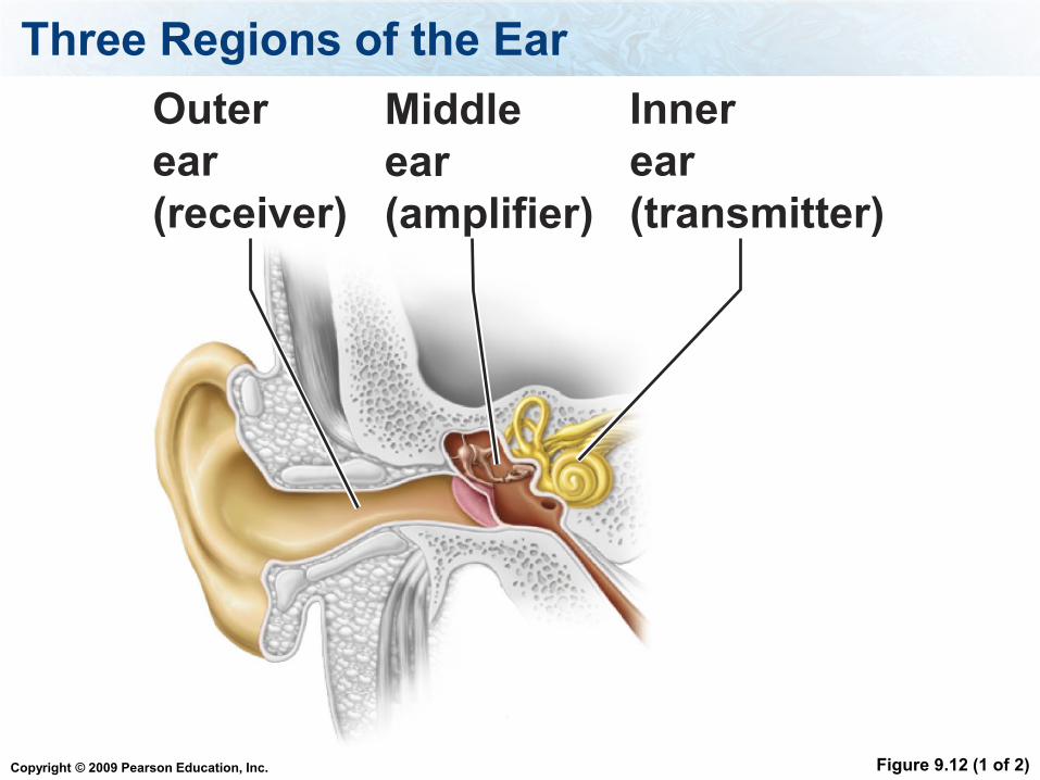
Three regions of the ear
Outer ear – the receiver
The middle ear – the amplifier
The inner ear – the transmitter
Copyright © 2009 Pearson Education, Inc.
Three Regions of the Ear
Figure 9.12 (1 of 2)
Outer
ear
(receiver)
Middle
ear
(amplifier)
Inner
ear
(transmitter)

Copyright © 2009 Pearson Education, Inc.
The Outer Ear
Consists of the:
Pinna – gathers the sound, acts like a
funnel
External auditory canal – brings the sound
from pinna to the tympanic membrane

Copyright © 2009 Pearson Education, Inc.
Middle Ear
Consists of the:
The tympanic membrane separates the outer
ear from the middle ear, vibrates when sound
waves hit it.
Three auditory bones – amplify the vibration
Malleus
Incus
Stapes
Auditory tube (eustachian tube) – equalizes
pressure between outer and middle ear
Copyright © 2009 Pearson Education, Inc.
Middle Ear
The tympanic membrane vibrates when
sound waves hit it and transmits the
vibration to the malleus
The vibrations are amplified by the three
bones and transmitted to the oval window

Copyright © 2009 Pearson Education, Inc.
Parts of the Inner Ear
Oval window – transmits sound from the stapes
to the fluid in the cochlea
Round window – relieves pressure
Cochlea – contains the receptor cells that
transform the signal from vibration to an
electrochemical signal to the neurons.
Vestibular apparatus – monitors position of the
head

Copyright © 2009 Pearson Education, Inc.
Hearing Depends on the Ear
Figure 9.12 (2 of 2)
The pinna gathers sound and funnels it into the external auditory canal to the tympanic membrane (eardrum).
The eardrum vibrates synchronously with sound waves, causing the bones of the middle ear to move.
The three bones of the middle ear amplify the pressure waves and convey the vibrations of the eardrum to the inner ear.
The cochlea converts pressure waves to neural messages that are sent to the brain for interpretation as sound.
Malleus(hammer)
Incus(anvil)
Stapes(stirrup)
Semicircular canals
Vestibular apparatus:
Auditory nerve
Cochlea
Oval window
Eardrum(tympanic membrane)
Round window
Auditory tube(Eustachiantube)
Outer ear(receiver)
Middle ear(amplifier)
Inner ear(transmitter)
External auditory canal
Vestibule

Copyright © 2009 Pearson Education, Inc.
Cochlea
It is in the cochlea where vibrations are
transformed into electrical signals that can be
sent by neurons
When the fluid in the cochlea moves, it moves
small “hair cells” against a membrane. This
allows ion channels to open
This leads to the release of neurotransmitters,
which trigger the neuron to send the message

Copyright © 2009 Pearson Education, Inc.
Hearing Depends on the Ear
Figure 9.13 (1 of 2)

Copyright © 2009 Pearson Education, Inc.
Hearing Depends on the Ear
Figure 9.13 (2 of 2)
Hair cell
Tectorial
membrane
Copyright © 2009 Pearson Education, Inc.
In the ear, the fluid filled coiled membrane that is responsible
transforming the vibrations into electrical signals. This structure is:
1. Tymphanic
membrane
2. Staples
3. Cochlea
4. Incus
Copyright © 2009 Pearson Education, Inc.
The tympanic membrane transmits the vibration to the ___.
Stapes
Malleus
Incus
Oval window

Copyright © 2009 Pearson Education, Inc.
The Vestibular Apparatus
Balance depends on the vestibular apparatus
of the inner ear
The vestibular apparatus is a fluid-filled maze
of chambers and canals within the inner ear

Copyright © 2009 Pearson Education, Inc.
The Vestibular Apparatus - Dynamic equilibrium
Fluid filled cupulas at base of the semicircular
canals have hair cells that are stimulated when
head moves. Hair cells send message to the
brain.

Copyright © 2009 Pearson Education, Inc.
The Vestibular Apparatus - Static equilibrium
Otoliths are small chalk like granules
When head is tilted the otoliths move and
stimulate hair cells that send message to the
brain

Copyright © 2009 Pearson Education, Inc.
Balance Depends on the Vestibular
Apparatus
Figure 9.16a (1 of 2)

Copyright © 2009 Pearson Education, Inc.
Balance Depends on the Vestibular
Apparatus
Figure 9.16a (2 of 2)

Copyright © 2009 Pearson Education, Inc.
The Vestibular Apparatus
Figure 9.16b (1 of 2)

Copyright © 2009 Pearson Education, Inc.
The Vestibular Apparatus
Figure 9.16b (2 of 2)
Copyright © 2009 Pearson Education, Inc.
Smell - olfaction
Sensory nerves for smell go directly to the cerebral cortex and to the amygdala and the hypothalamus.
They do not pass through the thalamus
Copyright © 2009 Pearson Education, Inc.
Smell - olfaction
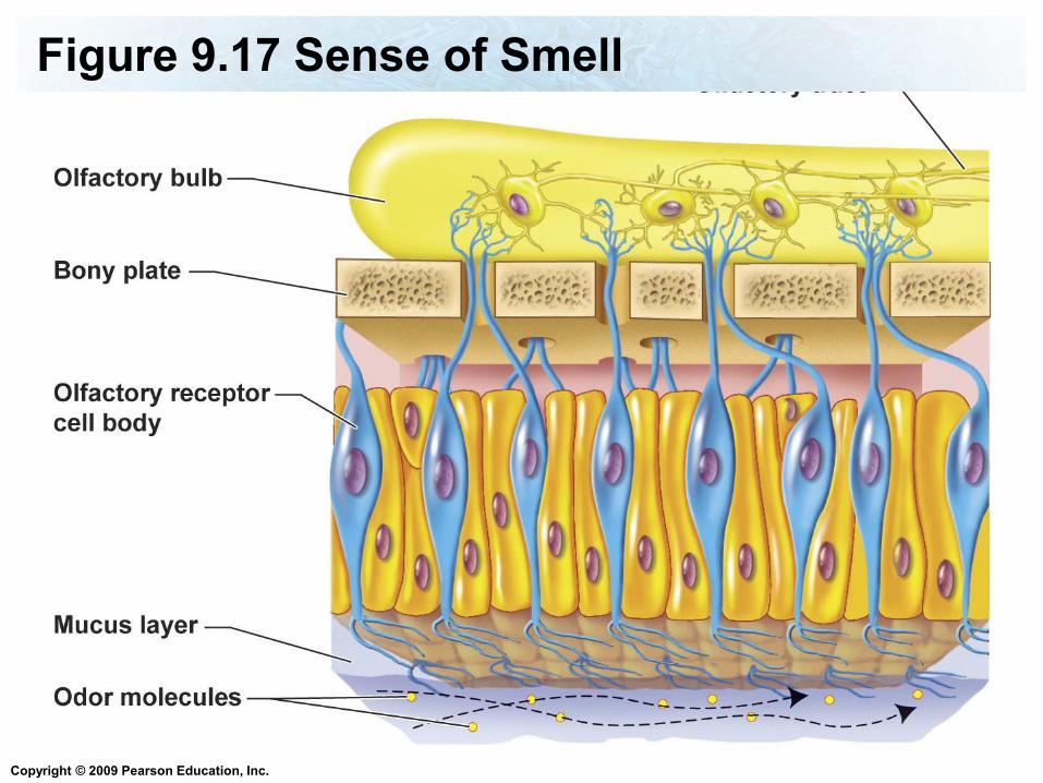
Odor molecules bind to the receptors in the cilia of olfactory receptor cells
The receptor cells send the message to the neurons in the olfactory bulb which carry the message to the brain.

Copyright © 2009 Pearson Education, Inc.
Figure 9.17 Sense of Smell
Copyright © 2009 Pearson Education, Inc.
Figure 9.17 Sense of Smell
Copyright © 2009 Pearson Education, Inc.
Taste
Taste and smell is very connected.
The tongue has taste buds on them
The taste buds have taste cells (receptor
cells) in them

Copyright © 2009 Pearson Education, Inc.
Taste
Food molecules bind to taste cells and
stimulate them. The taste cells send the
messages to the sensory neurons which
send the message to the brain.

Copyright © 2009 Pearson Education, Inc.
Smell and Taste
Figure 9.18
Copyright © 2009 Pearson Education, Inc.
Read Chapter 6
What is the function of sensory receptor
cells?
What is an example of sensory adaptation?
What are the types of senses of touch?
What are the types of sensory receptors in
skin, what type of touch do they detect, be
able to describe them?
Important Concepts

Copyright © 2009 Pearson Education, Inc.
What are all of the layers and structures
(including the fluids) of the eye and what are
their functions?
What is the blind spot?
How does the signal travel from the
photoreceptors to the brain, what part of the
brain receives the signal? Be able to describe
in detail this process, including the cells that
transmit the messages.
Important Concepts

Copyright © 2009 Pearson Education, Inc.
What are all the parts of the ear, are they part of the inner, middle or outer ear, and what is their functions? What is the path of sound waves and vibrations through the ear
How does the ear detect head movement and position?
How do we detect odor? What part of the brain receives the signal? Where are olfactory receptors found?
How do we detect tastes? What structures are responsible for taste?
Important Concepts

Copyright © 2009 Pearson Education, Inc.
Definitions
stimuli (stimulus), sensory adaptation, dilates,
constrict, bipolar cells, ganglion cells,
photoreceptors, transmits, amplifies, otoliths,
cupula, taste buds,