nervous system 1. 1. central nervous system 2. peripheral nervous system brain 2. spinal cord 1....
TRANSCRIPT

Nervous System
1.Central Nervous System 2. Peripheral Nervous System
1. Somatic 2. Autonomic 1.Brain 2. Spinal Cord

1. 5 parts
1 BRAIN
2 SPINAL CORD
3. AUTNMIC .NERVOUS.SYSTEM.
4.ENTERIC .NERVOUS.SYSTEM.
5.PERIPHERAL NERVES
2. 3 parts-
1.SENSOR
2.MOTOR
3. MOTIVATION
3. Central Nervous System is Organized in 3 ways
1. Hierarchical Organization
2. Lateralized Organization
3. Localized Organization
FUNCTIONALLY

GLIAL CELL CLASSFICATION
size=0.02µm
1.ASTROCYTES
Fibrous
Protoplasmic
Radial glial cells
Müller cells
Bergmann glia
2. EPENDYMAL CELLS
3.OLIGODENDROCYTES
4. MICROGLIAL CELLS
5.SATELLITE CELLS
6.SCHWANN CELLS
7.ENTERIC GLIAL
PNS CNS

Morphology of neuron
2 parts
Cell body (soma)
Processes
Dendrites
Axon
1.membrane
2.perikaryon
3.nucleus
Presynaptic terminals.terminal (bouton / button)
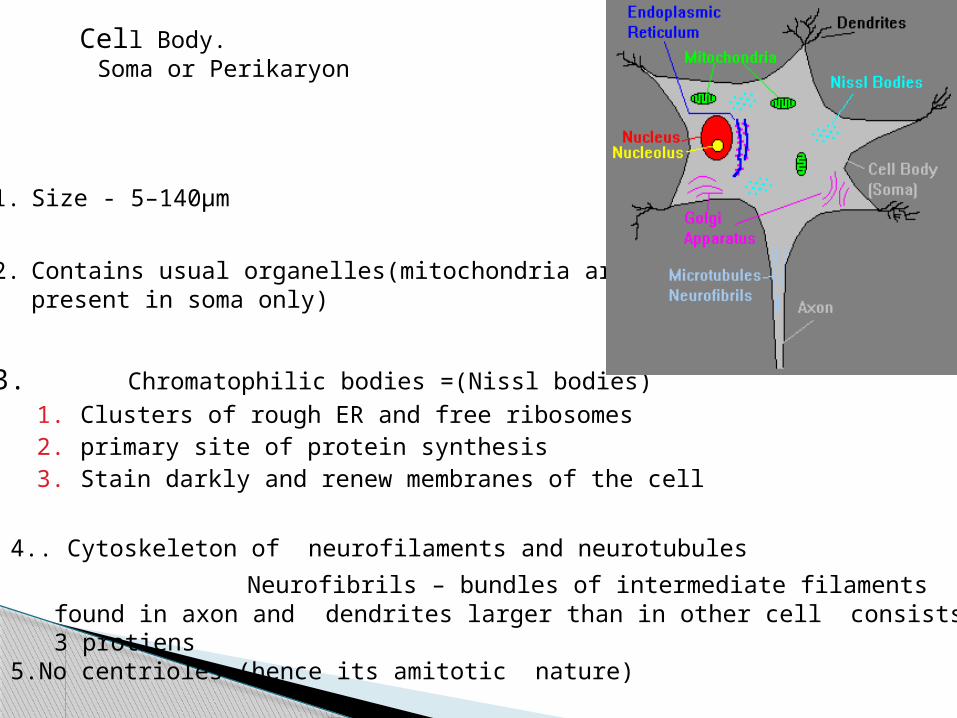
1. Size - 5–140µm
2. Contains usual organelles(mitochondria arepresent in soma only)
3. Chromatophilic bodies =(Nissl bodies)1. Clusters of rough ER and free ribosomes2. primary site of protein synthesis3. Stain darkly and renew membranes of the cell
4.. Cytoskeleton of neurofilaments and neurotubules
Neurofibrils – bundles of intermediate filaments found in axon and dendrites larger than in other cell consists of 3 protiens
5.No centrioles (hence its amitotic nature) 6. Tapers to form axon hillock
Cell Body. Soma or Perikaryon

1. Covered by neurolemma(:Made up of Schwann cells).
2. Often myelinated:( Myelin is formed by Schwann cells).3. Note: axon is only part of neuron that is ever myelinated.
Axon

composition is similar that To soma : neurofilaments ,microtubule , mitochondria, Nissl bodies, no Golgi apparatus
Dendritic spine: flattened and parallel cisternae
2- 7 processes
Dendrite
1. Branches off the cell body that carry information to the cell body.
2. Relatively short.3. Often branched.4. Have receptors for neurotransmitters.
5. Conduct local potentials.

1. Input2. Output3. Centre4. Integration5. Speed6. New ideas7. Energy8. Proper connections9. Insulation10. Day dreaming11. Output flexibility12. Orginality
COMPARISION WITH COMPUTORS

Neurons-encode information at the cellular level.
1. Frequency modulation
2. On and Off mechanism of electrical impulses,
Principle Binary code
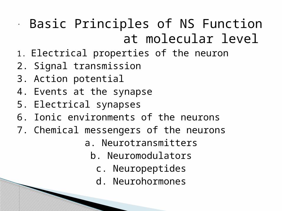
1. Electrical properties of the neuron2. Signal transmission3. Action potential4. Events at the synapse5. Electrical synapses6. Ionic environments of the neurons7. Chemical messengers of the neurons
a. Neurotransmittersb. Neuromodulators
c. Neuropeptidesd. Neurohormones
. Basic Principles of NS Function at molecular level


. PRIMARY 1.RMP 2.CONDUCTION 3. EXICITATION
RMP= -65mV
------
-65-65-65
-
65
12


3. EXCITIBILITY
Stimulus
1.Mechanical 2.Chemical 3.Thermal
4.Electric 5.Osmotic
4 ACCOMODATION
If the stimulus, even with stronger strength is applied very slowly to a nerve, these may have no response only due to l of attaining the threshold strength.
2. LATENT PERIOD
1. Velocity 2.Diameter
SECONDARY 1. Action Potential
1. Monopotential 2. Biphasic-potential 3.Compound-potential

TYPE
1.Law of forward conduction
2. Law of bell and magendie
3. Law of isolated conduction
4. Law of physical integeration
5.Law of 2 way conduction
FACTORS 1.Diameter
2. Mylenated
3.Temp;
4.Mechanical pressure
5. Chemical
6.Spike potential ^ conduction increases
7.PH
8.O2
9.Effect of ions
10.Level of RMP
11.Blood supply
5. CONDUCTION
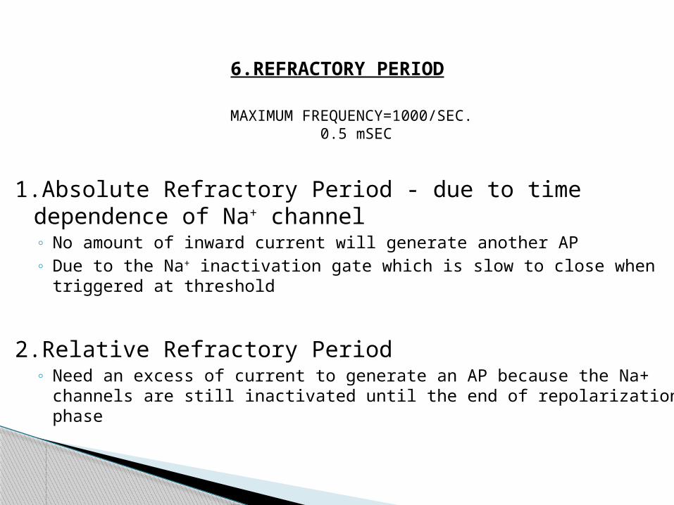
6.REFRACTORY PERIOD
MAXIMUM FREQUENCY=1000/SEC. 0.5 mSEC
1.Absolute Refractory Period - due to time dependence of Na+ channel◦ No amount of inward current will generate another AP◦ Due to the Na+ inactivation gate which is slow to close when triggered at threshold
2.Relative Refractory Period◦ Need an excess of current to generate an AP because the Na+ channels are still
inactivated until the end of repolarization phase

1. Long refractiory period
2. Can conduct only 1 impulse at a time
7. Infatigability
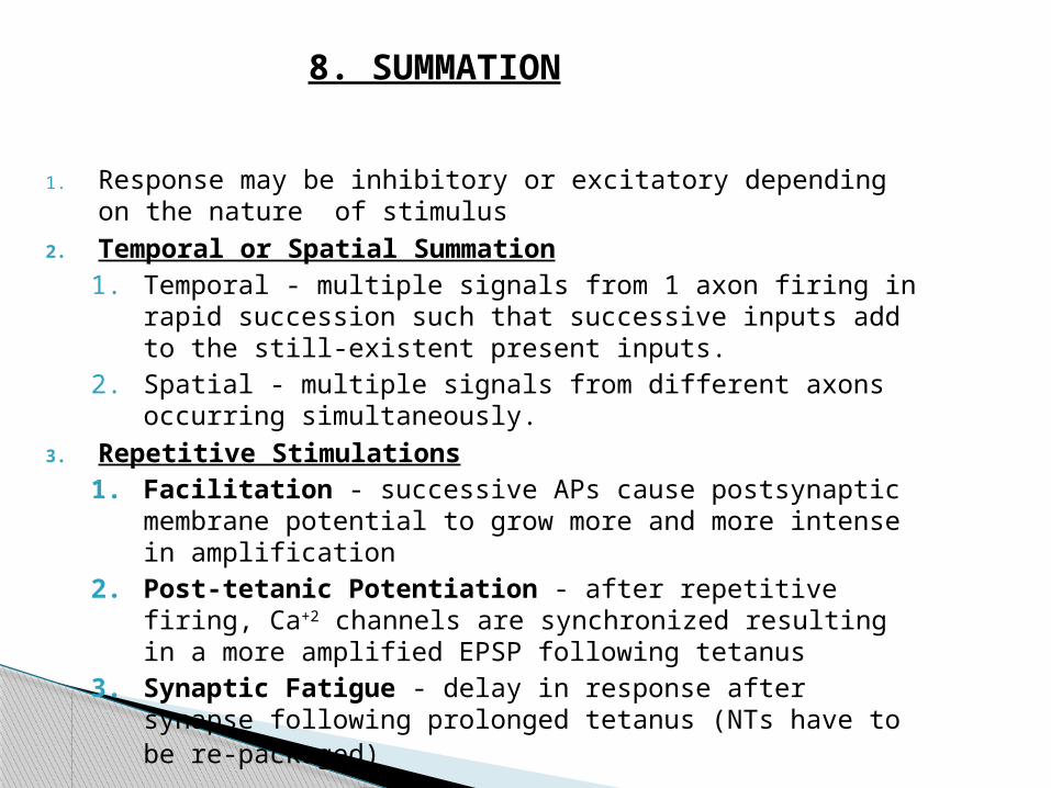
1. Response may be inhibitory or excitatory depending on the nature of stimulus
2. Temporal or Spatial Summation 1. Temporal - multiple signals from 1 axon firing in rapid succession
such that successive inputs add to the still-existent present inputs.2. Spatial - multiple signals from different axons occurring
simultaneously.
3. Repetitive Stimulations1. Facilitation - successive APs cause postsynaptic membrane
potential to grow more and more intense in amplification2. Post-tetanic Potentiation - after repetitive firing, Ca+2 channels
are synchronized resulting in a more amplified EPSP following tetanus
3. Synaptic Fatigue - delay in response after synapse following prolonged tetanus (NTs have to be re-packaged)
8. SUMMATION

9. ADAPTATION
The nerve fibre is quickly adapt itself, thus gradual changes to excite, but sudden alteration cause excitation.
10. ALL OR NONE LAW
a stimulus be adequate , a single nerve will always give a maximum response .If the duration or strength of the stimulus be further no alteration in the response will lake place.
NOTE;-(In the whole nerve this property differ ,but is true for a single nerve).
11. NEUROSECRETION/NEUROTROPINS

The Neuromuscular Junction

1. Motor Unit
1. Composed of an alpha motor neuron and all
the myofibers innervated by that neuron
2. Neuromuscular junction
1. Where the axon terminal and the motor
endplate meet
3. Motor Endplate-sole plate-postjunctional
membrane
1. The region of the myofiber directly under the
terminal axon branches
Nerve-Muscle Functional Unit
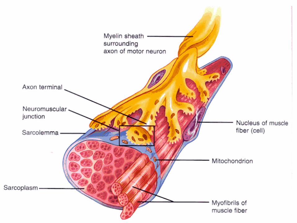

Neuromuscular Junction
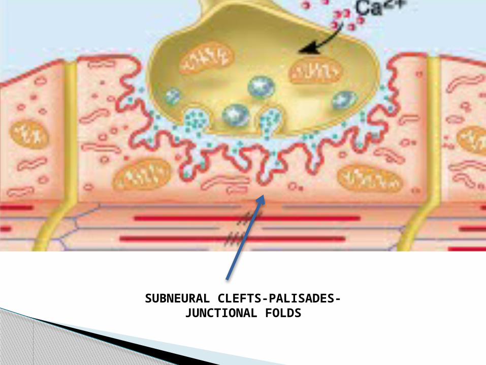
SUBNEURAL CLEFTS-PALISADES-JUNCTIONAL FOLDS


Events at NJM
• An action potential (AP), an electrical impulse, travels down the axon of the motor neuron to the end bulbs (synaptic terminals)
• The AP causes the synaptic vesicles to fuse with the end bulb membrane, resulting in the release of Acetylcholine (Ach) into the synaptic cleft
• Ach diffuses across the synaptic cleft & binds to Ach receptors on the motor end plate
• The binding of Ach to its receptors causes a new AP to be generated along the muscle cell membrane
• Immediately after it binds to its receptors, Ach will be broken down by Acetylcholinesterase (AchE) – an enzyme present in the synaptic cleft

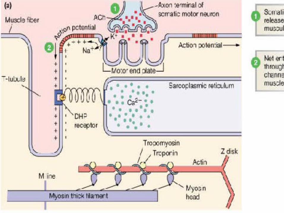
EXCITATION-CONTRACTION COUPLING:
1)Action Potential
2)Depolarization of the T-Tubules - Causes conformational change in the DHPR - opens Ca2+ channels(Ryr) on sacroplasmic reticulum
3)Ca2+ released from SR into ICF
4)Ca2+ binds to Troponin C cooperatively - causes conformational change
5)Tropomyosin is out of way
6)Cross-bridge cycling
7)Relaxation via Ca2+ ATPase

Transmission at this junction involves several steps:
1. When an action potential (inhibited by tetrodotoxin) reaches the axon terminal it causes Ca channels to open. Ca2+ rushes into the cell because Ca2+ outside is much higher than Ca2+ inside
2. The terminal region is loaded with vesicles containing the transmitter acetylcholine (ACh)
3. Ca2+ causes some of the vesicles to fuse with the membrane and release their ACh (inhibited by botulinum toxin)
4. ACh diffuses across the junction and binds to the ACh receptor protein (inhibited by curare) in the postsynaptic membrane
5. Binding causes an ion channel to open
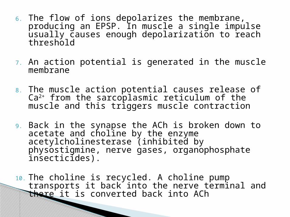
6. The flow of ions depolarizes the membrane, producing an EPSP. In muscle a single impulse usually causes enough depolarization to reach threshold
7. An action potential is generated in the muscle membrane
8. The muscle action potential causes release of Ca2+ from the sarcoplasmic reticulum of the muscle and this triggers muscle contraction
9. Back in the synapse the ACh is broken down to acetate and choline by the enzyme acetylcholinesterase (inhibited by physostigmine, nerve gases, organophosphate insecticides).
10. The choline is recycled. A choline pump transports it back into the nerve terminal and there it is converted back into ACh

Chemical Transmitters Are Made and Stored in the Presynaptic Terminal
1. The end of the nerve enlarges into an axon terminal
2. Transmitters are made in the terminal and are stored in tiny vesicles so that they can be released whenever an action potential comes along
3. Transmitters are made only by the incoming (presynaptic) nerve
4. Because the transmitter is only on one side the impulse can go in only one direction
Calcium is Required for Transmitter Release
1. Transmitter release requires Ca2+ ions
2. Normally Ca2+ in the cell is kept very low (by a Ca pump)- if the cell needs Ca2+ it must come from the outside
3. The action potential coming in to the terminal opens Ca channels -> Ca comes rushing in
4. The Ca2+ causes some of the vesicles to fuse to the membrane- then they open up and the transmitter is released
5. Botulinum and tetanus toxins block transmitter release