nephrogenic ascites:apoorlyunderstood...
TRANSCRIPT

EDITORIAL COMMI1TEE
Tomas Berl, Editor William Henrich Mark Paller Fred SilvaDenver, CO Dallas, 7X Minneapolis, MN Oklahoma City, OK
DESCRIPTION OF THE NEPHROLOGY-HYPERTENSION TRAINING PROGRAM AT THEUNIVERSITY OF CALIFORNIA, SAN DIEGO SCHOOL OF MEDICINE
UCSD offers a 2- or 3-yr program in clinical nephrology-hypertension and research training in nephrology-hyperten-sion. This program prepares physicians for careers in both academic and clinical nephrology. There are opportunitiesfor research fellows without clinical training for both MDs and PhDs, and approximately 20 fellows are in the currentnephrology-hypertension program. The program offers a balance between clinical training in a very active clinicalteaching program and a variety of research opportunities and disciplines. Clinical rotations are supervised by 1o facultymembers at UCSD Medical Center, the San Diego Veterans Affairs Medical Center. and the San Diego Naval RegionalMedical Center. There are four clinical rotations. which include a rotation on the UCSD Transplant and Pheresis Program.Approximately 120 transplants are performed each year. and several hundred renal transplants are monitored. TheNephrology-Hypertension Consultation Service exposes each fellow to consult experiences in pancreas. liver. lung. andheart transplants as appropriate for these disciplines. There is also an active vigorous medical intensive care unit
exposure during all ofthe rotations. The fellow has the primary responsibility for the care oftransplant patients during theacute phase of treatment. and there exists a strong working relationship with the Transplant Surgery Division. More than100 acute hemodialyses are performed each month at UCSD Medical Center. After clinical training. fellows areexposed to a broad range of laboratory experiences in a unique School of Medicine sethng. With the exception ofpharmacology. there are no basic science departments at the UCSD School of Medicine. and there is a vigorousinteraction among basic scientists and clinical investigators. Within the division. there are laboratories actively engagedin the study of renal physiology and pathophysiology. biochemistry. cellular physiology and molecular medicine. renaland molecular cellular immunology. research and treatment modalities for acute and chronic renal failure. andprograms on the clinical mechanisms of systemic hypertension as well as the genetics of hypertension. The School ofMedicine provides an extensive training in laboratory methods in research for all research fellows. and there areextensive opportunities for collaboration with Cell and Molecular Medicine and the Department of Pharmacology.There are also extended research opportunities through two institutional Physician/Scientist Training Grant Programs.Most of the faculty in the Division of Nephrology-Hypertension are actively involved in clinical and basic researchactivities. which span programs beyond the Division of Nephrology-Hypertension. Each of these laboratories ischaracterized by a high degree of fellow and faculty interaction. The University of California. San Diego. offers a broad,balanced program directed toward the preparation of individuals for academic careers and the provision of a broadbase of scientific techniques that will prepare them for the decades ahead.
Nephrogenic Ascites: A Poorly Understood Syndrome1Terry C. Hammond2 and Marwan A. Takiyyuddin
_________- � � - �-�- ascites formation is clearly understood. Patients fre-
TO. Hammond, Navy Medical Center San Diego, San quently present with hypertension, moderate to mas-Diego, CA sive ascites, minimal extremity edema, cachexia,
MA. Takiyyuddin, Division of Nephrology-Hyperten- and a historY of dialysis-associated hypotension. Thesion, Department of Medicine, Department of Veter- ascitic fluid is typically an exudate. Although treat-ans Affairs Medical Center, San Diego, CA ment options are limited, continuous ambulatory pen-
toneal dialysis, penitoneovenous shunt placement,(J. Am. Soc. Nephrol. 1994; 5:1 173-1 177) . .
and renal transplantation appear to be effective in---- �---��- controlling ascites formation. Nephrogenic ascites is
ABSTRACT associated with a grave prognosis, especially if treat-Nephnogenlc ascites is a condition characterized by ment Is not instituted. One patient with nephrogenic
the presence of massive ascites in a patient with ESRD. ascites iS described here.Neither the exact cause nor the pathogenesis of . . , .
Key Words: Nephrogenic ascites. hemodialysis. peritoneal1 dialysis. peritoneovenous shunt, kidney transplantReceived November 18, 1993. Accepted March 10, 1994.2 correspondence to Dr. ic. Hammond, Division of Nephrolo9y-Hypertension,
veterans Affairs Medical cenio�, Son Diego, CA 92161. ‘�T ephrogenic ascites is a syndrome of refractory
� of Nephrology I N ascites associated with ESRD. It is defined as
copynght 0 1994 by the American Society of Nephrolo9y clinically evident chronic ascites, occurring in pa-
Journal of the American Society of Nephrology 1173
Nephrogenic Ascites
1174 VolumeS’ NumberS’ 1994
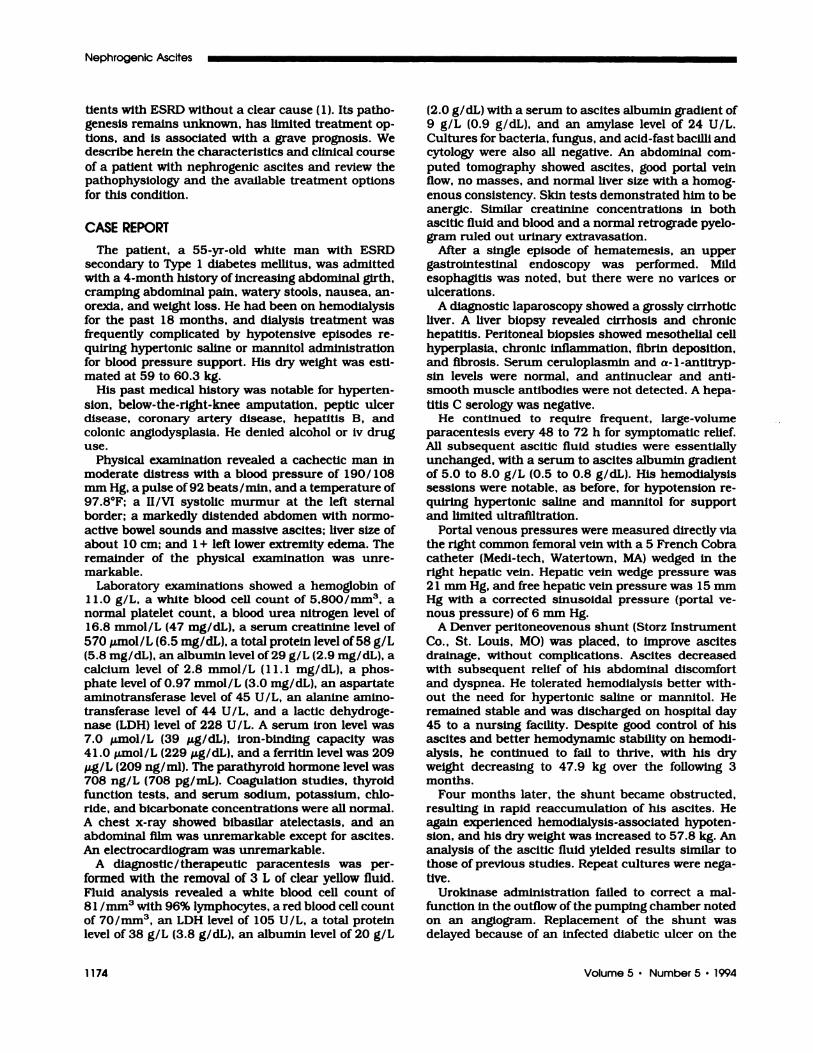
tients with ESRD without a clear cause ( 1 ). Its patho-
genesis remains unknown, has limited treatment op-tions, and is associated with a grave prognosis. Wedescribe herein the characteristics and clinical course
of a patient with nephrogenic ascites and review thepathophysiology and the available treatment optionsfor this condition.
CASE REPORT
The patient, a 55-yr-old white man with ESRDsecondary to Type 1 diabetes mellitus, was admitted
with a 4-month history of increasing abdominal girth,cramping abdominal pain, watery stools, nausea, an-orexia, and weight loss. He had been on hemodialysisfor the past 18 months, and dialysis treatment was
frequently complicated by hypotensive episodes re-
quiring hypertonic saline or mannitol administrationfor blood pressure support. His dry weight was esti-mated at 59 to 60.3 kg.
His past medical history was notable for hyperten-
sion, below-the-right-knee amputation, peptic ulcerdisease, coronary artery disease, hepatitis B, andcolonic angiodysplasia. He denied alcohol or iv drug
use.
Physical examination revealed a cachectic man in
moderate distress with a blood pressure of 190/108mm Hg. a pulse of92 beats/mm, and a temperature of97.8#{176}F; a 111W systolic murmur at the left sternal
border; a markedly distended abdomen with normo-active bowel sounds and massive ascites; liver size ofabout 10 cm; and 1 + left lower extremity edema. Theremainder of the physical examination was unre-
markable.Laboratory examinations showed a hemoglobin of
1 1 .0 g/L, a white blood cell count of 5,800/mm3, a
normal platelet count, a blood urea nitrogen level of16.8 mmol/L (47 mg/dL), a serum creatinine level of
570 �mol/L (6.5 mg/dL), a total protein level of58 g/L(5.8 mg/dL), an albumin level of29 g/L (2.9 mg/dL), a
calcium level of 2.8 mmol/L ( 1 1 . 1 mg/dL), a phos-
phate level of 0.97 mmol/L (3.0 mg/dL), an aspartateaminotransferase level of 45 U/L, an alanine amino-
transferase level of 44 U/L, and a lactic dehydroge-nase (LDH) level of 228 U/L. A serum iron level was7.0 j.�mol/L (39 �g/dL), iron-binding capacity was41 .0 �mol/L (229 �tg/dL), and a ferritin level was 209
�g/L (209 ng/ml). The parathyroid hormone level was708 ng/L (708 pg/mL). Coagulation studies, thyroid
function tests, and serum sodium, potassium, chlo-ride, and bicarbonate concentrations were all normal.A chest x-ray showed bibasilar atelectasis, and anabdominal film was unremarkable except for ascites.
An electrocardiogram was unremarkable.A diagnostic / therapeutic paracentesis was per-
formed with the removal of 3 L of clear yellow fluid.Fluid analysis revealed a white blood cell count of81/mm3 with 96% lymphocytes, a red blood cell countof 70/mm3, an LDH level of 105 U / L, a total protein
level of 38 g/L (3.8 g/dL), an albumin level of 20 g/L
(2.0 g/dL) with a serum to ascites albumin gradient of9 g/L (0.9 g/dL), and an amylase level of 24 U/L.Cultures for bacteria, fungus, and acid-fast bacilli and
cytology were also all negative. An abdominal com-
puted tomography showed ascites, good portal veinflow, no masses, and normal liver size with a homog-
enous consistency. Skin tests demonstrated him to beanergic. Similar creatinine concentrations in bothascitic fluid and blood and a normal retrograde pyelo-gram ruled out urinary extravasation.
After a single episode of hematemesis, an uppergastrointestinal endoscopy was performed. Mild
esophagitis was noted, but there were no varices or
ulcerations.
A diagnostic laparoscopy showed a grossly cirrhotic
liver. A liver biopsy revealed cirrhosis and chronichepatitis. Peritoneal biopsies showed mesothelial cellhyperplasia, chronic inflammation, fibrin deposition,and fibrosis. Serum ceruloplasmin and a- 1 -antitryp-sin levels were normal, and antinuclear and anti-
smooth muscle antibodies were not detected. A hepa-
titis C serology was negative.He continued to require frequent, large-volume
paracentesis every 48 to 72 h for symptomatic relief.All subsequent ascitic fluid studies were essentiallyunchanged, with a serum to ascites albumin gradientof 5.0 to 8.0 g/L (0.5 to 0.8 g/dL). His hemodialysissessions were notable, as before, for hypotension re-quiring hypertonic saline and mannitol for supportand limited ultraffitratlon.
Portal venous pressures were measured directly via
the right common femoral vein with a 5 French Cobracatheter (Medi-tech, Watertown, MA) wedged in theright hepatic vein. Hepatic vein wedge pressure was2 1 mm Hg, and free hepatic vein pressure was 1 5 mmHg with a corrected sinusoidal pressure (portal ye-
nous pressure) of 6 mm Hg.A Denver peritoneovenous shunt (Storz Instrument
Co. , St. Louis, MO) was placed, to improve ascitesdrainage, without complications. Ascites decreased
with subsequent relief of his abdominal discomfort
and dyspnea. He tolerated hemodialysis better with-
out the need for hypertonic saline or mannitol. He
remained stable and was discharged on hospital day
45 to a nursing facility. Despite good control of hisascites and better hemodynamic stability on hemodi-alysis, he continued to fail to thrive, with his dryweight decreasing to 47.9 kg over the following 3months.
Four months later, the shunt became obstructed,resulting in rapid reaccumulation of his ascites. He
again experienced hemodialysis-associated hypoten-
sion, and his dry weight was increased to 57.8 kg. Ananalysis of the ascitic fluid yielded results similar to
those of previous studies. Repeat cultures were nega-tive.
Urokinase administration failed to correct a mal-function in the outflow of the pumping chamber notedon an angiogram. Replacement of the shunt was
delayed because of an infected diabetic ulcer on the

Hammond and Takiyyuddin
Journal of the American Society of Nephrology 1175
left foot that ultimately required a below-the-knee
amputation.His condition continued to deteriorate, and the pa-
tient subsequently died, 6 months after the Initialdiagnosis was made. An autopsy was not performed.
DISCUSSION
Incidence, Diagnosis, and Patient’sCharacteristics
Nephrogenic ascites has been known by severalnames such as nephrogenous ascites, hemodialysis-associated ascites, dialysis ascites, idiopathic ascites,or ascites associated with ESRD ( 1 ). Nephrogenicascites is preferred, because the onset of ascites may
occur before the initiation of dialysis (2).Nephrogenic ascites is characterized by a marked
center-to-center variability in incidence (0.7 to 20%), awide age range of onset ( 1 1 to 7 1 yr; mean, 42 yr), anda male sex (male:female = 2: 1 ), but no race predilec-tion (white:black = 1 : 1 ) ( 1 ). Chronic ambulatory pen-toneal dialysis preceded hemodialysis in 69% of thepatients ( 1 ). Ascites accumulation can occur as earlyas 18 months before or as late as 69 months after theinitiation of hemodialysis (1). Approximately 70% ofreported cases in the past occurred in association withthe glomerulonephnitides and hypertensive renal dis-ease (1,3). The high percentage of those conditionsmay be lower today because there are significantly
more patients with diabetic nephropathy on hemodi-alysis today. Diagnostic criteria and recommended
evaluation for patients with suspected nephrogenic
ascites are outlined in Table 1.
Pathogenesis
The pathogenesis of ascites formation remains elu-sive. Several pathogenetic factors including elevatedhepatic vein hydrostatic pressure, fluid retention,increased penitoneal membrane permeability, andimpaired lymphatic drainage have been considered
(Table 2).
An Increase in hepatic outflow pressure secondary
to liver disease could result in the accumulation ofprotein-rich fluid because an intact sinusoidal endo-thelium is highly permeable to albumin. The questionarises whether ascites formation in the case understudy was the result of chronic liver disease and portalhypertension. The relationship between portal pres-sure elevation and ascites formation and/or gastroin-testinal bleeding from vanices has been previouslystudied in 124 patients with chronic liver disease (4).Patients with evidence of either ascites or bleedingfrom varices had mean portal pressures of 16.6 ± 3.4and 16.2 ± 3.0 mm Hg, respectively (4). On the otherhand, patients without evidence of these complica-tions had a mean pressure of 1 1 .7 ± 3.0 mm Hg (4).
Only 2 of 1 24 patients who had evidence of either
ascites or gastrointestinal bleeding had portal pres-sures of less than 10 mm Hg (8 and 9 mm Hg,respectively) (4). Clearly. these complications ofchronic liven disease are associated with portal pres-sunes above 8 to 9 mm Hg (4). In comparison, thepatient under study had marginally elevated portalpressure (6 mm Hg). Furthermore, the absence ofvarices on endoscopy and a serum-ascites albumingradient repeatedly of less than 11 g/L (1.1 g/dL) (5)
do not support the presence of a clinically significantdegree of portal hypertension. All of the above findings
TABLE 1. Diagnostic criteria and recommended evaluation for nephrogenic ascites
Criteria Recommended Evaluation
History/Physical SignsIncreasing abdominal girthAnorexia and early satietyDialysis-associated hypotenslonCachexlaMassive ascltes combined with minimal edema
Ascitic Fluid CharacteristicsStraw colorWhIte blood cell count of 25 to 1 600/mm3Serum-ascites gradient <9 g/L (0.9 g/dL)Negative culture and cytologies
No Evidence of:Portal hypertensionCardiac/perlcardial diseasePeritoneal infection or malignancyPancreatic pseudocystInferior vena cava obstructionBudd-Chiani syndromeUrinary extravasationHypothyroidism
History and physical examinationGeneral chemistries including BUN, creafinine, total
protein, and albumin
Paracentesis for complete blood cell count withdifferential, total albumin, amylase, culture,cytology, urea. protein, and creatinine
Thyroid function tests, iron studies, parathyroldhormone level, penitoneoscopy with
llver/perltoneal biopsy, abdominal computedtomography/magnetic resonance imaging,portal venous pressure measurement
Nephrogenic Ascites
1 176 Volume S ‘ Number S ‘ 1994
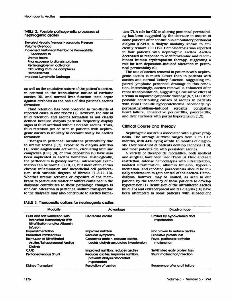
TABLE 2. Possible pathogenetic processes ofnephrogenic ascites
Elevated Hepatic Venous Hydrostatic Pressure
Volume OverloadIncreased Peritoneal Membrane Permeability
Secondary toUremic toxinsPrior exposure to dialysis solutionsRenin-angiotensin activationCirculating immune complexesHemosiderosis
Impaired Lymphatic Drainage
as well as the exudative nature of the patient’s ascites,in contrast to the transudative nature of cirrhotic
ascites (6), and normal liver function tests argueagainst cirrhosis as the basis of this patient’s ascites
formation.Fluid retention has been observed in two-thirds of
reported cases in one review ( 1 ). However, the role of
fluid retention and ascites formation is not clearly
defined because dialysis patients frequently display
signs of fluid overload without notable ascites. Thus,fluid retention per se seen In patients with nephno-genic ascites is unlikely to account solely for ascitesformation.
Changes in peritoneal membrane permeability dueto uremic toxins ( 1 ,7), exposure to dialysis solution( 1 ), renin-angiotensin activation, circulating immunecomplexes (CIC) (8). or iron deposition (9) have alsobeen implicated in ascites formation. Histologically,the peritoneum is grossly normal; microscopic exam-ination can be normal (3, 10, 1 1) but more often showschronic inflammation and mesothelial cell prolifera-tion with variable degrees of fibrosis ( 1-3, 1 1-13).
Whether uremic serositis or exposure of the mem-brane to particulate matter or buffers contained in thedialysate contributes to these pathologic changes is
unclear. Alteration in penitoneal sodium transport dueto the dialysate may also contribute to ascites forma-
TABLE 3. Therapeutic options for nephrogenic ascites
tion (7). A role for CIC in altering penitoneal permeabil-
ity has been suggested by the decrease in ascites in
some patients after continuous ambulatory penitonealdialysis (CAPD), a dialytic modality known to effi-ciently remove CIC ( 1 2). Hemosiderosis was reported
in four patients with nephrogenic ascites. Ascites
decreased in response to iv deferoxamine and recom-binant human erythropoietin therapy, suggesting arole for iron deposition-induced alteration in perito-neal permeability (9).
The rate of ascites removal in patients with nephro-
genic ascites is much slower than in patients withascites and normal kidney function, suggesting im-
pained lymphatic penitoneal drainage in this condi-tion. Interestingly, ascites removal is enhanced after
renal transplantation, suggesting a causative effect ofuremia In impaired lymphatic drainage (6,7, 14). Otherpossible contributing causes of ascites in patientswith ESRD include hypoproteinemia, secondary hy-
perparathynoidism-induced serositis, congestiveheart failure, constrictive penicarditis, pancreatitis,and liver cirrhosis with portal hypertension (1,2).
Clinical Course and Therapy
Nephrogenic ascites is associated with a grave prog-nosis. The average survival ranges from 7 to 10.7months, with 44% dying within 15 months of diagno-sis. Over one-third of patients develop cachexia (1,3),
and most patients die with persistent ascites.A variety of therapeutic modalities, both medical
and surgical, have been used (Table 3). Fluid and saltrestriction, intense hemodialysis with ultrafiltration,isolated ultrafiltration, albumin infusion, hypenali-
mentation, and repeated paracentesis should be ini-tially undertaken to gain control of the ascites. Hemo-dialysis, however, may be limited, as seen in ourpatient, by the tendency of these patients to develophypotension ( 1 ). Reinfusion of the ultrafiltered ascites
fluid ( 1 5) and extracorponeal ascites dialysis ( 1 6) havebeen attempted in some patients with subsequent
Modality Advantage Disadvantage
Fluid and Salt Restriction With Decreases ascltes Limited by hypovolemia andIntensified Hemodialysis With hypotensionUltrafiltration and/or AlbuminInfusion
Hyperalimentation improves nutrition Not proven to reduce ascitesRepeated Paracentesis Reduces symptoms Excessive protein lossReinfusion of Ultrafiltrated Conserves protein, reduces ascites, Fever, peritoneai catheter
Ascites/Extracorporeal Ascites avoids dialysis-associated hypotension malfunctionDialysis
CAPD Improved nutrition, reduces ascites Self-limited early protein lossPeritoneovenous Shunt Reduces ascites, improves nutrition,
prevents dialysis-associatedhypotension
Shunt malfunction/infection
Kidney Transplant Resolution of ascites Recurrence after graft failure
Hammond and Takiyyuddin
Journal of the American Society of Nephrology 1177
improvement in hemodynamic stability, a reduction inascites, and improvement in the quality of life as well.
The placement of either a Denver or LeVeen penito-neovenous shunt (Becton and Dickinson, Sunnyvale,CA), as in our patient, has been associated with areduction, although palliative, in ascites formationand improvement in hemodynamic stability duringhemodialysis (4, 1 7-1 9). Objective improvements are
also noted in the nutritional status with weight gainand subjective improvement in appetite and physicalactivities. Complications of shunt placement, seen inover one-half of the cases, include malfunction from
occlusion or migration of the venous end out of thesuperior vena cava and infection. These complicationsrequire either minor revisions or removal of the shunt.
Shunts remained functional for up to 18 months inone series (Denver) ( 1 7) and for more than 3 yr in
another (LeVeen) (9). Thus, shunt placement shouldbe seriously considered early in the treatment of suchpatients.
CAPD is also effective in the treatment of ascites(1,11,12,20,21). The combination ofroutine daily ex-
changes and control of fluid and salt intake allows
control of the ascites. The patients subjectively feelbetter, with improved caloric and protein intake andsubsequent weight gain. Within 6 months of contin-ued treatment, the amount of total protein excretion
in the dialysis effluent decreases from 26.5 to 50 to 7.8to 9.44 g/day, with a subsequent rise in the serumprotein and albumin levels and the resolution of as-cites. CAPD may be beneficial in controlling ascitesformation by reducing ip fluid protein concentration,
which normally would osmotically draw fluid into thepenitoneal cavity ( 13). Other treatments attemptedwith only partial success that are no longer recom-mended Include the ip administration of steroids(1,2,21) and bilateral nephrectomy (1,3,19,20).
Of all of the therapeutic modalities, kidney trans-plantation appears to be the most effective in control-
ling ascites formation. Nearly all reported cases (22
out of 24 cases) had complete resolution of the asciteswithin 2 to 6 wk after transplantation, with a singlecase requiring 1 2 months for the complete resolutionof ascites ( 1 ,6,7, 10, 1 1 , 18,20). Two patients, on theother hand, had a recurrence of ascites despite ade-quate renal graft function 2 yr (with creatinine of 1.6mg/dL) (13) and 4 months (with a creatinine clearanceof 52 mL/min) (8) after transplantation. Interestingly,ascites often recurs after the loss of graft function forwhatever reason (1,19,20). The ascites can recur atthe time of graft failure (20) or anytime thereafter forup to 3 yr (19,20).
In conclusion, nephrogenic ascites is a rare condi-tion with a grave prognosis and an unknown butprobably multilactonial cause. Although no prospec-tive studies have been performed comparing varioustreatment modalities, on the basis of current data,
CAPD, penitoneovenous shunt placement, and renaltransplantation offer the best hope for an improve-ment in the quality of life and recovery.
REFERENCES
1 . Gl#{252}ckZ, Nolph KD: Ascites associated with end-stagerenal disease. Am J Kidney Dis 1987; 10:9-18.
2. Mauk P, Schwartz JT, Lowe JE, Smith JL, Graham DY:Diagnosis and course ofnephrogenic ascites. Arch InternMed 1988:148:1577-1579.
3. Feingold LN, Gutman RA, Walsh FX, Gunnells C: Con-trol of cachexia and ascites in hemodialysis patients bybinephrectomy. Arch Intern Med 1974; 134:989 -997.
4. Rector WG: Portal hypertension: A permissive factor onlyin the development of ascites and variceal bleeding. Liver1986:6:221-226.
5. Runyon BA, Montano AA, Akrlviadis EA, Antillon MR,Irving MA, McHutchison JG: The Serum-ascites albumingradient is superior to the exudate-transudate conceptin the differential diagnosis of ascites. Ann Intern Medl992;1 17:215-220.
6. Craig R, Sparberg M, Ivanovich P, Rice L, Dordal E:Nephrogenic ascites. Arch Intern Med 1974; 134:276-279.
7. Gotloib L, Servadlo C: Ascites in patients undergoingmaintenance hemodialysis. Am J Med 1976:61:465-470.
8. Twardowski ZJ, Alpert MA, Gupta RC, Nolph LW, Mad-sen: Circulating immune complexes: Possible toxins re-sponsible for serositis (pericarditis, pleuritis, and perito-nitis) in renal failure. Nephron 1983:35:190-195.
9. Be�ba� N, Soylemezoglu 0, Saat#{231}i,U et aL: Peritonealhemosiderosis in pediatric patients with nephrogenicascites. Nephron 1992;62:292-295.
10. Wang F, Pillay VKG, Ing TS, Armbruster FKW, Rosen-berg JC: Ascites in patients treated with maintenancehemodialysis. Nephron 1974; 12:105-113.
1 1 . Slngh 5, Mltra 5, Berman LB: Ascites In patients onmaintenance hemodialysis. Nephron 1974; 12:114-120.
12. Rubin J, Kiley J, Ray R, McFarlan 5, Bower J: Contin-uous ambulatory peritoneal dialysis: Treatment of dial-ysis-related ascites. Arch Inter Med 1981 ; 141:1093-1095.
13. Rodriguez HJ, Walls J, Slatopoisky E, Klahr 5: Recur-rent ascites following peritoneal dialysis: A new syn-drome? Arch Intern Med 1974; 134:283-287.
14. Morgan AG, Terry SI: Impaired peritoneal fluid drainagein nephrogenic ascites. Clin Nephrol 1981:15:61-65.
15. Parbhoo SP, Adjukiewicz A, Sherlock 5: Treatment ofascites by continuous ultraffitration and reinfusion ofprotein concentrate. Lancet 1974; 1:949-952.
16. Adler AJ, Feldman J, Friedman EA, Berlyne GM: Use ofextracorporeal ascites dialysis in combined hepatic andrenal failure. Nephron 1 982;30:3 1-35.
1 7. Hobar PC, Turner WW, Valentine RJ: Successful use ofthe denver peritoneovenous shunt in patients with neph-rogenic ascites. Surgery 1987; 101:161-163.
18. Cadnapaphornchal P. Kuruvlla KC, Holmes J, SchrierR: Analysis of 5 year experience of home dialysis as atreatment modality for patients with end-stage renalfailure. Am J Med 1974;57:789-799.
19. Hoim A, Rutsky EA, Aldrete JS: Short- and long-termeffectiveness, morbidity, and mortality of peritone-ovenous shunt inserted to treat massive refractory as-cites of nephrogenic origin. Analysis of 14 cases. AmSurg 1989:55:645-652.
20. Popli 5, Chen WT, Nakamoto 5, Daugirdas JT, Ces-pedes LE, Ing TS: Hemodialysis ascites in anephricpatients. Clin Nephrol 1981 ; 15:203-205.
2 1 . Pascual JF, Melendez MT, Rivera-Viera JF: Local steroidtherapy of refractory ascites associated with dialysis. JPediatr 1979:94:319-320.
22. Korzets A, Danby P, Feehally J, Walls J: CAPD: Suc-cessful use in the treatment of nephrogenic ascites.Nephrol Dial Transplant 1989:4:918-919.