nephritis in childhood · (a) acute interstitial nephritis. (b) acute and subacute embolic...
TRANSCRIPT

NEPHRITIS IN CHILDHOODBY
NORMAN B. CAPON, M.D., M R.C.P.
(From The Royal Liverpool Chiild(ieii's Hospital,for thle Medical Researclh CouInci!)
The investigations upon which this paper is based were undertakenprimi-arily to exurnine the practical value of the so-called " renal efficiencytests " in children. Cases of kidney disease admitted to one of the medicalwards of the Royal Liverpool Children's Hospital were systematicallyexamined, and the results have been arfalysed. This has neceSsitated agenieral survey of kiidney dise'ases, and of the special peculiarities of thesediseases in children. So far as possible, the pathological and clinical aspectshave been correlated in the classification adopted; and -an attempt has beenimade to emphasise the necessity of taking a broad general view of each casestudied.
The paper is sub-divided under the following headings:-
1. Classification of nephropathies.2. Methods of investigation.3. Description of cases studied.
(A) Acute glomerulo-nephritis.(B) Chronic glomerulo-nephritis.(C) Diffuse tubular nephritis.
4. Pathological findings in diffuse tubular nephritis.5. Treatment.6. Suminary and conclusions.7. Bibliographical references.
1. ClAS SIFICATION OF NEPHROPATIIIES.It must be admitted that there is at the present time no satisfactory
classification of kidney diseases. Of the many schemes which have beensuggested (Heubner(12), Newburgh(27), Schwartz and Kohn(28), and Tripputi(31),etc.), one of the best-known is that of Volhard and Fahr(32), which is herereproduced in abridged form (Table A). This classificat'on has a pathologic'al
TABILE A.
CLASSIFICATION 1.(Modified froin Volhard and Fahr.)
A. Nephrosis. Miuller, 1905. (No increase of PP.)B. Nepliritis.
1. Diffuse glomerulo-nephritis. (Tnerease of B.P.)2. Focal nephritis. (No increase of B.P.)
C. Renal Sclerosis.
on March 23, 2020 by guest. P
rotected by copyright.http://adc.bm
j.com/
Arch D
is Child: first published as 10.1136/adc.1.3.141 on 1 January 1926. D
ownloaded from

ARCHIVES OF DISEASE IN CHILDHOOD
rather than a clinical foundation, and it marshals cases of kidney diseaseunder three main heads-degenerative, inflammatory and sclerotic; or, inother words, nephrosis, nephritis, and renal sclerosis.
(A) Nephrosis is Comparatively a rare condition in which the cells liningthe kidney tubules show degenerative changes, without any evidence ofinflammnation elsewhere in the kidney (e.g., glomerular changes). Thevarious types of degeneration which may be fotund are stated by Munk(25) tobe as follows:-Cloudy swelling -or albuminous, fatty, lipoid (cf. mnyelinkidnev of McNee(19)), necrotic, hyaline, a.myloid, and possibly glvcogen-deaerneration in diabetes irrellitus.
Poisoning with heavy metals and intestinal iiitoxication may benentioned as possible causes of nephrosis.
The studies of Epstein(9) and of Major and fHelwig(20) seem to show thatcther characteristic features of nephrosis include hypercholesterinammia, alow metabolic rate and a great increase of serum-albuumin as compared withsertumn-globulin.
(B) Nephritis is comparatively a common disease in which the kidneysshow signs of inflammntory response; for instance, lyvperammia and swellillnof the tufts, outpouring of exudate and of cells into the capsules of Bowman,le.-cocytic and lymphoeytic infiltration of the interstitial tissue, anddegenerat,ive changes in the cells lining the kidney tubules.
(c) Renal sclerosis is a common lesion in (aults, but is very rare inchildren. I s,hall not consider it, in this paper.
Clinically, one of the most important features of nephrosis is extensiveand persistent cedeina; whereas in nephritis, cedema is variable in degreeanld frequently is not a prominent feature.
Volhard and Fahr(32) dlivided their nephritic cases into two groups, diffuseglornierulo-nephritis and focal nephritis, the difference between them being,mainly clinicall and coneerning the blood pressure. Thev recognised, further-more, that focal nephritis may be (1) glomerular, for instance in endocarditis,especially if due to the streptococcus viridans; (2) embolVc; or (3) of -theseptic or acute interstitiall type (Councilman), seen occasionally in dipphtheriaand also after scarlet fever where the throat infection has been very severe.
If the distinction wlhich differenitiaites " nephrosis " from the better-known and older term " pa.renchynma.tous or tubular nlephritis " is notproperly appreciated, the rmodified classification of Volhard and Fahr appearsto be no advance upon the foriner terminology. A certain laxity in adheringto the terms of definition, together with the clinical fact that examples of
parenchymartous or tubular nephritis " frequently bear a close resemblanceto cases of nephrosis, ha.s led several writers to describe as " nephrosiscases in which there was evidence of inflarnmamtaon of the kidneys. My owninvestigta.tions, though compara.tively few in number, demonstrate theapplicability to childhood of Volhard and Fahr's statement that nephrosisis a rare condition; and in. order properly to record the pathological lesion
142
on March 23, 2020 by guest. P
rotected by copyright.http://adc.bm
j.com/
Arch D
is Child: first published as 10.1136/adc.1.3.141 on 1 January 1926. D
ownloaded from
NEPHRITIS IN CHILDHOOD
which exists in a majority of " large white children," I have suggested aclassification (Table B) which is in essence a reversion to the older scheme,but provides also a place for examples of true nephrosis.
TABLE B.
CLASSIFICATION 2.A. Nephrosis. (Some cases of parenchymatous nephritis.)r3. Nephritis.
( General1. Glomerulo-nephritis J Focal nephrit-i of Volhard and
I Local or Fahr.Padonephritis of Heubner.
2. Diffuse tubular nephritis. (Most cases of parenchymatous ortubular nephritis.)
3. Focal nephritis:(a) Acute interstitial nephritis.(b) Acute and subacute embolic nephritis.
C. Renal Sclerosis.
It is a truism to state that when a child's kidneys become inflamed, itis rare to find on histological examinIation that any one of the componentstructures of the kidney has escaped: there a-re few examples of nephritiswhich do not show diffuse nephritis.
I have employed the word " diffuse" to emphasise one special andimnportant feature found in the kidneys of " large white children," namely,the very widesprea.d extent of the lesion. At the same time, the clinicaland pathological advantages gained by emphasising in our terminology whichl)articular component of the kidney has been mainlv affected have not beenforgotten.
The difference between what I have called general and local glomerulo-nephritis is only a question of degree, namely, the number of -glomeruliaffected. It is said that successive reports on the state of the cardiovascularsystem provide a measure of this factor. Upon this topic, which is, I thin1k,relatively of small importance, a voluminous literature must be consulted;I have summarised it by accepting the view that Heubner's(13) pedonephritisis the sa.me condition as Volhard and Fahr's focal glomerulo-nephritis.
2. METHODS OF INVESTIGATION.Reference to Table C will give at a glanlce the general scheme of
investigation followed, though it will be obvious that for certain cases it wasr.ecessary, on medical grounds, to exercise discretion in the application oftests. Furthermore, it is well recognised that dift'iculties encountered duringthe investigation of renal cases are more frequent and more insurmountable
I.43
on March 23, 2020 by guest. P
rotected by copyright.http://adc.bm
j.com/
Arch D
is Child: first published as 10.1136/adc.1.3.141 on 1 January 1926. D
ownloaded from

ARCHIVES OF DISEASE IN CHILDHOOD
in children than in adults. For instance: (1) urine collection withoutcatheterisation may be impossible; and the total volume in 24 hours is often-inaccurately recorded even in older children; (2) blood vessels are small andone may have difficulty in obtaining blood for examination: in some cases Iused the patient's saliva for the estimation of urea; (3) emotional disturbancesare frequent in children; therefore blood pressure records are often not reliable;(4) accurate dieting is a great difficulty; one cannot expect the co-operationfrom a child which one receives from an adult.
For these reasons, tests were sometimes unsatisfactorv; and sometimesthe delay between repetitions of the same test was longer than one would-ha.ve wished.
TABLE C.
-: -. . ,PLAN OF INVESTIGATION.
1. Medical history of patient and of relatives.
2. General clinical exatmination, with special reference toBody-weight.(Edema.Aneemia.
- Heart and blood vessels. B.P.Optic fundi.Septic foci, e.g., teeth and tonsils.
3. Urine. Daily Examination,(a') General. Volume: sp, gr.: reaction.
(b) Chemical. Protein content.UJrea content.Chloride content.
(c) MVlicroscopical.
4.- Blood. Nonprotein nitrogen.- Wassermann reaction.
5. Renal efficiency tests.(a) Phenolsulphonephthalein excretion.
(b) Urea concentration test.
(c) Salt test.(d) Fixation of specific gravity test.
6. "Living kidney " examinaltion.
7. Autopsy. Histoloqy.
144
on March 23, 2020 by guest. P
rotected by copyright.http://adc.bm
j.com/
Arch D
is Child: first published as 10.1136/adc.1.3.141 on 1 January 1926. D
ownloaded from

NElPHRITIS IN CHILDHOOD
T'lhe following notes are supplied in amplification of the scheme shown inTable C:-
Urine. For some time I estirnated the diastase content of urines daily,but later discarded this test. The chloride content was estinmated by Harvey'smodification of Volhard's method (see M-yers (26)).
Blood. Non-protein nitrogen was estirmated y a modified Kjeldahlprocess, with subsequent nesslerisation and colorimetric comparison.
Ren(il Efliciency Tests. From the outset one kept in mind thelimnitations which workers on adults have shown to exist in the use of thesetests, as well as the caution which inust be exercised in making deductioinsfrom the results obtained. For instance, it is not yet accurately knownwhich particular functional capacity of the kidney is tested by the differentfunctional tests. Furthermore, it must be renmemabered that in health nosingle a,ctivity of the kidnev is exerted to the full: the organ possesses whatmay be described as a big reserve power " and frequently no evidence offunctional incapacity is revealed until damage is widespread and severe. Inother words, the kidney conceals its own weaknesses at a time when thephysician wishes to discover them; and when the lesions are no longer smallenough to be hidden, obvious clinical signs frequiently appear.
Another difficulty lies in the fact that the functional efficiency of kidneytissue which has developed by compensatory hypertrophy is unknown.Moreover, the functions of the kidney are disturbed by most severe diseasesof the viscera., especially of the heart, blood vessels, lungs and blood; asalso by all so-called " general diseases," for instance, fevers. Henc6impairment of function, a,s shown by efficiency tests, may be only the shadowand not the substance. Wilcox and Lyttle(33) and Gittings and GrameMitchell(1l) give interesting figures in relationship to this aspect.
In hea.lthy children the two-hour excretion of phenolsulphonephthalein,admiiinistered intrarmuscularly, should be at least 60%. T.. W. Hill(14) foundit to be 76%, Leopold and Bernhard(16) 70)%°', Curtis(6) 75%, Tileston andComfort(30) 89%, and Wilcox and Lyttle(33) 60%. When oedema is present,the test by intramuscular route is almost valueless; and when there is bloodin t,he urine the estimation of the excreted dye is generally impossible. Onseveral occasions I found the matching of colouirs in mea.suring the excreted'phthalein a very difficuilt matter even when there was no blood in the urine;the addition of alkali to bring out the colour of the dye precipitatesphosphates, which interfere with the colour comparison. The phosphatesmay be separated, but this renders the test less accurate. The ureaconcentration test was carried ouit according to the method describedby McLean(18), except that the dose of urea employed was 10 gra.ms.By the term " salt test " I refer te the exhibition of 10 grams of sodiurncliloride,. with subsequent investigation of increase of weight, due to aedema,and of the urinary excretion of sal]t. This test undoubtedly aggravates thepatient's condition temporarily; it was used on a few occasions only.
145
on March 23, 2020 by guest. P
rotected by copyright.http://adc.bm
j.com/
Arch D
is Child: first published as 10.1136/adc.1.3.141 on 1 January 1926. D
ownloaded from
ARCHIVES OF DISEASE IN CHILDHOOD
The fixation of specific gravity test was found to be applicable only tothe older children; I have little experience of it.
Living Kidney " Examtination. In certain cases, decapsulation of thekidneys was performed, and I am greatly indebted to Afr. R. C. Dun, SeniorSurgeon to the Roval Liverpool Children's Hospital, for his kindness inassisting mne by excising a small wedge of kidney substance during theoperation. This piece of tissue was dropped at once into formalin and waslater examined microscopically. By this method of examina.tion a fullerknowledge of the kidney lesion is gained.
3. DESCRIPTION OF CASES STUDIED.
])During the past three years I have had the opportunity of observingsome 20 cases of nephropa.thy in children between the ages of 2i and 12years. One of these cases has been published under the names of Dr.Dingwiall Fordyce and myself('0) and I shall not include it in this paper.
Four other cases could not, for various rea.sons, be studied adequately,and I have excluded them from this survey.
Of the remaining 15 cases I have classified three as acute glomerulc-nephritis, six as chronic glomerulo-nephritis, and six as diffuse tubularnephritis.
A. Acute Glomereulo-Nephritis.Only brief reference will be made to these three cases,
M. D. Female, age 34 years. For some weeks before the onset of nephritis thischild had been debilitated; there had also been otorrhoea and a vaginal discharge. Aneruption resembjiig " points of blood " was noticed on the body and legs two weeksb)efore the child was brought' to hospital suffering with pyrexia, oliguria, haematuriaand proteinuria- (7 gms. per litre). There was a moderate grade of oedema of thesubcutaneous tissues; and numerous impetiginous areas were scattered widely on theskin. Microscopical examination of the urine showed numerous casts of all descriptionsThe extreme gravity of the patient's condition made systematic examination impossible.The svstolic blood pressure was about 90 mm. of mercury; possibly this is too high anestimate because the child was in an irritable, frightened state. She died a few daysafter admission and an autopsy was refused. There does not appear to be sufficienteAidence to say that nephritis was secondary to impetigo, though this is probable.
D. S. Male, age 22 years. Oliguria and haematuria developed immediately afterI.neumonia. The urine contained 2 to 3 gms. of protein per litre and numerous castswere found. The temperature on admission was 1030, but it fell to normal almost atonce and recovery was rapid. When the patient was re-examined 12'months after hisattack, his general health was quite satisfactory, the urine was normal and the systolicblood pressure was 112 mm. of mercury. This is a high figure for a child of 3j years.
A. D. Male, age 9 $, years. In infancy this child had been circumcised and hadlhad slight measles. There was no illness precedino the attack of nephritis, except a"cold in the bead." I'he onset was characterised by rise of temperature, vomiting, andrain in the right side of the abdomen: these signs, together with anorexia -and 'veryslight jatiudice suggest that a catarrhal cholangitis had developed. Proteinuria, up to1 gm. per litre, was discovered: erythrccytes and leucocytes were present in the urine,but no casts were found. Slight oedema of the a,nkles was noted. The blood non-
146
on March 23, 2020 by guest. P
rotected by copyright.http://adc.bm
j.com/
Arch D
is Child: first published as 10.1136/adc.1.3.141 on 1 January 1926. D
ownloaded from

NEPHRITIS IN CHILDHOOD
Fig. 1.X 135.
X 130.
Fig. 2.X 135.
X 135.Fig. 4.
147
Fig. 3.
on March 23, 2020 by guest. P
rotected by copyright.http://adc.bm
j.com/
Arch D
is Child: first published as 10.1136/adc.1.3.141 on 1 January 1926. D
ownloaded from

ARCHIVES OF DISEASE IN CHILDHOOD
X 135 X 200.Fig. 5. Fig. 5a.
X 125. X 150.Fig. 6..
148
Fig. 7.
on March 23, 2020 by guest. P
rotected by copyright.http://adc.bm
j.com/
Arch D
is Child: first published as 10.1136/adc.1.3.141 on 1 January 1926. D
ownloaded from
NE1'HRITiS IN CHILDHOOD
rirotein nlitrogen was 37 lIlgs. per 100 c.cms. of whole blood, and the two-hour 'plithaleinexcretion was 94%. Rlecovery was rapid; and when the patient was re-exa.mined 1 Tyears after this attack he was found to be in normal lIealth, and his urine was normal.The systolic blood pressare was 117 mm. of mnercury (age of patient 114 years).
B. Chronic Glomierulo-Nephritis.This forin of nephritis affects especially children of school age. rjho
cnset may be sudden, with an acute nephritis, or it mafly be gradual. Ineither case the disease is frequently preceded by one of the exanthemata, orby somne infective condition, for instance, tonsillitis, to which special attentionhas been drawn by many observers, including CautleV (3), Marriott(2U,Allison(l), and D. L. Smith and C. W. Bailey(29).
XWhen the onset is insidious, anorexia, heaidache, vomitinig, oliguria andhLema.turia are frequent symptoms; cedeina is variable in degree, butt isgenera.lly not a prominent sign except during an acute exacerbation. Pallorand general ma.lnutrition are almost always found. Some cases bear astriking resemblance to tuberculosis of the lidneys and urinary tract.
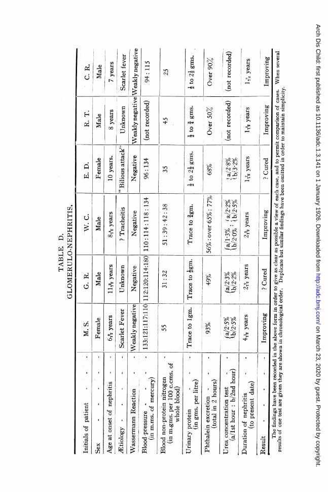
The following are abbreviated records of my cases; additional notes willbe found in Table D:-
M. S. The patient had whooping.cough, chicken-pox, measles with pneumonia,anid severe scarlet fever, all during one year. Bilateral otorrhcea developed as acomplication of scarlet fever, and the mother noticed that the urine was beccmingscanty and dark coloured. Headaches, anorexia, general malaise and some oedema ofthe face, body and legs were additional complaints. The parents noted that thesymptoms showed cyclical exacerbations, each attack lasting about two weeks. (Edemasoon disappeared and has not redeveloped at any time. She is a tllini, frail, nervouschild; her teeth and tonsils are not above suspicion.
G. R. Parenits are Russian Jews; healtlly. The patient had whooping-cougzh,chicken-pox, measles and mumps in early childhood. Adenoids removed at 3 years.
No illness immediately preceded the present complaint, which began with cough,anorexia and vomiting; also slight pains in the chest and abdomen on exertion. Therewas some headache, fever and sweating. The urine was noticed to be dark coloured.Shortly after the onset, right-sided pleurisy with effusion developed. The fluid wasstraw coloured and contained large numbers of small lymphocytes; neither tubercle,Jtcilli nor other micro-organisms were discovered and cultures were sterile. Later,radmographs of the chest showed no evidence of tuberculosis. Von Pirquet reactionriegative. Radiographic examination of kidney regions was negative. No tuberciebacilli were found in the urine or faeces; guinea-pig inoculation with urine was negativefor tubercle bacilli.
The patient is a well-developed, fat boy with slight cedema below the knees.Teeth and tonsils healthy.
W. C. The patient had whooping-cough in infancy, haemorrhagic measles when6i4 vears old and chicken-pox when 71 years old. He is stated to have bled freelyafter circumcision and after extraction of teeth. With the onset of tracheitis wheln8' years old, the parents inoticed that the urine was' dark coloured; there was alsosomne loss of appetite. Whenever he caught a " chest cold," which was a frequentoccurrence, the urine became bright red, with increased frequency of micturitio-durinig the day, and there was anorexia and increased pallor.
Between these attacks the urine did not become normal in colour. He is a thini,frail, alert child of unusually high intelligence. No redema has been noted. O)n
19
on March 23, 2020 by guest. P
rotected by copyright.http://adc.bm
j.com/
Arch D
is Child: first published as 10.1136/adc.1.3.141 on 1 January 1926. D
ownloaded from
ARCHIVE,S OF DISEASE IN CHILDHOOD
admission to hospital he was found to have calcareous lymphatic glands in theabdomen; these were confirmed by radiograph. There were no signs, clinical orradiographic, of tuberculosis elsewhere. Examination of the bladder by radiographyand cystoscopy, and of the urine by microscopy and by guinea-pig inoculation, were allnegative for tuberculosis. The teeth were satisfactory; tonsillectomy was required anldwas carried out in June, 1924.
E. D. The patient had not suffered with any previous illnesses except enuresi.s.The onset occurred with a " bilious attack," followed by puffiness around the eyes,aind possibly by a purpuric eruption on the legs. The patient was a thin, pale, nervousgirl, with spasmodic internal strabismus. There was no oedema. The teeth weresatisfactory; but the tonsils were found to be unhealthy and were removed in December,1923. Blood coagulation time and fragility tests were normal. No tuberele bacilliwere found in the urine.
R. T. Fairly hlealthy family history. There was no known cause for the onset ofba-maturia in this patient; it began fSve weeks before admission to hospital andcontinued intermittently. Von Pirquet reaction was negative. Radiograph of thechest showed a good deal of spottiness of and adjacent to the right lung root.Radiograplhs of the kidney regions showed nothing abnormal.
C. R. The patient's previous illnesses were measles, one year before the presenlttrouble, and frequent attacks of nocturnal enuresis. The complaint bcg3,aii withpuffiness of the face which was noticed after an attack of scarlet fever; at tlle sametiine the urinie was noticed to be dark red in colour. The appetite was greater thanuisual. The patient was found to be a fairly well-nourished, pale child, with slightoedemna of the shins. Four very septic teeth were extracted anid tonsillectomy waspetformed in May, 1924.
In the urine of these patients red blood corpuscles and leucocytes,hvaline, granular and epithelia.l casts were almost constantly present. Bloodcasts were less frequently seen. Epithelial cells, probably from the kidneytubules, were commonly found. It should be noted that the amount of)rotein passed in the urine in chronic glomerulo-nephritis is comparatively
small. The optic fundi were normal in all cases. The cardiac dulness wasnormal in extent in all patients except G. R., in whom it was possiblyincreased (31 inches from the mid-sternal line in the fifth intercostal space:age of patient at this time was 13 years). No case showed signs of organicheart disease.
It will be seen on reference to Table D that abnormally high bloodpressure readings were observed in each chil(d except R. T., in whom recordswere not made; but it must be pointed out that by the time G. R. 's systolicblood pressure was 180 mm. of mercury (age of patient=131 Years), his urinehad become normal; and similarly when E. D.'s systolic blood pressure was134 mm. of mercury (age of patient=11J years), her urine was normal.
In attempting to determine the prognosis in cases of chronic glomerulo-nephritis it is important to remember that two processes, one progressiveand the other regenerative, are probably taking place. The former includesepithelial inflammation, recognised bv finding protein in the urine, andkidnev sclerosis. So long as proteinuria continues one mra.y assume thatprogressive inflammatory changes are taking place; improvement ma.y berecorded when proteinuria is diminishina or has disappeared. Any
150
on March 23, 2020 by guest. P
rotected by copyright.http://adc.bm
j.com/
Arch D
is Child: first published as 10.1136/adc.1.3.141 on 1 January 1926. D
ownloaded from
4)
cn U
o En
~cn
0*
5-C)
504
00
'0 c
C0
C0
00
0~~
C00
'do
00
0
a)0ow
IUa)0u
(A
a)
IH
ni
ozbJ
Q)
0a)
0
to
a)
0
z
6
0
on March 23, 2020 by guest. P
rotected by copyright.http://adc.bm
j.com/
Arch D
is Child: first published as 10.1136/adc.1.3.141 on 1 January 1926. D
ownloaded from

l)
Cd
'-'H
H1
b,O
bDu -I- 0z
- °
Cd
>
(1 0 ui 4-
L
CldC
Cdd
4$4
Uf)$4
Cd 0a) 0
CC-111-~0
0 ,I.P4aCn cd
()Cd
,u >.1>,£+n1
a1)
CdQ)
cnIU)
0 >
O 4-
) )0
to
t-
L4 to), ..
O).00
00
to,I
'..to
to
o
in
on.)
NI
tot..I -
OC-< 0N
toY
to
to
I00N
to
00to
* -q
Unco
000
0(:7
too
Ns
(A
bDo
0
00
100OC)
cA
buo
0
0340D
bc
N
$-4a1)
0
(A
bo0
0340D
ho
004-0
(N
0C\Ti
0
a1)0
to
0N
00
0S t
a)C*l --
%#-
;,,1-4 L
4) 8
*~~~~~~~ cdo~~~~
)~~~~~~~~~~~~~~~a*---.---0 0
1
*
c0
o
ce.
0
0
Cd -
Cd
Cd .
NCd la
'-I C4..
0 's-
Cd ,laI..-'
ho ~~~~~~~~~~4U)~ ~~~~~~~~0)
Xi-4 .w 0.~~u'-' ~ (1 )
Cd *S 4 -4$.a)~~~44~~~0 v)O$4U 0 a, )
a)~~~~~~~~~x i
-4- 0 C.) $4bidLuCdIn -~0~ 1 0 Cd 4
ri0 0 0 4- 0bo13~ ~ ~ o?
o -
U)
Cda)
1-4
0
Cdd
Cd
Cil I0 c
a))
Cd
Cd
aL) U)
0 Cd
'-I
r O Y
| -
.o00Cd
Cdu
aL)
-4
Cd$4.
aL)Cd
a)
0
z
[U)
$.4
Cda)
U)$4
Cda)
4
CC
a)
Cd
~0
Cd
a)
1.-'-4
I4-' 4-
U
o U)
a1)
Cd
a1)
CL).-I-.4
1-4I".j
V
L)-1
.4
.4i
3
I
¢4H
P.40D0QHD0-
a)
04En
$40
En
Cd
4,
O
CC
0
$4
'0
a)
ad
14
0
0
0CCcn
0dCC-0U00
'. c
(A
aL).t
Cd04
0
Un
.-I._da)
-* E ;~~~~~~~~~~~~~~~~~~~~~~~~~~~~~~~~~~~~~
II
i
I
II
1-
1-
-11
on March 23, 2020 by guest. P
rotected by copyright.http://adc.bm
j.com/
Arch D
is Child: first published as 10.1136/adc.1.3.141 on 1 January 1926. D
ownloaded from

NEPHRITIS IN CHILDHOOD
effect which kidney sclerosis may have upon the cardiovascular systemseems to depend upon the distribution and stage of the lesion; hencethe blood pressure ma,y or may not be raised in chronic glomerulo-nephritis.WVhen it is permanently elevated serious kidney disease is present.
Regenera.tion of kidney tissue plays a greater part in the child thanin the adult, and its effect in concealing the true extent of the pathologicalchange is probably greater the younger the patient. In the two :patientsG. R.* and E. D. 'it is evident that at the time of the last urinaryexamination there was no active kidney inflammation; but the histories ofthe cases, together with the blood pressure records, furnished proof sufficientto classify them as chronic glomerulo-nephritis. If thev had been examplesof local glomerulo-nephritis, alternatively termed Volhard and Fahr's focalnephritis and -Heubner's pedonephritis. the blood pressure would not havebeen higher than norma.l [Heubner(13), Tripputi(31) and others]. Mendel(23'has pointed out that since the time when HIeubner gave his originaldescription of p'edonephritis it! has been shown that this syndrome has notthe clear cut boundaries which it was at first believed to possess. I submitthat if the term paedonephritis is to be, retained it must be regarded as a mildform of chronic glomerulo-nephritis, .and not as a separate entity. Thereare obvious disadvantages in introducing new words to differentiate caseswhich seem to vary only in the extent of the lesion.
'I'he possibility of a recurrence of a.cute nephritis in these cases is-considerable; and Ermerson(8) has emphasised that even minor symptomsm1ayV be of grave significance, because they furnish evidence of advancingepithelial disease. Hence it is; most difficult to say when a, ease of clhronic,glomerulo-nephritis is cured. Even if there has been no active inflamma.tionfor several years, it is possible that delayed vascular, complications mayaappear in a(lult life.
C. Diffuise Tutbular Nephritis.The most striking. feature of this form of nephritis is severe generalisedt
edeuna. The age of the-patient is usually younger than in chronic glomerulo-nephritis. Cold and exposuire have always been regarded a.s oetiologicalfactors;, a,t times tuberculosis, syphili.s and chronic suppurative conditionsare antecedents. Marriott(21) finds that staphylococca.l infection of the nasalsilntuses is often the primary cause. The onset of the disease is generallyinsidious, with pallor, oliguria and odema. Pyrexia is often only slight;sometimes it is absent. Heematurian is seldom noticed. Proteinuria isconsiderable, and many ca,sts of all descriptions-especially hyaline.granular, epithelial and fa.tty-are found in the urine. Leucocytes and cellsfrom the renal tract are numerous. Erythrocytes are scantv.
*A few days before the completion of this paper, the patient G. R. suffered an acuteexacerbation of nephritis, characterised by general malaise, oliguria and hbmaturia,with nwmerous urinary cnsfs.
C
153
on March 23, 2020 by guest. P
rotected by copyright.http://adc.bm
j.com/
Arch D
is Child: first published as 10.1136/adc.1.3.141 on 1 January 1926. D
ownloaded from

ARCHIVES OF DISEASE IN CHILDHOOD
Retention of nitrogen in the blood is infrequent; but the power of chlorideelimination is considerably diminished. The blood pressure is not raised andthe heart is not hypertrophied. There are no changes in the eye grounds.Death is frequently due to some intercurrent infection.
Attention must be drawn to the fact that the newer conception of thiscondlition makes it a " general metabolic disturbance" rather than a localkidney disorder with secondary cedema.
Linder, Lundsgaa.rd and van Slyke(17) have emphasised the great fall inconcentration of plasma proteins found in this disease; and Clausen(4) hasnoted the lowering of surface tension, both of the blood and of the urine.Furthermore, this observer has shown that the substance which has thiseffect can increase the permeability of collodion membranes for protein.
At a,utopsy the kidneys are generally found to be of the large, palevariety, with or without amyloid degeneration. The cortex is wider thannormal and is of a, lilght yellow or light red colour; it is clearlv differentiatedfrorn the medulla. Further reference to the pathology of this condition willbe miade later.
It is for this formn of nephritis that Edebohls'(7) operation of bilateralkidney decapsulation has been most strongly advocated; and there is evidenceto show that improvement sometimes follows this procedcure, though theaetual effect of the operation on the kidneys is not properly understood.Edebohls (loc. cit.) believed that decapsulation allowed new blood vesselsto grow from the perinephric tissues into the kidneys. He was aware of thefact that after decapsulation a new capsule develops-in fact herecomnmended re-deca,psulation if the patient's symptoms began to returnafter temnporarv improvement; but he held that the new capsule becomesorganised in from three weeks to three months, and that it is always moresucculent and vascular than the origina.l capstule. Other observers havedemonstrated tha,t the new ca,psule formed after decapsulation is avascular*and that any beneficial effects following the operation must rather be ascribedto the relief of kidney tension. This hyvpothesis may be. a.ppropriate to casesof aculte nephritis, but it is unlikely.that increa,se of kidney tension is animportant feature of suba.cute and chronic nephritis. Edebohls reports thathie never noticed any evidence of increased renal tension in cases of chronicnephritis.
'These difficulties have been discussed in a review of the subject ofdecapsulation in '' Medical Science "(22) and also by J. Lovett Morse(24).It is probable that renal decapsulation would give better results if performedeaiirlier in the history of the case. The -practical difficultv lies in selecting
*There is much truth in the contention of Edebohls that since most of the experimentalwork on this question -was carried out on the kidneys of normal, or acutelynephritic a.nimals, the findings cannot properly be oompaxed with the results inchronic nephritic human kidneys. As yet the reports oli human cages are very few,
154
on March 23, 2020 by guest. P
rotected by copyright.http://adc.bm
j.com/
Arch D
is Child: first published as 10.1136/adc.1.3.141 on 1 January 1926. D
ownloaded from

NEPHRITIS IN CHILDHOOD
X 200.
x 135.
Fig. 8.X 125.
x 1O0.Fig. 9.
Fig. 7a.
155
Fe.- to.
on March 23, 2020 by guest. P
rotected by copyright.http://adc.bm
j.com/
Arch D
is Child: first published as 10.1136/adc.1.3.141 on 1 January 1926. D
ownloaded from

156 ARCTIVES OF )DISEAtE IN CHILDHOOD
X 135. x 1ino.Fig. I1. Fig. 12.-:
X 125,Fi.-. 1 .1
X 125,F"ig. 14;9
on March 23, 2020 by guest. P
rotected by copyright.http://adc.bm
j.com/
Arch D
is Child: first published as 10.1136/adc.1.3.141 on 1 January 1926. D
ownloaded from

NEPPHRITIS IN CIIILDHOOD 157
those, cases of acute and early subacute nephritis which will not recoverunder medical treatment alone. Until we are able to do this it is unlikelythat renal decapsulation wNill be extensivelv employed except as 'a lastresource.
The operations upon the cases here reported were performed by MIr.It. C. Dun, who decapsulates only one kidney at a tirme and allows aboutone mionth to initervene between the two operations. He believes' thisprocedure to be safer than the double operation. Four patients in this series'underwent decapsulation; in one case it was unilateral only.
T'he following are brief notes of nmy own eases; additional details willbe found in Table E:
V. B. This boy came under observation at the age of 10 years. His father haddied of phtllisis when the patient was 11 months old; seven months later a tuberculoutsabscess de^-eloped on the right side of the boy's face. At the age of 2 years tuberculousdactylitis was diagnosed. Two months la-ter the left knee joint was similarly affectedand discharged through several sinuses. It is certain that the patient's kidneys werediseased by the age of 71 years; probably they were affected some considerable timebefore this.
When the child came under my observation he had been cedematous for about fiveweeks; the other chief symptoms were anorexia, lethargy, oliguria, headaches, epistaxis,vomiting and diarrhoea. He was found to be a very pale child, with numerous scarsand bone deformities due to former tuberculosis. There were some carious teeth, andthe tonsils were enlarged and slightly inflamed. The submaxillary and axillarylymphatic glands on both sides were enlarged. The heart was hypertrophied, thedeep cardiac dulness being in the fifth left intercostal space four inches from themidsternal line; the second aortic sound was accentuated. Ascites and slight bilateralhydrothorax were present in addition to generalised subcutaneous oedema.
The child improved under treatment (note diminution of non-protein nitrogen eofthe blood); this suggested that the symptoms were largely due to an acute exacerbationof nephritis in kidneys affected by chronic changes, probably witli amyloid degeneration.At a later examination the spleen was found to be palpable.
One week before death (age 11 3 years) the patient was re-admitted in ureemia,with severe headaches, oliguria and vomitiiig as main symptoms. CEdema was scarcelynoticeable; several ura.mic convulsions, with increasing drowsiness, ushered in the end.Unfortunately it was not p)ossible to obtain permission for anl autopsy.
WV. L. Onset of orbital cedema when 4 years old. It was variable in degreeSom.e monlths later albuminuria was discovered. The patienit was admitted to ahospital when 4 ' years old and was diagnosed " chronic nephiritis "; treatmenitincluded laparotomy for suspected tuberculous peritonitis. Some ascitic fluid wasliberated and enlarged meseniteric glands were noted.* When 41y years old thepatient contracted measles, which caused an exacerbation of cedema.
He was first admitted to hospital at the age of 5 years; and on four subsequentoccasions lie was- re-admitted witlh exacerbations of niephritis. CEdema was generallysevere. Several carious teeth were removed. The tonsils were slightly hypertrophied,but were not removed. The urine was always negative for tubercle bacilli: on oneoccasion a growth of non-hiemolytic streptococci was obtained and the patient-receiveda course of autogenous vaccine. The heart was normal; occasional bronchitis developed.The liver border was palpable 1" inches below the subcostal margin.in the xight nippleline. Its surface was very slightly granular. The eye grounds were normal.,
* Within the last few weeks I have seen a younger brother of this patient suffeting with advanced tuberculousperitonitis.
on March 23, 2020 by guest. P
rotected by copyright.http://adc.bm
j.com/
Arch D
is Child: first published as 10.1136/adc.1.3.141 on 1 January 1926. D
ownloaded from

158 ARCH1IVES OF DISEASE IN CHILDIIOODThe first decapsulation was performed 1 9. years after the onset of symptoms. The
left kidney was found to be enlarged and deep red in colour; but it must beremembered that the depth of colour noted at operation is undoubtedly influenced tosome extent by traction on the kidney pedicle during the process of bringing theorgan into the wound. The kidney capsule was thin and was easily stripped; it wasnot excised. Perinephric cedema was fairly considerable.
At the second operation (right kidney) the findings were similar to those recordedabove. There was only slight bulging of the kidney substance when the capsule wasincised; and the perinephric tissues were less oedematous than on the left side. Asmall wedge of kidney substance was removed for histological examination.
During the four months following the second operation there was undoubtedimprovement in the patient's general condition; cedema diminished to the extent ofbeing a slight puffiness of the face only, the skin colour improved, and the onlysymptom was same lethargy. Then the former symptonis returned; oedema increased,and after a fluctuating course the patient died in his own home nine months afteroperation. It was not possible to perform an autopsy.
M. S. The patient's previous illness was measles, without complications, abouttwo years before the onset of nephritis. This disease began when the child was 57vears old, with gradual swelling of the abdomen and external genitalia. Soon oedemaof the face, hands and legs developed. Except for occasional slight frontal headachethere were no other symptoms of any kind. Clinically there was much cedema,incluiding hydrothorax and ascites. Pyrexia occurred from time to time. The heartwas normal. Fourteen carioii3 teeth were removed. The tonsils were slightlyhvpertrophied; and the eye grounds were normal.
The left kidney was decapsulated 11 months after the onset of symptoms. It wasf normal size, with fairly deep sulci, and showed a few pale areas. The capsulestripped easily except at the sulci. Eighteen days after operation his conditionshowed no cedema of the face and only slight puffiness below the knees; skin colourunas healthier qnd the patient much more comfortable. Some bronchitis was present.Table F shows the results of salt excretion tests carried out before, and one monthafter, unilateral decapsulation. It is obvious that these tests alone do not demonstratethat the improved power of excreting sodium chloride was due to unilateraldecapsulation, but the findings are thought to be of sufficient interest to be recorded.
Decapsulation of the other kidney was deferred on account of recurring andilregular pyrexia which lasted about a week. It was decided to allow the patientto retuirn to his own home, on strict diet, for a short period before undergoing thesecond operation. UJnfortunately duringf this interval he became acutely ill with highfever, very intense cedema, vomiting, diarrheea and headaches. He did not respondto treatment. On the day before his death, blood culture showed a pure growth ofpneumococcus (proved bile-soluble).
Full post-mortem examination was carried out and showed a terminal pneumococcalperitonitis with a slightly turbid effusion. The wall of the left ventricle of the heartwas 3 inch thick. The lungs, which were compressed by pleural effusion, showednogrrossdisease. The spleen weighed 41 ounces. The kidneys are described later.
A. S. The patient had suffered no illnesses prior to the onset of orbital oedema,wllich began at the age of 2A years, and alternately disappeared and re-appearedabout six times before the patient was admitted to hospital. The only other symptomwas oliguria. At no time was blood seen in the urine. Clinically, generalisedoedemawas the most prominent feature. The heart and lungs were normal. The edge ofthe liver was palpable one inch below the right subcostal margin in the nipple line;its surface was smooth. The eye grounds were normal. No tubercle bacilli were foundir the urine, but a free growth of streptococci was cultured on one occasion.
on March 23, 2020 by guest. P
rotected by copyright.http://adc.bm
j.com/
Arch D
is Child: first published as 10.1136/adc.1.3.141 on 1 January 1926. D
ownloaded from

NEPHRITIS IN CHILDHOOD
TABLE F.
Showing results of salt excretion tests carried out on M. S. (A) beforedecapsulation, and (B) one month after unilateral decapsulation.
Weight in Vol. of urine NaCl percentage Increase in Total urinarykilos. in c.cms. in urine. NaCl percentage. NaCl in gms.
12/13-3-23 25-27 00 - -
A. 10 gms. NaCi given by mouth on 13-3-23.
13/14-3-23 26-3 1083 0.0 0.0 0 0
14/15-3-23 - 1140 0-06 G 06 0*68
15/16/3/23 _ 1282 0-35 0*35 448
Total urinary NaCi excretion in 24 hours 0 0 gms.to toof of 48 ,, 068
72 5 16
Date. Weight in Vol. of urine NaCl percentage Increase in Total urinarykilos. in c.cms. in urine. NaCl percentage. NaCl in gms.
16/17-4-23 Uncertain owing 798 0-5 _to surgicaldressings
B. 10 gms. NaCl given by mouth on 17-4-23.17/18-4-23 ,, 912 1104 054 4 92
18/19-4-23 712 *63 '13 992
19/220-4-23 684 .59 '09 '61
20/21-4-23 684 '93 .43 2-94
Total urinary NaCl excretion in 24 lhours 4-92 gins.,, It of,, , 48 5-84
it of,, ,, ,72 ,, 645
The first decapsulation operation was performed on the right kidney Iyear after the onset of symptoms. The kidney capsule was found to be slightly moreadlherent than usual to the perinepliic tissues, which were very cedematous. For fourdays following the operation the child was unusually irritable; obliguria was marked andcedema increased. By the sixth day cedema had considerably diminished again and thepatient's health had improved. There was progrcssive diminution of dropsy andincreased flow of urine.
The left kidney was decapsulated one month after the right. This viscus wasfound to be remarkably pale: it was larger than the right kidney and its capsulestripped easily. The surrounding tissues were intensely oedematous. A small wedgeof kidney tissue was removed from each organ for histological examination.
After the second operation there was a temporary exacerbation of symptoms,followed by progressive diminution of cedema, which had disappeared completely twomosntls after the second operation. The patient now looked very pale and wasted,but he was cheerful in spirits. Two months' treatment at a Convalescent Home
159
on March 23, 2020 by guest. P
rotected by copyright.http://adc.bm
j.com/
Arch D
is Child: first published as 10.1136/adc.1.3.141 on 1 January 1926. D
ownloaded from

ARCHIVEiS OF DISEASE IN CHILDHOOD
followed; after which the patient returned to his own home, where he has remainedfor the past two years, with the exception of a short visit to hospital for tonsillectomyand extraction of five carious teeth. It is interesting to note that only slight an'dtemporary cedema followed tlle operation of tonsillectomy under general anaesthesia.
Thle parents report that the patient's life is llow cyclical, witlh increasing generalo-dema up to a point when diarrhoea, vomiting and polyuria occur. The oedema thenvanishes quickly, leaving the patient very emaciated and hungry. Generally theprogressive swelling takes about two months to reach its climax; by the end of thefollowing two to three weeks all cedema has disappeared again. The patient remainsill good spirits even when he is cedematous.
The parents are satisfied that since tonsillectomy and dental treatment, the ^bove;vnmptoms have been much less noticeable; and the boy's present condition (Fig. 17)is very satisfactory. He has very slight cedema below the knees; his nutrition isgood and his colour is healthy. The heart is of normal size; the blood pressure is higherthlani normal. No ophthalmoscopic changes can be found. The urine contains 8 gms.of protein per litre; epithelial, granular and hyaline casts are numerous; erythrocytesare absent.
T. W. Both father (Chinese) and mother are healthy. The patient had sufferedonilv one previous illness, when at the age of 2 4 years she lost consciousness forlhalf an hour. The cause was unknown. From birth there had been continuousoscillatory nystagmoid movements of the right eye.
When 2 A98 years old she had " a cold in the head " and was in bed for one week.Three weeks later anorexia, thirst, and swellings around the eyes developed. Nochange in the urine was at first noticed by the parents. Later diarrhoea, and swellingfsof the abdomen and ankles, with dysuria and oligurirn, developed. The child was thenadmitted to the Royal Liverpool Children's Hospital. She was drowsy and irritabledluring examination. The skin was dry and pale. Well-marked generalised oedemawas )resent, but there were no signs of effusions into the serous sacs. The heart wasof normal size and the first cardiac sound was accentuated at all areas. It wasimpossible to examine the optic fundi satisfactorily on account of the extreme aedemaof the eyelids, and the difficuilty of handling the patient. After two months' treatmentin hospital, the child develcped rubella and was tranisferred to a Fever Hospital. Shewas readmitted at the age of 3 -?2 years with an acute exacerbation of nephritis.
The first decapsulation operation (right kidney) was performed five months afterthe onset of the primary attack; the viscus was larger and paler than normal andits capsule stripped easily. The left kidney was decapsulated six weeks later; thisorgan was found to be larger and paler than the right. Its capsule was adherent iIIone or two places and several small pieces of kidney tissue remained attached to thecapsule when this was removed. A small wedge of kidney tissue for microscopicalexamination was excised from both kidneys.
The patient's condition remained unchaniged. Severe general cedema persisted andmarked irritability on even the slightest disturbance was a prominent feature. Alluirine was passed into the bed. These two facts made the case a very unsatisfactoryone to follow.
The patient died six weeks after the second operation, and it was not possible toobtain permission for an autopsy.
J. E. The patient's previous illnesses were :--Tonsillitis, at 12 months, whoopingcough, at 2-1 years, and occasional mild bronchitic attacks. The present illnessdeveloped gradually with slight but variable puffiness around the eyes; this datedfrom the attack of whooping cough, and had been noted by the parents for severalmonths before the patient was admitted to hospital at the age of 3j years.
160
on March 23, 2020 by guest. P
rotected by copyright.http://adc.bm
j.com/
Arch D
is Child: first published as 10.1136/adc.1.3.141 on 1 January 1926. D
ownloaded from

NEPHRITIS IN CHILDHOOD 161
F . I-Fg1. X 135.F4ig. 1;5. l-3g. 6.
Fig. 17.
V I 1)
on March 23, 2020 by guest. P
rotected by copyright.http://adc.bm
j.com/
Arch D
is Child: first published as 10.1136/adc.1.3.141 on 1 January 1926. D
ownloaded from

ARCHIVE,S OF DISEASE IN CHILDHOOD
The child was pale but fairly well nourished. There was slight cedema ofeyelids, hands and wrists, legs and feet; no signs of free fluid in the pleural orperitoneal cavities were found. The heart was normal. Slight bronchitis was notedand the tonsils were hypertrophied. Shortly after admission the patient had anattack of acute follicular tonsillitis, and Mr. P. W. Leathart considered thattonsillectomy would be required later. The teeth were sound. The patient improvedand was discharged to his home almost free from cedenia. He cointinlued to attend theOut-Patieut Department of the lospital. At the age of 3 years he passed throughineasles and broncho-pneumonia without any recurrence of cedema or other specialkidney signs. In September, 1924 (age 5 years), the patient was in bed for five weekswith right-sided pleuro-pneumonia; again no special kidney signs were noted. Withthe exception of these illnesses the boy has enljoyed very good health. There have beenno swellings or headaches; the sight is goodl and the optic fundi are normal.
Clinical examination at the present time shows the patient to be well nourished,free from cedema and of healthy colour. Several teeth which recently became carioushave received attention. The systolic blood pressure is 115 mm. of mercury, which isIhigh for a child of 5`1 years. The urine contains 8 gms. of protein per litre, withgranular and lhyalo-granular casts, and a few leucocytes and large round epithelialcells. No erythrocytes can be found.
4. PATHOLOGICAL FINDINGS IN IDIFFUSE TUBULAR NEPHRITIS.
R:efer.ence has already been made to the naked eye appearance of thekidneys at operation. At the autopsy performned upon. M. S., who died 86 daysafter decapsulation of the left kidney, it was decided to remove this kidneyintact with its peri-renal fatty tissues, in order to investigate histologicalchanges resulting upon decapsulation. These findings are noted below. Theright kidney was found to be enlarged (weight 5 ozs.). The capsular vesseleswere congested; the capsule itself stripped easily and no cvsts were seen. Thesurface of the kidney was not granular. Cross section showed the cortexto be very pale in colour; the pyramids were only slightly paler than normal.There were no visible hemorrhages. The bladder and ureters were normal.The histological appearances of the kidnev wedges removed in differentca.ses were sufficiently alike to permit a single general description of ther,ondition.
The most promiinlent feature notedl is advanced granular degeneration ofthe cells lining the secreting tubules (Fig. 1), which are dilated. Most ofthe.se expanded tuLbules are empty, but in some portions the secreting cellshave been desquamated and the tubules are partially filled with theirdegenerated remains (Fig. 2). There are scattered areas of necrosis wheregroups of tubules have become entirely disintegrated. MIany of the collectingtubules contain granular masses (casts) and their lining cells show CloUdysswelling. The glomeruli show several different types of pathological change,in varying grades. Some are swollen and congested as the result of cellularinfiltration with mononuclears and, to ai lesser degree, with polymorphonu-clear leucocytes (Figs. 2, 1.0); the glomerulus may almost fill the capsuleof Bownman (Fig. 2). Occasionally a hvaline thrombus is visible within thetuft (Fig. 3). Exudation into the capsulil of Bowman is frequently noted;
162
on March 23, 2020 by guest. P
rotected by copyright.http://adc.bm
j.com/
Arch D
is Child: first published as 10.1136/adc.1.3.141 on 1 January 1926. D
ownloaded from

NEPHRMTIS IN CHIILDHOOD
most often it resembles " streamers " passing between the tuft and theinner surface of the capsule (Figs. 1, 4). Some glomeruli show a furtherstage of the sa.me process, with adhesion to the capsule (Fig. 6) and theforin,ation of epithelial crescents (Figs. 7, 7a). The capsule ma.y be linedby a thin hyaline, deposit (Fig. 3); in some cases this is laminated (Figs. 5, 5a).
'l'he most advanced staage of degeneration appears to consist offibroblalstic infiltration of gloineiuli which have undergone some or all of thechanges described above; the remnants of these tufts can be seen.as semi-slerotic masses (Figs. 5, 5a, 8, 9, 16). The fact that in places the glomeruliappear to be in closer juxtaposition than usual may point to shrinkage ofthie kidney. The connective tissue shows areas of infiltration with smallround cells; fibroblasts, polymoriphs and erythrocytes are less numerous(,Figs. 5, 5a, 7, 7a, 9, 11, 16). Tlhese areas form more or less circumscribedzones, and the adjacent epithelial (cells show advanced degrees of the changespreviously noted. Newly-formed fibrous tissue may be seen scattered insheaf-formation (Fig. 12); probablyv it is not yet contracting, because thetubules lying between its strands do not appear to be compressed.
The only pathological change noted in the portions of kidney capsuleremoved with the wedges of renal tissue was some slight thickening inlpatches. The larger blood vessels of the kidney showed no pathologicallchanges. The kidneys removed from M. S. at autopsy demand special notes.The left kidney, which had been deca.psulated three months previously, washardened in its bed of surrounding fat and sections were examinedmicroscopically (Figs. 13, 14, 15, 16). With the exception of thin fibrousstrands (see Fig. 13) there is no newlv-formed capsule. The peri-renal fatcontains numerous blood vessels, but careful search did not demonstrate thatany of these passed into the kidnev substance. Comparison of these sectionswith the illustration of a ca-se examined by Dr. John H. Larkin for Dr.Edebohls(7) shows most striking differences. In the latter case four mnonthsliad elapsed between decapsulation and the death of the patient. It wouldseem unlikely that the new vascular capsule which Larkin figures couldhave developed in the extra month of life which the patient enjoyed, incoiriparison with mny patient. Further-more, it should be noted that thepatient of Edebohls was an adult, and therefore less likelv than a childof 61J years to show reparative changes in her kidneys.
5. TREATMENT.
I do not propose to deal with all the well-recognised and accepted-nethods of treatment of nephritis. I shall draw attention to the followingpoints only:
(1) In diffuse tubular nephritis the diet should be rich in protein andshould have a high calorie content.
163
on March 23, 2020 by guest. P
rotected by copyright.http://adc.bm
j.com/
Arch D
is Child: first published as 10.1136/adc.1.3.141 on 1 January 1926. D
ownloaded from

ARCHIVES' OF ITSEASE IN CHI-LDHOOD
.(2) As diuretics for cefdemaitous patients, alkalis and urea bv mouthare undoubtedlv of value. Calcium lactate has been advocated byBoyd(2) and calcium chloride by Keith, Barrier and Whelan.05). I haveused the former without success.
(3) The removal of septic foci, e.g., teeth, tonsils, sinus disease,is of great importance in the treatment of the present attack and as asa.feguard ag,ainst recurrences.
(4) Parents must be warned of the possibility and danger of slightex-acerb)ations,a'nd m:u11st guacrd the, child against chills, intestinal(listurbances anld m-lild infeetions gener-ally. Too much emphasis cannotbe placed upon the eomparative mildness of the general symptoms whichto the parent may be the only- signs of prog,ressive kidney disease.
(5) Decapsulation of the kidneys wais followed by some temporaryim)provement in my eaises; and one c.hild (A. S.) has undloubtedly derivedgreat benefit.
6. SUMMARY AND CONCLUIoS1NS.
1. I have suggested what appears to me to be a suitable clinical and.pathological classification for the inflammatory kidney diseases of childhood.
2. I have reported my findings in a small group of cases.
3. On these findings I would say that:-(a) Renal efficiency tests, while certainly to be commended on
general principles as the rational procedure in investigation of disease,have not in mny experience furnished any diagnostic information ofilT-portance [see Crawford(5)]. On the other hand they are sometimesof help in determining the degree of the lesion, and whether it is-advancing or retrogressing. They are of most value in prognosis and indirecting diet and treatmnent. The tests must be repeated and takeninto consideration .long with clinical and other laboratory findings.
It can be stated definitely that continued nitrogen retentioni is ofiinportance and denotes serious damange; and that a persistently low'phthalein excretion is also serious.
(b) Kidney decapsulation is worthy of further trial. It is probablethat results would be better if the operation were cairied out sooner.
My best thanks are tendered to the MNledical Research Council foren-abling me to carry out these investigations, and for defraying thelaboratory expenses. With one exception the patients were studied in Dr.Dingwall Fordyce's ward at the Royal Liverpool Children's Hospital, candIam .greatly -ndebted to 'him for his helpful advice. Dr. J. Murray ]3lighkindly allowed me to study one of his cases (M. S.). I gladly acknowledge
164
on March 23, 2020 by guest. P
rotected by copyright.http://adc.bm
j.com/
Arch D
is Child: first published as 10.1136/adc.1.3.141 on 1 January 1926. D
ownloaded from

NEPHRITIS IN CHILIDHOOD 165
the help which the- follbwing gentlemen have given inf their own specialdepartments: Mr. R.. C. iDun, Professor E. E. Glynn, Mr. ThurstanHolland, Air. F. A. G. Jeans, Dr. H:owell Evais, Dr. P. W. Lea.thart, andMr. P. G. Capons. The microscopical sections were prepared in ProfessorGlynn's, laboratory.
REFERENCES.
1. llison, 1.. S. Practilioner, 1925, cxiv, 222.2.. Boyd,- G.: Am. Juil. IDis. Chtildrent, Chicago, 1922, xxiii, 3753 Cautley, E.: Arch. of Pediatrics, N.Y., 1923, xl, 84.4. Claiisen, S. W. : Am. J?'l. Dis. Children, Chicago, 1925, xxix, 581.5. Crawford, E. Gla.sgow Med. Jnl., 1924, ci, 72.(6. Curtis, R. D.: Abt's Pediatrics, Vol. IV, Chapter xcvi, Philadelpllia, 1924.7. Edebohls, G., M.: " The Surgical Treatment of Bright's Disease," New York, 1904.8. Em-erson, C. P.: Jnl. Am. Med. Assoc., Chicago,' 1921, lxxvii, 745.
*9. Epstein, A. A.: Am. Jnl. Med. Sci., Philadelphia, 1917, cliv, 638.10. Fordycee A. D., and Capon, N. B.: Brit. Jnl. CGild. Dis., 1924, xxi, 1.L1.. Gittings, 3. a., and Mitchell, A. Graeme: Am. Jnl. Dis. Children, Chicago, 1917,
xiv, 174.12. Heubner, :0. " Uber chronisehe Nephritis und Albuminurie 'im Kindesalter,"
Berlin, 1897.13. "Lehrbuch der' Kinderheilkunde," Vol. ii, leipzig, 1911.14. Hill, L. W.: Am. Jnl. Dis. Children', Chicago, 1917, xiv, 267.15. Keith, N. M., Barrier, C.: W., and Whelan, M.: Jnl. Am. Med. Assoc., Chicago,
1924, lxxxiii,. 666.16. Leoold, J. S., and Bernhard, A.: .4m. Jnl. Dis. Children, Chicago, 1916, xi, 432.-17. Linder, G. C., Lundsgaard, C., and van Slyke, D. D.. Jnl. Ei p. Med., N.Y., 1924,
xxxix, 887.18. McLean, H. " Modern Metlhods in the Diagnosis and Treatment of Renal
Disease,", London, 1924.19. McNee, J.]W.: Jnl. Path7. and Bac'., Edin., 1925, xxxvi, 260.
20. Major R.' H.., and Helwig, F. C.: Bull. J. Hopkins Hosp., Baltimore 1925,xxxvi, 260.
21. Marriott, W. M.: Med Clinics N. Am., Philadelphia, 1924, vii, 1413.22. Medical Science, 1920, ii, 50.23. Mendel, -L. : Monatschr. f. Kinderheilk., Leipzig, 1923, xxvii, 27.24. Morse,, J. Lovett: Jnl. Am. Med. Assoc., Chicago, 1917, lxix, 525.25. Munk, ,F.:: "Pathologie und Klinik der Neplirosen, Neplhritiden und
Schrumpfnieren," Berlin, 1918.26. Myers, V. C.: "Practical Chemical Analysis of Blood," London, 1921.27. Newbunrgh, L. H..: Mpdicine, Baltimore, 1923,. ii, 77.28. Schwa,rtz, H.,.and Kohbn,.J. L.: Am. Jnl. Dis. Children, Chicago. 1922, xxiv, 125.29. $mith, D.- L., and Bailey, C. W.: Arch. of Pediatrics, N.Y., 1923; xl, 525.30. Tileston, W., and Comfort, C.: Am. Jnl. Dis. Children, Chicago, 1915, x, 278.31. Tripputi, V.: La Pediatria, Naples, 1924, xxxii, 482.32. Vclhard, F., and Fahr, T.: "Die Brightische Nierenkrankheit," Berlin, 1914.33. VWilcox, H. B., and Lyttle, J. D.: Am. Jnl. Dis. Children, Chicago, 1923, xxvi, 195,
on March 23, 2020 by guest. P
rotected by copyright.http://adc.bm
j.com/
Arch D
is Child: first published as 10.1136/adc.1.3.141 on 1 January 1926. D
ownloaded from