necropsy study of fibrosis and rheumatic heart...
TRANSCRIPT

Brit. Heart J., 1968, 30, 391.
Necropsy Study of Endomyocardial Fibrosis andRheumatic Heart Disease in Uganda 1950-1965
A. G. SHAPER, M. S. R. HUTT, AND R. M. COLES
From the Departments of Pathology and Medicine, Makerere University College Medical School, Kampala, Uganda
Endomyocardial fibrosis is a relatively commonform of heart disease in Uganda (Davies, 1948;Shaper and Williams, 1960) and accounts for some10 per cent of heart disease seen at necropsy inKampala (Davies, 1961). It is characterized inthe established condition by fibrosis in the endo-cardium and subjacent myocardium affecting par-ticularly the inflow tract and the apex of one orboth ventricles. The aetiology of this disorder isnot known, and hypotheses have been put forwardin attempts to incriminate virus or filarial infections,plantain diets, and rheumatic heart disease. Thedisorder has also been described in West Africa,Ceylon, South India, and Central Africa, and well-authenticated cases have been seen in Europeansresident in tropical areas (Brockington, Olsen, andGoodwin, 1967).Mulago Hospital, Kampala, is situated in Bu-
ganda, the largest province of Uganda, and abouthalf the patients admitted to the hospital belong tothe local Ganda tribe (Fig. 1). There is also alarge immigrant population in Buganda, coming inparticular from Rwanda and Burundi (herein re-ferred to as 'Rwandans') and from the WesternProvince of Uganda (Kigezi, Ankole, Toro, andBunyoro districts). An analysis of the tribal originsof cases of endomyocardial fibrosis coming tonecropsy at Mulago Hospital in the period 1950-1961 showed a preponderance of this conditionamong those groups immigrant to Buganda, inparticular those originating from Rwanda andBurundi. The condition was far less common thanexpected among the indigenous Ganda people(Shaper and Coles, 1965).
This analysis of the tribal origins of subjects withendomyocardial fibrosis has now been extended tocover the period 1950-1965, and a similar analysis
Received November 6, 1967.391
has been made of subjects with rheumatic heartdisease over this 16-year period. The presentstudy examines the age and sex distribution andthe tribal origins of subjects in whom endomyo-cardial fibrosis or rheumatic heart disease has beenestablished at necropsy, attempts to assess thesignificance of tribal predominance in these condi-tions, and examines the possible significance of theconcurrence of these two disorders in the same per-son. The pattems of cardiac pathology seen inrheumatic heart disease and in endomyocardialfibrosis are described, and comment is made onintracardiac thrombosis, embolic phenomena, andbacterial endocarditis.
SUBJECTS AND METHODSThe necropsy records of Mulago Hospital, Kampala,
were studied for the years 1950-1965 and all cases inwhich a diagnosis of endomyocardial fibrosis or rheu-matic heart disease had been clearly established wereincluded in this study. Several cases in which endo-myocardial fibrosis or rheumatic heart disease was ofmoderate degree occur in this series and no attempt hasbeen made to include only advanced cases. However,isolated patches of endocardial thickening irregularlydistributed over the ventricular surface were not acceptedas evidence of endomyocardial fibrosis. These lesionsare frequently seen in other cardiac disorders, e.g.idiopathic cardiomegaly (Hutt et al., 1965). Death wasusually but not always due to the presence of endomyo-cardial fibrosis or rheumatic heart disease. Three sub-jects diagnosed at necropsy as having endomyocardialnecrosis have been included in this study, as they possiblyrepresent the more acute lesions of endomyocardialfibrosis. The material comprises 356 African subjects,and information on tribal origin was available in all except6, and an age assessment had been made in all except5 subjects.The Mulago Hospital register of deaths and necrop-
sies for the years 1950-1965 was studied and an analysismade by tribe, age, and sex to establish the necropsy
on 30 June 2018 by guest. Protected by copyright.
http://heart.bmj.com
/B
r Heart J: first published as 10.1136/hrt.30.3.391 on 1 M
ay 1968. Dow
nloaded from

Shaper, Hutt, and Coles
FIG. 1.-Map of Uganda: some of its provinces and neigh-bouring countries.
rates for these various groups. Data are not presentedfor stillbirths or for necropsies on subjects under 1 yearof age, but medico-legal necropsies, which compriseabout 15 per cent of all necropsies, are included. Thisanalysis of deaths and necropsies by sex, age, and tribewas made for each year separately and a table drawn upfor the whole 16-year period under review*. A sum-mary of this analysis is given in Table I.By reference to the observed figures for endomyo-
cardial fibrosis (Table II) and rheumatic heart disease(Table III), and the detailed table of necropsies fromwhich the summary in Table I is derived, the expectednumber of deaths due to either ofthese causes in each sexand age category for each tribal group (if there were nodifference in incidence between the tribal groups) canbe calculated by simple proportion. For example, 746necropsies were performed on men aged 15-24 years inthe 1950-1965 period and 19 cases of endomyocardialfibrosis were observed. Since 243 necropsies amongmen in this age-group were from the Rwandan tribalgroup, the number of endomyocardial fibrosis casesexpected for male Rwandans aged 15-24 years can be
* Copies of the full analysis of deaths and necropsies 1950-1965 are available on request.
calculated as 243 x 19/746 = 6-2. A similar calcula-tion for all other age, sex, and tribal groups leads to thefigures presented in Tables II and III in which theobserved distributions of necropsy cases of endomyo-cardial fibrosis and of rheumatic heart disease are com-pared with the expected distributions. AX2 (chi squared)test was carried out for men and women separately inboth series in order to assess the significance of any differ-ences between the observed and expected figures.
Concurrence. If endomyocardial fibrosis and rheu-matic heart disease are two completely independentconditions, it is possible to calculate the number ofoccasions on which both should occur in the samesubject, given the details of the necropsy populationand the detailed incidence of each condition in thispopulation. These expected concurrence rates havebeen calculated for both men and women for each age-group in each of the main tribal groups, and the differ-ences between the observed and expected figures sub-jected to a x2 test.
RESULTSEndomyocardial Fibrosis (Table II). Only 19
(11%) of the 172 cases of endomyocardial fibrosis
392
on 30 June 2018 by guest. Protected by copyright.
http://heart.bmj.com
/B
r Heart J: first published as 10.1136/hrt.30.3.391 on 1 M
ay 1968. Dow
nloaded from

Endomyocardial Fibrosis and Rheumatic Heart Disease in Uganda
TABLE INECROPSY RATES IN VARIOUS TRIBAL GROUPS AT MULAGO HOSPITAL 1950-1965
Ganda Rwanda Ankole Others Total
M F M F M F M F M F
Deaths 4250 2474 2129 613 442 178 3960 991 10781 4256Necropsies 1934 854 1583 391 298 104 2666 479 6481 1828
Necropsy rate (%) 46 35 74 64 67 58 67 48 60 43
Tribal necropsyrate (%) 42 72 65 64 55
TABLE IIOBSERVED AND EXPECTED DISTRIBUTION, BY SEX, AGE, AND TRIBE, OF SUBJECTS WITH
ENDOMYOCARDIAL FIBROSIS 1950-1965 (EXPECTED NUMBERS IN BRACKETS)
Age Ganda Rwanda Ankole Others(yr) M F M F M F M F
1-4 0 (1-0) 0 (0 3) 3 (0 9) 1 (0-3) 0 (0-1) 0 (0-1) 0 (0 9) 0 (0 3)5-14 2 (4 8) 1 (6-2) 7 (1-3) 10 (3-1) 1 (0 6) 1 (0 4) 2 (4 4) 1 (33)15-24 1 (4 2) 2 (2 9) 15 (6 2) 4 (1-5) 2 (1-2) 0 (0 4) 1 (7 4) 1 (2-1)25-34 2 (8 2) 2 (9 7) 30 (12-3) 11 (5 8) 1 (1-6) 5 (1-3) 6 (16-9) 4 (5-1)35-44 2 (7-1) 2 (3-5) 17 (6 8) 5 (1-7) 1 (1-3) 0 (0 6) 7 (11-8) 1 (22)45-54 2 (3 8) 2 (1-9) 3 (1-7) 1 (0 3) 2 (0 5) 0 (0-1) 3 (4 0) 0 (0 6)55-64 1 (2.9) 0 (1-4) 1 (0 8) 1 (0 2) 0 (0 3) 0 ( 0 ) 4 (2 0) 1 (0 3)
10 (32 0) 9 (25 9) 76 (30 0) 33 (12-9) 7 (5-6) 6 (2-9) 23 (47 4) 8 (13-9)
TABLE IIIOBSERVED AND EXPECTED DISTRIBUTION, BY SEX, AGE, AND TRIBE, OF SUBJECTS WITH
RHEUMATIC HEART DISEASE 1950-1965 (EXPECTED NUMBERS IN BRACKETS)
Age Ganda Rwanda Ankole Others(yr.)
M F M F M F M F
1-4 1 (0 7) 1 (0 7) 1 (0 6) 0 (0 7) 0 (0-1) 1 (0-1) 0 (0 6) 0 (0-6)5-14 5 (4-4) 9 (7 2) 3 (2-1) 2 (3 5) 1 (0 6) 0 (0 5) 2 (4-1) 4 (3 8)15-24 5 (3 3) 4 (4 2) 2 (4 9) 2 (2 2) 1 (1-0) 0 (0 6) 7 (5 9) 4 (3-1)25-34 17 (7 8) 8 (8 8) 9 (11-7) 3 (5 3) 2 (1-5) 2 (1-2) 8 (16-0) 7 (4.7)35-44 7 (6 8) 11 (4 8) 7 (6 6) 0 (2 4) 3 (1-2) 0 (0 9) 9 (11-4) 0 (3 0)45-54 7 (6 5) 11 (8 2) 0 (2-9) 0 (1-5) 2 (0-8) 0 (0 6) 8 (6 7) 2 (2 6)55-64 16 (9 6) 3 (2 9) 1 (2-7) 0 (0 4) 1 (0 9) 0 (0-1) 2 (6 8) 1 (0 7)65 + 2 (2 3) 3 (2-1) 1 (0 5) 0 (0-1) 0 (0-1) 0 (0-1) 1 (1 1) 0 (0 6)
60 (41-4) 50 (38 9) 24 (32 0) 7 (16-1) 10 (6 2) 3 (4-1) 37 (52 6) 18 (19 1)
over the 16-year period are from the local Gandatribe, who contribute 34 per cent to the totalnecropsies at Mulago Hospital. The remainderare from several tribal groups all of whom are im-migrant to Buganda. The subjects originatingfrom Rwanda and Burundi (Rwandans) provide109 (63%) of the cases and only 24 per cent of thetotal necropsies. If the number of cases found atnecropsy in the various tribal groups is expressedas a percentage of all necropsies performed onmembers of that tribe, the incidence is substantiallyhigher in the Rwandan group (5-8%) than in theGanda group (07%) or 'other tribes' (1 1%),while the Ankole occupy an intermediate position(3-5%).
The value of probability resulting from the x2test carried on the data in Table I is less than 0 001both for males (X2 = 100-8; f = 6) and females(X2 = 47-5;f = 6).
Direct comparison of the observed and expectednumbers shows that this is principally due to a
conspicuous excess of endomyocardial fibrosisamong the Rwandan group, apparent in both sexes
and in all age-groups. There is a suggestion ofsome excess occurrence in the female Ankole group.There are also far fewer cases observed in almostall Ganda male and female age-groups than expec-
ted.
Rheumatic Heart Disease (Table III). Half of
393
on 30 June 2018 by guest. Protected by copyright.
http://heart.bmj.com
/B
r Heart J: first published as 10.1136/hrt.30.3.391 on 1 M
ay 1968. Dow
nloaded from

Shaper, Hutt, and Coles
TABLE IVCHANGES IN INCIDENCE AT NECROPSY OF ENDO-MYOCARDIAL FIBROSIS AND RHEUMATIC HEART DIS-EASE OVER TWO 8-YEAR PERIODS, 1950-1957 AND 1958-
1965
Ganda Rwanda Others
M F M F M F
EndomyocardialFibrosis
1950-1957 8 2 43 11 15 4% necropsies 1-0 0-6 5-2 6-7 1-0 1-81958-1965 3 7 33 22 17 8% necropsies 0-3 1-4 4 4 9-6 1.1 2-2
Rheumatic HeartDisease
1950-1957 16 16 11 2 21 4% necropsies 2-0 4-5 1-3 1-2 1-5 1-81958-1965 44 34 15 5 28 17% necropsies 3 9 6 8 2-0 2-2 1-8 4-7
the rheumatic heart disease cases (52%) are fromthe local Ganda tribe who provide 34 per cent ofthe total necropsies at Mulago Hospital, while theRwandan group, who provide 24 per cent of thetotal necropsies, account for 15 per cent of thecases. If the numbers of cases found at necropsyin the various tribal groups is expressed as a
percentage of all necropsies performed on membersof that tribe, the incidence is lower in the Rwandangroup (1-7%) than in the Ganda group (4-2%),with the Ankole group (3-5%) again occupying anintermediate position. In the "other tribes", thepercentage incidence related to all necropsies (2.0%)is closer to the Rwandan figure than to the Gandaincidence.The value of probability resulting from the x2
test carried out on the data in Table III is <0X01both for males (X2 = 28-0, f = 12) and females(X2 = 23X7; f = 10). Direct comparison of theobserved and expected numbers shows that this isprincipally due to an excess number of cases in theGanda group and a diminished number in theRwandan group, while the male Ankole groupshows a slight excess of observed over expectedfigures. There were 9 cases of rheumatic heartdisease in subjects of the Toro tribal group overthis 16-year period, in contrast to a complete
absence of endomyocardial fibrosis in this group
over the same period. The Toro group come tonecropsy most frequently after the Ganda, Rwanda,and Ankole groups, and endomyocardial fibrosishas been recorded in several other tribal groups
coming to necropsy far less frequently.
Changing Incidence of Endomyocardial Fibrosisand Rhewnatic Heart Disease at Necropsy. If theincidence of these two lesions at necropsy in thetwo 8-year periods 1950-1957 and 1958-1965 arecompared, certain very striking changes are seen.We initially compared the necropsy rates for thesetwo 8-year periods, and there was virtually nochange seen in any group by tribe and sex, i.e. thenecropsy rates as shown in Table I were seen inboth of the two periods. In Table IV is shownthe number of cases of both conditions by tribeand sex in these two 8-year periods. The moststriking feature of the figures for endomyocardialfibrosis is the reversal of the sex ratio in the Gandaand Rwandan groups. In the figures for rheumaticheart disease there is a marked increase in the num-ber of cases of rheumatic heart disease seen atnecropsy, most strikingly evidenced in the Gandasubjects, both male and female, and in the female"Other" group.
Age Distribution of Endomyocardial Fibrosis andRheumatic Heart Disease. From Table V it can beseen that both these conditions present over a verywide age range, and that the similarities in age andsex distribution in these necropsies are more strikingthan the differences.
In men, a similar proportion of both disorders arefound in the under 15-year age-group. A slightlylarger proportion of endomyocardial fibrosis casespresent in the 15-34 year period compared with therheumatic series, but the difference between thesetwo proportions is not significant and the propor-tions are similar in the 35-54-year period. Signif-icantly more rheumatic heart cases come to necropsypast 55 years of age, and the observed difference(13-2%) is 3*3 times its standard error.
TABLE VAGE DISTRIBUTION OF SUBJECTS WITH ENDOMYOCARDIAL
FIBROSIS (EMF) AND RHEUMATIC HEART DISEASE (RHD) COMINGTO NECROPSY, 1950-1965
Age-groups (yr.)
1-4 5-14 15-24 25-34 35-44 45-54 55-64 65 +
Male EMF 2-6 10-3 16-4 33-6 23-3 8-6 5-2 0(%) RHD 1-5 8-4 11-4 27-5 19 8 13 0 15-3 3-1
Female EMF 1-8 23-2 12-5 39 3 14-3 5-4 3-6 0(%) RHD 2-6 19-2 12-8 25-6 14-1 16-7 5-1 3-8
394
on 30 June 2018 by guest. Protected by copyright.
http://heart.bmj.com
/B
r Heart J: first published as 10.1136/hrt.30.3.391 on 1 M
ay 1968. Dow
nloaded from

Endomyocardial Fibrosis and Rheumatic Heart Disease in Uganda
In women, in both the series there is twice thepercentage of cases found under 15 years of agecompared with the respective male series. Thedifference between the endomyocardial fibrosis andrheumatic series in the 15-34-year group is notsignificant. Over 35 years of age there is a smallerproportion of endomyocardial fibrosis (23 3%) thanof rheumatic heart disease (39 7%), and the observeddifference is 2 1 times its standard error. Whenthe 35-54 years and over 55-year groups are takenseparately, there is no significant difference be-tween the two series.
Heart Weights in Endomyocardial Fibrosis andRheumatic Heart Disease. The average heartweights in subjects with both conditions are shownin Table VI, and are compared with heart weightsfound at necropsy at Mulago Hospital in subjectswith normal hearts (Coles and Davies, 1959). Allhearts weighed with the aortas are excluded as wellas those hearts in which both disorders were concur-rently present. The heart weights refer only tosubjects aged 15 years or more in both series and tosubjects aged 20 years or more in the routinenecropsy series of Coles and Davies (1959).The findings in this study show that hearts with
endomyocardial fibrosis tend to be on average some80-110 g. heavier than normal hearts, while rheu-matic heart disease hearts weigh about 150-180 g.
more than normal hearts. The range, however, iswide and many hearts with either disorder fall withinthe normal range. No attempt has been made tocorrelate the type of heart lesion with the weightof the heart.
Pattern of Pathology in Rheumatic Heart Disease.The mitral valve was involved in 188 cases (88-3%),with mitral stenosis in 100, i.e. 47 per cent of allthose with rheumatic heart disease (Table VII).The aortic valve was involved in 94 cases (44%),with aortic stenosis in 21 cases. Tricuspid valveinvolvement was present in 31 cases (14%), withstenosis in only 3 cases, and in only 2 cases was thepulmonary valve involved.The age distribution of subjects with mitral
stenosis was not significantly different from that ofother rheumatic heart disease subjects, but mitralstenosis was present in 40 per cent of male subjectsand 59 per cent of female subjects. The observeddifference (19%) is 2-7 times its standard error,suggesting that this finding is of some significance.Lone mitral valve disease without stenosis oc-
curred in 39 cases and associated endomyocardialfibrosis was present in 7 of these. Of the 16 casesin Ganda subjects, only 2 were male (aged 7 and 10
TABLE VIHEART WEIGHTS OF ADULT SUBJECTS WITH ENDO-MYOCARDIAL FIBROSIS AND RHEUMATIC HEART DIS-EASE COMPARED WITH HEART WEIGHTS OF SUBJECTS
WITHOUT HEART DISEASE
Groups Sex No. Mean SD Rangeweight (g.) (g.)
(g.)
Normal subjects(Coles and Davies, SF 130 226 - 134-3201959) M 456 267 - 230-346
Endornyocardial fF 28 308 76 190-460fibrosis VM 65 379 100 145-680
Rheumatic heart JF 41 372 86 170-580disease M 90 444 138 160-760
TABLE VIIDISTRIBUTION OF LESIONS IN RHEUMATIC HEARTDISEASE, MULAGO HOSPITAL NECROPSIES, 1950-1965
Mitral valve alone = 99 (Mitral stenosis in 61)Mitral and aortic valves = 57 (Mitral stenosis in 25,
aortic stenosis in 15,combined mitral stenosisand aortic stenosis in 10)
Aortic valve alone = 25 (Aortic stenosis in 4)Mitral and tricuspid valves = 18 (Mitral stenosis in 10,
tricuspid stenosis in 2,both with mitral stenosis)
Mitral, tricuspid, aortic valves = 12 (Mitral stenosis in 4,aortic stenosis in 2,tricuspid stenosis in 1,with mitral stenosis)
Mitral and pulmonary valves = 1Mitral, tricuspid, andpulmonary valves = 1
years), indicating that in this study mitral incompe-tence as a lone lesion in a male Ganda subject over
15 years of age was singularly uncommon. Mitralincompetence and tricuspid incompetence as a
combined lesion occurred in 8 subjects, with associa-ted endomyocardial fibrosis in 2 of these.
Bacterial endocarditis occurred in 26 per cent ofrheumatic heart disease cases, affecting 33 per centof the male subjects and 14 per cent of the femalesubjects (Table VIII). The observed difference(19%) is 3-4 times its standard error.
Ante-mortem atrial and/or ventricular intracardiacthrombi were present in 31 cases (14-6%), but in
TABLE VIIIBACTERIAL ENDOCARDITIS IN NECROPSIED CASES OFRHEUMATIC HEART DISEASE, MULAGO HOSPITAL,
1950-1965
Male Female
Mitral valve 18 8Mitral and aortic valves 9 0Aortic valve 17 3Mitral, aortic, tricuspid valves 1 0
45 11
395
on 30 June 2018 by guest. Protected by copyright.
http://heart.bmj.com
/B
r Heart J: first published as 10.1136/hrt.30.3.391 on 1 M
ay 1968. Dow
nloaded from

Shaper, Hutt, and Coles
Q\rv)
Type IE M F a ffect ing apex
only
Type 2EMFextendinq from
a pex to valve
Type 3E M F affectingvalvular region
only
Type 4Lesions in apex andvalvular region.Unaffected endo-myocardium between
Type 5E M F patch or patchesin areas other thanapex or valves
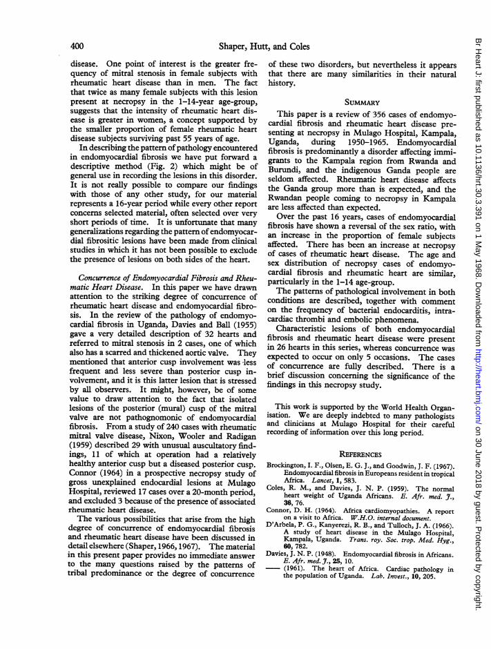
FIG. 2.-The distribution of endomyocardial fibrosis in the left or right ventricle.
only 5 were ventricular thrombi present (2 3%).Cases with associated endomyocardial fibrosis wereexcluded from this analysis (Table IX).
Embolic phenomena in the absence of bacterialendocarditis were present in 36 subjects with rheu-matic heart disease, 4 of whom also had endomyo-cardial fibrosis. If these latter 4 cases are excluded,embolic phenomena occurred in 15 per cent of therheumatic heart disease subjects. Eighteen ofthese 32 subjects had mitral stenosis.
Pattern of Pathology in Endomyocardial Fibrosis.The 172 hearts with endomyocardial fibrosis wereseparated into several descriptive groups based onthe distribution of the lesions (Fig. 2).
Type 1. Apex only involved, extending for a vari-able distance along the inflow tract.
Type 2. Lesion involving the apex and with exten-sion along the ventricular wall to involvethe mitral or tricuspid valve.
TABLE IXDISTRIBUTION OF INTRACARDIAC THROMBI INRHEUMATIC HEART DISEASE AT NECROPSY, 1950-1965
Site No.
Right atriurn 13Left atrium 10Right and left atria 3Right atriumn and right ventricle 2Right atrium, right ventricle, and
left ventricle 1Right ventricle 1Left ventricle 1
Type 3. Lesion of the ventricular wall involving theatrioventricular valve but with no apicallesion.
Type 4. Apical lesion and a separate lesion of theventricular wall involving the atrioventri-cular valve.
Type 5. Lesion of the ventricular wall away fromthe apex and the atrioventricular valve.
The following method of abbreviation will be usedin the ensuing text: Ri will indicate a Type 1 lesionon the right side of the heart and L2 will indicatea Type 2 lesion on the left side of the heart, and soon.
Pure right ventricular lesions were present in 19cases (11 %), with apical involvement alone in 6(Ri), apical involvement extending to the tricuspidvalve in 11 (R2), separate apical and tricuspid valvelesion in 1 (R4), and an isolated ventricular wallpatch in 1 (R5).
Pure left ventricular lesions were present in 66cases (38%), with apical involvement alone in 26(Li), and an apical lesion extending right up toinvolve the posterior mitral valve cusp in 25 (L2).In 10 cases without apical involvement the posteriormitral valve cusp was adherent to the ventricularwall fusing with an area of endomyocardial fibrosis(L3), and in 2 further cases this lesion was presentwith a separate lesion at the apex (L4). In 3 casesthere was a large area of endomyocardial fibrosis inthe left ventricular wall separate from the apex andthe mitral valve (L5).
396
on 30 June 2018 by guest. Protected by copyright.
http://heart.bmj.com
/B
r Heart J: first published as 10.1136/hrt.30.3.391 on 1 M
ay 1968. Dow
nloaded from

Endomyocardial Fibrosis and Rheumatic Heart Disease in Uganda
Combined Lesions. Both right and left ventricleswere affected by endomyocardial fibrosis in 88cases (51%), the involvement being predominantlyleft-sided in 39 cases, predominantly right-sided in22, and both sides being about equally affected inthe remaining 27 cases.
In Table X, the pattem of combined right- andleft-sided endomyocardial fibrosis is shown usingthe system described above, and in addition indi-cating (in brackets) which side of the heart was moreinvolved, or whether both sides were equally in-volved. From the Table it can be seen that inthose cases in which the right-sided lesion affectedonly the ventricular apex (Rl, 38 cases) the majorinvolvement by endomyocardial fibrosis was mostfrequently on the left side of the heart (28 cases),and in 29 of the 38 cases the mitral valve was in-volved. From the necropsy description the effectof the right ventricular apical lesion on the functionof the tricuspid valve was not always possible toassess. It must be presumed that in many of thesecases tricuspid incompetence was present, but itsfrequency or degree could not be estimated.
If one examines those right ventricular apicallesions which were sufficiently extensive as to in-volve the tricuspid valve and/or its attachments(R2, 35 cases), the predominant pathology was eitheron the right side of the heart (18 cases) or bothsides were equally affected (16 cases). In 29 ofthese 35 cases the mitral valve was involved so thatboth mitral and tricuspid incompetence were pre-sumably present. Several other combinations oflesions occurred apart from these main groupingsand the pattern of these can be seen from Table X.
Bacterial Endocarditis. Bacterial endocarditiswas present in 10 of the 173 cases of endomyocardialfibrosis, but in 7 of these cases rheumatic heart dis-ease was also present. In the 3 cases of bacterialendocarditis occurring in pure endomyocardialfibrosis the mitral valve was involved in one caseand the left ventricular endocardium in 2 cases. Inthe 7 RHD +EMF cases with bacterial endocarditisthe mitral valve was involved in 6 cases and theaortic valve in 1 case.
Ante-mortem atrial and/or ventricular intracardiacthrombi were present in 74 cases (43%) of endo-myocardial fibrosis and involved the ventricles in50 of these cases (29%). The left ventricle hadintracardiac thrombi in 43 subjects and the rightventricle in 16 subjects (Table XI).
Embolic phenomena in the absence of bacterialendocarditis were present in 36 cases, 9 of whichhad associated rheumatic heart disease. If these
TABLE XPATTERNS OF COMBINED RIGHT- AND LEFT-SIDED
ENDOMYOCARDIAL FIBROSIS
R-1 R-2 R-3 R-4 R-5
L-1 8 (5L/1R/2B) 8 (7R/1B) 3 (3L) 2 (2L)L-2 18 (16L/B2) 14 (3R/lL/lOB) 1 (1L) 2 (2L) 2 (2L)L-3 3 (1L/2R) 8 (7R/1B) 2 (2B) - -
LA 8 (6L/2B) 5 (4B/1R) - 2 (2B) -
L-5 1 (1R) I (iB)
Note: Figures in brackets indicate frequency with whichone or other side of heart is more severely affected, i.e. L,R, or B indicates that the more advanced lesion was on theleft (L) or right (R) side or that both sides (B) were equallyaffected.
TABLE XIDISTRIBUTION OF INTRACARDIAC THROMBI IN ENDO-MYOCARDIAL FIBROSIS AT NECROPSY, MULAGO
HOSPITAL, 1950-1965
Site No.
Left ventricle 29Right atrium 22Right and left ventricles 5Right ventricle 5Right atrium and both ventricles 4Left atrium and ventricle 2Right atrium and ventricle 2Right atrium, left ventricle 2Both atria, left ventricle 1Both atria 1Left atrium 1
74
TABLE XIIDISTRIBUTION OF 26 CASES WITH CONCURRENTENDOMYOCARDIAL FIBROSIS ANDRHEUMATIC HEART
DISEASE: MULAGO HOSPITAL, 1950-1965
Ganda Rwanda Ankole Others
1-4 - 2 -5-14 - 1 1 -15-24 - - -
25-34 - 5* 1 4t35-44 2t 2 - 145-54 2* - 1 155-64 - 1 - 2
4 11 3 8
* Includes 1 female. t = Includes 2 female.
latter are excluded, embolic phenomena occurredin 16 per cent of the endomyocardial fibrosis sub-jects. Aortic embolization occurred in 7, and asthese constitute a syndrome apparently peculiar inKampala to endomyocardial fibrosis and not seen inthis rheumatic heart disease series, they will be fullydescribed in a separate paper.
Concurrence of Endomyocardial Fibrosis andRheumatic Heart Disease (Table XII). Charac-teristic macroscopical lesions of both rheumatic
397
on 30 June 2018 by guest. Protected by copyright.
http://heart.bmj.com
/B
r Heart J: first published as 10.1136/hrt.30.3.391 on 1 M
ay 1968. Dow
nloaded from

Shaper, Hutt, and Coles
heart disease and endomyocardial fibrosis werepresent in 26 hearts: 20 male and 6 female. Weare aware of the problems inherent in retrospectivestudies ofnecropsy material, and in most cases underdiscussion the hearts had been fully described, oftenby several pathologists, and photographs werefrequently available.The number of occasions on which both condi-
tions might be expected to occur in the same subjectwas calculated separately for both male and femalesubjects for each age-group in each of the main
tribal groups. As the Ankole group had behavedin an intermediate way in the assessment of tribalpredominance in both series they were included in"other tribes" for the purpose of this particularstatistical evaluation. It was calculated that 2-2cases of combined lesions were to be expected inthe 6481 male necropsies and 2-5 cases in the 1828female necropsies, in striking contrast to the 20male and 6 female cases in which these two disorderswere considered to be concurrently present.
Description of Cases showing Concurrence of BothConditionsGroup A: (7 cases). In this group advanced mitral
stenosis was present without other valvular lesions of arheumatic nature. In addition, there were characteristiclesions ofendomyocardial fibrosis. In 3 cases the fibrosiswas quite separate from the mitral valve lesion, oblitera-ting the apex of the right ventricular cavity in all 3, and,in addition, involving the apex of the left ventricularcavity in 2.
In the other 4 there was typical rheumatic mitralstenosis, but in each case the posterior mitral cusp wasalso adherent to the ventricular wall, the chordae ten-dineae were short, thick, and often fibrosed, and dis-appeared into an area of endomyocardial fibrosis. In 2of these 4 cases, there was left ventricular apical endo-myocardial fibrosis in one case separate from the area onthe posterior wall, and in the other it was continuouswith the area behind the posterior mitral cusp. Inaddition, 2 of these 4 cases had severe endomyocardialfibrosis of the right ventricle.
Group B: (11 cases). In this group both the anteriorand posterior cusps of the mitral valve were consideredto be abnormal in a manner consistent with rheumaticfever, but there was no stenosis. In addition, therewere lesions in the ventricular cavities characteristic ofendomyocardial fibrosis, and the posterior cusp of themitral valve was usually involved in a lesion suggestiveof endomyocardial fibrosis.
In 9 cases the posterior mitral cusp, in addition tobeing thickened, irregular, and contracted in a rheu-matic manner, was associated with thickened and some-times fibrosed chordae tendineae and was adherent tothe posterior ventricular wall. Here it fused with apatch of endomyocardial fibrosis of varying size. In2 cases there were further but separate patches in the
left ventricle but not at the apex and in 4 cases therewas apical endomyocardial fibrosis-separated from thepatch on the posterior wall in 3 cases and continuouswith it in 1 case. In 7 cases there was fibrosis involvingthe right ventricular apex, 3 of a gross degree with dis-tortion and 3 of less degree.
In 2 cases there was rheumatic mitral incompetencewith involvement of both anterior and posterior cusps,but the posterior cusp was free and not affected in themanner described above. In both cases there wasfibrosis of the left ventricular apex quite separate fromthe mitral lesion and the right ventricular apex wassimilarly affected.
Group C: (3 cases). In this group the mitral andaortic valves were both involved in an apparent rheu-matic process, together with lesions of endomyocardialfibrosis.
In one subject with mitral stenosis there was well-defined fibrosis running from the posterior cusp to theposterior wall of the left ventricle and down to the apex.There were other patches within the left ventricularcavity and also a small patch near the right ventricularapex. All three valves of the aortic cusp were involvedin an apparent rheumatic process, with small pale vege-tations on the edges of two cusps and scarring of thethird cusp. In the second case both mitral cusps werethickened, nodular, and almost cartilagenous, withslight narrowing of the mitral ring. The anterior leaf-let was adherent to the ventricular wall by a mass oforganizing material, and there was a patch of fibrosis atthe apex of the cavity. The aortic valve was nodular,calcified, and incompetent. In the third case both themitral and aortic valves showed thickening and calcifica-tion without gross distortion, and there was a sharplydefined plaque of fibrosis on the wall of the right ventri-cular cavity.
Group D: (5 cases). In this group the aortic valvedisease was the main site of rheumatic heart disease,together with lesions of endomyocardial fibrosis.
In all 5 cases there was endomyocardial fibrosis ofthe left ventricular apex, often of a gross degree, withextensive fibrosis and occasional calcification and over-lying thrombus. Two of these cases showed, in addi-tion, fibrosis of the left ventricular wall behind theposterior mitral cusp, tethering the papillary muscles andchordae, and one of these had additional patches in theright ventricle. In one case the tricuspid valve showedslight thickening of the free margins, and the chordaeof the posterior cusp were adherent to each other andto the ventricular wall, with small patches of yellowish-white thickening over the papillary muscles. Aorticstenosis was present only in 1 of these 5 cases associatedwith a large and dense fibrotic scar at the apex of theleft ventricular cavity.
DISCUSSIONThis paper is intended to provide information
rather than to review the whole subject of endo-myocardial fibrosis or rheumatic heart disease in a
398
on 30 June 2018 by guest. Protected by copyright.
http://heart.bmj.com
/B
r Heart J: first published as 10.1136/hrt.30.3.391 on 1 M
ay 1968. Dow
nloaded from

Endomyocardial Fibrosis and Rheumatic Heart Disease in Uganda
tropical area. The discussion will be brief andwill merely indicate the possible significance ofsome of our observations.
In the previous study of the tribal origins ofsubjects coming to necropsy at Mulago Hospitalover a 12-year period, it was evident that endomyo-cardial fibrosis particularly affected the immigrantsto Buganda from Ruanda and Burundi (Shaper andColes, 1965). The condition occurred less fre-quently than expected among the local Ganda tribe.The present study over a 16-year period confirmsthe conspicuous preponderanceamong the Rwandansand the relative freedom from the disorder seen inthe indigenous group. Endomyocardial fibrosiswas present at the rate of 1 in every 147 necropsiesin the Ganda subjects and in 1 in every 18 necrop-sies in the Rwandan subjects. The observed inci-dence of cases in the Ankole female group is inexcess of the expected number, but the numbersinvolved are too small to do more than suggest anincreased susceptibility in this group to endomyo-cardial fibrosis.Rheumatic heart disease occurs even more fre-
quently at necropsy than endomyocardial fibrosis,and appears to affect the Ganda group more thanexpected and the Rwandan group less than expec-ted, though the level of statistical significance is notas high as in the endomyocardial fibrosis figures.In the Ganda subjects, rheumatic heart disease ispresent in 1 in every 25 necropsies; in the Rwan-dans in 1 in every 64 necropsies.
In the previous study, attention was drawn to theabsence of endomyocardial fibrosis in subjects fromthe Toro group, the tribal group coming to nec-ropsy most frequently after the Ganda, Rwanda,and Ankole groups, though endomyocardial fibrosishad been recorded in several other tribal groupscoming to necropsy far less frequently. In thepresent 16-year study this remains true, and nocase of endomyocardial fibrosis is recorded atnecropsy in a subject belonging to the Toro group.On the other hand, 9 cases of rheumatic heart dis-ease were observed at necropsy in people originatingin Toro.The migrant status of the people originating from
Rwanda and Burundi, and the history of their pat-tern of movement into Buganda, has been fullydescribed in our earlier publication. It must againbe emphasized that, according to the 1959 UgandaCensus, a considerable proportion of Rwandansliving in Buganda claim to have been born inBuganda, and this is almost certainly true of manyof those in the younger age-groups. We suggestedin our earlier study that, whatever the initiatingfactor in endomyocardial fibrosis, a state ofincreasedsusceptibility was associated with poorsocioeconomic
conditions such as is seen in the Rwandan,while a degree of protection was conferred onthe Ganda by a dietary and social backgroundwhich would however still be regarded as subopti-mal by western standards. In an attempt to movethe field of interest from nutritional factors tothose concerned with infection and immunity, ahypothesis was later put forward that emphasizedthe previous immunological experiences in theimmigrant community, in particular that resultingfrom parasitic infestation, such as malaria, andsuggested that endomyocardial fibrosis mightrepresent in the Rwandans an altered response toinfection with haemolytic streptococci (Shaper,1966).A comparison of the two 8-year periods at present
under review (1950-1957 and 1958-1965) showsthat there have been certain temporal changes inthe incidence and sex ratios of both endomyocardialfibrosis and rheumatic heart disease (Table IV).In the Rwanda group, the former constitutes 5-5per cent of necropsies in both 8-year periods, butwhereas in the first period there was a nearly similarsex ratio, by the second 8-year period, femalesubjects had more than double the incidence atnecropsy. In the Ganda group the proportion ofendomyocardial fibrosis at necropsy fell from 0*84per cent to 0-61 per cent over the two 8-year periods,but with a similar appearance of a conspicuousfemale predominance in the second 8-year period.In rheumatic heart disease neither the Ganda northe Rwanda groups showed any change in sex ratioover the years, but the proportion of rheumaticheart disease cases at necropsy rose from 2-7 to4-8 per cent in the Ganda, and the Rwanda showeda slight increase from 1-3 to 2-0 per cent of necrop-sies.
In the majority of cases of both conditions pre-senting at necropsy, we are confronted with achronic fibrotic lesion in the heart. As the agedistribution at necropsy of these two conditions issimilar, perhaps the least that can be concluded isthat both have a similar natural history.From these necropsy findings it appears that in
recent years both endomyocardial fibrosis andrheumatic heart disease have shown an increasingprediliction for female subjects, and that in bothdisorders mortality rates are much higher in femalesubjects in the 1-14 age period than in the male.Clinical studies at Mulago Hospital have also indi-cated a female preponderance in endomyocardialfibrosis of 2:1 (D'Arbela, Kanyerezi, and Tulloch,1966), and in recent unselected necropsies thefemale sex has predominated (Connor, 1964).
It is not intended to discuss in detail the patternof pathology seen in the hearts with rheumatic
399
on 30 June 2018 by guest. Protected by copyright.
http://heart.bmj.com
/B
r Heart J: first published as 10.1136/hrt.30.3.391 on 1 M
ay 1968. Dow
nloaded from
Shaper, Hutt, and Coles
disease. One point of interest is the greater fre-quency of mitral stenosis in female subjects withrheumatic heart disease than in men. The factthat twice as many female subjects with this lesionpresent at necropsy in the 1-14-year age-group,suggests that the intensity of rheumatic heart dis-ease is greater in women, a concept supported bythe smaller proportion of female rheumatic heartdisease subjects surviving past 55 years of age.
In describing the pattern ofpathology encounteredin endomyocardial fibrosis we have put forward adescriptive method (Fig. 2) which might be ofgeneral use in recording the lesions in this disorder.It is not really possible to compare our findingswith those of any other study, for our materialrepresents a 16-year period while every other reportconcerns selected material, often selected over veryshort periods of time. It is unfortunate that manygeneralizations regarding the pattern ofendomyocar-dial fibrositic lesions have been made from clinicalstudies in which it has not been possible to excludethe presence of lesions on both sides of the heart.
Concurrence of Endomyocardial Fibrosis and Rheu-matic Heart Disease. In this paper we have drawnattention to the striking degree of concurrence ofrheumatic heart disease and endomyocardial fibro-sis. In the review of the pathology of endomyo-cardial fibrosis in Uganda, Davies and Ball (1955)gave a very detailed description of 32 hearts andreferred to mitral stenosis in 2 cases, one of whichalso has a scarred and thickened aortic valve. Theymentioned that anterior cusp involvement was -lessfrequent and less severe than posterior cusp in-volvement, and it is this latter lesion that is stressedby all observers. It might, however, be of somevalue to draw attention to the fact that isolatedlesions of the posterior (mural) cusp of the mitralvalve are not pathognomonic of endomyocardialfibrosis. From a study of 240 cases with rheumaticmitral valve disease, Nixon, Wooler and Radigan(1959) described 29 with unusual auscultatory find-ings, 11 of which at operation had a relativelyhealthy anterior cusp but a diseased posterior cusp.Connor (1964) in a prospective necropsy study ofgross unexplained endocardial lesions at MulagoHospital, reviewed 17 cases over a 20-month period,and excluded 3 because of the presence of associatedrheumatic heart disease.The various possibilities that arise from the high
degree of concurrence of endomyocardial fibrosisand rheumatic heart disease have been discussed indetail elsewhere (Shaper, 1966,1967). The materialin this present paper provides no immediate answerto the many questions raised by the patterns oftribal predominance or the degree of concurrence
of these two disorders, but nevertheless it appearsthat there are many similarities in their naturalhistory.
SUMMARYThis paper is a review of 356 cases of endomyo-
cardial fibrosis and rheumatic heart disease pre-senting at necropsy in Mulago Hospital, Kampala,Uganda, during 1950-1965. Endomyocardialfibrosis is predominantly a disorder affecting immi-grants to the Kampala region from Rwanda andBurundi, and the indigenous Ganda people areseldom affected. Rheumatic heart disease affectsthe Ganda group more than is expected, and theRwandan people coming to necropsy in Kampalaare less affected than expected.Over the past 16 years, cases of endomyocardial
fibrosis have shown a reversal of the sex ratio, withan increase in the proportion of female subjectsaffected. There has been an increase at necropsyof cases of rheumatic heart disease. The age andsex distribution of necropsy cases of endomyo-cardial fibrosis and rheumatic heart are similar,particularly in the 1-14 age-group.The patterns of pathological involvement in both
conditions are described, together with commenton the frequency of bacterial endocarditis, intra-cardiac thrombi and embolic phenomena.
Characteristic lesions of both endomyocardialfibrosis and rheumatic heart disease were presentin 26 hearts in this series, whereas concurrence wasexpected to occur on only 5 occasions. The casesof concurrence are fully described. There is abrief discussion concerning the significance of thefindings in this necropsy study.
This work is supported by the World Health Organ-isation. We are deeply indebted to many pathologistsand clinicians at Mulago Hospital for their carefulrecording of information over this long period.
REFERENCESBrockington, I. F., Olsen, E. G. J., and Goodwin, J. F. (1967).
Endomyocardial fibrosis in Europeans resident in tropicalAfrica. Lancet, 1, 583.
Coles, R. M., and Davies, J. N. P. (1959). The normalheart weight of Uganda Africans. E. Afr. med. J.,36, 76.
Connor, D. H. (1964). Africa cardiomyopathies. A reporton a visit to Africa. W.H.O. internal document.
D'Arbela, P. G., Kanyerezi, R. B., and Tulloch, J. A. (1966).A study of heart disease in the Mulago Hospital,Kampala, Uganda. Trans. roy. Soc. trop. Med. Hyg.,60, 782.
Davies, J. N. P. (1948). Endomyocardial fibrosis in Africans.E. Afr. med. J3., 25, 10.(1961). The heart of Africa. Cardiac pathology inthe population of Uganda. Lab. Invest., 10, 205.
400
on 30 June 2018 by guest. Protected by copyright.
http://heart.bmj.com
/B
r Heart J: first published as 10.1136/hrt.30.3.391 on 1 M
ay 1968. Dow
nloaded from

Endomyocardial Fibrosis and Rheumatic Heart Disease in Uganda
-, and Ball, J. D. (1955). The pathology of endomyo-cardial fibrosis in Uganda. Brit. Heart J., 17, 337.
Hutt, M. S. R., Ikeme, A. C., Lucas, A. O., Prata, A.,Puigbo, J. J., Shaper, A. G., and Fejfar, Z. (1965).Cardiomyopathies. Bull. Wid Hlth Org., 33, 257.
Nixon, P. G. F., Wooler, E. H., and Radigan, L. R. (1959).Mitral incompetence caused by disease of the muralcusp. Circutation, 19, 839.
Shaper, A. G. (1966). Endomyocardial fibrosis and rheu-
matic heart-disease. Lancet, 1, 639.(1967). On the nature of some tropical cardiomyo-pathies. Trans. roy. Soc. trop. Med. Hyg., 61, 458.
-, and Coles, R. M. (1965). The tribal distribution ofendomyocardial fibrosis in Uganda. Brit. Heart J.,
27. 121.--, and Williams, A. W. (1960). Cardiovascular disorders
in an African hospital in Uganda. Trans. roy. Soc.trop. Med. Hyg., 54, 12.
401
on 30 June 2018 by guest. Protected by copyright.
http://heart.bmj.com
/B
r Heart J: first published as 10.1136/hrt.30.3.391 on 1 M
ay 1968. Dow
nloaded from