neck and arm pain syndromes || electrodiagnostic studies
TRANSCRIPT

Chapter
© 2011 Elsevier Ltd.
DOI: 10.1016/B978-0-7020-3528-9.00005-4
| 5 |
Electrodiagnostic studiesCaroline A Quartly
CHAPTER CONTENTS
Introduction 65
Education and certification inelectrodiagnostic medicine 68
Psychometric and economic considerations 69
Electrodiagnostic findings and clinicalcorrelation 70
Electrodiagnostic medicine in neck and armpain syndromes 70
Nerve conduction studies 71
Electromyography 73
Somatosensory evoked potentials 75
Autonomic nerve testing 75
Conclusion 75
Additional resources 76
INTRODUCTION
‘There are more things in heaven and earth, Horatio,
than are dreamt of in your philosophy’
Hamlet to Horatio in Act I, Scene V
an argument against the oversimplification of a doctrine
In the context of neck and arm pain syndromes, there aremany diagnostic categories where electrodiagnostic evalu-ation may be relevant (Box 5.1). However, as with referralfor every clinical test in medicine, initiation of referral toan electrodiagnostic service should always be prefaced byclinical reasoning. Will doing the test have a bearing onpatient outcome? It behoves us to always remember that
just because we have the ability to do a test, this doesn’tmean that doing the test serves the patient. When theopportunities for intervention are limited, such as when,for example, spinal MRI studies are done in the absenceof clinical signs and/or symptoms of neurological ororthopaedic compromise or of a history suggestive of sin-ister pathology, we are complicit in the creation/perpetua-tion of an expensive health care system that does notchange patient outcome. Clinical questions that mightthen be useful in deciding whether to refer for electrodiag-nostic testing include:
1. Is the accurate documentation of the presence andseverity of this lesion critical to surgical referral forconsultation to determine the merits of exploration,decompression, transposition, or nerve graft?
2. Is the accurate documentation of the presence andseverity of this condition essential to determiningwhether specific medication (such as, e.g.,prednisone) will change outcome?
3. Is the accurate documentation of the presence andseverity of this condition essential to determiningwhether further investigation will uncover apotentially treatable disorder, such as vitamin B12deficiency, vasculitis, or diabetes?
4. Is the accurate documentation of the presence andseverity of this condition essential to determiningwhether exercise prescriptions and/or work capacityrecommendations will require modification, such as,for example, in the case of severe axonal nerve damage(where potential metabolic exhaustion of recoveringor permanently damaged nerve is an issue) or inpatients with hereditary sensory-motor neuropathies,entrapment neuropathies, and myopathies?
5. Is the finding of a negative test useful or contextuallyharmful to patient management?
65
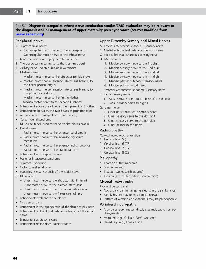
Box 5.1 Diagnostic categories where nerve conduction studies/EMG evaluation may be relevant tothe diagnosis and/or management of upper extremity pain syndromes (source: modified fromwww.aanem.org)
Peripheral nerves
1. Suprascapular nerve:
– Suprascapular motor nerve to the supraspinatus
– Suprascapular motor nerve to the infraspinatus
2. Long thoracic nerve injury: serratus anterior
3. Thoracodorsal motor nerve to the latissimus dorsi
4. Axillary nerve: isolated deltoid involvement
5. Median nerve:
– Median motor nerve to the abductor pollicis brevis
– Median motor nerve, anterior interosseus branch, to
the flexor pollicis longus
– Median motor nerve, anterior interosseus branch, to
the pronator quadratus
– Median motor nerve to the first lumbrical
Median motor nerve to the second lumbrical
• Entrapment above the elbow at the ligament of Struthers
• Entrapments between the two heads of pronator teres
• Anterior interosseus syndrome (pure motor)
• Carpal tunnel syndrome
6. Musculocutaneous motor nerve to the biceps brachii
7. Radial nerve:
– Radial motor nerve to the extensor carpi ulnaris
– Radial motor nerve to the extensor digitorum
communis
– Radial motor nerve to the extensor indicis proprius
– Radial motor nerve to the brachioradialis
• Entrapment at the spiral groove
• Posterior interosseus syndrome
• Supinator syndrome
• Radial tunnel syndrome
• Superficial sensory branch of the radial nerve
8. Ulnar nerve:
– Ulnar motor nerve to the abductor digiti minimi
– Ulnar motor nerve to the palmar interosseus
– Ulnar motor nerve to the first dorsal interosseus
– Ulnar motor nerve to the flexor carpi ulnaris
• Entrapments well above the elbow
• Tardy ulnar palsy
• Entrapment in the aponeurosis of the flexor carpi ulnaris
• Entrapment of the dorsal cutaneous branch of the ulnar
nerve
• Entrapment at Guyon’s canal
• Entrapment of the deep palmar branch
Upper Extremity Sensory and Mixed Nerves
A. Lateral antebrachial cutaneous sensory nerve
B. Medial antebrachial cutaneous sensory nerve
C. Medial brachial cutaneous sensory nerve
D. Median nerve
1. Median sensory nerve to the 1st digit
2. Median sensory nerve to the 2nd digit
3. Median sensory nerve to the 3rd digit
4. Median sensory nerve to the 4th digit
5. Median palmar cutaneous sensory nerve
6. Median palmar mixed nerve
E. Posterior antebrachial cutaneous sensory nerve
F. Radial sensory nerve
1. Radial sensory nerve to the base of the thumb
2. Radial sensory nerve to digit 1
G. Ulnar nerve
1. Ulnar dorsal cutaneous sensory nerve
2. Ulnar sensory nerve to the 4th digit
3. Ulnar sensory nerve to the 5th digit
4. Ulnar palmar mixed nerve
Radiculopathy
Cervical nerve root stimulation
1. Cervical level 5 (C5)
2. Cervical level 6 (C6)
3. Cervical level 7 (C7)
4. Cervical level 8 (C8)
Plexopathy
• Thoracic outlet syndrome
• Brachial neuritis
• Traction palsies (birth trauma)
• Trauma (stretch, laceration, compression)
Myopathy/dystrophy
Proximal versus distal
• Not usually painful unless related to muscle imbalance
• Family history may or may not be relevant
• Pattern of wasting and weakness may be pathognomic
Peripheral neuropathy
• May be sensory, motor, distal, proximal, axonal, and/or
demyelinating
• Acquired: e.g., Guillain–Barre syndrome
• Hereditary: e.g., HSMN I or II
Part | 1 | Introduction
66

Chapter | 5 | Electrodiagnostic studies
Aswithmost clinical tests,when results are takenoutof con-text the value of the test is greatly diminished. By extension,one can infer that utilizing tests out of context carries a higherlikelihoodof doingmore harm than good to the patient (She-kelle et al 2000). It is important to understand that – next totechnical issues – perhaps the largest source of error in electro-physiology lies in drawing conclusions from a limited data-base. The same can of course be said of the hallmarks of theclinical evaluation, history and clinical examination.
There seems to be a growing compulsion to use auto-mated software programs to analyse nerve conduction(Armstrong et al 2008) and even electromyographic data;a compulsion perhaps driven by the erroneous assump-tion that electrodiagnostic evaluation involves the analysisof a signal as simple and uncomplicated as is elicited byelectrocardiogram. The issue, of course, is that either lim-ited or incorrectly interpreted and applied data may leadto an increase in both false negative and false positivefindings. Either way, the patient may not be served. Tobe more specific: if, for example, a limited study is usedto identify the presence or absence of carpal tunnel syn-drome but the superimposition of other confoundingdiagnoses is neither considered nor evaluated, surgicaldecompression may result in poor outcome. Alternatively,the lack of surgical intervention may result in poor out-come if the data set is limited and incorrectly assumesa ‘normal’ study. A patient who returns with a label of‘normal’ after a technical study that is not accompaniedby a clinical evaluation may be completely misinter-preted (even to the extent of being called a malingerer)if the referring clinician or the third-party payer doesnot understand the indications and limitations of theprocedure.
Interpretation of the test as ‘normal’ without the contextof the clinical evaluation (history and physical examina-tion) carries with it an enormous burden of responsibility.The responsibility to the patient and to the healthcare sys-tem as a whole is in understanding that the results of anygiven individual clinical test should never be taken out ofcontext. If the clinician making the referral is not fully aware
Table 5.1 A few basic examples of electrodiagnostic terminolog
Configuration of motor unit recorded
on EMG
Polyphasic A po
in rei
Serrated Mult
Long duration Musc
May
Large
amplitude
Musc
Reco
norm
Small
amplitude
Musc
May
of the symptoms characteristic of a specific peripheral nerveinjury, and as such requests only evaluation of a limited andspecific diagnosis, it is that clinician who should perhapsbear full responsibility for not only the way in which the testis used technically but also the ultimate medical outcome ofthe patient.
One has to confront the fact that not all clinicians maybe aware that specific presentations may signify anaetiology or even technique of testing outside commonknowledge (Seror 2005). On the other hand, it mustalso be noted that the current literature is replete with amost-welcome focus on removing bias from analysis ofspecific disorders through more critical and representativestudies. For example, comparative analyses of tests such asthose used to diagnose entrapment ranging from that ascommon as carpal tunnel syndrome (Ayyar et al 2001,Bodofsky et al 2005, Chang et al 2006, Dan et al 2006,Aygul et al 2009, Brannagan et al 2009), to the moreelusive radial nerve entrapment (Beck et al 1998), and tochronic demyelinating inflammatory and immune-mediated neuropathies (Hattori et al 2005, Latov &Sander 2003) are now even appearing in literature searchengines of best evidence. Helpful studies confirming thatelectrodiagnostic evaluation can be blinded are also start-ing to emerge (Haig et al 2006).
It is the intent of this chapter that the reader comes toappreciate that the electrodiagnostic evaluation is mostappropriately utilized in clinical practice as an extensionof the clinical examination. It is not the intent of this chap-ter to discuss in detail the technical aspects of the ways inwhich electrodiagnostic studies are performed. Indeed,such information is clearly and extensively covered innumerous texts on the subject; perhaps the most compre-hensive of which is Electrodiagnostic Medicine (Dumitruet al 2002). More recent publications by acknowledgedexperts in the field offer invaluable perspective (Aminoff2005, Buschbacher & Prahlow 2005, Katirji 2007, Daube& Rubin 2009). To facilitate an understanding of the mate-rial in this chapter, some basic terminology not otherwiseexplained in the text is included in Table 5.1.
y
tential that crosses the baseline 5 or more times (may be seen
nnervation/remodelling)
iple phases that do not cross the baseline (same inference)
le specific and dependent on innervation ratio of specific muscle
be seen in collateral sprouting / indicative of prior axonal damage
le specific and dependent on innervation ratio of specific muscle
rding needle characteristics also influence amplitude, hence
ative data are critical
le specific and dependent on innervation ratio of specific muscle
indicate myopathy or chronic neuropathy
Continued
67

Table 5.1 A few basic examples of electrodiagnostic terminology—cont’d
Stigmatic electrode Recording electrode/cathode
The differential signal analysis requires a cathode and an anode
Terminal latency (motor or sensory) The time (in milliseconds) it takes for a stimulus to evoke a response from nerve to its
nearest recording site (nerve or muscle)
Spontaneous discharge quoted and
explained in text
Fibrillation Mostly indicative of recent or active denervation.
Positive sharp
wave
May occur as fibrillation or motor unit
Fasciculation Spontaneous discharge of motor unit
Recruitment When used to reflect firing frequency, can be directly correlated with motor unit
drop (or axonal loss)
Interference pattern Can vary with the presence or absence of superimposed upper motor neuron
involvement
The firing frequency is not abnormal in the presence of upper motor neuron
involvement alone
Part | 1 | Introduction
EDUCATION AND CERTIFICATIONIN ELECTRODIAGNOSTIC MEDICINE
Although not exclusively, two subspecialties of medicineare more likely to perform electrodiagnostic evaluations:neurology and physical medicine and rehabilitation(physiatry). An acknowledged minimal requirement ofsupervised training in the field of electromyography is sixmonths during residency training. In North America,certification of competency in electrodiagnosis is achievedafter successful written and oral examination throughorganizations such as the American Association of Neuro-muscular and Electrodiagnostic Medicine (AANEM) or itsCanadian equivalent, the Canadian Society of Clinical Neu-rophysiologists (CSCN). Basic eligibility to be consideredfor such examination requires both clinician reference anddocumentation of a minimum of 200 independent evalua-tions. During the six-month interval of residency trainingthe focus is primarily on gaining technical skills. The con-text in which the technical skill is applied is a backgroundof three to four years of medical school and four to fiveyears of specialty residency training (encompassing notonly more detailed neuroanatomy and neurophysiologybut also in-depth expressions of the influence of disease,trauma, ageing, and genetics upon the neuromuscular sys-tem). In rehabilitation medicine, such understanding isalso married with knowledge of the myriad presentationsof musculoskeletal disorders. In neurology, such under-standingmay bemarriedwith amore extensive backgroundin differential diagnosis of central and peripheral neurolog-ical disorders. The above information is inserted not toimpress but rather to impress the extent of the art form
68
upon those who would oversimplify the use and value ofelectrophysiology.
The easiest part of the electrodiagnostic evaluation isthe technical execution of the study (which of courserequires a comprehensive understanding of pitfalls inthe technical execution of the test). A poorly executed testis just as likely to miss a disease as it is to result in theinference of disease when there is none. Technical compe-tence, as well as understanding which data to collect andhow to interpret the findings in context, takes both skilland experience. This becomes the art of the specialty.
While in some jurisdictions (after a year’s apprentice-ship) a technologist may still become eligible forexamination to acquire certification of competency in exe-cution of nerve conduction studies (as an EMG technician),growing recognition of the extent of training required tooperate sophisticated electroneurodiagnostic equipmenthas led to the development of two-year diploma programsin electrophysiology (see, for example, BCIT 2009). Certifi-cation of competency within subsets of electroneurophy-siology is then acquired by specific examination. Suchdetailed training should prepare the technologist with thesavvy of how to avoid pitfalls and how to problem solvevarious technical challenges in the acquisition of data.
While technologists have some basic knowledge ofinstrumentation, they do not usually have the entire med-ical context to assess the appropriateness of the specificsof the evaluation, nor do they assume the legal respon-sibility for excluding or including coexisting competingmedical, pharmacological, or ageing effects on nervestructure and function. The most comprehensive electro-diagnostic evaluation is, therefore, done either personallyby a recognized specialist in the field with the privilegeof a comprehensive history and clinical examination or

Chapter | 5 | Electrodiagnostic studies
by a certified technologist in partnership with a specialist inthe field (who, again, should have access to a comprehen-sive history and clinical examination, i.e., the context).
PSYCHOMETRIC AND ECONOMICCONSIDERATIONS
The beauty of the title of this book as ‘evidence-informed’ as opposed to ‘evidence-based’ evaluation isthat the latter terminology has over time become hack-neyed, misunderstood, and misused. The intent of theterminology was indeed originally to raise the cultureof awareness of the value of critical appraisal of evidencein clinical decision making. A fundamental tenet of suchcritical appraisal deals with understanding how bias maypollute clinical decision making. Part of the beauty ofhow health care decision making has evolved is thatthe self-regulation of professional competence extendsto raising consciousness about the ongoing rigour withwhich each and every diagnostic test and interventionneeds to be examined (Shekelle et al 2000). Each andevery test that is used in medicine is limited as to whatit tests. It follows that it is important to understand whata test does not test. Sometimes a test is most useful whenit is positive; technical and biological variations maymake a negative test unreliable. It is always importantto bear in mind when assessing the value of a specificclinical tool that the absence of proof is not necessarilythe proof of absence. Specific tests may not lend them-selves well to rigorous scrutiny because of the rarenessof the disorder they reflect.
We know from inter- and intra-rater reliability, that themore explicitly the parameters of normal and/or specificrange of disease are described, the higher the likelihoodthat experts will agree with each other and with them-selves. Without such descriptors, inter-rater reliability,specifically in any test that involves perception, is oftenbetween 60 and 70%. Intra-rater reliability is known tobe higher (at around 80%), as over time we tend to agreewith ourselves more consistently.
With regard to the content of the electrodiagnostic eval-uation, organizations such as the AANEM and the CSCNare dedicated to assisting in the setting of standards forcompetent electrodiagnostic evaluation by setting guide-lines for the most reasonable number of parameters toinclude in any given study of a specific diagnosis or con-stellation of symptoms. These recommendations arebased on the strength of evidence after critical appraisalof the literature. Evidence may be robust enough to actu-ally publish guidelines (such as with carpal tunnel syn-drome) (Jablecki et al 2002a,b) or less robust (as in thecase of chronic idiopathic demyelinating polyneuropa-thy), forcing us to rely on consensus of expert opinion(Latov & Sander 2003).
When one examines how easily generalized results are,i.e., how closely any given test result represents one’sown patient population, one cannot escape considerationof how the business of medicine influences the outcomeas well as the practical application and utility of any giventest in medicine. What is actually practised in a commu-nity may be far different than the standards to which theAANEM and CSCN hold their diplomats. In some juris-dictions, such as British Columbia, payment of service isinherently tied to demonstration of competence by exam-ination. This is not universally the case. Clinics offeringelectrodiagnostic services may be limited by poor reim-bursement of the service, which may in some instanceslimit the amount of data gathering that can occur. Whenclinicians assess patients whom they have already seenin consultation, and so have had the privilege of clinicalexamination, testing may be quite specific, utilizing aselection of the most appropriate electrodiagnostic techni-ques to diagnose specific conditions in the context of asound knowledge of the patient’s limitations. Reimburse-ment fees that do not adequately reflect the time neededto gather data or differentiate between the time necessaryto rule out a specific disorder, particularly in the presenceof confounding disorders that also affect the nervoussystem (such as diabetes), may contribute to a higherincidence of false negative as well as false positive diag-noses. In either case, neither the patient nor the clinicianis well served. Furthermore, as reimbursement schedulesfrequently reflect neither the expertise of the examinernor the length of time taken for a test (except in grossterms), there may be no uniformity in what is in actualitygathered or delivered through any given test.
In the United States, reimbursement is directly tied tothe time required to execute the test. In order to preventabuse of the opposite sort (i.e., doing more than isrequired), the AANEM has culled the opinions of expertsto establish an appropriate number of tests to rule in orout a given diagnosis. Such guidelines assist third-partypayers in understanding reasonable and customary feeswhile at the same time addressing inadvertent abuse inan otherwise open-ended system.
In the interest of clarifying the interpretation of reportsof electrodiagnostic testing and in order to better servepatient care, caveats may be appended to the conclusionsprovided at the end of a given test. Such a caveat mightread: ‘. . . [such and such] nerves only were studied. Noabnormality was identified in these specific nerves. Clini-cal correlation is required. . .’ This caveat might be mostapplicable in the situation where the patient is referred,for example, to rule out carpal tunnel syndrome, but notfor electrodiagnostic consultation, meaning that the tes-ter/interpreter is working in a ‘vacuum.’
Other caveats may draw attention to the limitation ofthe test itself, regardless of how much data is gathered.This will help the referring clinician, third-party payer,and/or legal representative understand the context of an
69
Part | 1 | Introduction
interpretation. For instance, if a referral is made to assesswhether there is electrodiagnostic support for a diagnosisof peripheral neuropathy, a disclaimer should perhaps beadded to the interpretation of ‘normal’ that reads: ‘. . .a nor-mal nerve conduction velocity does not exclude the pres-ence of either an early dying-back neuropathy or a small-fibre neuropathy. Clinical correlation is recommended. . .’
Similarly, if the request is to rule out thoracic outlet syn-drome, the interpretation of ‘normal’ should perhapsinclude a statement such as: ‘. . .evidence of either axonaldamage or demyelination in thoracic outlet syndrome israre. A “normal” study does not rule out a symptomaticdynamic compression of the neurovascular bundle withinthe thoracic inlet or outlet (the result ofwhichmay be relatedto circumstances of compression resulting in temporarynerve ischaemia). Full clinical correlation is suggested. . .’
ELECTRODIAGNOSTIC FINDINGSAND CLINICAL CORRELATION
Recall that the restoration of homeostasis is the first orderof the day for all tissue damage. Any insult or injury to thehost will be met with an instantaneous attempt at repair.Sometimes the recovery is complete (such as skinresponse to a paper cut). Sometimes – if the lesion is dee-per – repair will be associated with the development ofscar tissue. In milder forms of injury, the scar tissue is suf-ficient for restoration of normal function. In more severeinjury, the repair process (scarring) may interfere with res-toration of normal function; the scar tissue will neverhave the same tensile property of the damaged tissue itreplaces. Each tissue in the body has the equivalent of scarformation when damage is repaired. For example, a sim-ple fracture to a bone may heal without eventual radiolog-ical evidence of prior trauma. By contrast, the morecomplicated the fracture, the higher the potential for per-manent deformity and impact on functional capacity.
The equivalent of the scar process is not necessarilysymptomatic. For example, trauma or loading thatexceeds the tensile properties of the vertebral body end-plate is thought to cause micro-fracture of the endplate,which in turn will be met with repair that results in bonysclerosis. This repair process is thought to contribute to adecreased ability of the nucleus pulposus to imbibe fluidat night, and hence to eventual dessication of the disc.In the process, the relative instability of the motion seg-ment is met with the development of osteophytes thatdevelop along force lines in an attempt to restore homeo-stasis and functional capacity (Bogduk & Twomey 1991).Although the radiological expression of this adaptivechange frequently carries the misnomer of degenerativedisc disease (misnomer because there is no disease), thespondylotic changes noted are frequently asymptomatic.In fact there is no evidence that there is a higher incidence
70
of symptoms in persons with spondylotic changes onX-ray than in persons without such radiological findings(van Tulder et al 1997).
This discourse about the interpretation of radiologicalchanges on X-ray is included to draw attention to a prac-tice in which anatomical changes such as, for example,those that may be identified on X-ray, are frequentlylabelled as the source of pain generation. By contrast,the absence of anatomical change upon imaging is ofteninterpreted as evidence that a given structure is not a paingenerator, somehow ignoring the fact that irritation of thepain fibres that innervate a given structure can in and ofthemselves be the source of pain. The source of pain doesnot necessarily have an anatomical correlate on imagingstudies. Similarly, for technical reasons, a nerve may bea source of pain but not necessarily show any structuralchange through electrodiagnostic testing. The converse isalso true: a structural change found during the electro-diagnostic examination is not necessarily symptomatic.
Some refer to the presence of motor unit configurationchanges, such as serration and/or polyphasicity (Table 5.1),as consistent with stable radiculopathy. In effect, one couldthink of this label as the equivalent of the presence of scartissue in the skin or of spondylotic changes reported in spinalX-rays. The changes reflect a repair process and are not nec-essarily symptomatic or functionally limiting. By contrast,the presence of large, long-duration potentials, as well asdecreased recruitment (reflecting axonal damage withoutrecovery or remodelling; Table 5.1), may have much moresignificant functional consequences. Amore limited comple-ment or motor axons trying to supply the same territory ofmuscle may be more vulnerable to metabolic exhaustionwith effort, resulting in decreased endurance and the capacityto act not only as a prime mover but also as a stabilizer. Thisinformation may have far more valuable prognostic andmanagement implications if employed thoughtfully towardthe clinical presentation. No one experiences such dimin-ished endurance more readily than patients who demon-strate the late effects of polio. Patients that suffer reflexmuscle inhibition of the supporting muscles of the suspen-sory system in post-whiplash-type injuries may also experi-ence a significant functional limitation in their capacity totolerate dynamic loading of the neck and shoulder girdle,particularly in the presence of coexistent profound single- ormulti-nerve axonal damage. Suchdamagemay include injuryto the nerve root, plexus, and/or peripheral nerve (such as thesuprascapular or long thoracic nerves) (see Box 5.1).
ELECTRODIAGNOSTIC MEDICINE INNECK AND ARM PAIN SYNDROMES
Electrodiagnosis in the context of neck and arm pain syn-dromes is taken to encompass nerve conduction studies,electromyography, and somatosensory evoked potentials,

Chapter | 5 | Electrodiagnostic studies
all of which are studies that evaluate the integrity of theperipheral nervous system. With the exception of isolatedacademic laboratories, most practitioners do not routinelyutilize electromagnetic stimulation of nerves and motorunit counting. Other electrodiagnostic studies, such asrepetitive nerve stimulation, have more relevance to disor-ders of the neuromuscular junction (such as myastheniagravis). Single-fibre electromyography is similarly mostfrequently used in disorders of the neuromuscular junc-tion and not directly in arm pain syndromes. Clearlysome arm pain syndromes are central in origin, the evalu-ation of which is outside the intent of this chapter.
Nerve conduction studies
Electrodiagnostic evaluation concerns itself with thestudy of sensory, motor, and autonomic nerve fibres.By common parlance, ‘EMG’ has come to be taken tomean both nerve conduction and EMG studies, eventhough there is a semantic difference between the two.
Table 5.2 Potential findings in confounding (superimposed) me
Primary types of painful sensoryneuropathy
Idiopathic small-fibre painful sensory neuropathy
Diabetic peripheral neuropathy
Inherited neuropathies
Peripheral neuropathy with connective tissue disease
Peripheral nerve vasculitis
MGUS neuropathy
Paraneoplastic sensory neuropathy
Familial amyloid polyneuropathy
Acquired amyloid polyneuropathy
Neuropathy with renal failure
Hereditary sensory autonomic neuropathy
Sarcoid polyneuropathy
Arsenic neuropathy
Fabry disease
Celiac disease
HIV-related neuropathy
*When limited studies are done to rule out entrapment neuropathies, th
abnormal results (MGUS-monoclonal gammopathy of undetermined sign
Technically, these are two entirely different tests thatpotentially reveal vastly different information aboutthe nervous system. Nerve conduction studies can onlybe performed on nerves that are readily accessible forreliable stimulation/recording and, as such, are limitedto the testing of a discrete set of peripheral nerves.By contrast, most muscles are accessible to needle EMGevaluation. The EMG, however, only reflects the stateof the muscle/innervating nerve supply and is, there-fore, limited to reflecting motor nerve/muscle integrity(meaning that conditions in which sensory abnor-malities alone are anticipated would not benefit fromEMG, but rather sensory nerve conduction alone).Table 5.2 lists potential nerve conduction findings inspecific diseases (further reinforcing the fact that inter-pretation of nerve conduction studies cannot be doneout of context, such as the frequent assumption thatcarpal tunnel syndrome can be comprehensively diag-nosed with limited data: ‘To a hammer everythinglooks like a nail’).
dical conditions (Mendell & Sahenk 2003)*
Electromyography (EMG) and nerve conductionstudy (NCS)
Normal
Abnormal EMG and NCS
Abnormal EMG and NCS
Abnormal EMG and NCS
Abnormal EMG and NCS
Abnormal EMG and NCS (may be normal)
Abnormal EMG and NCS
Abnormal EMG and NCS
Abnormal EMG and NCS
Abnormal EMG and NCS
Abnormal EMG and NCS
Abnormal EMG and NCS
Abnormal EMG and NCS
Normal EMG and NCS
EMG and NCS may be normal or abnormal
EMG and NCS usually abnormal
e coexistence of these disorders may influence interpretation of
ificance; HIV-human immunodeficiency virus)
71

Part | 1 | Introduction
Sensory nerves carry information from the externalenvironment towards the spinal cord and brain. When asensory nerve is stimulated distally and recordings aremade from the same nerve proximally, the process is saidto reveal orthodromic conduction (conduction in thedirection of intent of the impulse; ortho meaning straightand dromic referring to direction). By contrast, when thesame nerve is stimulated proximally and recordings aremade distally, the process is said to reveal antidromic con-duction (against the direction of conduction; anti mean-ing against).
The cell bodies of the sensory nerve fibres are located inthe dorsal root ganglion (DRG). If damage occurs to thesensory nerve proximal to the DRG, stimulation of thenerve in the periphery will fail to elicit an abnormalresponse. It then follows that an abnormal sensoryresponse reflects structural damage to the sensory nervedistal to the DRG. When easily accessible, a sensory nervemay be stimulated as part of a mixed nerve or as a puresensory nerve, and recordings can be made either from asensory nerve or from a mixed nerve. Interpretation ofabnormal sensory responses, however, carries with it theobligation of understanding how lack of rigour in tech-nique, in assumptions about instrumentation settings,in temperature control, in anatomical variation, andin pathological processes will influence the quality andintegrity of the data collected.
Motor nerves carry information from the spinal cord tomuscle, the conduction direction of which is orthodromicfor the motor nerves. The cell body of the motor nerve is,of course, the anterior horn cell within the spinal cord.While some nerve fibres carry only motor axons and someare purely sensory, many nerves readily accessible forstimulation to test conduction velocity carry all fibres(motor, sensory, and autonomic). The amplitude of theresponse recorded from a muscle reflects a compoundaction potential. The amplitude of the response is a sum-mation of amplitudes evoked by stimulating every motorunit (individual nerve and all of the muscle fibres itsupplies). If some axons are demyelinated, the time thatthe evoked response of those specific axons takes to arriveat the muscle stigmatic/recording electrode will vary, pro-ducing a response that is attenuated and of lower ampli-tude. As long as the area under the curve remains thesame, the drop in amplitude does not represent axonalloss. Normative data is electrode dependent (includingsuch factors as size of recording surface area, conductivity,and inter-electrode distance), technique and temperaturedependent, and instrumentation (filter setting) dependent.
When any given nerve is stimulated, the propagation ofthe impulse is bidirectional. A small stimulus (sensoryimpulse) may travel proximally along a given nerve tostimulate a motor nerve in the anterior horn cell, creatinga simple monosynaptic reflex. A motor response gener-ated by such a reflex is referred to as a late response, spe-cifically as an H- or Hoffman reflex. With the exception of
72
the H-reflex that is elicited by stimulating the mediannerve and recorded from the flexor carpi radialis (and spe-cific conditions associated with a loss of central inhibi-tion, such as MS), an H-reflex is rarely found in theupper extremity after early childhood. Carefully executed,the H-reflex may be an extremely helpful tool in diagnos-ing C6 sensory radiculopathy.
A supra-maximal stimulus applied to the nerve may alsocreate a late response. This response is referred to as theF-response (which actually stands for Foot response),a purely motor phenomenon thought to be generated bythe bouncing back of a motor-propagated impulse off theaxon hillock. The evaluation of F-responses are perhapsmost useful in conditions where there is suspected multifocalconduction block, such as Guillain–Barre Syndrome orCRDP (chronic relapsing demyelinating poly-radiculopathy).Because of technical challenges in the execution of theF-responses, controversy continues to exist as to the value ofthe F-response in conditions of proximal conduction block,such as thoracic outlet syndrome and/or radiculopathy. Someclinicians find the lack of persistence of a volley of F-responsesto hold greater significance, correlating with the likelihood ofproximal nerve pathology.
In executing a nerve conduction study, usually sensoryand/or motor nerves are systematically stimulated todetermine patterns of abnormality that may revealisolated nerve injury or entrapment, radiculopathy, plexo-pathy, peripheral neuropathy, and/or combinations ofthese conditions. The process is methodical and the par-ticular pattern of stimulation/results is compared to thatof normative data representative of the technique chosen.To calculate motor conduction velocity, a mixed nerve isstimulated at two separate points along the course of thenerve and recordings are made from a muscle. The dis-tance between two points is measured (usually with a tapemeasure), and the conduction velocity is calculated. Thetime that it takes for a stimulus to get to the muscleand cause contraction is termed the terminal motor latency.Variations in anatomical position during the acquisitionof data will greatly influence the results, a fact that is mostcommonly acknowledged in the elbow flexion angle (thedegree of which requires standardization for reproducibil-ity/validity of results when assessing ulnar nerve conduc-tion block/entrapment).
There are a limited number of ways in which a nervecan respond to injury. Within a given nerve a variablenumber of fibres will be unmyelinated. Of the myelinatedfibres, different diameters of nerves will conduct animpulse with variable rates. When a nerve is stimulated,the fastest conducting fibres are reflected in the calcula-tions of conduction velocity. It then follows that the smal-lest diameter fibres (the unmyelinated nerve fibres, whichcarry pain sensation) may be damaged, but that the nerveconduction velocity may be recorded as normal. Thisleads us to consider the following caveats when interpret-ing nerve conduction velocity study results:

Chapter | 5 | Electrodiagnostic studies
• One may have a small-fibre neuropathy and yet thenerve conduction study may be ‘normal.’
• One may have damage to unmyelinated fibres(transmitting pain sensation) and yet the nerveconduction study may be ‘normal.’ This, of course, ismost commonly seen in diabetes.
• One may have damage to the most distal end of asensory nerve (as in dying-back neuropathies) and yetthe nerve conduction may be ‘normal.’ This is afunction of the practical limitations imposed by thelocation of stimulating and stigmatic electrodes.
• One may have damage to a sensory nerve proximalto the DRG (as in the case of root avulsion) and yetthe sensory nerve conduction velocity may be‘normal.’
The number of axons in a given nerve also influences nerveconduction velocity. Indeed, axonal loss can produce aslowing of conduction velocity of about 30% of the base-line normal for a specific nerve. Conduction velocityreduced by more than 30%, however, is more likely toreflect some degree of demyelination. A prolongation ofterminal motor latency may reflect either demyelinationor more proximal axonal loss. That axonal loss can occuranywhere along the course of the nerve. A prolongationin the terminal motor latency of the median mixed nerveof the thenar muscle may reflect either compression (caus-ing demyelinization) of the median nerve at the level of thecarpal tunnel or proximal axonal loss (an entirely differentmechanism) within the parent median nerve, lower plexus,or root. The process of compression of the median nerve atthe level of the carpal tunnel usually preferentially involvessensory fibres. It thus becomes easy to see that taking pro-longation of the terminal motor latency of the mediannerve out of context may lead to potential over-diagnosisof carpal tunnel syndrome and under-diagnosis of, forexample, a lower plexus lesion.
Different injuries and disease states will affect the pat-tern of involvement of the peripheral nervous system dif-ferently. To facilitate comprehension, two simplistic (butnot entirely accurate) models are used to explain nervefunction: one referring to a nerve as a cable of wires, thesecond likening the nerve to a garden hose. It behovesone to never lose sight of how rich in metabolic activitynerves are. Within each axon (or wire or hose) are multi-ple channels of axoplasm, rich in mitochondria, trans-porting nutrients at different rates throughout the courseof every nerve fibre. Damage to any part of the nerve sig-nals the cell body to initiate repair. The process is adynamic one. The more extensive the damage and thecloser that damage is to the cell body, the less likely themetabolic demands on the cell body will be able toachieve full repair, and the more likely the cell body willdie (as in the case of polio). In an attempt to achievehomeostasis and maximally restore function, neighbour-ing intact nerves will automatically sprout nerve fibres inan attempt to take over the territory of the damaged nerve.
Similarly, damage to the most distal end of the nerveat the neuromuscular junction is met with an immediateresponse of nerve repair (such as when botulinum toxintype A is incorporated into the terminal nerve twig, inter-fering with the SNAP 25 receptor and release of acetyl-choline across the synaptic cleft). When the cell bodyproduces enough material to repair the SNAP 25 recep-tor, the sprouted nerve twigs disappear and full restora-tion of function occurs in the sense of completereinnervation and remodelling. In other forms of nervedamage, the capacity of the nerve to achieve full repairis dependent upon the number of axons that remainintact and so can receive the regenerating nerve. Crushinjury to nerve, by destroying axonal architecture, islikely to produce a far greater functional loss if therecovering nerve cannot find its skeletal framework. Asa nerve sprout forms into a tumour-like ending with nopathway of meaningful end, one or multiple neuromasmay form. Depending on their location, these may beprofoundly disabling. Electrodiagnostic studies are rarelyhelpful in diagnosing neuromas, except in ruling outother potential sources of entrapment/pain generation.Again, an index of suspicion and careful clinical exami-nation are indicated.
The results of any given nerve conduction study repre-sent a point in time. In isolation and out of context, itmay be impossible to know whether an abnormalfinding reflects a process that is deteriorating, improving,or remaining static. By gathering sufficient data (throughstimulating an adequate number of motor and sensorynerves as well as nerves that receive different peripheraland root innervations), it is possible to categorize nerveresponse to injury as reflective of damage to an individ-ual peripheral nerve (such as through entrapment), agroup of motor and sensory nerves (such as in peripheralneuropathy), predominantly axonal or demyelinatingnerves, sensory or motor nerves, and proximal or distalnerves. By categorizing the pattern of involvement itbecomes possible to offer a likelihood of diagnosis thatis consistent with a specific disease process.
Electromyography
Perhaps no part of the electrodiagnostic examination is assubject to variability in data interpretation as the EMG.Variations in training, technique, and sampling (bothtime of sampling in relationship to insult and the actualchoice of muscles sampled) will profoundly affect inter-pretation. Technically, the term electromyography refersto the insertion of a microphone in the shape of a pinthrough muscle fascia and into a muscle belly. The ampli-fied sounds generated by needle/pin (microphone) inser-tion with the muscle at rest and from muscle contractionare interpreted and compared to normative data. The nor-mative data is injury-stage specific, muscle specific, elec-trode specific, and age specific.
73

Part | 1 | Introduction
While electromyographic data may give informationabout primary disorders of muscle as well as reflectingacute/chronic axonal damage and/or repair of motornerves, it would be considered of limited use in diagnos-ing arm pain syndromes, except where weakness isassociated with pain or where sensory and motor involve-ment coexist in the same disease process. In the latter case,the use of electromyography (needle examination) is mosthelpful by reflecting the severity and acuity of the disor-der. A neurosurgeon might be more inclined to performsurgery for radiculopathy in the presence of diffuse fibril-lation activity than in a case where there was no evidenceof acute or ongoing nerve damage.
Making a decision to undergo surgical intervention inthe case of certain entrapment neuropathies on the basisof fibrillation activity alone should arguably be a farmore complicated and educated decision-making process.Except when a nerve is incontrovertibly transected, as longas there is clear evidence of some degree of function distalto the lesion, one has to weigh the possibility that nervegrafting will have a worse outcome than the extent ofnatural recovery (even if incomplete). Exploration anddecompression of peripheral nerve injuries other than car-pal tunnel syndrome (where the process of damage ismost frequently primarily demyelinating), is generallyconsidered to have a more tempered (if not inconsistent)outcome. In practical terms, when damage to a nerveinvolves demyelination (rather than isolated axonaldamage), identification of the point of conduction blockis often easy.
The same cannot be said of selective fascicular axonaldamage, the localization of which may not be possible(particularly when there are competing hypotheses regard-ing cause). The thoughtful clinician will carefully examinemotor and sensory function of the peripheral nerve inquestion in an attempt to localize the most likely locationof the lesion. In this attempt, the clinician will be guidedby the clinical findings on initial examination. Whensubsequent examination confirms some degree of recov-ery, there may well be cause to be optimistic. A recoveringnerve, however, may become easily metabolicallyexhausted, particularly if inadvertent abuse/overuse occursduring the recovery process. If at that eventual stage nerverecovery plateaus or appears worse and surgical explora-tion occurs (out of context of the preceding clinical find-ings), inaccurate conclusions may be drawn that do notactually reflect the truth or care of the decision-makingprocess.
Fibrillation is the name given to spontaneous activityrecorded by a monopolar or concentric needle insertedinto a muscle belly while the muscle is at rest. Fascicula-tion is the name given to a spontaneous firing of an entiremotor unit, i.e., a motor axon and all the muscle fibresthat it supplies. Fasciculations may be seen in denervatednerves, but are not pathognomonic of that process, asthey may also be seen in some central states where there
74
appears to be a loss of central inhibition. The presenceof fibrillation is taken to infer a separation of a nerve twigfrom an individual muscle fibre. There is a time delaybetween insult to a nerve and the anticipated time thatfibrillation activity will be demonstrable. Examining anextremity too soon after injury may fail to identify theseverity of the lesion. Recent or active denervation isquantified by the intensity of fibrillation activity noted,the pattern and significance of which will vary with theinnervation ratio of each muscle. As a nerve starts tore-establish connection with muscle (reinnervation), thedegree of fibrillation activity decreases. Fibrillation canbe categorized by characteristic firing frequency and byconfiguration. The presence of fibrillation may reflectrecent, ongoing, or prior axonal disruption. Disruptionrefers to separation of the nerve from a muscle fibre.Inflammation of muscle may also cause it to be separatedfrom the nerve fibre, such as in polymyositis where multi-ple muscle fibres are in essence denervated.
A change in firing frequency of voluntarily recruitedmotor units may precede the presence of fibrillation andmay be the first sign of axonal damage. As soon as theaxon is separated from the muscle fibre, however, thebody will attempt to repair the process. The cell bodybecomes quite metabolically active while preparing forthe repair process. Initially, nerve sprouts form fromproximally intact nerves to reconnect with the muscle.The regenerating nerve seeks an intact endoneurium,eventually re-establishing continuity with the endplate.When substrates are delivered to the nerve terminus, re-establishing viable connection, the sprouts disappearand the process of remodelling and repair begins. If theaxon cannot be repatriated, sprouts from neighbouringintact nerves take over the territory of the muscle fibresof the damaged nerve. This will result in fibre grouping,which is observable on muscle biopsy. This process andthe obligatory fibre grouping will be evident in the changein duration and/or amplitude as well as firing frequencycharacteristics of volitional motor units. Clearly, unlessmotor fibres are involved, the EMG will not be abnormal,and (needle) EMG will not lend any further informationto the understanding or management of the process athand. If, however, there is both pain and weakness, theninsertion of an EMG needle into muscle will certainly helpestimate how severe the axonal damage is to the mixednerve. Severe axonal damage may indeed have significantconsequences upon the treatment options as well as theanticipated degree of disability/impairment.
When electromyography first became part of clinicaltesting of nerve and muscle disorders, analogueequipment was used. Learning instrumentation wasfundamental to being able to execute a test. The analogueequipment allowed easy manipulation of the signal bychanging filter settings. A system of assessment developed.With the advent of digital equipment, the ability tomanipulate filter settings became more remote. Variations

Chapter | 5 | Electrodiagnostic studies
in filter settings unique to equipment manufacturersreflected company assumptions of how the signal-to-noise ratio could be manipulated to give cleaner dataor at least the appearance of cleaner data. The ability tofeel confident about whether essential parts of the signalwere being lost became challenged, (and hence the pos-sibility of, for example, missing fibrillations on EMGrecordings, if present). To the best of my knowledgethere is no standardized regulation of filter setting indigital equipment. Regulation of filter settings is at thediscretion of the clinician gathering data. The progres-sion of digital equipment has certainly generated moreattractive reports (recording nerve conduction values tothe hundredth decimal place), giving the appearance ofincreased accuracy, despite the fact that a low-tech tapemeasure is still used to measure the inter-stimulationinterval. The largest source of error in nerve conductionvelocity calculation will occur with variations in the pre-cision with which the inter-stimulus distance is recordedwith a tape measure.
Other sources of error in the acquisition of neurophysio-logical data include failure to adjust results for variation intemperature. A cold extremity may slow down the speed ofconduction of impulses, increase the amplitude of sensorynerve responses, and extinguish fibrillation activity. Failureto achieve a supra-maximal stimulus, too short a distancebetween anode and cathode, filter settings, and lack of skinpreparation may all impede accurate recording of data. Lackof appreciation of anatomical variation may lead to confu-sion and misinterpretation of data. Examples of such varia-tion include the presence of neural anastomosis ascommon variant, variations in peripheral nerve or in rootinnervations patterns, pre- or post-fixed plexuses, and angu-lated courses of spinal nerve roots (associated with ageing).Given the variations in technical examination montagesincluding electrode placement and choice of nerve combina-tions studied, normative datawill vary. Published normativedata can be used provided the technique of electrode place-ment ismirrored exactly. Otherwise only the normative datathat is associated with technique choice should be used.
Somatosensory evoked potentials
Somatosensory evoked potentials use a computer averagingtechnique to sift out a common signal from backgroundnoise. Recording electrodes follow a lab-specific standar-dized montage over the skull. The peripheral sensory nervepathway up to the somatosensory cortex is evaluated.Somatosensory evoked potentials are commonly used toreflect ongoing nerve integrity during spinal surgery. Theremay be differences in recording montages from lab to lab,which contributes impediment to reliability and validitytesting. Somatosensory responses have been used to eval-uate sensory nerves that are not commonly tested suchas the lateral femoral cutaneous nerve of the thigh. Unfortu-nately, technical challenges thwart confidence in the
reliability of data. It is fair to say that one of the great valuesof this test is achieved when a response is elicited. An absentresponse unfortunately does not necessarily infer pathology.There are many factors that mitigate accurate elicitation of aresponse. Perhaps as a result, somatosensory evoked poten-tials have not been found to be helpful in diagnosing manyarm pain syndromes where not done routinely.
Autonomic nerve testing
Autonomic nerves can be easily stimulated and recordingsmade from the arm or leg. The sympathetic skin response(SSR) is studied. The test is a bit finicky to perform, andthe evoked responses can be somewhat frustrating torecord. As such, the diagnostic usefulness is good butnot great, and its usefulness in prospective and longitudi-nal studies is yet to be determined (Ravits 1990). This testis also somewhat time consuming, sensitive to a hugenumber of variables, and may not reflect any consistencydespite disease. At the present time the testing does notgive more information than can be elicited by standardclinical tests developed to assess autonomic dysfunction.
CONCLUSION
Electrodiagnostic evaluation can be an invaluable tool inestablishing pathology as well as the presence or absenceof coexisting confounding conditions, in assisting withthe determination of the appropriate vigour with whichrehabilitation effort is pursued, and in determining prog-nosis and treatment options. As with all clinical tests,the actual value will depend to a great deal on clarificationof the clinical question, the technical execution of the test,and the skills and experience of both the examiner andthe interpreter of the data. To best serve the patient, theexamination cannot and should not be taken out ofcontext.
There is nothing wrong with doing a limited examina-tion as long as the results are not taken to reflect anythingbut that fact; (in context) there is nothing wrong with theconcept of minimalist evaluation as discussed by Johnson(Pease et al 2007) provided everyone – including thepatient – understands that a sometimes limited/focusedevaluation was used to exclude a specific pathology, noth-ing more and nothing less. The important communicationto patient and colleague alike must be that other potentialcauses of the symptoms have not been excluded by thisapproach. Communication is of paramount importance.Because of the habitual use of the word EMG when onlya nerve conduction study is done, clarification of the clini-cal question will allow the examiner to utilize the tool cor-rectly and with the greatest advantage. The execution ofthe test must also be properly compensated to ensurethat data gathering is comprehensive and appropriate.
75

Part | 1 | Introduction
The following are recommendations to assist in effec-tive referral for electrodiagnostic testing:
I. Develop a relationship with the electromyographer.II. Think about what you want to know from the
evaluation and convey that query as succinctly andclearly as possible.
III. Give the electromyographer as much data as possibleabout potentially confounding insults to theneuromuscular system, the nature of the onset ofsymptoms (e.g., trauma), coexisting disease, thetime frame of the occurrence, and the progressionof symptoms, as well as copies of any priorelectrodiagnostic data.
IV. Ask for copies of the data sheets and review whichnerves were examined to determine if this correlateswith your clinical query. If not, pick up the phone,discuss with the specialist, and if necessary re-referfor further testing. Most electromyographers arequick to indicate which tests they have limitedcomfort in doing accurately (simply from exposureor experience) and know which colleagues may havemore advanced experience if the test is deemednecessary.
V. Maintain an open line of communication; mostspecialists are happy to explain the limitations of thetest and/or confirm the lack of need for furtherevaluation.
VI. Remember that the test represents a point in time.If the neurological condition deteriorates, further orrepeat evaluation may be necessary.
76
VII. Remember that each lab/electromyographer may usedifferent paradigms when studying the absence orpresence of a disorder, that normative data istechnique, electrode, temperature, age, andsometimes even equipment specific, and that resultsof data are not necessarily interchangeable from labto lab or clinician to clinician (depending on theirtraining).
VIII. Organizations such as the AANEM will direct you tothe latest published studies and/or consensusguidelines on specific disorders (Brandstater et al1999, Jablecki et al 2002a,b).
Additional resources
With a picture as good as a thousand words, the reader isdirected to two excellent websites designed to inform thegeneral public on what to expect when referred for electro-diagnostic testing:
• search under emg or nerve conduction at http://www.healthlinkbc.ca/
• patient resources at http://aanem.org/
Under the patient resources icon of the American Associa-tion of Neuromuscular and Electrodiagnostic Medicinewebsite is a link to a video clip that clearly demonstratesthe application of nerve conduction/EMG testing as a diag-nostic tool. Indeed, the AANEM website (www.aanem.org)and the organization itself may be the single most compre-hensive international resource of its kind, for patients andclinicians alike.
REFERENCES
Aminoff, M.J., 2005. Electrodiagnosis inclinical neurology, fifth ed. ChurchillLivingstone, New York.
Armstrong, T.N., Dale, A.M., Al-Lozi,M.T., Franzblau, A., Evanoff, B.A.,2008. Median and ulnar nerveconduction studies at the wrist:Criterion validity of the NC-statautomated device. J. Occup. Environ.Med. 50, 758–764.
Aygul, R., Demir, R., Kotan, D.,Kuyucu, M., Ulvi, H., 2009.Sensitivities of conventional andnew electrophysiological techniquesin carpal tunnel syndrome and theirrelationship to body mass index. J.Brachial Plex. Peripher. Nerve Inj. 4, 12.
Ayyar, D.R., Romano, J., Rotta, F.,Sharma, K.R., 2001. Early diagnosisof carpal tunnel syndrome:Comparison of digit 1 with wrist and
distoproximal ratio. Neurol. Clin.Neurophysiol. 2, 2–10.
BCIT, 2009. Electroneurophysiology.http://www.bcit.ca/study/programs/5750diplt.
Beck, J., Bronson, J., Gillet, J., Kupfer, D.M.,Lee, G.W., 1998. Differential latencytesting: a more sensitive test for radialtunnel syndrome. J. Hand Surg.23, 859–864.
Bodofsky, E.B., Campellone, J.V.,Greenburg, W.M., Tomaio, A.C.,Wu, K.D., 2005. A sensitive newmedian-ulnar technique fordiagnosing mild carpal tunnelsyndrome. Electromyogr. Clin.Neurophysiol. 45, 139–144.
Brannagan, T.H., Chin, R.L.,De Sousa, E.A.,Latov, N., Sander, H.W., 2009.Demyelinating findings in typical andatypical chronic inflammatory
demyelinating polyneuropathy:sensitivity and specificity. J. Clin.Neuromuscul. Dis. 10, 163–169.
Brandstater, M.E., Busis, N.A., Carroll, D.J., Chokroverty, S., Chiou-Tan,F.Y., Cockrell, J.L., 1999. Practiceparameter for needleelectromyographic evaluation ofpatients with suspected cervicalradiculopathy. Muscle Nerve22, S225–S229.
Bogduk, N., Twomey, L.T., 1991.Clinical anatomy of the lumbarspine. Churchill Livingstone,Melbourne.
Buschbacher, R.M., Prahlow, N.D., 2005.Manual of nerve conduction studies,second ed. Demos MedicalPublishing, New York.
Chang, M.H., Chiang, H.L., Hsieh, P.F.,Lee, Y.C., Liu, L.H., Wei, S.J., 2006.

Chapter | 5 | Electrodiagnostic studies
Comparison of sensitivity oftranscarpal median motorconduction velocity andconventional conduction techniquesin electrodiagnosis of carpal tunnelsyndrome. Clin. Neurophysiol.117, 984–991.
Dan, Y.F., Fook-Chong, S., Leoh, T.H.,Lo, Y.L., Nurjannah, S., Prakash, K.M.,2006. Sensitivities of sensory nerveconduction study parameters in carpaltunnel syndrome. J. Clin.Neurophysiol. 23, 565–567.
Daube, J.R., Rubin, D.I., 2009. Clinicalneurophysiology. Oxford UniversityPress, New York.
Dumitru, D., Amato, A.A., Zwarts, M.J.,2002. Electrodiagnostic medicine,second ed. Hanley & Belfus, St. Louis.
Haig, A.J., Harris, M., Kendall, R.,Miner, J., Parres, C.M., 2006.Assessment of the validity ofmasking in electrodiagnosticresearch. Am. J. Phys. Med. Rehabil.85, 475–481.
Hattori, T., Kuwabara, S., Misawa, S.,Mori, M., Nakata, M., Tamura, N.,2005. Superficial radial sensory nervepotentials in immune-mediated and
diabetic neuropathies. Clin.Neurophysiol. 116, 2330–2333.
Jablecki, C.K., Andary, M.T., Floeter, M.K.,Miller, R.G., Quartly, C.A., Vennix, M.J.,et al., 2002a. Practice parameter:Electrodiagnostic studies in carpaltunnel syndrome: Report of theAmerican Association ofElectrodiagnostic Medicine, AmericanAcademy of Neurology, and theAmerican Academy of PhysicalMedicine and Rehabilitation.Neurology 58, 1589–1592.
Jablecki, C.K., Andary, M.T., Floeter, M.K.,Miller, R.G., Quartly, C.A., Vennix, M.J.,Wilson, J.R., 2002b. Practice parameterfor electrodiagnostic studies in carpaltunnel syndrome: Summary statement.Muscle Nerve 25, 918–922.
Katirji, B., 2007. Electromyography inclinical practice: A case studyapproach, second ed. Mosby,Philadelphia.
Latov, N., Sander, H.W., 2003. Researchcriteria for defining patients withCIDP. Neurology 60, S8–S15.
Mendell, J.R., Sahenk, Z., 2003. Painfulsensory neuropathy. N. Engl. J. Med.348, 1243–1255.
Pease, W., Lew, H., Johnson, E.W., 2007.Johnson’s practicalelectromyography, fourth ed.Lippincott Williams and Wilkins,Baltimore.
Ravits, J., 1990. Autonomic nervoussystem testing: An AAEM Workshop.AAEM, Milwaukee, pp. 1–8.
Shekelle, P.G., Kravitz, R.L., Beart, J.,Marger, M., Wang, M., Lee, M., 2000.Are nonspecific practice guidelinespotentially harmful? A randomizedcomparison of the effect ofnonspecific versus specific guidelineson physician decision making.Health Serv. Res. 34, 1429–1448.
Seror, P., 2005. Frequency of neurogenicthoracic outlet syndrome in patientswith definite carpal tunnel syndrome:An electrophysiological evaluation in100 women. Clin. Neurophysiol.116, 259–263.
Van Tulder, M.W., Assendelft, W.J.,Koes, B.W., Bouter, L.M., 1997.Spinal radiographic findingsand non-specific low back pain:A systematic review ofobservational studies. Spine22, 427–434.
77