nct02560012 version date: 08/11/2016
TRANSCRIPT

IRB NUMBER: HSC-MS-14-0665
IRB APPROVAL DATE: 08/11/2016
Targeting of Renal Cell Cancer with Specific Inhibitors: A Model for Selective Adaptive Medicine Based on Molecular Alterations
NCT02560012
Version Date: 08/11/2016

IRB NUMBER: HSC-MS-14-0665
IRB APPROVAL DATE: 08/11/2016
Targeting of Renal Cell Cancer with Specific Inhibitors: A Model for Selective Adaptive Medicine Based on Molecular Alterations
Clinical Protocol No. GU-14-102
Principal Investigator: Robert Amato, D.O.
Professor, Department of Internal Medicine, Division of Oncology
Co-Investigators:
Interventional Radiology
Alan Cohen, M.D. The University of Texas Health Science Center at Houston
Diagnostic Radiology
Varaha S. Tammisetti, M.D. The University of Texas Health Science Center at Houston
Eduardo Matta, M.D.
The University of Texas Health Science Center at Houston
Collaborators: Reynolds Brobey, Ph.D. Postdoctoral
Research Fellow, The University of Texas Health Science Center at Houston
Mehdi Dehghani, Ph.D. Research and Development Director, CompanionDx Reference Laboratory
Nwabugwu S. Ochuwa, Pharm.D., BCOP
Clinical Specialist in Oncology, UT Memorial Hermann Cancer Center
Version 1: October 3, 2014 Version 2: May 10, 2016 Version 3: July 14, 2016
The study is to be conducted according to the protocol and in compliance with Good Clinical Practice (GCP) and other applicable regulatory requirements.
Signature: Date:
Robert Amato, D.O.
IRB NUMBER: HSC-MS-14-0665
IRB APPROVAL DATE: 08/11/2016
Confidential Protocol No. GU-14-102
Page 2 Ver. 3, dated 7/14/2016
TABLE OF CONTENTS
TABLE OF CONTENTS .............................................................................................................2 LIST OF ABBREVIATIONS ........................................................................................................5 SYNOPSIS .................................................................................................................................7 1 BACKGROUND ............................................................................................................11
1.1 Current and Novel Response Assessment Methodologies (Figure 1)..........................11 1.2 PRELIMINARY STUDIES ...........................................................................................13
1.2.1 Background On Therapeutic Agents ....................................................................15 2 OBJECTIVES................................................................................................................16
2.1 Primary .......................................................................................................................16 3 PATIENT ELIGIBILITY .................................................................................................17
3.1 Inclusion Criteria .........................................................................................................17 3.2 Exclusion Criteria ........................................................................................................18
4 STUDY DESIGN ...........................................................................................................19 4.1 DETERMINATION OF MOLECULAR ALTERATIONS ................................................19
4.1.1 First-Line Molecular Targeted Agents ..................................................................20 4.1.2 Gene Mutation Detection and mRNA Expression Profiling Using NGS ................23 4.1.3 Proteomics...........................................................................................................23
4.2 First-Line Therapy.......................................................................................................24 4.3 Second-Line Therapy..................................................................................................25 4.4 Dosing ........................................................................................................................26 4.5 Definition of End of Study............................................................................................26
5 VISIT SCHEDULE AND ASSESSMENTS ....................................................................26 5.1 Study Flow ..................................................................................................................26
5.1.1 Schedule of Assessments (See Appendix D) .......................................................26 5.1.2 Screening ............................................................................................................27 5.1.3 Treatment Period .................................................................................................27
5.2 Tumor Assessment .....................................................................................................29 5.2.1 Safety ..................................................................................................................29
6 REMOVAL FROM STUDY ............................................................................................29 6.1 Off-Treatment .............................................................................................................29 6.2 Off-Study ....................................................................................................................29
7 SAFETY MONITORING AND REPORTING..................................................................30 7.1 Adverse Events...........................................................................................................30 7.2 Adverse Event Definition.............................................................................................30 7.3 Evaluating Adverse Events .........................................................................................30
7.3.1 Determination of Severity.....................................................................................31 7.3.2 Determination of Relatedness ..............................................................................31
7.4 Serious Adverse Events..............................................................................................32 7.5 Reporting ....................................................................................................................32
8
7.6 Unanticipated Problem Reporting ...............................................................................33 DATA AND PROTOCOL MANAGEMENT ....................................................................33
8.1 Protocol Compliance ...................................................................................................33
8.2 Data Collection ...........................................................................................................33
8.3 Database Management...............................................................................................34 9
8.4 Site Monitoring ............................................................................................................34 STATISTICAL CONSIDERATIONS ..............................................................................34
9.1 Study Design ..............................................................................................................34
9.2 Analysis of Primary Endpoint ......................................................................................34
9.3 Interim Analysis and Monitoring ..................................................................................35

IRB NUMBER: HSC-MS-14-0665
IRB APPROVAL DATE: 08/11/2016
Confidential Protocol No. GU-14-102
Page 3 Ver. 3, dated 7/14/2016
10 ETHICAL CONSIDERATIONS......................................................................................35
10.1 Ethical Compliance .....................................................................................................35 10.2 IRB Review .................................................................................................................35 10.3 Recruitment ................................................................................................................35 10.4 Informed Consent .......................................................................................................36 10.5 Confidentiality .............................................................................................................36 10.6 Publication of Study Results .......................................................................................37 10.7 Retention of Documents..............................................................................................37
11 REFERENCES ..............................................................................................................38 12 APPENDIX A: NCI COMMON TERMINOLOGY CRITERIA FOR ADVERSE EVENTS 41 13 APPENDIX B: ECOG Performance Status..................................................................42 14 APPENDIX C: RECIST 1.1 Guidelines ........................................................................43 15 APPENDIX D: SCHEDULE OF ASSESSMENTS .........................................................44 16 APPENDIX E: DRUG INFORMATION ..........................................................................47
16.1 Everolimus ..................................................................................................................47 16.2 Sunitinib ......................................................................................................................47 16.3 Sorafenib ....................................................................................................................47 16.4 Temsirolimus ..............................................................................................................48 16.5 Pazopanib...................................................................................................................48 16.6 Axitinib ........................................................................................................................49 16.7 Cabozatinib.................................................................................................................49 16.8 How Supplied..............................................................................................................49
16.8.1 Everolimus ...........................................................................................................49 16.8.2 Sunitinib ...............................................................................................................49 16.8.3 Sorafenib .............................................................................................................50 16.8.4 Temsirolimus .......................................................................................................50 16.8.5 Pazopanib............................................................................................................50 16.8.6 Axitinib .................................................................................................................50 16.8.7 Cabozatinib..........................................................................................................50
16.9 Storage .......................................................................................................................50 16.9.1 Everolimus ...........................................................................................................50 16.9.2 Sunitinib ...............................................................................................................50 16.9.3 Sorafenib .............................................................................................................50 16.9.4 Temsirolimus .......................................................................................................51 16.9.5 Pazopanib............................................................................................................51 16.9.6 Axitinib .................................................................................................................51 16.9.7 Cabozatinib..........................................................................................................51
16.10 Monitoring of Toxicities ............................................................................................51 16.10.1 Management of stomatitis/oral mucositis/mouth ulcers.....................................51 16.10.2 Management of nausea/vomiting......................................................................52 16.10.3 Management of anemia....................................................................................52 16.10.4 Management of diarrhea ..................................................................................52 16.10.5 Management of Hypertension...........................................................................52 16.10.6 Management of Fatigue (lethargy, malaise, asthenia) ......................................53 16.10.7 Management of infections ................................................................................53 16.10.8 Management of noninfectious pneumonitis.......................................................53 16.10.9 Management of thyroid dysfunction ..................................................................53 16.10.10 Hemorrhage/bleeding/coagulopathy .................................................................53 16.10.11 Venous thrombosis...........................................................................................54 16.10.12 Arterial thrombosis ...........................................................................................54
16.11 Management of Everolimus-specific toxicities .........................................................54

IRB NUMBER: HSC-MS-14-0665
IRB APPROVAL DATE: 08/11/2016
Confidential Protocol No. GU-14-102
Page 4 Ver. 3, dated 7/14/2016
16.12 Management of sunitinib-specific toxicities ..............................................................55 16.13 Follow-up for toxicities .............................................................................................56 16.14 Dose interruption for surgery or surgical procedures ...............................................56 16.15 Other concomitant medications ...............................................................................56
16.15.1 Everolimus .......................................................................................................57 16.15.2 Sunitinib ...........................................................................................................57 16.15.3 Sorafenib..........................................................................................................58 16.15.4 Temsirolimus ....................................................................................................58 16.15.5 Pazopanib ........................................................................................................58 16.15.6 Axitinib .............................................................................................................59 16.15.7 Cabozatinib ......................................................................................................59
16.16 STUDY MEDICATION COMPLIANCE ....................................................................59 16.17 References..............................................................................................................60
17 Appendix F: OBTAINING BIOPSY TISSUE FOR MOLECULAR DIAGNOSITCS .......61

IRB NUMBER: HSC-MS-14-0665
IRB APPROVAL DATE: 08/11/2016
Confidential Protocol No. GU-14-102
Page 5 Ver. 3, dated 7/14/2016
LIST OF ABBREVIATIONS
Abbreviation Term ADR adverse drug reaction
AE adverse event
ALP alkaline phosphatase
ALT alanine aminotransferase
AST aspartate aminotransferase
BID Twice a day
BP blood pressure
BUN blood urea nitrogen
C Celsius
CBC complete blood cell count
CFR Code of Federal Regulations
CI Confidence interval
CNS Central nervous system
eCRF Electronic Case Report Form
CT computed tomography
CTCAE Common Terminology Criteria for Adverse Events
ECOG Eastern Cooperative Oncology Group
FDA Food and Drug Administration
GCP Good Clinical Practice
HIV Human immunodeficiency virus
IFN Interferon
IL-6 Interleukin-6
IRB Institutional Review Board
IV Intravenous(ly)
L Liter
LDH Lactate dehydrogenase
mL Milliliter(s)
MRI Magnetic resonance imaging
NGS Next-generation sequencing
NCI National Cancer Institute
NK (cell) Natural killer cell
OS Overall survival
PCR Polymerase chain reaction
PFS Progression-free survival
PT Prothrombin time
PTT Partial thromboplastin time
RCC Renal cell cancer
SAE Serious adverse event
TNF Tumor necrosis factor
IRB NUMBER: HSC-MS-14-0665
IRB APPROVAL DATE: 08/11/2016
Confidential Protocol No. GU-14-102
Page 6 Ver. 3, dated 7/14/2016
Abbreviation Term ULN Upper limit of normal
US United States
VEGRF Vascular endothelial growth factor receptor
WBC White blood cell count

IRB NUMBER: HSC-MS-14-0665
IRB APPROVAL DATE: 08/11/2016
Confidential
Protocol No. GU-14-102
Page 7
Ver. 3, dated 7/14/2016
SYNOPSIS
Protocol number: Title: Targeting of Renal Cell Cancer with Specific Inhibitors: A Model for Se-
lective Adaptive Medicine Based on Molecular Alterations
Study Sites: Single center, US
Patient Population: Renal cell cancer patients
Number of Patients: 100
Study Duration: 32 months
Study Design: This will be a prospective, one-arm, proof of concept study designed to evaluate the efficacy of algorithm-based allocation (based on genom- ic/proteomic profile) of first-line therapy in renal cell carcinoma (RCC).
After eligibility review, patients will receive one of the four first-line ther- apy agents based on their tumor’s genomic/proteomic profile. Upon dis- ease progression, patients will then receive one of two second-line agents based on their tumor’s genomic/proteomic profile.
Because this is a proof-of-concept study, the sample size is based on feasibility of accrual. The clinic should be able to recruit 100 patients within a reasonable timeframe for the study. The number of patients receiving each drug will vary based on the frequency of molecular alte- rations in the population. Therefore, groups will not be compared with one another – our research goal is to determine whether the progres- sion-free survival (PFS) for each drug is improved over the PFS re- ported in FDA approval trials for each drug when they are assigned based on molecular analysis.
Rationale Treatment for RCC is administered empirically. Administration of FDA- approved drugs based on results of testing for DNA, RNA, and protein markers should result in better outcomes for patients and reduce the burden of treatment relative to empiric therapy.
Objectives: Primary
Biomarker-guided selection of molecular-targeted agents for the treatment of renal cell carcinoma
Dosing and Administration: See Appendix E
Eligibility Criteria: Inclusion Criteria Subjects may be included in the study only if they meet all of the follow- ing inclusion criteria:
Pathologically confirmed renal cell carcinoma.
No prior systemic and/or investigative therapy of any kind.
o Patients with primary tumor in place are strongly encouraged to undergo nephrectomy prior to initiation of study agent.
o Prior palliative radiotherapy to metastatic lesion(s) is permitted. Patient must have adequately recovered from the acute toxicities of this treatment.
o All major surgery of any type and/or radiotherapy must be completed at least 4 weeks prior to registration.

IRB NUMBER: HSC-MS-14-0665
IRB APPROVAL DATE: 08/11/2016
Confidential
Protocol No. GU-14-102
Page 8
Ver. 3, dated 7/14/2016
Must have progressive metastatic disease
ECOG performance status ≤2
Women of childbearing potential and male patients must use acceptable methods of contraception—tubal ligation, vasecto- my, barrier contraceptive with spermicide—while on study and for 3 months after the last dose of study therapy. Oral, implant- able, or injectable contraceptives may be affected by cytoch- rome P450 interactions, and are therefore not considered effec- tive for this study.
Age ≥18 years
Required Initial Laboratory Values:
o Granulocytes ≥1,500/µL
o Platelet Count ≥100,000/µL
o Hemoglobin ≥9 g/dL
o AST/ALT ≤ 2.5 times the upper limit of normal (ULN)
o Alk. Phos. ≤ 2.5 x ULN
o Serum bilirubin ≤ 1.5 x ULN
o Amylase/Lipase within normal range
o Urinalysis ≤ 1+ protein
o Pregnancy test for womenNegative
o Serum creatinine ≤ 1.5 x ULN
o Electrocardiogram (ECG) no active ischemia
o Echocardiogram ejection fraction ≥40%
o Pulmonary function tests
o Fasting serum cholesterol ≤300 mg/dL OR ≤7.75
mmol/L AND fasting triglycerides ≤2.5 x ULN. NOTE: In case one or both of these thresholds are exceeded, the patient can only be included after initiation of appropri- ate lipid lowering medication.
No clinical symptoms of hypothyroidism
Signed informed consent prior to the performance of any study- specific procedures
Exclusion Criteria
Ongoing hemoptysis, or cerebrovascular accident within 12 months prior to study entry, or peripheral vascular disease with claudication occurring upon walking less than one city block, or history of clinically significant bleeding.
Deep venous thrombosis or pulmonary embolus within 12 months prior to study entry and no ongoing need for full-dose oral or parenteral anticoagulation. For maintenance of catheter patency daily prophylactic aspirin or low-dose coumadin (1-2 mg) is allowed.
Evidence of current central nervous system (CNS) metastases.

IRB NUMBER: HSC-MS-14-0665
IRB APPROVAL DATE: 08/11/2016
Confidential
Protocol No. GU-14-102
Page 9
Ver. 3, dated 7/14/2016
All patients must undergo a CT scan of the brain (with contrast,
if possible) within 42 days prior to registration. Any imaging ab- normality indicative of active CNS metastases will exclude the patient from the study.
Significant cardiovascular disease defined as congestive heart failure (New York Heart Association Class II, II or IV) angina pectoris requiring nitrate therapy, or recent myocardial infarction (within the preceding 6 months prior to study entry).
Uncontrolled hypertension (defined as blood pressure of ≥160 mmHg systolic and/or ≥90 mmHg diastolic on medication). Document over 48 hours with minimum of 3 readings.
Ongoing requirement for systemic corticosteroid therapy (ex- cept replacement therapy for adrenal insufficiency) or other im- munosuppressants are not permitted. Topical and/or inhaled steroids are allowed.
Uncontrolled psychiatric disorder.
Delayed healing of wounds, ulcers, and/or bone fractures.
―Currently active‖ second malignancy other than non-melanoma skin cancers. Patients are not considered to have a ―currently active‖ malignancy if they have completed anti-cancer therapy and are considered by their physician to be a less than 30% risk of relapse.
Pregnant or nursing. Because there is an unknown but potential risk for adverse events in nursing infants secondary to treat- ment of the mother, breastfeeding should be discontinued if the mother is treated.
Other concurrent severe and/or uncontrolled medical disease which could compromise participation in the study (i.e., uncon- trolled diabetes, severe infection, severe malnutrition, ventricu- lar arrhythmias, chronic liver or renal disease, active upper GI tract ulceration).
A known history of HIV seropositivity.
Impairment of gastrointestinal function or gastrointestinal dis- ease that may significantly alter the absorption of oral molecular targeted agents (e.g., ulcerative disease, uncontrolled nausea, vomiting, diarrhea, malabsorption syndrome or small bowel re- section).
Study Evaluations: Safety Safety will be assessed by vital sign measurements, clinical laboratory tests, physical examinations and the incidence and severity of adverse events (graded according to CTCAE v 4.0.).
Anti-tumor Activity Anti-tumor activity will be assessed via PFS. Tumor response and pro- gression will be defined according to RECIST 1.1 criteria.
Correlative studies Next-generation sequencing analysis using a specific RCC panel will be conducted to assess molecular alterations in patient samples.

IRB NUMBER: HSC-MS-14-0665
IRB APPROVAL DATE: 08/11/2016
Confidential
Protocol No. GU-14-102
Page 10
Ver. 3, dated 7/14/2016
Protocol Discontinuation Criteria:
Off-treatment may occur due to one or more of the following (note: patients off-treatment should continue to be followed for all protocol assessments):
Adverse event
Progression of disease (compared to baseline assessment)
Investigator decision
Non-compliance with protocol
Off-study may occur due to one or more of the following:
Death
Lost to follow-up
Withdrawal of consent
Completion of protocol study period
Administrative study closure
Statistical Methods Analysis of Primary Endpoint
The primary endpoint will be PFS on both first-line and second-line therapy. The PFS is defined as the time elapsed between treatment initiation and tumor progression or death from any cause, with censor- ing of patients who are lost to follow-up. The median PFS will be deter- mined for each drug in both first-line and second-line therapy, and sur- vival curves will be plotted using the Kaplan-Meier method. PFS will be reported as median and 95% confidence interval.
Analysis of the secondary endpoint, exploratory endpoints, and sub- group will be carried out using appropriate statistical methods and de- scriptive statistics.
Interim Analysis and Monitoring
The data will be reviewed by the expert committee after 50% of the pa- tients are enrolled. The committee will review safety and efficacy data. Ineffective treatment arms, if any, will be dropped.

IRB NUMBER: HSC-MS-14-0665
IRB APPROVAL DATE: 08/11/2016
Confidential
Protocol No. GU-14-102
Page 11
Ver. 3, dated 7/14/2016
1 BACKGROUND Significant issues remain unanswered at key decision points in the management of patients with renal cell cancer (RCC). As many as 20%–30% of RCC patients who are treated surgically de- velop metastases. In addition, up to 50% of patients with RCC present with stage III (locally ad-
vanced) or stage IV disease, and the 5-year survival rates for these patients are <2%.1 The ap- proval of molecular targeted agents for the treatment of advanced RCC has provided many the- rapeutic options. Improved understanding of cancer biology and advances in biotechnology in recent years have brought us ever closer to the concept of personalized treatment of RCC. A key component of personalized treatment is the development of biomarkers that can guide the application of new and existing treatments. This requires a thorough understanding of the rela-
tionship between the biomarker and a treatment effect.2
The goal of our multidisciplinary research program is to improve therapeutic outcomes in pa- tients with renal epithelial neoplasms by using a genomics and proteomics approach to detect mutational target abnormalities in these tumors and thus to improve therapeutic success by se- lecting the treatments most appropriate for an individual’s molecular profile. These gene and protein alterations can be classified into a number of clinically targetable pathways or functional groups: cell-cycle-associated factor alterations, PI3K/mammalian target of rapamycin (mTOR)
pathway alterations, growth factor receptor alterations, or Ras/MAPK pathway alterations.1
Targeted therapies have introduced a paradigm shift in the management of metastatic RCC. Efficacy and tolerability data for sunitinib, sorafenib, pazopanib, temsirolimus, everolimus, axiti- nib, and cabozatinib have provided pivotal first -and second-line data supporting the benefits of novel targeted therapies for the management of metastatic RCC. It is difficult to sort out the ra- tionale supporting the use of one first-line agent relative to another. It is even more challenging to identify a rationale for choosing the subsequent line of therapy. Sequencing schemes for pa- tients with clear cell or even non-clear cell histological subtype are generally chosen empirically,
and it remains unknown which sequence(s) are more effective than others.2
There are several issues of clinical importance that have not been adequately addressed to date regarding targeted therapy for RCC. (1) The optimum sequence of administration of effec- tive agents remains unknown. (2) Biomarkers known to be predictive of treatment response will be used for selecting treatments. (3) Biomarkers predictive of therapeutic effectiveness need to be established. Prognostic biomarkers separate patients into subgroups with expected failure risk and demonstrate that this separation can improve outcome by indicating the need for more aggressive treatment. Biomarker-assessed assignment is restricted to patients with the specific biomarker values.
Traditionally, two important endpoints in clinical trials are tumor shrinkage (objective response) and time to disease progression (progression-free survival [PFS]). Targeted antiangiogenic therapy for metastatic RCC is cytostatic rather than cytotoxic, which results in decreased angi- ogenesis/tumor growth, therefore decreasing tumor enhancement but not significantly shrinking the tumor. RECIST 1.1 is a size-based response evaluation criteria; thus, there is poor correla- tion between response category assessed on RECIST 1.1 and clinical benefit from targeted therapy. Therefore, newer response assessments have been developed to incorporate changes in enhancement and development of necrosis rather than relying on tumor regression. This would help better discriminate patients with early tumor progression from those with progression
free disease and increased survival.3-9
1.1 Current and Novel Response Assessment Methodologies (Figure 1)

Confidential
Protocol No. GU-14-102
Page 12
Ver. 3, dated 7/14/2016
._.•....._...,...n.i.._ ......... ....
re po
RECIST 1.1- SIZE based CHOI criteria -SIZE OR DENSITY
BASED
Target
I ions
Imaging
ca tegorie R ponse valua tion
Ta rget
I ion
imaging r
ponse
categories
·Tumor ize IOmm
-Maximum o/5
larget lesions (maxlmum2 P"rorgon)
-Tumor ze ;>15
mm -Maximum of 10
target lesions -Portal phase
-R I
Cool noc lnNt tht crltedl tot CR. PR
or PO No aymptocn.toc dcotrriontion •ttn"bu....,.tonunorr
Prugn' ..,\. • _r
"*"" .._.._ .. NII
1
Modified Choi criteria -SIZE and
ENHANCEMENT
SACT criteria -SIZE,DENSITY,
ENHANCEMENT AND 3D
VOLUMETRIC MEASUREMENTS
Target
l ions
Imaging Re ponse eva luation
Targ t lesions
imaging
response
ca t gories
Respon
valuation
Tu m or ize15 m m
-Maxim um of
10 targ II i ns -A rterial phase
-ROI
-Tumor ize
10m m
-Maximum
oflO target
lesions (maxim umS
per organ)
-P rtal pha -VOl
-Lung lesions were not measu red using VOl
:!'!: IRB NUMBER: HSC-MS-14-0665
UTH'cildi IRB APPROVAL DATE: 08/11/2016

Confidential
Protocol No. GU-14-102
Page 13
Ver. 3, dated 7/14/2016
IRB NUMBER: HSC-MS-14-0665
IRB APPROVAL DATE: 08/11/2016
Figure 1. Diagrams summarizing various response criteria used in response assessment to targeted an- tiangiogenic therapy in metastatic renal cell carcinoma. Reprinted from Blanca et al, Metastatic Renal Cell Carcinoma: Radiologic Findings and Assessment of Response to Targeted Antiangiogenic Therapy by
using Multidetector CT, RadioGraphics 2013; 33:1691–1716
Molecular profiling holds promise for guiding oncologists in the choice of individualized therapy for each patient. This profiling can be explored at different levels, such as the gene expression (genomics) and protein expression (proteomics) level. Each of these levels gives different but complementary information about the molecular characteristics of an individual tumor. The clini- cal applications of molecular profiling include categorizing prognosis, predicting treatment effi- cacy, and subgrouping of patients. Molecular profiling is the key to better understanding the init- iation and prognosis of RCC, which can lead to the development of targeted therapy options.
1.2 PRELIMINARY STUDIES
Comprehensive analysis of the genome sequence of individual cancers has helped uncover the specific mutations that contribute to the malignant phenotype, identify new targets for therapy, and increase the opportunities for choosing the optimal treatment for each patient. Dividing each tissue sample into subtypes with unique molecular alterations associated with different out- comes and different responses to particular therapies will improve outcomes.
Emerging preclinical evidence suggests that resistance is mediated via tumor and environmen- tal changes that enable continued perfusion and tumor growth that becomes less reliant on
VEGF over time.10 Furthermore, elements downstream of receptor blockade, such as hypoxia-

Confidential
Protocol No. GU-14-102
Page 14
Ver. 3, dated 7/14/2016
IRB NUMBER: HSC-MS-14-0665
IRB APPROVAL DATE: 08/11/2016
inducible factor (HIF) and protein kinase B (Akt), in addition to pathways independent of VEGF or mTOR, could drive tumor growth despite adequate blockade of these targets. For example,
von Hippel-Lindau (VHL) renal cysts can be precursors to RCC.11 The VHL gene encodes a tu-
mor suppressor protein (pVHL) that interacts with HIF, a ―master regulator‖ of many genes.12
The inactive form of the VHL gene is linked to tumor development through loss of pVHL. Thus, loss of pVHL is a key signaling event leading to tumor progression in RCC that involves multiple growth factors.13,14
These considerations provide a rational basis for sequential therapy targeting these escape elements. Specifically, drugs targeting VEGF ligand and its downstream effectors, including the VEGF receptor 2 and PI3K-mTOR, have become the cornerstone of renal cancer treatment. Understanding these key molecular pathways implicated in the tumorigenesis of RCC has crys- tallized in the development of more effective therapies (Figure 2). We will concentrate on sur- veying the targets and attendant pathways of the following drugs and their effects on interre- lated and downstream signaling molecules: first-line therapies approved for metastatic RCC,
including Sorafenib (a Raf kinase inhibitor),15 Sunitinib (inhibits VEGFR2, PDGF receptor [PDGFR], FMS-like tyrosine kinase 3 [FLT-3], and c-Kit; down-regulates the expression of p-
STAT3; and inhibits T regulatory cells and myeloid-derived suppressor cells),16 Temsirolimus
(highly specific for inhibition of mTOR kinase),17 and Pazopanib (inhibits a broad spectrum of
kinases, including VEGFR1-3, PDGFR-α and -β, and c-Kit).18 Approved second-line therapies
include Everolimus (highly specific for inhibition of mTOR kinase),19 Axitinib (an inhibitor of
VEGFR1-3, PDGF receptor, and cKIT),20 and Cabozatinib (an inhibitor of the tyrosine kinases
MET, VEGF receptors, AXL, and RET).21
We hypothesize that assessing the functionality and activity of key cancer signaling proteins can identify biomarkers that will assist oncologists in selecting the most appropriate therapy for indi- vidual patients. We further propose that detecting driver mutations, gene expression profiling, and evaluating the activity of involved signaling proteins will permit a better understanding of the

Confidential
Protocol No. GU-14-102
Page 15
Ver. 3, dated 7/14/2016
IRB NUMBER: HSC-MS-14-0665
IRB APPROVAL DATE: 08/11/2016
key signaling networks associated with cancer cell survival, uncontrolled cell growth, metastas- es, and therapeutic resistance; this information could be used to more accurately predict patient outcomes.
Previous studies have indicated that the tissue of origin may be less relevant to response to therapy and prognosis than the causative mutations from a metastatic site. Therefore, we will collect tissue throughout the trial and incorporate a real-time assessment of biomarkers to assist in directing therapy. This is particularly important in RCC, which is often treated with a sequence of agents. Repeated testing of tissues will provide evidence supporting the best order in which to administer treatment agents.
1.2.1 Background On Therapeutic Agents Table 1. Approval studies for RCC drugs.
Drug
Development stage
Approval trial
Targets
PFS, mos.
Setting
Reference
Sorafenib
Approved
Phase III: vs. placebo
VEGFR1-3, PDGFR, Kit, FLT3, RET, RAF
5.5
Second line, ccRCC after cytokines
15
Sunitinib
Approved
Phase III: vs. IFN-α
VEGFR1-3, PDGFR, Kit, FLT3, RET
11
First line, ccRCC
16
Temsirolimus
Approved
Phase III: alone vs. IFN- α vs. plus IFN-α
mTOR
5.5
First line, poor prog- nosis, any histology
17
Pazopanib
Approved
Phase III: vs. placebo
VEGFR1-3, PDGFR, Kit
9.2
First line, ccRCC
18
Axitinib
Approved
Phase III: vs. sorafenib
VEGFR1-3, PDGFR, Kit
4.8
Second line, after TKI or cytokines
19
Everolimus
Approved
Phase III: vs. placebo
mTOR
4
After TKI progression or intoler- ance
20
Cabozatinib
Approved
Phase III: vs. everolimus
MET, VEGFRs, AXL, RET
7.4
After TKI progression, ccRCC
21
ccRCC, clear cell renal cell carcinoma
By treating based on molecular alterations instead of pathology, we will be applying rational treatment assignment to less-prevalent forms of RCC such as papillary and chromophobic types. Non-clear cell RCCs are under-studied, and effective treatment options for this population are lacking.
The successful accomplishment of this proposal has the potential to yield treatment algorithms for rational treatment selection that assign the optimal treatment to the optimal patient, using FDA-approved agents and assigning them based on individual tumor markers. This treatment optimization would benefit patients directly by improving outcome as well as by decreasing ex- posure to toxic yet ineffective agents. Administering the molecular targeted agent(s) most likely to be effective for each patient has the additional benefit of also decreasing their economic
Confidential
Protocol No. GU-14-102
Page 16
Ver. 3, dated 7/14/2016
IRB NUMBER: HSC-MS-14-0665
IRB APPROVAL DATE: 08/11/2016
health burden. Successful proof of our concept has the promise for application to other solid tu- mors.
2 OBJECTIVES
2.1 Primary
Biomarker-guided selection of molecular-targeted agents for the treatment of RCC (Figure 3)
Assess efficiency of algorithm-based allocation to treatment in first-line and second-line treatment of metastatic renal cell carcinoma, using progression free survival as the pri- mary endpoint
o Molecularly characterize initial metastatic biopsy to choose a first-line targeted agent
o Upon tumor progression, obtain a second metastatic biopsy to determine ac- quired resistance and adaption in molecular alterations and select the appropri- ate second-line agent for treatment
Figure 3. Biomarker-guided treatment selection
Prospectively compare RECIST 1.1 with MASS criteria and 3D segmentation and quanti-
fication on follow-up scans to assess which criteria or parameter performs better in iden-
tifying progressive disease.
o We hypothesize that the degree of enhancement on pre-treatment imaging could
prospectively predict response to antiangiogenic therapy and PFS

Confidential
Protocol No. GU-14-102
Page 17
Ver. 3, dated 7/14/2016
IRB NUMBER: HSC-MS-14-0665
IRB APPROVAL DATE: 08/11/2016
o We hypothesize that degree of response on the first post-therapy scan by MASS cri-
teria and 3D quantification could identify early response assessment of an antiangi-
ogenic effect, which could serve as a biomarker to prospectively predict PFS 3 PATIENT ELIGIBILITY
3.1 Inclusion Criteria
Pathologically confirmed renal cell carcinoma.
No prior systemic and/or investigative therapy of any kind.
o Patients with primary tumor in place are strongly encouraged to undergo nephrectomy prior to initiation of study agent.
o Prior palliative radiotherapy to metastatic lesion(s) is permitted. Patient must have adequately recovered from the acute toxicities of this treatment.
o All major surgery of any type and/or radiotherapy must be completed at least 4 weeks prior to registration.
Must have progressive metastatic disease
ECOG performance status ≤2
Women of childbearing potential and male patients must use acceptable methods of contraception; tubal ligation, vasectomy, barrier contraceptive with spermicide while on study and for 3 months after the last dose of study therapy. Oral, implantable, or injecta- ble contraceptives may be affected by cytochrome P450 interactions, and are therefore not considered effective for this study.
Age ≥18 years
Required Initial Laboratory Values:
o
o
Granulocytes
Platelet Count
≥1,500/µL
≥100,000/µL
o
o
Hemoglobin
AST/ALT
≥9 g/dL
≤2.5 x upper limit of normal (ULN)
o
o
o
Alk. Phos.
Serum bilirubin
Amylase/Lipase
≤2.5 ULN
≤1.5 x ULN
within normal range
o
o
Urinalysis
Pregnancy test for women
≤1+ protein
Negative
o
o
o
Serum creatinine
Electrocardiogram (ECG)
Echocardiogram
≤1.5 x ULN
no active ischemia
ejection fraction ≥40%
o Pulmonary function tests

Confidential
Protocol No. GU-14-102
Page 18
Ver. 3, dated 7/14/2016
IRB NUMBER: HSC-MS-14-0665
IRB APPROVAL DATE: 08/11/2016
o Fasting serum cholesterol ≤300 mg/dL OR ≤7.75 mmol/L AND fasting triglyce- rides ≤2.5 x ULN. NOTE: In case one or both of these thresholds are exceeded, the patient can only be included after initiation of appropriate lipid lowering medi- cation.
No clinical symptoms of hypothyroidism
Signed informed consent prior to the performance of any study-specific procedures 3.2 Exclusion Criteria
Ongoing hemoptysis, or cerebrovascular accident within 12 months prior to study entry,
or peripheral vascular disease with claudication occurring upon walking less than one city block, or history of clinically significant bleeding.
Deep venous thrombosis or pulmonary embolus within 12 months prior to study entry and no ongoing need for full-dose oral or parenteral anticoagulation. For maintenance of catheter patency daily prophylactic aspirin or low-dose coumadin (1-2 mg) is allowed.
Evidence of current central nervous system (CNS) metastases. All patients must under- go a CT scan of the brain (with contrast, if possible) within 42 days prior to registration. Any imaging abnormality indicative of active CNS metastases will exclude the patient from the study.
Significant cardiovascular disease defined as congestive heart failure (New York Heart Association Class II, II or IV) angina pectoris requiring nitrate therapy, or recent myocar- dial infarction (within the preceding 6 months prior to study entry).
Uncontrolled hypertension (defined as blood pressure of ≥160 mmHg systolic and/or ≥90 mmHg diastolic on medication). Document over 48 hours with minimum of 3 readings.
Ongoing requirement for systemic corticosteroid therapy (except replacement therapy for adrenal insufficiency) or other immunosuppressants are not permitted. Topical and/or in- haled steroids are allowed.
Uncontrolled psychiatric disorder.
Delayed healing of wounds, ulcers, and/or bone fractures.
―Currently active‖ second malignancy other than non-melanoma skin cancers. Patients are not considered to have a ―currently active‖ malignancy if they have completed anti- cancer therapy and are considered by their physician to be a less than 30% risk of re- lapse.
Pregnant or nursing. Because there is an unknown but potential risk for adverse events in nursing infants secondary to treatment of the mother, breastfeeding should be discon- tinued if the mother is treated.
Other concurrent severe and/or uncontrolled medical disease which could compromise participation in the study (i.e., uncontrolled diabetes, severe infection, severe malnutri- tion, ventricular arrhythmias, chronic liver or renal disease, active upper GI tract ulcera- tion).
A known history of HIV seropositivity.
Impairment of gastrointestinal function or gastrointestinal disease that may significantly alter the absorption of oral molecular targeted agents (e.g., ulcerative disease, uncon- trolled nausea, vomiting, diarrhea, malabsorption syndrome or small bowel resection).

Confidential
Protocol No. GU-14-102
Page 19
Ver. 3, dated 7/14/2016
IRB NUMBER: HSC-MS-14-0665
IRB APPROVAL DATE: 08/11/2016
4 STUDY DESIGN
4.1 DETERMINATION OF MOLECULAR ALTERATIONS
We will assess biopsy samples using a targeted panel sensitive to the known pathways listed in Table 1 (see also Appendix E). All patients will undergo molecular alterations testing. Then, based on the results, we will select the most appropriate drug for first-line therapy of RCC. Once each patient’s disease progresses, we will use their second biopsies to determine which mark- ers are present in the evolved tumor and assign second-line treatment accordingly.
Molecular profiling of tumor samples will follow two lines of assessment: genomics (next- generation sequencing [NGS]) and proteomics (selected reaction monitoring [SRM]) mass spec- trometry analyses. A targeted genomic and proteomic panel actionable in RCC will be used to determine molecular alterations in metastatic biopsy samples (Table 2).
Table 2. Panel of actionable alterations
| |
| |
| | |
|||||||||||| ||||||||||||
| | | | | | | | | | | | ||||||||||||
||||||||||
| | | | | |
| | | | | | | |
| | | | | | | | | |
| | |||| | |
| | | | | | | | | | | | | | | | | | | | | | | | | | | |
| | | | ||||
| | | | | | | | | | | | | |
||| | | | | | | | | |||| | | || | || | | | || | || | | | | | |
|||||||| | | | | | |
||| | | | | ||||||
| | | | |||| | | || | || | | | || | || | | | | | |
|||||||| ||||||||||||
| | | | | | | | | |

Confidential
Protocol No. GU-14-102
Page 20
Ver. 3, dated 7/14/2016
IRB NUMBER: HSC-MS-14-0665
IRB APPROVAL DATE: 08/11/2016
| | | | | | | | | | | |
||||||||||||
| |||||||||||||||||||||||||||||||||||||||||||||||||||||||| ||||||||||||||||||||||||||||||||||||||||||||||||||||||| ||||||| ||||||| |||||||| |||||||| || || ||| ||| ||||||| |||||||
|||||||| | | | | | | | | | | | | | | | | | |
|||||||| |||||||| ||||||||| |||||||||
|||||||| |||||||| | |
|| || || || || || || || || ||
|||||||| | | | | | | | | | | | | | |
| | | | |||| | | | | | | |||| |||| | | | | | | | | | | | |
||||||||| ||||||||| ||||||||| ||||||||| || || | |
| | | | | | | | |||||| ||||
| | | | | |
| | | | | | | | |||||| | | |||||| | | |||||| | | | | |
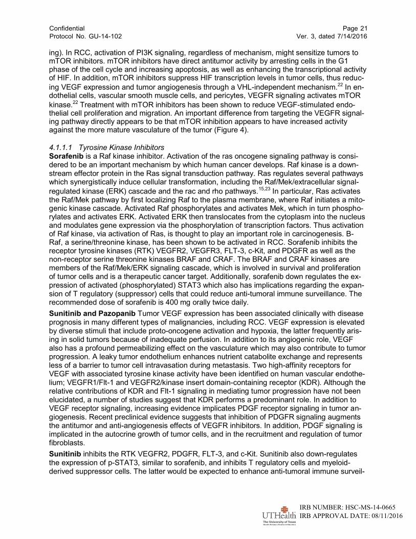
4.1.1 First-Line Molecular Targeted Agents
The tyrosine kinases cause the phosphorylation of tyrosine residues that initiate cellular signal- ing pathways to promote cell survival, proliferation and angiogenesis. There are three small mo- lecule inhibitors of tyrosine kinases that have been Food and Drug Administration approved for use in metastatic RCC based on clinical evidence of efficacy in several recent trials: sorafenib, sunitinib, and pazopanib.
The mTOR pathway can be activated in tumor cells by a variety of mechanisms, including those that enhance the PI3K/Akt signaling pathway (e.g., PTEN loss or activated receptor TK signal-
Confidential
Protocol No. GU-14-102
Page 21
Ver. 3, dated 7/14/2016
IRB NUMBER: HSC-MS-14-0665
IRB APPROVAL DATE: 08/11/2016
ing). In RCC, activation of PI3K signaling, regardless of mechanism, might sensitize tumors to mTOR inhibitors. mTOR inhibitors have direct antitumor activity by arresting cells in the G1 phase of the cell cycle and increasing apoptosis, as well as enhancing the transcriptional activity of HIF. In addition, mTOR inhibitors suppress HIF transcription levels in tumor cells, thus reduc-
ing VEGF expression and tumor angiogenesis through a VHL-independent mechanism.22 In en- dothelial cells, vascular smooth muscle cells, and pericytes, VEGFR signaling activates mTOR
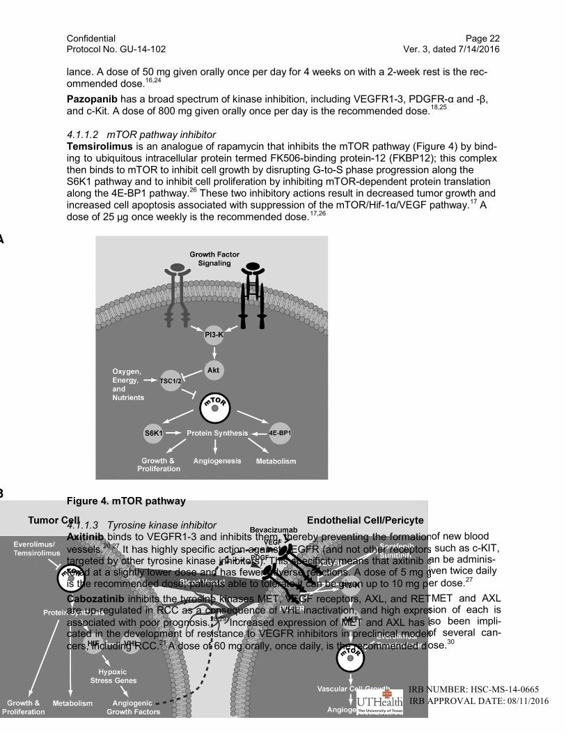
kinase.22 Treatment with mTOR inhibitors has been shown to reduce VEGF-stimulated endo- thelial cell proliferation and migration. An important difference from targeting the VEGFR signal- ing pathway directly appears to be that mTOR inhibition appears to have increased activity against the more mature vasculature of the tumor (Figure 4).
4.1.1.1 Tyrosine Kinase Inhibitors Sorafenib is a Raf kinase inhibitor. Activation of the ras oncogene signaling pathway is consi- dered to be an important mechanism by which human cancer develops. Raf kinase is a down- stream effector protein in the Ras signal transduction pathway. Ras regulates several pathways which synergistically induce cellular transformation, including the Raf/Mek/extracellular signal-
regulated kinase (ERK) cascade and the rac and rho pathways.15,23 In particular, Ras activates the Raf/Mek pathway by first localizing Raf to the plasma membrane, where Raf initiates a mito- genic kinase cascade. Activated Raf phosphorylates and activates Mek, which in turn phospho- rylates and activates ERK. Activated ERK then translocates from the cytoplasm into the nucleus and modulates gene expression via the phosphorylation of transcription factors. Thus activation of Raf kinase, via activation of Ras, is thought to play an important role in carcinogenesis. B- Raf, a serine/threonine kinase, has been shown to be activated in RCC. Sorafenib inhibits the receptor tyrosine kinases (RTK) VEGFR2, VEGFR3, FLT-3, c-Kit, and PDGFR as well as the non-receptor serine threonine kinases BRAF and CRAF. The BRAF and CRAF kinases are members of the Raf/Mek/ERK signaling cascade, which is involved in survival and proliferation of tumor cells and is a therapeutic cancer target. Additionally, sorafenib down regulates the ex- pression of activated (phosphorylated) STAT3 which also has implications regarding the expan- sion of T regulatory (suppressor) cells that could reduce anti-tumoral immune surveillance. The recommended dose of sorafenib is 400 mg orally twice daily.
Sunitinib and Pazopanib Tumor VEGF expression has been associated clinically with disease prognosis in many different types of malignancies, including RCC. VEGF expression is elevated by diverse stimuli that include proto-oncogene activation and hypoxia, the latter frequently aris- ing in solid tumors because of inadequate perfusion. In addition to its angiogenic role, VEGF also has a profound permeabilizing effect on the vasculature which may also contribute to tumor progression. A leaky tumor endothelium enhances nutrient catabolite exchange and represents less of a barrier to tumor cell intravasation during metastasis. Two high-affinity receptors for VEGF with associated tyrosine kinase activity have been identified on human vascular endothe- lium; VEGFR1/Flt-1 and VEGFR2/kinase insert domain-containing receptor (KDR). Although the relative contributions of KDR and Flt-1 signaling in mediating tumor progression have not been elucidated, a number of studies suggest that KDR performs a predominant role. In addition to VEGF receptor signaling, increasing evidence implicates PDGF receptor signaling in tumor an- giogenesis. Recent preclinical evidence suggests that inhibition of PDGFR signaling augments the antitumor and anti-angiogenesis effects of VEGFR inhibitors. In addition, PDGF signaling is implicated in the autocrine growth of tumor cells, and in the recruitment and regulation of tumor fibroblasts.
Sunitinib inhibits the RTK VEGFR2, PDGFR, FLT-3, and c-Kit. Sunitinib also down-regulates the expression of p-STAT3, similar to sorafenib, and inhibits T regulatory cells and myeloid- derived suppressor cells. The latter would be expected to enhance anti-tumoral immune surveil-
Confidential Protocol No. GU-14-102
Page 22 Ver. 3, dated 7/14/2016
Figure 4. mTOR pathway
4.1.1.3 Tyrosine kinase inhibitor Axitinib binds to VEGFR1-3 and inhibits them, thereby preventing the formation
vessels.20,27 It has highly specific action against VEGFR (and not other receptors targeted by other tyrosine kinase inhibitors). This specificity means that axitinib c tered at a slightly lower dose and has fewer adverse reactions. A dose of 5 mg g is the recommended dose; patients able to tolerate it can be given up to 10 mg p
Cabozatinib inhibits the tyrosine kinases MET, VEGF receptors, AXL, and RET. are up-regulated in RCC as a consequence of VHL inactivation, and high expres
associated with poor prognosis.28,29 Increased expression of MET and AXL has a cated in the development of resistance to VEGFR inhibitors in preclinical models
cers, including RCC.21 A dose of 60 mg orally, once daily, is the recommended d
lance. A dose of 50 mg given orally once per day for 4 weeks on with a 2-week rest is the rec- ommended dose.16,24
Pazopanib has a broad spectrum of kinase inhibition, including VEGFR1-3, PDGFR-α and -β,
and c-Kit. A dose of 800 mg given orally once per day is the recommended dose.18,25
4.1.1.2 mTOR pathway inhibitor Temsirolimus is an analogue of rapamycin that inhibits the mTOR pathway (Figure 4) by bind- ing to ubiquitous intracellular protein termed FK506-binding protein-12 (FKBP12); this complex then binds to mTOR to inhibit cell growth by disrupting G-to-S phase progression along the S6K1 pathway and to inhibit cell proliferation by inhibiting mTOR-dependent protein translation along the 4E-BP1 pathway.26 These two inhibitory actions result in decreased tumor growth and increased cell apoptosis associated with suppression of the mTOR/Hif-1α/VEGF pathway.17 A dose of 25 µg once weekly is the recommended dose.17,26
of new blood , such as c-KIT, an be adminis- iven twice daily
er dose.27
MET and AXL sion of each is lso been impli- of several can-
ose.30
IRB NUMBER: HSC-MS-14-0665
IRB APPROVAL DATE: 08/11/2016

Confidential Protocol No. GU-14-102
Page 23 Ver. 3, dated 7/14/2016
IRB NUMBER: HSC-MS-14-0665
IRB APPROVAL DATE: 08/11/2016
4.1.1.4 mTOR pathway inhibitor Everolimus is, like temsirolimus, an analogue of rapamycin (Figure 3). Everolimus works along
three pathways of action.19 (1) It inhibits cell growth and proliferation by reducing expression of proteins (such as Hif and VHL) that regulate the cell cycle. (2) It inhibits cell metabolism by de- creasing the activity of cell nutrient transporters via the PI3K-Akt-TSC1/2 pathway, thus reduc- ing the ability of cells to take up nutrients. (3) It reduces angiogenesis by inhibiting the growth of proliferation factors and preventing the release of VEGF and other angiogenic factors. A dose of 10 mg given orally once daily is the recommended dose.19,31
4.1.2 Gene Mutation Detection and mRNA Expression Profiling Using NGS
|||||||||||||||||||||||||||||||||||||||||||||||||||||||||||||||||||||||||||||||||||||||||||||||||||||||||||||||||||||||||||||||||||||||||||||||||||||||||||||||||||||||||||||||||||||||||||||||||||||||||| |||||||||||||||||||||||||||||||||||||||||||||||||||||||||||||||||||||||||||||||||||||||||||||||||||||||||||||||||||||||||||||||||||||||||||||||||||||||||||||||||||||||||||||||||||||||||||||||||||| |||||||||||||||||||||||||||||||||||||||||||||||||||||||||||||||||||||||||||||||||||||||||||||||||||||||||||||||||||||||||||||||||||||||||||||||||||||||||||||||||||||||||||||||||||||||||||||||||||||||||| |||||||||||||||||||||||||||||||||||||||||||||||||||||||||||||||||||||||||||||||||||||||||||||||||||||||||||||||||||||||||||||||||||||||||||||||||||||||||||||||||||||||||||||||||||||||||||||||||||||||||||| |||||||||||||||||||||||||||||||||||||||||||||||||||||||||||||||||||||||||||||||||||||||||||||||||||||||||||||||||||||||||||||||||||||||||||||||||||||||||||||||||||||||||||||||||||||||||||||||||||||||||||||| |||||||||||||||||||||||||||||||||||||||||||||||||||||||||||||||||||||||||||||||||||||||||||||||||||||||||||||||||||||||||||||||||||||||||||||||||||||||||||||||||||||||||||||||||||||||||||||||||||||||| |||||||||||||||||||||||||||||||||||||||||||||||||||||||||||||||||||||||||||||||||||||||||||||||||||||||||||||||||||||||||||||||||||||||||||||||||||||||||||||||||||||||||||||||||||||||||||||||||||||||||| |||||||||||||||||||||||||||||||||||||||||||||||||||||||||||||||||||||||||||||||||||||||||||||||||||||||||||||||||||||||||||||||||||||||||||||||||||||||||||||||||||||||||||||||||||||||||||||||||||||||||| |||||||||||||||||||||||||||||||||||||||||||||||||||||||||||||||||||||||||||||||||||||||||||||||||||||||||||||||||||||||||||||||||||||||||||||||||||||||||||||||||||||||||||||||||||||||||||||||||||||||||| |||||||||||||||||||||||||||||||||||||||||||||||||||||||||||||||||||||||||||||||||||||||||||||||||||||||||||||||||||||||||||||||||||||||||||||||||||||||||||||||||||||||||||||||||||||||||||||||||||||||||||| |||||||||||||||||||||||||||||||||||||||||||||||||||||||||||||||||||||||||||||||||||||||||||||||||||||||||||||||||||||||||||||||||||||||||||||||||||||||||||||||||||||||||||||||||||||||||||||||||||||||||||| |||||||||||||||||||||||||||||||||||||||||||||||||||||||||||||||||||||||||||||||||||||||||||||||||||||||||||||||||||||||||||||||||||||||||||||||||||||||||||||||||||||||||||||||||||||||||||||||||||||||| |||||||||||||||||||||||||||||||||||||||||||||||||||||||||||||||||||||||||||||||||||||||||||||||||||||||||||||||||||||||||||||||||||||||||||||||||||||||||||||||||||||||||||||||||||||||||||||||| |||||||||||||||||||||||||||||||||||||||||||||||||||||||||||||||||||||||||||||||||||||||||||||||||||||||||||||||||||||||||||||||||||||||||||||||||||||||||||||||||||||||||||||||||||||||||||||||||||||||||| |||||||||||||||||||||||||||||||||||||||||||||||||||||||||||||||||||||||||||||||||||||||||||||||||||||||||||||||||||||||||||||||||||||||||||||||||||||||||||||||||||||||||||||||||||||||||||||||||||||||||| ||||||||||||||||||||||||||||||||||||||||||||||||||||||||||||||||||||||||||||||||||||||||||||||||||||||||||||||||||||||||||||||||||||||
4.1.3 Proteomics
Our targeted proteomics approach (SRM or multiple SRM [MRM]) is a non-scanning mass spec- trometry technology that can quantify both proteins and their post-translational modifications, such as phosphorylation. This technique enables the direct measurement of peptides derived
from intact proteins without the need for antibodies or other affinity reagents;32,33
several groups
have used this approach to target cellular signaling molecules and their regulation by phospho- rylation to understand signaling networks. Our Center for Clinical Proteomics has significant ex- perience designing SRMs for signaling molecules in fresh/frozen tissues and in biofluids. We are already using this technique with LMD cancer cells to couple quantitative protein measure- ments and activation profiles with morphology on the same patient specimens as above. Though our team has experience with these assays, we have been working with Expression Pathology, Inc. to develop assays to signaling molecules within archival, FFPE tissues from the patients as they have patented methods for working with fixed tissues. This approach will be used to complement the studies described above, adding quantitative data to the morphological information and providing assays for particular proteins and activation sites when immunological reagents are not available for key markers.
Our SRM-based targeted strategy utilizes morphologic analysis (from LMD), quantitative pro- teomics, and high target specificity to discern the state of activation of signal transduction path- ways in tumor cells and companionate tissues. Sequence and phospho-specific assays are de- signed in silico and directed against putative sites of activation of a signaling protein. The unique LMD system we have developed uses conventional hematoxylin-eosin staining and a nonglass coverslip to ensure high-quality morphological assessment for selecting diagnostic cells from tissue sections cut from formalin-fixed, paraffin-embedded patient tissues. Stained

Confidential Protocol No. GU-14-102
Page 24 Ver. 3, dated 7/14/2016
IRB NUMBER: HSC-MS-14-0665
IRB APPROVAL DATE: 08/11/2016
tissues mounted without coverslips often exhibit degraded morphology, which prevents accurate dissection of the desired cells. In addition, although a general region of a tumor can be dis- sected using approaches other than LMD, image-oriented, cell-specific correlations cannot be made from such dissections. In addition, digital images of target cells are captured during our LMD process; they are then annotated and stored for later correlation with SRM measurements. Our newly developed technique enables the selection of immunofluorescently labeled cells us- ing fluorescent optics and the coverslip system to couple subcellular marker expression with ac- curate marker quantification or with downstream signaling patterns.
|||||||||||||||||||||||||||||||||||||||||||||||||||||||||||||||||||||||||||||||||||||||||||||||||||||||||||||||||||||||||||||||||||||||||||||||||||||||||||||||||||||||||||||||||||||||||||||||||||||||||||| |||||||||||||||||||||||||||||||||||||||||||||||||||||||||||||||||||||||||||||||||||||||||||||||||||||||||||||||||||||||||||||||||||||||||||||||||||||||||||||||||||||||||||||||||||||||||||||||||| |||||||||||||||||||||||||||||||||||||||||||||||||||||||||||||||||||||||||||||||||||||||||||||||||||||||||||||||||||||||||||||||||||||||||||||||||||||||||||||||||||||||||||||||||||||||||||||||||||||| |||||||||||||||||||||||||||||||||||||||||||||||||||||||||||||||||||||||||||||||||||||||||||||||||||||||||||||||||||||||||||||||||||||||||||||||||||||||||||||||||||||||||||||||||||||||||||||||||||||||| |||||||||||||||||||||||||||||||||||||||||||||||||||||||||||||||||||||||||||||||||||||||||||||||||||||||||||||||||||||||||||||||||||||||||||||||||||||||||||||||||||||||||||||||||||||||||||||||||||||||||| |||||||||||||||||||||||||||||||||||||||||||||||||||||||||||||||||||||||||||||||||||||||||||||||||||||||||||||||||||||||||||||||||||||||||||||||||||||||||||||||||||||||||||||||||||||||||||||||||||| |||||||||||||||||||||||||||||||||||||||||||||||||||||||||||||||||||||||||||||||||||||||||||||||||||||||||||||||||||||||||||||||||||||||||||||||||||||||||||||||||||||||||||||||||||||||||||||||||||||||||| |||||||||||||||||||||||||||||||||||||||||||||||||||||||||||||||||||||||||||||||||||||||||||||||||||||||||||||||||||||||||||||||||||||||||||||||||||||||||||||||||||||||||||||||||||||||||||||||||||||||||| ||||||||||||||||||||||||||||||||||||||||||||||||||||||||||||||||||||||||||||||||||||||||||||
4.2 First-Line Therapy
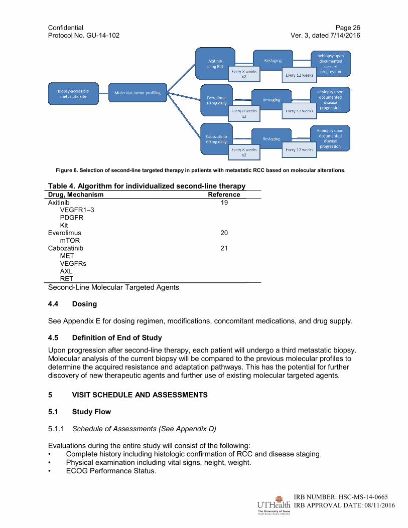
It is hypothesized that a more effective treatment protocol can be obtained and applied to pa- tients based on molecular alterations and the use of known targeted agents that inhibit the TK- dependent pathways or the mTOR-dependent pathways. A molecular targeted agent for each patient will be selected based on their molecular alteration profile; patients will receive that mo- lecular targeted agent and undergo radiographic evaluation according to RECIST criteria to de- termine response. The radiographic staging evaluation will be performed at baseline; restaging images will be obtained after two 6-week periods of sunitinib treatment or after two 8-week pe- riods for temsirolimus, sorafenib, or pazopanib treatment, then every 12 weeks thereafter in all patients (Figure 5, Table 3).
Figure 5. Selection of first-line targeted therapy in patients with metastatic RCC based on molecular alterations.
Table 3. Algorithm for individualized first-line therapy
Confidential Protocol No. GU-14-102
Page 25 Ver. 3, dated 7/14/2016
IRB NUMBER: HSC-MS-14-0665
IRB APPROVAL DATE: 08/11/2016
Drug, Mechanism Reference
First-line treatment Pazopanib 18
VEGFR1–3 PDGFR Kit
Sunitinib 16 VEGFR1–3
Predominantly VEGFR2 (KDR)
PDGFR Kit Flt3 RET STAT3
Sorafenib 15 VEGFR1–3 PDGFR Kit Flt3 RET B-Raf, C-Raf
Temsirolimus 17 mTOR
B-Raf and C-Raf, signal transduction protein kinases of the Raf kinase family; Flt, Fms-like tyrosine ki- nase; Kit, a stem cell signaling pathway; mTOR, mammalian target of rapamycin; PDGFR, platelet- derived growth factor receptor; RET, rearranged during transfection (an oncogene); STAT, signal trans- ducer and activator of transcription; VEGFR, vascular endothelial growth factor receptor
Similar testing will be performed on biopsies obtained after disease progression. Samples will undergo molecular analysis to determine molecular alterations and adaptations conferring treatment resistance. Depending on the molecular alterations found, patients will be assigned second-line therapy with everolimus, axitinib, or cabozatinib.
4.3 Second-Line Therapy
Upon radiographic progression after first-line therapy, we will obtain a second metastatic biopsy and, based on molecular alterations second-line therapy with the next molecular targeted agent (everolimus, axitinib, or cabozatinib) will be selected. This will allow us to determine the appro- priate second-line treatment based on the mechanisms of acquired resistance and adaptation.
Based on RECIST criteria to determine response, radiographic evaluation will be performed af- ter two 8-week periods of treatment and every 12 weeks thereafter in all patients (Figure 6, Ta- ble 4).
Confidential Protocol No. GU-14-102
Page 26 Ver. 3, dated 7/14/2016
IRB NUMBER: HSC-MS-14-0665
IRB APPROVAL DATE: 08/11/2016
Figure 6. Selection of second-line targeted therapy in patients with metastatic RCC based on molecular alterations.
Table 4. Algorithm for individualized second-line therapy
Drug, Mechanism Reference
Axitinib 19 VEGFR1–3 PDGFR Kit
Everolimus 20 mTOR
Cabozatinib 21 MET VEGFRs AXL RET
Second-Line Molecular Targeted Agents
4.4 Dosing
See Appendix E for dosing regimen, modifications, concomitant medications, and drug supply.
4.5 Definition of End of Study
Upon progression after second-line therapy, each patient will undergo a third metastatic biopsy. Molecular analysis of the current biopsy will be compared to the previous molecular profiles to determine the acquired resistance and adaptation pathways. This has the potential for further discovery of new therapeutic agents and further use of existing molecular targeted agents.
5 VISIT SCHEDULE AND ASSESSMENTS
5.1 Study Flow
5.1.1 Schedule of Assessments (See Appendix D)
Evaluations during the entire study will consist of the following: • Complete history including histologic confirmation of RCC and disease staging. • Physical examination including vital signs, height, weight. • ECOG Performance Status.

Confidential Protocol No. GU-14-102
Page 27 Ver. 3, dated 7/14/2016
IRB NUMBER: HSC-MS-14-0665
IRB APPROVAL DATE: 08/11/2016
• Imaging and diagnostic studies. • Hematology, coagulation, and chemistry studies. • Urinalysis. • Fasting triglycerides and cholesterol. • β-HCG (females of childbearing potential).
These assessments should be performed within ±3 days of the scheduled day of assessment. Every effort should be made to ensure that the protocol required tests and procedures are com- pleted as described. However, it is anticipated that from time to time there may be circums- tances, outside of the control of the investigator that may make it unfeasible to perform the test. In these cases, the investigator will take all steps necessary to ensure the safety and well-being of the patient. When a protocol-required test cannot be performed the investigator will document the reason for this and any corrective and preventive actions which he/she has taken to ensure that normal processes are adhered to as soon as possible.
5.1.2 Screening
The screening assessments will be performed within 28 days prior to receiving study medica- tion:
• Signed informed consent • Vital signs • Concomitant medications assessment • Medical History: Includes histologic confirmation of malignancy, and best response(s),
if applicable. Patient information will be entered into the Memorial Sloan-Kettering Cancer Center prognostic model (refer to J Clin Oncol, July 1999).
• Physical Examination: Includes height, weight. A full neurologic examination (mental status, cranial nerves, motor, reflexes, sensory, gait, cerebella) will be performed at baseline only.
• ECOG Performance Status: Refer to ECOG Performance Status Criteria. • Hematology Profile: Includes CBC with differential and platelets. • Coagulation Profile: Includes PT, INR, and PTT. • Chemistry Profile: Includes total protein, uric acid, BUN, creatinine, LDH with isoen-
zymes, AST, ALT, alkaline phosphatase with isoenzymes, phosphorus, magnesium, total bilirubin, calcium, albumin, and glucose.
• Electrolytes: sodium, potassium, chloride, and bicarbonate. • TSH level. • Amylase/lipase. • Miscellaneous Tests: VEGF, IL-6, TNF-α, C-reactive protein, sedimentation rate • Fasting triglycerides and cholesterol. • Cardiac Profile: Includes EKG and echocardiogram. • Pulmonary Profile: Includes spirometry with DLCO. • Pregnancy screening test: Includes beta-HCG pregnancy test within 7 days of the first
treatment for females of childbearing potential • Urinalysis: Includes routine measurements. A microscopic analysis will be conducted
pretreatment. Subsequent microscopic analyses will be done only if clinically indicated. • Imaging and Diagnostic Studies: CT of the chest and abdomen with IV contrast in ar-
terial phase or CT of the abdomen and pelvis with IV contrast in portal venous phase. 5.1.3 Treatment Period
Confidential Protocol No. GU-14-102
Page 28 Ver. 3, dated 7/14/2016
IRB NUMBER: HSC-MS-14-0665
IRB APPROVAL DATE: 08/11/2016
All study visits beyond screening, including study drug administration and safety laboratory evaluations must be obtained within ±3 days of the day specified in the protocol schedule.
• Hematology Profile: CBC, differential, and platelets will be obtained weekly for the first 6-8 weeks, then every 2 weeks.
• Chemistry Profile: Total protein, uric acid, BUN, creatinine, LDH with isoenzymes, AST, ALT, alkaline phosphatase with isoenzymes, phosphorus, magnesium, total bili- rubin, calcium, albumin, and glucose will be obtained weekly for the first 6-8 weeks, then every 2 weeks.
• Electrolytes: sodium, potassium, chloride, and bicarbonate will be obtained weekly for the first 6-8 weeks, then every 2 weeks.
• Amylase/lipase will be obtained every 4 weeks.
• VEGF, IL-6, TNF-α, C-reactive protein, and sedimentation rate will be obtained at each restaging.
• Fasting triglycerides and cholesterol will be obtained every 4 weeks.
• TSH level will be obtained every 6-8 weeks.
• Urinalysis will be obtained every 4 weeks.
• Cardiac and pulmonary function tests as clinically indicated.
• Tumor restaging will be performed every 8 weeks (twice) for patients receiving temsi- rolimus, sorafenib, pazopanib, axitinib, everolimus, or cabozatinib and every 6 weeks (twice) for those receiving sunitinib and every 12 weeks thereafter.
• Interim history and exam with performance status and weight will be recorded every 2 weeks for the first 4 weeks, then every 4 weeks thereafter.
• Information regarding drug dosages, laboratory examinations and treatment related toxicities will be recorded before each treatment cycle is given.
• Since all of the drugs in this trial are standard of care, there may be instances when subjects will receive their treatment at other infusion centers as well as their standard blood draws due to insurance network provider determinations, cost, etc. Dr. Amato will work closely with referring physicians and infusion centers. The lab results and clinic/infusion notes will be obtained and reviewed by Dr. Amato. All subjects will still come to the cancer center for restaging study visits.
Evaluations 30 (±3) Days after Last Dose of Study Therapy or Withdrawal from The Study
• Interim history and physical examination. • Adverse event assessment. • Hematology Profile: CBC with differential, platelet count. • Coagulation Profile: PT, INR, and PTT. • Chemistry Profile: Total protein, uric acid, BUN, creatinine, LDH with isoenzymes,
AST, ALT, alkaline phosphatase with isoenzymes, phosphorus, magnesium, total bili- rubin, calcium, albumin, and glucose.
• Electrolytes: sodium, potassium, chloride, and bicarbonate. • TSH level. • Fasting triglycerides and cholesterol. • Amylase/lipase. • Urinalysis.
Confidential Protocol No. GU-14-102
Page 29 Ver. 3, dated 7/14/2016
IRB NUMBER: HSC-MS-14-0665
IRB APPROVAL DATE: 08/11/2016
• Vital Signs, ECOG performance status.
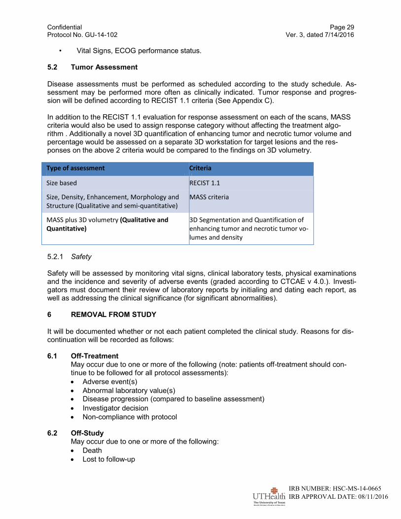
5.2 Tumor Assessment
Disease assessments must be performed as scheduled according to the study schedule. As- sessment may be performed more often as clinically indicated. Tumor response and progres- sion will be defined according to RECIST 1.1 criteria (See Appendix C).
In addition to the RECIST 1.1 evaluation for response assessment on each of the scans, MASS criteria would also be used to assign response category without affecting the treatment algo- rithm . Additionally a novel 3D quantification of enhancing tumor and necrotic tumor volume and percentage would be assessed on a separate 3D workstation for target lesions and the res- ponses on the above 2 criteria would be compared to the findings on 3D volumetry.
Type of assessment Criteria
Size based RECIST 1.1
Size, Density, Enhancement, Morphology and Structure (Qualitative and semi-quantitative)
MASS criteria
MASS plus 3D volumetry (Qualitative and Quantitative)
3D Segmentation and Quantification of enhancing tumor and necrotic tumor vo- lumes and density
5.2.1 Safety
Safety will be assessed by monitoring vital signs, clinical laboratory tests, physical examinations and the incidence and severity of adverse events (graded according to CTCAE v 4.0.). Investi- gators must document their review of laboratory reports by initialing and dating each report, as well as addressing the clinical significance (for significant abnormalities).
6 REMOVAL FROM STUDY
It will be documented whether or not each patient completed the clinical study. Reasons for dis- continuation will be recorded as follows:
6.1 Off-Treatment
May occur due to one or more of the following (note: patients off-treatment should con- tinue to be followed for all protocol assessments):
Adverse event(s)
Abnormal laboratory value(s)
Disease progression (compared to baseline assessment)
Investigator decision
Non-compliance with protocol
6.2 Off-Study May occur due to one or more of the following:
Death
Lost to follow-up

Confidential Protocol No. GU-14-102
Page 30 Ver. 3, dated 7/14/2016
IRB NUMBER: HSC-MS-14-0665
IRB APPROVAL DATE: 08/11/2016
Withdrawal of consent
Completion of protocol study period
Administrative study closure
7 SAFETY MONITORING AND REPORTING
The investigator will monitor each patient for clinical and laboratory evidence of adverse events on a routine basis throughout the study following the NCI Common Terminology Criteria for Ad- verse Events (CTCAE), Version 4.0. A link to the electronic version of the CTCAE can be found in Appendix A. The investigator will assess and record any adverse event (serious and non- serious) in detail on the adverse event form including the date of onset, description, severity, time course, duration, outcome and relationship to the study drug from the time the patient signs the informed consent until 4 weeks after the patient has stopped study treatment.
7.1 Adverse Events
All adverse events (AEs) should be treated appropriately. Such treatment may include interrup- tion or discontinuation of study drug, starting or stopping concomitant treatments, changes in the frequency or nature of assessments, hospitalization, or any other medically required inter- vention.
Information about common side effects already known about the study drug can be found in Ap- pendix E and package inserts. This information will be included in the patient informed consent and should be discussed with the patient during the study as needed.
7.2 Adverse Event Definition
An AE is defined as any unintended or undesirable, noxious, or pathological change, compared to pre-existing conditions, experienced by a patient during a clinical study or the follow-up pe- riod, regardless of relationship to study drug. AEs include:
Suspected adverse drug reactions.
Reactions from drug overdose, abuse, withdrawal, sensitivity, or toxicity.
Significant changes or abnormalities, when compared to baseline, in structure (sign), function (symptom), clinical laboratory results, or physiological testing. This includes any worsening of a pre-existing condition temporally associated with the use of study drug.
Other medical events, regardless of their relationship to the study drug, such as injury, surgery, accidents, extensions of symptomatology, or apparently unrelated illnesses.
7.3 Evaluating Adverse Events
Each AE will be evaluated to determine:
the severity grade (mild, moderate, severe) or (grade 1-4)
its relationship to the study drug(s) (suspected/not suspected)
its duration (start and end dates or if continuing at final exam)
action taken (no action taken; study drug dosage adjusted/temporarily interrupted; study drug permanently discontinued due to this adverse event; concomitant medication taken; non-drug therapy given; hospitalization/prolonged hospitalization)
outcome
whether it constitutes a serious adverse event (SAE)
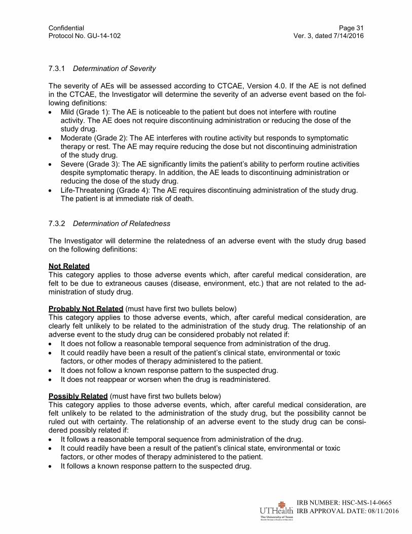
Confidential Protocol No. GU-14-102
Page 31 Ver. 3, dated 7/14/2016
IRB NUMBER: HSC-MS-14-0665
IRB APPROVAL DATE: 08/11/2016
7.3.1 Determination of Severity
The severity of AEs will be assessed according to CTCAE, Version 4.0. If the AE is not defined in the CTCAE, the Investigator will determine the severity of an adverse event based on the fol- lowing definitions:
Mild (Grade 1): The AE is noticeable to the patient but does not interfere with routine activity. The AE does not require discontinuing administration or reducing the dose of the study drug.
Moderate (Grade 2): The AE interferes with routine activity but responds to symptomatic therapy or rest. The AE may require reducing the dose but not discontinuing administration of the study drug.
Severe (Grade 3): The AE significantly limits the patient’s ability to perform routine activities despite symptomatic therapy. In addition, the AE leads to discontinuing administration or reducing the dose of the study drug.
Life-Threatening (Grade 4): The AE requires discontinuing administration of the study drug. The patient is at immediate risk of death.
7.3.2 Determination of Relatedness
The Investigator will determine the relatedness of an adverse event with the study drug based on the following definitions:
Not Related This category applies to those adverse events which, after careful medical consideration, are felt to be due to extraneous causes (disease, environment, etc.) that are not related to the ad- ministration of study drug.
Probably Not Related (must have first two bullets below) This category applies to those adverse events, which, after careful medical consideration, are clearly felt unlikely to be related to the administration of the study drug. The relationship of an adverse event to the study drug can be considered probably not related if:
It does not follow a reasonable temporal sequence from administration of the drug.
It could readily have been a result of the patient’s clinical state, environmental or toxic factors, or other modes of therapy administered to the patient.
It does not follow a known response pattern to the suspected drug.
It does not reappear or worsen when the drug is readministered. Possibly Related (must have first two bullets below) This category applies to those adverse events, which, after careful medical consideration, are felt unlikely to be related to the administration of the study drug, but the possibility cannot be ruled out with certainty. The relationship of an adverse event to the study drug can be consi- dered possibly related if:
It follows a reasonable temporal sequence from administration of the drug.
It could readily have been a result of the patient’s clinical state, environmental or toxic factors, or other modes of therapy administered to the patient.
It follows a known response pattern to the suspected drug.

Confidential Protocol No. GU-14-102
Page 32 Ver. 3, dated 7/14/2016
IRB NUMBER: HSC-MS-14-0665
IRB APPROVAL DATE: 08/11/2016
Probably Related (must have first three bullets below)
This category applies to those adverse events which, after careful medical consideration, are felt with a high degree of certainty to be related to the administration of the study drug. The rela- tionship of an adverse event to the study drug can be considered probably related if:
It follows a reasonable temporal sequence from administration of the drug.
It could not be reasonably explained by the known characteristics of the patient’s clinical state, environmental or toxic factors or other modes of therapy administered to the patient.
It disappears or decreases upon cessation of drug or reduction in dose.*
It follows a known response pattern to the suspected drug. Definitely Related (must have first three bullets below) This category applies to those adverse events, which, after careful medical consideration, are felt to be related to the administration of the drug. The relationship of an adverse event to the study drug can be considered definitely related if:
It follows a reasonable temporal sequence from administration of the drug or drug levels have been established in body fluids or tissues.
It could not be reasonably explained by the known characteristics of the patient’s clinical state, environmental or toxic factors or other modes of therapy administered to the patient.
It disappears or decreases upon cessation of drug or reduction in dose and, if applicable, appears upon rechallenge.*
It follows a known response pattern to the suspected drug.
*There are exceptions when an adverse event does not disappear upon discontinuation of the drug, yet drug relatedness clearly exists; e.g., 1) tardive dyskinesia, 2) fixed drug eruptions.
7.4 Serious Adverse Events
Information about all serious adverse events (SAEs) will be collected and recorded. A SAE is an undesirable sign, symptom or medical condition which:
is fatal or life-threatening
results in persistent or significant disability/incapacity
constitutes a congenital anomaly/birth defect
requires inpatient hospitalization or prolongation of existing hospitalization, unless hospitalization is for: routine treatment or monitoring of the studied indication, not associated with any deteri-
oration in condition (specify what this includes) elective or pre-planned treatment for a pre-existing condition that is unrelated to the indi-
cation under study and has not worsened since the start of study drug treatment on an emergency outpatient basis for an event not fulfilling any of the definitions
of a SAE given above and not resulting in hospital admission social reasons and respite care in the absence of any deterioration in the patient’s general
condition
is medically significant, i.e., defined as an event that jeopardizes the patient or may require medical or surgical intervention to prevent one of the outcomes listed above
7.5 Reporting
The principal investigator has the obligation to report all serious adverse events to the IRB and DSMB according to their respective requirements.

Confidential Protocol No. GU-14-102
Page 33 Ver. 3, dated 7/14/2016
IRB NUMBER: HSC-MS-14-0665
IRB APPROVAL DATE: 08/11/2016
Serious Adverse Events (SAE) that are determined by the PI to be unexpected + related will be submitted to the DSMB within 7 calendar days of the determination by telephone or fax; written report no later than 15 calendar days of the determination. Deaths should be reported to the IRB within 24 hours of investigator knowledge. Any unexpected, serious, related adverse expe- riences should be reported to the IRB within 7 calendar days of investigator knowledge.
Any pregnancy that occurs during study participation should be reported. The pregnancy should be followed up to determine outcome, including spontaneous or voluntary termination, details of birth, and the presence or absence of any birth defects, congenital abnormalities or maternal and newborn complications.
7.6 Unanticipated Problem Reporting
The Principal Investigator (PI) must notify the IRB of unexpected problems that might arise dur- ing the study within 7 calendar days. The PI is must make a judgment call regarding the expec- tedness and causality of the problem. Examples include:
any adverse event which in the opinion of the PI is both unexpected and related and places patients or others at risk of harm
Protocol deviation that harmed patients or placed patients in increased risk of harm
Unanticipated adverse device effect
A breach of confidentiality
Change in FDA labeling or withdrawal from marketing of a drug, device, or biologic used in a research protocol
Information that indicates a change to the risks or potential benefits of the research
8 DATA AND PROTOCOL MANAGEMENT
8.1 Protocol Compliance
Written informed consent must be obtained from the patient prior to study specific screening tests or procedures. Results of all baseline evaluations which assure that all inclusion and ex- clusion criteria have been satisfied, must be reviewed by the Investigator prior to enrollment of that patient. The investigator and/or research coordinator must see each patient prior to drug administration. All required interim and pre-treatment data should be available and the investi- gator must have made a designation as to tumor response and toxicity grade. The Principal In- vestigator will be the final arbitrator of response or toxicity should a difference of opinion exist.
8.2 Data Collection
Investigators or their designee must enter the data required by the protocol onto Case Report Forms (CRFs). A brief explanation for required but missing data should be recorded as a com- ment. The Principal Investigator is ultimately responsible for assuring that data entered into the CRFs are accurate, authentic/original, attributable, complete, consistent, legible, timely (con- temporaneous), enduring and available when required.
Entries made in the CRF must be either verifiable against source documents, or have been di- rectly entered into the CRF, in which case the entry in the CRF will be considered as the source data. Any corrections to entries made in the CRFs or source documents must be dated, initialed
Confidential Protocol No. GU-14-102
Page 34 Ver. 3, dated 7/14/2016
IRB NUMBER: HSC-MS-14-0665
IRB APPROVAL DATE: 08/11/2016
and explained (if necessary) and should not obscure the original entry. The Investigator will sign the CRFs to indicate that, to his/her knowledge, they are complete and accurate.
8.3 Database Management
The data manager will review the CRF data entered by study staff for completeness and accu- racy. Data queries stating the nature of the problem and requesting clarification will be created for discrepancies and missing values and sent to the study coordinator. The study coordinator will respond promptly to queries and make any necessary changes to the CRFs.
8.4 Site Monitoring
To ensure that the protocol and Good Clinical Practices (GCP) are being followed and that study data are accurate, complete and reliable, the trial will be monitored by a Data Safety Monitoring Board (DSMB), Medical Monitor and CRA monitoring as specified in the Data Safety Monitoring Plan (DSMP) for the study and the DSMB’s charter.
Through ongoing monitoring the designee will periodically check the patient data recorded in the CRFs against source documents, to ensure accuracy, completeness, and adherence to the pro- tocol, regulatory compliance, and the maintenance of comprehensive clinical records. The study coordinator will provide access to source documents to perform this verification.
9 STATISTICAL CONSIDERATIONS
9.1 Study Design
This will be a prospective, one-arm, proof of concept study designed to evaluate the efficacy of algorithm-based allocation (based on genomic/proteomic profile) of first-line therapy in RCC.
After eligibility review, patients will receive one of the four first-line therapy agents based on their tumor’s genomic/proteomic profile. Upon disease progression, patients will then receive one of two second-line agents based on their tumor’s genomic/proteomic profile.
Because this is a proof-of-concept study, the sample size is based on feasibility of accrual. The clinic should be able to recruit 100 patients within a reasonable timeframe for the study. Number of patients receiving each drug will vary based on the frequency of molecular alterations in the population. Therefore, groups will not be compared with one another – our research goal is to determine whether the PFS for each drug is improved over the PFS reported in FDA approval trials for each drug when they are assigned based on molecular analysis.
9.2 Analysis of Primary Endpoint The primary endpoint will be progression-free survival (PFS) on both first-line and second-line therapy. The PFS is defined as the time elapsed between treatment initiation and tumor pro- gression or death from any cause, with censoring of patients who are lost to follow-up. The me- dian PFS will be determined for each drug in both first-line and second-line therapy, and survival curves will be plotted using the Kaplan-Meier method. PFS will be reported as median and 95% confidence interval.
Analysis of the secondary endpoint, exploratory endpoints, and subgroup will be carried out us- ing appropriate statistical methods and descriptive statistics.

Confidential Protocol No. GU-14-102
Page 35 Ver. 3, dated 7/14/2016
IRB NUMBER: HSC-MS-14-0665
IRB APPROVAL DATE: 08/11/2016
9.3 Interim Analysis and Monitoring
The data will be reviewed by the expert committee after 50% of the patients are enrolled. The committee will review safety and efficacy data. Ineffective treatment arms, if any, will be dropped.
10 ETHICAL CONSIDERATIONS
10.1 Ethical Compliance
The study will be conducted in accordance with legal and regulatory (21 CFR 50, 56, 312 as applicable) requirements, as well as the general principles set forth in the Guidelines for GCP (ICH 1996) and the Declaration of Helsinki (World Medical Association 1996 and 2008).
The principal investigator is responsible for ensuring that all participating staff members are adequately trained and competent to perform his/her assigned tasks.
10.2 IRB Review
Before implementing this study, the protocol, the proposed informed consent form, and other required information, must be reviewed and approved by a properly constituted Institutional Re- view Board (IRB). Any amendments to the protocol, other than administrative ones, must be re- viewed and approved by this committee before implementation. If an immediate change to the protocol is implemented for safety reasons by the investigator, the IRB must be informed imme- diately. The study must be reviewed and approved at least annually as well.
10.3 Recruitment
Upon obtaining approval form external agencies and internal review committees, we will begin recruitment for the clinical trial. Based on our previous experience with the regulatory review process, we estimate that this will be completed in approximately 4-6 months. Once approved, patients with RCC seen at the UT Memorial Hermann Cancer Center will be accrued; we esti- mate that accrual to the first-line molecular-targeted agent will be completed in approximately 18 months. Upon radiographic progression, patients will undergo a second metastatic biopsy to determine the molecular tumor profile and initiate a second-line molecular targeted agent. Our recruitment plan for enrolling the 100 patients follows a multi-step approach:
Houston recruitment:
o We will accrue patients from our direct clinic population: The University of Texas Health Science Center at Houston; Memorial Hermann Cancer Center – Texas Medical Center; Memorial Hermann Cancer Center – Southwest; Memorial Hermann Cancer Center – Northeast.
We have referral relationships with local oncology faculty at primary care clin- ics.
We have referral relationships with local medical oncologists. We employ a Nurse Navigator and Physician Liaison who work with local
providers to increase referrals. Our referral area is extremely broad. Memorial Hermann pulls from the great-
er Houston area. Based on the strength of its reputation, The University of Texas pulls from the entire state of Texas as well as from surrounding states.
Statewide recruitment:
Confidential Protocol No. GU-14-102
Page 36 Ver. 3, dated 7/14/2016
IRB NUMBER: HSC-MS-14-0665
IRB APPROVAL DATE: 08/11/2016
o We have strong referral relationships for consults with medical oncologists in several areas of Texas:
Austin Abilene Dallas-Fort Worth The Rio Grande Valley (McAllen-Harlingen-Brownsville) East Texas (Beaumont-Port Arthur-Lake Charles, LA)
Regional recruitment: o We have strong referral relationships for consults with medical oncologists in Louisi-
ana, Arkansas, Oklahoma, and Colorado
National recruitment:
o The Kidney Cancer Association (http://www.kidneycancer.org/) has an online clinical trial matching service for patients that reaches an international audience. We plan to register our trial with the association.
o The National Institutes of Health’s clinical trials registry, ClinicalTrials.gov
(http://clinicaltrials.gov/), is a large worldwide registry. All US human clinical trials are required to register on the site, which is searchable by the public. The World Health Organization has a similar clinical trials registry (http://apps.who.int/trialsearch/Default.aspx).
o Dr. Amato has more than 30 years of experience as a treating physician and re- searcher. As such, he has a broad acquaintance within the urology and oncology communities. This results in a number of referrals each month from fellow urologists around the world.
10.4 Informed Consent
The Investigator will be responsible for obtaining consent, documented on the Informed Consent Form (ICF) signed and dated by each patient or his/her legally authorized representative, prior to his/her participation in the study, in accordance with ICH GCP guidelines. The ICF will be written in non-technical language. The patient should read and be given as much time as they need to consider their participation before signing and dating it.
The investigator or study staff designee must explain to each patient the nature of the study, its purpose, the procedures involved, the expected duration, the potential risks and benefits in- volved and any discomfort it may entail. Each patient must be informed that participation in the study is voluntary and that he/she may withdraw from the study at any time and that withdrawal of consent will not affect his/her subsequent medical treatment or relationship with the treating physician.
Participation in the study and date of informed consent should be documented appropriately in the patient’s files. The original ICF will be maintained in the research files and a copy must be maintained in the institution’s medical records. The patient or his/her legally authorized repre- sentative will also be given a copy of the signed consent form.
10.5 Confidentiality
All records identifying the patient will be kept confidential and, to the extent permitted by the ap- plicable laws and/or regulations, will not be made publicly available. Patient names will not be supplied to third parties. A unique study accession number will be assigned to each patient on study and will be used on the CRFs. Identifiable data on any document (e.g., pathologist report)
Confidential Protocol No. GU-14-102
Page 37 Ver. 3, dated 7/14/2016
IRB NUMBER: HSC-MS-14-0665
IRB APPROVAL DATE: 08/11/2016
must be redacted before a copy of the document is supplied to third parties. The study coordina- tor will maintain a list to enable patients’ records to be identified for verification purposes.
Study data stored electronically will be stored in zone 100, on password-protected, encrypted computers. Paper study data will be maintained by the study coordinator in the locked research offices.
10.6 Publication of Study Results
The investigator will assure that the key elements of this protocol will be posted in a publicly ac- cessible database such as www.ClinicalTrials.gov. In addition, upon study completion and finali- zation of the study report the results of this study will be submitted for publication in scientific journals and/or scientific meetings. If the results of the study are published, the patient’s identity will remain confidential.
10.7 Retention of Documents
To enable evaluations and/or audits from regulatory authorities or sponsors, the investigator agrees to keep records, including the identity of all participating patients (sufficient information to link records, e.g., CRFs and hospital records), all original signed informed consent docu- ments, copies of all CRFs, safety reporting forms, source documents, detailed records of treat- ment disposition, and adequate documentation of relevant correspondence in a secure storage facility.
Essential documents (written and electronic) should be retained for at least three (3) years after the completion of the study. The records should be retained by the investigator according to lo- cal regulations or as specified in the Clinical Study Agreement (CSA), whichever is longer.

Confidential Protocol No. GU-14-102
Page 38 Ver. 3, dated 7/14/2016
IRB NUMBER: HSC-MS-14-0665
IRB APPROVAL DATE: 08/11/2016
11 REFERENCES
1. Rodríguez-Antona C, García-Donas J. Constitutional genetic variants as predictors of
antiangiogenic therapy outcome in renal cell carcinoma. Pharmacogenomics 2012;13:1621
2. De Mattos-Arruda L, Rodon J. Pilot studies for personalized cancer medicine: focusing on the patient for treatment selection. Oncologist 2013;18:1180
3. Brufau BP, Cerqueda CS, Villalba LB, et al. Metastatic renal cell carcinoma: radiologic findings and assessment of response to targeted antiangiogenic therapy by using multi- detector CT. Radiographics 2013;33(6):1691
4. Katabathina VS, Lassau N, Pedrosa I, et al. Evaluation of treatment response in patients with metastatic renal cell carcinoma: role of state-of-the-art cross-sectional imaging. Curr Urol Rep 2012;13(1):70
5. Shinagare AB, Krajewski KM, Jagannathan JP, et al. Genitourinary imaging: part 2, role of imaging in medical management of advanced renal cell carcinoma. AJR Am J Roent- genol 2012;199(5):W554
6. Kang HC, Tan KS, Keefe SM, et al. MRI assessment of early tumor response in metas- tatic renal cell carcinoma patients treated with sorafenib. AJR Am J Roentgenol 2013;200(1):120
7. Yaghmai V, Miller FH, Rezai P, et al. Response to treatment series: part 2, tumor re- sponse assessment--using new and conventional criteria. AJR Am J Roentgenol 2011;197(1):18
8. Smith AD, Shah SN, Rini BI, et al. Morphology, attenuation, size, and structure (MASS) criteria: assessing response and predicting clinical outcome in metastatic renal cell car- cinoma on antiangiogenic targeted therapy. AJR Am J Roentgenol 2010;194(6):1470
9. Smith AD, Lieber ML, Shah SN. Assessing tumor response and detecting recurrence in metastatic renal cell carcinoma on targeted therapy: importance of size and attenuation on contrast-enhanced CT. AJR Am J Roentgenol 2010;194(1):157
10. Latif F, Tory K, Gnarra J, et al. Identification of the von Hippel-Lindau disease tumor suppressor gene. Science 1993;260:1317
11. Clifford SC, Prowse AH, Affara NA, et al. Inactivation of the von Hippel-Lindau (VHL) tumour suppressor gene and allelic losses at chromosome arm 3p in primary renal cell carcinoma: evidence for a VHL-independent pathway in clear cell renal tumourigenesis. Genes Chromosomes Cancer 1998;22:200
12. de Paulsen N, Brychzy A, Fournier MC, et al. Role of transforming growth factor-alpha in von Hippel-Lindau (VHL)(-/-) clear cell renal carcinoma cell proliferation: a possible mechanism coupling VHL tumor suppressor inactivation and tumorigenesis. Proc Natl Acad Sci USA 2001;98:1387
13. Patel PH, Chadalavada RS, Chaganti RS, et al. Targeting von Hippel-Lindau pathway in renal cell carcinoma. Clin Cancer Res 2006;12:7215
14. O'Reilly KE, Rojo F, She QB, et al. mTOR inhibition induces upstream receptor tyrosine kinase signaling and activates Akt. Cancer Res 2006;66:1500
15. Wilhelm SM, Carter C, Tang L, et al. BAY 43-9006 exhibits broad spectrum oral antitumor activity and targets the RAF/MEK/ERK pathway and receptor tyrosine kinases involved in tumor progression and angiogenesis. Cancer Res 2004;64:7099
16. Abrams TJ, Lee LB, Murray LJ, Pryer NK, Cherrington JM. SU11248 inhibits KIT and platelet-derived growth factor receptor beta in preclinical models of human small cell lung cancer. Mol Cancer Ther 2003;2:471-8.
17. Wan X, Shen N, Mendoza A, et al. CCI-779 inhibits rhabdomyosarcoma xenograft growth by an antiangiogenic mechanism linked to the targeting of mTOR/Hif- 1alpha/VEGF signaling. Neoplasia 2006;8:394-401
Confidential Protocol No. GU-14-102
Page 39 Ver. 3, dated 7/14/2016
IRB NUMBER: HSC-MS-14-0665
IRB APPROVAL DATE: 08/11/2016
18. Hurwitz H, Dowlati A, Savage S, et al: Safety, tolerability and pharmacokinetics of oral administration of GW786034 in pts with solid tumors. J Clin Oncol 23:195s, 2005 (suppl; abstr 3012)
19. Lane HA, Wood JM, McSheehy PM, et al. mTOR inhibitor RAD001 (everolimus) has antiangiogenic/vascular properties distinct from a VEGFR tyrosine kinase inhibitor. Clin Cancer Res 2009;15:1612
20. Rini BI, Rixe O, Bukowski RM, et al. AG-013736, a multi-target tyrosine kinase receptor inhibitor, demonstrates anti-tumor activity in a phase 2 study of cytokine-refractory, metastatic renal cell cancer (RCC). J Clin Oncol 2005; 23: Abstr 4509.
21. Choueiri TK, Escudier B, Powles T, et al. Cabozantinib versus everolimus in advanced renal-cell carcinoma. N Engl J Med 2015;373(19):1814
22. Meric-Bernstam F, Gonzalez-Angulo AM. Targeting the mTOR signaling network for cancer therapy. J Clin Oncol 2009;27: 2278
23. Escudier B, Eisen T, Stadler WM, Szczylik C, Oudard S, Staehler M, Negrier S, Chevreau C, Desai AA, Rolland F, Demkow T, Hutson TE, Gore M, Anderson S, Hofilena G, Shan M, Pena C, Lathia C, Bukowski RM. Sorafenib for treatment of renal cell carcinoma: Final efficacy and safety results of the phase III treatment approaches in renal cancer global evaluation trial. J Clin Oncol. 2009 Jul 10;27(20):3312-8.
24. Motzer RJ, Hutson TE, Tomczak P, Michaelson MD, Bukowski RM, Rixe O, Oudard S, Negrier S, Szczylik C, Kim ST, Chen I, Bycott PW, Baum CM, Figlin RA. Sunitinib versus interferon alfa in metastatic renal-cell carcinoma. N Engl J Med 2007;356(2):115-24
25. Sternberg CN, Davis ID, Mardiak J, Szczylik C, Lee E, Wagstaff J, Barrios CH, Salman P, Gladkov OA, Kavina A, Zarbá JJ, Chen M, McCann L, Pandite L, Roychowdhury DF, Hawkins RE. Pazopanib in locally advanced or metastatic renal cell carcinoma: results of a randomized phase III trial. J Clin Oncol. 2010 Feb 20;28(6):1061-8.
26. Kwitkowski VE, Prowell TM, Ibrahim A, Farrell AT, Justice R, Mitchell SS, Sridhara R, Pazdur R. FDA approval summary: temsirolimus as treatment for advanced renal cell carcinoma. Oncologist. 2010;15(4):428-35.
27. Rini BI, Escudier B, Tomczak P, Kaprin A, Szczylik C, Hutson TE, Michaelson MD, Gorbunova VA, Gore ME, Rusakov IG, Negrier S, Ou YC, Castellano D, Lim HY, Uemura H, Tarazi J, Cella D, Chen C, Rosbrook B, Kim S, Motzer RJ. Comparative effectiveness of axitinib versus sorafenib in advanced renal cell carcinoma (AXIS): a randomised phase 3 trial. Lancet. 2011;378(9807):1931-9. Erratum in: Lancet. 2012 Nov 24;380(9856):1818.
28. Rankin EB, Fuh KC, Castellini L, et al. Direct regulation of GAS6/AXL signaling by HIF promotes renal metastasis through SRC and MET. Proc Natl Acad Sci USA 2014;111:13373
29. Gibney GT, Aziz SA, Camp RL, et al. c-Met is a prognostic marker and potential therapeutic target in clear cell renal cell carcinoma. Ann Oncol 2013;24:343
30. Cabozatinib package insert, available at: http://www.exelixis.com/sites/default/files/pdf/COMETRIQ%20Prescribing%20Informatio n.pdf
31. Motzer RJ, Escudier B, Oudard S, Hutson TE, Porta C, Bracarda S, Grünwald V, Thompson JA, Figlin RA, Hollaender N, Urbanowitz G, Berg WJ, Kay A, Lebwohl D, Ravaud A; RECORD-1 Study Group. Efficacy of everolimus in advanced renal cell carcinoma: a double-blind, randomised, placebo-controlled phase III trial. Lancet 2008;372(9637):449-56.
32. Anderson L, Hunter CL. Quantitative mass spectrometric multiple reaction monitoring assays for major plasma proteins. Mol Cell Proteomics 2006;5:573
33. Miyazawa M, Yasuda M, Fujita M, Kajiwara H, Hirabayashi K, Takekoshi S, Hirasawa T, Murakami M, Ogane N, Kiguchi K, Ishiwata I, Mikami M, Osamura RY. Therapeutic

Confidential Protocol No. GU-14-102
Page 40 Ver. 3, dated 7/14/2016
IRB NUMBER: HSC-MS-14-0665
IRB APPROVAL DATE: 08/11/2016
strategy targeting the mTOR-HIF-1alpha-VEGF pathway in ovarian clear cell adenocarcinoma. Pathol Int. 2009 Jan;59(1):19-27

Confidential Protocol No. GU-14-102
Page 41 Ver. 3, dated 7/14/2016
IRB NUMBER: HSC-MS-14-0665
IRB APPROVAL DATE: 08/11/2016
12 APPENDIX A: NCI COMMON TERMINOLOGY CRITERIA FOR ADVERSE EVENTS
Safety and tolerability will be assessed according to Version 4.0 of the National Cancer Institute’s Common Terminology Criteria for Adverse Events (CTCAE). An electronic version may be found at: http://ctep.cancer.gov/reporting/ctc.html.

IRB NUMBER: HSC-MS-14-0665
IRB APPROVAL DATE: 08/11/2016
Confidential Protocol No. GU-14-102
Page 42 Ver. 3, dated 7/14/2016
13 APPENDIX B: ECOG PERFORMANCE STATUS
Grade ECOG 0 Fully active, able to carry on all pre-disease performance without restriction 1 Restricted in physically strenuous activity but ambulatory and able to carry out work of
a light or sedentary nature, e.g., light house work, office work 2 Ambulatory and capable of all self care but unable to carry out any work activities. Up
and about more than 50% of waking hours 3 Capable of only limited self care, confined to bed or chair more than 50% of waking
hours 4 Completely disabled. Cannot carry on any self care. Totally confined to bed or chair 5 Dead

IRB NUMBER: HSC-MS-14-0665
IRB APPROVAL DATE: 08/11/2016
Confidential Protocol No. GU-14-102
Page 43 Ver. 3, dated 7/14/2016
14 APPENDIX C: RECIST 1.1 GUIDELINES
Tumor response and progression will be defined according to RECIST 1.1 criteria. Electronic guidelines may be found at: http://www.eortc.be/recist/documents/RECISTGuidelines.pdf.

Confidential Protocol No. GU-14-102
Page 44 Ver. 3, dated 7/14/2016
IRB NUMBER: HSC-MS-14-0665
IRB APPROVAL DATE: 08/11/2016
15 APPENDIX D: SCHEDULE OF ASSESSMENTS
First-line Temsirolimus/Sorafinib/Pazopanib Schedule of Assessments (8 week cycle) until progression
Visit Name Pre-treat Cycle 1 Subsequent Cycles
Visit Day -28 to -1 1 8 15 22 29 36 43 50 1 8 15 22 29 36 43 50 Informed consent X Documentation of histologic di- agnosis and extent of cancer
X
Inclusion/Exclusion criteria X
History & Physical exam with height, weight, complete neuro- X
logical exam
Interim physical exam X X X X
ECOG Performance status X X X X X
Concomitant Med Assessments X X
Study drug dosages, AE’s, drug
toxicities X
CBC with diff, platelets X X X X X X X X X X X X X
Coagulation profile X
Electrolyte panel X X X X X X X X X X X X X
Comprehensive metabolic panel X X X X X X X X X X X X X
Amylase/Lipase X X X X
Fasting triglycerides and choles-
terol X X X X
Tumor markers X X
Beta HCG (for females) -7 to -1
TSH X X
Urinalysis X X X X
MRI or CT Brain (if indicated) X X1
CT Chest with RECIST tumor
measurements X X1
MRI or CT Abdomen and Pelvis
with RECIST measurements X X1
Bone Scan if indicated X X1
Spirometry with DLCO X
12 Lead EKG X
Echocardiogram X
As Indicated During Treatment
Molecular Tumor Profiling X After Documentation of Progression of Disease
Biopsy of Metastatic Tissue X 1. Tumor assessments will be every 8 weeks for two cycles then every 12 weeks. This will fall just prior to D1 in Cycle 2, Cycles 3, 6, and every 3
rd cycle thereafter.
This will fall mid-cycle during Cycles 4, 7, and every 3rd
cycle thereafter. There are no scans during Cycles 5, 8, and every 3rd
cycle thereafter.
Confidential Protocol No. GU-14-102
Page 45 Ver. 3, dated 7/14/2016
IRB NUMBER: HSC-MS-14-0665
IRB APPROVAL DATE: 08/11/2016
First-line Sunitinib Schedule of Assessments (6 week cycle) until progression
Visit Name Pre-treat Cycle 1 Cycles 2, 4, 6, etc. Cycles 3, 5, 7, etc.
Visit Day -28 to -1 1 8 15 22 29 36 1 8 15 22 29 36 1 8 15 22 29 36
Informed consent X Documentation of histologic
diagnosis and extent of cancer X
Inclusion/Exclusion criteria X History & Physical exam with
height, weight, complete neuro- X logical exam
Interim physical exam X X X X X X
ECOG Performance status X X X X X X X Concomitant Med Assess-
ments X X X
Study drug dosages, AE’s,
drug toxicities X X
CBC with diff, platelets X X X X X X X X X X X X X Coagulation profile X Electrolyte panel X X X X X X X X X X X X X Comprehensive metabolic
panel X X X X X X X X X X X X X
Amylase/Lipase X X X X X Fasting triglycerides and cho-
lesterol X X X X X
Tumor markers X X X
Beta HCG (for females) -7 to -1
TSH X X X
Urinalysis X X X X X
MRI or CT Brain (if indicated) X X2 X CT Chest with RECIST tumor
measurements X X2 X
MRI or CT Abdomen and Pel-
vis with RECIST measure- ments
X X2 X
Bone Scan if indicated X X2 X
Spirometry with DLCO X
12 Lead EKG X
Echocardiogram X
As Indicated During Treatment
Molecular Tumor Profiling X After Documentation of Progression of Disease
Biopsy of Metastatic Tissue X 2. Tumor assessment only on Cycle 2.

Confidential Protocol No. GU-14-102
Page 46 Ver. 3, dated 7/14/2016
IRB NUMBER: HSC-MS-14-0665
IRB APPROVAL DATE: 08/11/2016
X
X
X X X
X X X X
Second-Line Axitinib/Everolimus/Carbozatinib (8 week cycle) until progression
Visit Name Cycle 1 Subsequent Cycles Post Tx
or EOS
Visit Day 1 8 15 22 29 36 43 50 1 8 15 22 29 36 43 50 30 day
± 3d
Interim history & physical exam X X X X X X ECOG Performance status X X X X X X Concomitant Med Assessments X X Study drug dosages, AE’s, drug toxicities
CBC with diff, platelets X X X X X X X X X X X X X Coagulation profile X Electrolyte panel X X X X X X X X X X X X X Comprehensive metabolic panel X X X X X X X X X X X X X Amylase/Lipase X X X X X Fasting triglycerides and choles- terol
Tumor markers X Beta HCG (for females)
TSH X X X X X Urinalysis X X X X X MRI or CT Brain X1
CT Chest with RECIST tumor measurements 1
MRI or CT Abdomen and Pelvis with RECIST measurements 1
Bone Scan if indicated X1
Spirometry with DLCO
12 Lead EKG
Echocardiogram
As Indicated During Treatment
Molecular Tumor Profiling After Documentation of Progression of Disease
Biopsy of Metastatic Tissue
1. Tumor assessments will be every 8 weeks for two cycles then every 12 weeks. This will fall just prior to D1 in Cycles 2, 3, 6, and every 3rd
cycle thereafter.
This will fall mid-cycle during Cycles 4, 7, and every 3rd
cycle thereafter. There are no scans during Cycles 5, 8, and every 3rd
cycle thereafter.

Confidential Protocol No. GU-14-102
Page 47 Ver. 3, dated 7/14/2016
IRB NUMBER: HSC-MS-14-0665
IRB APPROVAL DATE: 08/11/2016
16 APPENDIX E: DRUG INFORMATION
16.1 Everolimus
The side effects most commonly reported at a frequency >20% includes the following:
Peripheral edema (13%-39%)
Dermatologic: acne (10%-22%) and rash (5%-59%)
Gastrointestinal: decrease in appetite (6%-30%), diarrhea (14%-50%), nausea (16%-29%),
vomiting (15%-29%), and stomatitis (44%-78%)
Neurologic: asthenia (13%-33%)
Renal: increased serum creatinine (19%-50%)
Respiratory: cough (20%-30%) and dyspnea (20%-24%),
Other: fatigue (14%-45%) and fever (15%-31%) The most common laboratory abnormalities include the following:
Endocrine: hypercholesterolemia (66%-85%), hypertriglyceridemia (27%-73%), and hyper-
glycemia (all grades, 14%-75%)
Hematologic: anemia (all grades, 41%-86%), lymphopenia (all grades, 20%-54%), and
thrombocytopenia (all grades, 19%-54%)
Hepatic: increased levels of alkaline phosphatase (32%-74%), ALT/SGPT (18%-51%), and
AST/SGOT (23%-69%) Grade 3 or 4 adverse effects include anemia (6.6%) and lymphopenia (1%-16%). Serious adverse effects include hemorrhage (3%), leukopenia (37%-58%) , infectious disease (37%-50%), hemolytic uremic syndrome, renal failure (3%), interstitial lung disease, pleural effusion (7%), pneumonia (6%), and non-infectious pneumonitis (1%-19%). See everolimus drug label for more information.
16.2 Sunitinib
The most common adverse events occurring at ≥20% frequency are as follows:
Dermatologic: discoloration of skin, yellow (25%), dry skin (23%), and rash (29%)
Gastrointestinal: abdominal pain (30%), constipation (23%), diarrhea (66%), indigestion
(34%), inflammatory disease of mucous membrane (47%), loss of appetite (48%), nausea
(58%), vomiting (39%), and taste sense altered (47%)
Musculoskeletal: pain in limb (40%)
Neurologic: asthenia (26%)
Renal: increased uric acid level (46%)
Respiratory: cough (27%)
Other: fatigue (62%)
The most common laboratory abnormalities include the following:
Hematologic: anemia (79%), bleeding (37%), leukopenia (78%), lymphocytopenia (68%),
neutropenic disorder (77%)
Hypothyroidism (16%) Serious adverse effects include hypertension (34%), left ventricular cardiac dysfunction (27%), pro- longed QT interval, Torsades de pointes (<0.1%), severe hypothyroidism, increased serum lipase level (56%), pancreatitis (1%), thrombocytopenia (68%), hepatotoxicity, pulmonary embolism (2%), radiation recall syndrome, and pneumonitis. See sunitinib drug label for more information.
16.3 Sorafenib
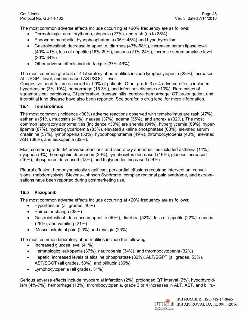
Confidential Protocol No. GU-14-102
Page 48 Ver. 3, dated 7/14/2016
IRB NUMBER: HSC-MS-14-0665
IRB APPROVAL DATE: 08/11/2016
The most common adverse effects include occurring at >20% frequency are as follows:
Dermatologic: acral erythema, alopecia (27%), and rash (up to 35%)
Endocrine metabolic: hypophosphatemia (35%-45%) and hypothyroidism
Gastrointestinal: decrease in appetite, diarrhea (43%-68%), increased serum lipase level
(40%-41%), loss of appetite (16%-29%), nausea (21%-24%), increase serum amylase level
(30%-34%)
Other adverse effects include fatigue (37%-46%) The most common grade 3 or 4 laboratory abnormalities include lymphocytopenia (23%), increased ALT/SGPT level, and increased AST/SGOT level. Congestive heart failure occurred in 1.9% of patients. Other grade 3 or 4 adverse effects included hypertension (3%-10%), hemorrhage (15.3%), and infectious disease (>10%). Rare cases of squamous cell carcinoma, GI perforation, transaminitis, cerebral hemorrhage, QT prolongation, and interstitial lung disease have also been reported. See sorafenib drug label for more information.
16.4 Temsirolimus
The most common (incidence ≥30%) adverse reactions observed with temsirolimus are rash (47%), asthenia (51%), mucositis (41%), nausea (37%), edema (35%), and anorexia (32%). The most common laboratory abnormalities (incidence ≥30%) are anemia (94%), hyperglycemia (89%), hyper- lipemia (87%), hypertriglyceridemia (83%), elevated alkaline phosphatase (68%), elevated serum creatinine (57%), lymphopenia (53%), hypophosphatemia (49%), thrombocytopenia (40%), elevated AST (38%), and leukopenia (32%).
Most common grade 3/4 adverse reactions and laboratory abnormalities included asthenia (11%), dyspnea (9%), hemoglobin decreased (20%), lymphocytes decreased (16%), glucose increased (16%), phosphorus decreased (18%), and triglycerides increased (44%).
Pleural effusion, hemodynamically significant pericardial effusions requiring intervention, convul- sions, rhabdomyolysis, Stevens-Johnson Syndrome, complex regional pain syndrome, and extrava- sations have been reported during postmarketing use.
16.5 Pazopanib
The most common adverse effects include occurring at >20% frequency are as follows:
Hypertension (all grades, 40%)
Hair color change (38%)
Gastrointestinal: decrease in appetite (40%), diarrhea (52%), loss of appetite (22%), nausea
(26%), and vomiting (21%)
Musculoskeletal pain (23%) and myalgia (23%) The most common laboratory abnormalities include the following:
Increased glucose level (41%)
Hematologic: leukopenia (37%), neutropenia (34%), and thrombocytopenia (32%)
Hepatic: increased levels of alkaline phosphatase (32%), ALT/SGPT (all grades, 53%),
AST/SGOT (all grades, 53%), and bilirubin (36%)
Lymphocytopenia (all grades, 31%)
Serious adverse effects include myocardial infarction (2%), prolonged QT interval (2%), hypothyroid- ism (4%-7%), hemorrhage (13%), thrombocytopenia, grade 3 or 4 increases in ALT, AST, and biliru-
Confidential Protocol No. GU-14-102
Page 49 Ver. 3, dated 7/14/2016
IRB NUMBER: HSC-MS-14-0665
IRB APPROVAL DATE: 08/11/2016
bin levels, infectious disease, lymphocytopenia, cerebrovascular accident, transient ischemic attack, pneumothorax, and pulmonary embolism. See pazopanib drug label for more information.
16.6 Axitinib
The most common adverse effects include occurring at >20% frequency are as follows:
Hypertension (all grades, 40%)
Hand-foot syndrome (27%)
Decreased weight (all grades, 25%)
Gastrointestinal: constipation (all grades, 20%), diarrhea (all grades, 55%), loss of appetite
(all grades, 34%), nausea (all grades, 32%), vomiting (all grades, 24%)
Asthenia (all grades, 21%)
Other: dysphonia (31%) and fatigue (all grades, 39%) The most common laboratory abnormalities include the following:
Hepatic: increased ALT/SGPT level (all grades, 22%; grade 3, <1%) Serious adverse effects include hypertensive crisis (<1%), GI fistula (1%), arterial thrombosis (1%- 2%), DVT (1%), hemorrhage (grade 3 or 4, 1%), venous thrombosis (grade 3 or 4, 3%), transient ischemic attack (1%), and pulmonary embolism (2%).
16.7 Cabozatinib
The most commonly reported adverse drug reactions (≥25%) are diarrhea, stomatitis, palmar-plantar erythrodysesthesia syndrome (PPES), decreased weight, decreased appetite, nausea, fatigue, oral pain, hair color changes, dysgeusia, hypertension, abdominal pain, and constipation.
The most common laboratory abnormalities (≥25%) are increased AST, increased ALT, lymphope- nia, increased alkaline phosphatase, hypocalcemia, neutropenia, thrombocytopenia, hypophospha- temia, and hyperbilirubinemia.
Serious side effects include hand-foot syndrome (42%-50%), impaired wound healing (2%), gas- trointestinal fistula (1%-1.2%), gastrointestinal perforation (0.9%-3%), pancreatitis (<1%), Tracho- esophageal fistula (4%), anemia, Grade 3 or 4 (5%), arterial thromboembolism (0.9%-2%), bleeding, Grade 3 or higher (2.1%-3%), lymphocytopenia, Grade 3 or 4 (16%), neutropenia, Grade 3 or 4 (3%), venous thromboembolism (6%-7.3%), cholestatic hepatitis (<1%), aseptic necrosis of bone of jaw (≤1%),posterior reversible encephalopathy syndrome (<1%), seizure (<1%), and pulmonary embolism (3.9%).
16.8 How Supplied
16.8.1 Everolimus
Everolimus is supplied as white to slightly yellow, elongated tablets with a beveled edge and no score of 2.5 mg, 5 mg, 7.5 mg, and 10 mg strengths. The 2.5 mg tab is engraved with ―LCL‖ on one side and ―NVR‖ on the other. The 5 mg tablet is engraved with ―5‖ on one side and ―NVR‖. The 7.5 mg tablet is engraved with ―7P5‖ on one side and ―NVR‖ on the other. The 10 mg tab is engraved with ―UHE‖ on one side and ―NVR‖ on the other. Everolimus will be supplied according to local prac- tice.
16.8.2 Sunitinib
Sunitinib is supplied as hard gelatin capsules in either 12.5 mg, 25 mg, or 50 mg strength. The 12.5 mg and 25 mg capsules consist of a caramel-colored top orange body, printed with white ink "Pfizer" on the cap and either "STN 12.5 MG" or "STN25 MG" on the body. The 50 mg capsule consists of a
Confidential Protocol No. GU-14-102
Page 50 Ver. 3, dated 7/14/2016
IRB NUMBER: HSC-MS-14-0665
IRB APPROVAL DATE: 08/11/2016
caramel top and caramel body, printed with white ink "Pfizer" on the cap and "STN50 MG" on the body. Sunitinib will be supplied according to local practice.
16.8.3 Sorafenib
Sorafenib is supplied as tablets containing sorafenib tosylate (274 mg) equivalent to 200 mg of sora- fenib. The tablet is round, biconvex, red film-coated tablets, debossed with the ―Bayer cross‖ on one side and ―200‖ on the other side. Sunitinib will be supplied according to local practice.
16.8.4 Temsirolimus
Temsirolimus is supplied as a 25 mg/mL injection with a 1.8 mL diluent designed for injection. The recommended initial dose of the drug is 25 mg infused over a 30-60 minute period once a week. Treat until disease progression or unacceptable toxicity.Temsirolimus injection vial contents must first be diluted with the enclosed diluent before diluting the resultant solution with 250 mL of 0.9% sodium chloride injection. Patients should receive prophylactic intravenous diphenhydramine 25 to 50 mg (or similar antihistamine) approximately 30 minutes before the start of each dose. Temsiroli- mus will be supplied according to local practice.
16.8.5 Pazopanib
Pazopanib is supplied as tablet containing 216.7 mg of pazopanib hydrochloride equivalent to 200 mg of pazopanib. The tablet 200 mg tablet is modified capsule-shaped, gray, film-coated with ―GS JT‖ debossed on one side. Pazopanib will be supplied according to local practice.
16.8.6 Axitinib
Axitinib is supplied as red, film-coated 1 mg and 5 mg tablets. The 1 mg tablet is oval-shaped and debossed with ―Pfizer‖ on one side and ―1 XNB‖ on the other side. The 5 mg tablet is triangular- shaped, debossed with ―Pfizer‖ on one side and ―5 XNB‖ on the other side. Axitinib will be supplied according to local practice.
16.8.7 Cabozatinib
Cabozantinib tablets are supplied as 60mg, 40mg, and 20mg yellow, film-coated tablets. The 60-mg tablets are oval shaped with no score, debossed with ―XL‖ on one side and ―60‖ on the other side of the tablet. The 40 mg tablets are triangle shaped with no score, debossed with ―XL‖ on one side and ―40‖ on the other side of the tablet. The 20 mg tablets are round shaped with no score, de- bossed with ―XL‖ on one side and ―20‖ on the other side of the tablet. Cabozatinib is supplied ac- cording to local practice.
16.9 Storage
16.9.1 Everolimus
Everolimus tablets should be stored at 25°C (77°F); excursions permitted between 15°C–30°C (59°F–86°F). Tablets should be stored in the original container, protected from light and moisture. Follow special handling and disposal procedures for anticancer pharmaceuticals.
16.9.2 Sunitinib
Sunitinib may be light-sensitive; capsules will be stored in the bottles in which they are received to protect the compound from light. Sunitinib capsules should be stored at 25°C (77°F); excursions permitted to 15°C-30°C (59°F-86°F).
16.9.3 Sorafenib
Confidential Protocol No. GU-14-102
Page 51 Ver. 3, dated 7/14/2016
IRB NUMBER: HSC-MS-14-0665
IRB APPROVAL DATE: 08/11/2016
Sorafenib tablets will be stored in a dry place at 25°C (77°F); excursions permitted to 15°C–30°C (59°F–86°F).
16.9.4 Temsirolimus
Temsirolimus must be stored under refrigeration at 2°–8°C (36°–46°F) and protected from light. Dur- ing handling and preparation of admixtures, temsirolimus should be protected from excessive room light and sunlight. Parenteral drug products should be inspected visually for particulate matter and discoloration prior to administration, whenever solution and container permit.
16.9.5 Pazopanib
Pazopanib tablets will be stored at room temperature between 20°C and 25°C (68°F to 77°F); ex- cursions permitted to 15°C-30°C (59°F-86°F).
16.9.6 Axitinib
Axitinib tablets will be stored at 20°C-25°C (68°F-77°F); excursions permitted to 15°C-30°C (59°F- 86°F).
16.9.7 Cabozatinib
Cabozatinib is stored at 20°C to 25°C (68°F to 77°F); excursions are permitted from 15°C to 30°C (59°F to 86°F).
16.10 Monitoring of Toxicities
Any AE/SAE not listed in the CTCAE version 4.0 will be graded as follows:
Severity of Event
Grade Definition
1 Mild: Symptoms which do not interfere with patient’s daily activities
2 Moderate: Symptoms which may interfere with patient’s daily activities
3 Severe: Events which interrupt patient’s usual daily activities
4 Life-threatening or disabling
5 Death
Medically significant adverse events considered related to the study treatment will be followed until resolved or considered stable. It will be left to the investigator’s clinical judgment to determine whether an adverse event is related and of sufficient severity to require the subject’s removal from treatment or from the study. A subject may also voluntarily withdraw from treatment due to what he or she perceives as an intolerable ad- verse event. If either of these situations arises, the subject should be strongly encouraged to under- go medical supervision until symptoms cease or the condition becomes stable.
16.10.1 Management of stomatitis/oral mucositis/mouth ulcers
Stomatitis/oral mucositis/mouth ulcers due to everolimus should be treated using local supportive care. Please note that investigators in earlier trials have described the oral toxicities associated with the everolimus as mouth ulcers, rather than mucositis or stomatitis. If your examination reveals mouth ulcers rather than a more general inflammation of the mouth, please classified the adverse event as such. Please follow the paradigm below for treatment of stomatitis/oral mucositis/mouth ulcers:
1. For mild toxicity (grade 1), use conservative measures such as non-alcoholic mouthwash or
salt water (0.9%) mouthwash several times a day until resolution.
2. For more severe toxicity (grade 2 in which case patients have pain but are able to maintain
adequate oral alimentation, or grade 3 in which case patients cannot maintain adequate oral
alimentation), the suggested treatments are topical analgesic mouth treatments (i.e., local
Confidential Protocol No. GU-14-102
Page 52 Ver. 3, dated 7/14/2016
IRB NUMBER: HSC-MS-14-0665
IRB APPROVAL DATE: 08/11/2016
anesthetics such as benzocaine, butyl amino benzoate, tetracaine hydrochloride, menthol, or
phenol) with or without topical corticosteroids, such as triamcinolone oral paste 0.1% (Kena-
log in orabase).
3. Agents containing hydrogen peroxide, iodine, and thyme derivatives may tend to worsen
mouth ulcers. It is preferable to avoid these agents.
4. Antifungal agents should be avoided unless a fungal infection is diagnosed. In particular, sys-
temic imidazole antifungal agents (ketoconazole, fluconazole, itraconazole, etc.) should be
avoided in all patients due to their strong inhibition of the everolimus metabolism, thereby
leading to higher everolimus exposures. Therefore, topical antifungal agents are preferred if
an infection is diagnosed. Similarly, antiviral agents such as acyclovir should be avoided un-
less a viral infection is diagnosed.
Note: stomatitis/oral mucositis should be appropriately graded using the functional grading given on the NCI-CTC for adverse events, version 4.0.
16.10.2 Management of nausea/vomiting
Investigators should manage anemia as medically indicated per local standards.
16.10.3 Management of anemia
Investigators should manage anemia as medically indicated per local standards.
16.10.4 Management of diarrhea
Appearance of diarrhea attributed to therapy-related toxicity may be treated with loperamide or per local standards.
16.10.5 Management of Hypertension
Severity Action
A: symptomatic sustained SBP ≥160 and <170
mmHg, or DBP ≥90 and <110 mmHg, or clini-
cally significant decrease in tech DBP of ≥20
mg of mercury.
Step 1. Continue steady medication at the
same dose. Step 2. Adjust current dose of or
initiate new antihypertensive medication(s).
Step 3. Titrate antihypertensive medications
during next 2 weeks as indicated to achieve
well-controlled blood pressure. If blood pres-
sure is not well-controlled within 2 weeks, fol-
low step 1 in scenario B.
B: Symptomatic, or SBP ≥170 mg of mercury
or DBP ≥110 mg of mercury, or failure to
achieve well-controlled blood pressure within 2
weeks in scenario A.
Step 1. Interrupt study medication. Step 2. Ad-
just current or initiate new antihypertensive
medications. Step 3. Titrate antihypertensive
medications during next 2 weeks as indicated
to achieve well-controlled BP. Step 4. Restart
study medication at the same dose or lower
dose at discretion of investigator once BP is
well-controlled. Dose adjustment for discontin-
uation of antihypertensive medications may be
necessary during scheduled to week off-
treatment..

Confidential Protocol No. GU-14-102
Page 53 Ver. 3, dated 7/14/2016
IRB NUMBER: HSC-MS-14-0665
IRB APPROVAL DATE: 08/11/2016
C: Two or more symptomatic episodes of
hypertension despite modification of antihyper-
tensive medications and production of study
medication dose.
Discontinuation of study medication and fol-
low-up per protocol.
Well-controlled blood pressure defined as meaning SBP <160 mmHg and meaning DBP <90 mmHg.
16.10.6 Management of Fatigue (lethargy, malaise, asthenia)
Severity Action
Grades 1 and 2 Continue study medication at same dose;
monitor as clinically indicated.
Grades 3 and 4
Step 1. Interrupt study medication until toxicity
reduced to ≤grade 2. Step 2. Restart study
medication at the same dose or lower dose at
discretion of investigator.
16.10.7 Management of infections
Tyrosine kinase and mTOR inhibitors have immunosuppressive properties and may predispose pa- tients to infections, especially infections with opportunistic pathogens. Localized and systemic infec- tions, including pneumonia, other bacterial infections and invasive fungal infections, such as asper- gillosis or candidiasis, have been reported. Some of these infections have been severe (e.g., leading to respiratory failure) and occasionally have had a fatal outcome. Investigator will monitor close for symptoms and signs of infection, and institute treatment promptly. If a diagnosis of invasive systemic fungal infection is made, therapy will be stopped and permanently discontinued and the patient treated with appropriate antifungal therapy.
16.10.8 Management of noninfectious pneumonitis
Both asymptomatic radiological changes (grade 1 = radiological long changes only) and symptomat- ic noninfectious pneumonitis (grade 2 = not interfering with activities of daily living; grade 3 = inter- fering with activities of daily living and oxygen indicated) have been noted in patients receiving eve- rolimus and sorafenib therapy. If noninfectious pneumonitis develops, consultation with a pulmonol- ogist is recommended. Record the non-infectious pneumonitis on the adverse event eCRF. If inves- tigator finds that noninfectious pneumonitis associated with therapy, dose modification will be made and corticosteroid initiated as appropriate per local standards.
16.10.9 Management of thyroid dysfunction
Baseline laboratory measurement of thyroid function will be obtained prior to therapy initiation. Pa- tients will be observed closely for signs and symptoms of thyroid dysfunction. Routine thyroid func- tion test will be conducted at each appointment. Treatment will follow standard medical practice.
16.10.10 Hemorrhage/bleeding/coagulopathy
Severity Action
Grade 1 Continue study medication at same dose;
monitor as clinically indicated.
Grade 2
Step 1. Interrupt study medication until the ad-
verse event resolves to ≤Grade 1. Step 2.
Restart medication with lower dose; monitor as

Confidential Protocol No. GU-14-102
Page 54 Ver. 3, dated 7/14/2016
IRB NUMBER: HSC-MS-14-0665
IRB APPROVAL DATE: 08/11/2016
clinically indicated.
Grade 3 or 4, or recurrent ≥Grade 2 event after
dose interruptions/reduction
Discontinuation of study medication and fol-
low-up per protocol. Note: if abnormality is not
clearly associated with clinical consequences,
contact the Novartis medical monitor to dis-
cuss the potential for continuation of study
treatment. If agreed, subject may restart medi-
cation at lower dose.
16.10.11 Venous thrombosis
Severity Action
Grade 2 Continue study medication with same dose;
monitor as clinically indicated.
Grade 3 or asymptomatic Grade 4
Step 1. Interrupt study medication at same
goes during the period of full-dose anticoagu-
lant and if all of the following criteria are met:
the subject must have been treated with an
anticoagulant (enoxaparin preferred, but war-
farin may be used) for at least 1 week. No
Grade 3 or 4 hemorrhagic events have oc-
curred while on anticoagulant should treat-
ment. Subject should be monitored as clinical-
ly indicated during anticoagulant should treat-
ment and after resuming study medication.
Symptomatic Grade 4
Discontinuation of study medication and fol-
low-up per protocol.
16.10.12 Arterial thrombosis
Severity Action
Any Grade Discontinuation of study medication and fol-
low-up per protocol
16.11 Management of Everolimus-specific toxicities
Management of hyperlipidemia and hyperglycemia
Treatment of hyperlipidemia should take into account the pre-treatment status and dietary habits. Lead tests to monitor hyperlipidemia must be taken in the fasting state. Grade 2 hypercholesterole- mia (>300 milligrams per deciliter or 7.75 mmol/L) or grade 2 hypertriglyceridemia (greater than 2.5 times ULN) will be treated with a 3-hydroxy-3-methyl-glutaryl (HMG)-CoA reductase inhibitor (e.g.,
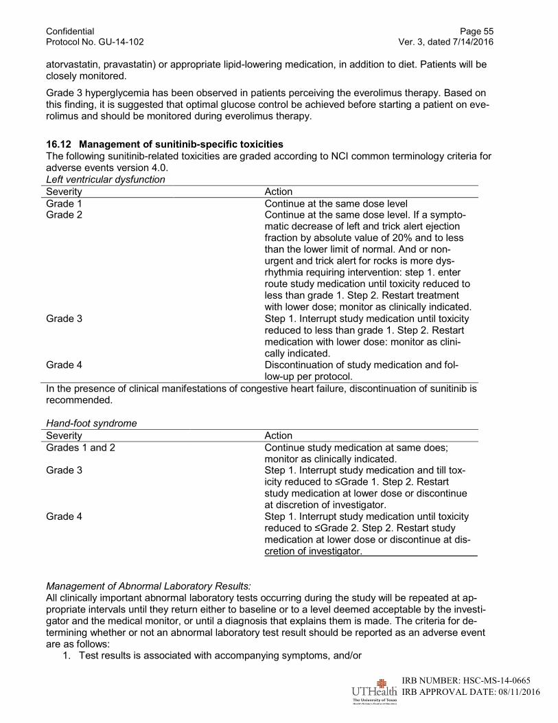
Confidential Protocol No. GU-14-102
Page 55 Ver. 3, dated 7/14/2016
IRB NUMBER: HSC-MS-14-0665
IRB APPROVAL DATE: 08/11/2016
atorvastatin, pravastatin) or appropriate lipid-lowering medication, in addition to diet. Patients will be closely monitored.
Grade 3 hyperglycemia has been observed in patients perceiving the everolimus therapy. Based on this finding, it is suggested that optimal glucose control be achieved before starting a patient on eve- rolimus and should be monitored during everolimus therapy.
16.12 Management of sunitinib-specific toxicities
The following sunitinib-related toxicities are graded according to NCI common terminology criteria for adverse events version 4.0. Left ventricular dysfunction
Severity Action
Grade 1 Continue at the same dose level Grade 2 Continue at the same dose level. If a sympto-
matic decrease of left and trick alert ejection fraction by absolute value of 20% and to less than the lower limit of normal. And or non- urgent and trick alert for rocks is more dys- rhythmia requiring intervention: step 1. enter route study medication until toxicity reduced to less than grade 1. Step 2. Restart treatment with lower dose; monitor as clinically indicated.
Grade 3 Step 1. Interrupt study medication until toxicity reduced to less than grade 1. Step 2. Restart medication with lower dose: monitor as clini- cally indicated.
Grade 4 Discontinuation of study medication and fol- low-up per protocol.
In the presence of clinical manifestations of congestive heart failure, discontinuation of sunitinib is recommended.
Hand-foot syndrome
Severity Action
Grades 1 and 2 Continue study medication at same does; monitor as clinically indicated.
Grade 3 Step 1. Interrupt study medication and till tox- icity reduced to ≤Grade 1. Step 2. Restart study medication at lower dose or discontinue at discretion of investigator.
Grade 4 Step 1. Interrupt study medication until toxicity reduced to ≤Grade 2. Step 2. Restart study medication at lower dose or discontinue at dis- cretion of investigator.
Management of Abnormal Laboratory Results: All clinically important abnormal laboratory tests occurring during the study will be repeated at ap- propriate intervals until they return either to baseline or to a level deemed acceptable by the investi- gator and the medical monitor, or until a diagnosis that explains them is made. The criteria for de- termining whether or not an abnormal laboratory test result should be reported as an adverse event are as follows:
1. Test results is associated with accompanying symptoms, and/or
Confidential Protocol No. GU-14-102
Page 56 Ver. 3, dated 7/14/2016
IRB NUMBER: HSC-MS-14-0665
IRB APPROVAL DATE: 08/11/2016
2. Test result requires additional diagnostic testing or medical/surgical intervention (merely re-
peating an abnormal test, in the absence of any of the above conditions, does not meet crite-
ria for reporting and an AE), and/or
3. Test result leads to a change in study dosing or discontinuation from the study, significant
additional concomitant drug treatment or other therapy, and/or
4. Test result leads to any of the outcomes included in the definition of a serious adverse event,
and/or
5. Test result is considered to be an adverse event by the investigator or sponsor Any abnormal test result that is determined to be an error does not require reporting as an adverse event, even if it did meet one of the above conditions except for condition #4. Clinically significant laboratory results must be recorded in the patient’s CRF.
16.13 Follow-up for toxicities
Patients whose study medication is temporarily interrupted or discontinued due to an adverse event or abnormal laboratory value, should be followed at least once per week for 4 weeks, and subse- quently at 2-week intervals, until resolution, stabilization of the event, or until the initiation of another anticancer therapy (second-line or initiation of a post-study third-line anticancer therapy), whichever comes first.
If the patient requires a dose delay of >21 days for everolimus due to an adverse event or abnormal laboratory value, from the intended day of the next scheduled dose, the patient should be discontin- ued from the specific line of treatment (first-line and/or second-line).
If the patient requires a dose delay of >21 days for sunitinib or if the delay coincides with the 2-week arrest and is >28 days (14 days of rest and 14 additional), the patient should be discontinued from the specific line of treatment (first-line and/or second-line).
For unexpected events that are not related to study medication toxicity (i.e., elective surgeries, per- sonal reasons), up to a maximum of 14 days is acceptable for a dose delay from therapy.
16.14 Dose interruption for surgery or surgical procedures
Temporary interruption of therapy is recommended in patients undergoing major surgical proce- dures. The decision to resume therapy following major surgical intervention should be based on clin- ical judgment of adequate wound healing.
16.15 Other concomitant medications
Patients must be instructed not to take any additional medications (over the counter or other prod- ucts, including nutritional supplements) during the study without prior consultation with approval from the investigator. All medications taken within 35 days of starting study treatment must be reported on the concomitant medication/significations non-drug therapy prior to start of study drug eCRF. The investigator or designee will continue collecting information on the initiation of additional anticancer therapies until the data cutoff date for the final analysis. All new anticancer therapies after the last dose of study treatment will be recorded in the appropriate eCRF.
The investigator should instruct the patient to notify the study center about any new medications he or she takes after the start of the study treatment. All medications (other than study treatment) and significant non-drug therapies (including physical therapy and blood transfusions) administered after the patient starts treatment in the first-line and during the second-line. Must be listed on the conco- mitant dedications/significant non-drug hairpiece after start of study drug eCRF.

Confidential Protocol No. GU-14-102
Page 57 Ver. 3, dated 7/14/2016
IRB NUMBER: HSC-MS-14-0665
IRB APPROVAL DATE: 08/11/2016
Investigational or commercial anticancer agents other than those included in this trial, are not al- lowed during the study. The initiation of any non-protocol specific anti-tumor treatment or surgery is considered an indication of disease progression and must be recorded appropriately. Patients would then only be followed for survival.
Everolimus, sunitinib, pazopanib, axitinib, and cabozatinib are metabolized primarily by the cytoch- rome P450 isoenzyme CYP3A4, to produce its primary active metabolite. Drugs or substances known to be strong inhibitors, strong inducers, or substrates of the isoenzyme CYP3A4 should be avoided unless use of the drug is essential and no substitute is available. For a complete list of drugs which are substrates for CYP3A4, please refer to this website: http://medicine.iupui.edu/flockhart/clinlist.htm.
A strong inhibitor is one that causes a >5-fold increase in the plasma AUC values or >80% decrease in clearance. A moderate inhibitor is one that causes a >2-fold increase in plasma AUC values or 50%-80% decrease in clearance. A weak inhibitor is one that causes a >1.25-fold increase in plas- ma AUC values or 20%-50% decrease in clearance. (Distinction is not always categorical as interac- tion can vary according to conditions.)
16.15.1 Everolimus
Strong inhibitors should not be co-administered with everolimus. Drugs or substances known to be inhibitors, inducers, or substrates of the isoenzyme CYP3A4 should be avoided unless use of the drug is essential and no substitute is available.
The following concomitant treatment are not allowed during the study:
Co-administration with strong CYP3A4 inhibitors (e.g., ketoconazole, itraconazole ritonavir),
and strong inducers (e.g., rifampin, rifabutin).
Seville orange, grapefruit, and their juices affect cytochrome P450 and PgP activity.
Co-administration with moderate CYP3A4 inhibitors (e.g., erythromycin, fluconazole, calcium chan- nel blockers, benzodiazepines) and strong inducers (e.g., carbamazepine, Phenobarbital, and phe- nytoin) should also be avoided if possible or otherwise subject to caution (e.g., increased frequency of safety monitoring) and dose adjustment.
If patients have to co-administer strong inducers of CYP3A4, start the dose at 10 mg daily and if well tolerated for the first 2 weeks of treatment, escalate as tolerated by 5 mg dose every 2 weeks to a maximum dose of 20 mg daily. If patient does not tolerate higher doses, reduction to highest tole- rated dose is recommended. If the strong inducer is discontinued, the everolimus dose should be returned to the dose used prior to initiation of the strong CYP3A4 inducer.
If patients require co-administration of a moderate CYP3A4 or PgP inhibitor, the everolimus dose should be reduced to 5 mg daily. Further dose reduction to 5 mg every other day may be required to manage adverse reactions.
16.15.2 Sunitinib
Strong inhibitors of CYP3A4 may increase sunitinib plasma concentrations and should be avoided unless there is an overriding clinical need. In particular, ketoconazole should be avoided if possible, since a clinical interaction study of sunitinib indicated that up to a 2-fold increase in plasma levels of sunitinib was induced by ketoconazole. Selection of an alternate concomitant medication with no or minimal enzyme inhibition potential is recommended. A dose reduction to a minimum of 37.5 mg dai- ly should be considered if sunitinib must be co-administered with a strong CYP3A4 inhibitor.
Inducers of CYP3A4 may decrease sunitinib plasma concentrations and should be avoided. For ex- ample, St. John’s wort may decrease the plasma concentrations unpredictably and should not be taken concomitantly. Selection of an alternate concomitant medication with no or minimal enzyme induction potential is recommended.
Confidential Protocol No. GU-14-102
Page 58 Ver. 3, dated 7/14/2016
IRB NUMBER: HSC-MS-14-0665
IRB APPROVAL DATE: 08/11/2016
In addition, concomitant treatment with the following drugs with dysrhythimic potential (i.e., terfena- dine, quinidine, procainamide, disopyramide, sotalol, probucol, bepridil, haloperidol, risperidone, and indapamide) is not recommended.
16.15.3 Sorafenib
Avoid rifampin as it is a strong CYP3A4 inducer. Co-administration with sorafenib resulted in 37% decrease in the mean AUC of sorafenib. Avoid concomitant use of strong CYP3A4 inducers (such as, carbamazepine, dexamethasone, phenobarbital, phenytoin, rifampin, rifabutin, St. John’s wort), when possible, because these drugs can decrease the systemic exposure to sorafenib. Strong inhi- bitors of CYP3A4 and P-glycoprotein, did not alter the mean AUC of sorafenib.
The aqueous solubility of sorafenib is pH dependent, with higher pH resulting in lower solubility. However, omeprazole, a proton pump inhibitor, administered at a dose of 40 mg once daily for 5 days, did not result in a clinically meaningful change in sorafenib single dose exposure. No dose ad- justment for sorafenib is necessary.
16.15.4 Temsirolimus
Strong inducers of CYP3A4/5 (eg, dexamethasone, rifampin) and strong inhibitors of CYP3A4 (eg, ketoconazole, atazanavir) may decrease and increase concentrations of the major metabolite of temsirolimus, respectively. If alternatives cannot be used, dose modifications of temsirolimus are recommended.
Avoid St. John's Wort which may decrease temsirolimus plasma concentrations, and grapefruit juice which may increase plasma concentrations of the major metabolite of temsirolimus.
The combination of temsirolimus and sunitinib resulted in dose-limiting toxicity (grade 3/4 erythe- matous maculopapular rash, and gout/cellulitis requiring hospitalization).
16.15.5 Pazopanib
In vitro studies suggested that the oxidative metabolism of pazopanib in human liver microsomes is mediated primarily by CYP3A4, with minor contributions from CYP1A2 and CYP2C8. So, co- administration with strong inhibitors of CYP3A4 (e.g., ketoconazole, ritonavir, clarithromycin) in- creases pazopanib concentrations and should be avoided. Alternate therapy that does not inhibit CYP3A4 should be considered. If co-administration of a strong CYP3A4 inhibitor is warranted, re- duce pazopanib dose to 400 mg.
Grapefruit or grapefruit juice should be avoided as it inhibits CYP3A4 activity and may also increase plasma concentrations of pazopanib.
CYP3A4 inducers CYP3A4 inducers such as rifampin may decrease plasma pazopanib concentra- tions. Consider an alternate concomitant medication with no or minimal enzyme induction potential. Pazopanib should not be used if chronic use of strong CYP3A4 inducers cannot be avoided. The concomitant use of strong CYP3A4 inducers (e.g., rifampin) may decrease pazopanib concentra- tions and should be avoided. Consider an alternate concomitant medication with no or minimal en- zyme induction potential. Pazopanib should not be used in patients who cannot avoid chronic use of strong CYP3A4 inducers.
Concomitant administration of pazopanib with esomeprazole, a proton pump inhibitor (PPI), de- creased the exposure of pazopanib by approximately 40% (AUC and Cmax). As a result, concomi- tant use of pazopanib with drugs that raise gastric pH should be avoided. If such drugs are needed, short-acting antacids should be considered in place of PPIs and H2-receptor antagonists. Separate antacid and pazopanib dosing by several hours to avoid a reduction in pazopanib exposure.

Confidential Protocol No. GU-14-102
Page 59 Ver. 3, dated 7/14/2016
IRB NUMBER: HSC-MS-14-0665
IRB APPROVAL DATE: 08/11/2016
16.15.6 Axitinib
Concomitant administration of ketoconazole, a strong inhibitor of CYP3A4/5, increased the plasma AUC. Co-administration of axitinib with strong CYP3A4/5 inhibitors should be avoided. Grapefruit or grapefruit juice may also increase axitinib plasma concentrations and should be avoided. Selection of concomitant medication with no or minimal CYP3A4/5 inhibition potential is recommended. If a strong CYP3A4/5 inhibitor must be co-administered, the dose should be reduced to half the normal dose as this dose reduction is predicted to adjust the axitinib area under the plasma concentration vs time curve (AUC) to the range observed without inhibitors. The subsequent doses can be in- creased or decreased based on individual safety and tolerability.
Co-administration of rifampin, a strong inducer of CYP3A4/5, reduced the plasma exposure of axiti- nib Co-administration of axitinib with strong CYP3A4/5 inducers (e.g., rifampin, dexamethasone, phenytoin, carbamazepine, rifabutin, rifapentin, phenobarbital, and St. John’s wort) should be avoided. Selection of concomitant medication with no or minimal CYP3A4/5 induction potential is recommended. Moderate CYP3A4/5 inducers (e.g., bosentan, efavirenz, etravirine, modafinil, and nafcillin) may also reduce the plasma exposure of axitinib and should be avoided if possible.
16.15.7 Cabozatinib Concomitant use of a strong CYP3A4 inhibitors such as grapefruit juice, indinavir, itraconazole, or ketoconazole should be avoided. Ketoconazole (400 mg daily for 27 days) increased single-dose plasma cabozantinib exposure (AUC0-inf) by 38%. If concomitant administration cannot be avoided, reduce cabozatinib dose by 40mg daily. Resume dose that was used prior to initiating the CYP3A4 inhibitor 2 to 3 days after discontinuation of inhibitor.
Concomitant use of a strong CYP3A4 inducer such as rifampin, phenytoin, carbamazepine, pheno- barbital, rifabutin, rifapentine, and St. John’s Wort should be avoided. Rifampin (600 mg daily for 31 days) decreased single-dose plasma cabozantinib exposure (AUC0-inf) by 77%. If concomitant ad- ministration cannot be avoided, increase cabozantinib dose by 40mg daily as tolerated with a maxi- mum dose of 180mg/day. Resume the dose that was used prior to initiating the CYP3A4 inducer 2- 3 days after discontinuation of inducer.
Concomitant use with gastric pH modifying agents such as proton pump inhibitor (PPI) esomepra- zole (40 mg daily for 6 days) did not have a clinically-significant effect on plasma cabozantinib expo- sure (AUC).
16.16 STUDY MEDICATION COMPLIANCE
Compliance will be assessed by the investigator or designee at each visit using pill counts for both everolimus and sunitinib. This information should be captured in the source document at each visit.
Patients will be requested to bring their unused medication, including empty packaging, to
the clinic at each visit.
All doses taken by the patient and all dose changes during the study must be recorded on
the Dosage Administration Record eCRF.
The investigator or designee must keep documentation (overall drug accountability log for
the study as well as individual study drug accountability records for each patient). of tablets
administered, tablets used, dose changes, dates dispensed, and intervals between visits.
Drug accountability will be monitored by the field monitor during site visits and at the comple-
tion of the study.

Confidential Protocol No. GU-14-102
Page 60 Ver. 3, dated 7/14/2016
IRB NUMBER: HSC-MS-14-0665
IRB APPROVAL DATE: 08/11/2016
16.17 References 1. Faivre S, Delbaldo C, Vera K, Robert C, Lozahic S, Lassau N, et al. Safety, pharmacokinetic, and antitumor activity of SU11248, a novel oral multitarget tyrosine kinase inhibitor, in patients with can- cer. J Clin Oncol 2006;24(1):25-35. 2. Robert C, Soria JC, Spatz A, Le Cesne A, Malka D, Pautier P, et al. Cutaneous side-effects of ki- nase inhibitors and blocking antibodies. Lancet Oncol 2005;6(7):491-500. 3. Mendel DB, Laird AD, Xin X, Li G, Schreck R, Carver J, et al. Development of a preclinical phar- macokinetic/pharmacodynamic relationship for the angiogenesis inhibitor SU11248, a selective inhi- bitor of VEGF and PDGF receptor tyrosine kinases in clinical development. Proc Am Soc Clin Oncol 2002;21:94. 4. Everolimus (Afinitor) prescribing information; revised February 2014 5. Axitinib (Inlyta) prescribing information; issued September 2013 6. Sunitinib (Sutent) prescribing information 7. Pazopanib (Votrient) prescribing information 8. Sorafenib (Nexavar) prescribing information 9. Cabozantinib (Cabometyx) prescribing information; issued April 2016 10. Micromedex 2.0; Accessed 1 June 2014
Confidential Protocol No. GU-14-102
Page 61 Ver. 3, dated 7/14/2016
IRB NUMBER: HSC-MS-14-0665
IRB APPROVAL DATE: 08/11/2016
17 APPENDIX F: OBTAINING BIOPSY TISSUE FOR MOLECULAR DIAGNOSITCS Biopsy tissue samples are processed by flash freezing in liquid nitrogen (LN2) and embedding the sample in OCT medium for subsequent frozen tissue sectioning. Embedding in OCT embedding compound followed by flash freezing at -80°C not only preserves DNA, RNA, and protein integrity, but also enables sectioning of the frozen tissue. The advantage of this method is that the frozen OCT containing the specimen can be placed into a cryo-microtome chuck and sectioned. Frozen sections can be mounted on microscope slides and stained to characterize the tissue sample, de- termine the ratio of tumor to healthy tissure, and, if need be, microdissected to isolate specific cell populations. Expediting the processing is essential in order to ensure tissue integrity, which is the primary goal of the fresh/frozen tissue study. Tissue processing should be done immediately by a trained technician after biopsy is collected.
Procedures to be followed:
1. The biopsy sample will be obtained by the interventional radiology and handed to a research
coordinator.
2. The OCT compound is poured into a small plastic tray, the tissue sample is submerged in the
compound, and the tray is placed on dry ice for freezing. After hardening of the OCT, the Di-
vision of Oncology research coordinator will, if possible, transport OCT-embedded tissue
immediately to CompanionDx for molecular analysis. If the tissue needs to be held at
UTHealth for any reason (e.g., off-hours), the tissue will be held at -20°C no longer than
overnight and transported on dry ice to the Division of Oncology’s -80°C freezer at IMM the
next day and delivered to CompanionDx at the earliest opportunity.
3. Samples will be de-identified and given a barcode for tracking.
4. At CompanionDx, OCT-embedded tissues will be sectioned using the following prodecure:
a) Clean work area and cryostat before beginning, wearing clean, sterile gloves.
b) Clear all tissue debris from cyrostat with pressurized air and a vacuum and then wipe
inside surfaces with 100% ethanol.
c) Clean all instruments (forceps, brushes, pencils, etc) with 100% ethanol.
d) Change blade on cyrostat between each block or at least between each different pa-
tient or surgical pathology case number. If using nondisposable blades, clean careful-
ly with 100% ethanol or RNA Zap, followed by a rinse with RNase-free water.
e) Similar to sectioning paraffin sections place a chuck in stage and advance to fresh
blade. Adjust the thickness dial to 5 microns. Shave blocks’ face until full representa-
tion of tissue appears in shavings. Tease fine sections away from block with cold,
fine-tipped paintbrush. Invert slide over section and allow section to melt to slide. Dip
section 5 times in room temperatures acetone; briefly air dry, then replace slide in
cryostat. Store cut slides either in cryostat or on dry ice until permanent storage.
5. Storing cut slides: Cut frozen sections should be stored in slide boxes wrapped air-tight in
parafilm at -80°C for long-term storage and at -20°C for short-term storage (overnight).
6. After the completion of molecular analysis, tissues will be returned to the Division of Oncolo-
gy lab for storage, in case molecular analysis needs to run a second time on that tissue
block. Storage will be at -80°C.