navigated surgery with fully digital · case courtesy of dr. armando lopes 3/15 radiographic and...
TRANSCRIPT
Case courtesy of Dr. Armando Lopes 1/15
Navigated surgery with fully digital workflow
Armando LopesPortugal
CLINICAL CASE
Case courtesy of Dr. Armando Lopes 2/15
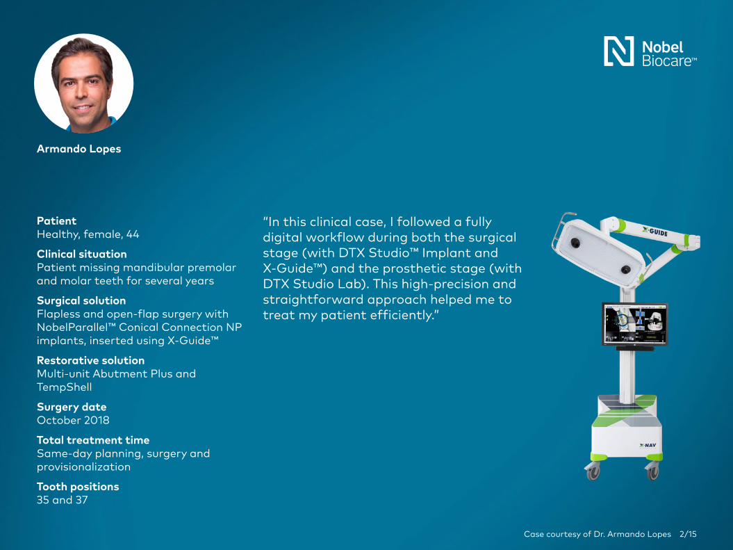
Armando Lopes
PatientHealthy, female, 44
Clinical situationPatient missing mandibular premolar and molar teeth for several years
Surgical solutionFlapless and open-flap surgery with NobelParallel™ Conical Connection NP implants, inserted using X‑Guide™
Restorative solutionMulti‑unit Abutment Plus and TempShell
Surgery dateOctober 2018
Total treatment timeSame‑day planning, surgery and provisionalization
Tooth positions35 and 37
“In this clinical case, I followed a fully digital workflow during both the surgical stage (with DTX Studio™ Implant and X-Guide™) and the prosthetic stage (with DTX Studio Lab). This high‑precision and straightforward approach helped me to treat my patient efficiently.”

Case courtesy of Dr. Armando Lopes 3/15
Radiographic and clinical images at presentation showing defects in both mandibular quadrants, where the patient has been missing premolar and molar teeth for several years. This clinical case will focus only on restoration of the narrow ridge between tooth positions 35–37, where free-hand, open flap surgery could be challenging.
Initial clinical situation
Treatment planning
Surgical procedure
Outcome
Courtesy of Dr Armando Lopes
Courtesy of Dr Armando Lopes
Courtesy of Dr Armando Lopes
Courtesy of Dr Armando Lopes
Courtesy of Dr Armando Lopes
Courtesy of Dr Armando Lopes
Courtesy of Dr Armando Lopes
Courtesy of Dr Armando Lopes
Courtesy of Dr Armando Lopes
Courtesy of Dr Armando Lopes
Courtesy of Dr Armando Lopes

Case courtesy of Dr. Armando Lopes 4/15
The SmartFusion function in DTX Studio Implant was used to combine data from a CBCT and an intraoral scanner, to integrate both the hard and soft tissue in the planning interface. The SmartSetup function in DTX Studio Implant was then used to automatically create a virtual restoration. The 8.5 mm implant in position 37 needs placement in proximity to nerve and buccal wall. X-Guide navigated surgery helps placing the implant at high precision.
Initial clinical situation
Treatment planning
Surgical procedure
Outcome
Courtesy of Dr Armando Lopes
Courtesy of Dr Armando Lopes
Courtesy of Dr Armando Lopes
Courtesy of Dr Armando Lopes
Courtesy of Dr Armando Lopes
Courtesy of Dr Armando Lopes
Courtesy of Dr Armando LopesCourtesy of Dr Armando Lopes
Case courtesy of Dr. Armando Lopes 5/15
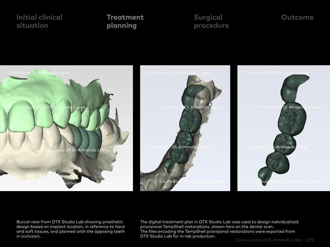
Buccal view from DTX Studio Lab showing prosthetic design based on implant location, in reference to hard and soft tissues, and planned with the opposing teeth in occlusion.
The digital treatment plan in DTX Studio Lab was used to design individualized provisional TempShell restorations, shown here on the dental scan. The files encoding the TempShell provisional restorations were exported from DTX Studio Lab for in‑lab production.
Initial clinical situation
Treatment planning
Surgical procedure
Outcome
Courtesy of Dr Armando Lopes Courtesy of Dr Armando Lopes
Courtesy of Dr Armando Lopes Courtesy of Dr Armando Lopes
Courtesy of Dr Armando Lopes Courtesy of Dr Armando Lopes
Courtesy of Dr Armando Lopes Courtesy of Dr Armando Lopes
Courtesy of Dr Armando Lopes
Courtesy of Dr Armando Lopes
Courtesy of Dr Armando Lopes
Courtesy of Dr Armando Lopes
Case courtesy of Dr. Armando Lopes 6/15
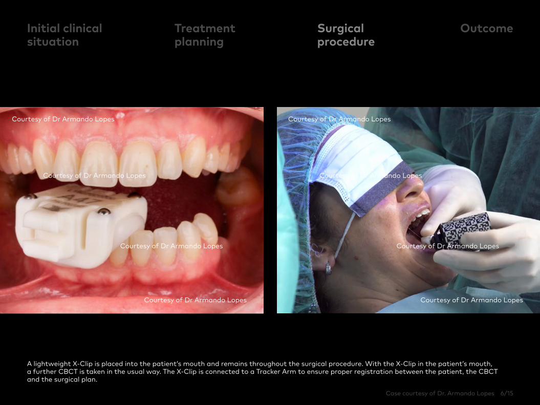
A lightweight X-Clip is placed into the patient’s mouth and remains throughout the surgical procedure. With the X-Clip in the patient’s mouth, a further CBCT is taken in the usual way. The X-Clip is connected to a Tracker Arm to ensure proper registration between the patient, the CBCT and the surgical plan.
Initial clinical situation
Surgical procedure
OutcomeTreatment planning
Courtesy of Dr Armando Lopes
Courtesy of Dr Armando Lopes
Courtesy of Dr Armando Lopes
Courtesy of Dr Armando Lopes
Courtesy of Dr Armando Lopes
Courtesy of Dr Armando Lopes
Courtesy of Dr Armando Lopes
Courtesy of Dr Armando Lopes
Case courtesy of Dr. Armando Lopes 7/15
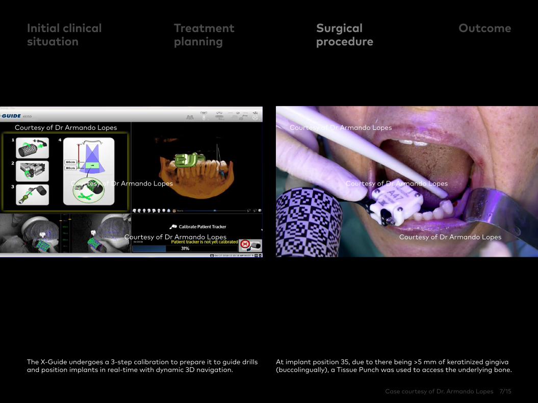
The X‑Guide undergoes a 3‑step calibration to prepare it to guide drills and position implants in real-time with dynamic 3D navigation.
At implant position 35, due to there being >5 mm of keratinized gingiva (buccolingually), a Tissue Punch was used to access the underlying bone.
Initial clinical situation
Surgical procedure
OutcomeTreatment planning
Courtesy of Dr Armando Lopes
Courtesy of Dr Armando Lopes
Courtesy of Dr Armando Lopes
Courtesy of Dr Armando Lopes
Courtesy of Dr Armando Lopes
Courtesy of Dr Armando Lopes
Case courtesy of Dr. Armando Lopes 8/15
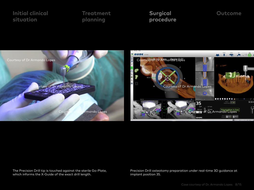
The Precision Drill tip is touched against the sterile Go‑Plate, which informs the X-Guide of the exact drill length.
Precision Drill osteotomy preparation under real‑time 3D guidance at implant position 35.
Initial clinical situation
Surgical procedure
OutcomeTreatment planning
Courtesy of Dr Armando Lopes
Courtesy of Dr Armando Lopes
Courtesy of Dr Armando Lopes
Courtesy of Dr Armando Lopes
Courtesy of Dr Armando Lopes
Courtesy of Dr Armando Lopes
Case courtesy of Dr. Armando Lopes 9/15
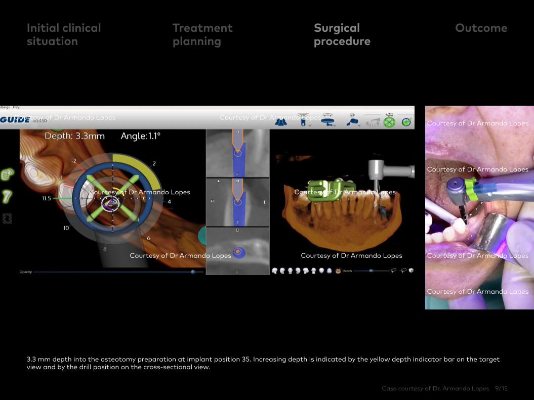
3.3 mm depth into the osteotomy preparation at implant position 35. Increasing depth is indicated by the yellow depth indicator bar on the target view and by the drill position on the cross-sectional view.
Initial clinical situation
Surgical procedure
OutcomeTreatment planning
Courtesy of Dr Armando Lopes Courtesy of Dr Armando Lopes
Courtesy of Dr Armando Lopes Courtesy of Dr Armando Lopes
Courtesy of Dr Armando Lopes Courtesy of Dr Armando Lopes
Courtesy of Dr Armando Lopes
Courtesy of Dr Armando Lopes
Courtesy of Dr Armando Lopes
Courtesy of Dr Armando Lopes

Case courtesy of Dr. Armando Lopes 10/15
The depth indicator bar turns red when the planned implant position of 11.5 mm depth is reached. Camera views, 3D view and the implant information panel are shown in parallel.
Initial clinical situation
Surgical procedure
OutcomeTreatment planning
Courtesy of Dr Armando Lopes Courtesy of Dr Armando Lopes
Courtesy of Dr Armando Lopes Courtesy of Dr Armando Lopes
Courtesy of Dr Armando Lopes Courtesy of Dr Armando Lopes
Courtesy of Dr Armando Lopes Courtesy of Dr Armando Lopes
Case courtesy of Dr. Armando Lopes 11/15
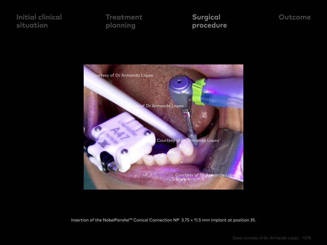
Insertion of the NobelParallel™ Conical Connection NP 3.75 × 11.5 mm implant at position 35.
Initial clinical situation
Surgical procedure
OutcomeTreatment planning
Courtesy of Dr Armando Lopes
Courtesy of Dr Armando Lopes
Courtesy of Dr Armando Lopes
Courtesy of Dr Armando Lopes

Case courtesy of Dr. Armando Lopes 12/15
At implant position 37, there was <5 mm of keratinized gingiva (buccolingually), so a flap was raised to access the bone and maximize soft tissue preservation.
The very precise X-Guide navigation allows for implant placement in proximity of buccal wall according to digital planning. NobelParallel CC NP 3.75 x 8.5 mm implant was chosen to fit the prosthetic design.
Initial clinical situation
Surgical procedure
OutcomeTreatment planning
Courtesy of Dr Armando Lopes
Courtesy of Dr Armando Lopes
Courtesy of Dr Armando Lopes
Courtesy of Dr Armando Lopes
Courtesy of Dr Armando Lopes
Courtesy of Dr Armando Lopes
Courtesy of Dr Armando Lopes
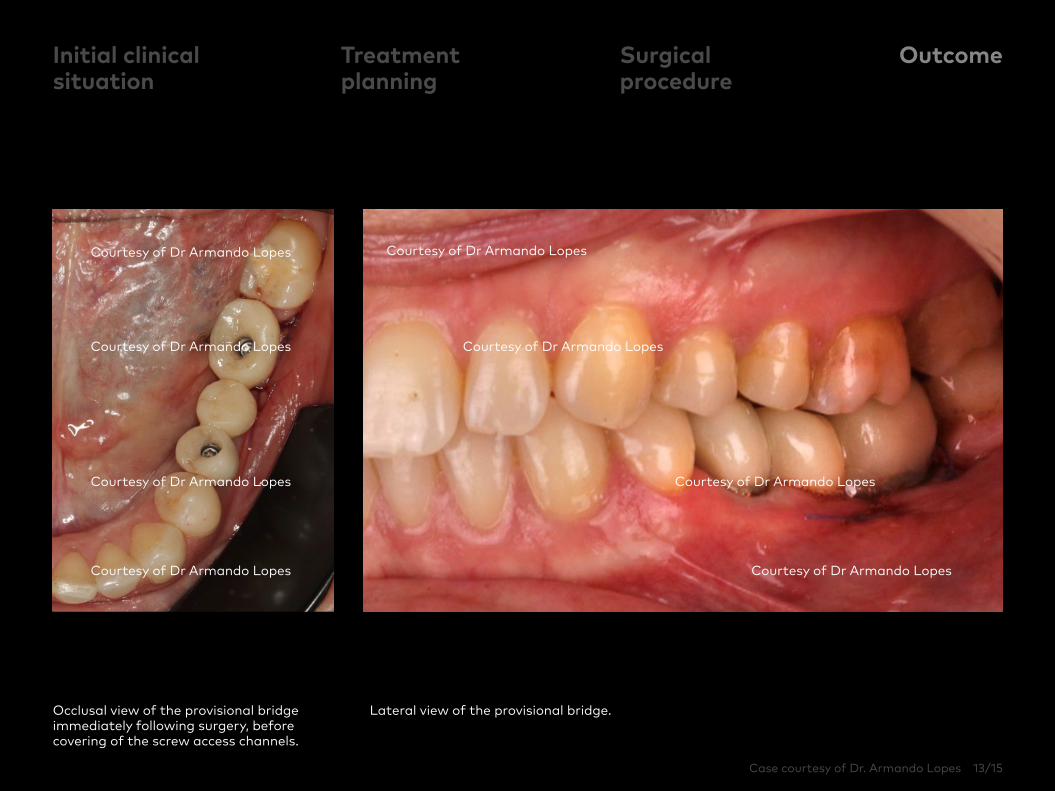
Case courtesy of Dr. Armando Lopes 13/15
Occlusal view of the provisional bridge immediately following surgery, before covering of the screw access channels.
Lateral view of the provisional bridge.
Initial clinical situation
Treatment planning
Surgical procedure
Outcome
Courtesy of Dr Armando Lopes Courtesy of Dr Armando Lopes
Courtesy of Dr Armando Lopes Courtesy of Dr Armando Lopes
Courtesy of Dr Armando Lopes Courtesy of Dr Armando Lopes
Courtesy of Dr Armando Lopes Courtesy of Dr Armando Lopes
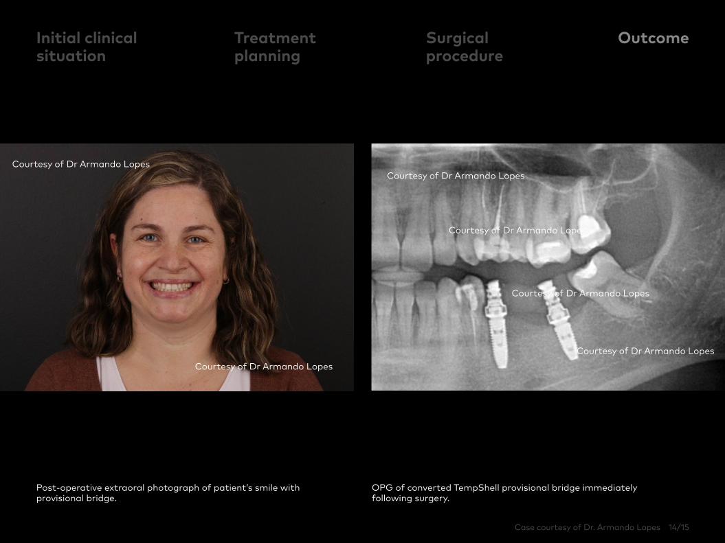
Case courtesy of Dr. Armando Lopes 14/15
Post-operative extraoral photograph of patient’s smile with provisional bridge.
OPG of converted TempShell provisional bridge immediately following surgery.
Initial clinical situation
Treatment planning
Surgical procedure
Outcome
Courtesy of Dr Armando Lopes
Courtesy of Dr Armando Lopes
Courtesy of Dr Armando Lopes
Courtesy of Dr Armando Lopes
Courtesy of Dr Armando Lopes
Courtesy of Dr Armando Lopes

Case courtesy of Armando Lopes
GMT 63808 GB 2003 © Nobel Biocare Services AG, 2020. All rights reserved. X-Guide distributed by: Nobel Biocare. Legal manufacturer: X-NAV Technologies, LLC, 1555 Bustard Road, Suite 75, Lansdale, PA, United States. KaVo is either registered trademark or trademark of Kaltenbach & Voigt GmbH. DTX Studio, Nobel Biocare, the Nobel Biocare logotype and all other trademarks are, if nothing else is stated or is evident from the context in a certain case, trademarks of Nobel Biocare. Please refer to nobelbiocare.com/trademarks for more information. Other trademarks are either registered trademarks or trademarks of their respective owners. Product images are not necessarily to scale. Disclaimer: Some products may not be regulatory cleared/released for sale in all markets. Please contact the local Nobel Biocare sales office for current product assortment and availability. For prescription use only. Caution: Federal (United States) law restricts this device to sale by or on the order of a licensed clinician, medical professional or physician. See Instructions For Use for full prescribing information, including indications, contraindications, warnings and precautions.