natural zeolite clinoptilolite: new adjuvant in anticancer ... · natural zeolite clinoptilolite:...
TRANSCRIPT
Journal of Molecular Medicine
DOI 10.1007/s001090000176
Original Article
Natural zeolite clinoptilolite: new adjuvant inanticancer therapyKre imir Paveli ( ) · Mirko Had ija · Ljiljana Bedrica · Jasminka Paveli · Ivan Ðiki · Ma a Kati · Marijeta Kralj · Maja Herak Bosnar · Sanja Kapitanovi · Marija Poljak-Bla i ·
imun Kri anac · Ranko Stojkovi · Mislav Jurin · Boris Suboti · Miroslav oli
K. Paveli · M. Had ija · J. Paveli · M. Kati · M. Kralj · M.H. Bosnar · S. Kapitanovi · M. Poljak-Bla i · R. Stojkovi · M. Jurin · B. SubotiRu er Bo kovi Institute, Division of Molecular Medicine and Division of Material Chemistry, Bijeni ka 54, Zagreb, Croatia
L. BedricaVeterinary Faculty, University of Zagreb, Heinzelova 55, 10001 Zagreb, Croatia
I. ÐikiLudwig Institute for Cancer Research, Biomedical Center, Husargatan 3, Uppsala, Sweden
. Kri anac
Institute of Pathology, Faculty of Medicine, University of Zagreb, alata 11, Zagreb, Croatia
M. oliMolecular Technologies Inc., 6512 Segovia, Goleta, CA 93117, USA
E-mail: [email protected]: +3851-46-80094Fax: +3851-46-80094
Received: 17 April 2000 / Accepted: 15 October 2000 / Published online:
- 1 -

KRE IMIR PAVELI
received his M.D./Ph.D. degree in medicine and experimental oncology from the University of Zagreb,Croatia. He received research fellowship at the RPMI in Buffalo, N.Y., University of Cincinnati, Ohio,and Fullbright fellowship at the Mayo Clinic, Rochester, Minn., USA. He is director of the Division ofMolecular Medicine at Ru er Bo kovi Institute and Director of the National Cancer ResearchProgram of the Republic of Croatia. Dr. Paveli is also Professor of Molecular Biology at theDepartment of Pharmacy and Biochemistry at the University of Zagreb. His research interests includemolecular medicine, particularly cancer genetic.
MIROSLAV OLI
received his Ph.D. in applied surface chemistry with minors in molecular biology and biophysics fromthe University of California at Berkeley, USA. He is presently Vice President of the Research andDevelopment Division, Molecutec Corporation, Goleta, California. His research interests include freeradicals chemistry and biology, environmental chemistry, biomedical effects of dietetic products, andsmall molecules drug discovery.
- 2 -

Abstract. Natural silicate materials, including zeolite clinoptilolite, have been shown to exhibitdiverse biological activities and have been used successfully as a vaccine adjuvant and for thetreatment of diarrhea. We report a novel use of finely ground clinoptilolite as a potential adjuvant inanticancer therapy. Clinoptilolite treatment of mice and dogs suffering from a variety of tumor typesled to improvement in the overall health status, prolongation of life-span, and decrease in tumors size.Local application of clinoptilolite to skin cancers of dogs effectively reduced tumor formation andgrowth. In addition, toxicology studies on mice and rats demonstrated that the treatment does not havenegative effects. In vitro tissue culture studies showed that finely ground clinoptilolite inhibits proteinkinase B (c-Akt), induces expression of p21WAF1/CIP1 and p27KIP1 tumor suppressor proteins, andblocks cell growth in several cancer cell lines. These data indicate that clinoptilolite treatment mightaffect cancer growth by attenuating survival signals and inducing tumor suppressor genes in treated cells.
Key words. Clinoptilolite - Adjuvant - Anticancer - Treatment
Abbreviations. EGF: Epidermal growth factor FBS: Fetal bovine serum MAPK: Mitogen-activatedprotein kinases PDGF: Platelet-derived growth factor SDS: Sodium dodecyl sulfate
IntroductionZeolites are hydrated natural and synthetic microporous crystals with well-defined structurescontaining AlO4 and SiO4 tetrahedra linked through the common oxygen atoms [1]. Zeolites have
been extensively used in various industrial applications based on their properties to act as catalysts, ionexchangers, adsorbents, and detergent builders [2, 3, 4, 5, 6]. It is also known that silicates andaluminosilicates possess biological activity, either positive or negative. Talc and silica have been usedin skin care for many decades, while well defined structures and catalytic activity makealuminosilicates an attractive model system for protein and enzyme mimetics [7]. Recent results havealso demonstrated that natural, biologically nontoxic clinoptilolite from Cuba deposits is very effectiveas glucose adsorbent, and this has been suggested as a potential medication for individuals sufferingfrom diabetes mellitus [8].
The best known positive biological activity of natural clinoptilolite is its action as antidiarrheal drug(see [9] and references therein). Clinoptilolite lowers the incidence of death and sickness (diarrhealsyndrome) produced by intestinal diseases in swine, rats, and calves (see [9] and references therein).Based on these results a comprehensive study was carried out on antidiarrheal drugs based on naturalclinoptilolite as an active material, in the therapy of acute diarrheal diseases in humans [9]. Theresearch lead to approval of the antidiarrheal drug Enterex for use in humans. In addition,accumulating evidence has indicated that zeolites play an important role in regulating the immunesystem. Ueki et al. [10] and Aikoh et al. [11] have reported that silica, silicates, and aluminosilicatesact as nonspecific immunostimulators similarly to superantigens. Superantigens are a class ofimmunostimulatory and disease-causing proteins of bacterial and viral origin with the ability toactivate relatively large fractions (5-20%) of the T cell population. Activation requires simultaneousinteraction of the superantigens with V domain of T cell receptor and with major histocompatibility
complex class II molecules on the surface of antigen presenting cells [10]. Proinflammatorymacrophages, which belong to class II MHC antigen-presenting cells, are activated by fibrogenicsilicate particulates [12, 13, 14, 15]. Indeed, experiments carried out by Ueki and coworkers [10] haveshown that removal of MHC class II DP/DR positive cells results in a lack of macrophage stimulationby asbestos.
- 3 -
Direct interaction of silicate particles with cells other than lymphocytes has also been identified anddescribed. It seems that mineral particles can trigger alterations in gene expression by initiatingsignaling events upstream of gene transactivation [16]. Exposure of cells to silicate particles has beenshown to lead to activation of mitogen-activated protein kinases (MAPK), protein kinase C, andstress-activated protein kinases [17]. Important transcription factors such as activator protein 1 andnuclear factor B are also activated, and expression of proinflammatory cytokines such as interleukin 1 , interleukin 6, and tumor necrosis factor is enhanced [18]. Modifications in receptor activationkinetics or activity of integrins may be responsible for the observed behavior. Alternatively, particlesengulfed by phagocytosis have been reported to stimulate production of reactive oxygen species [19].It was recently shown that redox regulation of gene expression is a general phenomenon in most cells.
The above knowledge of zeolites and other silicates led us to test the biological activity of naturalclinoptilolite. Mechanical treatment of natural clinoptilolite was used to produce small-sized particles(MZ) that were tested for possible toxicity and anticancer activity in vivo. Here we provide evidencethat orally administered natural clinoptilolite is nontoxic and useful in cancer treatment in animalmodels. Additional in vitro tissue culture experiments with various cancer cell lines indicated that MZtreatment modifies intracellular signaling pathways leading to inhibition of survival signals andinduction of tumor suppressor genes.
Materials and methods
Natural clinoptilolite
The fine powder of natural clinoptilolite was obtained by tribomechanical micronization. Chemicalcomposition of the MZ was determined by the atomic absorption spectroscopy. Qualitative andquantitative phase analyses of the MZ were performed by powder X-ray diffractometry using aSiemens 5000D diffractometer with CuK radiation in the region 2 =4-80°. Thermogravimetric and
differential thermogravimetric analysis of the MZ was performed using a TA 4000 System(Mettler-Toledo) apparatus. The heating rate was 10 K/min in nitrogen atmosphere. Particle sizedistribution curves of the MZ were taken by a Mastersize XLB (Malvern) laser light-scatteringparticle-size analyzer.
Cell lines and proliferation assay
Effect of MZ on in vitro cell proliferation was studied on several human cell lines: diploid fibroblasts(Hef522), cervical carcinoma (HeLa), colon carcinomas (CaCo-2, HT-29, and SW 620), mammarycarcinomas (MCF-7 and SkBr-3), and one mouse fibrosarcoma cell line. The cells were maintained byculturing in Dulbecco’s modified Eagle’s medium supplemented with 10% fetal bovine serum (FBS),2 mM L-glutamine, 100 U/ml penicillin, and 100 µg/ml streptomycin in a humidified atmosphere with5% CO2 at 37°C. For the purpose of proliferation assay experiments the cells were plated at a
concentration of 1 104 cells/ml onto 96-microwell plates (200 µl/well). After overnight incubation
the standard medium was replaced with the medium which was pretreated with either 0.5, 5, or50 mg/ml MZ. For this purpose the medium and MZ were mixed, and after 18 h of shaking MZ waspelleted by centrifugation (5000 g for 10 min).
The cells were then incubated for additional 72 h, when cell viability (cell growth) was measuredusing MTT assay which detects dehydrogenase activity in viable cells. For this purpose the mediumwas discarded, and MTT was added to each well at concentration of 20 µg/40 µl. After 4 h of
- 4 -

incubation at 37°C the precipitates were dissolved in 160 µl DMSO. The absorbance was measured onan enzyme-linked immunosorbent assay reader at 570 nm. The cell proliferation is expressed as apercentage of absorbance, recorded in cell line treated with particular concentration of MZ, in relationto the absorbance of control, nontreated, cells which was expressed as 100%.
Analysis of p21WAF1/CIP1 and p27KIP1
Experiments with p21WAF1/CIP1 and p27KIP1 were carried out on human adenocarcinoma (CaCo-2)and human cervical carcinoma (HeLa) cell lines. The cells, originally grown in tissue culture flasks,were collected and seeded onto glass slides. After 24 h the medium was replaced either with the freshstandard medium (control cells) or with the medium pretreated with 50 mg/ml MZ. After 72 h ofincubation the cells were washed with PBS and fixed in methanol with 3% hydrogen peroxide(Kemika, Zagreb, Croatia).
Proteins, p21WAF1/CIP1 and p27KIP1, expression was analyzed immunocytochemically. Nonspecificbinding was blocked by applying normal rabbit serum (1:10) for 30 min. Primary antibodies p21(5 µg/ml, PharMingen) and p27 (2 µg/ml, Transduction Laboratories) were allowed to bind overnightat 4°C. Slides were washed three times in PBS. Secondary antibody (rabbit anti-mouse; Dako,Denmark) was applied for 1 h at room temperature. Finally, peroxidase-antiperoxidase (Dako)conjugate diluted 1:100 in PBS was applied for 1 h at room temperature. After washing with PBS theslides were stained with 0.025% diaminobenzidine tetrahydrochloride (Sigma) containing 4% H2O2
for 7 min and counterstained with hematoxylin for 30 s. The slides were analyzed with a lightmicroscope (Olympus). The level of nonspecific background staining was established for eachmeasurement using control cells processed in the same way but without exposure to the primary antibody.
The concentration of antigen was assessed by estimating the relative visual intensity of a chromogeniclabel, and the results are expressed on a three-point scale as follows: -, negative staining; +, weakstaining and ++, moderate staining.
Biochemical studies of signaling pathways
The following were used: epidermal growth factor (EGF; Intergen), platelet-derived growth factor(PDGF) BB (Amgen), protein ladder markers (10-200 kDa; Life Technologies), leupeptin and aminiprotease inhibitor kit (Boehringer-Mannheim), Pefabloc (Fluka), aprotinin (Trasylol, Bayer), andnitrocellulose membranes (Millipore). Affinity-purified rabbit polyclonal anti-Akt, anti-pAkt,anti-JNK, anti-pJNK and anti-pERK2 (MAPK) antibodies were purchased from New England Biolabs.The rabbit polyclonal anti-ERK2 (C-14) antibodies were from Santa Cruz Biotechnology. Secondaryantibodies, peroxide-conjugated swine anti-rabbit were from New England Biolabs,peroxide-conjugated sheep anti-mouse immunoglobulin from Amersham/Pharmacia, andperoxide-conjugated protein A from Kirkegaard and Perry Laboratories.
Murine fibrosarcoma cells were grown in Petri dishes (6 cm in diameter) in RPMI medium with 10%FBS up to the 80% confluence. Before starting the experiments the cells were starved for 24 h.Subsequently the cells were treated with MZ pretreated medium with or without 10% FBS for 0, 5, 30,and 60 min or with EGF (100 µg/ml) and PDGF (40 µg/ml). After the indicated time of treatment thecells were washed with PBS and scraped into ice-cold lysis buffer containing 50 mMhydroxyethylpiperazine ethane sulfonic acid, pH 7.2, 150 mM NaCl, 1 mM EDTA, 20 mM NaF,2 mM sodium orthovanadate, 1% (w/v) Triton X-100, 10% (w/v) glycerol, and protease inhibitors(1 mM Pefabloc, 10 µg/ml leupeptin, and 1% Trasylol). Following 45 min at 4°C with gentle rocking asoluble fraction was prepared by centrifugation at 4°C for 15 min at 13,000 g. Equal amounts of cell
- 5 -

lysates (measured by the Bradford assay) were mixed with 3 sodium dodecyl sulfate (SDS) sample
buffer and heated for 2 min at 98°C. Proteins were separated by SDS polyacrylamide gelelectrophoresis and transferred onto nitrocellulose membrane. Immunoblots were blocked with 5%bovine serum albumin in TBS (10 mM Tris-HCl, pH 7.4; 150 mM NaCl) for 1 h, incubated for 1 hwith primary antibodies (anti-pAkt, anti-pJNK, anti-pERK2) in TBS, washed six times for 10 mineach in TBS 0.05% Triton X-100, and then incubated for 1 h with appropriate secondary antibody.Following further washes, immunoblots were visualized by using enhanced chemiluminescencereagents. To reprobe blots they were incubated in stripping buffer (62.5 mM Tris-HCl, pH 6.7; 2%SDS; 100 mM 2-mercaptoethanol) at 58°C for 25 min, washed extensively with TBS, reblocked asdescribed above, and reblotted with the appropriate antibodies.
Isolation of apoptotic DNA fragments
HeLa cells (1 105) were grown in a 10-ml flask for 24 h, after which the medium was discarded
and replaced with the MZ pretreated medium (see above). After 24 h the cells were tripsinized,pelleted by centrifugation (1200 g), and washed twice in PBS. Afterwards the cells were resuspended10 s in 100 µl lysis buffer (1% NP-40 in 20 mM EDTA, 50 mM Tris-HCl, pH 7.5) and centrifuged5 min at 3000 g. The supernatant was transferred to a new Eppendorf tube while the pellet wasincubated once more with 100 µl lysis buffer and centrifuged as before. The supernatants were pooledtogether and incubated 2 h in 1% SDS and RNase (5 µg/µl) at 56°C, after which the proteinase K wasadded in final concentration 2.5 µg/µl overnight. DNA fragments were pelleted by addition of 1/2volume of 10 M ammonium acetate and 2.5 volume of prechilled absolute ethanol. Aftercentrifugation (30 min, 12,000 g), the pellet was washed with 70% ethanol, centrifuged 10 min at 12000 g, dried, and dissolved in 20 µl TE buffer (10 mM Tris-HCl pH 7.4; 1 mM EDTA pH 8). TheDNA was visualized on 1.5% agarose gel.
Animals
Mice
CBA/HZgr and C57BL/6 mice of both sexes were used. Toxicity study experiments were performedon the CBA/HZgr strain, while experiments with tumors were performed on both strains. Fornonclinical tolerance testing male mice of the BALB/c strain were used. At the beginning of theexperiments the animals were about 4 months old, weighing 25-28 g. Until beginning the experimentsthe mice were maintained in standard conditions with unrestricted access to food and water.
Rats
Wistar rats of both sexes from the animal breeding colony at the Institute for Medical Research,Zagreb, Croatia were used for toxicity and nonclinical tolerance testing studies. At the beginning ofthe experiments they were 2-3 months old, weighing in average 300 g (males) and 200 g (females).
Dogs
Twenty-two dogs were used in the experiments. They were of various breeds, weighing from 3 to42 kg. The animals were of both sexes, 5-14 years old. The data on the 14 dogs in which diseaseimprovement was observed, are presented in Table 2.
- 6 -

Application of mechanically treated natural clinoptilolite (MZ)
Because of the insolubility of the tested substance, it was administered to the animals either orally bygavage or in their diet (mice, rats), supplemented as powder to the conventional food, or in capsules(dogs) which were again admixed to food. When testing the growth of mammary aplastic carcinoma ormammary aplastic carcinoma metastases formation MZ and standard food for laboratory mice (Pliva,Zagreb, Croatia) were mixed in the ratio 20%:80%. Each mouse on average ate about 4 g food daily,thus consuming about 800 mg MZ. When testing the growth of melanoma, MZ was given to miceorally (gavage) at doses of 20, 30, and 40 mg/mice five times per day (tested doses were 100, 150, and200 mg/mice, respectively). In toxicity studies MZ was applied in diet mixed with standard food.
Tumors
Mammary carcinoma occurred spontaneously in CBA/HZgr mice, maintained in the animal breedingsection of the Division of Molecular Medicine, Ru er Bo kovi Institute, Zagreb, Croatia. Thetumor is a highly anaplastic carcinoma with very high incidence of mitoses; it does not form anyglandular structures and leads to spontaneous metastases in the lungs. After transplantation of 1 106
viable tumor cells into the animals a growing tumor is obtained which causes the mouse’s death afterabout 4 weeks. For the purpose of the experiments tumor cell suspension was always prepared from invivo growing tumor.
Melanoma B16, originally obtained from Holt Radium Institute, Manchester, United Kingdom, hasbeen maintained at the Ru er Bo kovi Institute since 1975 by subcutaneous inoculations ofsuspension containing 2 106 tumor cells into flanks of C57BL/6 mice.
Spontaneous tumors in dogs were of various origins, sizes, and locations. The data on 14 tumors arepresented in Table 2. In another 8 tumors, not presented in Table 2, there were two lymphomas, twoautoimmune hemolytic anemias, and one each of prostate tumor, osteosarcoma, mammaryfibrochondroadenocarcinoma, and epulis.
To obtain tumor cells in suspension large pieces of tumor removed from the mice were cut up in verysmall pieces (Hank’s solution). The particles were allowed to settle, and the supernatant (cellsuspension) was removed and spun down at 150 g for 10 min. The pellet was resuspended and cellviability was tested by Trypan blue exclusion test: more than 90% of tumor cells were scored asviable. To obtain locally growing tumor, an inoculum of 0.1 ml, containing 1 106 viable tumor
cells, was injected subcutaneously into the right thigh of recipient mice. Tumor growth was checkedeach day after tumor cell inoculation into the mice. When the tumor was established, its size wasmeasured by a caliper. Three diameters were measured, and tumor volume was calculated. To obtainexperimental lung metastases 0.25 ml, containing 1 105 mammary aplastic carcinoma cells, was
injected into mouse tail vein. The mice were killed 18 days later. The lungs were removed, washed inwater, separated into lobules, and immersed in a fixative. Macroscopically visible nodules on lung’ssurface were counted.
- 7 -

Toxicology studies
Preclinical toxicology was performed according to standards and regulations of the Organization forEconomic Cooperation and Development principles of food laboratory practice (Paris 1998). Thetesting was approached by setting the "limit" test - applying the high doses of MZ, 2 200 and
2 500 mg/mouse per day orally (gavage) for 6, 14, and 30 days. Since the MZ did not cause the
death of mice in a "limit" test, an "up and down" test was performed on mice, with daily doses rangingfrom 60 to 400 mg/mouse (MZ given orally, gavage, for 30 days). Again, no toxicity was observed.Therefore a classical acute, subchronic and chronic toxicity study of mice and rats of both sexes(separately) was performed.
Mice
The mice were of the CBA/HZgr strain. MZ was given in a diet (powdered MZ mixed with standardfood at the ratio of 25:75%). The duration of study was as follows: acute toxicity, 1 month; subchronictoxicity, up to 3 months; chronic toxicity, up to 6 months. Animals were monitored for: phenotypicchanges, changes in behavior, and survival (every day), changes in body weight (weekly), amount offood and water consumed (checked on days 14 and 28 when mice were kept for 24 h in metaboliccages, five mice per cage), changes in hematological and serum clinical chemistry parameters(erythrocytes, leukocytes, plateletes, hematocrit, hemoglobin, glucose, alkaline phosphatase, aspartateaminotransferase, alanine aminotransferase, bilirubin, inorganic phosphorous, and calcium; after 1, 3,and 6 months); and urine clinical chemistry parameters (glucose, proteins, urobilinogen, bilirubin,nitrites, erythrocytes, leukocytes, pH, and specific gravity; urine was collected while the animals werekept, once a month for 24 h, in metabolic cages). Pathohistological analysis of liver, spleen, kidney,brain, lung, testes, ovary, duodenum, eye, stomach, large and small intestine, muscles, myocard,pancreas, thymus and axillary lymph node was carried out on killed experimental and control mice.
Rats
Wistar rats were used. MZ was given in a diet (mixed with standard food at ratios of 25:75 and 50:50).The duration of study was as follows: acute toxicity, 1 month; subchronic toxicity, 3 months; chronictoxicity, 12 months. Animals were monitored for: phenotypic changes, changes in behavior andsurvival (every day), changes in body weight (every 4 days), amount of food (every day) and waterconsumed (every 4 days), and changes in hematological and serum clinical chemistry parameters (thesame as for mice; once a month). Pathohistological analysis of liver, spleen, lung, kindey, testes, ovaryand brain, was performed on killed experimental and control rats after 1, 6, and 12 months.
The reproductive/developmental toxicity was tested on mice (CBA/HZgr) due to their short gestationperiod and larger litter size. MZ was given in a diet (powdered MZ mixed with standard food at theratio of 25:75%). For reproductive toxicity study ten male and ten female mice were fed with the foodsupplemented with the MZ for 50 and at least 14 days, respectively, before mating. The treatmentcontinued during the prepregnancy and pregnancy period (one cycle) and to the point of weaningoffspring. The same pair of animals was fed with the MZ and monitored during four consecutivecycles (approximately 4-5 months). The same schedule was applied for control, nontreated, animals.The parental generation was monitored for duration of cycle period (prepregnancy and pregnancyperiod), fertility (presence or absence of litter in particular cycle), delivery incidence, mortality, andpathohistological appearance of ovaries, after 4th cycle. Number of total and viable pups born as wellgain in pups body weight and pups mortality until weaning was also scored.
- 8 -

For teratology study healthy, untreated pregnant mice were fed with MZ mixed to the conventionalfood from day 6 through day 16 of gestation and the mice were killed 1 day before parturition. Thefetuses were analyzed for microscopic pathology.
Local tolerance was evaluated to ascertain whether the test substance is tolerated at the sites in thebody which may come into contact with the product as a result of its administration.
Repeated-dose dermal tolerance testing was performed on male Wistar rats and male BALB/c mice.MZ was applied on the shaved skin of the whole dorsal region of animals in three ways: (a) as originalpowder, (b) mixed with neutral creme at the ratio of 1:1, (c) mixed with paraffin oil at the ratio of 1:1.The animals were treated twice a day during 28 days. Macroscopic changes in the treated skin wereexamined daily. The left dorsal region of the animal was used as control. For microscopic analysis ofthe possible changes the skin samples were collected 1 day after the last treatment.
Results
Properties of mechanically treated natural clinoptilolite
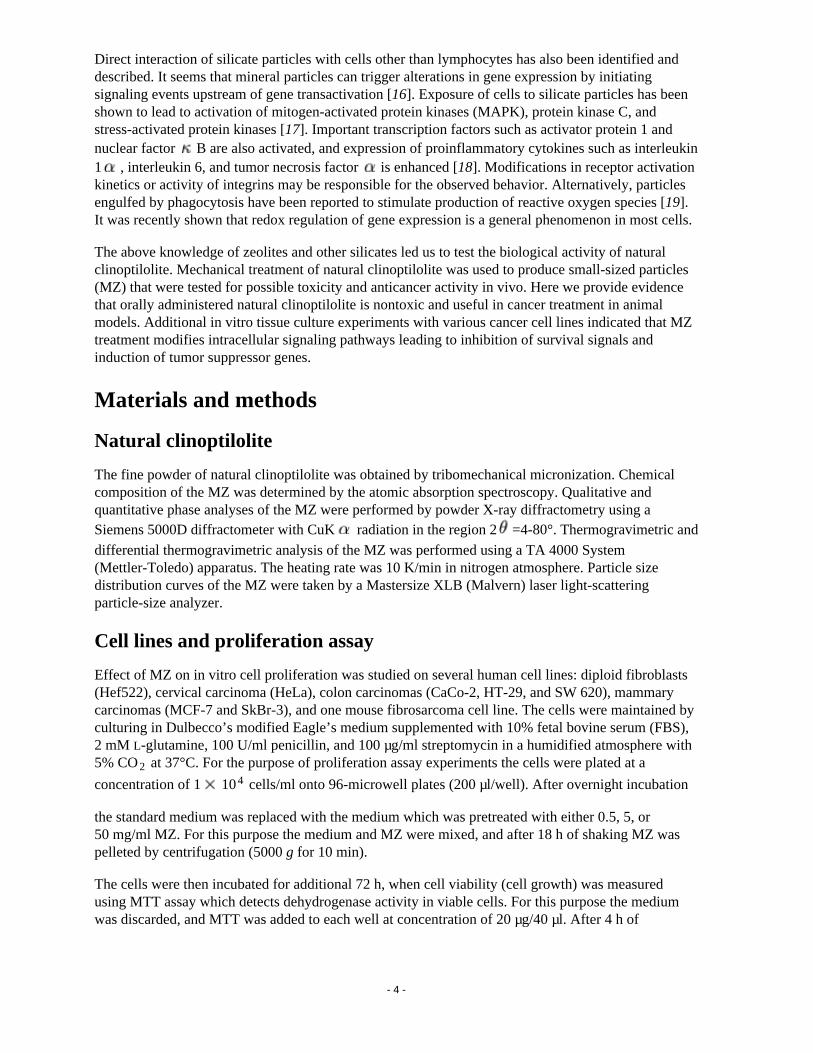
Mechanically treated natural clinoptilolite (MZ) contained approximately 85 wt.% clinoptilolite. Theremaining 15% consisted of silica, montmorillonite and mainly mordenite zeolite. The chemicalcomposition of the natural clinoptilolite is presented in Table 1. Differential thermal analysis(differential thermogravimetric) of the MZ shows that the maximum rate of water desorption occurredat 50°C, indicating that the change in sample weight during heating to 50°C corresponds to theremoval of loosely held moisture within the solid microstructure. Analysis of the water desorptioncurve shows that the MZ contains approx. 16 wt.% of water (loosely held moisture + zeolitic water) ofwhich approx. 2 wt.% is loosely held moisture (Fig. 1A, B). No phase transformation was observedduring the heating of MZ to 800°C. Particle size analysis of the MZ showed that maximum frequencyof particles (approx. 13%) appeared at 1.5 µm with average size of 2.9 µm. In 25% of particles the sizewas up to 1.5 µm, in 50% up to 2 µm, and in 75% up to 3 µm (Fig. 1C, D).
Table 1. Chemical composition of the mechanically treated natural clinoptilolite (MZ)
Oxide wt.%
SiO2 50-55
Al 2O3 9.3-11.4
Fe2O3 2.2-2.8
Na2O 0.8-1.1
K 2O 2.9-4.3
MgO 0.8-1-2
CaO 13.7-17.2
MnO 0.07-0.90
TiO2 0.14-0.22
Water (800°C) 14-16
- 9 -
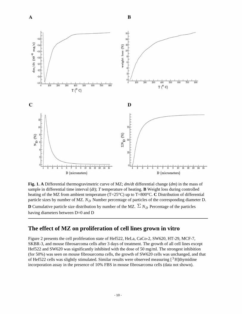
Fig. 1. A Differential thermogravimetric curve of MZ; dm/dt differential change (dm) in the mass ofsample in differential time interval (dt); T temperature of heating. B Weight loss during controlledheating of the MZ from ambient temperature (T=25°C) up to T=800°C. C Distribution of differentialparticle sizes by number of MZ. ND Number percentage of particles of the corresponding diameter D.
D Cumulative particle size distribution by number of the MZ. ND Percentage of the particles
having diameters between D=0 and D
The effect of MZ on proliferation of cell lines grown in vitro
Figure 2 presents the cell proliferation state of Hef522, HeLa, CaCo-2, SW620, HT-29, MCF-7,SKBR-3, and mouse fibrosarcoma cells after 3 days of treatment. The growth of all cell lines exceptHef522 and SW620 was significantly inhibited with the dose of 50 mg/ml. The strongest inhibition(for 50%) was seen on mouse fibrosarcoma cells, the growth of SW620 cells was unchanged, and thatof Hef522 cells was slightly stimulated. Similar results were observed measuring [3H]thymidineincorporation assay in the presence of 10% FBS in mouse fibrosarcoma cells (data not shown).
- 10 -
Fig. 2. Effect of the medium pretreated with 0.5, 5.0, and 50.0 mg/ml MZ on growth of various celllines. Vertical bars Standard deviations; arrows statistical difference in comparison to control (P<0.001, Student’s t test)
Analysis of intracellular signaling pathways in MZ-treated cells
Since previous studies have indicated that exposure of cells to silicate particles leads to activation ofMAPK, protein kinase C, and stress-activated protein kinases/JNK [17], we further analyzed whetherMZ treatment also affects mitogenic and survival signaling pathways in these cell models.
The most significant results were detected measuring the activity of Akt protein. Akt, or protein kinaseB, has been recently shown to mediate survival signals downstream of phosphoinositide-3 kinase byphosphorylating Bad proteins. We have observed an increase in Akt phosphorylation in response toserum, EGF, or insulin treatment. The addition of the MZ pretreated medium containing 10% FBS tothe cells decreased Akt phosphorylation in comparison to the cells treated with only serum containingmedium, while the addition of growth factors EGF and PDGF restored its activity (Fig. 3A) andovercame the effects of MZ on cell growth. Determination of the activity of Akt at various times afterthe addition of MZ pretreated medium with 10% FBS showed slight decrease in pAkt level after5 min. This decrease was more pronounced after 30 and 60 min of treatment (Fig. 3B). However, theaddition of MZ pretreated medium without serum to the cells increased activity of Akt compared onlyto the serum-starved cells. Overnight treatment of the cells with EGF also increased Akt activity.However, combined overnight treatment of the cells with EGF and MZ pretreated medium decreasedAkt activity, indicating that inhibition of Akt might be linked to MZ inhibition of the EGF-triggeredpathways.
- 11 -

Fig. 3. A Activity of Akt protein 5 min after addition of the MZ pretreated medium to murinefibrosarcoma cells. B Decreased Akt protein activity at various times after treatment of murinefibrosarcoma cells with MZ pretreated medium. C Effect of serum free MZ-pretreated medium on theactivity of MAPK in murine fibrosarcoma cells. WB Western blot; FBS fetal bovine serum; MZmechanically activated clinoptilolite zeolite; pAkt phosphorylated Akt; EGF epidermal growth factor; PDGF platelet-derived growth factor; MAPK mitogen-activated protein kinase; pMAPKphosphorylated mitogen-activated protein kinase
- 12 -

MAP kinase activity was increased in serum-starved cells in response to EGF, PDGF, or serum.Addition of only MZ pretreated medium to the serum-starved cells increased MAPK activity onlytemporarily (after 5 min); in the next 30 min MAPK activity returned to the normal level (Fig. 3C). Incontrast, addition of MZ pretreated medium plus 10% serum slightly decreased MAPK activitycompared only to serum-treated cells or cells incubated only with MZ pretreated medium. Theseresults are in agreement with those of the previously performed thymidine test.
Medium pretreated with MZ added to the cells either alone or in combination with serum caused nochange in JNK activity (data not shown).
The effect of MZ on expression of inhibitors of cycline-dependent kinases, p21WAF1CIP1 and p27KIP1,was tested using immunocytochemical method, in HeLa and CaCo-2 cells. Treatment with MZinduced the expression of p21WAF1/CIP1 in CaCo-2 cells and p27KIP1 in HeLa cells, while nontreatedcells were negative for expression of p21WAF1/CIP1/p27KIP1 (Fig. 4).
- 13 -
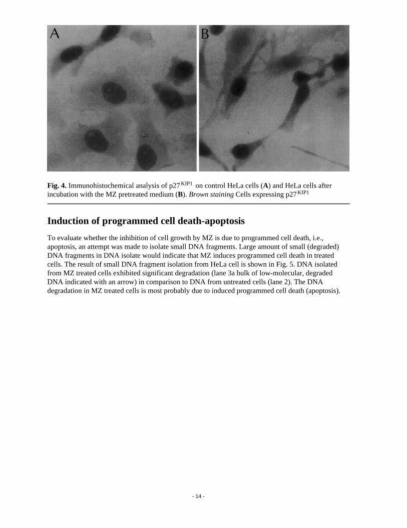
Fig. 4. Immunohistochemical analysis of p27KIP1 on control HeLa cells (A) and HeLa cells afterincubation with the MZ pretreated medium (B). Brown staining Cells expressing p27KIP1
Induction of programmed cell death-apoptosis
To evaluate whether the inhibition of cell growth by MZ is due to programmed cell death, i.e.,apoptosis, an attempt was made to isolate small DNA fragments. Large amount of small (degraded)DNA fragments in DNA isolate would indicate that MZ induces programmed cell death in treatedcells. The result of small DNA fragment isolation from HeLa cell is shown in Fig. 5. DNA isolatedfrom MZ treated cells exhibited significant degradation (lane 3a bulk of low-molecular, degradedDNA indicated with an arrow) in comparison to DNA from untreated cells (lane 2). The DNAdegradation in MZ treated cells is most probably due to induced programmed cell death (apoptosis).
- 14 -

Fig. 5. Apoptotic DNA fragments in 1.5% agarose gel. Lane 1 DNA molecular weight marker IX (Øx 174/HindIII); lane 2 DNA isolated from untreated HeLa cells; lane 3 DNA isolated from theMZ-treated HeLa cells. Arrow Degraded, low-molecular DNA fragments
Toxicology
Oral (in diet) administration of MZ to mice and rats for 6 and 12 months, respectively, caused nochanges that could be considered a toxic effect of treatment. The MZ equalized (regulated) andshortened the prepregnancy period. The number of pups per litter was increased in MZ-treated mice.Probably for this reason the gain in pups’ body weight until weaning was decreased. As a final
- 15 -

consequence higher mortality of pups between days 8 and 21 of the neonatal period was observed.However, there are no differences between control and treated animals that would suggestreproductive toxicity attributable to the MZ administration. The MZ did not elicit toxicity during theperiod of organogenesis. The test substance, MZ, was not toxic or allergenic for the skin.
Effect of MZ on tumor growth in animal models
Previous studies in cultured cells have suggested that MZ inhibits growth of cancer cells in vitro. Tostudy the effect of MZ in vivo studies on mice, rats, and dogs were undertaken. Subsequent studieswere performed on murine transplantable tumors, melanoma B16, and mammary carcinoma.Mammary aplastic carcinoma cells were injected into the right thigh of two groups of mice. One group (n=14) was fed with food supplemented with MZ starting from 15 days prior tumor transplantationuntil the animal’s death; the other group (n=14) was fed with MZ from the day of tumortransplantation until the animal’s death. A group of five tumor-bearing mice receiving standard foodwas used as control. Tumor growth was significantly inhibited in both groups of animals fed with MZsupplemented food (Fig. 6). The tumor growth curves for individual animals were uniform,particularly when MZ was given prior to the tumor transplantation. However, there was no differencein mice survival among the groups.
- 16 -

Fig. 6. Tumor growth following injection of 1 106 mammary aplastic carcinoma cells into the right
thigh of CBA/HZgr mice. The animals were exposed to 20% of MZ in the food either from the day oftumor transplantation (n=14) or 15 days prior to tumor transplantation (n=14). Control mice receivedstandard food. Vertical bars Standard deviation. The differences between control and bothexperimental groups were statistically significant (P<0.001, Student’s t test) for the days 25, 30, and 35
Melanoma B16 cells were inoculated subcutaneously in C57BL mice on day 0. For the next 30 daysthe mice were given MZ orally five times per day. Tumor volume was recorded; it was markedlylower in 5 of 80 mice (daily dose 150 mg/mouse) than in the control group (Fig. 7A). Despite the factthat the tumors started to grow more rapidly after the therapy with MZ was abrogated (between days30 and 60 after tumor transplantation), the mice lived a statistically significantly longer period whentreated with 200 and 150 mg MZ than control animals (Fig. 7B). The mice used for experimentalmammary aplastic carcinoma lung metastases formation were fed with MZ diet from 15 days prior totumor cell injection until to the end of the experiment, i.e., 18 days after tumor transplantation. Thecontrols consumed standard food. Each of these two groups comprised 20 animals. About 20-40nodules per animal were scored, but there was no difference between the groups (data not shown).
- 17 -

Fig. 7. Growth rate (A) of melanoma B16 treated with 150 mg MZ/mouse per day and survival (B) ofmelanoma-bearing mice treated with three different doses of MZ
There was no effect of MZ treatment on in vivo growth of two mammary carcinomas which differedfrom that showed in Fig. 6 (data not shown).
Of 22 dogs suffering from various kinds of spontaneous tumors that were treated with MZ, 14responded to therapy, i.e., the tumor disappeared completely, or the tumor size was significantlyreduced (presented in Table 2). Among three dogs which had prostate tumor there was one that wasstated sonography showed to have (in addition to prostate tumor) a prostate cyst (case 3). The dog wasconspicuously quiet, without appetite, and hardly moved. When the usual therapy did not work, MZtherapy was started. After only 2 days of treatment the dog became active; on the third day it beganeating normally, and on the fourth day the dog urinated normally, blood-free urine. On day 10 the cystand the tumor were reduced in size, and after 1 month they had disappeared completely. Although theprostate became only insignificantly smaller, the dog showed no signs of illness. At this point it isinteresting to note that the very high pretherapy serum values for aspartate aminotransferase(497 µmol/l) and alanine aminotransferase (433 µmol/l) decreased after 1 month of MZ therapy tonormal levels (16 and 43 µmol/l) and remained in the normal range for entire observation period (5 months).
Table 2. The effect of MZ treatment on growth of spontaneous tumors of dogs ( values before andafter treatment with MZ, a.t. after beginning of treatment, HMT hematocrit, ALT alanine
- 18 -

aminotransferase, AST aspartate aminotransferase, ALP alkaline phosphatase, GGT -glutamyl
transferase, L number of leukocytes)
No. BreedAge (years)a
Weight (kg)
Sex DiagnosisPrevious treatment
MZ treatment
Biochemicalandhematological changes
Therapeuticeffects
1 Schnauzer 8 15 MProstate adenocarcinomab
Castration3 200 mg/day,
28 days
HMT 61 45;ALT 103 62
7 days a.t.generalimprovement;withdraw ofcatheter;14 days a.t.no signs of disease
2 Poodle 12 16 M
Prostateadenocarcinoma (4 3 cm) and
testis tumor (20 cm)
-
3 200 and
2 200 mg/day,
6 months
AST 55 10;GGT 4 1
90 days a.t.reduction intumor mass(testis) to 1/3
3German shepherd
8 42 M
Prostateadenocarcinoma (5 5 cm) and
cyst
Antibiotics3 1200 mg/day
Bilirubin 25.8 6.2; AST
497 16;ALT 433 43;ALP 79 33
29 days a.t.tumordisappeared
4MixedGerman shepherd
14 20 F
Mammaryadenocarcinoma,multiple - 5nodes (0.5-3 cm)
-3 400 mg/day,
1 monthNo changes
10 days a.t.all nodesdisappeared;12 monthslater no signsof disease
5Englishcocker spaniel
8 15 F
Mammaryadenocarcinoma,multiple - 4nodes (0.5-3 cm)
-3 400 mg/day,
58 daysNo changes
58 days a.t.all tumornodesreduced insize 50%
6 Poodle 11 F
Mammaryadenocarcinoma,multiple - 4nodes (0.5-3 cm)
-3 400 mg/day,
2.5 monthsNo changes
2-3 months(smallernodules);4-6 months(larger nodules)
7Dobermann pinscher
8 F
Mammaryadenocarcinoma,multiple - 4nodes (0.5-3 cm)
-5 400 mg/day,
3 monthsNo changes
2-3 months(smallernodules);4-6 months(larger nodules)
- 19 -

No. BreedAge (years)a
Weight (kg)
Sex DiagnosisPrevious treatment
MZ treatment
Biochemicalandhematological changes
Therapeuticeffects
8Englishcocker spaniel
9 F
Mammaryadenocarcinoma,multiple - 4nodes (0.5-3 cm)
-3 400 mg/day,
4 monthsNo changes
2-3 months(smallernodules);4-6 months(larger nodules)
9Airedale terrier
9 F
Mammaryadenocarcinoma,multiple - 4nodes (0.5-3 cm)
Antibiotics5 400 mg/day,
10 monthsNo changes
2-3 months(smallernodules);4-6 months(larger nodules)
10German shepherd
8 38 MSkinadenocarcinoma (tail)
Surgicallyremoved,resectionwound didnot heal
6 400 mg/day,
62 days, andlocal appl. ofpowdered substance
Glucose 6.9 3.8; AST
50 38
3 days a.t.normalhealingstarted andcompleted7 days later
11MixedGerman shepherd
10 35 M
Carcinomaplanocellulare ofthe skin (tail), 3 tumor
Two nodessurgically removed
4 100 mg/day,
93 daysUrea 17.5 6.3
remainingnodedisappeared67 days a.t.
12 Malamute 12 40 MCarcinomaplanocellulare ofthe tongue
Surgicallyremoved,resectionwound didnot heal
3 100 mg/day,
32 days-
3 days aftertreatmentwoundstarted toheal, and2 days laterno furthersigns ofwoundvisible; dogstarted to eat
13German pinch
5 3 MHypertrophy andhyperplasia ofsalivary gland
Antibiotics3 100 mg/day,
147 days
Urea 9.5 7.5;AST 40 27;ALT 54 36; L 3.1 12
7 days a.t.node becamesofter andsmaller(75%);14 days laterno signs of hypertrophy
- 20 -

No. BreedAge (years)a
Weight (kg)
Sex DiagnosisPrevious treatment
MZ treatment
Biochemicalandhematological changes
Therapeuticeffects
14Berner sennenhund
8 40 M Lung cancer -4 400 mg/day,
35 days
AST 35 16;bilirubin 8.5
2.8
7 days a.t.generalimprovement;7 days laterno signs oftumor(obtained by X-ray)
aAt the beginning of therapy
bHormone dependent
Another dog (case 2) had, in addition to prostate tumor, a testis tumor. The testis was approximately20 cm in diameter when the therapy with MZ was started. After 1 month therapy the testis size wasreduced by one-third. After 2 months of therapy the testis was reduced in size to one-half and after3 months to one-third of its pretreatment size (Fig. 8A). However, the prostate remained equally large.
- 21 -

Fig. 8. Growth rate of testicular tumor (A) and salivary gland hyperplasia (B) in dogs. A Poodle,12 years old, case 2. B Pinch, 5 years old, case 13. Arrow Day of therapy cessation. All other detailsare indicated in Table 1
The third dog (case 1) diagnosed to have prostate adenocarcinoma came to the clinic in a very badgeneral condition. It urinated only with great difficulty. After 1 month of classical therapy noimprovement was observed. A catheter was placed in the dog’s urethra. The therapy was continued fora further 2 weeks but did not work. The dog was ante finem and the owners asked for euthanasia.Classical therapy was then replaced by MZ therapy (3 200 mg/day). After 1 week a general
improvement was observed, and the catheter was removed. After 14 days of therapy no signs ofdisease were still visible. The therapy continued for an additional 14 days, with daily healthimprovement. Then the owners decided on castration (in most cases castration eliminates problemsrelated to the prostate), and the therapy with MZ was stopped. Eight months later the dog is still alivewithout any major health problems.
- 22 -

Three dogs suffered from skin tumors. One of these (case 11) had three lesions nodules on the skinabove the tail. Two were removed, and the third, the smallest, was left. Histologically the tumor wasdiagnosed as carcinoma planocellulare. After 1 month of therapy with MZ the cherry-sized tumor wasreduced in size by one-third. Over following 5 weeks the lesion disappeared completely. The dog isstill (7 months latter) under therapy. The presently 11-year-old dog is very vivacious and in unusuallygood condition.
Another dog (case 10) suffered from adenocarcinoma on the skin of the tail, which was surgicallyremoved. However, even 2 weeks after surgery the wound did not heal, and amputation wasconsidered. The dog was then given MZ in capsules, and powdered MZ was also scattered on thewound. The wound healed within 1 week.
The third dog (case 12) had a growth on its tongue of approx. 2 cm diameter. Histologically it wascarcinoma planocellulare. After surgical removal of the tumor the wound did not heal. The dog wasgiven MZ orally in capsules, and powdered MZ was also applied locally. Five days later the biopsywound was no longer visible.
A 5-year-old dog (case 13), diagnosed to a have enlarged (walnut-size node) left salivary gland, wastreated with conventional therapy for 4 months, without success. During that time the gland becomelarger and larger, and the dog developed serious problems with swallowing and salivation. After only1 week of MZ therapy the node become softer and smaller by one-third. After a further 1 week thenode disappeared completely, and only the capsule was palpable (Fig. 8B).
Mammary adenocarcinomas, in the form of multiple nodules (in sizes between that of green beans andlarge walnuts), were diagnosed in six female dogs. After the therapy with MZ was started, the nodulesdisappeared completely: in one dog after 10 days, with no signs of disease even after 12 months; infour dogs after 2-3 months (smaller nodules) and 4-6 months (larger nodules), with no signs of diseasethereafter, at the present, 2 months; and in one dog the nodules were reduced in size to 50% after58 days of treatment.
In one case of a dog (case 14) with lung cancer, again, after only 14 days of treatment with MZ (4 400 mg/day) signs of tumor disappeared completely.
In addition to the effects of MZ expressed on the primary disease, all dogs, even those in whichprimary disease was not cured, responded to MZ therapy in only about 7 days with generalconstitutional and behavioral improvement lasting even after the therapy was interrupted. The samewas observed for some hematological and serum clinical parameters measured before and after thetherapy. Hematocrit decreased to the normal range in case 1. Very high total serum bilirubin valuesfell to the normal range in cases 3 and 14, while serum urea concentration change was noted in cases11 and 13. The most pronounced improvement was noted for aspartate aminotransferase, alanineaminotransferase, and alkaline leukocyte phosphatase, with pretherapy values normalized after thetherapy was started in almost all cases (nos. 1, 2, 3, 10, 13, and 14; Table 2).
DiscussionNumerous natural compounds are commonly used for the treatment of various diseases, includinggreen tea and soybean extracts (for review see [20]). Recent findings indicate that dietetic products andantioxidant compounds also have a beneficial effect particularly in cancer patients. In many cases theexact mechanism of their action is not fully understood. In this report we studied the effect of naturalclinoptilolite zeolite particles on development of several cancer models in vivo and in vitro. We found
- 23 -

that mechanically activated clinoptilolite zeolites act as anticancer therapeutic agents in in vivo animalstudies and in tissue culture cell models. Clinoptilolite applied orally in mice and dogs suffering froma variety of tumor types led to a significant shrinkage of some tumors and improvement in overallhealth status in some animals.
The range of effects was diverse, ranging from negative antitumor response, to normalization ofbiochemical parameters, prolongation of life span, and decrease in tumor size. The best results inanimal models were observed in the treatment of skin cancer in dogs, suggesting that adsorption ofsome active components is responsible for MZ activity (direct contact action). Complementary studiesperformed in tissue culture indicated that MZ treatment affects proliferation and survival of severalcancer cell lines. Addition of MZ inhibited cell proliferation in a concentration-dependent manner, inpart due to induction of inhibitors of cycline dependent kinases, inhibition of B/Akt expression andinduction of programmed cell death.
The work described here was performed with the nontoxic natural, high silica content zeolite,clinoptilolite. The zeolite particles were negatively charged in the entire pH range studied (pH 1-11).Electron microscopy showed the absence of fibers, and most particles were round with very roughsurface (data not shown). The absence of fibrous, positively charged particles was encouraging sincesuch particles are present in asbestos and erionite zeolites, which are highly carcinogenic andmutagenic. In addition, activated zeolite particles did not catalyze the production of hydroxyl radicals,unlike asbestos or erionite (data not shown). It seems that absence of fibrous particles capable ofproducing hydroxyl radicals makes this zeolite sample nontoxic and noncarcinogenic, at least whenapplied orally.
Silicate and aluminosilicate particulates can interact directly with specific cells and modify theirintracellular pathways, leading to the regulation of gene expression. MZ was particularly successful ininhibiting protein kinase B/Akt in in vitro experiments with cancer cells. Such inactivation resulted ingrowth inhibition and increase in apoptosis of cancer cells. Inhibition of Akt by MZ treatment wasshown only in the presence of serum. This indicated that adsorption of serum components can be oneof the mechanisms of MZ action in these experiments. Indeed, the addition of EGF to serum-freemedium led to activation of Akt, which was also blocked by MZ pretreatment. Adsorption ofmolecules involved in signal transduction cascades, such as inositol phosphatides and calcium, mightalso contribute to its therapeutic efficiency. Preliminary lipid adsorption studies show that MZ arestrong lipid sorbents. Similar results are observed with adsorption of proteins. Modifications ofmembrane ordering and interactions of other proteins with membrane proteins might also be involved [21], since membrane translocation is needed for activation of protein kinase B/Akt. It has alsorecently been shown that the activation of phosphoinositide-3 kinase and Akt is responsible for theability of transformed epithelial cells to survive without cell attachment. Recent results indicate thatconstitutive activation of phosphoinositide-3 kinase in five small-cell lung cancers cell lines studiedwas responsible for fast growth and anchorage independence of small-cell lung cancer cells [22]. Inaccordance with this, MZ treatment leads to inhibition of protein kinase B/Akt pathways andsubsequent apoptosis in our cell model. Akt has recently been demonstated to inactivate an importantcyclin inhibitor and tumor suppressor molecule, p27KIP1 [22].
Here we provide evidence that MZ treatment increases levels of p21WAF1CIP1 and p27KIP1 in tumorcell models. It is not yet clear whether inhibition of Akt is involved in regulation of expression of p21WAF1CIP1 and p27KIP1 cell cycle inhibitors. Preliminary results also show that MZ adsorbs anddeactivates nitric oxide and other oxidants. In addition, it has recently been reported that antioxidantsstimulate the activation of cyclin inhibitor p21WAF1/CIP1 [23]. This molecule is responsible for thearrest of cell growth, and its expression in adenocarcinomas of lung is positively correlated withoptimistic survival prognosis. The present study observed that activated clinoptilolite induces tumor
- 24 -

suppressor molecules (both p21 and p27).
The mechanisms of action of MZ in vivo remain largely unknown at this time. The results presentedhere indicate that inhibition of proliferation and survival of cancer cells may be part of mechanismsinvolved in anticancer effect of MZ compounds. More studies on several other aspects of their actionincluding possible immunomodulatory action of MZ will be performed in the future. Taken together,this report characterizes cellular effects of the MZ compounds in tissue culture cell models andprovides data supporting a role of natural zeolite as an anticancer therapeutic agent in in vivo tumor models.
References1.Breck DW (1964) Crystalline molecular sieves. J Chem Educ 41:678-689
2.Flanigen EM (1980) Molecular sieve zeolite technology-the first twenty-five years. In: Rees LVC(ed) Proceedings of the 5th International Conference on Zeolites. Heyden, London, pp 760-780
3.Sersale R (1985) Natural zeolites: processing, present and possible applications. Stud Surface SciCatalysis 24:503-512
4.Naber JE, De Jong KP, Stork WHJ, Kuipers HPCE, Post MFM (1994) Industrial application ofzeolite catalysis. Stud Surface Sci Catalysis 84C:2197-2220
5.Garces JM (1999) Observations on zeolite applications. In: Treacz MMJ, Marcus BK, Misher ME,Higgins JB (eds) Proceedings of the 12th International Conference on Zeolites. Materials ResearchSociety, Warrendale, pp 551-566
6.Colella C (1999) Natural zeolites in environmentally friendly processes and applications. StudSurface Sci Catalysis 125:641-655
7.Bedioui F (1995) Zeolite-encapsulated and clay-intercalated metal porphyrin, phthalocyanine andSchiff-base complexes as models for biomimetic oxidation catalysts: an overview. Coordination ChemRev 144:39-68
8.Concepcion-Rosabal B, Rodriguez-Fuentes G, Simon-Carballo R (1997) Development and featuringof the zeolitic active principle FZ: a glucose adsorbent. Zeolites 19:47-50
9.Rodriguez-Fuentes G, Barrios MA, Iraizoz A, Perdomo I, Cedre B (1997) Enterex - anti-diarrheicdrug based on purified natural clinoptilolite. Zeolites 19:441-448
10.Ueki A, Yamaguchi M, Ueki H, Watanabe Y, Ohsawa G, Kinugawa K, Kawakami Y, Hyodoh F(1994) Polyclonal human T-cell activation by silicate in vitro. Immunology 82:332-335
11.Aikoh T, Tomokuni A, Matsukii T, Hyodoh F, Ueki H, Otsuki T, Ueki A (1998)Activation-induced cell death in human peripheral blood lymphocytes after stimulation with silicate invitro. Int J Oncol 12:1355-1359
12.Drumm K, Oettinger R, Smolarski R, Bay M, Kienast K (1998) In vitro study of human alveolarmacrophages inflammatory mediator transcriptions and releases induced by soot FR 101, Printex 90,titandioxide and Chrysotile B. Eur J Med Res 3:432-438
- 25 -

13.Holian A, Uthman MO, Goltsova T, Brown SD, Hamilton RF (1997) Asbestos and silica-inducedchanges in human alveolar macrophage phenotype. Environ Health Perspect 105 [Suppl 5]:1139-1142
14.Schimmelpfeng J, Seidel A (1991) Cytotoxic effects of quartz and chrysotile asbestos: in vitrointerspecies comparison with alveolar macrophages. J Toxicol Environ Health 33:131-140
15.Allison AC, Harrington JS, Birbeck M (1966) An examination of the cytotoxic effects of silica onmacrophages. J Exp Med 124:141-154
16.Tsuda T, Morimoto Y, Yamato H, Nakamura H, Hori H, Nagata N, Kido M, Higashi T, Tanaka I(1997) Effects of mineral fibers on the expression of genes whose products may play a role in fiberpathogenesis. Environ Health Perspect 105 [Suppl 5]:1173-1178
17.Lim Y, Kim SH, Kim KA, Oh MW, Lee KH (1997) Involvement of protein kinase C,phospholipase C, and protein tyrosine kinase pathways in oxygen radical generation byasbestos-stimulated alveolar macrophages. Environ Health Perspect 105 [Suppl 5]:1325-1327
18.Simeonova P, Torium W, Kommineni C, Erkan M, Muson AE, Rom WN, Luster MI (1997)Molecular regulation of IL-6 activation by asbestos in lung epithelial cells-role of reactive oxygenspecies. J Immunol 159:3921-3928
19.Martin LD, Krunkosky TM, Dye JA, Fischer BM, Jiang NF, Rochelle LG, Akley NJ, Dreher KL,Adler KB (1997) The role of reactive oxygen and nitrogen species in the response of airwayepithelium to particulates. Environ Health Perspect 105 [Suppl 5]:1301-1307
20. oli M, Paveli K (2000) Molecular mechanisms of anticancer activity of some natural dieteticproducts. J Mol Med 78:333-336
21.Peterson MW, Kirschbaum J (1998) Asbestos-induced lung epithelial permeability: potential roleof nonoxidant pathways. Am J Physiol 19:L262-L268
22.Moore SM, Rintoul RC, Walker R, Chilvers ER, Haslett C, Sethi T (1998) The presence of aconstitutively active phosphoinositide 3-kinase in small cell lung cancer cells mediatesanchorage-independent proliferation via a protein kinase B and p70s6k-dependent pathway. CancerRes 58:5239-5247
23.Chinery R, Brockman JA, Peeler MO, Shyr Y, Beauchamp RD, Coffey RJ (1997) Antioxidantsenhance the cytotoxicity of chemotherapeutic agents in colorectal cancer-a p53 independent inductionof p21WAF1/CIP1 via C/EBP . Nat Med 3:1233-1241
- 26 -