natural selection and early changes of phenotype of tumor ... · natural selection and early...
TRANSCRIPT
The problem of interrelations between neoplasticcells and in vivo systems of the innate and specific immu-nity includes three main aspects: 1) the host�s protection,i.e., mechanisms of recognition and elimination ofmutant, transformed, and tumor cells by effectors of thesystem of the innate antitumor immunity (and of thespecific antitumor immunity in the case of antigenictumors); 2) the defense mechanisms of the transformedand tumor cells against effectors of the innate antitumorimmunity system; 3) the effect of selective pressure of thehost�s innate and specific antitumor immunity on thetumor progression.
The first aspect of the problem has been studied formore than 30 years mainly in experiments on tumortransplantation in laboratory animals including thosewith certain genetic deficiencies of the immune systemand on in vitro cytotoxic activity (CTA) of resident andactivated macrophages (MPh), dendrite cells (DC), nat-
ural killers (NK), neutrophils (NPh), and also of T-lym-phocytes directed against transformed and tumor cellsof various origin. The nonspecific recognition of tumorcells by effectors of the innate antitumor immunity andthe inhibition of their growth (or, on the contrary, thestimulation of their proliferation) are associated with acomplex of intercellular interactions and the inductionof various cytokine networks which are produced incooperation with DC, MPh, NK, NPh, and the tumorcells and with the involvement (or without it during theearly stages of carcinogenesis) of activated T-cells. Thisaspect of the innate antitumor immunity has been longand intensively studied and is considered in variousreviews [1-8].
Tumor cells during their in vivo appearance,growth, and dissemination suffer permanent stresscaused by various growth-inhibiting and cell-damagingsignals, including the CTA of DC, MPh, NK, and NPh.
REVIEW
0006-2979/00/6501-0078$25.00 ©2000 ÌÀÈÊ �Íàóêà/Interperiodica�
Biochemistry (Moscow), Vol. 65, No. 1, 2000, pp. 78-94. Translated from Biokhimiya, Vol. 65, No. 1, 2000, pp. 92-111.Original Russian Text Copyright © 2000 by Deichman.
Natural Selection and Early Changes of Phenotype of Tumor Cells in vivo:
Acquisition of New Defense Mechanisms
G. I. Deichman
Institute of Carcinogenesis, Blokhin Russian Cancer Research Center, Russian Academy of Medical Sciences, Kashirskoe Shosse 24, Moscow, 115478 Russia; fax: (095) 324-1205; E-mail: [email protected]
Received October 5, 1999
Abstract�This review summarizes results obtained in the author�s and collaborating laboratories within the last decadeand is designed to attract the attention of researchers to discrete biochemical mechanisms of protection acquired in vivoby cells of malignant tumors against effectors of innate antitumor immunity. Tumor progression in vivo is associated withthe appearance and selection of tumor cells with new specific characteristics: a high level of H2O2-catabolizing (antioxi-dant) activity (H2O2
CA) and the ability for immediate release of E2-type prostaglandin (PGES) on contact with naturalkillers, macrophages, and neutrophils; the expression of the [H2O2
CA + PGES] phenotype provides the tumor cells withtwo mechanisms of local protection against effectors of innate and acquired antitumor immunity. This results in a 10-100-fold less effective rejection of tumor cells in immune and normal animals and corresponding increase of tumori-genicity. The in vitro transformation of normal fibroblasts, spontaneous or induced by oncogenes LTSV40, E1a,b, Ha-ras, myc, and also by p53175 and bcl-2 does not result in the [H2O2
CA + PGES] phenotype expression, but during subse-quent in vivo growth of the above-mentioned transformants the selection of tumor cells of the [H2O2
CA + PGES] pheno-type is correlated with a 30-200-fold increase in their tumorigenicity (accompanied or not accompanied by spontaneousmetastatic activity). Unlike the transformation induced by the above-mentioned oncogenes, the transformation of nor-mal cells by the v-src gene results in the [H2O2
CA + PGES] phenotype expression. The data presented confirm the deter-mining role of the v-src gene in the expression of the [H2O2
CA + PGES] phenotype. In various primary viral carcinogene-ses (SV40, SA7(C8)) the natural selection of cells expressing the [H2O2
CA + PGES] phenotype begins even within the latentperiod and can be completed by the appearance of primary tumors.
Key words: transformation, carcinogenesis, in vivo tumor progression, antioxidant and PGE2-releasing activity of tumorcells, protection mechanisms of tumor cells, innate and specific antitumor immunity
NATURAL SELECTION AND CHANGES OF PHENOTYPE OF TUMOR CELLS 79
BIOCHEMISTRY (Moscow) Vol. 65 No. 1 2000
Apoptosis is the mechanism of death for the greatmajority of tumor cells. Tumor cells that survive in theprimary focus of tumor growth (and also in remotedeposits) develop under conditions of continuous stressand selective pressure of effectors of the innate antitu-mor immunity. Highly malignant tumor cells selected invivo usually show 10-100-fold increased tumorigenicactivity (TGA) compared to their in vitro transformedprecursors, whereas the spontaneous and experimentalmetastatic activities (SMA, EMA) and also some othernew characteristics arise less regularly. These new char-acteristics include the increased resistance of tumor cellsto the CTA of MPh, NK, and NPh. Obviously, theappearance of a monoclonal tumor is associated withselection of extremely rare variants of tumor cells whichhave different (sometimes very specific) mechanisms toovercome the host�s control.
Growth-regulating positive and negative signals ofnormal surrounding cells are another type of selectivepressure of the host on the heterogenous population oftumor cells [9]. But unlike the effectors of the innateantitumor immunity, such as DC, MPh, NK, and NPh,the normal surrounding cells seem to be not activatedduring the contact interaction with the tumor cells (atleast, this is not shown) and their effect on the tumorcells is, as a rule, either cytostatic or growth-stimulating,but not cytolytic.
The great advantage of the innate antitumorimmunity system versus the specific antitumor immuni-ty is its permanent readiness to recognize in vivo theappearance of a very small number of transformed andtumor cells (even single cells), no matter whether theyexpress the specific tumor antigens or not. It is obviousa priori that primary tumors can be most efficiently con-trolled by the innate antitumor immunity within the ini-tial latent period when the number of tumor cells is notgreat and their evolutionary genetic changes are, as arule, minimal [4, 5]. During this period, only a few ofthe in vivo transformed cells can escape the recognitionand elimination by the innate antitumor immunity sys-tem and start the proliferation. Therefore the latentperiod in the tumor development seems to be crucial incarcinogenesis because it is just the time when the colli-sion is solved in favor of either the proliferative activityof the in vivo transformed cells or the ability of thehost�s immunity system to detect their appearance andto reject them.
Until recently, the role of natural (innate) immuni-ty in the protection of the host organism, intensivelystudied in bacterial and viral infections, was consideredseparately from mechanisms of the specific T-cellimmunity. The current experimental and theoreticalanalysis of innate immunity reactions during infectiousand autoimmune processes suggests the innate immu-nity not only involves the mechanisms of inherent non-clonal recognition and elimination of dangerous exoge-
nous agents but plays instructive and controlling rolesin the induction of specific (adoptive) immunity [10-13].
It seems that these new concepts concerning theinterrelations between innate and acquired immunitymay be also applied to the problem of antitumorimmunity; this especially concerns the nonclonal mech-anisms of recognition of tumor cells by effectors of theinnate immunity. But unlike bacterial and viral infec-tions which usually have short latent periods, acutecourse, and the effective development of humoraland/or cell immune reactions, in the case of sponta-neous tumors the system of innate antitumor immunitydeals with the absent or insignificant antigenic differ-ences between the normal and autologous tumor cellsand, as a rule, with the long-term carcinogenesis devel-oping by a number of oncogenetic stages in the tumorevolution which significantly change biological proper-ties of the tumor cells.
The present review considers phenotypic changes intumor cells which are associated with their in vivo acqui-sition of discrete resistance mechanisms to the CTA ofeffectors of the innate antitumor immunity system andalso the role of these mechanisms in the in vivo survivalof tumor cells under conditions of normal and immuneorganism, natural selection, and progression of tumors.This aspect of carcinogenesis has been the area of maininterest and experimental study in our laboratory and inthe laboratories of our collaborators for the last decade.
In these studies our main approach included thefollowing of tumor progression beginning from in vitro(or in vivo) transformation of normal cells by variousoncogenes or spontaneously, with subsequent naturalselection in vivo of their descendants and their acquisi-tion of malignant phenotype. This approach allows usto study the in vivo dynamics of the progression of indi-vidual tumors and to identify discrete changes in thetumor cells which regularly appear during their malig-nant development, i.e., the secondary phenotypicchanges of tumor cells specific for the tumor progres-sion. It was found that during in vivo growth of cellstransformed in vitro, natural selection resulted intumor cell variants with high antioxidant activity thatwas displayed by H2O2-catabolizing activity (H2O2
CA)and newly acquired ability to immediately release E2-type prostaglandin (PGES) during contact interactionwith NK, MPh, and NPh. The remarkable selective invivo advantages of tumor cells due to acquisition of the[H2O2
CA + PGES] phenotype and, respectively, the localprotection against effectors of the innate and adoptedT-cell immunity, the indispensable acquisition of theseproperties during the tumor progression, and the rela-tion with tumorigenicity are unique for the above-described phenotype compared to some other second-ary changes in tumor cells which more or less regularlyappear during tumor progression in vivo.
80 DEICHMAN
BIOCHEMISTRY (Moscow) Vol. 65 No. 1 2000
I. EXPRESSION OF [H2O2CA + PGES] PHENOTYPE
DURING in vivo PROGRESSION OF CELLS SPONTANEOUSLY TRANSFORMED in vitro
We first observed [H2O2CA + PGES] phenotype
expression more than 10 years ago during natural selec-tion in vivo of cell variants of Syrian hamster tumorswhich were different in the levels of TGA and SMA.These cells were originated from spontaneously trans-formed in vitro hamster embryo cells of the STHE strainobtained in our laboratory. The cells of this strain havelow levels of TGA and EMA, fail to spontaneouslymetastasize, lack immunogenicity, and are highly sensi-tive to the cytotoxic activity of MPh and NK. They hadnot been selected in vivo before being used in that work.It seemed important to determine the discrete differ-ences between the parental cells of the STHE strain andtheir in vivo selected malignant descendants.
In preliminary experiments in vivo selected variantsof these cells of high tumorigenicity, metastasizing ornot metastasizing (SMA+ and SMA�), were found byquantitative tests on TGA (determined as the logarithmof 50% transplantation dose (log TrD50)), EMA, andSMA to have a significantly higher resistance to theCTA of MPh and NK than the parental cells, and thus,the previous reports of other authors [14-19] were con-firmed. In studies [20] and [21] the resistance of variouscell lines of mouse tumors of different histogenesis to theCTA of MPh was found to correlate with their antioxi-dant activity determined by the total activity of catalase,CuZn-superoxide dismutase [CuZn-SOD], and glu-tathione peroxidase. In the same period, the resistanceof some tumor cell lines to the CTA of NK was inde-pendently reported to be associated with their ability tosecrete E2-type prostaglandins which neutralized theCTA of NK [22-25].
In connection with these data, twenty-six individualcell lines of the same origin (strain STHE) selected invivo were compared by the following characteristics: thesensitivity (or resistance) of the cells to damage by H2O2
(H2O2R); the ability of extracts (standardized against the
protein) from the cells to catabolize H2O2 (H2O2CA); the
activities of catalase, CuZn-SOD, glutathione, and glu-tathione oxidase and peroxidase (in cell extracts); theability of tumor cells to secrete PGE2 during contactinteraction with NK and inactivate the CTA of the NK;the possible relation of PGE2-secreting activity (PGE2
S)with the cell sensitivity to the CTA of NK and also tothe TGA, EMA, and SMA [5, 26-28].
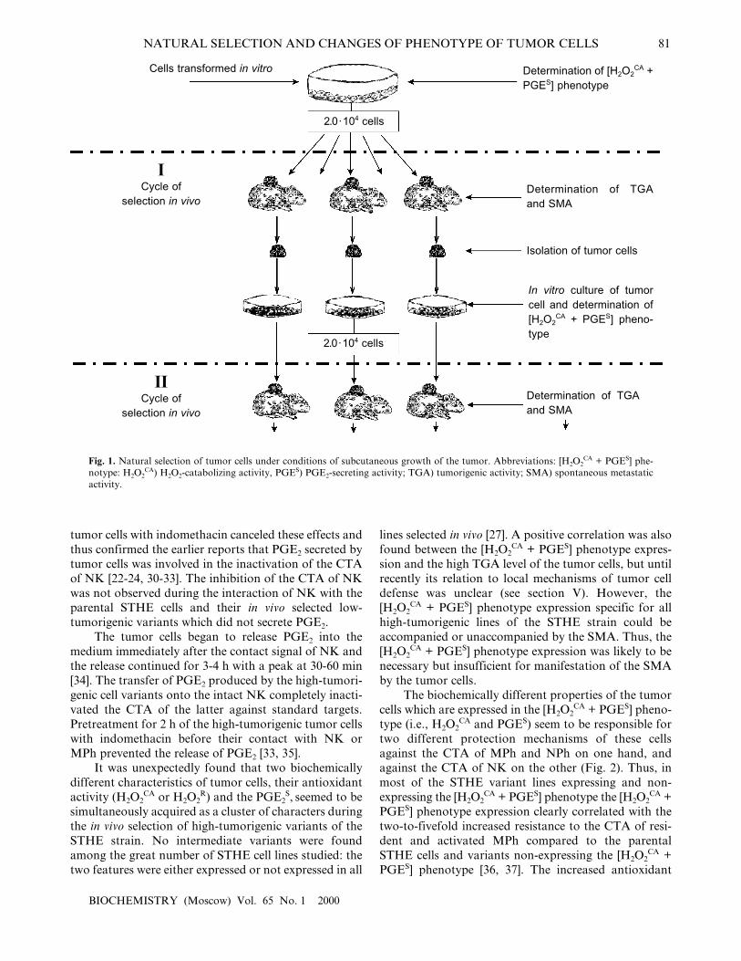
A procedure was elaborated for in vivo selection ofvariants of these cells during their subcutaneous growthin normal animals. Each cycle of the selection was start-ed with a subcutaneous transplantation of 2.0⋅104 tumorcells grown in the in vitro culture; their growth in vivowas continued for 55-60 days (Fig. 1). Then the animalswere sacrificed, cells from the tumor nodes from indi-
vidual animals were again put into tissue culture, andtheir characteristics were studied after 3-5 in vitro pas-sages (to remove the host�s stroma cells).
The sensitivity of tumor cells to damage by H2O2
(H2O2R) was tested by incorporation of [3H]TdR into
intact cells (control) and into cells treated with fivetwofold different doses of H2O2 (from 1.65 to 27.0 mM).The H2O2
CA was determined using luminol-dependentchemiluminescence which was recorded at the timerequired for inactivation of 95% of H2O2 in the presenceof an extract from the cells under study (versus the timerequired for inactivation of H2O2 in solution without thecell extract). The H2O2
CA was considered positive whenthe inactivation of 95% of H2O2 added to the tumorextract required 20 sec - 3 min. These two tests were con-venient for the evaluation of total antioxidant activity ofthe cells, and their results were essentially the same. Thestandard methods used for determination of the H2O2
R
and H2O2CA and also of some enzymatic antioxidants are
described elsewhere [26-29].The PGE2
S activity of tumor cells was determinedwith a standard radioimmunoassay (RIA) [24] and avery sensitive and reproducible biological test based onthe ability of PGE2 to inactivate the CTA of NK wasespecially elaborated for this work. This method makesit possible to determine the biological activity of nativePGE2 secreted by tumor cells and has some importantbenefits versus the RIA [30].
The quantitative test of the TGA includes the sub-cutaneous transplantation into five or six normal Syrianhamsters with four tenfold different doses of the tumorcells with subsequent recording of final (55-60 dayslater) results of the transplantation and determination of50% transplantation dose (log TrD50). This approachappears to be very informative, and its high repro-ducibility enables the correct evaluation of the TGAlevel of tumor cell lines under study. Standardapproaches were also used for quantitative determina-tion of the EMA, SMA, and also of the cell sensitivity tothe CTA of MPh, NPh, and NK.
Using these methods the in vivo selected high-tumorigenic variants of STHE cells were shown to havemore than the tenfold increased level of H2O2
R com-pared to the low-tumorigenic variants and the parentalSTHE cells and also significantly accelerated H2O2
CA
(from 15-20 min characteristic for the extract from theparent STHE cells to 20-180 sec for the high-tumori-genic variants). The high-tumorigenic cell variants alsohad constantly increased activity of catalase by 20-60%and decreased activity of CuZn-SOD and of lipid perox-idation. Changes in other antioxidant enzymes were lessregular [26-28].
The in vivo selected high-tumorigenic variants ofSTHE cells were significantly more resistant to the CTAof NK, and, moreover, direct contact resulted in therapid loss of the CTA by the NK. Pretreatment of the
NATURAL SELECTION AND CHANGES OF PHENOTYPE OF TUMOR CELLS 81
BIOCHEMISTRY (Moscow) Vol. 65 No. 1 2000
tumor cells with indomethacin canceled these effects andthus confirmed the earlier reports that PGE2 secreted bytumor cells was involved in the inactivation of the CTAof NK [22-24, 30-33]. The inhibition of the CTA of NKwas not observed during the interaction of NK with theparental STHE cells and their in vivo selected low-tumorigenic variants which did not secrete PGE2.
The tumor cells began to release PGE2 into themedium immediately after the contact signal of NK andthe release continued for 3-4 h with a peak at 30-60 min[34]. The transfer of PGE2 produced by the high-tumori-genic cell variants onto the intact NK completely inacti-vated the CTA of the latter against standard targets.Pretreatment for 2 h of the high-tumorigenic tumor cellswith indomethacin before their contact with NK orMPh prevented the release of PGE2 [33, 35].
It was unexpectedly found that two biochemicallydifferent characteristics of tumor cells, their antioxidantactivity (H2O2
CA or H2O2R) and the PGE2
S, seemed to besimultaneously acquired as a cluster of characters duringthe in vivo selection of high-tumorigenic variants of theSTHE strain. No intermediate variants were foundamong the great number of STHE cell lines studied: thetwo features were either expressed or not expressed in all
lines selected in vivo [27]. A positive correlation was alsofound between the [H2O2
CA + PGES] phenotype expres-sion and the high TGA level of the tumor cells, but untilrecently its relation to local mechanisms of tumor celldefense was unclear (see section V). However, the[H2O2
CA + PGES] phenotype expression specific for allhigh-tumorigenic lines of the STHE strain could beaccompanied or unaccompanied by the SMA. Thus, the[H2O2
CA + PGES] phenotype expression was likely to benecessary but insufficient for manifestation of the SMAby the tumor cells.
The biochemically different properties of the tumorcells which are expressed in the [H2O2
CA + PGES] pheno-type (i.e., H2O2
CA and PGES) seem to be responsible fortwo different protection mechanisms of these cellsagainst the CTA of MPh and NPh on one hand, andagainst the CTA of NK on the other (Fig. 2). Thus, inmost of the STHE variant lines expressing and non-expressing the [H2O2
CA + PGES] phenotype the [H2O2CA +
PGES] phenotype expression clearly correlated with thetwo-to-fivefold increased resistance to the CTA of resi-dent and activated MPh compared to the parentalSTHE cells and variants non-expressing the [H2O2
CA +PGES] phenotype [36, 37]. The increased antioxidant
Cells transformed in vitro Determination of [H2O2CA +
PGES] phenotype
2.0· 104 cells
2.0· 104 cells
Determination of TGAand SMA
Isolation of tumor cells
In vitro culture of tumorcell and determination of[H2O2
CA + PGES] pheno-type
ICycle of
selection in vivo
IICycle of
selection in vivo
Fig. 1. Natural selection of tumor cells under conditions of subcutaneous growth of the tumor. Abbreviations: [H2O2CA + PGES] phe-
notype: H2O2CA) H2O2-catabolizing activity, PGES) PGE2-secreting activity; TGA) tumorigenic activity; SMA) spontaneous metastatic
activity.
Determination of TGAand SMA

82 DEICHMAN
BIOCHEMISTRY (Moscow) Vol. 65 No. 1 2000
activity of malignant variants of the tumor cells corre-lated, as a rule, with the increased activity of catalase,and this probably protected the tumor cells against dam-age by reactive oxygen forms and H2O2 produced byMPh and NPh.
In contrast, the release of PGE2 by the same tumorcells during contact interaction with NK and MPhinhibited the CTA of NK and thus, an active and essen-tially aggressive protection of the tumor cells against theCTA of NK cells was displayed. Moreover, pretreat-ment of such tumor cells with indomethacin recoverstheir sensitivity to the CTA of NK [38]. Thus, the dis-crete mechanism of resistance of the STHE high-tumori-genic variants to the CTA of NK is determined by theirability to inactivate the CTA of NK by release of PGE2
in response to contact interaction with these effectors.Paradoxically, during this interaction, the tumor cellsand NK interchange their roles: the first play as effec-tors, whereas NK become their unprotected targets. TheCTA of MPh was significantly (by two orders of magni-tude) less sensitive to the suppression by PGE2; howev-er, the PGE2-induced decrease in the production bymacrophages of H2O2 which is toxic for tumor cells andmay be an additional protection mechanism of tumorcells against these effectors. Such an autoregulationmechanism of H2O2 release and production of PGE2 was
described for activated MPh. Its biological significanceseems to be in protecting MPh against excess damagewith H2O2 [21]. Certain mechanisms of tumor cell pro-tection against MPh and NK are schematically shown inFig. 2.
The two described protection mechanisms of malig-nant variants of tumor cells against MPh, NPh, and NKwhich are based on the [H2O2
CA + PGES] phenotypeexpression do not exhaust all possibilities of tumor cellprotection; in particular, tumor cells can produceunidentified factors which suppress the innate antitumorimmunity and thus allow the tumor to �sneak through�the host�s protection barriers [39]. Apoptosis of NK andT cells induced during the FAS�FASL-interaction ofthese effectors with tumor cells should be noted amongrecently found new mechanisms of tumor cell protec-tion. This mechanism of tumor cell protection hasalready been described elsewhere [40-43].
The mechanism of genetic regulation of charactersresponsible for the [H2O2
CA + PGES] phenotype is stillunknown. While the H2O2
R (and H2O2CA) of tumor cells
presents the total activity of various genes responsiblefor catalase, CuZn-SOD, and glutathione peroxidase,the releasing of PGE2 (which is one of ultimate metabo-lites of arachidonic acid) is determined by the activity ofone enzyme, cyclooxygenase. Until recently, these char-
Protection mechanisms
of the organism of the tumor
by activation by selection of cells which have
--------------------------------------------------------------------------------------------------------------------↓ ↓ ↓ ↓ ↓ ↓ ↓ ↓ ↓ ↓
Fig. 2. Innate immunity of the host and tumor progression.
A. of macrophages and neutrophils (pro-ducing various cytotoxic factors includingreactive forms of O2 (H2O2, O2
�))
A. high antioxidant activity(the H2O2
R, the H2O2CA)
B. of natural killers (NK) B. ability to release PGE2 duringcontact with NK and MPh (PGE2)
Arrest of the growth, the tumor suppressionand/or prevention
Expression of new stable phenotype[H2O2
CA + PGES]
immunosuppression and tumor growth↓ ↓

NATURAL SELECTION AND CHANGES OF PHENOTYPE OF TUMOR CELLS 83
BIOCHEMISTRY (Moscow) Vol. 65 No. 1 2000
acters (the antioxidant activity and production of PGE2)were usually considered separately, disregarding theirinterrelations. However, the co-expression of the char-acters which determine the antioxidant and PGE2-secreting activities suggest a common regulation of thesetwo biochemically different properties of tumor cells. Inthis case, the in vivo selection of H2O2
R variants of tumorcells (e.g., by activated MPh or NPh) would result in theacquisition of PGE2
S without involvement of NK, and,on the contrary, NK would be responsible for selectionof cells expressing both these characteristics.
It is still unclear whether cell variants expressingthe [H2O2
CA + PGES] phenotype preexist in the popula-tion of the parental STHE strain. However, it is obviousthat the in vitro cultivation fails to reveal their selectiveadvantages. We were unsuccessful in our attempts toselect in vitro cells of the STHE strain expressing[H2O2
CA + PGES] phenotype by a long-term treatmentwith low cytotoxic doses of H2O2 (unpublished data) orby a long-term co-culture of the tumor cells with theresident or activated MPh. In these experiments theacquisition by the STHE cells of a certain resistance tothe H2O2-induced damage was not accompanied by theexpression of PGES [44]. It seems to indicate that cellvariants expressing the [H2O2
CA + PGES] phenotype donot preexist in the parental population of cells whichwere spontaneously transformed in vitro and that theexpression of this phenotype more likely is a new char-acteristic of the tumor cells which is acquired underconditions of their in vivo growth and natural selection(including the selection determined by the CTA ofMPh, NK, and NPh) and provides them selectiveadvantages in vivo during the interaction with theseeffectors. The [H2O2
CA + PGES] phenotype expressioncorrelated with a 30-200-fold increase in the TGA(accompanied or unaccompanied with SMA) of variantlines of the STHE strain, and this prompted us to studyother models of in vitro cell transformation especiallythose specified by the high TGA level induced duringtransformation.
II. EXPRESSION OF [H2O2CA + PGES]
PHENOTYPE BY CELLS TRANSFORMED in vitroBY ROUS SARCOMA VIRUS; POSSIBLE ROLE
OF THE v-src GENE
It was shown for the first time in the 1960s and thenconfirmed that cells of mice, rats, and hamsters trans-formed in vitro by the Rous Sarcoma Virus(Schmidt�Ruppin strain) (RSV-SR) had an extremelyhigh level of TGA and often of the SMA [45-47]. Muchlater, the v-src-induced carcinogenesis was characterizedas an exclusive one-step type of neoplasm different fromother types of carcinogenesis. The mechanism of one-step carcinogenesis still remains unclear [48].
Therefore, in the light of the data presented in sec-tion I, it was interesting to study characteristics of cellstransformed in vitro by RSV-SR, including the possibleexpression of the [H2O2
CA + PGES] phenotype, TGA,SMA, and the sensitivity to the CTA of MPh and NK.For this purpose we subjected normal embryonic cells ofSyrian hamster to in vitro transformation with RSV-SRand thus acquired four independent strains of v-src-transformants.
It was shown that during the in vitro transformationthese four strains acquired the expression of the [H2O2
CA +PGES] phenotype, extremely high levels of catalase,TGA, and SMA (the latter was found in three of thefour lines), high resistance to the CTA of MPh and NK.The [H2O2
CA + PGES] phenotype of these cells wasexpressed on the level of both, i.e., cell populations andclones [28, 29, 49, 50]. Some characteristics of two linesof the v-src-transformants are presented in Table 1.
A nonrandom correlation between the [H2O2CA +
PGES] phenotype expression and a high level of TGA ofcells of RSV-SR-transformants was confirmed by exper-iments when the antioxidant activity (H2O2
R) of thesecells and PGES were preliminary suppressed in vitro, sep-arately or combined, by nontoxic doses of BCNU andindomethacin and the TGA level of the treated andintact cells was subsequently determined in vivo. Thetreatment with both agents or only with BCNU morethan 150-fold suppressed the TGA of the cells; a singletreatment with indomethacin alone insignificantly sup-pressed the TGA [49].
The unusually high antioxidant activity of the cellstrains RSV-SR-transformed in vitro is mainly due to thehigh activity of catalase which in these cells is 1.8-2.5-fold higher than in spontaneously transformed cells ofthe STHE strain and fivefold higher than in normalfibroblasts (Table 1).
The [H2O2CA + PGES] phenotype expression by cells
of the RSV-SR-transformants (similarly to the earliershown pattern for the STHE variants selected in vivo)was directly responsible for the cell resistance to the CTAof MPh and NK. Pretreatment of the RSV-SR-transfor-mants with indomethacin made them highly sensitive tothe CTA of NK [38]. The initially high resistance of v-src-transformants to the CTA of MPh and NK was espe-cially surprising because these cells had not been selectedin vivo and had no preliminary experience of interactionwith these effectors of the innate antitumor immunity.
Evidently, in the RSV-SR-transformants the[H2O2
CA + PGES] phenotype expression, high level of theTGA, and the acquisition of resistance to the CTA ofMPh and NK are determined by the transforming activ-ity of the v-src gene and do not depend on their selectionin vivo by MPh or NK, as it was earlier supposed for thein vivo selection of STHE variants (see section I).
The specific role of the v-src gene and its trans-forming activity in the induction of the [H2O2
CA + PGES]

84 DEICHMAN
BIOCHEMISTRY (Moscow) Vol. 65 No. 1 2000
phenotype and also of high levels of the TGA, SMA,and resistance to the CTA of MPh and NK becameclearer due to following studies. It occurred that two ofour RSV-SR-transformants (lowly metastatic HET-SRand highly metastatic HET-SR-1) were transfected withplasmid DNA which contained the activated gene N-ras(neo). It was shown earlier that p21ras and pp60v-src usedthe same signal pathway�via phosphatidylinositol-3-kinase [51, 52]. Expression of the v-src gene (mRNA andenolase activity of pp60) was significantly (nearly com-pletely) inhibited in the HET-SR cells transfected withN-ras, whereas the expression of p21N-ras increased ten-fold and more [53]. Simultaneously with the inhibitionof activity of the v-src gene, the N-ras-transfected HET-SR cells completely lost the expression of the [H2O2
CA +PGES] phenotype [54]. In the N-ras-transfectants of theHET-SR-1 strain the expression of pp60v-src was signifi-cantly less suppressed and the expression of the gene N-ras (p21ras) increased only 2.0-2.5-fold. In this case cellsof most of the transfected clones under study retainedthe H2O2
CA but lost the PGES. It should be noted thatthe N-ras-transfection of the RSV-SR-transformed cellswas found to be the only condition that resulted either inthe complete inhibition of activity of the v-src-gene andthe complete loss of the [H2O2
CA + PGES] phenotype or,in the case of partial inhibition of the v-src-gene and lowactivity of the transfected N-ras, in the dissociation ofcharacters which contribute to the [H2O2
CA + PGES]phenotype (that always occurred only due to the loss ofPGES).
The interaction of pp60v-src and p21N-ras in variousrecipient cells can occur in more than one site of the
signal transduction pathway initiating from the G-protein and moving through PI-3 [55]; the level ofgenetic instability of transfectant cells is suggested toinfluence this interaction. The complete inhibition ofthe [H2O2
CA + PGES] phenotype during the N-ras-transfection of the HET-SR cells is likely to be associ-ated with the initial part of the signal pathway abovethe activation of PLC (phospholipase C), while theinhibition only of PGE2 with the retained H2O2
CA inthe N-ras-transfected HET-SR-1 cells seems to beassociated with the interaction of pp60v-src and p21N-ras
downstream of the signal pathway [53, 54]. In anycase, the complete or partial inhibition of the v-src-gene activity in the RSV-SR-transformants in most N-ras-transfectant clones is accompanied by complete orpartial suppression of the [H2O2
CA + PGES] phenotype,respectively, and this indicates that the expression ofthis phenotype in the RSV-SR-transformants dependson v-src-gene activity. Later we showed that the trans-fection of cells of both the HET-SR and STHE strainswith the genes N-ras and Ha-ras suppressed theirantioxidant activity due to significant (virtually com-plete) inhibition of catalase which was graduallyrecovered during the subsequent two-three cycles of invivo selection [28]. Thus, it was shown for the first timethat the significantly increased activity of catalasealong with the PGE2
S are specific biochemical mani-festations of cell transformation in vitro by the v-src-gene, whereas genes N-ras and Ha-ras inhibited thesetwo v-src-induced cell characteristics.
It seems that only one type of normal cells canexpress a similar [H2O2
CA + PGES] phenotype, namely,
GP6 (nmoles/min per mg
protein)
19.4 ± 0.01
67.0 ± 2.2
144.6 ± 7.8
99.0 ± 7.7
CuZn-SOD5
(IU/mg protein)
1587 ± 42
803 ± 22
320 ± 17
293 ± 6
catalase4
(U/mg protein)
4.0 ± 0.3
8.6 ± 0.6
17.7 ± 1.1
16.3 ± 0.7
3PGE2
S
(+) or (�)
�
�
+
+
2
H2O2CA
≥ 10 min
> 5 min
10-20 sec
10-20 sec
Transforma-tion in vitro
�
spontaneous
RSV-SR
Cell line
HEF1
STHE
HET-SR
HET-SR-1
SMÀ8
(+) or (�)
�
�
�
+++
Table 1. Characteristics of Syrian hamster cell lines transformed in vitro by the Rous sarcoma virus (RSV-SR) com-pared to normal cells and spontaneously transformed cells of the STHE strain
Normal fibroblasts of Syrian hamster.H2O2-catabolizing activity of cell extracts (min, sec); details in [28].PGE2-secreting activity, (+) or (�), during contact interaction of target cells with NK [28].Spectrophotometric determination, units per mg protein (after Aebi, 1984; details in [28]).Spectrophotometric determination, international units per mg protein (after Beauchamp and Fridovich, 1971; details in [28]).Glutathione peroxidase (GP) was determined in nanomoles per mg protein per min (after Tietze, 1969; details in [28]).Tumorigenic activity determined as log of 50% transplantation dose of tumor cells; details in [49].Spontaneous metastatic activity determined with standard tests (+) or (�); details in [49].
TGA7
(log TrD50)
�
2.7-3.4
0.5-1.4
Antioxidant activity of enzymesExpression in vitro
}}1
2
3
4
5
6
7
8
NATURAL SELECTION AND CHANGES OF PHENOTYPE OF TUMOR CELLS 85
BIOCHEMISTRY (Moscow) Vol. 65 No. 1 2000
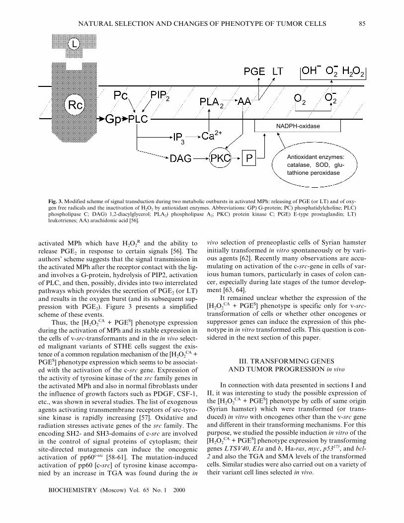
activated MPh which have H2O2R and the ability to
release PGE2 in response to certain signals [56]. Theauthors� scheme suggests that the signal transmission inthe activated MPh after the receptor contact with the lig-and involves a G-protein, hydrolysis of PIP2, activationof PLC, and then, possibly, divides into two interrelatedpathways which provides the secretion of PGE2 (or LT)and results in the oxygen burst (and its subsequent sup-pression with PGE2). Figure 3 presents a simplifiedscheme of these events.
Thus, the [H2O2CA + PGES] phenotype expression
during the activation of MPh and its stable expression inthe cells of v-src-transformants and in the in vivo select-ed malignant variants of STHE cells suggest the exis-tence of a common regulation mechanism of the [H2O2
CA +PGES] phenotype expression which seems to be associat-ed with the activation of the c-src gene. Expression ofthe activity of tyrosine kinase of the src family genes inthe activated MPh and also in normal fibroblasts underthe influence of growth factors such as PDGF, CSF-1,etc., was shown in several studies. The list of exogenousagents activating transmembrane receptors of src-tyro-sine kinase is rapidly increasing [57]. Oxidative andradiation stresses activate genes of the src family. Theencoding SH2- and SH3-domains of c-src are involvedin the control of signal proteins of cytoplasm; theirsite-directed mutagenesis can induce the oncogenicactivation of pp60c-src [58-61]. The mutation-inducedactivation of pp60 [c-src] of tyrosine kinase accompa-nied by an increase in TGA was found during the in
vivo selection of preneoplastic cells of Syrian hamsterinitially transformed in vitro spontaneously or by vari-ous agents [62]. Recently many observations are accu-mulating on activation of the c-src-gene in cells of var-ious human tumors, particularly in cases of colon can-cer, especially during late stages of the tumor develop-ment [63, 64].
It remained unclear whether the expression of the[H2O2
CA + PGES] phenotype is specific only for v-src-transformation of cells or whether other oncogenes orsuppressor genes can induce the expression of this phe-notype in in vitro transformed cells. This question is con-sidered in the next section of this paper.
III. TRANSFORMING GENES AND TUMOR PROGRESSION in vivo
In connection with data presented in sections I andII, it was interesting to study the possible expression ofthe [H2O2
CA + PGES] phenotype by cells of same origin(Syrian hamster) which were transformed (or trans-duced) in vitro with oncogenes other than the v-src geneand different in their transforming mechanisms. For thispurpose, we studied the possible induction in vitro of the[H2O2
CA + PGES] phenotype expression by transforminggenes LTSV40, E1a and b, Ha-ras, myc, p53175, and bcl-2 and also the TGA and SMA levels of the transformedcells. Similar studies were also carried out on a variety oftheir variant cell lines selected in vivo.
Rc
L
2
3
2+
2 2 2
2 22
Fig. 3. Modified scheme of signal transduction during two metabolic outbursts in activated MPh: releasing of PGE (or LT) and of oxy-gen free radicals and the inactivation of H2O2 by antioxidant enzymes. Abbreviations: GP) G-protein; PC) phosphatidylcholine; PLC)phospholipase C; DAG) 1,2-diacylglycerol; PLA2) phospholipase A2; PKC) protein kinase C; PGE) E-type prostaglandin; LT)leukotrienes; AA) arachidonic acid [56].
NADPH-oxidase
Antioxidant enzymes:catalase, SOD, glu-tathione peroxidase
86 DEICHMAN
BIOCHEMISTRY (Moscow) Vol. 65 No. 1 2000
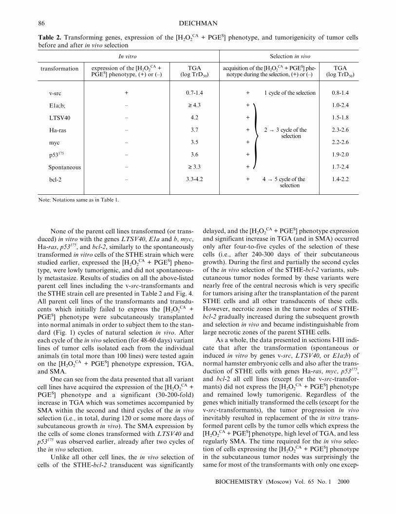
None of the parent cell lines transformed (or trans-duced) in vitro with the genes LTSV40, E1a and b, myc,Ha-ras, p53175, and bcl-2, similarly to the spontaneouslytransformed in vitro cells of the STHE strain which werestudied earlier, expressed the [H2O2
CA + PGES] pheno-type, were lowly tumorigenic, and did not spontaneous-ly metastasize. Results of studies on all the above-listedparent cell lines including the v-src-transformants andthe STHE strain cell are presented in Table 2 and Fig. 4.All parent cell lines of the transformants and transdu-cents which initially failed to express the [H2O2
CA +PGES] phenotype were subcutaneously transplantedinto normal animals in order to subject them to the stan-dard (Fig. 1) cycles of natural selection in vivo. Aftereach cycle of the in vivo selection (for 48-60 days) variantlines of tumor cells isolated each from the individualanimals (in total more than 100 lines) were tested againon the [H2O2
CA + PGES] phenotype expression, TGA,and SMA.
One can see from the data presented that all variantcell lines have acquired the expression of the [H2O2
CA +PGES] phenotype and a significant (30-200-fold)increase in TGA which was sometimes accompanied bySMA within the second and third cycles of the in vivoselection (i.e., in total, during 120 or some more days ofsubcutaneous growth in vivo). The SMA expression bythe cells of some clones transformed with LTSV40 andp53175 was observed earlier, already after two cycles ofthe in vivo selection.
Unlike all other cell lines, the in vivo selection ofcells of the STHE-bcl-2 transducent was significantly
delayed, and the [H2O2CA + PGES] phenotype expression
and significant increase in TGA (and in SMA) occurredonly after four-to-five cycles of the selection of thesecells (i.e., after 240-300 days of their subcutaneousgrowth). During the first and partially the second cyclesof the in vivo selection of the STHE-bcl-2 variants, sub-cutaneous tumor nodes formed by these variants werenearly free of the central necrosis which is very specificfor tumors arising after the transplantation of the parentSTHE cells and all other transducents of these cells.However, necrotic zones in the tumor nodes of STHE-bcl-2 gradually increased during the subsequent growthand selection in vivo and became indistinguishable fromlarge necrotic zones of the parent STHE cells.
As a whole, the data presented in sections I-III indi-cate that after the transformation (spontaneous orinduced in vitro by genes v-src, LTSV40, or E1a;b) ofnormal hamster embryonic cells and also after the trans-duction of STHE cells with genes Ha-ras, myc, p53175,and bcl-2 all cell lines (except for the v-src-transfor-mants) did not express the [H2O2
CA + PGES] phenotypeand remained lowly tumorigenic. Regardless of thegenes which initially transformed the cells (except for thev-src-transformants), the tumor progression in vivoinevitably resulted in replacement of the in vitro trans-formed parent cells by the tumor cells which express the[H2O2
CA + PGES] phenotype, high level of TGA, and lessregularly SMA. The time required for the in vivo selec-tion of cells expressing the [H2O2
CA + PGES] phenotypein the subcutaneous tumor nodes was surprisingly thesame for most of the transformants with only one excep-
transformation
v-src
E1a;b;
LTSV40
Ha-ras
myc
p53175
Spontaneous
bcl-2
TGA (log TrD50)
0.8-1.4
1.0-2.4
1.5-1.8
2.3-2.6
2.2-2.6
1.9-2.0
1.7-2.4
1.4-2.2
Table 2. Transforming genes, expression of the [H2O2CA + PGES] phenotype, and tumorigenicity of tumor cells
before and after in vivo selection
acquisition of the [H2O2CA + PGES] phe-
notype during the selection, (+) or (�)
+ 1 cycle of the selection
+
+
+ 2 → 3 cycle of the selection
+
+
+
+ 4 → 5 cycle of the selection
TGA (log TrD50)
0.7-1.4
≥ 4.3
4.2
3.7
3.5
3.6
≥ 3.3
3.3-4.2
expression of the [H2O2CA +
PGES] phenotype, (+) or (�)
+
�
�
�
�
�
�
�
Selection in vivo
Note: Notations same as in Table 1.
In vitro
}
NATURAL SELECTION AND CHANGES OF PHENOTYPE OF TUMOR CELLS 87
BIOCHEMISTRY (Moscow) Vol. 65 No. 1 2000
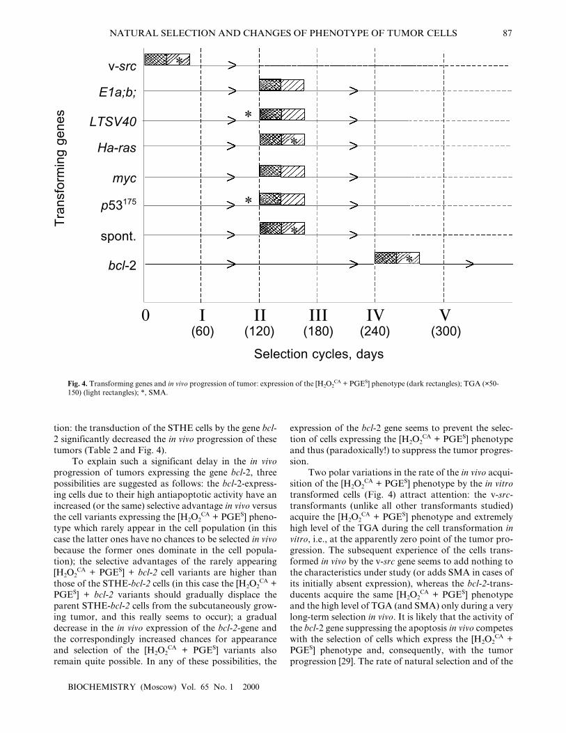
tion: the transduction of the STHE cells by the gene bcl-2 significantly decreased the in vivo progression of thesetumors (Table 2 and Fig. 4).
To explain such a significant delay in the in vivoprogression of tumors expressing the gene bcl-2, threepossibilities are suggested as follows: the bcl-2-express-ing cells due to their high antiapoptotic activity have anincreased (or the same) selective advantage in vivo versusthe cell variants expressing the [H2O2
CA + PGES] pheno-type which rarely appear in the cell population (in thiscase the latter ones have no chances to be selected in vivobecause the former ones dominate in the cell popula-tion); the selective advantages of the rarely appearing[H2O2
CA + PGES] + bcl-2 cell variants are higher thanthose of the STHE-bcl-2 cells (in this case the [H2O2
CA +PGES] + bcl-2 variants should gradually displace theparent STHE-bcl-2 cells from the subcutaneously grow-ing tumor, and this really seems to occur); a gradualdecrease in the in vivo expression of the bcl-2-gene andthe correspondingly increased chances for appearanceand selection of the [H2O2
CA + PGES] variants alsoremain quite possible. In any of these possibilities, the
expression of the bcl-2 gene seems to prevent the selec-tion of cells expressing the [H2O2
CA + PGES] phenotypeand thus (paradoxically!) to suppress the tumor progres-sion.
Two polar variations in the rate of the in vivo acqui-sition of the [H2O2
CA + PGES] phenotype by the in vitrotransformed cells (Fig. 4) attract attention: the v-src-transformants (unlike all other transformants studied)acquire the [H2O2
CA + PGES] phenotype and extremelyhigh level of the TGA during the cell transformation invitro, i.e., at the apparently zero point of the tumor pro-gression. The subsequent experience of the cells trans-formed in vivo by the v-src gene seems to add nothing tothe characteristics under study (or adds SMA in cases ofits initially absent expression), whereas the bcl-2-trans-ducents acquire the same [H2O2
CA + PGES] phenotypeand the high level of TGA (and SMA) only during a verylong-term selection in vivo. It is likely that the activity ofthe bcl-2 gene suppressing the apoptosis in vivo competeswith the selection of cells which express the [H2O2
CA +PGES] phenotype and, consequently, with the tumorprogression [29]. The rate of natural selection and of the
Tra
nsfo
rmin
g ge
nes
LTSV40
v-src
E1a;b;
Ha-ras
myc
p53175
spont.
bcl-2
0(60)
*
I II III IV V
*
*
*
*
*
(120) (180) (240) (300)
Selection cycles, days
Fig. 4. Transforming genes and in vivo progression of tumor: expression of the [H2O2CA + PGES] phenotype (dark rectangles); TGA (×50-
150) (light rectangles); *, SMA.
88 DEICHMAN
BIOCHEMISTRY (Moscow) Vol. 65 No. 1 2000
in vivo acquisition of the [H2O2CA + PGES] phenotype
expression by the cells transformed in vitro with thegenes LTSV40, E1a,b, Ha-ras, myc, and p53175 wasapproximately the same and did not depend on the typeof transforming genes.
In general, the findings permit the [H2O2CA + PGES]
phenotype expression as a marker of a definite relative-ly early (premetastatic) step in the in vivo progression oftumors.
IV. SELECTION OF CELLS BY THE [H2O2
CA + PGES] PHENOTYPE DURING PRIMARY VIRAL CARCINOGENESIS
Until recently the earliest steps of primary carcino-genesis in vivo and phenotypical changes in tumor cellswhich seem to occur during its latent period were inac-cessible for investigation. However, according to Nowell[65], �the fundamental nature of this initial step, anddegree to which it is specific for each neoplasm, remainsa basic problem in cancer research�. To study this prob-lem, we have used a new opportunity, namely, the deter-mination of the [H2O2
CA + PGES] phenotype as a mark-er of the definite and, possibly, one of the earliest stepsin tumor progression in vivo after transformation.
As shown in the previous sections, the acquisitionof the [H2O2
CA + PGES] phenotype expression alongwith the 30-200-fold increase in TGA by the cells whichwere initially transformed or transduced in vitro withvarious antigens occurred during the in vivo selection inthe subcutaneous tumors at surprisingly similar rates(with the above-described two polar variations). Thiscould indicate that the initial population of cells trans-formed in vitro with various agents did not contain vari-ants expressing the [H2O2
CA + PGES] phenotype and/orthere was no conditions for their selection in vitro. Thishypothesis is supported by the finding that the probabil-
ity of their appearance de novo and the selection rateduring the tumor growth in vivo were nearly the same forvarious transformants. However, the possibility andtime of the acquisition of the [H2O2
CA + PGES] pheno-type expression by the cells during the appearance of pri-mary tumors remained unknown.
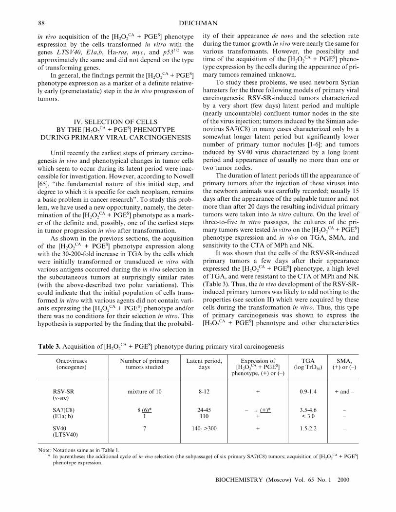
To study these problems, we used newborn Syrianhamsters for the three following models of primary viralcarcinogenesis: RSV-SR-induced tumors characterizedby a very short (few days) latent period and multiple(nearly uncountable) confluent tumor nodes in the siteof the virus injection; tumors induced by the Simian ade-novirus SA7(C8) in many cases characterized only by asomewhat longer latent period but significantly lowernumber of primary tumor nodules [1-6]; and tumorsinduced by SV40 virus characterized by a long latentperiod and appearance of usually no more than one ortwo tumor nodes.
The duration of latent periods till the appearance ofprimary tumors after the injection of these viruses intothe newborn animals was carefully recorded; usually 15days after the appearance of the palpable tumor and notmore than after 20 days the resulting individual primarytumors were taken into in vitro culture. On the level ofthree-to-five in vitro passages, the cultures of the pri-mary tumors were tested in vitro on the [H2O2
CA + PGES]phenotype expression and in vivo on TGA, SMA, andsensitivity to the CTA of MPh and NK.
It was shown that the cells of the RSV-SR-inducedprimary tumors a few days after their appearanceexpressed the [H2O2
CA + PGES] phenotype, a high levelof TGA, and were resistant to the CTA of MPh and NK(Table 3). Thus, the in vivo development of the RSV-SR-induced primary tumors was likely to add nothing to theproperties (see section II) which were acquired by thesecells during the transformation in vitro. Thus, this typeof primary carcinogenesis was shown to express the[H2O2
CA + PGES] phenotype and other characteristics
Oncoviruses (oncogenes)
RSV-SR(v-src)
SA7(C8)(E1a; b)
SV40 (LTSV40)
SMA, (+) or (�)
+ and �
��
�
Table 3. Acquisition of [H2O2CA + PGES] phenotype during primary viral carcinogenesis
TGA (log TrD50)
0.9-1.4
3.5-4.6< 3.0
1.5-2.2
Expression of[H2O2
CA + PGES]phenotype, (+) or (�)
+
� → (+)*+
+
Latent period,days
8-12
24-45110
140- >300
Number of primarytumors studied
mixture of 10
8 (6)*1
7
Notations same as in Table 1.In parentheses the additional cycle of in vivo selection (the subpassage) of six primary SA7(C8) tumors; acquisition of [H2O2
CA + PGES]phenotype expression.
Note:*

NATURAL SELECTION AND CHANGES OF PHENOTYPE OF TUMOR CELLS 89
BIOCHEMISTRY (Moscow) Vol. 65 No. 1 2000
under study independently of conditions of the in vivogrowth. This conclusion was also supported by findingof the extremely short latent period (8-12 days) of theRSV-SR-induced primary tumors (correlating with thetransformation period of the cells in vitro) and the highmultiplicity of these neoplasms.
Unlike them, eight of nine primary SA7(C8) tumorswhich appeared somewhat later (24-45 days after thevirus inoculation to newborn animals) and were studiedafter 39-65 days of their in vivo development did notexpress the [H2O2
CA + PGES] phenotype and were two-to-three orders of magnitude less tumorigenic (Table 3).However, six of these eight tumors transplanted intoanimals and thus subjected to an additional cycle of invivo selection (during 62 days of subcutaneous growth,i.e., in total, 100-120 days of their development in vivo)demonstrated the [H2O2
CA + PGES] phenotype expres-sion and about tenfold increase in TGA. It is suggestedthat by the end of latent periods of the early SA7(C8)primary tumors or shortly after their appearance, i.e.,within 39-65 days of the tumor development in vivo sometumor cells in the primary tumor nodes (it seems to be aminor undetectable fraction) already expressed the[H2O2
CA + PGES] phenotype, but the period of 39-65days seemed to be too short for these cells to replacetheir parent cells in the tumor node. This hypothesis wasconfirmed by findings on the ninth primary SA7(C8)tumor which appeared after 110 days of the latent peri-od: the cells of this tumor expressed the [H2O2
CA +PGES] phenotype immediately after their appearance(and also an increased level of TGA) (Table 3).
The duration of the latent periods of seven SV40-induced primary tumors varied from 140 to more than300 days, and each of these tumors after its appearanceexpressed the [H2O2
CA + PGES] phenotype (Table 3).The TGA level of primary SV40 tumors was about twoorders of magnitude higher than both, i.e., the earlySA7(C8) primary tumors and the cells transformed bySV40 in vitro, and varied from 1.5 to 2.2 log TrD50.
Based on the time of acquisition of the [H2O2CA +
PGES] phenotype by the cells of SA7(C8)- and SV40-induced primary tumors, the minimal time could beapproximately determined that was required for thereplacement in vivo of the [H2O2
CA + PGES] phenotype-negative cells in the subcutaneous nodes by cells express-ing this phenotype. This time was more than 65 days,and periods about 100-140 days of the tumor growth invivo appeared to be sufficient. Thus, the dynamics of theprocess is the function of time required for the appear-ance and in vivo selection of tumor cells expressing the[H2O2
CA + PGES] phenotype.Unlike the growth of the in vitro transformed cells
in tissue culture, the appearance of primary monoclonaltumors in vivo is usually a multistep process whichincludes the transformation of cells and selection of newvariants of the tumor cells possessing some selective
advantages for growth in vivo. The RSV-SR-inducedone-step carcinogenesis is the exception from this rule.Findings on the primary SA7(C8)- and SV40-carcino-genesis indicated that the appearance and selection ofcell variants which express the [H2O2
CA + PGES] pheno-type and, correspondingly, can protect themselvesagainst effectors of the innate antitumor immunity startsduring the latent period and, depending on its duration,could be completed before its termination. Primarytumors appearing in vivo are usually monoclonal (andneoplasms induced by SV40 and adenoviruses are not anexclusion from this rule), and this supports the sugges-tion that the occasional appearance of rare cell variantsexpressing the [H2O2
CA + PGES] phenotype is associatedwith their strict selection that provide the maintenanceof tumor cells with selective advantages in vivo.
The RSV-SR-transformed cells are the strikingexception from this scenario: the cell transformation bythe v-src gene both in vitro and in vivo occurs simultane-ously with their acquisition (without a stage of selectionin vivo) of the [H2O2
CA + PGES] phenotype, the maximalTGA, and even SMA in some cases. The strikingly shortlatent period (8-12 days) till the appearance of the pri-mary palpable tumors induced with RSV-SR, theirappearance in all inoculated animals, and the multiplic-ity of these neoplasms suggest a nonrandom associationbetween the expression by these cells of the v-src gene,the [H2O2
CA + PGES] phenotype, and the high level oftheir TGA. It remains important to elucidate to whatdegree the probability and incidence of various tumorsand also the duration of their latent periods are deter-mined by the appearance of cells expressing the [H2O2
CA +PGES] phenotype.
V. EXPRESSION OF [H2O2CA + PGES] PHENOTYPE
AND EFFICIENCY OF SPECIFIC ANTITUMOR IMMUNITY
It remained unclear whether the local protection oftumor cells based on the [H2O2
CA + PGES] phenotypeexpression and directed against effectors of the innateantitumor immunity is related to specific antitumorimmunity. In particular, two questions were interestingfor us: a possible effect of the specific antitumor immu-nity on the rate of in vivo selection of tumor cellsexpressing the [H2O2
CA + PGES] phenotype and the pos-sible effect of the [H2O2
CA + PGES] phenotype expres-sion by the tumor cells on the efficiency of their immunerejection under conditions of normal and immune hostorganisms.
To study the first question, cells of Syrian hamstertransformed in vitro with the SV40 virus (strain HE-wtSV40) which expressed TSTA (Tumor SpecificTransplantation Antigen) and did not express the[H2O2
CA + PGES] phenotype were subjected to standard

90 DEICHMAN
BIOCHEMISTRY (Moscow) Vol. 65 No. 1 2000
cycles of selection in vivo in normal and SV40-immu-nized animals. After each cycle of the selection in vivo,cells of the tumor nodes which appeared in the normaland SV40-immunized animals were transferred to invitro tissue culture and tested for the [H2O2
CA + PGES]phenotype expression, whereas the expression of TGA,SMA, and TSTA was tested in vivo. The TSTA expres-sion was determined by the specific immune sensitivityof the tumor cells (the difference between TGA (in logTrD50) of the immune and normal animals) [68]. The[H2O2
CA + PGES] phenotype and high TGA wereacquired during the selection in both normal andimmune animals nearly simultaneously (between the sec-ond and third cycles of the in vivo selection of tumorcells); the rate of in vivo selection of immune resistanttumor cells (in the immune animals) was significantlydecreased compared to the selection by the [H2O2
CA +PGES] phenotype and in most cases required four selec-tion cycles. Apparently, the selective pressure of effec-tors of the specific antitumor immunity on the TSTA-expressing cells is less efficient than the natural selectionof the same tumor cells expressing the [H2O2
CA + PGES]phenotype, and the combination of these two types ofselection (on the immune animals) neither accelerates,nor decreases the rate of the process. Thus, the selectivepressure of the effectors of the innate and specific anti-tumor immunity in vivo on the tumor cells was realizedin parallel and independently.
Nevertheless, the probable effect of the [H2O2CA +
PGES] phenotype expression on the cell sensitivity torejection due to effectors of the specific antitumor
immunity must not be ruled out. This probability wasstudied using two approaches as follows.
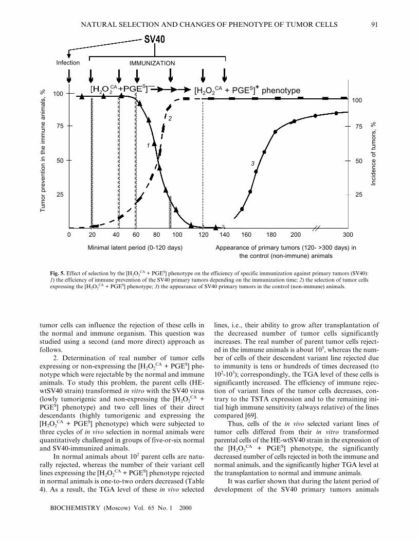
1. The possible prevention of SV40 primary tumorsby a single immunization of the infected newborn ani-mals with the SV40 virus within the latent period or afterits termination. Figure 5 shows summarized results ofsix experiments on such prevention and includes theabove-presented (Table 4) data on the approximatetimes of the [H2O2
CA + PGES] phenotype acquisition bythe cells of SV40 and SA7(C8) primary tumors duringthe latent period.
The specific immunization of animals within thefirst 60-70 days was highly efficient and prevented theappearance of SV40 tumors in 90-100% of the animals(Fig. 5, grey columns). However, the efficiency ofimmune tumor prevention rapidly decreased on the 90-100th days of the latent period and was inefficient fromthe first day after the end of the minimal latent period(although it was long before the peak of appearance ofthe primary tumors) [66-68].
Thus, the period of maximal efficiency of the pre-ventive specific immunization against the SV40 primarytumors (the first 60-70 days of the latent period) wasstrikingly the same as the early [H2O2
CA + PGES] pheno-type-negative stage in the development of these tumorswhen they could not yet protect themselves. And, on thecontrary, the end of this �naive� stage (within 60-100days of the latent period) correlated with the rapiddecrease in the efficiency of the specific immunizationagainst the SV40 tumors (Fig. 5). And the question ishow the [H2O2
CA + PGES] phenotype expression by the
104
5/5
2/5
5/6
1/6
5/5
4/6
5/5
5/5
Animals
normal
immune (experiment 1)
normal
immune (experiment 2)
normal
immune
normal
immune
Expression of[H2O2
CA + PGES]phenotype, (+) or (�)
�
+
+
Numberof selec-
tion cyclesin vivo
1
3
3
Cell line
HE-wtSV40 (initial)
HE-wtSV40- 2SC-4
HE-wtSV40-2SC-5
IndexIS*
≥1.5
≥1.4
1.6
≥1.2
Table 4. Expression of the [H2O2CA + PGES] phenotype, tumorigenicity, and efficiency of immune rejection
Notations same as in Table 1.Index of immune sensitivity (IS) of tumor cells was determined as the difference between values of log TrD50 for immune and normal ani-mals.
TGA (log TrD50)
3.1
≥4.6
3.5
≥4.9
1.7
3.3
<1.2
2.4
103
5/5
0/5
3/6
0/6
5/5
4/6
5/5
4/5
102
0/5
0/5
0/6
0/6
5/5
2/6
5/5
3/5
101
0/5
0/5
0/6
0/6
2/5
0/6
5/5
2/5
Transplantational test
Note:*
NATURAL SELECTION AND CHANGES OF PHENOTYPE OF TUMOR CELLS 91
BIOCHEMISTRY (Moscow) Vol. 65 No. 1 2000
tumor cells can influence the rejection of these cells inthe normal and immune organism. This question wasstudied using a second (and more direct) approach asfollows.
2. Determination of real number of tumor cellsexpressing or non-expressing the [H2O2
CA + PGES] phe-notype which were rejectable by the normal and immuneanimals. To study this problem, the parent cells (HE-wtSV40 strain) transformed in vitro with the SV40 virus(lowly tumorigenic and non-expressing the [H2O2
CA +PGES] phenotype) and two cell lines of their directdescendants (highly tumorigenic and expressing the[H2O2
CA + PGES] phenotype) which were subjected tothree cycles of in vivo selection in normal animals werequantitatively challenged in groups of five-or-six normaland SV40-immunized animals.
In normal animals about 102 parent cells are natu-rally rejected, whereas the number of their variant celllines expressing the [H2O2
CA + PGES] phenotype rejectedin normal animals is one-to-two orders decreased (Table4). As a result, the TGA level of these in vivo selected
lines, i.e., their ability to grow after transplantation ofthe decreased number of tumor cells significantlyincreases. The real number of parent tumor cells reject-ed in the immune animals is about 105, whereas the num-ber of cells of their descendent variant line rejected dueto immunity is tens or hundreds of times decreased (to102-103); correspondingly, the TGA level of these cells issignificantly increased. The efficiency of immune rejec-tion of variant lines of the tumor cells decreases, con-trary to the TSTA expression and to the remaining ini-tial high immune sensitivity (always relative) of the linescompared [69].
Thus, cells of the in vivo selected variant lines oftumor cells differed from their in vitro transformedparental cells of the HE-wtSV40 strain in the expression ofthe [H2O2
CA + PGES] phenotype, the significantlydecreased number of cells rejected in both the immune andnormal animals, and the significantly higher TGA level atthe transplantation to normal and immune animals.
It was earlier shown that during the latent period ofdevelopment of the SV40 primary tumors animals
Fig. 5. Effect of selection by the [H2O2CA + PGES] phenotype on the efficiency of specific immunization against primary tumors (SV40):
1) the efficiency of immune prevention of the SV40 primary tumors depending on the immunization time; 2) the selection of tumor cellsexpressing the [H2O2
CA + PGES] phenotype; 3) the appearance of SV40 primary tumors in the control (non-immune) animals.
Tum
or p
reve
ntio
n in
the
imm
une
anim
als,
% 100
Inci
denc
e of
tum
ors,
%75
50
25
2 2ÑÀ S
1
2
3
100
75
50
25
0 20 40 60 80 100 120 140 160 180 200 300
Minimal latent period (0-120 days) Appearance of primary tumors (120- >300 days) inthe control (non-immune) animals
Infection IMMUNIZATION
[H2O2CA + PGES]+ phenotype
92 DEICHMAN
BIOCHEMISTRY (Moscow) Vol. 65 No. 1 2000
infected with the SV40 virus were neither tolerant to theTSTA of SV40 nor immune [67, 68]. The comparison ofdata presented in Fig. 5 and Table 4 suggests an expla-nation of the rapidly decreased efficiency of the specificimmunoprevention of the SV40 primary tumors duringthe second half of the latent period: during this time thenumber of the [H2O2
CA + PGES] phenotype-expressingtumor cells is gradually increasing, while their real num-ber which is rejectable in the immune organism is simul-taneously decreasing tens or hundreds of times (despitethe retained immune sensitivity). Thus, the acquiredwith the [H2O2
CA + PGES] phenotype ability of tumorcells for local defense against effectors of the innate anti-tumor immunity and for their suppression at the site oftumor growth is suggested to affect the interaction andinterchange of instructive information between thetumor cells and T-cell effectors of the specific antitumorimmunity. Direct local inhibition of T-helpers andkillers by the tumor cells expressing the [H2O2
CA +PGES] phenotype is also not excluded.
The data presented in this section seem to be relat-ed to one of the basic and unsolved problems inimmunology of tumors: why the efficiency of immuneprevention and immune therapy of tumors and metas-tases is, as a rule, lower than expected [6-8, 70, 71]. Itseems likely that local mechanisms of tumor cell defenseagainst effectors of the innate and specific antitumorimmunity which are acquired together with the expres-sion of the [H2O2
CA + PGES] phenotype are an impor-tant and underestimated factor responsible for the lowefficiency of immune treatment of tumors in vivo.
In conclusion, the results of the 10 years of studiesin our laboratory presented in this review have shownthat the secondary phenotypic changes in the trans-formed and tumor cells regularly appear during the nat-ural selection and tumor progression in vivo. Thesechanges are manifested by the highly increased H2O2-catabolizing (antioxidant) activity (H2O2
CA) of tumorcells and the simultaneous acquisition of a new discretecharacter�the ability for the immediate releasing ofprostaglandin E2 (PGES) on contact interaction withNK and MPh. The simultaneous acquisition in vivo ofthese two biochemically different characteristics oftumor cells was found during comparative studies onparent cell lines from Syrian hamster which were trans-formed in vitro with various agents and on more than100 of their descendent lines naturally selected in vivo.The expression of the [H2O2
CA + PGES] phenotype wasshown to provide the cells of both transplantable andprimary tumors of various origin with mechanisms ofactive local protection against the CTA of MPh, NK,NPh, and T-lymphocytes. This significantly increasesthe probability of their survival in vivo and, respectively,their TGA; as well it inhibits the efficiency of their spe-cific and nonspecific rejection in vivo caused by effectorsof the host immunity. Expression of the [H2O2
CA +
PGES] phenotype was also found in the in vitro culturesof various human tumor cell lines (data not presented).
Apparently, the in vivo selection of rare variants oftumor cells expressing the [H2O2
CA + PGES] phenotypesuggests its association with the direct cytotoxic influ-ence of effectors of the innate antitumor immunity:MPh, NPh, and NK. However, experiments with cellstransformed with the v-src gene in vitro (or in vivo) seemto suggest the more complicated pattern of events result-ing in the appearance of the [H2O2
CA + PGES] pheno-type-expressing cells.
The specific role of the v-src gene in induction ofthe [H2O2
CA + PGES] phenotype during the transforma-tion of normal cells with the Rous Sarcoma Virus, theinability of other transforming genes studied (includingthe suppressor genes and bcl-2) to induce in vitro theexpression of this phenotype, and the obligatory selec-tion in vivo of cells expressing the [H2O2
CA + PGES] phe-notype by the same neoplasms of other oncogenesis(i.e., unrelated with the v-src gene) suggest that thetumor cells expressing the [H2O2
CA + PGES] phenotypeappear de novo possibly in relation with occasionaldamage of DNA of the transformed and tumor cells, inparticular, caused by products of the CTA of MPh,NPh, and NK. Such damage can, in particular, result inthe activation of the c-src gene (or other genes of the srcfamily) and in the expression by rare variants of tumorcells of the [H2O2
CA + PGES] phenotype which is specif-ic for the v-src-transformants; the in vivo selectiveadvantages of the tumor cells expressing the [H2O2
CA +PGES] phenotype provide the conditions for survival,growth, and gradual replacement of the initial (non-expressing [H2O2
CA + PGES] phenotype) cells by theirdescendants of this phenotype. Thus in vivo cytotoxiceffectors of the host�s innate immunity system seem toplay the double and crucial role in the selection oftumor cells: under in vivo conditions, the appearance denovo and natual selection of the tumor cells expressingthe [H2O2
CA + PGES] phenotype seem to be preceded byactivation of certain regulatory cell genes specific forthe v-src-transformed cells. Such src-like changes in thetumor phenotype in vivo are likely to be one of the ear-liest (premetastatic) secondary transformations oftumor cells of various origin which are functionallyrequired for their survival in vivo during natural selec-tion and progression.
This work was supported by the State Priorities inScience and Technique program (grant Nos. 47, 46, 22),the Russian Foundation for Basic Research (grant Nos.93-04-20708, 96-04-48054, 99-04-48358), and also by theSoros International Science Foundation (grant No.J3Z100). The author heartily thanks G. I. Abelev and A.D. Altstein for their valuable remarks and discussion ofsome results presented in this review and also N. A.Dyakova for her great help in preparation of the manu-script for press.
NATURAL SELECTION AND CHANGES OF PHENOTYPE OF TUMOR CELLS 93
BIOCHEMISTRY (Moscow) Vol. 65 No. 1 2000
REFERENCES
1. Klein, G. (1975) in Immunology of Tumor�HostRelationship (Smith, R. T., and Landy, M., eds.)Academic Press Inc., N. Y., pp. 201-213.
2. Old, L. J. (1981) Cancer Res., 47, 261-275. 3. Alexander, P., and Eccles, S. (1984) in Cancer Invasion and
Metastasis: Biological and Therapeutic Aspects (Nicolson,G. L., and Milas, L., eds.) Raven Press, N. Y., pp. 293-308.
4. Deichman, G. I. (1984) Advances in Science andTechnology. Oncology [in Russian], Vol. 13, VINITI,Moscow, pp. 46-97.
5. Deichman, G. I. (1988) Cancer Surveys, 7, 675-690. 6. Schrieber, H. (1993) in Fundamental Immunology, 3rd ed.
(Paul, W. E., ed.) Raven Press, N. Y., pp. 1143-1178.7. Klein, E., and Mantovani, A. (1993) Curr. Opin.
Immunol., 5, 714-718.8. Boon, T., Cerrottini, J.-C., van den Eynde, B., van den
Bruggen, P., and van Pel, A. (1994) Ann. Rev. Immunol.,12, 337-366.
9. Weinberg, R. (1989) Cancer Res., 49, 3713-3721.10. Janeway, C. A., and Travers, P. (1997) Immunobiology,
3rd ed., Current Biology Ltd., Garland Publishing Inc., N.Y.-London.
11. Medzhitov, R., and Janeway, C. A. (1997) Curr. Opin.Immunol., 9, 4-9.
12. Bandelac, A., and Fearon, D. T. (1997) Curr. Opin.Immunol., 9, 1-3.
13. Fearon, D. T., and Locksley, R. M. (1996) Science, 272,50-54.
14. Sniderman, R., and Pike, M. C. (1976) Science, 192, 370-372.15. Gorelik, E., Fogel, M., Segal, A., and Feldman, M. (1979)
J. Natl. Cancer Inst., 63, 1397-1404.16. Gorelik, E., Feldman, M., and Segal, S. (1982) Cancer
Immunol. Immunother., 12, 105-109.17. Gronberg, A., Kiessling, R., Ericksson, E., and Hansson,
M. (1981) J. Immunol., 127, 1734-1739.18. Yamashina, K., Fulton, A., and Heppner, G. (1985) J.
Natl. Cancer Inst., 75, 765-770.19. Patek, P. O., Lin, Y., Collins, J. L., and Cohn, M. (1986)
J. Immunol., 136, 741-745.20. Nathan, C. F., Arrick, B. A., Murray, H. W., de Santia,
N. M., and Cohn, Z. A. (1981) J. Exp. Med., 153, 766-782.21. Adams, D. O., and Nathan, C. F. (1983) Immunol. Today,
4, 166-169.22. Droller, M., Schneider, M., and Perlman, P. (1979) Cell
Immunol., 39, 165-177.23. Brunda, M. J., Herberman, R. B., and Holden, H. T.
(1980) J. Immunol., 124, 2682-2687.24. Fulton, A. M. (1984) Int. J. Cancer, 33, 375-379.25. Young, R., and Knies, S. (1984) J. Natl. Cancer Inst., 72,
919-922.26. Deichman, G. I., and Vendrov, E. L. (1986) Int. J. Cancer,
37, 401-409.27. Deichman, G. I., Kluchareva, T. E., Matveeva, V. A.,
Kushlinsky, N. E., Bassalyk, L. S., and Vendrov, E. L.(1989) Int. J. Cancer, 44, 904-907.
28. Deichman, G. I., Kashkina, L. M., Mizenina, O. A.,Gorojanskaya, E. G., Nikiforov, M. A., Gudkov, A. V.,Dyakova, N. A., Komelkov, A. V., Prilutskaya, M. O.,Kushlinsky, N. E., and Tatosyan, A. G. (1996) Int. J.Cancer, 66, 747-752.
29. Deichman, G. I., Matveeva, V. A., Kashkina, L. M.,Dyakova, N. A., Uvarova, E. N., Nikiforov, M. A., andGudkov, A. V. (1998) Int. J. Cancer, 75, 277-283.
30. Kluchareva, T. E., Matveeva, V. A., and Kushlinsky, N.E. (1992) Immunol. Lett., 33, 239-246.
31. Kluchareva, T. E., and Matveeva, V. A. (1983) Byull.Eksp. Biol. Med., 10, 86-89.
32. Kluchareva, T. E., and Matveeva, V. A. (1985) Byull.Eksp. Biol. Med., 6, 732-734.
33. Kluchareva, T. E., Matveeva, V. A., Bassalyk, L. S., andKushlinsky, N. E. (1988) Byull. Eksp. Biol. Med., 2, 204-206.
34. Matveeva, V. A., Kluchareva, T. E., and Uvarova, E. N.(1993) Byull. Eksp. Biol. Med., 2, 193-194.
35. Kluchareva, T. E., Matveeva, V. A., Kashkina, L. M.,Kushlinsky, N. F., Bassalyk, L. S., and Prilutskaya, M. O.(1989) Eksp. Onkol., 11, 53-56.
36. Lavnikova, N. A., and Burdelya, L. G. (1990) Byull. Eksp.Biol. Med., 109, 954-956.
37. Burdelya, L. G. (1997) Neoplasma, 44, 31-35.38. Kluchareva, T. E., Matveeva, V. A., and Uvarova, E. N.
(1990) Byull. Eksp. Biol. Med., 9, 308-310.39. Deichman, G. I., Kluchareva, T. E., Kashkina, L. M., and
Matveeva, V. A. (1979) Int. J. Cancer, 23, 571-584.40. Hahne, M., Rimoldi, D., Schröter, M., Romero, P.,
Schreier, M., French, L. E., Schneider, P., Bornand, T.,Fontana, A., Lienard, D., Cerottini, J.-C., and Tschopp,J. (1996) Science, 274, 1363-1366.
41. Walker, P. R., Saas, P., and Dietrich, P.-Y. (1997) J.Immun., 158, 4521-4524.
42. Von Reyher, U., Strater, J., Kittstein, W., Gschwendt, M.,Krammer, P. H., and Moller, P. (1998) Cancer Res., 58,526-534.
43. O�Connel, J., Bennet, M. W., O�Sullivan, G. C., Collins, J.K., and Shanahan, F. (1999) Nature Med., 5, 267-268.
44. Volpe, E. A. (1992) J. Exp. Clin. Cancer Res., 11, 109-122.45. Svoboda, J. (1964) Natl. Cancer Inst. Monograph, 17, 277-
298.46. Alström, C. G. (1964) Natl. Cancer Inst. Monograph, 17,
299-317.47. Obukh, J. B., Kryukova, I. N., Biryulina, T. I., and
Kuznetzova, N. N. (1969) Int. J. Cancer, 4, 799-808.48. Varmus, H. (1984) Ann. Rev. Genet., 18, 553-612.49. Deichman, G. I., Kashleva, E. A., Kluchareva, T. E., and
Matveeva, V. A. (1989) Int. J. Cancer, 44, 908-910.50. Kashleva, E. A., Matveeva, V. A., Uvarova, E. N., and
Kluchareva, T. E. (1990) Eksp. Onkol., 12, 32-34.51. Sjolander, A., Yamamoto, K., Huber, B. E., and Larenha,
E. G. (1991) Proc. Natl. Acad. Sci. USA, 88, 7908-7912.52. Fukui, Y., and Hanafusa, H. (1991) Mol. Cell Biol., 11,
1972-1979.53. Topol, L. Z., Kisseljova, N. P., Gutierres, M. L.,
Deichman, G. I., Musatkina, E. A., Shtutman, M. S.,Zakamaldina, T. Z., Blair, D. G., and Tatosyan, A. G.(1993) Mol. Carcinogenesis, 8, 167-176.
54. Deichman, G. I., Topol, L. Z., Kluchareva, T. E.,Matveeva, V. A., Zakamaldina, T. A., Uvarova, E. N.,and Tatosyan, A. G. (1992) Int. J. Cancer, 51, 903-908.
55. Yu, C.-L., Tsai, M. N., and Stacey, D. W. (1988) Cell, 52,63-71.
56. Hamilton, T. A., and Adams, D. O. (1987) Immunol.Today, 3, 151-158.

94 DEICHMAN
BIOCHEMISTRY (Moscow) Vol. 65 No. 1 2000
57. Brown, M. T., and Cooper, J. A. (1996) Biochim. Biophys.Acta, 1287, 121-149.
58. Hirai, H., and Varmus, H. E. (1990) Mol. Cell. Biol., 10,1307-1318.
59. Koch, C. A., Anderson, D., Moran, M. F., Ellis, C., andPowson, T. (1991) Science, 252, 668-674.
60. Ralston, R., and Bishop, J. M. (1985) Proc. Natl. Acad.Sci. USA, 82, 7845-7849.
61. Mukhopadhyay, D., Tsiokas, L., Zhou, X. M., Foster, D.,Brugge, J. S., and Sukhatme, V. P. (1995) Nature, 375,577-581.
62. Kanner, S. B., Parsons, S. J., Parsons, J. T., and Gilmer,T. M. (1988) Oncogene, 2, 327-335.
63. Littrell, D. K., Lee, A., Lansing, T. J., Crosby, R. M.,Jung, K. D., Willard, D., Luther, M., Rodrigues, M.,Berman, J., and Gilmer, T. M. (1994) Proc. Natl. Acad.Sci. USA, 91, 83-87.
64. Irby, R. B., Weiguang Mao, Coppola, D., Kang, J.,Laubeau, J. M., Trudeau, W., Karl, R., Fujita, D. J., Jove,R., and Yeatman, T. J. (1999) Nature Gen., 21, 187-190.
65. Nowell, P. S. (1986) Science, 194, 23-28.66. Deichman, G. I., and Kluchareva, T. E. (1964) Nature
(Lond.), 4937, 1126-1128.67. Deichman, G. I., and Kluchareva, T. E. (1964) Virology,
24, 131-137.68. Deichman, G. I. (1969) Adv. Cancer Res., 12, 101-136.69. Deichman, G. I., Dyakova, N. A., Kashkina, L. M.,
Matveeva, V. A., and Uvarova, E. N. (1999) Immunol.Lett., 70/1, 37-42.
70. Kedar, E., and Klein, E. (1992) Adv. Cancer Res., 59, 245-294.
71. Wick, M., Dubey, P., Koeppen, H., Siegel, C. T., Fields,P. E., Chen, L., Bluestone, J. A., and Schreiber, H. (1997)J. Exp. Med., 186, 229-238.