natural and engineered kallikrein inhibitors: an emerging...
TRANSCRIPT

Biol. Chem., Vol. 391, pp. 357–374, April 2010 • Copyright � by Walter de Gruyter • Berlin • New York. DOI 10.1515/BC.2010.037
2010/300
Article in press - uncorrected proof
Review
Natural and engineered kallikrein inhibitors: an emerging
pharmacopoeia
Joakim E. Swedberg, Simon J. de Veer andJonathan M. Harris*
Institute of Health and Biomedical Innovation, QueenslandUniversity of Technology, Brisbane, Queensland 4059,Australia
* Corresponding authore-mail: [email protected]
Abstract
The kallikreins and kallikrein-related peptidases are serineproteases that control a plethora of developmental and home-ostatic phenomena, ranging from semen liquefaction to skindesquamation and blood pressure. The diversity of rolesplayed by kallikreins has stimulated considerable interest inthese enzymes from the perspective of diagnostics and drugdesign. Kallikreins already have well-established credentialsas targets for therapeutic intervention and there is increasingappreciation of their potential both as biomarkers and as tar-gets for inhibitor design. Here, we explore the current statusof naturally occurring kallikrein protease-inhibitor complex-es and illustrate how this knowledge can interface with strat-egies for rational re-engineering of bioscaffolds and designof small-molecule inhibitors.
Keywords: bioscaffold; drug design; inhibitor; phagedisplay; protease positional scanning syntheticcombinatorial library; sunflower trypsin inhibitor.
Introduction
The kallikrein peptidase family
The extended kallikrein peptidases are defined by homologyto either of two ancestral serine proteases, plasma kallikreinor tissue kallikrein, which differ in their gene location,sequence and structure. Whereas plasma kallikrein (KLKB1,located at 4q34-q35) has no related homologue, the tissuekallikrein-related peptidases (KLKs) form a highly conservedmulti-gene locus encoding enzymes with either trypsin- orchymotrypsin-like activity (Yousef and Diamandis, 2001;Clements et al., 2004). Fifteen KLKs have been characterisedto date, arranged as a tandemly clustered array on chromo-some 19q13.3-13.4 (Clements et al., 2004). This representsthe largest known continuous collection of proteases withinthe human genome (Puente et al., 2003).
Typically, KLK proteins are produced according to stricttemporal and spatial expression patterns and function in reg-
ulated activation cascades, suggesting an involvement in adiverse range of physiological processes (Pampalakis andSotiropoulou, 2007). Both liver-derived KLKB1 and tissue-derived KLK1, as well as KLK2 and KLK12 in vitro (Giustiet al., 2005), participate in the progressive activation ofbradykinin, a bioactive peptide involved in blood pressurehomeostasis and inflammation initiation (Bhoola et al.,1992). Although this is the only demonstration of classicalkininogenic activity that was the original hallmark of kallik-rein proteases, the contribution of subsets of KLKs to vitalphysiological processes is well appreciated. Prostate-expressed KLK2, 3, 4, 5 and 14 are involved in seminogelinhydrolysis (Lilja, 1985; Deperthes et al., 1996; Takayama etal., 2001b; Michael et al., 2006; Emami and Diamandis,2008), KLK6 and 8 have reported functions in defining neu-ral plasticity (Shimizu et al., 1998; Scarisbrick et al., 2002;Tamura et al., 2006; Ishikawa et al., 2008) and KLK5, 7, 8and 14 assist in epidermal remodelling via controlled pro-teolysis of corneodesmosomal proteins (Caubet et al., 2004;Brattsand et al., 2005; Stefansson et al., 2006; Kishibe et al.,2007). These kallikrein-driven phenomena are critical pro-cesses within the prostate, central nervous system and skin,respectively.
In parallel to identification of the kallikrein locus (andserine proteases generally), attempts have been made to pro-duce inhibitors to regulate these enzymes and thus maintainthe delicate homeostatic balance between degradationand synthesis in crucial biological pathways. It has been un-equivocally shown that failure of inhibition in kallikrein-related pathways causes detrimental effects that in turn haveled to a burgeoning interest in the design and synthesis ofkallikrein-specific inhibitors.
Kallikrein structure
Collectively, the KLKs exhibit a number of structural andfunctional similarities that need to be considered and can beexploited in targeted inhibitor design. All tissue KLK genesconsist of five exons of almost identical size with intermit-tent introns displaying a fully conserved phase (Yousef andDiamandis, 2001). This has consequences in terms of trans-lated protease sequence; each KLK contains the conservedserine protease catalytic triad (His57, Asp102, Ser195) andN-terminal regulatory pre- and pro- sequences, meaning thatKLKs are expressed as inactive precursors (zymogens).Although the sequence homology within the classical KLKcluster (KLK1–3) is markedly higher (62–72%) than simi-larities with any of the more recently characterised KLKs(25–49%) (Harvey et al., 2000), emerging crystallographic

358 J.E. Swedberg et al.
Article in press - uncorrected proof
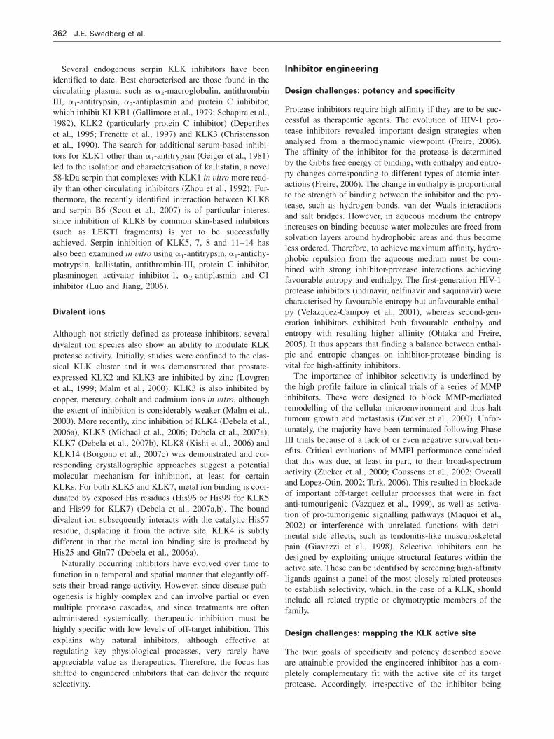
Figure 1 Structural alignment of KLK1–15, KLKB1 and b-trypsin.(A) Ribbon plot of KLK1 (PDB accession no. 1SPJ) as a representative KLK structure. b-Sheets and a-helices are shown in yellow andblue, respectively, with important features highlighted in green and purple, with catalytic residues displayed as stick models. (B) Sequencesimilarity between KLK1–15 and KLKB1 mapped onto the surface of b-trypsin with conservation visualised as a gradient from blue (high),cyan, green and light green to yellow (low). The alignment was produced in Swiss-PdbViewer 4.0.1 using the KLK1, 3-7, KLKB1 and b-trypsin crystal structures (PDB accession nos. 1SPJ, 2ANY, 2ZCH, 2BDH, 2PSX, 1L2E and 2QXI, 1SFI) and KLK2 and 8–15 modelscreated using Swiss Model (Guex et al., 2009). (C) Structural alignment of sequence conservation between KLK1–15, KLKB1 and b-trypsin. Location of b-sheets and a-helices are indicated by yellow arrows and blue cylinders, respectively, with catalytic residues shownin cyan. Important features are highlighted in green and purple and cysteine residues participating in disulfide bonds are shown in yellow.Completely conserved residues and conservative substitutions are shown in dark grey and light grey, respectively.
studies have revealed that KLK tertiary structure across thelocus is highly conserved (Figure 1). Indeed, the kallikrein‘molecular chassis’ conforms to the canonical trypsin fold,consisting of an almost entirely b-sheet arrangement (SCOPtrypsin-like serine proteases; SCOP ID 50493). Furthermore,although the KLKB1 gene encodes four additional N-terminaldomains, its level of sequence similarity across the catalyticdomain is not dissimilar to that observed for the tissue KLKlocus (Figure 1).
Rationale for kallikrein inhibition
Consistent with the important role of kallikrein proteases innormal physiology, defective control of expression or activityis strongly linked to development of disease. Most prominentis the potential significance of KLKs to certain cancers typ-ified by aberrant KLK expression including lung, pancreatic,colon (Yousef et al., 2004) and hormone-dependent cancers,particularly those of the prostate (Magklara et al., 2000;

Kallikrein inhibitors 359
Article in press - uncorrected proof
Petraki et al., 2003; Veveris-Lowe et al., 2005), ovaries(Dong et al., 2001; Yousef et al., 2003; Prezas et al., 2006)and breast (Yousef et al., 2002; Zhang et al., 2006; Pampa-lakis et al., 2009). Contrasting effects of KLK overexpres-sion on cancer physiology have been proposed. KLKoverexpression can provide a pathway for tumour develop-ment and metastasis by degrading the restrictive localisedtissue architecture and shifting the cellular signalling axis todrive increased proliferation. Alternatively, differential KLKexpression can have a protective function in certain cancers.A considerable amount of research has been devoted toexploring the utility of KLK panels as hormone-dependentcancer biomarkers (Borgono and Diamandis, 2004; Clementset al., 2004). KLK3 (prostate-specific antigen, PSA) remainsthe gold-standard biomarker for prostate cancer diagnosisand prognosis (Stamey et al., 1987; Catalona et al., 1991),albeit with some degree of controversy.
In the context of the skin, a role for KLKs has been iden-tified in the rare but severe disorder Netherton syndrome(OMIM: 256500) (Chavanas et al., 2000; Descargues et al.,2005; Briot et al., 2009), as well as the milder but muchmore frequent condition acne rosacea (Yamasaki et al., 2007;Stefansson et al., 2008). Furthermore, KLK1 and KLKB1have been implicated in a number of diseases associated withdysregulation of the kallikrein/kinin system, such as cardio-vascular, renal, inflammatory and gastrointestinal tract dis-ease (Devani et al., 2002; Stadnicki et al., 2003). Finally,KLK6 and KLK8 have potential roles in neurodegenerativeconditions such as Alzheimer’s disease (Little et al., 1997;Shimizu-Okabe et al., 2001), multiple sclerosis (Terayama etal., 2005; Scarisbrick et al., 2008), epilepsy (Momota et al.,1998), synucleinopathies (Iwata et al., 2003) and generalcentral nervous system inflammation (Blaber et al., 2004).The identification of dysfunctional KLK activity as a majorpathological influence suggests that targeted inhibition of rel-evant KLKs is likely to be an effective therapeutic strategy.
Transient inhibition of certain kallikrein-mediated pro-cesses has already been explored and the use of aprotinin toreduce peri-operative bleeding in cardiac surgery by inhib-iting proteases involved in haemostatic and inflammatoryprocesses was widespread (Fritz and Wunderer, 1983; Terrellet al., 1996). Although initially thought to be successful, thistherapy has recently been withdrawn from use following theresult of several larger clinical trials that were terminatedbefore completion because of a persistent trend of increasedmortality (Murkin, 2009). Since this outcome is very recent,the probable physiological explanation is yet to be deter-mined, although one might speculate that it involves inter-ference with unrelated biological pathways owing to thebroad range activity of aprotinin, much like the first-gener-ation matrix metalloprotease (MMP) inhibitors.
Naturally occurring kallikrein inhibitors
Although proteases drive numerous vital physiological pro-cesses, activity must be tightly regulated to ensure correctspatial and temporal delivery. The function of mature pro-
teases is commonly controlled by protein-based inhibitorsthat adopt the form of a non-hydrolysable substrate. Whereasalmost all inhibitors bind to their cognate protease via anexposed motif, the mechanism by which they achieve inhi-bition varies from canonical inhibitors that simply block theprotease active site to serpins that irreversibly disturb thedelicate conformation of the protease (Laskowski and Kato,1980). A number of canonical and serpin inhibitors of KLKproteases have been identified. These include physiologicalinhibitors that provide an essential regulatory influence onKLK-mediated processes and activation cascades, as well asbroad-range, exogenous inhibitors that function as valuableresearch tools for determining the significance of KLK activ-ity in experimental systems. Notable KLK inhibitors are dis-cussed in more detail below and a comprehensive inhibitoryprofile of natural and engineered inhibitors is provided inFigure 2A–D.
Canonical inhibitors
Standard-mechanism or canonical inhibitors currently formthe largest class of protein-based inhibitors. Here, proteaseinhibition generally involves contact with the target proteasevia the canonical loop, a characteristic exposed binding motifon the inhibitor surface that is complementary to the active-site cleft. This interaction is often referred to as analogousto the formation of a protease-substrate complex, since bothare non-covalent, tight-binding and reversible with contactoccurring across extended b-sheets (Tyndall et al., 2005).However, in the protease-inhibitor complex, hydrolysis of thereactive-site P1-P19 peptide bond is rare, preventing subse-quent dissociation of the complex and temporarily removingthe ability of the protease to bind substrate (Laskowski andKato, 1980). The importance of this binding mechanism ishighlighted by the fact that the conformation of the canonicalloop is very similar, even among diverse families of proteaseinhibitors (Figure 2E).
Kunitz-domain inhibitors
Canonical inhibitors of this class are characterised by theKunitz domain, a motif spanning 50–60 residues that con-tains the inhibitory binding loop within a central anti-parallelb-sheet (Figure 2E). Considerable rigidity across the domainis imparted by a network of conserved disulfide bonds(Hynes et al., 1990; Perona et al., 1993). Most prominent isbovine pancreatic trypsin inhibitor (BPTI), also previouslyreferred to as kallikrein inactivator, aprotinin or trasylol. Fol-lowing isolation and crystallisation from bovine pancreatictissue extracts, biochemical characterisation revealed thatBPTI was a potent inhibitor of not only bovine trypsin, butalso KLKB1 (Kraut et al., 1930). Later studies that extendedto the classical tissue kallikrein locus reported inhibitoryactivity against KLK1 (Hofmann and Geiger, 1983) andKLK2 (Geiger et al., 1980). More recently, BPTI/aprotininhas been widely used as a non-specific reagent to block theactivity of kallikrein proteases in vitro, including KLK7(Lundstrom and Egelrud, 1988), KLK5 and 14 (Brattsand etal., 2005), and KLK2 and 4 (Mize et al., 2008).

360 J.E. Swedberg et al.
Article in press - uncorrected proof
BPTI has no homologue expressed in humans and there-fore is not an endogenous modulator of kallikrein proteases,but several potent inhibitors of KLKB1 and KLK1 have beenisolated from human tissue, including tissue factor pathwayinhibitor-2 (TFPI-2) (Petersen et al., 1996) and Kunitz-typeprotease inhibitor domains from Alzheimer amyloid precur-
sor protein (APP) and its homologue APPH (Petersen et al.,1994). A further KLKB1 and KLK1 inhibitor, serine pepti-dase inhibitor Kunitz-type 2 (SPINT2) was later isolatedfrom human placental tissue and termed bikunin because ofits double-headed Kunitz domain structure (Delaria et al.,1997). Plant material has also proven to be a rich source of

Kallikrein inhibitors 361
Article in press - uncorrected proof
.
Figure 2 Comparison of the activity of naturally occurring and engineered kallikrein inhibitors and structural overlay of the conservedbinding loop of protein kallikrein inhibitors.Representation of the relative kallikrein inhibitory activity for an extensive selection of reported naturally occurring (blue), engineeredprotein (orange) and synthetic small-molecule (green) inhibitors. Data were compiled using the reported Ki (canonical inhibitors, panel Aand synthetic inhibitors, panel B), IC50 (metal ions, panel C) or ka (covalent inhibitors, panel D) value for each kallikrein-inhibitor complex.Importantly, the relative inhibition values encompass different modes of inhibition and, undoubtedly, different experimental conditions, andtherefore solely provide a relative comparison and should not be taken as absolute. Abbreviations: APP(H) KPI, Alzheimer amyloid precursorprotein (homologue) Kunitz protease inhibitor; BbTI, Bauhinia bauhinioides trypsin inhibitor; BfTI, Bauhinia forficata trypsin inhibitor;BuXI, Bauhinia ungulate factor Xa inhibitor; BvTIp, Bauhinia variegata trypsin inhibitor, purple variety; CeKI, Caesalpinia echinatakallikrein inhibitor; CMTI, Cucurbita maxima trypsin inhibitor; EcTI, Enterolobium contortisiliquum trypsin inhibitor; LlTI, Leucaenaleucocephala trypsin inhibitor; PAI-1, plasminogen activator inhibitor-1, STI, soybean trypsin inhibitor; SFTI, sunflower trypsin inhibitor;SLPI, secretory leukocyte protease inhibitor; SwTI, Swartzia pickellii trypsin inhibitor. (E) Overlay of the binding loops from various classesof naturally occurring kallikrein inhibitors (inhibitor/class/PDB accession number): LEKTI domain 6/Kazal/1HOZ; aprotinin/Kunitz/2FTL;Barley Bowman-Birk inhibitor/Bowman-Birk inhibitor/1TX6; a1-antitrypsin/Serpin/1OPH; SFTI/Bowman-Birk-like/1SFI; E. coli trypsininhibitor/Ecotin/1EZU; bound to the active site of b-trypsin. Trypsin is shown as grey ribbons, with catalytic residues as stick models. Theinhibitors are shown as yellow b-sheets and green coils. Note that all inhibitors interact with the protease via an extended b-sheet (blackarrow).
Kunitz-type compounds that inhibit KLKs; KLKB1 (Olivaet al., 1999), KLK7 (Lundstrom and Egelrud, 1988), KLK5and 14 (Brattsand et al., 2005) are inhibited by soybean tryp-sin inhibitor (SBTI). Inhibition of KLKB1 has also beendemonstrated using novel compounds from Enterolobium,Leucaena, Swartzia and several Bauhinia species (Sampaioet al., 1996), as well as Caesalpinia echinata kallikrein inhib-itor (CeKI) (Cruz-Silva et al., 2004).
Kazal-domain inhibitors
Lympho-epithelial Kazal-type-related inhibitor (LEKTI) isproduced as a multi-domain serine protease inhibitor encod-ed by the SPINK5 gene (chromosome 5q31-32) (Magert etal., 1999). Within the skin and thymus, the LEKTI pro-pro-tein is processed into up to 15 individual subunits. Structur-ally, the resulting fragments are typified by a Kazal-likedomain arrangement; an anti-parallel b-sheet bordered bytwo short a-helices is held together by conserved disulfidelinks. Most LEKTI domains carry basic residues (Arg orLys) at their putative P1 sites, suggesting that trypsin-likeproteases are likely targets for LEKTI-derived peptides(Magert et al., 1999). A range of LEKTI fragments can mod-ulate KLK activity, albeit with differing efficiency; KLK5,7 and 14 are inhibited by LEKTI D8–11 and LEKTI D5(Deraison et al., 2007), and KLK5 and 7 are inhibited byLEKTI D6–99 (Schechter et al., 2005) and LEKTI D6 (Egel-rud et al., 2005). This activity, in synergy with physiologicalfactors such as pH, provides an essential regulatory influenceon the cascade of kallikrein proteases associated withdesquamation (Borgono et al., 2007b; Deraison et al., 2007).An additional SPINK-related product, LEKTI2 encoded bySPINK9, has recently been identified within the skin andshows KLK5-specific inhibition restricted to the palmoplan-tar epidermis (Brattsand et al., 2009; Meyer-Hoffert et al.,2009).
Other canonical inhibitors
Aside from the classical Kunitz- and Kazal-type inhibitors,several other prominent canonical inhibitors of KLKs have
been identified. Purified from the periplasm of Escherichiacoli, ecotin is a 32-kDa dimeric serine protease inhibitor withbroad-range activity. Characterisation revealed that it is avery potent inhibitor of proteases of the contact activationsystem, including KLKB1 (Ulmer et al., 1995). Hirustasin,a serine protease inhibitor isolated from the medical leechHirudo medicinalis, contains a single antistasin-like domainand is a potent inhibitor of KLK1, but not KLKB1 (Sollneret al., 1994). A detailed investigation of KLK inhibitors iso-lated and purified from stratum corneum extracts identifiedthe elafin-like protease inhibitor antileukoprotease as a skin-expressed KLK7 inhibitor and the potato type-1 inhibitoreglin C as an exogenous inhibitor (Franzke et al., 1996).More recently, several KLK inhibitors have been isolatedfrom plant material; Cucurbita maxima trypsin inhibitor(CMTI) -I and -II exhibit inhibitory activity against serineproteases of the clotting cascade, including KLKB1(Grzesiak et al., 2000a), and sunflower trypsin inhibitor(SFTI) is a non-specific Bowman-Birk-like inhibitor ofKLK4 (Swedberg et al., 2009).
Serpins
Abundant in circulating human plasma, serpins are largeprotein-based inhibitors that irreversibly attenuate the activ-ity of their target protease. Like canonical inhibitors, contactwith the protease is made via an extended b-sheet (Figure2E). However, the serpin mechanism of inhibition is uniquein that it is characterised by catalysis of the P1-P19 peptidebond, which causes both a change in the serpin conformationand formation of a protease-inhibitor covalent bond (Loe-bermann et al., 1984). Recent crystallographic studies havefurther clarified this phenomenon. Since the protease is tight-ly linked to the inhibitor, the serpin structural change due tocleavage at the reactive centre induces a shift in conforma-tion of the bound protease (Huntington et al., 2000). Theresulting loss of protease structure has terminal effects oncatalytic ability, namely distortion of the catalytic triad viadisplacement of Ser195 and collapse of vital interactionsformed during zymogen activation (Huntington et al., 2000).
362 J.E. Swedberg et al.
Article in press - uncorrected proof
Several endogenous serpin KLK inhibitors have beenidentified to date. Best characterised are those found in thecirculating plasma, such as a2-macroglobulin, antithrombinIII, a1-antitrypsin, a2-antiplasmin and protein C inhibitor,which inhibit KLKB1 (Gallimore et al., 1979; Schapira et al.,1982), KLK2 (particularly protein C inhibitor) (Depertheset al., 1995; Frenette et al., 1997) and KLK3 (Christenssonet al., 1990). The search for additional serum-based inhibi-tors for KLK1 other than a1-antitrypsin (Geiger et al., 1981)led to the isolation and characterisation of kallistatin, a novel58-kDa serpin that complexes with KLK1 in vitro more read-ily than other circulating inhibitors (Zhou et al., 1992). Fur-thermore, the recently identified interaction between KLK8and serpin B6 (Scott et al., 2007) is of particular interestsince inhibition of KLK8 by common skin-based inhibitors(such as LEKTI fragments) is yet to be successfullyachieved. Serpin inhibition of KLK5, 7, 8 and 11–14 hasalso been examined in vitro using a1-antitrypsin, a1-antichy-motrypsin, kallistatin, antithrombin-III, protein C inhibitor,plasminogen activator inhibitor-1, a2-antiplasmin and C1inhibitor (Luo and Jiang, 2006).
Divalent ions
Although not strictly defined as protease inhibitors, severaldivalent ion species also show an ability to modulate KLKprotease activity. Initially, studies were confined to the clas-sical KLK cluster and it was demonstrated that prostate-expressed KLK2 and KLK3 are inhibited by zinc (Lovgrenet al., 1999; Malm et al., 2000). KLK3 is also inhibited bycopper, mercury, cobalt and cadmium ions in vitro, althoughthe extent of inhibition is considerably weaker (Malm et al.,2000). More recently, zinc inhibition of KLK4 (Debela et al.,2006a), KLK5 (Michael et al., 2006; Debela et al., 2007a),KLK7 (Debela et al., 2007b), KLK8 (Kishi et al., 2006) andKLK14 (Borgono et al., 2007c) was demonstrated and cor-responding crystallographic approaches suggest a potentialmolecular mechanism for inhibition, at least for certainKLKs. For both KLK5 and KLK7, metal ion binding is coor-dinated by exposed His residues (His96 or His99 for KLK5and His99 for KLK7) (Debela et al., 2007a,b). The bounddivalent ion subsequently interacts with the catalytic His57residue, displacing it from the active site. KLK4 is subtlydifferent in that the metal ion binding site is produced byHis25 and Gln77 (Debela et al., 2006a).
Naturally occurring inhibitors have evolved over time tofunction in a temporal and spatial manner that elegantly off-sets their broad-range activity. However, since disease path-ogenesis is highly complex and can involve partial or evenmultiple protease cascades, and since treatments are oftenadministered systemically, therapeutic inhibition must behighly specific with low levels of off-target inhibition. Thisexplains why natural inhibitors, although effective atregulating key physiological processes, very rarely haveappreciable value as therapeutics. Therefore, the focus hasshifted to engineered inhibitors that can deliver the requireselectivity.
Inhibitor engineering
Design challenges: potency and specificity
Protease inhibitors require high affinity if they are to be suc-cessful as therapeutic agents. The evolution of HIV-1 pro-tease inhibitors revealed important design strategies whenanalysed from a thermodynamic viewpoint (Freire, 2006).The affinity of the inhibitor for the protease is determinedby the Gibbs free energy of binding, with enthalpy and entro-py changes corresponding to different types of atomic inter-actions (Freire, 2006). The change in enthalpy is proportionalto the strength of binding between the inhibitor and the pro-tease, such as hydrogen bonds, van der Waals interactionsand salt bridges. However, in aqueous medium the entropyincreases on binding because water molecules are freed fromsolvation layers around hydrophobic areas and thus becomeless ordered. Therefore, to achieve maximum affinity, hydro-phobic repulsion from the aqueous medium must be com-bined with strong inhibitor-protease interactions achievingfavourable entropy and enthalpy. The first-generation HIV-1protease inhibitors (indinavir, nelfinavir and saquinavir) werecharacterised by favourable entropy but unfavourable enthal-py (Velazquez-Campoy et al., 2001), whereas second-gen-eration inhibitors exhibited both favourable enthalpy andentropy with resulting higher affinity (Ohtaka and Freire,2005). It thus appears that finding a balance between enthal-pic and entropic changes on inhibitor-protease binding isvital for high-affinity inhibitors.
The importance of inhibitor selectivity is underlined bythe high profile failure in clinical trials of a series of MMPinhibitors. These were designed to block MMP-mediatedremodelling of the cellular microenvironment and thus halttumour growth and metastasis (Zucker et al., 2000). Unfor-tunately, the majority have been terminated following PhaseIII trials because of a lack of or even negative survival ben-efits. Critical evaluations of MMPI performance concludedthat this was due, at least in part, to their broad-spectrumactivity (Zucker et al., 2000; Coussens et al., 2002; Overalland Lopez-Otin, 2002; Turk, 2006). This resulted in blockadeof important off-target cellular processes that were in factanti-tumourigenic (Vazquez et al., 1999), as well as activa-tion of pro-tumorigenic signalling pathways (Maquoi et al.,2002) or interference with unrelated functions with detri-mental side effects, such as tendonitis-like musculoskeletalpain (Giavazzi et al., 1998). Selective inhibitors can bedesigned by exploiting unique structural features within theactive site. These can be identified by screening high-affinityligands against a panel of the most closely related proteasesto establish selectivity, which, in the case of a KLK, shouldinclude all related tryptic or chymotryptic members of thefamily.
Design challenges: mapping the KLK active site
The twin goals of specificity and potency described aboveare attainable provided the engineered inhibitor has a com-pletely complementary fit with the active site of its targetprotease. Accordingly, irrespective of the inhibitor being

Kallikrein inhibitors 363
Article in press - uncorrected proof
Figure 3 Phage-display cycle.Genetic diversity is generated by ligating degenerate oligonucleo-tides into the genome of filamentous bacteriophage (typically M13)to produce fusion proteins with pIII, pV or pVII bacteriophage coatproteins. The resultant bacteriophage library is then allowed to inter-act with an immobilised bait molecule (BINDING) after whichweakly bound bacteriophages are removed by washes of increasingstringency (WASH) prior to harvesting of more specifically boundbacteriophage particles by cycles of high- and low-pH washing(ELUTE). The resulting eluate is then reamplified in an E. colibacteriophage propagation strain (AMPLIFY) and iterativelyscreened and amplified through a further three or four cycles toproduce a final library through pseudo-evolutionary selection. Indi-vidual clones from this library are then sequenced and validatedthrough interaction or inhibition assays.
designed, the first step is to probe the active site of the targetenzyme. The most common strategies revolve around map-ping the active site of the target protease through bacterio-phage display, positional scanning and sparse matrixsubstrate libraries.
Bacteriophage display (or biopanning) exploits the phys-ical link between peptides on the exterior of the bacterio-phage particle and the genetic material that carries the codefor it. In its simplest form, phage display parallels naturalevolution, with subpopulations of genetically diverse bacte-riophage libraries iteratively selected and amplified by bind-ing to an immobilised target; see Smith and Petrenko (1997)for a review. This technique has been spectacularly success-ful as a tool for sampling the chemical landscape of shortpeptides and monoclonal antibodies, resulting in a series ofdrugs currently available in the clinic, including Avastin,Rituxan and Lucentis (Dimitrov and Marks, 2009). Aningenious twist in the application of phage display has refo-cused this technique as a tool for discovering protease cleav-age specificities. This strategy turns the affinity maturationconcept on its head so that non-selected sequences are immo-bilised on a solid support and sequences are selected follow-ing cleavage from the solid support. This technique, whichwas pioneered by Matthews and Wells (1993) revolvesaround expression of the bacteriophage pIII protein as afusion protein with an affinity tag, with the pIII portion ofthe fusion protein being separated from the affinity tag by arandomised sequence. The resulting bacteriophage library isthen immobilised on an affinity matrix specific for the fusionpartner on the modified pIII protein. Bacteriophages thathave linker sequences that are substrates for a given proteasewill be cleaved off the solid support and can be harvestedby filtration and subject to affinity maturation through iter-ative cycles of amplification, immobilisation and cleavage(Figure 3). To date, this technology has been applied toKLKB1 (Dennis et al., 1995), KLK2 (Cloutier et al., 2002),KLK3 (Coombs et al., 1998), KLK6 (Sharma et al., 2008)and KLK14 (Felber et al., 2006). These studies in turn guid-ed the design of the engineered bioscaffolds discussed below.
The strength of phage display lies in the magnitude anddiversity of the libraries that can be screened. However, thescreening technique itself is not without inherent bias andblind spots. In particular, pIII fusion peptides have the poten-tial to modulate bacteriophage infectivity. In turn, this canlead to under- or over-representation of particular sequencesduring screening through their ability to propagate duringreamplification rather than by virtue of their affinity for animmobilised ligand (Wilson and Finlay, 1998). Peptidesequences can also be strongly selected by the solid supportused for immobilisation of the target protease. Immobilisedmetal affinity chromatography (IMAC) is notorious for itsability to select for pIII fusion proteins with multiple histi-dine residues. These issues are highlighted by studies byCloutier et al. (2002) that identified peptide substrates forKLK2 using phage display. Although a number of substratesfor the enzyme were identified, none of these achieved thecatalytic efficiency of the control substrate (TFRSA), norwas this sequence selected. However, this may reflect the
fact that assays were based on peptide hydrolysis whereasselection was carried out on the basis of protein cleavage.
Selection bias inherent in biological libraries is circum-vented by the positional scanning synthetic combinatoriallibrary (PS-SCL) approach, which uses solid-phase chemis-try to generate diversity. It consists of sub-libraries of pep-tides for each binding subsite of the protease; thus, if theP1–P4 sites are to be investigated, four sub-libraries areneeded. These contain 20 pools of peptides, each with a dif-ferent fixed amino acid for that protease subsite (P1, P2, P3or P4), whereas the other positions have a mixture of allamino acids (Dooley and Houghten, 1993). By screening therate of proteolysis against all 80 pools, the individual con-tribution of each amino acid at each subsite to substrate rec-ognition or inhibitor binding is revealed. Positional scanninghas been used to probe the active site of a number of KLKs;the outcomes are summarised in Table 1.
Although this information is invaluable, it is importantwhen interpreting PS-SCL data to be conscious of theassumptions on which the method is based. First, it isassumed that the contributions of each position to biologicalactivity are independent of each other. One argument againstthis is what can be termed P-site overlap: although the resultsfrom the screen indicate that the same residue is favoured inadjacent positions, when assayed as an individual peptide,this is not the case. Two PS-SCL studies of KLK4 rankedGln favourably for the P2 and P3 positions (Matsumura etal., 2005; Debela et al., 2006b), whereas assays of individ-ually synthesised peptides revealed that Gln is only favoured
364 J.E. Swedberg et al.
Article in press - uncorrected proof
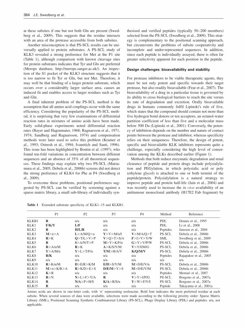
Table 1 Extended substrate specificity of KLK1–15 and KLKB1.
P1 P2 P3 P4 Method Reference
KLKB1 R n/a n/a n/a PDL Dennis et al., 1995KLK1 F/K/Y L/F n/a n/a PDL Li et al., 2008KLK2 R H/L/R n/a n/a Peptides Janssen et al., 2004KLK3 M)n)A L)A/M/Q)n Y)V)M/n/I V)M/A/Q)T PS-SCL Debela et al., 2006bKLK4 R)K Q)T/L)V)P V)Q)T)S/A F)I)V)Y/W SML Swedberg et al., 2009KLK5 R S)A/N/T)P M)Y)K/F/n G)Y)V/P/W PS-SCL Debela et al., 2006bKLK6 R)A/n/M R)K A)K/S/Y/M V)Y/I/M/G PS-SCL Debela et al., 2006bKLK7 Y)A/M/n Y)L)T/F/n T/M)H/A/V K/Q/M/V PS-SCL Debela et al., 2006bKLK8 R/K n/a n/a n/a Peptides Rajapakse et al., 2005KLK9 n/a n/a n/a n/a n/a n/aKLK10 R)K/n/M D)E/R)K/M E/D)S/Y/M M)D/E/V/n PS-SCL Debela et al., 2006bKLK11 M)n)K/R)A R)K/D)E)S D/E/M)Y)S M)D/E/V/M PS-SCL Debela et al., 2006bKLK12 K)R n/a n/a n/a Peptides Memari et al., 2007KLK13 R)N N)L)F)Y/A R V)Y)I/P/G PS-SCL Borgono et al., 2007aKLK14 R N/A)P)H/S K/A)R/S/n Y)W)F/V/I PS-SCL Borgono et al., 2007aKLK15 R n/a n/a n/a Peptide Takayama et al., 2001a
Amino acids are shown in one-letter code, with ‘n’ representing norleucine. Bold font indicates the most preferred residue at eachsubsite. When several sources of data were available, selections were made according to the following priority order: Sparse MatrixLibrary (SML), Positional Scanning Synthetic Combinatorial Library (PS-SCL), Phage Display Library (PDL) and peptides. n/a, notapplicable.
at these subsites if one but not both Gln are present (Swed-berg et al., 2009). This suggests that the residue interactswith an area of the protease accessible from both subsites.
Another misconception is that PS-SCL results can be uni-versally applied to protein substrates. A PS-SCL study ofKLK3 revealed a strong preference for Met at the P1 site(Table 1), although comparison with known cleavage sitesfor protein substrates indicates that Tyr and Gln are preferred(Merops database, http://merops.sanger.ac.uk/). An inspec-tion of the S1 pocket of the KLK3 structure suggests that itis too narrow to fit Tyr or Gln, but not Met. Therefore, itmay well be that binding of a larger protein substrate, whichoccurs over a considerably larger surface area, causes aninduced fit and enables access to larger residues such as Tyrand Gln.
A final inherent problem of the PS-SCL method is theassumption that all amino acid couplings occur with the sameefficiency. Considering the popularity of the PS-SCL meth-od, it is surprising that very few examinations of differentialreaction rates in mixtures of amino acids have been made.Early solid-phase experiments noted differential reactionrates (Bayer and Hagenmaier, 1968; Ragnarsson et al., 1971,1974; Sandberg and Ragnarsson, 1974) and compensationmethods were later used to solve this problem (Kramer etal., 1993; Ostresh et al., 1994; Ivanetich and Santi, 1996).This issue has been highlighted by Boutin et al. (1997), whofound ten-fold variations in concentration between differentsequences and an absence of 35% of all theoretical sequen-ces. These findings may explain why two PS-SCL (Matsu-mura et al., 2005; Debela et al., 2006b) screens did not detectthe strong preference of KLK4 for Phe at P4 (Swedberg etal., 2009).
To overcome these problems, positional preferences sug-gested by PS-SCL can be verified by screening against asparse matrix library, a small sub-library of individually syn-
thesised and verified peptides (typically 50–200 members)selected from the PS-SCL (Swedberg et al., 2009). This strat-egy is complementary to the positional scanning approach,but circumvents the problems of subsite cooperativity andincomplete and under-represented sequences. In addition,since each peptide is individually assayed, there is often fargreater selectivity apparent for each position in the peptide.
Design challenges: bioavailability and stability
For protease inhibitors to be viable therapeutic agents, theymust be not only potent and specific towards their targetprotease, but also readily bioavailable (Fear et al., 2007). Thebioavailability of a drug in a particular tissue is governed byits ability to cross biological barriers to reach the site versusits rate of degradation and excretion. Orally bioavailabledrugs in humans commonly fulfil Lipinski’s rule of five,which states that the compound should contain not more thanfive hydrogen bond donors or ten acceptors, an octanol-waterpartition coefficient of less than five and a molecular massbelow 500 Da (Lipinski et al., 2001). Conversely, the poten-cy of inhibition depends on the number and nature of contactpoints between the protease and inhibitor, whereas specificityrelies on their uniqueness. Therefore, the design of potent,specific and bioavailable KLK inhibitors represents quite achallenge, especially considering the high level of conser-vation among the KLKs described earlier (Figure 1).
Methods that both reduce enzymatic degradation and renalclearance of peptide and protein drugs include polysialyla-tion and PEGylation, in which polysialic acid or poly(ethylene glycol) is attached to one or both termini of thepeptide/protein. Polysialylation is a natural strategy toimprove peptide and protein half-life (Jain et al., 2004) andwas recently used to increase the in vivo availability of anantitumour monoclonal antibody (H17E2 Fab fragment) by

Kallikrein inhibitors 365
Article in press - uncorrected proof
a factor of five (Constantinou et al., 2008). PEGylation isapproved by the FDA as a vehicle for pharmaceuticals(Harris and Chess, 2003) and is used in a number of protein-based drugs currently on the market (Veronese and Harris,2008) with an annual market value of over $US4 billion(Krishan, 2007).
Peptide-based inhibitors particularly suffer from shorthalf-lives since they are often hydrophilic and thus showpoor oral absorption and are rapidly cleared by the kidneys(Werle and Bernkop-Schnurch, 2006). In addition, peptidedrugs are often quickly degraded by the numerous exo- andendo-proteases present in most tissues (Werle and Bernkop-Schnurch, 2006). Even if intravenously administered, mostpeptides are cleared from the bloodstream within minutes.To overcome these particular challenges, peptide lead com-pounds often need to be modified in various ways, dependingon the route of administration and the target tissue. N- andC-terminal modifications such as N-acetylation and C-ami-dation can reduce degradation by exoproteases (McGregor,2008). For example, the natural peptide thymopoietininvolved in T-cell maturation has a half-life of 1 min in plas-ma, whereas a double end-capped version shows no detect-able degradation (Heavner et al., 1986). Similarly, cappingwith fatty acids can improve both the half-life and lipophi-licity of a peptide, a technique used to improve serumstability of an anti-proliferative somatostatin analogue (Das-gupta et al., 2002).
Backbone- and/or disulfide bond-circularised peptides areintrinsically stable to degradation, a common feature in nat-urally occurring peptides. A diverse range of plants producecyclotides, circular miniproteins with a cysteine knot of threeintertwined disulfides, believed to be involved in hostdefence (Craik et al., 2002; Gruber et al., 2008). Many bac-teria also produce circular peptides (bacteriocins) to targetcompeting species (Mercedes et al., 2008), as do manymarine microorganisms (Hamada and Shioiri, 2005). Thisapproach was recently used by Clark et al. (2005) to producea backbone-cyclised snail venom peptide (a-conotoxin)analogue with improved stability in serum.
A more significant challenge is to improve peptide resis-tance to endoprotease activity, since this involves peptidebackbone and/or side chain modifications that are likely tohave major effects on inhibitor potency and specificity. Onestrategy involves the substitution of certain amino acids thatare known to be targets of proteolysis with non-natural D-amino acids, as has been used to produce a variety of pro-tease-resistant reagents including small peptides (Silvia et al.,2008) and antibodies (Webb et al., 2005). Alternatively, sub-stitution of peptide backbone atoms, in particular participantsof the amide bond, can be used to produce what are com-monly referred to as pseudopeptides. For example, the amidenitrogens of scissile bonds can be methylated (N-methyla-tion) to prevent hydrolysis, as for the fungal peptide immuno-suppressant cyclosporine A. This peptide has seven N-meth-ylations, as well as one D-amino acid, and is highly bioa-vailable since it is chiefly degraded by the hepaticcytochrome P450 3A enzyme system rather than by prote-ases (Dunn et al., 2001). Recent advances in methods for
synthesis of multi N-methylated peptides (Biron et al., 2006)have yielded a somatostatin analogue with four-fold higherhalf-life after oral administration (Biron et al., 2008). A num-ber of less commonly used amide bond surrogates also exist,including retro bonds (NH-CO), carba bonds (CH2CH2), azabonds (CO-NH-NR-CO), reduced amide (CH2NH) and ureabonds (HN-CO-NH). These have previously been reviewedelsewhere (Adessi and Soto, 2002; Lozano et al., 2006).
Curiously, certain naturally occurring small protein inhib-itors that deviate from Lipinski’s rule of five are readily bio-available. For example, orally administrated Bowman-Birkinhibitors (;8 kDa) are readily bioavailable (Kennedy,1998) and the same is true for cyclotides (;3 kDa), at leastin insects (Whetstone and Hammock, 2007). There is a grow-ing realisation that many of the issues above can be solvedat a stroke by using naturally occurring inhibitors of serineproteases. For example, the inhibitors aprotinin (Terrell et al.,1996) and hirudin (Zeymer, 1998) have seen clinical use asmodulators of bleeding after thoracic surgery and thrombo-lysis, respectively. The obvious next step for these naturallyoccurring inhibitors is to produce variants with redirectedinhibitory specificity and enhanced potency. Use of thesebioscaffolds is becoming increasingly common as theiradvantages in terms of stability, specificity and potency arerecognised.
Engineered inhibitors: bioscaffolding
Bioscaffolding revolves around the use of natural templatestructures (bioscaffolds) in contrast to more conventionalsmall-molecule approaches. Extensive use is made of pre-existing natural protease inhibitors, which are re-engineeredusing recombinant DNA techniques or solid-phase peptidesynthesis. The chief features of successful bioscaffolds are astructure that is both robust and flexible enough to toleratemultiple amino acid substitutions, and a template structurethat has intrinsic protease inhibitor activity. Thus, the engi-neering goal is simply to redirect and enhance existinginhibitory activity. Further advantages come from facile pro-duction of inhibitor molecules in living systems, thus facil-itating biopanning (phage display) and cheap and efficientlarge-scale bioproduction in microbial or plant systems.
Currently there are four classes of bioscaffold with rele-vance to kallikreins: serpins, Kunitz-domain inhibitors, eco-tin and SFTI derivatives. All four classes of inhibitor interactwith their target proteases through the canonical loopdescribed above (Tyndall et al., 2005; Figure 2E), formingan extension of the protease b-sheet structure. However, thescaffolding presenting the reactive loop shows considerablestructural diversity and plays varying roles in stabilising theprotease-inhibitor complex. In addition to these four estab-lished bioscaffolds, there is also a move towards productionof specific monoclonal antibodies that can inhibit kallikreins,with Dyax developing a specific KLK1 inhibitor for treat-ment of asthma (Sexton et al., 2009). This antibody wasselected from a so-called Fab-on-phage library (Steukers etal., 2006), which contains genes encoding the antibody

366 J.E. Swedberg et al.
Article in press - uncorrected proof
heavy- and light-chain variable regions that are thendisplayed on the phage surface as Fabs and selected usingsurface plasmon resonance screening. Other non-clinicalexamples include antibody-mediated inhibition of KLK13 ina cell-based model of extracellular matrix degradation(Kapadia et al., 2004) and use in biochemical characterisa-tion of KLK12 (Memari et al., 2007). Both the bioscaffoldand monoclonal antibody approaches make extensive use ofphage display as a primary design tool. However, bioscaffolddesign is also driven by information from positional scanningand sparse matrix libraries, as outlined below.
Serpins
To date, three serpins have been exploited as bioscaffolds;C1-inhibitor protein, which inhibits the complement system(Kase and Pospisil, 1983); human a1-antitrypsin (AAT),which protects lung tissue from the effects of human neutro-phil elastase (Sun and Yang, 2004); and human a1-antichy-motrypsin (ACT), which is associated with the developmentof Alzheimer’s disease (Potter et al., 2001). All three inhib-itors are high-molecular-weight ()50 kDa) plasma proteinsthat contact their target proteases via a canonical loop, whichis termed a reactive loop since it is cut by the target protease.Although the reactive loop does in theory control the major-ity of the specificity of a given serpin, there is also a signif-icant contribution from serpin exosites such that swappingreactive loops alone between serpin subtypes is not sufficientto completely redirect their inhibitory activity (Djie et al.,1997). Nonetheless, the reactive loop is very tolerant of sub-stitution and this has led to the development of a series ofrecombinant kallikrein inhibitors.
Initially, an inhibitor for KLKB1 was developed using avariant of C1-inhibitor (Sulikowski et al., 2002). C1-inhibitoreffectively blocks KLKB1 activity and the aim of the re-engineering was to increase the inhibition specificity andprevent the blockade of other complement proteases. Accord-ingly, a reactive loop variant was designed on the basis ofthe plasma KLK substrate specificity, which was previouslyassessed using a relatively small pool of arginine methylesters (Levison and Tomalin, 1982). The resulting variantshowed the specificity required and maintained potency, withka of 382 180 M-1 s-1. Following this success, a KLK2 inhib-itor was designed using ACT as bioscaffold (Cloutier et al.,2004). The ACT reactive loop was substituted with a KLK2-specific sequence found through phage display (Cloutieret al., 2002), yielding an inhibitor with a ka value of 6261M-1 s-1.
More recently, Felber et al. (2006) re-engineered the reac-tive loop of both AAT and ACT serpins using pentapeptidesderived from a phage display scan against immobilisedKLK14 (Felber et al., 2005). Currently, KLK2 ACT-derivedinhibitors are being developed by Med Discovery, with thelead compound, MDKP67b, close to entering clinical trialsas a targeted treatment for prostate cancer. The success ofthis approach owes much to the adaptability of the serpinreactive loop. In addition, using a human serpin as a bio-scaffold, the problem of patient immune response to a proteintherapeutic is neatly avoided.
Kunitz domain
Originally identified in BPTI, the Kunitz-domain inhibitorymotif is remarkably compact and shows considerable thermalstability (Moses and Hinz, 1983; Makhatadze et al., 1993).Both BPTI and the Alzheimer’s amyloid b-protein precursor(APPI), which has a similar structure to BPTI, have beenexploited as bioscaffolds. APPI was the first bioscaffold sub-jected to phage display. Screening against KLKB1 achieveda Ki value of 15 pM (Dennis et al., 1995). A later studysearched for potent inhibitors of six distinct serine proteases,including KLKB1. This study focused on the BPTI canonicalloop, substituting positions P1, P3 and P4 with amino-acidside chains gauged to explore the diversity of charge, shapeand hydrophobicity. The best inhibitor against KLKB1showed a ki value of 5.4 nM, a modest 2.2-fold improvementover the wild-type inhibitor; considerable thermal destabilis-ation was observed when P4 mutations were undertaken(Grzesiak et al., 2000b).
The Kunitz-domain approach has also had some successin the world of commercial drug design and development,with a Dyax product, ecallantide (DX-88), reaching PhaseIII clinical trials for treatment of hereditary angioedema.Ecallantide was derived from a phage display screen of theKunitz domain of human lipoprotein-associated coagulationinhibitor (LACI) using immobilised KLKB1 as bait. It has areported Ki value of 44 pM and is highly specific for KLKB1(Williams and Baird, 2003). At the time of writing, FDAapproval for use of ecallantide was awaiting completion ofa risk evaluation and mitigation strategy.
Ecotin
Ecotin (E. coli trypsin inhibitor) was isolated from the peri-plasmic space of E. coli and is a dimeric, bidentate proteaseinhibitor (Chung et al., 1983). It is thought to provide pro-tection against the effects of neutrophil elastase (Eggers etal., 2004), which it inhibits with a Ki value of 12 pM. Inter-action between the protease and ecotin is driven by fourloops (two from each ecotin monomer) and results in theformation of a heterotetramer with a very extensive buriedsurface area of 2850 A2. Interestingly, binding by the contactloops is not restricted to the trypsin active site, but alsoincludes residues that are highly divergent within the trypsinsuperfamily. Accordingly, these secondary contacts have thepotential to provide greater selectivity than that of inhibitorsthat bind at the active site alone. This prompted Craik andco-workers (Stoop and Craik, 2003) to re-engineer the ecotinbioscaffold to produce a series of variant molecules that caninhibit KLKB1, thrombin, MT-SP1 and FXIIa. Two distinctdesign strategies were used, a simple progressive substitutionof the ecotin apparent P1 residue and biopanning using phagedisplay. Ecotin, like the C1-inhbitor bioscaffold, is a potentinhibitor of KLKB1 but lacks selectivity against other con-tact activation proteases. Given the role of the contact loops,as well as the canonical loop, in protease complex formation,a library of ecotin variants was produced by gene shufflingto target all five motifs. This library was screened againstimmobilised KLKB1 as described above and, in a novel

Kallikrein inhibitors 367
Article in press - uncorrected proof
Figure 4 Tetrahedral transition-state analogues of serine proteases.(A) Structure of the P1-P19 residues and adjoining peptide bond ona typical amide substrate (left) and putative tetrahedral intermediateformed coordinately with the Ser195 (catalytic) side chain prior tocleavage of the peptide bond (right). (B) Representation of a boronicacid inhibitor (e.g., P8720, Figure 2B) (left) and subsequent com-plex formation via covalent bond formation with the Ser195 (cata-lytic) hydroxyl group (right). (C) Structure of a peptide aldehydeinhibitor (e.g., hK2p01, Figure 2B) (left) and covalent bond for-mation with the Ser195 (catalytic) hydroxyl group (right). (D) Rep-resentation of a chloromethyl ketone inhibitor (e.g., CH-2856,Figure 2B) (left) and mechanism of inhibition via dual linkage tothe target protease at Ser195 (catalytic) as well as the adjacent His57(catalytic) by the exposed CH2-Cl group (right).
deviation from the normal phage display procedure, thelibrary was pre-blocked with soluble proteases from classesagainst which inhibitory activity was to be selected. This ledto the isolation of an ecotin variant with a Ki value forKLKB1 of 11 pM but with activity against factor Xa, factorXIa, urokinase-type plasminogen activator thrombin, andmembrane-type serine protease 1 that is four to seven ordersof magnitude lower (Stoop and Craik, 2003).
Sunflower trypsin inhibitor
SFTI belongs to the Bowman-Birk serine protease inhibitorfamily and is a potent inhibitor of trypsin, cathepsin G andsuppressor of tumourigenesis (14 ST14/matriptase/MT-SP1).It was originally discovered in sunflower (Helianthusannuus) seeds and characterised by determination of itsthree-dimensional structure in complex with bovine b-tryp-sin (Luckett et al., 1999). Consisting of just 14 cyclised ami-no acids bisected by a disulfide bond, SFTI is intermediatebetween the macromolecular scaffolds represented by thebioscaffolds above and the small-molecule inhibitorsdescribed below. Its smaller size allows for facile chemicalsynthesis (Zablotna et al., 2002; Korsinczky et al., 2005) andrapid generation of variants. In addition, a very recent pub-lication described expression of a library of fully cyclic SFTIvariants in E. coli (Austin et al., 2009), opening up the pos-sibility of rapid in vivo screening.
Similar to the other bioscaffolds described above, SFTIinteracts with its target proteases via a canonical-type loop.However, unlike the larger molecules, SFTI has essentiallyno molecular scaffolding behind it, relying on its fully cyclicbackbone and a disulfide bond for stability. Interestingly, thestructure of SFTI is maintained even when the disulfide isremoved (by substituting glycine or aminobutyric acid),being preserved by an internal hydrogen bond network(Korsinczky et al., 2005). However, this variant is not stableto cleavage by proteases. Surprisingly, there have been fewattempts to use the SFTI molecule as a bioscaffold and thesmall number of variants produced have not been tested incell culture or animal systems. To date, only KLK4 has beenactively targeted; Swedberg et al. (2009) used a sparse matrixlibrary approach to probe the KLK4 active site, and thensubstituted the optimal sequence obtained into the SFTI bio-scaffold. This strategy improved both the potency and spec-ificity of KLK4 inhibition by SFTI, increasing its potencyby nearly two orders of magnitude to give a Ki value of3.6 nM. Moreover, the specificity of inhibition improved togive 500-fold selectivity against the closely related KLK14.Thus, in contrast to the situation for serpins described above,re-engineering of just three residues is sufficient to complete-ly redirect the inhibitory activity of SFTI. This reflects thelack of interactions with regions outside the active-site cleftin the SFTI/protease complex.
Despite the inhibitory potency of these bioscaffolds, onlyecallantide has successfully made the transition to clinicaluse. This may reflect the fact that ecotin, Kunitz-domain andserpin-based inhibitors are all macromolecular inhibitorsrequiring intravenous administration. Although the much small-er SFTI may yet prove to be readily bioavailable, detailed
pharmacokinetics are currently unavailable for either wild-type or engineered versions of this scaffold. In addition, theother bioscaffold-based inhibitors show considerable cross-reactivity between the kallikreins. Clearly there is consider-able scope for further investigation focused on theseremarkable reagents, which have the potential to become assuccessful as the small-molecule approaches conventionallyapplied to protease inhibitor design.
Engineered inhibitors: small-molecule inhibitors
The most commonly used small-molecule kallikrein inhibi-tors are serine protease-specific tetrahedral transition-stateanalogues. These include peptides in which the C-terminalcarboxyl acid is substituted for boronic acid, an aldehyde orhalogenated methyl ketones to form a tetrahedral adduct withSer 195 (Figure 4). For example, Fareed et al. (1981) usedKLK2 and KLKB1 kininogen cleavage sites to design pep-tide aldehyde inhibitors with Ki values of 10–20 mM,although these inhibitors showed little specificity against anumber of serine proteases of the blood clotting cascade. Aboronic acid inhibitor based on a similar peptide sequenceproduced a potent (Ki 150 pM) and more specific KLKB1

368 J.E. Swedberg et al.
Article in press - uncorrected proof
inhibitor (Dela Cadena et al., 1995). Concurrently, Evans etal. (1996a,b) reported conservative non-natural amino acidsubstitutions of KLKB1 kininogen cleavage sites combinedwith a fluoroalkyloxymethyl ketone functionality to developa series of specific KLK1 inhibitors with Ki values in thelower nM range. These inhibitors attenuated breast cancercell invasion in a Matrigel-based model (Wolf et al., 2001).More recently, the boronic acid approach was used to devel-op inhibitors to PSA; the best has a Ki value of 65 nM
(LeBeau et al., 2008). However, there are limited specificitydata available for these compounds and their biologicalimpact is ambiguous.
Using a different strategy, Wanaka et al. (1990) cappedphenylalanine N-terminally with benzylamine to yield acompetitive KLKB1 inhibitor (Ki 810 nM). This strategy, pre-viously used to produce trypsin, plasmin and thrombin inhib-itors (Markwardt et al., 1968), capitalises on mimicking theP1 arginine by the benzylamine group; the ‘peptide bond’ isdisplaced, resulting in a non-hydrolysable compound. Vari-ous derivatives of this compound were later screened toproduce a more potent KLKB1 inhibitor with a Ki value of130 nM (Teno et al., 1993), although it suffers from the samelack of specificity as previous benzylamine derivatives(Markwardt et al., 1968). b-Lactam analogues have also beenused to produce a number of serine protease inhibitors(Konaklieva, 2002), including a series of KLK3 inhibitorswith inhibition in the nM range, although the specificity ofthis inhibition was not evaluated (Adlington et al., 1997,2001).
Recently, phage display has been used to identify a seriesof KLK2 peptide inhibitors with inhibition constants in thelower micromolar range and selectivity over a number oftryptic serine proteases, including KLKB1 (Hekim et al.,2006). The stability of these peptides was later improved byhead-to-tail cyclisation and/or internal disulfide bond for-mation, without loss of potency (Pakkala et al., 2007). Thesame group also produced three non-peptide KLK3 inhibitorswith inhibition in the nanomolar range by screening againsta library of small drug-like molecules (Koistinen et al.,2008b). The development of these inhibitory peptides hasrecently been reviewed (Koistinen et al., 2008a).
Small-molecule kallikrein inhibitors have not had the sameclinical success as bioscaffold-based inhibitors. However,transition-state analogues are currently being investigated foruse as activity-based probes, utilising the platform initiallyestablished for organophosphate peptide inhibitors (Liu et al.,1999), which were developed to detect KLK6 (Oikonomo-poulou et al., 2008). Clearly, the bioscaffold approachand conventional small-molecule design techniques arecomplementary.
Conclusions
There is increasing appreciation of the potential roles playedby the kallikrein proteases as regulators of human physiol-ogy, as biomarkers in disease diagnosis and as points for
therapeutic intervention. With application of the strategieswe have discussed here, the next decade could see a blos-soming of research in this area and the production of a newgeneration of protease inhibitor drugs to rival the antiviralprotease inhibitors.
Acknowledgements
Work in the corresponding author’s laboratory is supported by theCancer Council Queensland (Grant �44323) and the Prostate Can-cer Foundation of Australia (Grant �PR09). SJD receives fundingfrom the Smart Futures Fund (Queensland Government, Australia).
References
Adessi, C. and Soto, C. (2002). Converting a peptide into a drug:strategies to improve stability and bioavailability. Curr. Med.Chem. 9, 963–978.
Adlington, R.M., Baldwin, J.E., Chen, B., Cooper, S.L., McCoull,W., and Pritchard, G.J. (1997). Design and synthesis of novelmonocyclic b-lactam inhibitors of prostate-specific antigen.Bioorg. Med. Chem. Lett. 7, 1689–1694.
Adlington, R.M., Baldwin, J.E., Becker, G.W., Chen, B., Cheng, L.,Cooper, S.L., Hermann, R.B., Howe, T.J., McCoull, W.,McNulty, A.M., et al. (2001). Design, synthesis and proposedactive site binding analysis of monocyclic 2-azetidinone inhib-itors of prostate-specific antigen. J. Med. Chem. 44, 1491–1508.
Austin, J., Kimura, R.H., Woo, Y.H., and Camarero, J.A. (2009). Invivo biosynthesis of an Ala-scan library based on the cyclic pep-tide SFTI-1. Amino Acids, in press. DOI: 10.1007/s00726-009-0338-4.
Bayer, E. and Hagenmaier, H. (1968). Solid phase synthesis of oxy-tocin. Tetrahedron Lett. 17, 2037–2039.
Bhoola, K.D., Figueroa, C.D., and Worthy, K. (1992). Bioregulationof kinins: kallikreins, kininogens, and kininases. Pharmacol.Rev. 44, 1–80.
Biron, E., Chatterjee, J., and Kessler, H. (2006). Optimized selectiveN-methylation of peptides on solid support. J. Pept. Sci. 12,213–219.
Biron, E., Chatterjee, J., Ovadia, O., Langenegger, D., Brueggen, J.,Hoyer, D., Schmid, H.A., Jelinek, R., Gilon, C., Hoffman, A.,et al. (2008). Improving oral bioavailability of peptides by mul-tiple N-methylation: somatostatin analogues. Angew. Chem. Int.Ed. 47, 2595–2599.
Blaber, S.I., Ciric, B., Christophi, G.P., Bernett, M.J., Blaber, M.,Rodriguez, M., and Scarisbrick, I.A. (2004). Targeting kallikrein6 proteolysis attenuates CNS inflammatory disease. FASEB J.18, 920–922.
Borgono, C.A. and Diamandis, E.P. (2004). The emerging roles ofhuman tissue kallikreins in cancer. Nat. Rev. Cancer 4, 876–890.
Borgono, C.A., Gavigan, J.A., Alves, J., Bowles, B., Harris, J.L.,Sotiropoulou, G., and Diamandis, E.P. (2007a). Defining theextended substrate specificity of kallikrein 1-related peptidases.Biol. Chem. 388, 1215–1225.
Borgono, C.A., Michael, I.P., Komatsu, N., Jayakumar, A., Kapadia,R., Clayman, G.L., Sotiropoulou, G., and Diamandis, E.P.(2007b). A potential role for multiple tissue kallikrein serineproteases in epidermal desquamation. J. Biol. Chem. 282, 3640–3652.
Borgono, C.A., Michael, I.P., Shaw, J.L., Luo, L.Y., Ghosh, M.C.,Soosaipillai, A., Grass, L., Katsaros, D., and Diamandis, E.P.(2007c). Expression and functional characterization of the can-

Kallikrein inhibitors 369
Article in press - uncorrected proof
cer-related serine protease, human tissue kallikrein 14. J. Biol.Chem. 282, 2405–2422.
Boutin, J.A., Gesson, I., Henlin, J.M., Bertin, S., Lambert, P.H.,Volland, J.P., and Fauchere, J.L. (1997). Limitations of the cou-pling of amino acid mixtures for the preparation of equimolarpeptide libraries. Mol. Divers. 3, 43–60.
Brattsand, M., Stefansson, K., Hubiche, T., Nilsson, S.K., and Egel-rud, T. (2009). SPINK9: a selective, skin-specific Kazal-typeserine protease inhibitor. J. Invest. Dermatol. 129, 1656–1665.
Brattsand, M., Stefansson, K., Lundh, C., Haasum, Y., and Egelrud,T. (2005). A proteolytic cascade of kallikreins in the stratumcorneum. J. Invest. Dermatol. 124, 198–203.
Briot, A., Deraison, C., Lacroix, M., Bonnart, C., Robin, A., Besson,C., Dubus, P., and Hovnanian, A. (2009). Kallikrein 5 inducesatopic dermatitis-like lesions through PAR2-mediated thymicstromal lymphopoietin expression in Netherton syndrome. J.Exp. Med. 206, 1135–1147.
Catalona, W.J., Smith, D.S., Ratliff, T.L., Dodds, K.M., Coplen,D.E., Yuan, J.J., Petros, J.A., and Andriole, G.L. (1991). Meas-urement of prostate-specific antigen in serum as a screening testfor prostate cancer. N. Engl. J. Med. 324, 1156–1161.
Caubet, C., Jonca, N., Brattsand, M., Guerrin, M., Bernard, D.,Schmidt, R., Egelrud, T., Simon, M., and Serre, G. (2004). Deg-radation of corneodesmosome proteins by two serine proteasesof the kallikrein family, SCTE/KLK5/hK5 and SCCE/KLK7/hK7. J. Invest. Dermatol. 122, 1235–1244.
Chavanas, S., Bodemer, C., Rochat, A., Hamel-Teillac, D., Ali, M.,Irvine, A.D., Bonafe, J.L., Wilkinson, J., Taieb, A., Barrandon,Y., et al. (2000). Mutations in SPINK5, encoding a serine pro-tease inhibitor, cause Netherton syndrome. Nat. Genet. 25,141–142.
Christensson, A., Laurell, C.B., and Lilja, H. (1990). Enzymaticactivity of prostate-specific antigen and its reactions with extra-cellular serine proteinase inhibitors. Eur. J. Biochem. 194, 755–763.
Chung, C.H., Ives, H.E., Almeda, S., and Goldberg, A.L. (1983).Purification from Escherichia coli of a periplasmic protein thatis a potent inhibitor of pancreatic proteases. J. Biol. Chem. 258,11032–11038.
Clark, R.J., Fischer, H., Dempster, L., Daly, N.L., Rosengren, K.J.,Nevin, S.T., Meunier, F.A., Adams, D.J., and Craik, D.J. (2005).Engineering stable peptide toxins by means of backbone cycli-zation: stabilization of the a-conotoxin MII. Proc. Natl. Acad.Sci. USA 102, 13767–13772.
Clements, J.A., Willemsen, N.M., Myers, S.A., and Dong, Y.(2004). The tissue kallikrein family of serine proteases: func-tional roles in human disease and potential as clinical biomar-kers. Crit. Rev. Clin. Lab. Sci. 41, 265–312.
Cloutier, S.M., Chagas, J.R., Mach, J.P., Gygi, C.M., Leisinger, H.J.,and Deperthes, D. (2002). Substrate specificity of human kallik-rein 2 (hK2) as determined by phage display technology. Eur. J.Biochem. 269, 2747–2754.
Cloutier, S.M., Kundig, C., Felber, L.M., Fattah, O.M., Chagas, J.R.,Gygi, C.M., Jichlinski, P., Leisinger, H.J., and Deperthes, D.(2004). Development of recombinant inhibitors specific tohuman kallikrein 2 using phage-display selected substrates. Eur.J. Biochem. 271, 607–613.
Constantinou, A., Epenetos, A.A., Hreczuk-Hirst, D., Jain, S., andDeonarain, M.P. (2008). Modulation of antibody pharmacoki-netics by chemical polysialylation. Bioconjug. Chem. 19, 643–650.
Coombs, G.S., Bergstrom, R.C., Pellequer, J.L., Baker, S.I., Navre,M., Smith, M.M., Tainer, J.A., Madison, E.L., and Corey, D.R.
(1998). Substrate specificity of prostate-specific antigen (PSA).Chem. Biol. 5, 475–488.
Coussens, L.M., Fingleton, B., and Matrisian, L.M. (2002). Matrixmetalloproteinase inhibitors and cancer: trials and tribulations.Science 295, 2387–2392.
Craik, D.J., Simonsen, S., and Daly, N.L. (2002). The cyclotides:novel macrocyclic peptides as scaffolds in drug design. Curr.Opin. Drug Discov. Dev. 5, 251–260.
Cruz-Silva, I., Gozzo, A.J., Nunes, V.A., Carmona, A.K., Faljoni-Alario, A., Oliva, M.L., Sampaio, M.U., Sampaio, C.A., andAraujo, M.S. (2004). A proteinase inhibitor from Caesalpiniaechinata (pau-brasil) seeds for plasma kallikrein, plasmin andfactor XIIa. Biol. Chem. 385, 1083–1086.
Dasgupta, P., Singh, A., and Mukherjee, R. (2002). N-Terminal acy-lation of somatostatin analog with long chain fatty acids enhan-ces its stability and anti-proliferative activity in human breastadenocarcinoma cells. Biol. Pharm. Bull. 25, 29–36.
Debela, M., Magdolen, V., Grimminger, V., Sommerhoff, C., Mes-serschmidt, A., Huber, R., Friedrich, R., Bode, W., and Goettig,P. (2006a). Crystal structures of human tissue kallikrein 4: activ-ity modulation by a specific zinc binding site. J. Mol. Biol. 362,1094–1107.
Debela, M., Magdolen, V., Schechter, N., Valachova, M., Lottspeich,F., Craik, C.S., Choe, Y., Bode, W., and Goettig, P. (2006b).Specificity profiling of seven human tissue kallikreins revealsindividual subsite preferences. J. Biol. Chem. 281, 25678–25688.
Debela, M., Goettig, P., Magdolen, V., Huber, R., Schechter, N.M.,and Bode, W. (2007a). Structural basis of the zinc inhibition ofhuman tissue kallikrein 5. J. Mol. Biol. 373, 1017–1031.
Debela, M., Hess, P., Magdolen, V., Schechter, N.M., Steiner, T.,Huber, R., Bode, W., and Goettig, P. (2007b). Chymotrypticspecificity determinants in the 1.0 A structure of the zinc-inhib-ited human tissue kallikrein 7. Proc. Natl. Acad. Sci. USA 104,16086–16091.
Dela Cadena, R.A., Stadnicki, A., Uknis, A.B., Sartor, R.B., Kettner,C.A., Adam, A., and Colman, R.W. (1995). Inhibition of plasmakallikrein prevents peptidoglycan-induced arthritis in the Lewisrat. FASEB J. 9, 446–452.
Delaria, K.A., Muller, D.K., Marlor, C.W., Brown, J.E., Das, R.C.,Roczniak, S.O., and Tamburini, P.P. (1997). Characterization ofplacental bikunin, a novel human serine protease inhibitor. J.Biol. Chem. 272, 12209–12214.
Dennis, M.S., Herzka, A., and Lazarus, R.A. (1995). Potent andselective Kunitz domain inhibitors of plasma kallikrein designedby phage display. J. Biol. Chem. 270, 25411–25417.
Deperthes, D., Chapdelaine, P., Tremblay, R.R., Brunet, C., Berton,J., Hebert, J., Lazure, C., and Dube, J.Y. (1995). Isolation ofprostatic kallikrein hK2, also known as hGK-1, in human sem-inal plasma. Biochim. Biophys. Acta 1245, 311–316.
Deperthes, D., Frenette, G., Brillard-Bourdet, M., Bourgeois, L.,Gauthier, F., Tremblay, R.R., and Dube, J.Y. (1996). Potentialinvolvement of kallikrein hK2 in the hydrolysis of the humanseminal vesicle proteins after ejaculation. J. Androl. 17, 659–665.
Deraison, C., Bonnart, C., Lopez, F., Besson, C., Robinson, R., Jaya-kumar, A., Wagberg, F., Brattsand, M., Hachem, J.P., Leonards-son, G., et al. (2007). LEKTI fragments specifically inhibitKLK5, KLK7, and KLK14 and control desquamation through apH-dependent interaction. Mol. Biol. Cell. 18, 3607–3619.
Descargues, P., Deraison, C., Bonnart, C., Kreft, M., Kishibe, M.,Ishida-Yamamoto, A., Elias, P., Barrandon, Y., Zambruno, G.,Sonnenberg, A., et al. (2005). Spink5-deficient mice mimic
370 J.E. Swedberg et al.
Article in press - uncorrected proof
Netherton syndrome through degradation of desmoglein 1 byepidermal protease hyperactivity. Nat. Genet. 37, 56–65.
Devani, M., Cugno, M., Vecchi, M., Ferrero, S., Di Berardino, F.,Avesani, E.C., de Franchis, R., and Colman, R.W. (2002). Kal-likrein-kinin system activation in Crohn’s disease: differences inintestinal and systemic markers. Am. J. Gastroenterol. 97,2026–2032.
Dimitrov, D.S. and Marks, J.D. (2009). Therapeutic antibodies: cur-rent state and future trends-is a paradigm change coming soon?Methods Mol. Biol. 525, 1–27.
Djie, M.Z., Stone, S.R., and Le Bonniec, B.F. (1997). Intrinsic spec-ificity of the reactive site loop of a1-antitrypsin, a1-antichy-motrypsin, antithrombin III, and protease nexin I. J. Biol. Chem.272, 16268–16273.
Dong, Y., Kaushal, A., Bui, L., Chu, S., Fuller, P.J., Nicklin, J.,Samaratunga, H., and Clements, J.A. (2001). Human kallikrein4 (KLK4) is highly expressed in serous ovarian carcinomas.Clin. Cancer Res. 7, 2363–2371.
Dooley, C.T. and Houghten, R.A. (1993). The use of positional scan-ning synthetic peptide combinatorial libraries for the rapid deter-mination of opioid receptor ligands. Life Sci. 52, 1509–1517.
Dunn, C.J., Wagstaff, A.J., Perry, C.M., Plosker, G.L., and Goa, K.L.(2001). Cyclosporin: an updated review of the pharmacokineticproperties, clinical efficacy and tolerability of a microemulsion-based formulation (Neoral) in organ transplantation. Drugs 61,1957–2016.
Egelrud, T., Brattsand, M., Kreutzmann, P., Walden, M., Vitzithum,K., Marx, U.C., Forssmann, W.G., and Magert, H.J. (2005). hK5and hK7, two serine proteinases abundant in human skin, areinhibited by LEKTI domain 6. Br. J. Dermatol. 153, 1200–1203.
Eggers, C.T., Murray, I.A., Delmar, V.A., Day, A.G., and Craik, C.S.(2004). The periplasmic serine protease inhibitor ecotin protectsbacteria against neutrophil elastase. Biochem. J. 379, 107–118.
Emami, N. and Diamandis, E.P. (2008). Human kallikrein-relatedpeptidase 14 (KLK14) is a new activator component of the KLKproteolytic cascade. Possible function in seminal plasma andskin. J. Biol. Chem. 283, 3031–3041.
Evans, D.M., Jones, D.M., Pitt, G.R., Ashworth, D., De Clerck, F.,Verheyen, F., and Szelke, M. (1996a). Synthetic inhibitors ofhuman tissue kallikrein. Immunopharmacology 32, 117–118.
Evans, D.M., Jones, D.M., Pitt, G.R., Sueiras-Diaz, J., Horton, J.,Ashworth, D., Olsson, H., and Szelke, M. (1996b). Selectiveinhibitors of plasma kallikrein. Immunopharmacology 32, 115–116.
Fareed, J., Messmore, H.L., Kindel, G., and Balis, J.U. (1981). Inhi-bition of serine proteases by low molecular weight peptides andtheir derivatives. Ann. N.Y. Acad. Sci. 370, 765–784.
Fear, G., Komarnytsky, S., and Raskin, I. (2007). Protease inhibitorsand their peptidomimetic derivatives as potential drugs. Phar-macol. Ther. 113, 354–368.
Felber, L.M., Borgono, C.A., Cloutier, S.M., Kundig, C., Kishi, T.,Ribeiro Chagas, J., Jichlinski, P., Gygi, C.M., Leisinger, H.J.,Diamandis, E.P., et al. (2005). Enzymatic profiling of humankallikrein 14 using phage-display substrate technology. Biol.Chem. 386, 291–298.
Felber, L.M., Kundig, C., Borgono, C.A., Chagas, J.R., Tasinato, A.,Jichlinski, P., Gygi, C.M., Leisinger, H.J., Diamandis, E.P.,Deperthes, D., et al. (2006). Mutant recombinant serpins as high-ly specific inhibitors of human kallikrein 14. FEBS J. 273,2505–2514.
Franzke, C.W., Baici, A., Bartels, J., Christophers, E., and Wiedow,O. (1996). Antileukoprotease inhibits stratum corneum chymo-tryptic enzyme. evidence for a regulative function in desquam-ation. J. Biol. Chem. 271, 21886–21890.
Freire, E. (2006). Overcoming HIV-1 resistance to protease inhibi-tors. Drug Discov. Today Dis. Mech. 3, 281–286.
Frenette, G., Deperthes, D., Tremblay, R.R., Lazure, C., and Dube,J.Y. (1997). Purification of enzymatically active kallikrein hK2from human seminal plasma. Biochim. Biophys. Acta 1334,109–115.
Fritz, H. and Wunderer, G. (1983). Biochemistry and applicationsof aprotinin, the kallikrein inhibitor from bovine organs. Arz-neimittelforschung 33, 479–494.
Gallimore, M.J., Amundsen, E., Larsbraaten, M., Lyngaas, K., andFareid, E. (1979). Studies on plasma inhibitors of plasma kallik-rein using chromogenic peptide substrate assays. Thromb. Res.16, 695–703.
Geiger, R., Clausnitzer, B., Fink, E., and Fritz, H. (1980). Isolationof an enzymatically active glandular kallikrein from humanplasma by immunoaffinity chromatography. Hoppe-Seyler’s Z.Physiol. Chem. 361, 1795–1803.
Geiger, R., Stuckstedte, U., Clausnitzer, B., and Fritz, H. (1981).Progressive inhibition of human glandular (urinary) kallikrein byhuman serum and identification of the progressive antikallikreinas a1-antitrypsin (a1-protease inhibitor). Hoppe-Seyler’s Z.Physiol. Chem. 362, 317–325.
Giavazzi, R., Garofalo, A., Ferri, C., Lucchini, V., Bone, E.A., Chia-ri, S., Brown, P.D., Nicoletti, M.I., and Taraboletti, G. (1998).Batimastat, a synthetic inhibitor of matrix metalloproteinases,potentiates the antitumor activity of cisplatin in ovarian carci-noma xenografts. Clin. Cancer Res. 4, 985–992.
Giusti, B., Serrati, S., Margheri, F., Papucci, L., Rossi, L., Poggi,F., Magi, A., Del Rosso, A., Cinelli, M., Guiducci, S., et al.(2005). The antiangiogenic tissue kallikrein pattern of endothe-lial cells in systemic sclerosis. Arthritis Rheum. 52, 3618–3628.
Gruber, C.W., Elliott, A.G., Ireland, D.C., Delprete, P.G., Dessein,S., Goransson, U., Trabi, M., Wang, C.K., Kinghorn, A.B., Rob-brecht, E., et al. (2008). Distribution and evolution of circularminiproteins in flowering plants. Plant Cell 20, 2471–2483.
Grzesiak, A., Buczek, O., Petry, I., Szewczuk, Z., and Otlewski, J.(2000a). Inhibition of serine proteinases from human blood clot-ting system by squash inhibitor mutants. Biochim. Biophys. Acta1478, 318–324.
Grzesiak, A., Krokoszynska, I., Krowarsch, D., Buczek, O., Dadlez,M., and Otlewski, J. (2000b). Inhibition of six serine proteinasesof the human coagulation system by mutants of bovine pancre-atic trypsin inhibitor. J. Biol. Chem. 275, 33346–33352.
Guex, N., Peitsch, M.C., and Schwede, T. (2009). Automated com-parative protein structure modeling with SWISS-MODEL andSwiss-PdbViewer: a historical perspective. Electrophoresis 30,S162–S173.
Hamada, Y. and Shioiri, T. (2005). Recent progress of the syntheticstudies of biologically active marine cyclic peptides and depsi-peptides. Chem. Rev. 105, 4441–4482.
Harris, J.M. and Chess, R.B. (2003). Effect of pegylation on phar-maceuticals. Nat. Rev. Drug Discov. 2, 214–221.
Harvey, T.J., Hooper, J.D., Myers, S.A., Stephenson, S.A., Ash-worth, L.K., and Clements, J.A. (2000). Tissue-specific expres-sion patterns and fine mapping of the human kallikrein (KLK)locus on proximal 19q13.4. J. Biol. Chem. 275, 37397–37406.
Heavner, G.A., Kroon, D.J., Audhya, T., and Goldstein, G. (1986).Biologically active analogs of thymopentin with enhanced enzy-matic stability. Peptides 7, 1015–1019.
Hekim, C., Leinonen, J., Narvanen, A., Koistinen, H., Zhu, L., Koi-vunen, E., Vaisanen, V., and Stenman, U.H. (2006). Novel pep-tide inhibitors of human kallikrein 2. J. Biol. Chem. 281, 12555–12560.

Kallikrein inhibitors 371
Article in press - uncorrected proof
Hofmann, W. and Geiger, R. (1983). Human tissue kallikrein. I.Isolation and characterization of human pancreatic kallikreinfrom duodenal juice. Hoppe-Seyler’s Z. Physiol. Chem. 364,413–423.
Huntington, J.A., Read, R.J., and Carrell, R.W. (2000). Structure ofa serpin-protease complex shows inhibition by deformation.Nature 407, 923–926.
Hynes, T.R., Randal, M., Kennedy, L.A., Eigenbrot, C., and Kos-siakoff, A.A. (1990). X-ray crystal structure of the proteaseinhibitor domain of Alzheimer’s amyloid b-protein precursor.Biochemistry 29, 10018–10022.
Ishikawa, Y., Horii, Y., Tamura, H., and Shiosaka, S. (2008). Neu-ropsin (KLK8)-dependent and -independent synaptic tagging inthe Schaffer-collateral pathway of mouse hippocampus. J. Neu-rosci. 28, 843–849.
Ivanetich, K.M. and Santi, D.V. (1996). Preparation of equimolarmixtures of peptides by adjustment of activated amino acid con-centrations. Methods Enzymol. 267, 247–260.
Iwata, A., Maruyama, M., Akagi, T., Hashikawa, T., Kanazawa, I.,Tsuji, S., and Nukina, N. (2003). Alpha-synuclein degradationby serine protease neurosin: implication for pathogenesis ofsynucleinopathies. Hum. Mol. Genet. 12, 2625–2635.
Jain, S., Hreczuk-Hirst, D., Laing, P., and Gregoriadis, G. (2004).Polysialylation: the natural way to improve the stability andpharmacokinetics of protein and peptide drugs. Drug Deliv. Syst.Sci. 4, 3–10.
Janssen, S., Jakobsen, C.M., Rosen, D.M., Ricklis, R.M., Reineke,U., Christensen, S.B., Lilja, H., and Denmeade, S.R. (2004).Screening a combinatorial peptide library to develop a humanglandular kallikrein 2-activated prodrug as targeted therapy forprostate cancer. Mol. Cancer. Ther. 3, 1439–1450.
Kapadia, C., Ghosh, M.C., Grass, L., and Diamandis, E.P. (2004).Human kallikrein 13 involvement in extracellular matrix deg-radation. Biochem. Biophys. Res. Commun. 323, 1084–1090.
Kase, F. and Pospisil, J. (1983). wPhysiologic inhibitors of bloodcoagulation. 2. a2-macroglobulin, a-antitrypsin and the C1inhibitor of complementx. Folia Haematol. Int. Mag. Klin. Mor-phol. Blutforsch. 110, 634–650.
Kennedy, A.R. (1998). Chemopreventive agents: protease inhibitors.Pharmacol. Ther. 78, 167–209.
Kishi, T., Cloutier, S.M., Kundig, C., Deperthes, D., and Diamandis,E.P. (2006). Activation and enzymatic characterization of recom-binant human kallikrein 8. Biol. Chem. 387, 723–731.
Kishibe, M., Bando, Y., Terayama, R., Namikawa, K., Takahashi,H., Hashimoto, Y., Ishida-Yamamoto, A., Jiang, Y.P., Mitrovic,B., Perez, D., et al. (2007). Kallikrein 8 is involved in skindesquamation in cooperation with other kallikreins. J. Biol.Chem. 282, 5834–5841.
Koistinen, H., Narvanen, A., Pakkala, M., Hekim, C., Mattsson,J.M., Zhu, L., Laakkonen, P., and Stenman, U.H. (2008a). Devel-opment of peptides specifically modulating the activity of KLK2and KLK3. Biol. Chem. 389, 633–642.
Koistinen, H., Wohlfahrt, G., Mattsson, J.M., Wu, P., Lahdenpera,J., and Stenman, U.H. (2008b). Novel small molecule inhibitorsfor prostate-specific antigen. Prostate 68, 1143–1151.
Konaklieva, M.I. (2002). b-Lactams as inhibitors of serine enzymes.Curr. Med. Chem. Anti Infect. Agents 1, 215–238.
Korsinczky, M.L., Clark, R.J., and Craik, D.J. (2005). Disulfidebond mutagenesis and the structure and function of the head-to-tail macrocyclic trypsin inhibitor SFTI-1. Biochemistry 44,1145–1153.
Kramer, A., Volkmer-Engert, R., Malin, R., Reineke, U., andSchneider-Mergener, J. (1993). Simultaneous synthesis of pep-tide libraries on single resin and continuous cellulose membrane
supports: examples for the identification of protein, metal andDNA binding peptide mixtures. Pept. Res. 6, 314–319.
Kraut, H., Frey, E.K., and Bauer, E. (1930). Uber die inaktivierungdes kallikreins. Hoppe-Seyler’s Z. Physiol. Chem. 192, 1–21.
Krishan, M. (2007). R&D paradigm shift and billion-dollar biolo-gics. In: Handbook of Pharmaceutical Biotechnology, S. CoxGad, ed., (Hoboleen, NJ, United States: John Wiley and SonsInc), pp. 161–199.
Laskowski, M. Jr. and Kato, I. (1980). Protein inhibitors of protein-ases. Annu. Rev. Biochem. 49, 593–626.
LeBeau, A.M., Singh, P., Isaacs, J.T., and Denmeade, S.R. (2008).Potent and selective peptidyl boronic acid inhibitors of the serineprotease prostate-specific antigen. Chem. Biol. 15, 665–674.
Levison, P.R. and Tomalin, G. (1982). The kinetics of hydrolysis ofsome extended N-aminoacyl-L-arginine methyl esters by humanplasma kallikrein. Evidence for subsites S2 and S3. Biochem. J.203, 149–153.
Li, H.X., Hwang, B.Y., Laxmikanthan, G., Blaber, S.I., Blaber, M.,Golubkov, P.A., Ren, P., Iverson, B.L., and Georgiou, G. (2008).Substrate specificity of human kallikreins 1 and 6 determinedby phage display. Protein Sci. 17, 664–672.
Lilja, H. (1985). A kallikrein-like serine protease in prostatic fluidcleaves the predominant seminal vesicle protein. J. Clin. Invest.76, 1899–1903.
Lipinski, C.A., Lombardo, F., Dominy, B.W., and Feeney, P.J.(2001). Experimental and computational approaches to estimatesolubility and permeability in drug discovery and developmentsettings. Adv. Drug Deliv. Rev. 46, 3–26.
Little, S.P., Dixon, E.P., Norris, F., Buckley, W., Becker, G.W., John-son, M., Dobbins, J.R., Wyrick, T., Miller, J.R., MacKellar, W.,et al. (1997). Zyme, a novel and potentially amyloidogenicenzyme cDNA isolated from Alzheimer’s disease brain. J. Biol.Chem. 272, 25135–25142.
Liu, Y., Patricelli, M.P., and Cravatt, B.F. (1999). Activity-basedprotein profiling: the serine hydrolases. Proc. Natl. Acad. Sci.USA 96, 14694–14699.
Loebermann, H., Tokuoka, R., Deisenhofer, J., and Huber, R.(1984). Human a1-proteinase inhibitor. Crystal structure analy-sis of two crystal modifications, molecular model and prelimi-nary analysis of the implications for function. J. Mol. Biol. 177,531–557.
Lovgren, J., Airas, K., and Lilja, H. (1999). Enzymatic action ofhuman glandular kallikrein 2 (hK2). Substrate specificity andregulation by Zn2q and extracellular protease inhibitors. Eur. J.Biochem. 262, 781–789.
Lozano, J.M., Salazar, L.M., Rivera, Z., and Patarroyo, M.E. (2006).What is hidden behind peptide bond restriction and carbonasymmetry of conserved antigens? Peptide bond isosters andchirally transformed pseudopeptides as novel elements for syn-thetic vaccines and therapeutic agents against malaria. Curr. Org.Chem. 10, 433–456.
Luckett, S., Garcia, R.S., Barker, J.J., Konarev, A.V., Shewry, P.R.,Clarke, A.R., and Brady, R.L. (1999). High-resolution structureof a potent, cyclic proteinase inhibitor from sunflower seeds. J.Mol. Biol. 290, 525–533.
Lundstrom, A. and Egelrud, T. (1988). Cell shedding from humanplantar skin in vitro: evidence of its dependence on endogenousproteolysis. J. Invest. Dermatol. 91, 340–343.
Luo, L.Y. and Jiang, W. (2006). Inhibition profiles of human tissuekallikreins by serine protease inhibitors. Biol. Chem. 387, 813–816.
Magert, H.J., Standker, L., Kreutzmann, P., Zucht, H.D., Reinecke,M., Sommerhoff, C.P., Fritz, H., and Forssmann, W.G. (1999).

372 J.E. Swedberg et al.
Article in press - uncorrected proof
LEKTI, a novel 15-domain type of human serine proteinaseinhibitor. J. Biol. Chem. 274, 21499–21502.
Magklara, A., Scorilas, A., Stephan, C., Kristiansen, G.O., Haupt-mann, S., Jung, K., and Diamandis, E.P. (2000). Decreased con-centrations of prostate-specific antigen and human glandularkallikrein 2 in malignant versus nonmalignant prostatic tissue.Urology 56, 527–532.
Makhatadze, G.I., Kim, K.S., Woodward, C., and Privalov, P.L.(1993). Thermodynamics of BPTI folding. Protein Sci. 2, 2028–2036.
Malm, J., Hellman, J., Hogg, P., and Lilja, H. (2000). Enzymaticaction of prostate-specific antigen (PSA or hK3): substrate spec-ificity and regulation by Zn2q, a tight-binding inhibitor. Prostate45, 132–139.
Maquoi, E., Munaut, C., Colige, A., Lambert, C., Frankenne, F.,Noel, A., Grams, F., Krell, H.W., and Foidart, J.M. (2002). Stim-ulation of matrix metalloproteinase-9 expression in human fibro-sarcoma cells by synthetic matrix metalloproteinase inhibitors.Exp. Cell. Res. 275, 110–121.
Markwardt, F., Landmann, H., and Walsmann, P. (1968). Compar-ative studies on the inhibition of trypsin, plasmin, and thrombinby derivatives of benzylamine and benzamidine. Eur. J. Bio-chem. 6, 502–506.
Matsumura, M., Bhatt, A.S., Andress, D., Clegg, N., Takayama,T.K., Craik, C.S., and Nelson, P.S. (2005). Substrates of the pros-tate-specific serine protease prostase/KLK4 defined by position-al-scanning peptide libraries. Prostate 62, 1–13.
Matthews, D.J. and Wells, J.A. (1993). Substrate phage: selectionof protease substrates by monovalent phage display. Science260, 1113–1117.
McGregor, D.P. (2008). Discovering and improving novel peptidetherapeutics. Curr. Opin. Pharmacol. 8, 616–619.
Memari, N., Jiang, W., Diamandis, E.P., and Luo, L.Y. (2007). Enzy-matic properties of human kallikrein-related peptidase 12(KLK12). Biol. Chem. 388, 427–435.
Mercedes, M., Marina, S.H., Matilde, F., Manuel, M.L., Eva, V., andManuel, M.B. (2008). Genetic features of circular bacteriocinsproduced by Gram-positive bacteria. FEMS Microbiol. Rev. 32,2–22.
Meyer-Hoffert, U., Wu, Z., and Schroder, J.M. (2009). Identificationof lympho-epithelial Kazal-type inhibitor 2 in human skin as akallikrein-related peptidase 5-specific protease inhibitor. PLoSOne 4, e4372.
Michael, I.P., Pampalakis, G., Mikolajczyk, S.D., Malm, J., Sotiro-poulou, G., and Diamandis, E.P. (2006). Human tissue kallikrein5 is a member of a proteolytic cascade pathway involved inseminal clot liquefaction and potentially in prostate cancer pro-gression. J. Biol. Chem. 281, 12743–12750.
Mize, G.J., Wang, W., and Takayama, T.K. (2008). Prostate-specifickallikreins-2 and -4 enhance the proliferation of DU-145 prostatecancer cells through protease-activated receptors-1 and -2. Mol.Cancer Res. 6, 1043–1051.
Momota, Y., Yoshida, S., Ito, J., Shibata, M., Kato, K., Sakurai, K.,Matsumoto, K., and Shiosaka, S. (1998). Blockade of neuropsin,a serine protease, ameliorates kindling epilepsy. Eur. J. Neurosci.10, 760–764.
Moses, E. and Hinz, H.J. (1983). Basic pancreatic trypsin inhibitorhas unusual thermodynamic stability parameters. J. Mol. Biol.170, 765–776.
Murkin, J.M. (2009). Lessons learned in antifibrinolytic therapy: theBART trial. Semin. Cardiothorac. Vasc. Anesth. 13, 127–131.
Ohtaka, H. and Freire, E. (2005). Adaptive inhibitors of the HIV-1protease. Prog. Biophys. Mol. Biol. 88, 193–208.
Oikonomopoulou, K., Hansen, K.K., Baruch, A., Hollenberg, M.D.,and Diamandis, E.P. (2008). Immunofluorometric activity-basedprobe analysis of active KLK6 in biological fluids. Biol. Chem.389, 747–756.
Oliva, M.L., Andrade, S.A., Batista, I.F., Sampaio, M.U., Juliano,M., Fritz, H., Auerswald, E.A., and Sampaio, C.A. (1999).Human plasma kallikrein and tissue kallikrein binding to a sub-strate based on the reactive site of a factor Xa inhibitor isolatedfrom Bauhinia ungulata seeds. Immunopharmacology 45, 145–149.
Ostresh, J.M., Winkle, J.H., Hamashin, V.T., and Houghten, R.A.(1994). Peptide libraries: determination of relative reaction ratesof protected amino acids in competitive couplings. Biopolymers34, 1681–1689.
Overall, C.M. and Lopez-Otin, C. (2002). Strategies for MMPinhibition in cancer: innovations for the post-trial era. Nat. Rev.Cancer 2, 657–672.
Pakkala, M., Hekim, C., Soininen, P., Leinonen, J., Koistinen, H.,Weisell, J., Stenman, U.H., Vepsalainen, J., and Narvanen, A.(2007). Activity and stability of human kallikrein-2-specific lin-ear and cyclic peptide inhibitors. J. Pept. Sci. 13, 348–353.
Pampalakis, G. and Sotiropoulou, G. (2007). Tissue kallikreinproteolytic cascade pathways in normal physiology and cancer.Biochim. Biophys. Acta 1776, 22–31.
Pampalakis, G., Prosnikli, E., Agalioti, T., Vlahou, A., Zoumpourlis,V., and Sotiropoulou, G. (2009). A tumor-protective role forhuman kallikrein-related peptidase 6 in breast cancer mediatedby inhibition of epithelial-to-mesenchymal transition. CancerRes. 69, 3779–3787.
Perona, J.J., Tsu, C.A., Craik, C.S., and Fletterick, R.J. (1993). Crys-tal structures of rat anionic trypsin complexed with the proteininhibitors APPI and BPTI. J. Mol. Biol. 230, 919–933.
Petersen, L.C., Bjorn, S.E., Norris, F., Norris, K., Sprecher, C., andFoster, D.C. (1994). Expression, purification and characteriza-tion of a Kunitz-type protease inhibitor domain from humanamyloid precursor protein homolog. FEBS Lett. 338, 53–57.
Petersen, L.C., Sprecher, C.A., Foster, D.C., Blumberg, H., Hama-moto, T., and Kisiel, W. (1996). Inhibitory properties of a novelhuman Kunitz-type protease inhibitor homologous to tissue fac-tor pathway inhibitor. Biochemistry 35, 266–272.
Petraki, C.D., Gregorakis, A.K., Papanastasiou, P.A., Karavana,V.N., Luo, L.Y., and Diamandis, E.P. (2003). Immunohistoche-mical localization of human kallikreins 6, 10 and 13 in benignand malignant prostatic tissues. Prostate Cancer Prostatic Dis. 6,223–227.
Potter, H., Wefes, I.M., and Nilsson, L.N. (2001). The inflammation-induced pathological chaperones ACT and apo-E are necessarycatalysts of Alzheimer amyloid formation. Neurobiol. Aging 22,923–930.
Prezas, P., Arlt, M.J., Viktorov, P., Soosaipillai, A., Holzscheiter, L.,Schmitt, M., Talieri, M., Diamandis, E.P., Kruger, A., and Mag-dolen, V. (2006). Overexpression of the human tissue kallikreingenes KLK4, 5, 6, and 7 increases the malignant phenotype ofovarian cancer cells. Biol. Chem. 387, 807–811.
Puente, X.S., Sanchez, L.M., Overall, C.M., and Lopez-Otin, C.(2003). Human and mouse proteases: a comparative genomicapproach. Nat. Rev. Genet. 4, 544–558.
Ragnarsson, U., Karlsson, S., and Sandberg, B. (1971). Studies onthe coupling step in solid phase peptide synthesis. Some prelim-inary results from competition experiments. Acta Chem. Scand.25, 1487–1489.
Ragnarsson, U., Karlsson, S.M., and Sandberg, B.E. (1974). Studieson the coupling step in solid phase peptide synthesis. Further

Kallikrein inhibitors 373
Article in press - uncorrected proof
competition experiments and attempts to assess formation of ionpairs. J. Org. Chem. 39, 3837–3841.
Rajapakse, S., Ogiwara, K., Takano, N., Moriyama, A., and Taka-hashi, T. (2005). Biochemical characterization of human kallik-rein 8 and its possible involvement in the degradation ofextracellular matrix proteins. FEBS Lett. 579, 6879–6884.
Sampaio, C.A., Oliva, M.L., Sampaio, M.U., Batista, I.F., Bueno,N.R., Tanaka, A.S., Auerswald, E.A., and Fritz, H. (1996). Plantserine proteinase inhibitors. Structure and biochemical applica-tions on plasma kallikrein and related enzymes. Immunophar-macology 32, 62–66.
Sandberg, B.E. and Ragnarsson, U. (1974). Preparation of some 2-phenylisopropyl-oxycarbonyl amino acids. Evaluation of theirproperties with particular reference to application in solid phasepeptide synthesis. Int. J. Pept. Protein Res. 6, 111–119.
Scarisbrick, I.A., Blaber, S.I., Lucchinetti, C.F., Genain, C.P., Blaber,M., and Rodriguez, M. (2002). Activity of a newly identifiedserine protease in CNS demyelination. Brain 125, 1283–1296.
Scarisbrick, I.A., Linbo, R., Vandell, A.G., Keegan, M., Blaber, S.I.,Blaber, M., Sneve, D., Lucchinetti, C.F., Rodriguez, M., and Dia-mandis, E.P. (2008). Kallikreins are associated with secondaryprogressive multiple sclerosis and promote neurodegeneration.Biol. Chem. 389, 739–745.
Schapira, M., Scott, C.F., James, A., Silver, L.D., Kueppers, F.,James, H.L., and Colman, R.W. (1982). High molecular weightkininogen or its light chain protects human plasma kallikreinfrom inactivation by plasma protease inhibitors. Biochemistry21, 567–572.
Schechter, N.M., Choi, E.J., Wang, Z.M., Hanakawa, Y., Stanley,J.R., Kang, Y., Clayman, G.L., and Jayakumar, A. (2005). Inhi-bition of human kallikreins 5 and 7 by the serine protease inhib-itor lympho-epithelial Kazal-type inhibitor (LEKTI). Biol.Chem. 386, 1173–1184.
Scott, F.L., Sun, J., Whisstock, J.C., Kato, K., and Bird, P.I. (2007).SerpinB6 is an inhibitor of kallikrein-8 in keratinocytes. J. Bio-chem. 142, 435–442.
Sexton, D.J., Chen, T., Martik, D., Kuzmic, P., Kuang, G., Chen, J.,Nixon, A.E., Zuraw, B.L., Forteza, R.M., Abraham, W.M., et al.(2009). Specific inhibition of tissue kallikrein 1 with a humanmonoclonal antibody reveals a potential role in airway diseases.Biochem. J. 422, 383–392.
Sharma, N., Oikonomopoulou, K., Ito, K., Renaux, B., Diamandis,E.P., Hollenberg, M.D., and Rancourt, D.E. (2008). Substratespecificity determination of mouse implantation serine protein-ase and human kallikrein-related peptidase 6 by phage display.Biol. Chem. 389, 1097–1105.
Shimizu, C., Yoshida, S., Shibata, M., Kato, K., Momota, Y., Mat-sumoto, K., Shiosaka, T., Midorikawa, R., Kamachi, T., Kawabe,A., et al. (1998). Characterization of recombinant and brain neu-ropsin, a plasticity-related serine protease. J. Biol. Chem. 273,11189–11196.
Shimizu-Okabe, C., Yousef, G.M., Diamandis, E.P., Yoshida, S.,Shiosaka, S., and Fahnestock, M. (2001). Expression of the kal-likrein gene family in normal and Alzheimer’s disease brain.Neuroreport 12, 2747–2751.
Silvia, P., Jimena, F.-C., Ludevid, M.D., and Ernest, G. (2008).D-SAP: a new, noncytotoxic and fully protease resistant cell-penetrating peptide. ChemMedChem 3, 296–301.
Smith, G.P. and Petrenko, V.A. (1997). Phage display. Chem. Rev.97, 391–410.
Sollner, C., Mentele, R., Eckerskorn, C., Fritz, H., and Sommerhoff,C.P. (1994). Isolation and characterization of hirustasin, an anti-stasin-type serine-proteinase inhibitor from the medical leechHirudo medicinalis. Eur. J. Biochem. 219, 937–943.
Stadnicki, A., Mazurek, U., Plewka, D., and Wilczok, T. (2003).Intestinal tissue kallikrein-kallistatin profile in inflammatorybowel disease. Int. Immunopharmacol. 3, 939–944.
Stamey, T.A., Yang, N., Hay, A.R., McNeal, J.E., Freiha, F.S., andRedwine, E. (1987). Prostate-specific antigen as a serum markerfor adenocarcinoma of the prostate. N. Engl. J. Med. 317, 909–916.
Stefansson, K., Brattsand, M., Ny, A., Glas, B., and Egelrud, T.(2006). Kallikrein-related peptidase 14 may be a major contrib-utor to trypsin-like proteolytic activity in human stratum cor-neum. Biol. Chem. 387, 761–768.
Stefansson, K., Brattsand, M., Roosterman, D., Kempkes, C.,Bocheva, G., Steinhoff, M., and Egelrud, T. (2008). Activationof proteinase-activated receptor-2 by human kallikrein-relatedpeptidases. J. Invest. Dermatol. 128, 18–25.
Steukers, M., Schaus, J.M., van Gool, R., Hoyoux, A., Richalet, P.,Sexton, D.J., Nixon, A.E., and Vanhove, M. (2006). Rapid kinet-ic-based screening of human Fab fragments. J. Immunol. Meth-ods 310, 126–135.
Stoop, A.A. and Craik, C.S. (2003). Engineering of a macromolec-ular scaffold to develop specific protease inhibitors. Nat. Bio-technol. 21, 1063–1068.
Sulikowski, T., Bauer, B.A., and Patston, P.A. (2002). a1-Proteinaseinhibitor mutants with specificity for plasma kallikrein and C1sbut not C1. Protein Sci. 11, 2230–2236.
Sun, Z. and Yang, P. (2004). Role of imbalance between neutrophilelastase and a1-antitrypsin in cancer development and progres-sion. Lancet Oncol. 5, 182–190.
Swedberg, J.E., Nigon, L.V., Reid, J.C., de Veer, S.J., Walpole,C.M., Stephens, C.R., Walsh, T.P., Takayama, T.K., Hooper, J.D.,Clements, J.A., et al. (2009). Substrate-guided design of a potentand selective kallikrein-related peptidase inhibitor for kallikrein4. Chem. Biol. 16, 633–643.
Takayama, T.K., Carter, C.A., and Deng, T. (2001a). Activation ofprostate-specific antigen precursor (pro-PSA) by prostin, a novelhuman prostatic serine protease identified by degenerate PCR.Biochemistry 40, 1679–1687.
Takayama, T.K., McMullen, B.A., Nelson, P.S., Matsumura, M., andFujikawa, K. (2001b). Characterization of hK4 (prostase), aprostate-specific serine protease: activation of the precursor ofprostate specific antigen (pro-PSA) and single-chain urokinase-type plasminogen activator and degradation of prostatic acidphosphatase. Biochemistry 40, 15341–15348.
Tamura, H., Ishikawa, Y., Hino, N., Maeda, M., Yoshida, S., Kaku,S., and Shiosaka, S. (2006). Neuropsin is essential for early pro-cesses of memory acquisition and Schaffer collateral long-termpotentiation in adult mouse hippocampus in vivo. J. Physiol.570, 541–551.
Teno, N., Wanaka, K., Okada, Y., Taguchi, H., Okamoto, U., Hiji-kata-Okunomiya, A., and Okamoto, S. (1993). Development ofactive center-directed plasmin and plasma kallikrein inhibitorsand studies on the structure-inhibitory activity relationship.Chem. Pharm. Bull. (Tokyo) 41, 1079–1090.
Terayama, R., Bando, Y., Jiang, Y.P., Mitrovic, B., and Yoshida, S.(2005). Differential expression of protease M/neurosin in oli-godendrocytes and their progenitors in an animal model of mul-tiple sclerosis. Neurosci. Lett. 382, 82–87.
Terrell, M.R., Walenga, J.M., Koza, M.J., and Pifarre, R. (1996).Efficacy of aprotinin with various anticoagulant agents in car-diopulmonary bypass. Ann. Thorac. Surg. 62, 506–511.
Turk, B. (2006). Targeting proteases: successes, failures and futureprospects. Nat. Rev. Drug Discov. 5, 785–799.
Tyndall, J.D., Nall, T., and Fairlie, D.P. (2005). Proteases universally

374 J.E. Swedberg et al.
Article in press - uncorrected proof
recognize b strands in their active sites. Chem. Rev. 105, 973–999.
Ulmer, J.S., Lindquist, R.N., Dennis, M.S., and Lazarus, R.A.(1995). Ecotin is a potent inhibitor of the contact system pro-teases factor XIIa and plasma kallikrein. FEBS Lett. 365, 159–163.
Vazquez, F., Hastings, G., Ortega, M.A., Lane, T.F., Oikemus, S.,Lombardo, M., and Iruela-Arispe, M.L. (1999). METH-1, ahuman ortholog of ADAMTS-1, and METH-2 are members ofa new family of proteins with angio-inhibitory activity. J. Biol.Chem. 274, 23349–23357.
Velazquez-Campoy, A., Kiso, Y., and Freire, E. (2001). The bindingenergetics of first- and second-generation HIV-1 protease inhib-itors: implications for drug design. Arch. Biochem. Biophys.390, 169–175.
Veronese, F.M. and Harris, J.M. (2008). Peptide and protein PEGy-lation III: advances in chemistry and clinical applications. Adv.Drug Deliv. Rev. 60, 1–2.
Veveris-Lowe, T.L., Lawrence, M.G., Collard, R.L., Bui, L., Hering-ton, A.C., Nicol, D.L., and Clements, J.A. (2005). Kallikrein 4(hK4) and prostate-specific antigen (PSA) are associated withthe loss of E-cadherin and an epithelial-mesenchymal transition(EMT)-like effect in prostate cancer cells. Endocr. Relat. Cancer12, 631–643.
Wanaka, K., Okamoto, S., Bohgaki, M., Hijikata-Okunomiya, A.,Naito, T., and Okada, Y. (1990). Effect of a highly selectiveplasma-kallikrein synthetic inhibitor on contact activation relat-ing to kinin generation, coagulation and fibrinolysis. Thromb.Res. 57, 889–895.
Webb, A.I., Dunstone, M.A., Williamson, N.A., Price, J.D., deKauwe, A., Chen, W., Oakley, A., Perlmutter, P., McCluskey, J.,Aguilar, M.-I., et al. (2005). T Cell determinants incorporatingb-amino acid residues are protease resistant and remain immu-nogenic in vivo. J. Immunol. 175, 3810–3818.
Werle, M. and Bernkop-Schnurch, A. (2006). Strategies to improveplasma half life time of peptide and protein drugs. Amino Acids30, 351–367.
Whetstone, P.A. and Hammock, B.D. (2007). Delivery methods forpeptide and protein toxins in insect control. Toxicon 49, 576–596.
Williams, A. and Baird, L.G. (2003). DX-88 and HAE: a develop-mental perspective. Transfus. Apher. Sci. 29, 255–258.
Wilson, D.R. and Finlay, B.B. (1998). Phage display: applications,innovations, and issues in phage and host biology. Can. J. Micro-biol. 44, 313–329.
Wolf, W.C., Evans, D.M., Chao, L., and Chao, J. (2001). A synthetictissue kallikrein inhibitor suppresses cancer cell invasiveness.Am. J. Pathol. 159, 1797–1805.
Yamasaki, K., Di Nardo, A., Bardan, A., Murakami, M., Ohtake, T.,Coda, A., Dorschner, R.A., Bonnart, C., Descargues, P., Hov-nanian, A., et al. (2007). Increased serine protease activity andcathelicidin promotes skin inflammation in rosacea. Nat. Med.13, 975–980.
Yousef, G.M., Borgono, C.A., Popalis, C., Yacoub, G.M., Polymeris,M.E., Soosaipillai, A., and Diamandis, E.P. (2004). In silicoanalysis of kallikrein gene expression in pancreatic and coloncancers. Anticancer Res. 24, 43–51.
Yousef, G.M. and Diamandis, E.P. (2001). The new human tissuekallikrein gene family: structure, function, and association to dis-ease. Endocr. Rev. 22, 184–204.
Yousef, G.M., Scorilas, A., Kyriakopoulou, L.G., Rendl, L., Dia-mandis, M., Ponzone, R., Biglia, N., Giai, M., Roagna, R., Sis-mondi, P., et al. (2002). Human kallikrein gene 5 (KLK5)expression by quantitative PCR: an independent indicator ofpoor prognosis in breast cancer. Clin. Chem. 48, 1241–1250.
Yousef, G.M., Polymeris, M.E., Yacoub, G.M., Scorilas, A., Soo-saipillai, A., Popalis, C., Fracchioli, S., Katsaros, D., and Dia-mandis, E.P. (2003). Parallel overexpression of seven kallikreingenes in ovarian cancer. Cancer Res. 63, 2223–2227.
Zablotna, E., Kazmierczak, K., Jaskiewicz, A., Stawikowski, M.,Kupryszewski, G., and Rolka, K. (2002). Chemical synthesis andkinetic study of the smallest naturally occurring trypsin inhibitorSFTI-1 isolated from sunflower seeds and its analogues. Bio-chem. Biophys. Res. Commun. 292, 855–859.
Zeymer, U. (1998). Recombinant hirudins: an overview of recentdevelopments. BioDrugs 10, 425–436.
Zhang, Y., Bhat, I., Zeng, M., Jayal, G., Wazer, D.E., Band, H., andBand, V. (2006). Human kallikrein 10, a predictive marker forbreast cancer. Biol. Chem. 387, 715–721.
Zhou, G.X., Chao, L., and Chao, J. (1992). Kallistatin: a novelhuman tissue kallikrein inhibitor. Purification, characterization,and reactive center sequence. J. Biol. Chem. 267, 25873–25880.
Zucker, S., Cao, J., and Chen, W.T. (2000). Critical appraisal of theuse of matrix metalloproteinase inhibitors in cancer treatment.Oncogene 19, 6642–6650.
Received October 16, 2009; accepted January 5, 2010

Copyright of Biological Chemistry is the property of De Gruyter and its content may not be copied or emailed
to multiple sites or posted to a listserv without the copyright holder's express written permission. However,
users may print, download, or email articles for individual use.