national kidney foundation primer on kidney diseases || metabolic acidosis
TRANSCRIPT

Metabolic AcidosisHarold M. Szerlip
13
Metabolic acidosis describes a process in which nonvolatile acids accumulate in the body. For practical purposes, this can result from either the addition of protons or the loss of base. The consequence of this process is a decline in the major extracellular buffer, bicarbonate, and, if unopposed, a decrease in extracellular pH. However, depending on the existence and the magnitude of other acid-base disturbances, the extracellular pH may be low, normal, or even high. Nor-mal blood pH is between 7.36 and 7.44, corresponding to a hydrogen ion concentration of 44 to 36 nmol/L.Because the body tightly defends against changes in pH, decreased pH sensitizes peripheral chemoreceptors, and that triggers an increase in minute ventilation. This com-pensatory respiratory alkalosis helps offset what would otherwise be a marked fall in pH. Because increased ventila-tion is a compensatory mechanism stimulated by acidemia, increased ventilation never returns the pH to normal. The expected partial pressure of carbon dioxide (PCO2) for any given degree of metabolic acidosis can be predicted using Winter’s formula:
P HCOCO 32 1 5 8 2= × ( ) + ±−. .
OVERVIEW OF ACID-BASE BALANCE
To maintain extracellular pH within the normal range, the entire daily production of acid must be excreted from the body (Fig. 13.1). The vast majority of acid production results from the metabolism of dietary carbohydrates and fats. Complete oxidation of these metabolic substrates produces CO2 and water. The 15,000 mmol of CO2 produced daily are efficiently exhaled by the lungs, and are therefore known as volatile acid. As long as ventilatory function remains nor-mal, this volatile acid does not contribute to changes in acid-base balance. Nonvolatile, or fixed, acids are produced by the metabolism of sulfate- and phosphate-containing amino acids. In addition, incomplete oxidation of fats and carbo-hydrates results in the production of small quantities of lac-tate and other organic anions, which, when excreted in the urine, represent loss of base. Individuals consuming a typi-cal meat-based diet produce approximately 1 mmol/kg/day of hydrogen ions. Fecal excretion of a small amount of base also contributes to total daily acid production.
The kidney is responsible not only for the excretion of the daily production of fixed acid, but also for the reclamation of the filtered bicarbonate. Bicarbonate reclamation occurs predominantly in the proximal tubule, mainly through the Na+-H+ exchanger. Active transporters in the distal tubule secrete hydrogen ion against a concentration gradient. Although urinary pH can be as low as 4.5, if there were no
123
urinary buffers, this would account for very little acid excre-tion. For example, excretion of 100 mmol of H+ into unbuf-fered urine at a minimum urine pH of 4.5 would require a daily urine volume of 5000 L. Fortunately, proton accepters, including urinary phosphate and creatinine, help buffer these protons, allowing the kidney to excrete approximately 40% to 50% of the daily fixed acid load as titratable acid (TA), so called because these acids are quantitated by titrat-ing the urine pH back to the 7.4 pH of plasma. In addition to TA, renal excretion of acid is supported by ammoniagen-esis. NH3 is generated in the proximal tubule by the deami-dation of glutamine to glutamate, which is subsequently deaminated to yield NH3 and α-ketoglutarate. The enzymes responsible for these reactions are upregulated by acido-sis and hypokalemia. Hyperkalemia, on the other hand, reduces ammoniagenesis. NH3 builds up in the renal inter-stitium and passively diffuses into the tubule lumen along the length of the collecting duct where it is trapped by H+.
Under conditions of increased acid production, the nor-mal kidney primarily increases acid excretion by augment-ing NH3 production. Renal acid excretion varies directly with the rate of acid production. Net renal acid excretion (NAE) is equal to the sum of TA and NH4
+ minus any secreted HCO3
−
Net renal acid excretion T HCO3= +( )−+ −A NH4
Thus, the etiology of a metabolic acidosis can be divided into four broad categories: (1) overproduction of fixed acids, (2) increased extrarenal loss of base, (3) decrease in the kidney’s ability to secrete hydrogen ions, and (4) inability of the kidney to reclaim the filtered bicarbonate (Fig. 13.2).
EVALUATION OF URINARY ACIDIFICATION
The cause of metabolic acidosis is often evident from the clinical situation. However, because the kidney is respon-sible for both the reclamation of filtered HCO3
− and the excretion of the daily production of fixed acid, to evaluate a metabolic acidosis it may be necessary to assess whether the kidney is appropriately able to reabsorb HCO3
−, secrete H+ against a gradient, and excrete NH4
+ (Box 13.1). The simplest test is to measure urine pH. Although urine pH can be measured using a dipstick, the lack of precision of this technique prevents it from being useful in clinical decision-making. Ideally the urine should be collected under oil and the pH measured using a pH electrode. Under conditions of acid loading, urine pH should be below 5.5. A pH higher than 5.5 usually reflects impaired distal hydrogen ion secre-tion. Measuring the pH after challenging the patient with

124 SECTION 2 — ACID-BASE, FLUID, AND ELECTROLYTE DISORD
Acid frommetabolism of
proteins
Base lost in stoolBase lost in urine
(HCO3�, organic anions)
ECF spaceH��40 nmol/L
pH�7.40
Urinary titrateable acids
Urinary NH4�
Acidaddition
Acidexcretion
H�
HCO3�
Rec
lam
atio
n
Filt
ratio
n
Figure 13.1 Maintenance of acid-base homeostasis requires that the addition of acid to the body is balanced by excretion of acid. Production of fixed nonvolatile acid occurs mainly through the metabolism of proteins. A small quantity of base is also lost in the stool and urine. Acid excretion occurs in the kidney through the se-cretion of H+ buffered by titratable acids and NH4
+. Bicarbonate filtra-tion and reclamation by the kidney is normally a neutral process. ECF, Extracellular fluid.
Base lost in stool DiarrheaBase lost in urine
(HCO3�, organic anions)
Acidproduction
ECF spaceH� > 42 nmol/L
Urinary titratable acids
Urinary NH4�
H� secretion
Distal RTAHypoaldosteronism
Kidney failureVoltage defects
Proximal RTAUreteral diversion
Lactic acidosisKetoacidosisDrugs/toxins
Acidaddition
Acidexcretion
HCO3�
Rec
lam
atio
n
Filt
ratio
n
Figure 13.2 Metabolic acidosis can result from increased acid production, increased loss of base in stool or urine, or decreased H+ secretion in the distal tubule. The causes of these processes are shown. ECF, Extracellular fluid; RTA, renal tubular acidosis.
Urine pH (Enhanced by Furosemide)NH4
+ ExcretionUrine NH4
+
Urine anion gapUrine osmol gap
Urine Pco2 with Bicarbonate LoadingFractional Excretion of HCO3
−
Box 13.1 Tests of Renal Acid Excretion
ERS
the loop diuretic, furosemide, will increase the sensitivity of this test by providing Na+ to the distal tubule for reab-sorption. The reabsorption of Na+ creates a negative electri-cal potential in the lumen and enhances H+ secretion. It is important, however, to rule out urinary infections with urea-splitting organisms, which will increase pH. An elevated urine pH may also be misleading in conditions associated with volume depletion and hypokalemia, as can occur in diarrhea. In contradistinction to furosemide, volume deple-tion with decreased sodium delivery to the distal tubule impairs distal H+ secretion. Furthermore, hypokalemia, by enhancing ammoniagenesis, raises the urine pH.
Because renal excretion of NH4+ accounts for the major-
ity of acid excretion, measurement of urine NH4+ provides
important information. Urinary NH4+ excretion can be
decreased by a variety of mechanisms, including a primary decrease in ammoniagenesis by the proximal tubule as seen in chronic kidney disease (CKD), or by decreased trap-ping in the distal tubule either secondary to decreased H+ secretion or increased delivery of HCO3
−, which will pref-erentially buffer H+, making it unavailable to form NH4
+. Although direct measurement of NH4
+ is becoming more readily available in clinical laboratories and is the true gold standard, many laboratories still do not perform this assay. Fortunately, an estimate of NH4
+ excretion is easily obtained by calculating the urine anion gap (UAG) or urine osmole gap. If, as is usual, the anion balancing the charge of the NH4
+ is Cl−, then
UAG (Na+ Cl= + ) − −K+
should be negative, because the chloride is greater than the sum of Na+ and K+ (Fig. 13.3). Although the measurement of the UAG in conditions of acid loading is often reflective of NH4
+ excretion, the presence of anions other than Cl− (such as keto anions or hippurate) makes it a less reliable assess-ment of NH4
+ than the urine osmole gap. The urine osmole gap, from the measured urine osmolality, is calculated as follows:
Urine osmole gap U Na K Urea nitrogen
/2
osm+ += − +( ) + 22 8
Glucose+ 1 8
.
The osmole gap is composed primarily of NH4+ salts. Thus,
half of the gap represents NH4+. An osmole gap greater than
100 mmol/L signifies normal NH4+ excretion.
Another test of distal H+ ion secretory ability is measure-ment of urine PCO2 during bicarbonate loading. Distal delivery of HCO3
− in the presence of normal H+ secretory capacity results in elevated PCO2 in the urine. When there is a secretory defect, urine PCO2 does not increase. Accu-rate measurement of urine PCO2 requires that the urine be collected under oil to prevent the loss of CO2 into the air.
COMPLICATIONS OF ACIDOSIS
Although most accept that a decrease in extracellular pH has detrimental effects on numerous physiologic param-eters and should be aggressively treated, this dogma has been challenged. The proponents of treatment argue that

125 CHAPTER 13 — METABOLIC ACIDOSIS
NH4�
90 mmol/L
K�
45 mmol/L
Otheranions
UA
G =
�40
Cl�
135 mmol/L
Na�
50 mmol/L
Normal NH4� Excretion
NH4�
20 mmol/L
K�
45 mmol/L
Otheranions
Cl�
85 mmol/L
Na�
50 mmol/L
Decreased NH4�
UA
G =
+10
NH4�
90 mmol/L
K�
45 mmol/L
Otheranions
Cl�
85 mmol/L
Na�
50 mmol/L
Normal NH4� Excretion with
Increased Organic Anions
UA
G =
+10
A B CFigure 13.3 In the presence of acidemia, the kidney increases NH4
+ excretion. The urine anion gap (UAG) is an indirect method for esti-mating urine NH4
+. A, Normal NH4+ excretion. If the accompanying anion is chloride, the UAG (Na+ + K+ − Cl−) will be negative, reflecting the
large quantity of NH4+ in the urine. B, Decreased NH4
+ excretion. A decrease in NH4+ secretion occurs when ammoniagenesis is diminished, H+
secretion is impaired, or HCO3− is delivered to the distal tubule. In these cases, the UAG will be inappropriately positive. C, Normal NH4
+ excre-tion with increased organic anions. If anions other than Cl− are excreted (e.g., ketones, hippurate), the UAG will be positive despite increased NH4
+ excretion, because these anions are not used in calculation of the gap.
acidemia depresses cardiac contractility, blocks activation of adrenergic receptors, and inhibits the action of key enzymes. However, uncontrolled clinical studies are not easy to inter-pret because of the difficulties in separating the effects of the acidosis from the effects of the underlying illness, and most controlled studies investigating the role of acidosis on cellular processes have been undertaken in isolated cells or organs; therefore, the effects of acidemia on whole-body physiology and their applicability to humans are unclear.
The effect of pH on cardiac function has been strongly debated. Cardiac output is determined by multiple compo-nents, and it is the sum of the effects on these individual components that determines the net effect of acidemia on cardiac function. Myocardial contractile strength and changes in vascular tone determine cardiovascular perfor-mance, and the relative contributions of each in the con-text of acidemia remain to be clarified. Because of differing effects of acidemia on contractile force, vascular tone, and sympathetic discharge, it is difficult to predict what happens to cardiac output from studies using isolated myocytes or perfused hearts. In one study, during continuous infusion of lactic acid, cardiac output increased; however, this was not confirmed in other studies. Notably, fractional shorten-ing of the left ventricle as assessed by transthoracic echocar-diography appears normal even in cases of severe acidemia, with the pH at which cardiac output and blood pressure fall remaining unclear.
APPROACH TO ACID-BASE DISORDERS
Complete evaluation of acid-base status requires a routine electrolyte panel, measurement of serum albumin, and arte-rial blood gas analysis (see Chapter 12). The traditional
approach to the diagnosis of metabolic acidosis relies on the calculation of the anion gap (AG) and subsequent separa-tion of metabolic acidosis into those with an elevated AG and those where the AG is normal, or so-called hyperchloremic metabolic acidosis (HCMA; Fig. 13.4). The AG is defined as the difference between the concentration of sodium, the major cation, and the sum of the concentrations of chloride and bicarbonate, and the major anions
AG Na Cl HCO= − + ( )+ − −3
Because the concentration of potassium changes mini-mally, its contribution is ignored for convenience. Because electrical neutrality must exist, the sum of the anions must equal the sum of the cations. The AG occurs because the unmeasured anions, such as sulfate, phosphate, organic anions, and especially the weak acid proteins, particularly albumin, are greater than the unmeasured cations, such as potassium, calcium, and magnesium. Thus, on examina-tion of the results from a basic chemistry panel, it appears that cations exceed anions, creating an AG. The normal AG is 10 ± 2 mEq/L, and any increase in the AG even in the face of a normal or frankly alkalemic pH represents the accumulation of acids and the presence of an acidosis. In many cases, the anions that make up the gap are not easily identifiable.
One caveat in using the AG is to recognize that the nor-mal gap is predominantly composed of the negative charge on albumin. When hypoalbuminemia is present, the AG must be corrected for the serum albumin. For each 1 g/dl decrease in the serum albumin, the calculated AG should be increased by 2.5 mEq/L. Thus, the corrected AG (AGc) is
AGc AG= + 2 5 (4 − Serum albumin).

126 SECTION 2 — ACID-BASE, FLUID, AND ELECTROLYTE DISORDERS
Figure 13.4 Normal anion gap (AG), AG acidosis, and hyperchlor-emic acidosis. A, In a normal case, the AG is equal to [Na+] − ([Cl−] + [HCO3
−]), which is equal to the un-measured anions minus the unmea-sured cations. B, In an AG acidosis there is a decrease in [HCO3
−] and an increase in organic anions (e.g., lactate), which results in an elevated AG. C, In a hyperchloremic acidosis, there is a decrease in [HCO3
−] and an increase in [Cl−] with no change in the AG.A B C
Normalgap 10
HCO325
Ani
on g
ap 1
0
Unm
easuredcations
(K, C
a, Mg)
Na140
Cl105
Normalgap 10
HCO310
Lactate20 A
nion
gap
30
Na140
Cl100
Unm
easu
red
anio
ns(p
rote
ins,
SO
4, P
O4,
orga
nic
anio
ns)
Normalgap 10
HCO310
Ani
on g
ap 1
0
Na140
Cl120
If the AG is not corrected, the presence of a metabolic acidosis may be masked. This is especially true in critically ill patients who typically have decreased serum albumin.
ANION GAP ACIDOSIS
As previously described, an increased AG represents the accumulation of nonchloride acids. The mnemonic GOLD MARK is a useful tool that helps to identify the causes of an AG acidosis (Fig. 13.5). AG acidosis can be divided into four major categories (Box 13.2): (1) lactic acidosis, (2) ketoaci-dosis, (3) toxins/drugs, and (4) severe kidney failure. In all but kidney failure, the accumulation of acids is caused by their overproduction. These acids dissociate into protons, which are quickly buffered by HCO3
− and their respective conjugate bases, the unmeasured anions. As long as these anions are retained in the body and not excreted, they contribute to the elevation in the AG.
LACTIC ACIDOSIS
Lactic acidosis is a common AG acidosis, and it is by far the most serious of all AG acidoses. Anaerobic metabolism of glucose (glycolysis) occurs in the extramitochondrial cyto-plasm and produces pyruvate as an intermediary. If this were the end of the glycolytic process, there would be a net pro-duction of two protons and a metabolically unsatisfactory reduction of nicotinamide adenine dinucleotide (NAD) to NADH (reduced form). Fortunately, pyruvate rapidly undergoes one of two metabolic fates: (1) under anaerobic conditions, because of the high NADH/NAD ratio, pyru-vate is quickly reduced by lactate dehydrogenase to lactate, releasing energy, consuming a proton, and decreasing the NADH/NAD ratio, thus allowing for continued glycolysis; or (2) in the presence of oxygen, pyruvate diffuses into the mitochondria, and, after oxidation by the pyruvate dehydro-genase (PDH) complex, enters the tricarboxylic acid cycle,
where it is completely oxidized to CO2 and water. Neither of these pathways results in the production of H+. During gly-colysis, glucose metabolism produces two molecules of lac-tate and two molecules of adenosine triphosphate (ATP). It is the hydrolysis of ATP (ATP = ADP + H+ + Pi) that releases
• Glycols• Oxoproline (pyroglutamic acid – acetaminophen)• L-lactate• D-lactate
• Methanol• Aspirin• Renal failure• Ketoacidosis
Figure 13.5 GOLD MARK is a useful mnemonic to remember the common causes of an anion gap metabolic acidosis.
Lactic AcidosisType AType Bd-Lactic acidosis
KetoacidosisDKAAKAStarvation ketosis
Toxins/DrugsMethanolEthylene glycolAcetaminophenSalicylate
Kidney Failure (with Severe Reductions in GFR)
AKA, Alcoholic ketoacidosis; DKA, diabetic ketoacidosis; GFR, glomerular filtration rate.
Box 13.2 Causes of a High Anion Gap Metabolic Acidosis

protons. Therefore, the acidosis does not occur because of the production of lactate, but rather because, under hypoxic conditions, the hydrolysis of ATP is greater than ATP pro-duction. Thus, the buildup of lactate is a surrogate marker for ATP consumption during hypoxic states.
Although lactate production averages about 1300 mmol/day, serum lactate levels are normally less than 1 mmol/L because lactate is either reoxidized to pyruvate and enters the tricarboxylic acid cycle or is used by the liver and kidney via the Cori cycle for gluconeogenesis. An increased concen-tration of lactate can therefore result from decreased oxi-dative phosphorylation, increased glycolysis, or decreased gluconeogenesis. Lactate levels between 2 and 3 mmol/L are frequently found in hospitalized patients. Some of these patients will go on to develop frank acidosis, but others will experience no adverse events. Lactic acidosis is defined as the presence of a lactate level greater than 5 mmol/L.
There is a poor correlation among arterial pH, calculated AG, and serum lactate levels, even in those patients with a serum lactic acid level greater than 5 mmol/L. Approxi-mately 25% of patients with serum lactate levels between 5 and 9.9 mmol/L have a pH greater than 7.35, and as many as half have AGs less than 12.
Lactic acidosis has been traditionally divided into types A and B (Box 13.3). Type A, or hypoxic lactic acidosis, results from an imbalance between oxygen supply and oxy-gen demand. In type B lactic acidosis, oxygen delivery is normal, but oxidative phosphorylation is impaired. This is seen in patients who have inborn errors of metabolism or who have ingested drugs or toxins. It is increasingly clear that lactic acidosis is often caused by the simultaneous exis-tence of both hypoxic and nonhypoxic factors, and, in many cases, it is difficult to separate one from the other. For exam-ple, hereditary partial defects in mitochondrial metabolism as well as age-related declines in cytochrome IV complex activity may result in lactic acidosis with a lesser degree of hypoxia than in patients without such defects. Even in cases of shock in which tissue oxygen delivery is obviously inad-equate, decreased portal blood flow and reduced hepatic clearance of lactate contribute to the acidosis. Similarly, in sepsis there is a decrease in both tissue perfusion and in the ability to use oxygen. Therefore, this dichotomy into type A and type B, based solely on cause, is largely of historical and conceptual interest.
The presence of lactic acidosis is considered a poor prog-nostic sign. Studies have shown that, as lactate levels increase above 4 mmol/L, the probability of survival decreases pre-cipitously. However, it remains unclear whether the blood lactate level is an independent contributor to mortality or whether it is a marker of the severity of the patient’s illness. Just as important to prognosis is the body’s ability to metabo-lize lactate after restoration of tissue perfusion. Patients able to reduce their lactate by half within 18 hours of resusci-tation have a significantly greater chance of survival. In all likelihood, the inability to metabolize lactate is a surrogate marker for organ dysfunction.
TYPE A LACTIC ACIDOSISLactic acidosis is commonly observed in conditions where oxygen delivery is inadequate, such as low cardiac output, hypotension, severe anemia, and carbon monoxide poison-ing. States of hypoperfusion are more prone to the accumula-tion of lactate than hypoxemic states. In the latter state, tissue
127 CHAPTER 13 — METABOLIC ACIDOSIS
oxygenation is often preserved because of compensatory mechanisms such as increased cardiac output, augmented red blood cell production, and a reduced affinity of hemo-globin for oxygen. In all cases of type A lactic acidosis, oxy-gen is unavailable to the mitochondria, and pyruvate, unable to enter the tricarboxylic acid cycle, is reduced to lactate.
TYPE B LACTIC ACIDOSISSepsis
Although sepsis is frequently associated with hypotension and thus with type A lactic acidosis, type B lactic acidosis may also develop during sepsis, even when oxygen delivery and tissue perfusion appear unimpeded. In fact, in the right clinical setting, a lactate level greater than 4 mmol/L has become a surrogate marker for severe sepsis independent of hypotension, so-called compensated shock. It has been postulated that in sepsis there is both an overproduction of pyruvate and an inhibition of PDH activity (the rate-limiting
Type A
Generalized seizureExtreme exerciseShockCardiac arrestLow cardiac outputSevere anemiaSevere hypoxemiaCarbon monoxide poisoning
Type B
SepsisThiamine deficiencyUncontrolled diabetes mellitusMalignancyHypoglycemiaDrugs/toxins
EthanolMetforminReverse transcriptase inhibitors (e.g., Zidovudine)SalicylateLinezolidPropofolNiacinIsoniazidNitroprussideCyanideCatecholaminesCocaineAcetaminophenStreptozotocinSorbitol/fructose
PheochromocytomaMalariaInborn errors of metabolism
Other
Hepatic failureRespiratory or metabolic alkalosisPropylene glycold-Lactic acidosis
Box 13.3 Types and Causes of Lactic Acidosis

175
128 SECTION 2 — ACID-BASE, FLUID, AND ELECTROLYTE DISORDER
state in oxidative phosphorylation). Because of the increased NADH/NAD ratio, pyruvate is rapidly reduced to lactate. In septic patients with lactic acidosis, dichloroacetate, an acti-vator of the PDH complex, lowers lactate levels significantly, suggesting that tissue oxygenation is adequate to support oxidative phosphorylation and therefore is not the limiting factor.
Drugs
Numerous drugs and toxins can cause lactic acidosis. The biguanide derivatives phenformin and metformin are recognized causes of lactic acidosis. Notably, phenformin was withdrawn from the U.S. market in 1976 because of the high frequency of lactic acidosis associated with its use. Both of these agents bind to complex 1 of the mitochondrial respiratory chain, inhibiting its activity. Metformin, a newer biguanide, has a markedly lower incidence of lactic acidosis, possibly because it is less lipid soluble and thus has limited ability to cross the mitochondrial membrane and bind to the mitochondrial complex. Almost all reported cases of metformin-associated lactic acidosis have occurred in patients with underlying CKD. It has been suggested that the present incidence of lactic acidosis in diabetics is no greater than the incidence of lactic acidosis before the introduction of metformin, and thus the association of metformin with lactic acidosis is more “guilt by association.” A causative role, however, is suggested by the observations that, in isolated mitochondria, metfor-min inhibits the respiratory chain, and that the incidence of lactic acidosis approaches zero when the drug is prescribed according to recommendations.
Lactic acidosis is increasingly recognized in patients with human immunodeficiency virus infection who are taking nucleoside reverse-transcriptase inhibitors. These agents, including stavudine, zidovudine, didanosine, abacavir, and lami-vudine, have been associated with severe lactic acidosis, often with concomitant hepatic steatosis. Nucleoside analogues inhibit mitochondrial DNA polymerase-γ. This causes mito-chondrial toxicity and a decrease in oxidative phosphoryla-tion, resulting in both lipid accumulation within the liver and decreased oxidation of pyruvate. Of note, hyperlactate-mia without frank lactic acidosis is often present in patients on these medications. What converts these mild elevations in lactate levels into frank lactic acidosis remains unknown.
Salicylate intoxication often produces lactic acidosis. This occurs both because the salicylate-induced respiratory alka-losis stimulates lactate production and because of the inhibi-tory effects of salicylates on oxidative metabolism. Ethanol ingestion may cause mild elevations in lactate levels second-ary to impaired hepatic conversion of lactate to glucose. In addition, the metabolism of ethanol increases the NADH/NAD ratio, favoring the conversion of pyruvate to lactate. Concomitant thiamine deficiency, as is often seen in alcohol abusers, may exacerbate the acidosis. Linezolid, an oxazolidi-none antibiotic approved for use against methicillin and vancomycin-resistant gram-positive organisms, is reportedly associated with lactic acidosis. The presumed mechanism is mitochondrial toxicity.
Vitamin Deficiency
Deficiency of thiamine, a cofactor for PDH, also can result in lactic acidosis. Patients requiring total parental nutrition may develop thiamine deficiency if not supplemented with
S
this vitamin. During a national shortage of parenteral vita-min preparations, numerous cases of lactic acidosis were reported because of inadequate thiamine supplementation.
Systemic Disease
Diabetes is often associated with lactic acidosis. Even under basal conditions, patients with diabetes experience mildly elevated lactate levels. This is thought to be secondary to decreased PDH activity caused by free fatty acid oxidation by liver and muscle. Lactate levels increase even more dur-ing diabetic ketoacidosis (DKA), possibly secondary to decreased hepatic clearance. This accumulation of lactate contributes to the elevated AG present in ketoacidosis.
Malignancy
Lactic acidosis has been detected in patients with acute rap-idly progressive hematologic malignancies such as leukemia or lymphoma. Lactate levels usually parallel disease activity. The increased blood viscosity and microvascular aggregates that are frequently found in acute leukemia cause regional hypoperfusion. Overproduction of lactate may also result from a large tumor burden and rapid cell lysis.
Alternate Sugars
The use of intravenous sorbitol or fructose, as irrigants dur-ing prostate surgery or in tube feedings, can cause lactic acidosis. The metabolism of these sugars consumes ATP, inhibits gluconeogenesis, and stimulates glycolysis, leading to the accumulation of excess lactate.
Propylene Glycol
Propylene glycol is a common vehicle for many drugs, including topical silver sulfadiazine and intravenous prepa-rations of nitroglycerin, diazepam, lorazepam, phenytoin, etomidate, and trimethoprim-sulfamethoxazole, among others. Although it is considered relatively safe, multiple case reports have verified the association of propylene glycol with lactic acidosis. Approximately 40% to 50% of administered propylene glycol is oxidized by alcohol dehy-drogenase to lactic acid. Toxic patients commonly develop an unexplained AG acidosis with increased serum osmolal-ity. Considering that patients who frequently receive many of the medications solubilized with propylene glycol have other possible causes for their acidosis, it is important to be aware of this iatrogenic cause of lactic acidosis. Correction of the metabolic abnormalities quickly occurs quickly fol-lowing discontinuation of the medication.
d-LACTIC ACIDOSISThis unusual form of AG acidosis is the result of the accu-mulation of the d-isomer of lactate. Unlike the lactate pro-duced by glycolysis in animals, which is the l-isomer, colonic bacteria produce both the l- and the d-isomers. Overpro-duction of d-lactate occurs in patients with short-bowel syndrome and is usually precipitated by high carbohydrate intake, with increased colonic delivery of carbohydrate due to the shortened bowel along with bacterial overgrowth responsible for overproduction of d-lactate. Mammalian clearance of d-lactate is far less efficient than that of l-lac-tate, and the increased d-lactate produced within the gut accumulates in the blood. Because d-lactate is not detected on routine lactic acid assays, which measure only l-lactate,
p0

diagnosis requires a high clinical suspicion. Patients typi-cally present with mental status changes, ataxia, and nys-tagmus. Treatment consists of an oral fast with intravenous nutrition and restoration of gut flora to normal through the administration of oral antibiotics. In severe cases, hemodi-alysis can decrease the concentration of d-lactate.
TREATMENT OF LACTIC ACIDOSISThe treatment of lactic acidosis is fraught with controversy. The most important step is correction of the underlying cause. In sepsis, restoring oxygenation via mechanical venti-lation and restoring perfusion via vasopressors or inotropes are of paramount importance, although these interventions do not always improve the lactic acidosis. In some patients with medication-induced lactic acidosis, withdrawal of the offending agent may be sufficient to correct the problem. There are anecdotal case reports of successful use of ribo-flavin or l-carnitine to treat lactic acidosis associated with nucleoside analogues in patients with acquired immunode-ficiency syndrome.
Often these measures fail, and the clinician is faced with the decision of whether or not to give sodium bicarbonate in an effort to increase serum pH. There are several potential problems with this approach. First, as previously discussed, it is not clear to what extent acidosis is deleterious and there-fore whether normalizing pH is of any benefit. Also, increas-ing pH may actually increase lactic acid production. Sodium bicarbonate is often administered as a hypertonic solution, which can lead to hyperosmolality and cellular dehydration. Perhaps most important is the possibility that the administra-tion of HCO3
− can cause a paradoxical decrease in intracellu-lar pH despite an increase in extracellular pH. Bicarbonate combines with hydrogen to form carbonic acid, which is then converted to CO2 and water. Thus, PCO2 increases with the titration of acid by bicarbonate and rapidly dif-fuses into cells, causing acidification, whereas bicarbonate remains extracellular. Thus, it is difficult to recommend the use of bicarbonate for the treatment of a low-serum pH alone. However, if the serum pH is less than 7.1, many clini-cians, despite the lack of supporting data, opt for treatment, because a further small decline in serum bicarbonate can have a profound effect on serum pH.
Other buffers may be better tolerated insofar as they buf-fer hydrogen ions without increasing CO2. One such buffer is tris-hydroxymethyl aminomethane (THAM), a biologically inert amino acid that can buffer both CO2 and protons. It does not lead to production of CO2 and thus works well in a closed system. The protonated molecule is excreted by the kidney and should be used cautiously in patients with kidney failure. Potential side effects include hyperkalemia, hypoglycemia, ven-tilatory depression, and hepatic necrosis in neonates. Despite its having been available for many years, there are no studies demonstrating improved outcomes with the use of THAM. The acute dose in milliliters of 0.3 mol/L solution can be derived using the following formula: dose in milliliters = lean body weight (kg) × decrease in HCO3
− from normal (mmol/L). The first 25% to 50% of the dose is administered over 5 minutes, and the remainder during 1 hour. Alternatively, a steady infu-sion of no more than 3.5 L/day can be administered for several days.
Dichloroacetate has also been used in the treatment of lactic acidosis. This agent stimulates the activity of PDH,
129 CHAPTER 13 — METABOLIC ACIDOSIS
increasing the rate of pyruvate oxidation and thereby decreasing lactate levels. A large multicenter trial in humans showed a reduction in serum lactate, an increase in pH, and an increase in the number of patients able to resolve their hyperlactatemia. Despite these favorable changes, no improvement in hemodynamic parameters or mortality was found.
Various modes of kidney replacement therapy have been used in the treatment of lactic acidosis. Standard bicarbon-ate hemodialysis treats acidosis primarily by diffusion of bicarbonate from the bath into the blood, and it is thus another form of bicarbonate administration, albeit with several advantages. In contrast to intravenous administra-tion of bicarbonate, hyperosmolality and volume overload are not a concern with hemodialysis. Also, hemodialysis, in addition to adding bicarbonate, removes lactate. Although the removal of lactate does not increase serum pH, there is some evidence that the lactate ion itself is harmful. Unfor-tunately, there are no randomized, prospective trials dem-onstrating the benefit of dialysis in lactic acidosis, and its use in the absence of other indications cannot be routinely recommended.
Several studies have shown that high-volume hemofiltra-tion using either lactate or bicarbonate-buffered replace-ment fluid can rapidly correct metabolic acidosis. These studies have been small, and the degree and type of aci-dosis have been poorly characterized. In addition, other treatment measures have usually been instituted, making it difficult to draw conclusions about the effectiveness of this treatment. Nevertheless, hemofiltration remains a viable therapeutic option.
Peritoneal dialysis has also been used in the treatment of metabolic acidosis. Although there are case reports of success using this modality, a randomized study comparing lactate-buffered peritoneal dialysis to continuous hemo-filtration showed that hemofiltration corrected acidosis more quickly and more effectively than peritoneal dialysis. Whether newer bicarbonate-buffered peritoneal dialysis solution is more efficacious remains to be determined.
KETOACIDOSIS
DIABETIC KETOACIDOSISDiabetic ketoacidosis is another common cause of a high AG acidosis. Although DKA may be the initial presenta-tion of diabetes mellitus, patients more commonly have a known diagnosis of diabetes and either were noncompliant with their insulin regimen or have some other precipitating factor such as infection. Patients are generally polyuric and polydipsic, but, if volume depletion becomes severe enough, polyuria may not be seen. Although DKA classically occurs in type 1 diabetes, it can also occur in patients with type 2 diabetes. DKA results from insulin deficiency and concomi-tant increase in counterregulatory hormones such as gluca-gon, epinephrine, and cortisol. This hormonal milieu leads to an inability of cells to use glucose, causing them to oxidize fatty acids as fuel, and it results in the production of large amounts of ketoacids. A diagnosis of DKA requires a pH less than 7.35, elevated AG, positive serum ketones of at least 1:2 dilutions, and decreased serum bicarbonate; however, not all patients with DKA meet these criteria. Ketones (anions) are rapidly excreted by the kidney in place of chloride if

130 SECTION 2 — ACID-BASE, FLUID, AND ELECTROLYTE DISORD
kidney perfusion and glomerular filtration rate (GFR) are well maintained. With the loss of these anions in the urine, the AG acidosis may be replaced by a mixed AG/hyperchlo-remic acidosis, or even with a pure hyperchloremic acidosis. Furthermore, an increase in the NADH/NAD ratio, which frequently occurs during DKA, causes ketones to shift from acetoacetate to β-hydroxybutyrate, which is not detected on the standard nitroprusside test used to identify serum and urinary ketones. If this occurs, serum ketones may appear negative or only trace positive. Finally, vomiting may result in a metabolic alkalosis, which would raise the serum bicar-bonate toward the normal range. In this case, the serum AG would almost certainly be elevated and the astute clinician would not be fooled.
Treatment
The treatment of DKA consists of three parts: fluid resusci-tation, insulin administration, and correction of potassium deficits. Patients with DKA often experience profound defi-cits of both sodium and free water. Hypovolemia, as dem-onstrated by hemodynamic compromise, should always be treated first. Patients should rapidly receive 1 to 2 L of 0.9% saline until their blood pressure has stabilized. There-after, hypotonic fluids in the form of 0.45% saline should be administered to correct free water deficits while continu-ing to provide volume. Insulin should be administered only after fluid resuscitation is well under way. If insulin is admin-istered precipitously, the rapid uptake of glucose by the cells will cause water to follow because of the fall in extracellular osmolality, potentially resulting in cardiovascular collapse. A regular insulin bolus of 0.1 unit/kg intravenously is pro-vided, followed by a continuous infusion of 0.1 unit/kg/h. If the glucose does not decline by 50 to 100 mg/dl/h, the infusion should be increased by 50%. As tissue perfusion improves, β-hydroxybutyrate is converted to acetoacetate, and serum ketones paradoxically increase, but then should decrease. Serum glucose usually approaches normal before ketosis is resolved. When glucose is less than 250 mg/dl, intravenous fluids should be changed to 5% dextrose to avoid hypoglycemia while awaiting resolution of ketogen-esis. The insulin infusion should be continued until the AG closes, the HCO3
− rises above 14 mmol/L, and the patient is taking food orally. Although the American Diabetes Association recommends continuing the insulin infusion until the HCO3
− is greater than 18 mmol/L, regeneration of HCO3
− may take up to 24 hours after the termination of ketogenesis, and this process is not hastened by insulin. A subcutaneous insulin dose should be given at least 1 hour before stopping the intravenous insulin infusion to avoid rebound ketosis.
Most patients with DKA have total-body potassium deple-tion. Nevertheless, their serum potassium may be normal or high because of a shift from cell stores caused by the pro-found insulinopenia. When insulin is restored, extracellular potassium is rapidly taken up by cells, and severe hypoka-lemia may ensue. Therefore, the addition of potassium to the intravenous fluids is recommended at a concentration of 10 to 20 mEq/L as soon as serum potassium falls below 4.5 mEq/L. Needless to say, this management algorithm requires frequent laboratory tests.
Although bicarbonate therapy has been used in severe DKA, this use is not supported by the literature. In fact,
ERS
bicarbonate administration even in patients with pH less than 7.0 has not been shown advantageous. In almost all cases, the acidosis rapidly improves with appropriate man-agement without the use of bicarbonate. Thus, the adminis-tration of sodium bicarbonate to patients with DKA cannot be routinely recommended. However, it is important that these patients be closely monitored with frequent analyses of arterial blood gases and electrolytes.
ALCOHOLIC KETOACIDOSISAlcoholic ketoacidosis (AKA) usually presents with an AG acidosis and ketonemia, but without significant hypergly-cemia. The classic presentation is that of a patient who has been on an alcohol binge, who develops nausea and vomiting, and stops eating. The patient typically presents 24 to 48 hours after the cessation of oral intake and may also complain of abdominal pain and shortness of breath. Alcohol levels are low or even immeasurable by the time AKA develops. AKA is similar to DKA in that it is a state of insulinopenia and increased counterregulatory hor-mones; in fact, the levels of these hormones are similar in both disorders. In AKA, normo- to hypoglycemia is usually observed despite a hormonal milieu favoring hyperglyce-mia, because decreased NAD curtails hepatic gluconeo-genesis and starvation depletes glycogen stores. However, patients with AKA can occasionally present with hypergly-cemia, and in those cases distinguishing it from DKA can be difficult. AKA almost always presents with a high AG, but acidemia is less universal. Patients often have concurrent metabolic alkalosis from vomiting or respiratory alkalosis from liver disease. Thus, patients with AKA may not be aci-demic and rarely have a simple metabolic acidosis. Because of the increased NADH/NAD ratio, the primary ketoacid present is β-hydroxybutyrate, and thus serum ketones may be reported as negative. This ratio also favors the forma-tion of lactic acid. Finally, electrolyte disorders, including hypokalemia, hypophosphatemia, and hypomagnesemia, are common.
Treatment
Therapy for AKA is straightforward and consists of volume repletion, provision of glucose (except for those patients with hyperglycemia), and correction of electrolyte abnor-malities. Patients are often volume depleted from vomiting combined with poor oral intake. Thiamine must be pro-vided before or concurrently with glucose to avoid precipi-tating Wernicke encephalopathy. Acidosis resolves as insulin increases and counterregulatory hormones are turned off in response to glucose infusion. The clinician must maintain a high degree of suspicion for this disorder, because the acid-base disturbance may be subtle on routine laboratory analy-ses, with patients often demonstrating only an elevated AG. Chronic alcoholics often have hypoalbuminemia, which can further obscure the interpretation of the AG. Any patient with nausea and vomiting with a recent history of alcohol abuse should considered for treatment of presumptive AKA until the diagnosis is excluded.
STARVATION KETOSISDuring prolonged fasting, insulin levels are suppressed, whereas glucagon, epinephrine, growth hormone, and cor-tisol levels are increased. This hormonal milieu results in

increased lipolysis with release of free fatty acids into the blood and stimulation of hepatic ketogenesis. The concen-trations of both β-hydroxybutyrate and acetoacetate increase during the course of several weeks, resulting in a mild AG metabolic acidosis.
TOXINS AND DRUGS
ETHYLENE GLYCOLIngestion of various toxins can cause severe metabolic aci-dosis with an increased AG and should always be suspected in these cases. Ethylene glycol is a sweet liquid that is found in antifreeze. Ingestion of 100 mL or more can be fatal. Eth-ylene glycol is metabolized by alcohol dehydrogenase into glycolic acid and subsequently into oxalic acid. This gener-ates NADH, which encourages the formation of lactic acid. The AG acidosis results from the accumulation of the vari-ous acid metabolites of ethylene glycol as well as lactic acido-sis. Diagnosis can be difficult, because ethylene glycol is not detected on routine toxicology assays. It should be suspected in anyone who presents with intoxication, a low blood alco-hol level, and a markedly increased AG metabolic acidosis without ketonemia. The serum osmolar gap may help detect ethylene glycol. The serum osmolar gap is the difference between the calculated serum osmolarity [([Na+] * 2) + (glucose/18) + (BUN/2.8)] and the actual serum osmo-lality as measured by the laboratory. A difference greater than about 10 to 15 mOsm/kg suggests the presence of an unmeasured, osmotically active substance, which in the right clinical setting could be a toxin. However, it is impor-tant to understand the limitations of this approach. Some laboratories measure serum osmolality using vapor pressure methodology rather than freezing point depression, and volatile substances such as alcohols may not be detected. As the osmotically active alcohol is metabolized into the various acids, the osmolar gap disappears. Thus, early after ingestion, the osmolar gap is elevated without a significant increase in the AG. As the alcohol is metabolized, the osmo-lar gap decreases while the AG increases. Examination of the urine may show calcium oxalate crystals, a finding that can be considered pathognomonic; however, the absence of these crystals does not rule out the ingestion of ethylene gly-col. Precipitation of calcium oxalate may occasionally cause hypocalcemia. Because fluorescein is added as a colorant to antifreeze, the urine of a patient with antifreeze ingestion may fluoresce under a Wood lamp.
METHANOLMethanol is an alcohol often found in solvents or as an adul-terant in alcoholic beverages. Toxicity is usually caused by ingestion of as little as 30 mL, and toxicity has also been reported after inhalation. Methanol is metabolized by alco-hol dehydrogenase to formaldehyde and then to formic acid, resulting in an elevated AG acidosis. As with ingestions of other alcohols, NAD depletion favors the production of lactate. Methanol is less intoxicating than either ethanol or ethylene glycol. The most characteristic symptom of metha-nol toxicity is blurry vision. Blindness may occur because of optic nerve involvement, and pancreatitis may be seen in up to two thirds of patients. As described previously, early after ingestion an osmolar gap may be found. The diag-nosis of both ethylene glycol and methanol poisoning can
131 CHAPTER 13 — METABOLIC ACIDOSIS
be confirmed by specific toxicologic assays, but treatment should never be delayed while awaiting these results.
Treatment of Toxic Alcohol Ingestions
Treatment of both ethylene glycol and methanol toxicity is based on the fact that it is the metabolites of these alco-hols that are actually harmful and that both substances are metabolized by alcohol dehydrogenase; therefore, blocking the activity of this enzyme will prevent the metabolic acidosis and will allow the alcohol to be excreted by the kidneys or to be removed by dialysis. Because alcohol dehydrogenase has a much higher affinity for ethanol than for either ethyl-ene glycol or methanol, the use of ethanol as a competitive inhibitor is the traditional treatment. Ethanol is supplied as a 10% solution in 5% dextrose in water (D5W). A loading dose of 0.8 to 1.0 g/kg body weight followed by an infusion of 100 mg/kg/h should be sufficient to maintain a blood alcohol level of 100 to 150 mg/dl. However, in some patients with marked ethanol tolerance this rate will need to be dou-bled. Fomepizole (4-methylpyrazole), a competitive inhibi-tor of alcohol dehydrogenase, has replaced ethanol as the treatment of choice. Fomepizole is a more potent inhibitor of alcohol dehydrogenase than ethanol, and it does not lead to central nervous system (CNS) depression. An initial load-ing dose of 15 mg/kg body weight is followed 12 hours later by 10 mg/kg every 12 hours for four doses, then 15 mg/kg every 12 hours for four more doses. Although fomepizole, because of its potency, has begun to call into question the need for dialysis, until more studies are available it is recom-mended that dialysis be instituted in all patients with sus-pected ingestions of ethylene glycol or methanol who have end-organ damage (kidney failure or visual impairment) and whose pH is less than 7.2. Both compounds can be rap-idly removed by hemodialysis. Hemodialysis can also help improve the acidosis by providing a source of bicarbonate. It is important to double the rate of any ethanol infusion or to increase the dose of fomepizole while the patient is receiv-ing hemodialysis. For either ingestion, gastric lavage with charcoal should be performed when ingestion has occurred within the preceding 2 to 3 hours.
SALICYLATE TOXICITYThe ingestion of salicylates is an important cause of mixed acid-base disturbances, producing both a respiratory alkalo-sis and a metabolic acidosis. Salicylate is a direct respiratory stimulant. Metabolic acidosis results from the accumula-tion of both lactic and ketoacids, whereas salicylic acid itself accounts for only a small quantity of the acid load. The common presenting sign of salicylate toxicity is tachypnea. The patient may also complain of tinnitus when serum con-centrations of salicylic acid reach 20 to 45 mg/dl or higher. Other CNS manifestations are agitation, seizures, and even coma. Both noncardiogenic pulmonary edema and upper gastrointestinal bleeding may occur. Hypoglycemia occurs in children but is rare in adults. Other symptoms include nausea, vomiting, and hyperpyrexia.
In the setting of salicylate overdose, peak serum concen-trations are achieved 4 to 6 hours after ingestion. The sever-ity of the ingestion can be predicted by the Done nomogram, which plots the toxic salicylate level at varying points follow-ing ingestion. This nomogram cannot be used with chronic ingestions or with the ingestion of enteric-coated aspirin.
132 SECTION 2 — ACID-BASE, FLUID, AND ELECTROLYTE DISORDE
The treatment of salicylate toxicity consists of supportive care, removal of unabsorbed compounds using charcoal lavage, administration of bicarbonate, and hemodialysis if necessary. Because the dissociation constant (pK) of sali-cylic acid is 3.0, alkalinization keeps the drug in its polar dissociated form, preventing diffusion into the CNS. In addition, because tissue salicylic acid is in equilibrium with the nondissociated compound in the plasma, alkalinization also decreases tissue levels. Concurrent alkalinization of the urine traps salicylate in the tubule, promoting its excretion. Hemodialysis is indicated in all patients with altered mental status, kidney failure that decreases renal excretion, volume overload that prevents the administration of bicarbonate, or salicylate levels greater than 100 mg/dl.
PYROGLUTAMIC ACIDOSISIt is becoming increasingly recognized that glutathione depletion can cause an AG acidosis. This underreported acidosis occurs in patients who usually have underlying infections and are treated with acetaminophen even at ther-apeutic doses. Glutathione depletion decreases the negative feedback inhibition on γ-glutamylcysteine synthetase, result-ing in an increase in pyroglutamic acid (5-oxoproline). Mea-surement of 5-oxoproline levels in the urine will confirm the diagnosis.
KIDNEY FAILURE
Kidney failure is a well-recognized cause of metabolic aci-dosis. With the reduction in nephron mass that occurs in CKD, there is decreased ammoniagenesis in the proximal tubule. Many patients with diminished kidney function may also have specific acidification defects in the form of a renal tubular acidosis (RTA). As the GFR declines, the kidney is unable to secrete the daily production of fixed acid. Serum bicarbonate may begin to decline when the GFR falls below 40 mL/min/1.73 m2.
The acidosis of kidney failure can be associated with either an elevated AG or a normal AG. With mild-to-mod-erate reductions in GFR, the anions that comprise the gap are excreted normally, and the acidosis reflects decreased ammoniagenesis and is therefore hyperchloremic. As kid-ney failure worsens, the kidney loses its ability to filter and excrete various anions, and the accumulation of sulfate, phosphate, and other anions produce an elevated AG. Because of the better control of phosphorus, more intensive
RS
dietary modifications, and earlier initiation of dialysis pro-vided today, even patients who are beginning kidney replace-ment therapy will often not manifest an increased AG.
Despite daily net positive acid balance, it is unusual for HCO3
− to fall below 15 mmol/L. Why the acidosis of CKD is rarely severe is unclear. Whether this lack of severity is secondary to buffering of the retained protons in bone, or to retention of organic anions usually lost in the urine that are instead subsequently converted to HCO3
−, is controver-sial. The buffering of protons by bone results in the loss of calcium and negative calcium balance. In addition, chronic acidosis causes protein breakdown, muscle wasting, and negative nitrogen balance. Maintaining acid-base balance close to normal can prevent these consequences.
The metabolic acidosis commonly found in patients with CKD can easily be corrected by prescribing oral bicarbon-ate. Usually two 650-mg (7.8 mEq) tablets three times daily will keep the serum bicarbonate in the normal range. It is rare for maintenance hemodialysis to be initiated solely for the purpose of correcting acidosis.
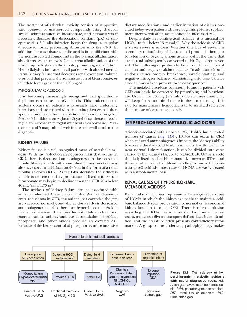
HYPERCHLOREMIC METABOLIC ACIDOSIS
Acidosis associated with a normal AG, HCMA, has a limited number of causes (Fig. 13.6). HCMA can occur in CKD when reduced ammoniagenesis impairs the kidney’s ability to excrete the daily acid load. In individuals with normal or near normal kidney function, it can be divided into cases caused by the kidney’s failure to reabsorb HCO3
− or secrete the daily fixed load of H+, commonly known as RTAs, and those in which renal acid-base handling is normal. In con-trast to AG acidosis, most cases of HCMA are easily treated with a supplemental base.
RENAL CAUSES OF HYPERCHLOREMIC METABOLIC ACIDOSIS
Renal tubular acidoses represent a heterogeneous cause of HCMA in which the kidney is unable to maintain acid-base balance despite preservation of normal or near-normal kidney function (normal GFR). There is often confusion regarding the RTAs, because no standard nomenclature exists, numerous diverse transport defects have been identi-fied, and the literature often presents contradictory infor-mation. A grasp of the underlying pathophysiology makes
Figure 13.6 The etiology of hy-perchloremic metabolic acidosis with useful diagnostic tools. AG, Anion gap; DKA, diabetic ketoacido-sis; PHA, pseudohypoaldosteronism; RTA, renal tubular acidosis; UAG, urine anion gap.
Hyperchloremic metabolic acidosis
InadequateNH3 production
Kidney failureHypoaldosteronism
PHA
Defect in HCO3�
reclamationDefect in H�
secretionExtrarenal loss of
base acid load
DiarrheaPancreatic fistula
Ureteral diversionsNH4CI/HCLNaCl load
Tolueneingestion
DKA
Excretion oforganic anions
Proximal RTA Distal RTA
Urine pH <5.5Positive UAG
Fractional excretionof HCO3 >15%
NegativeUAG
High urineosmole gap
Urine pH >5.5 Positive UAG

0
the approach to these disorders more comprehensible. The RTAs can be divided into four major categories: (1) primary defects in ammoniagenesis, (2) hypoaldosteronism, (3) dis-orders of the proximal tubule, and (4) disorders of the dis-tal tubule. The distal tubule defects can be further divided into those with hypokalemia and those with hyperkalemia (Fig. 13.7).
DEFECTIVE AMMONIAGENESISOne of the most common causes of an HCMA is the inabil-ity of the kidney to generate ammonia because of CKD. By definition, RTA refers to a specific acid excretory defect occurring despite the presence of normal or near-normal kidney function. Thus, it bears emphasis that the HCMA of CKD is not classified as RTA. As the number of nephrons decreases with CKD, there is a proportional decrease in the production of ammonia. As mentioned in the section on AG acidosis, when GFR falls below 40 mL/min/1.73 m2, the kidney is unable to excrete the daily acid load, and HCO3
− begins to decline with a concomitant increase in the serum Cl−, producing HCMA. Only when the GFR falls below 15 to 20 mL/min/1.73 m2 does the kidney lose the ability to secrete anions, thus converting this HCMA into an AG acidosis. It needs to be stressed that the acidosis in kidney failure, whether manifested by hyperchloremia or an AG, is primarily caused by defective ammoniagenesis. As such, the UAG will be positive because of the decrease in ammonia excretion, whereas urine pH will be less than 5.5.
HYPOALDOSTERONISMPrimary or secondary hypoaldosteronism is a common disor-der causing hyperkalemia and metabolic acidosis (Box 13.4). Hyporeninemic hypoaldosteronism (type IV RTA) is the most frequently encountered variety of this disorder, which is most common in patients with diabetes and mild CKD. The precise cause of hyporeninemia has not been clearly defined, but the findings that hypertension is frequently present and that the disorder may be partly reversed with chronic furosemide use suggest that renin suppression may be secondary to chronic volume overload. Neither has the cause of the hypoaldosteronism been fully explained. Renin suppression alone should not cause hypoaldosteronism,
133 CHAPTER 13 — METABOLIC ACIDOSIS
because hyperkalemia is a potent stimulus of aldosterone secretion and anephric individuals still secrete aldosterone. The acidosis is primarily caused by decreased ammoniagen-esis as a result of the associated hyperkalemia induced by the aldosterone deficiency. Hypoaldosteronism, by diminishing distal sodium reabsorption, also results in a less negative lumen potential, thus decreasing the rate of H+ secretion but not the electromotive force of the pump. Because the hydro-gen pump is not defective, urine pH is usually less than 5.5.
Patients with type IV RTA are usually asymptomatic, with only minor laboratory abnormalities (mild hyperkalemia and decreased HCO3
−). However, when renal potassium handling is further perturbed by various stressors, marked hyperkalemia ensues with a decline in ammoniagenesis. These stressors include sodium depletion, which decreases delivery of sodium to the distal tubule; high potassium diet; and potassium-sparing diuretics or medications that further decrease renin and aldosterone levels, such as angiotensin-converting enzyme inhibitors, angiotensin receptor block-ers, nonsteroidal antiinflammatory drugs, or heparin. Most patients can be treated by removing the insult to potassium homeostasis, restricting potassium intake, and providing supplemental bicarbonate. Proving that type IV RTA is pres-ent requires the demonstration of low renin and aldosterone
p032
Primary
Addison diseaseCongenital enzyme defectsDrugs
HeparinAngiotensin converting enzyme inhibitorsAngiotensin receptor blockers
Hyporeninemic Hypoaldosteronism (Type IV RTA)
PHA I (Autosomal Dominant)—Mineralocorticoid Resistance
PHA, Pseudohypoaldosteronism; RTA, renal tubular acidosis.
Box 13.4 Causes of Hypoaldosteronism
Figure 13.7 Evaluation of RTA. *Extrarenal loss of base is not a form of RTA. Findings shown, because the use of this algorithm may lead to its diagnosis. FEHCO3
, Fractional excre-tion of bicarbonate; PHA, pseudohy-poaldosteronism; RTA, renal tubular acidosis.
Renal tubular acidosis
Urine anion gap Serum K
Serum K
NegativePositive
Extrarenal loss of base*
Urine pH
Bicarbonateinfusion
Urine pH may be >5.5
Distal RTAPco2–backleakPco2–classic
Generalizedtubular defect
Ureteral obstruction
>5.5 <5.5
K K
HypoaldosteronismPHA Type 1 (autosomal dominant)PHA Type 1 (autosomal recessive)
Gordon’s syndrome (PHA II)Kidney failure
FeHCO3 >15%
Proximal RTA
KK

134 SECTION 2 — ACID-BASE, FLUID, AND ELECTROLYTE DISORDE
levels after sodium depletion. Because of practical consider-ations, these tests are rarely ordered, and most patients will be treated empirically.
Autosomal-dominant pseudohypoaldosteronism (PHA) type I is an uncommon disorder caused by a mutation in the renal mineralocorticoid receptor, which results in decreased affinity for aldosterone. This genetic disorder presents in childhood with hyperaldosteronism, hyperka-lemia, metabolic acidosis, salt wasting, and hypotension. Autosomal-dominant PHA type I becomes less severe with age. Carbenoxolone and glycyrrhizic acid (found in true licorice) both inhibit 11-β-hydroxysteroid dehydrogenase, the enzyme in the kidney that converts cortisol, which binds the mineralocorticoid receptor, to cortisone, which does not bind to the mineralocorticoid receptor. They can be used to treat this disorder by increasing the intrarenal supply of mineralocorticoid.
PROXIMAL RENAL TUBULAR ACIDOSISProximal RTA, often called type II RTA (because it was the second type described), is a defect in the ability of the proximal tubule to reclaim filtered HCO3
− (Box 13.5). In type II RTA, the proximal tubule has a diminished thresh-old (approximately 15 mmol/L instead of the normal 24 mmol/L) for HCO3
− reabsorption. When plasma HCO3−
falls below this threshold, complete reabsorption occurs. Proximal RTA can be congenital or acquired, and it may exist as an isolated defect in HCO3
− reabsorption or as part of a more generalized transport defect known as Fanconi syndrome, in which there is diminished reabsorption of other solutes across the proximal tubule. Patients with prox-imal RTA from Fanconi syndrome, in addition to the loss of HCO3
−, inappropriately excrete amino acids, glucose, phos-phorus, and uric acid in their urine.
As would be expected, mutations in the Na+-H+ exchanger on the luminal membrane, the Na+- HCO3
− cotransporter on the basolateral membrane, and cytosolic carbonic anhydrase have all been implicated in the isolated hereditary and spo-radic forms of proximal RTA. Several drugs that block car-bonic anhydrase, including the diuretic acetazolamide and the anticonvulsant topiramate, also cause isolated HCO3
− wasting. Proximal RTA with Fanconi syndrome is frequently found in patients with cystinosis, Wilson disease, Lowe syn-drome, multiple myeloma, and light chain disease, among other conditions. A decrease in ATP production, which reduces basolateral Na+-K+-ATPase activity, is the presumed etiology of this global transport defect. Drugs, particularly the cyclophosphamide analogue ifosfamide, and cidofovir, used in the treatment of cytomegalovirus retinitis, have also been associated with a generalized proximal tubulopathy.
Because distal H+ excretion is normal, urine pH during steady state will be less than 5.5 when the HCO3
− is below the lowered threshold and bicarbonaturia is absent. In this set-ting, the serum HCO3
− will be between 15 and 18 mEq/L. It is important to recognize that whenever the HCO3
− increases above the reabsorptive threshold, HCO3
− will appear in the urine, and the pH will be greater than 6.5. Although ammo-niagenesis is preserved in proximal RTA, direct or indirect measurement of urine NH4
+ may show an inappropriately low excretion. This occurs because HCO3
−, which escapes proximal reabsorption, serves as a buffer sink for secreted H+, thus reducing the trapping of NH4
+. The diagnosis of
RS
proximal RTA is established by demonstrating a fractional excretion of HCO3
− greater than 15% when supplemental bicarbonate is administered in an attempt to increase the serum bicarbonate to normal.
Treatment of proximal RTA is difficult, because admin-istered base is rapidly excreted in the urine. Extremely large amounts of base (10 to 15 mmol/kg/day) are fre-quently needed, and therefore compliance is limited. The increased delivery of HCO3
− to the distal nephron induces or exacerbates hypokalemia. It is recommended that fre-quent doses of a mixture of Na and K salts of bicarbonate and citrate be used.
DISTAL RENAL TUBULAR ACIDOSISClassic Distal Renal Tubular Acidosis with Hypokalemia
Distal RTA, also known as type I RTA, represents the inabil-ity of the distal tubule to acidify the urine (Box 13.6). As with proximal RTA, the distal variety can be congenital or acquired. Abnormalities have been identified in both the luminal H+-ATPase and the basolateral Cl−-HCO3
− exchanger. The acquired form is associated with autoimmune diseases, especially systemic lupus erythematosus and Sjögren syn-drome, dysproteinemia, and kidney transplant rejection. Immunocytochemical studies have shown decreased staining of the H+-ATPase and Cl−-HCO3
− exchanger in patients with the acquired form of distal RTA. Ifosfamide, which is also associated with a proximal RTA, can cause a distal defect. Amphotericin, which disrupts membrane-forming ion chan-nels, causes a distal RTA by allowing the backleak of protons across the luminal membrane. The classic finding in type I RTA is an inappropriately high urine pH (greater than 5.5).
Because H+ secretion is defective in distal RTA, less NH4+
can be trapped in the lumen of the tubule, and the UAG will be positive, reflecting this decrease in NH4
+ excre-tion. Besides having an inappropriately high urine pH and a positive UAG, distal RTA can be further characterized by
Isolated Defects in HCO3 ReabsorptionCarbonic anhydrase inhibitorsAcetazolamideTopiramateSulfamylonCarbonic anhydrase deficiency
Generalized Defects in Proximal Tubular TransportCystinosisWilson diseaseLowe syndromeGalactosemiaMultiple myelomaLight chain diseaseAmyloidosisVitamin D deficiencyIfosfamideCidofovirLeadAminoglycosides
Box 13.5 Causes of Proximal Renal Tubular Acidosis

measuring urine PCO2 during an HCO3− infusion. Distal
delivery of HCO3− in the presence of a normal H+ secre-
tory capacity results in elevated pCO2 in the urine. When there is a H+ secretory defect, urine pCO2 will not increase. As would be expected, in amphotericin-induced RTA where H+ ion secretion is unaffected, urine pCO2 increases nor-mally. Occasionally it may be difficult to distinguish HCMA induced by diarrhea from a distal RTA. Diarrhea results in HCMA and hypokalemia. Because the hypokalemia increases renal ammoniagenesis, urine pH may be inappro-priately elevated. Thus, on the surface, both forms of acido-sis appear similar. However, measurement of the UAG will easily distinguish the markedly elevated urine NH4
+ with its negative AG found in diarrheal illness from the low NH4
+ excretion and positive AG found with distal RTA. The one caveat is that sodium must be delivered to the distal tubule as indicated by urine Na+ that is greater than 20 mmol/L.
Classic distal RTA is associated with hypokalemia (due to augmented distal K+ secretion in lieu of H+ secretion in exchange for Na+ reabsorption), hypocitraturia (from enhanced proximal tubule cell reabsorption), hypercalci-uria (from the buffering of H+ in bone and loss of calcium), and nephrocalcinosis. The treatment of distal RTA is simply to supply enough base (2 to 3 mmol/kg/day) to counter the daily fixed production of acid. This can be administered as a mixture of sodium and potassium salts of either bicarbonate or citrate.
Distal Renal Tubular Acidosis with Hyperkalemia
Distal RTA with hyperkalemia can be further divided into two broad general categories: (1) a generalized defect of both distal tubular H+ and K+ secretion, or (2) a primary defect in Na+ transport, often referred to as a “voltage defect” (Box 13.7).
Generalized Distal Tubule Defect
Unlike classic distal RTA, a more generalized defect of the distal tubule can occur in which both H+ and K+ secretion
FamilialDefective HCO3
−-Cl− exchanger (autosomal dominant)Defective H+-ATPase (autosomal recessive)
EndemicThai endemic distal RTA
DrugsAmphotericinTolueneLithiumIfosfamideFoscarnetVanadium
Systemic disordersSjögren syndromeCryoglobulinemiaSystemic lupus erythematosusKidney transplant rejection
RTA, Renal tubular acidosis.
Box 13.6 Causes of Distal Renal Tubular Acidosis With Hypokalemia
135 CHAPTER 13 — METABOLIC ACIDOSIS
is impaired. This has been best characterized in cases of ureteral obstruction and in patients with interstitial kid-ney disease resulting from sickle cell anemia or systemic lupus erythematosus. In animals with ureteral obstruction, immunocytochemical staining has shown loss of the apical H+-ATPase. Why hyperkalemia occurs is less clear. Because K+ excretion cannot be augmented by diuretics, a primary defect in K+ transport is likely. Similar to classic distal RTA, urine pH is greater than 5.5.
Distal Sodium Transport Defects
Several disorders have been characterized by defective sodium transport in the distal tubule. The reabsorption of Na+ by the distal tubule generates a lumen-negative poten-tial. This electrical negativity helps promote the secretion of K+ and H+. Any drug or disorder that interferes with the creation of this lumen-negative potential will diminish both K+ and H+ secretion. These are commonly classified as volt-age defects. Autosomal-recessive PHA type I is a syndrome in which there is loss of function of the epithelial sodium channel (ENaC) in the distal tubule. Numerous mutations have been described in various subunits of this channel. This disease manifests in childhood with marked hyperkale-mia, metabolic acidosis, hyperaldosteronism, and salt wast-ing. Because the ENaC also exists in other tissue, including the lung, colon, and sweat glands, patients with this disor-der often have defects related to these organs. Treatment consists of providing a high salt intake. Drugs that block ENaC produce a similar metabolic picture. These include the potassium-sparing diuretics amiloride and triamterene, as well as trimethoprim and pentamidine.
Another well-recognized disorder of distal transport is PHA type II, also known as Gordon syndrome (see Chapters 39 and 67). Individuals with this condition have mild volume overload with suppressed renin and aldosterone, hyperten-sion, hyperkalemia, and metabolic acidosis. Mutations in two members of a family of serine-threonine kinases, WNK1 and WNK4, have been shown as the cause of this syndrome. These kinases appear to play an important role in the regulation of Cl− transport in numerous different tissues. It appears that defects in these kinases result in an increase in the number of neutral NaCl transporters (NCCT) and thus increase NaCl transport across the distal convoluted tubule. Less delivery
Lupus NephritisObstructive NephropathySickle Cell AnemiaVoltage DefectsFamilial
PHA type I (autosomal recessive)PHA type II (autosomal recessive)—Gordon syndrome
DrugsAmilorideTriamtereneTrimethoprimPentamidine
PHA, Pseudohypoaldosteronism.
Box 13.7 Causes of Distal Renal Tubular Acidosis With Hyperkalemia

136 SECTION 2 — ACID-BASE, FLUID, AND ELECTROLYTE DISORD
of sodium to the more distal tubule segments for reabsorp-tion curtails the generation of the lumen-negative potential. This results in decreased H+ and K+ secretion. Supporting this hypothesis is the fact that PHA type II can be treated with thiazide-type diuretics, which block the NCCT.
The acidosis in all of these sodium transport disorders is secondary to decreased H+ secretion caused by an unfa-vorable electrical gradient in the distal tubule as well as decreased ammoniagenesis caused by the hyperkalemia. Whether the urine pH is less than 5.5 depends on how severely H+ secretion is affected.
COMBINED PROXIMAL AND DISTAL RENAL TUBULAR ACIDOSISCombined proximal and distal renal tubular acidosis is an extremely uncommon disorder, which has been called type 3 RTA. As would be expected, both proximal HCO3
− reab-sorption and distal H+ secretion are impaired. Mutations in the gene for cytosolic carbonic anhydrase results in such a defect. As already discussed, ifosfamide can also cause a combined defect.
INCOMPLETE DISTAL RENAL TUBULAR ACIDOSISPatients with incomplete distal RTA typically come to medi-cal attention because of calcium stone disease and neph-rocalcinosis. Serum HCO3
− is normal, but urine pH never falls below 5.5, even after acid loading with NH4Cl or CaCl2. This disorder likely represents a milder form of distal RTA. Frank metabolic acidosis may become evident when patients are stressed by diarrhea or other conditions that require compensation by augmented renal proton secretion.
EXTRARENAL CAUSES OF HYPERCHLOREMIC METABOLIC ACIDOSIS
EXTRARENAL BICARBONATE LOSSLoss of base during episodes of diarrhea or with overzeal-ous use of laxatives is associated with HCMA. Loss of HCO3
− can also occur with pancreatic fistulae or following pancreas transplantation if drainage of the pancreatic duct occurs into the bladder. Ureteral diversions using an isolated sigmoid loop were frequently associated with bicarbonate loss because of Cl-HCO3
− exchange in the bowel loop. These ureteral-sigmoidostomies have largely been replaced with ureteral diversions using ileal conduits, which have less surface area and contact time for loss of HCO3
− to occur; however, if these become obstructed, HCMA can still develop.
ACID LOADAn obvious cause of an HCMA is ingestion or infusion of a chloride salt of an acid. Both NH4Cl and CaCl2 can result in a metabolic acidosis and can be used as a provocative test to assess urinary acidification. In addition, total parenteral nutrition using hydrochloric acid salts of various amino acids can produce a metabolic acidosis if an insufficient quantity of base (usually acetate) is added to the infusion mixture. Another form of acid load is saline (NaCl). Volume resuscita-tion with 0.9% NaCl will often produce an HCMA, referred to as a dilutional acidosis. This occurs because of “dilution” of the plasma HCO3
− by the more acidic saline solution (pH 7.0) and because volume expansion diminishes proximal HCO3
− reabsorption.
ERS
URINARY LOSS OF ANIONSAs previously discussed, if organic anions are excreted in the urine, they represent a source of base lost from the body. Although involving the kidney, this cannot be viewed as being caused by an intrinsic kidney defect. Because of the low renal threshold for the excretion of ketoacids, patients with DKA, if they are able to maintain their intravascular volume or are volume resuscitated, will excrete these anions in place of Cl−, resulting in HCMA. A similar metabolic dis-turbance exists after toluene exposure. Toluene is a com-mon solvent found in paint products and glues. Exposure is generally by inhalation, either accidental or intentional. Toluene is rapidly absorbed through the skin and mucous membranes and is metabolized to hippuric acid. Hippurate is quickly excreted by the kidney, leaving behind an HCMA. Although hippurate is not a base, its rapid excretion into the urine conceals the AG origins of this disturbance. Both of these disorders are usually easily discovered after an adequate history has been obtained.
KEY BIBLIOGRAPHYAdrogue HJ, Madias NE: Management of life-threatening acid-base dis-
orders, N Engl J Med 338:26-34, 1998, and 107-111.Alper SL: Genetic diseases of acid-base transporters, Annu Rev Physiol
64:899-923, 2002.Bonny O, Rossier B: Disturbances of Na/K balance: pseudohypoaldo-
steronism revisited, J Am Soc Nephrol 13:2399-2414, 2002.Brent J, McMartin K, Phillips S, et al: Fomepizole for the treatment of
methanol poisoning, N Engl J Med 344:424-429, 2001.Carlisle EJ, Donnelly SM, Vasuvattakul S, et al: Glue-sniffing and distal
renal tubular acidosis: sticking to the facts, J Am Soc Nephrol 1:1019-1027, 1991.
Chang CT, Chen YC, Fang JT, et al: Metformin-associated lactic acid-osis: case reports and literature review, J Nephrol 15; 398-394, 2002.
Claessens YE, Cariou A, Monchi M, et al: Detecting life-threatening lac-tic acidosis related to nucleoside-analog treatment of human immu-nodeficiency virus-infected patients, and treatment with l-carnitine, Crit Care Med 31:1042-1047, 2003.
Dargan PI, Wallace CI, Jones AL: An evidence based flowchart to guide the management of acute salicylate (aspirin) overdose, Emerg Med J 19:206-209, 2002.
DuBose TD Jr, Mcdonald GA: Renal tubular acidosis. In Dubose TD, Hamm LL Jr, editors: Acid-base and electrolyte disorders: a companion to Brenners and Rector’s The Kidney, Philadelphia, 2002, WB Saunders, pp 189-206.
Figge J, Jabor A, Kazda A: Anion gap and hypoalbuminemia, Crit Care Med 26:1807-1810, 1998.
Fraser AD: Clinical toxicologic implications of ethylene glycol and gly-colic acid poisoning, Ther Drug Monit 24:232-238, 2002.
Han J, Kim G-H, Kim J, et al: Secretory-defect distal renal tubular aci-dosis is associated with transporter defect in H+-ATPase and anion exchanger-1, J Am Soc Nephrol 13:1425-1432, 2002.
Hood VL, Tannen RL: Protection of acid-base balance by pH regulation of acid production, N Engl J Med 339:819-826, 1998.
Igarashi T, Sekine T, Inatomi J, et al: Unraveling the molecular patho-genesis of isolated proximal renal tubular acidosis, J Am Soc Nephrol 13:2171-2177, 2002.
Ishihara K, Szerlip HM: Anion gap acidosis, Semin Nephrol 18:83-89, 1998.Izzedine H, Launay-Vacher V, Isnard-Bagnis C, et al: Drug-induced Fan-
coni’s syndrome, Am J Kidney Dis 41:292-309, 2003.Karet FE: Inherited distal renal tubular acidosis, J Am Soc Nephrol
13:2178-2184, 2002.Kirschbaum B, Sica D, Anderson F: Urine electrolytes and the urine
anion and osmolar gaps, J Lab Clin Med 133:597-604, 1999.Lemann J Jr, Bushinsky DA, Hamm LL: Bone buffering of acid and base
in humans, Am J Physiol Renal Physiol 285:F811-F832, 2003.Levraut J, Grimaud D: Treatment of metabolic acidosis, Curr Opin Crit
Care 9:260-265, 2003.
Full bibliography can be found on www.expertconsult.com.