nasogastric tube insertion · testing placement wash hands and put on clean gloves draw up 30cc of...
TRANSCRIPT

NOAA Diving Medical Technician 2015 Nasogastric tubes
Overview Types of Tubes Indications for their use How to insert NG tubes Complications of NG tubes Enteral Feedings Indications and Complications. Gastrostomy

Types of Tubes Short tubes: passed through the nose into the stomach
− Levin tube: range in size from 14 to 18 Fr, single lumen made of plastic or rubber with holes near the tip.
− Gastric Sump (Salem): is radiopaque, clear plastic double lumen
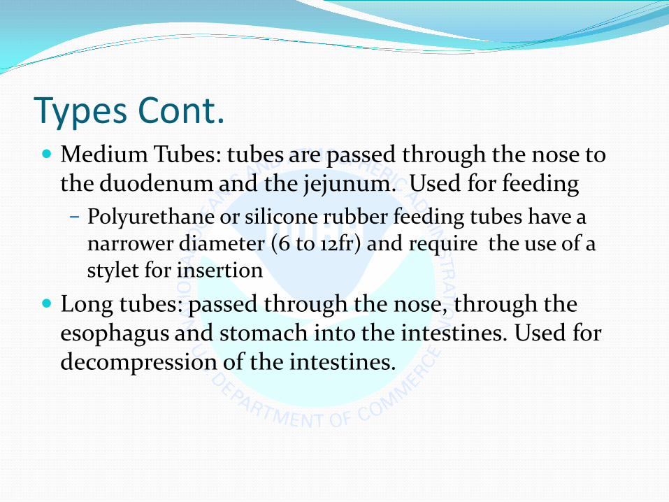
Types Cont. Medium Tubes: tubes are passed through the nose to
the duodenum and the jejunum. Used for feeding − Polyurethane or silicone rubber feeding tubes have a
narrower diameter (6 to 12fr) and require the use of a stylet for insertion
Long tubes: passed through the nose, through the esophagus and stomach into the intestines. Used for decompression of the intestines.
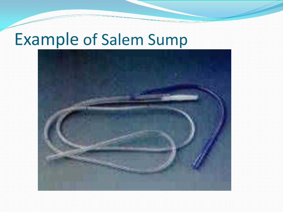
Example of Salem Sump

Indications for GI Intubation To decompress the stomach and remove gas and
fluid To lavage the stomach and remove ingested toxins To diagnose disorders of GI motility and other
disorders To administer medications and feedings To treat an obstruction To compress a bleeding site To aspirate gastric contents for analysis

Intubating the client with an NG tube Assessment:
− Who needs an NG: Surgical clients Ventilated client Neuromuscular impairment . Clients who are unable to maintain adequate oral
intake to meet metabolic demands.
− Assess patency of nares.
Assessment cont.
− Assess client’s medical history: Nosebleeds Nasal surgery Deviated septum Anticoagulation therapy
− Assess client’s gag reflex. − Assess client’s mental status. − Assess bowel sounds.
Planning Gather equipment:
14 0r 16 Fr NG tube Lubricating jelly PH test strips Tongue blade Flashlight Emesis basin Catheter tipped syringe 1 inch wide tape or commercial fixation device Suctioning available and ready

Planning Cont. − Explain procedure to client − Position the client in a sitting or high fowlers
position. If comatose-semi fowlers. − Examine feeding tube for flaws. − Determine the length of tube to be inserted. − Measure distance from the tip of the nose to the
earlobe and to the xyphoid process of the sternum.
− Prepare NG tube for insertion.

Measurement

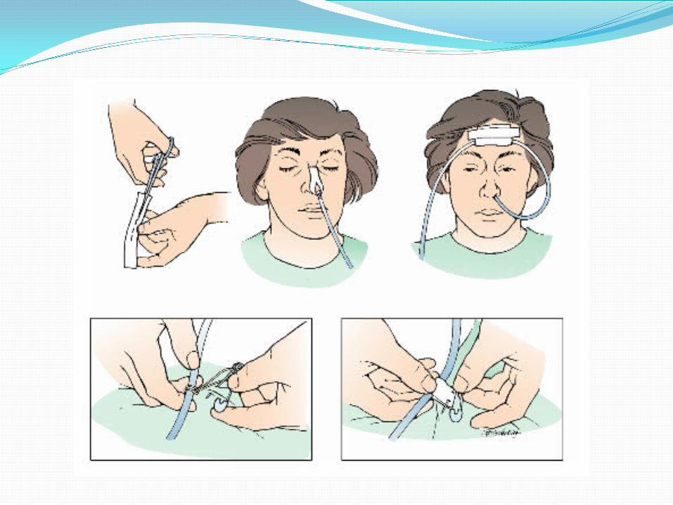
Implementation − Wash Hands − Put on clean gloves − Lubricate the tube − Hand the client a glass of water − Gently insert tube through nostril to back of
throat (posterior nasopharnyx). Aim back and down toward the ear.
− Have client flex head toward chest after tube has passed through nasopharynx


Implementation Cont. Emphasize the need to mouth breathe and swallow during
the procedure. Swallowing facilitates the passage of the tube through the
oropharnyx. When the tip of the tube reaches the carnia stop and listen
for air exchange from the distal end of the tube. If air is heard remove the tube.
Advance tube each time client swallows until desired length has been reached.
Do not force tube. If resistance is met or client starts to cough, choke or become cyanotic stop advancing the tube and pull back.
Implenentation Cont. Check placement of the tube.
− X-ray confirmation − Testing pH of aspirate
Secure the tube with tape or commercial device
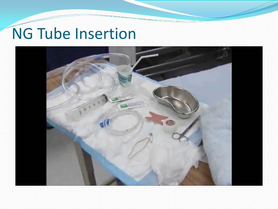
NG Tube Insertion
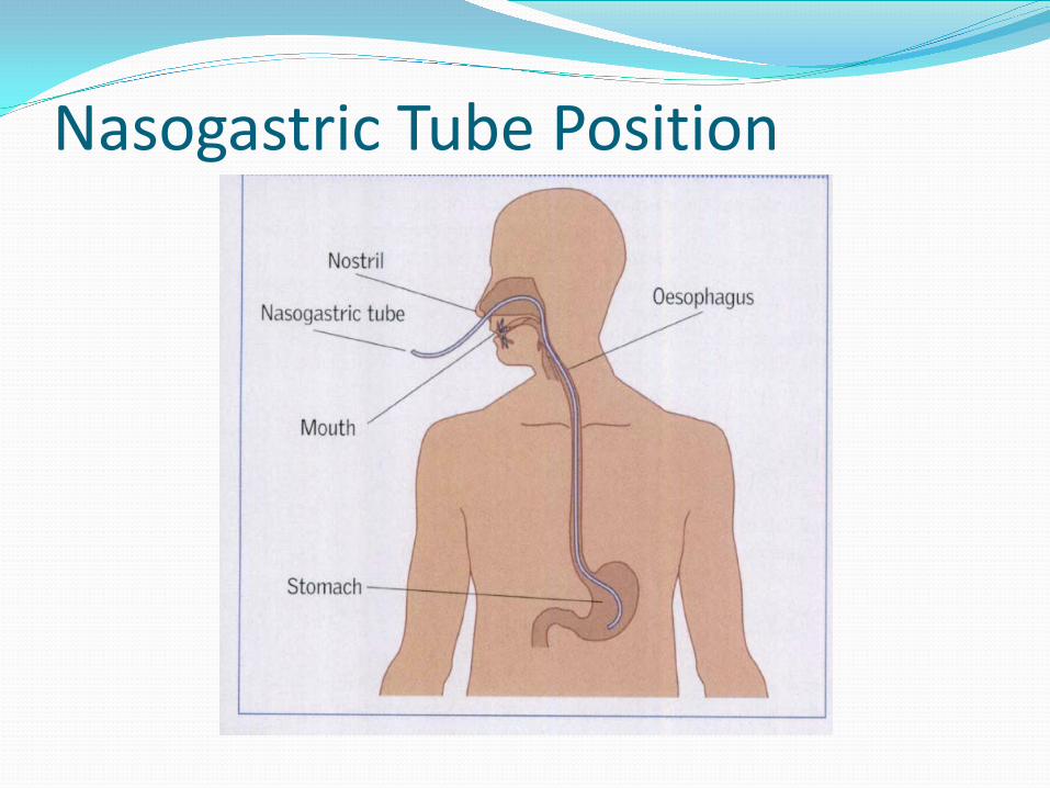
Nasogastric Tube Position
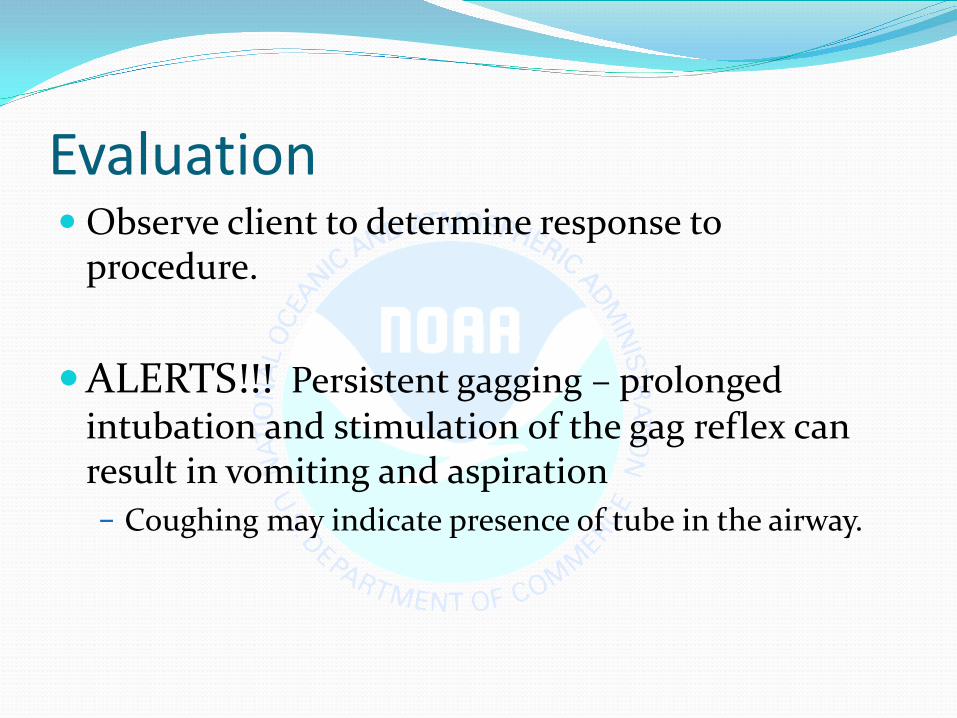
Evaluation Observe client to determine response to
procedure.
ALERTS!!! Persistent gagging – prolonged intubation and stimulation of the gag reflex can result in vomiting and aspiration − Coughing may indicate presence of tube in the airway.
Evaluation Cont. Note location of external site marking on the tube Documentation
− Size of tube, which nostril and client’s response. − Record length of tube from the nostril to end of
tube − Record aspirate pH and characteristics
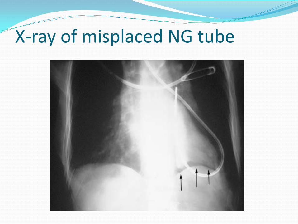
X-ray of misplaced NG tube

Testing Placement Wash hands and put on clean gloves Draw up 30cc of air into the syringe and attach to end of
the NG tube. Flush tube with 30cc of air prior to attempting to aspirate fluid. Draw back on the syringe to obtain 5 to 10 cc of gastric aspirate.
If unable to aspirate: − Advance tube – may be in air space above aspirate level − If intestinal placement suspected (pH 4-6) withdraw tube 5 to 10
cm − Have client lie on his/her left side wait 10-15 mins and attempt
aspiration again.

Testing Placement cont. Observe appearance of aspirate:
From client with enteral feeding – appearance of curdled enteral feed
From nasointestinal – bile stained From stomach (non feed)– green, tan, bloody, brown. Pleural fluid – pale yellow and serous Gently mix aspirate in syringe

Testing Placement cont. Note:
− In a study by Metheny et al (1994) the gastric aspirate of 880 clients were
examined: > gastric aspirate ranged in color from green to
yellow, tan/brown or bloody > respiratory aspirate was described as tan or
yellow/green (Best 2005)
Testing Placement Cont. Measure pH of aspirated GI contents by dipping pH
strip into the fluid or by applying a few drops of the fluid to the strip. Compare the color of the strip with the color on the chart.
Gastric fluid from a client who has fasted for at least 4 hours usually has a pH range from 1 to 4 but may be increased if the client is receiving acid inhibiting medications (pH 4-6)

Testing Placement Cont. Fluid from nasointestinal tube of fasting client usually
has a pH greater than 6. intestinal contents are less acidic than stomach.
Clients with a continuous tube feed may have a pH of 5 or higher.
Pleural fluid from the tracheubronchial tree is generally greater than 7.
National Patient Safety Association(2005a) recommend a pH of less than 5.5 feedings can be
initiated (Best, 2005)
Testing Placement Cont. Measure the length of the tube from nostril to tip. If after repeated attempts, it is not possible to
aspirate fluid from a tube that was originally established by x-ray examination to be in the desired position and there are NO risk factors for dislocation, tube has remained in original position and the client is NOT experiencing any difficulty you may assume the tube is correctly placed.

Responsibilities Identify signs and symptoms of inadvertent
respiratory migration. Identify conditions that increase the risk for
spontaneous tube dislocation from the intended position (retching, vomiting, nasotracheal suctioning, severe coughing)

Enteral Nutrition What is it:
− The administration of nutrients directly into the GI tract. The most desirable and appropriate method of providing nutrition is the oral route, but this is not always possible.
− Nasogastric feeding is the most common route − Nurses are the main healthcare professional
responsible for intubation

Questions?