nasal manifestations of systemic diseasesrhinologychair.org/assets/=nasal.pdf · the ent...
TRANSCRIPT

NASAL MANIFESTATIONS
OF SYSTEMIC DISEASES
DR. SHARIF ALMATRAFI , R2 .
9 MARCH 2016

OBJECTIVES
- WEGENER’S GRANULOMATOSIS .
- T-CELL LYMPHOMA.
- CHURG-STRAUSS SYNDROME.
- RHINOSCLEROMA.
- TUBERCULOSIS.
- LEPROSY.
- SYPHILIS.

WEGENER’S GRANULOMATOSIS

WEGENER’S
GRANULOMATOSIS (WG)
Granulomatosis with polyangiitis (GPA) , is a rare
multisystem autoimmune disease of unknown etiology.
- necrotizing granulomatous inflammation and vasculitis in
small- and medium-sized blood vessels.


This retrospective analysis is concerned with 10 patients
with Wegener's disease , FEB 2015 , by Rogister et al .
The ENT manifestations:
- rhinologic (osetocartilaginous--6 cases; mucosal--9 cases)
- the otologic (3 cases)
- laryngeal area (2 cases).


The study encompassed 43 patients with GPA of the mean
age of 47.7 ± 12.8 years .
They found that inflammation occurred mainly in the
maxillary sinuses (72 %). The mean L-M score was 5.8 ± 6.1.

DIAGNOSIS
1. CBC: Anemia and high ESR.
2. Urine: Red cells, casts and albumin in urine and high
creatinine levels.
3. X-ray chest: Single or multiple cavity lesions.
5. Cytoplasmic antineutrophilic cytoplasmic antibody (c-
ANCA).

4. Biopsy : epithelioid granuloma and necrotizing vasculitis.

TREATMENT
1- Immunosuppressive therapy: Oral cyclophosphamide
prednisone , retuximab.
2. Trimethoprim-sulfamethoxazole (Bactrim/Septran): good
results with limited disease.
3. Plasma exchange and intravenous immunoglobulin.


retrospective analysis of 975 visits from 99 GPA patients ,
2014 Sep , lally et al .
48 subjects had never received RTX and 51 received RTX at
least once. There was no active ENT disease during 92.4% of
the observational period (days) for subjects receiving RTX,
compared with 53.7% of the observational period for subjects
not receiving RTX
- Conclusion : Patients being treated with RTX were 11 times
less likely to have active ENT disease than patients being
treated with other therapies.

4- Local treatment :upper airway hygiene, local antibiotics
and nasal irrigation (These methods lead to the reduction of
nasal bacterial colonization).
- lubricants like glyceryl monoleate (reducing symptoms from
dry mucosa).
- large crusts must be mechanically removed..

5- Surgical.


METHODS:
During a 5-year-period, four women with an average age of 33 years underwent reconstructive rhinoplasty of their saddle-nose performed by an L-shaped rib cartilage graft.
- After follow-up period of 42 months for all the patients , the external form and function were preserved.
CONCLUSION:
- External nasal reconstruction for patients affected by WG appears to be safe and effective if the disease is in remission before any operation.

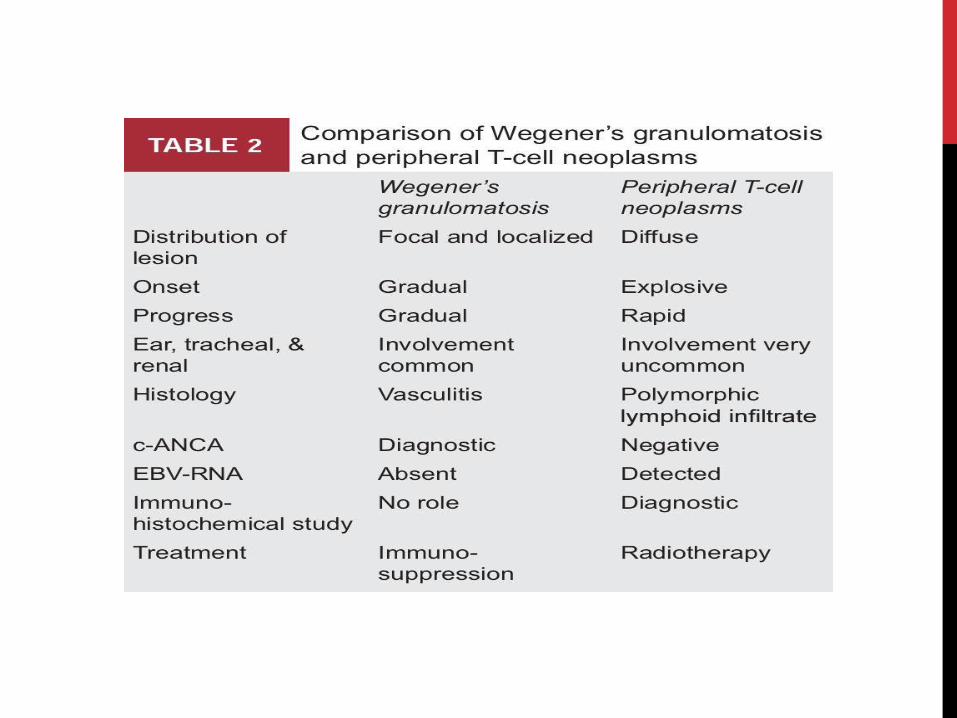
PERIPERERAL T-CELL NEOPLASM ( NON-HEALING
MIDLINE GRANULOMA, POLYMORPHIC RETICULOSIS)
- A destructive disease of the nose and mid-facial region.
- Absence of pulmonary and renal involvement.

CLINICAL FEATURES
- Unilateral lesions in nose extending to soft tissue of nose,
upper lip, oral cavity, maxillary sinus and orbit.
- Lesions are explosive and rapidly progressive.
- Secondary infection of lesions by gram-negative and
anaerobic organisms is common.

DIAGNOSIS
1- Biopsy: It will show mixed population of cells (mature
lymphocytes, plasma cells and large lymphoreticular cells),
which resembles picture of lymphoma.
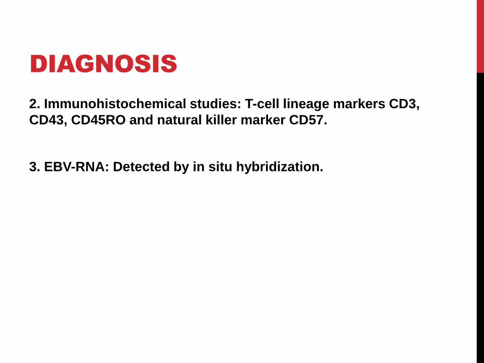
DIAGNOSIS
2. Immunohistochemical studies: T-cell lineage markers CD3,
CD43, CD45RO and natural killer marker CD57.
3. EBV-RNA: Detected by in situ hybridization.


1995 , LEARY ET AL
- Sections of 22 lymphoid proliferations arising in the
sinonasal region or Waldeyer's ring were studied with EBV
encoded RNAs (EBER-1 and -2) using in situ hybridisation.
RESULTS:
- EBV was detected in nuclei of tumour cells of five of
seven T cell lymphomas .

TREATMENT
1- Localized lesion: Radiotherapy followed by surgical
debridement .
2. Multiorgan disease: Standard leukemia protocol.
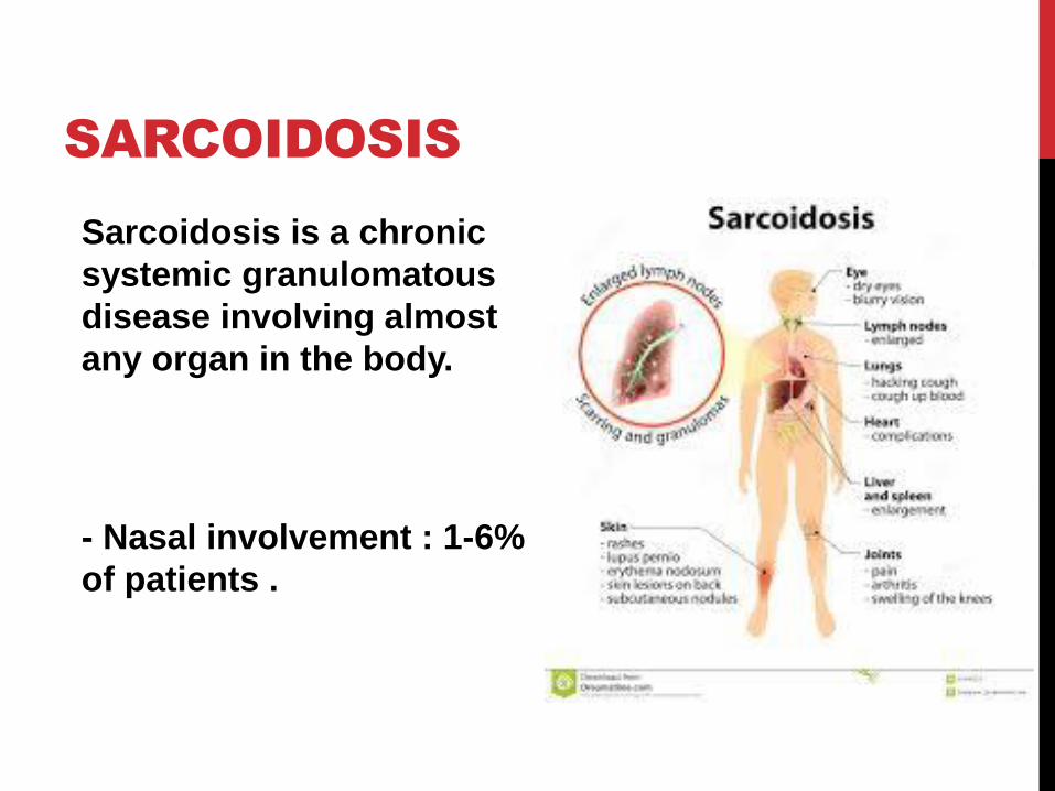
SARCOIDOSIS
Sarcoidosis is a chronic
systemic granulomatous
disease involving almost
any organ in the body.
- Nasal involvement : 1-6%
of patients .

ORL-HNS
- Salivary glands (parotid swelling)
- Oropharynx (tonsillar hypertrophy),
- Larynx (epiglottic or subglottic swelling).
- Neuropathy (sudden deafness and unilateral or bilateral
facial nerve palsy).

NASAL INVOLVEMENT
Externally : papular lesions on the nose that can
coalesce to form bluish-red swellings.
- lupus pernio that describes violaceous cutaneous
lesions on the cold-sensitive areas such as the nose,
cheeks, ears and fingers.

NASAL INVOLVEMENT
Intranasal : diffuse nasal crusting , diffuse mucosal swelling
of the septum and inferior turbinate , submucosal nodules.
- nasal polyps that are friable and bleed readily.

DIAGNOSIS1- X-ray chest: Diffuse pulmonary infiltrate with hilar adenopathy.
2. Biopsy: Non-caseating granuloma.

3. Gallium-67 scanning.
4. Angiotensin-converting enzyme: Elevated.
5. Serum and urinary calcium levels.

TREATMENT
1. Systemic steroids.
2. Methotrexate: ( unresponsive to steroids ).
3. Local treatment.
-recurrence is high .

CHURG-STRAUSS SYNDROME
- An eosinophil-rich , granulomatous inflammation and
necrotizing vasculitis of the upper respiratory tract, affecting
small to medium vessels .

There are three phases:
1. a prodromal phase consists of allergic disease (allergic
rhinitis, nasal polyposis)
2. peripheral blood and tissue eosinophilia (eosinophilic
pneumonia or gastroenteritis)
3. a life-threatening systemic vasculitis.
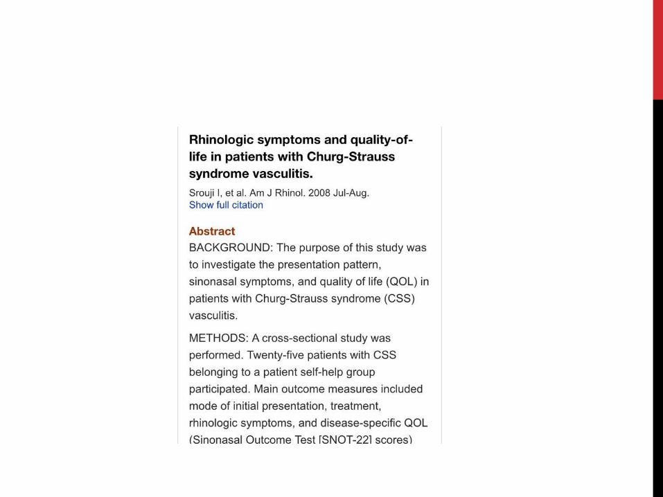

A cross-sectional study , 25 patients with CSS.
- mode of initial presentation, treatment, rhinologic symptoms, and disease-specific QOL (Sinonasal Outcome Test [SNOT-22] scores) and comparisons were made with general rhinosinusitis.
RESULTS:
80% had active sinonasal symptoms at the time of the study.
28 % reported worsening of their nasal symptoms as the main event leading to their diagnosis.
48 % had undergone nasal surgery.
Nasal symptoms:
nasal obstruction (95%), rhinorrhea (95%), anosmia (90%), and excessive sneezing (80%). Other symptoms included nasal crusting (75%), purulent nasal discharge (65%), and epistaxis (60%).
- SNOT-22 scores were significantly higher than normal.
CONCLUSION:
Sinonasal symptoms are common at initial presentation of CSS, emphasizing the role of otolaryngologists in its diagnosis

- p-ANCA is positive in 70% of patients, while c-ANCA is
negative.

TREATMENT
The management of CSS is represented by glucocorticoids
as standard treatment. CSS does not respond to
cyclophosphamide, as does WG.


a 33-year-old female suffering from Churg-Strauss syndrome having
had multiple operations in the past for recurrent polyps.
NDRP was performed on the left nostril only. The mucosa of the
left nasal vault was replaced by a split-thickness skin graft
(modified dermoplasty). On the right nostril, polyps were removed
and the ostia of the paranasal sinuses were enlarged as in typical
endoscopic sinus surgery.
- after eight months after the operation no polyps are detected on
the left side while polyps have recurred on the right nasal cavity.

RHINOSCLEROMA
- Chronic granulomatous bacterial disease.
- Endemic in several parts of the world (India).

- The causative microorganism, Klebsiella rhinoscleromatis ,
is a Gram-negative bacillus.
- The disease begins in the nose but extends to nasopharynx,
oropharynx, larynx (mostly subglottic region), trachea and
bronchi.

CLINICAL FEATURES
1. Catarrhal: Foul smelling purulent nasal discharge for
weeks to months.
2. Atrophic stage: crusting, which resembles atrophic
rhinitis.

3. Granulomatous stage: Multiple granulomatous nodules in
the nasal mucosa .
- These painless nodules are non-ulcerative and can be
found in pharynx, larynx, trachea and bronchi.
4. Cicatricial stage: Fibrosis leads to stenosis of nares,
distortion of upper lip and adhesions in the nose,
nasopharynx, oropharynx and larynx.

DIAGNOSIS
- Biopsy: Submucosa is infiltrated with plasma cells,
lymphocytes, eosinophils, Mikulicz cells and Russell bodies.
- Cultures of infected tissue.

MIKULICZ CELLS
Vacuolated histiocytes containing the causative bacilli.

RUSSELL BODIES
- Homogeneous eosinophilic inclusion bodies (accumulation
of immunoglobulins secreted by the plasma cells).

TREATMENT
- Combination of surgical debridement and long term
antibiotic therapy
- The antibiotic treatment consists of Streptomycin (1 g/day
for 4 weeks) and tetracycline (2 g/ day).
- NO role for radiotherapy or corticosteroids.


17-year-old female patient from Tehran, Iran, with
rhinoscleroma
- Prior treatment with streptomycin and tetracycline had been
unsuccessful.
- A three-month course of high-dose oral ciprofloxacin (750
mg b.i.d.) led to prompt cessation of the growth of the
granuloma which was removed later by plastic surgery.

TB
Tuberculosis is a chronic granulomatous infectious disease
caused by Mycobacterium tuberculosis and Mycobacterium
bovis.

TUBERCULOSIS
- Anterior part of nasal septum and anterior end of
inferior turbinate.
- Nodular infiltration ulceration perforation of
cartilaginous part of nasal septum.

- Diagnosis: Biopsy and special staining for acidfast bacilli,
culture of organisms.
- Treatment is antitubercular therapy.


A 48-year-old man presented with postnasal drip and an unpleasant nasal odour.
- Endoscopic examination revealed irregular thickening of the left lateral and posterior wall of the nasopharynx, partially covered with crusts and necrotic tissue.
- Histopathological : giant cell epithelioid granulomas with caseous necrosis.
- Direct examination after Ziehl-Neelsen staining was positive for tuberculosis. After six months of antituberculous triple therapy, endoscopic examination revealed a completely normal nasopharynx.

LEPROSY
- Tropics , Mycobacterium leprae.
- The nose is involved more commonly in lepromatous
type in comparison to tuberculoid form of leprosy.
- Anterior part of nasal septum and anterior end of
inferior turbinate.


CLINICAL FEATURES
- Excessive nasal discharge with red and swollen mucosa ,
Crusting and bleeding occurs later , nodular lesions on the
septum ulcerate and cause perforation of cartilaginous part
of septum.
- Late sequelae: Atrophic rhinitis, depression of nasal bridge.

DIAGNOSIS
�Scrapings of nasal mucosa and biopsy: Acid fast lepra
bacilli are present in the foamy appearing macrophages
called lepra cells.

TREATMENT
1- Antibiotics: Dapsone, rifampicin and isoniazid.
2. Reconstruction procedures: They are performed when
disease is inactive.


57 leprosy patients having moderately collapsed nose were taken
for nasal reconstruction.
- The bone graft was obtained from the second metatarsal of the
foot, placed in between lining and the nasal skin.
48 patients were followed in 3 to 5 year intervals.
26 patients were completely satisfied, 14 patients were happy with
shape of the nose along with some other problems. 6 cases showed
the poor results.


Rhino-sinus abnormalities were investigated in 13 ex-lepromatous leprosy patients
All patients had turbinate atrophy ( 100%) and 6 of the 13 (46.2 %) had septal perforation.
Paranasal sinus involvement was noted in 9 of 12 examined patients (75 %). The most commonly affected sinus was the maxillary sinus 8 of 12( 66.7 %).
All three patients treated by endoscopic sinus surgery experienced relapse and required further surgery
SYPHILIS
Acquired Syphilis.
1. Primary: Primary chancre of the vestibule is rare.
2. Secondary: Presents in nose with simple rhinitis with
crusting and fissuring in the nasal vestibule.

3. Tertiary: Nose is commonly involved.
- nasal septum gumma, which destroys both bony
and cartilaginous parts of nasal septum (saddle
nose deformity and perforation of palate ).

CONGENITAL
1-Early form: In the first 3 months of life, it
manifests as “snuffles”�and subsequently
other findings appear such as purulent
nasal discharge, fissuring and excoriations
of nasal vestibule and skin of upper lip.

2. Late form: In puberty, clinical features of tertiary syphilis
manifest such as gumma and perforation of nasal septum.
Other stigmata of syphilis (corneal opacities, deafness and
Hutchinson’s teeth) are also present.

DIAGNOSIS
1- Serological tests: VDRL.
2. Biopsy of the tissue: Special stains demonstrate
Trepenoma pallidum.

TREATMENT
- Penicillin: Benzathine penicillin.
- Nasal alkaline wash , Removal of nasal crusts.
- Surgery.


KLEBSIELLA
RHINOSCLEROMA

PERIPERERAL T-CELL
NEOPLASM (NONHEALING
MIDLINE GRANULOMA)

SARCOIDOSIS

WEGENER
GRANULOMATOSIS

SARCOIDOSIS

LEPROSY

DO YOUR BEST !!

TAKE HOME
MESSAGES …
- Ent surgeons have an important rule in diagnosing
systemic disease.
- Patient focused team approach provides the best care for
the patient .

THANK YOU