nanotechnology-applied curcumin for different diseases therapy
TRANSCRIPT

Review ArticleNanotechnology-Applied Curcumin forDifferent Diseases Therapy
Negar Ghalandarlaki,1 Ali Mohammad Alizadeh,1 and Soheil Ashkani-Esfahani2
1 Cancer Research Center, Tehran University of Medical Sciences, Tehran, Iran2 Student Research Committee, Shiraz University of Medical Sciences, Shiraz, Iran
Correspondence should be addressed to Ali Mohammad Alizadeh; [email protected]
Received 4 February 2014; Revised 21 April 2014; Accepted 25 April 2014; Published 5 June 2014
Academic Editor: Yoshinori Marunaka
Copyright © 2014 Negar Ghalandarlaki et al. &is is an open access article distributed under the Creative Commons AttributionLicense, which permits unrestricted use, distribution, and reproduction in any medium, provided the original work is properlycited.
Curcumin is a lipophilic molecule with an active ingredient in the herbal remedy and dietary spice turmeric. It is used by di'erentfolks for treatment of many diseases. Recent studies have discussed poor bioavailability of curcumin because of poor absorption,rapid metabolism, and rapid systemic elimination. Nanotechnology is an emerging (eld that is potentially changing the way wecan treat diseases through drug delivery with curcumin. &e recent investigations established several approaches to improve thebioavailability, to increase the plasma concentration, and to enhance the cellular permeability processes of curcumin. Severaltypes of nanoparticles have been found to be suitable for the encapsulation or loading of curcumin to improve its therapeutice'ects in di'erent diseases. Nanoparticles such as liposomes, polymeric nanoparticles, micelles, nanogels, niosomes, cyclodextrins,dendrimers, silvers, and solid lipids are emerging as one of the useful alternatives that have been shown to deliver therapeuticconcentrations of curcumin. &is review shows that curcumin’s therapeutic e'ects may increase to some extent in the presenceof nanotechnology. &e presented board of evidence focuses on the valuable special e'ects of curcumin on di'erent diseases andcandidates it for future clinical studies in the realm of these diseases.
1. Introduction
Curcumin, 1,7-bis(4-hydroxy-3-methoxyphenyl)-1,6-hepta-dien-3,5-dione, is a lipophilic molecule that rapidly per-meates cell membrane [1]. Typical extract of Curcumalonga L. contains the structures I to III: (I) diferuloyl-methane/curcumin (curcumin I, 75%), (II) demethoxycur-cumin (curcumin II, 20%), and (III) bisdemethoxycurcumin(curcumin III, 5%) [2, 3] (Figure 1). Curcumin is an activeingredient in the herbal remedy and dietary spice turmeric[4]. It has a long history of administration by di'erent folks ofChina, India, and Iran for the treatment ofmanydiseases suchas diabetes, liver diseases, rheumatoid diseases, atheroscle-rosis, infectious diseases, cancers, and digestive disorderssuch as indigestion, dyspepsia, ,atulence, and gastric andduodenal ulcers [5, 6]. Many researchers have worked oncurcumin due to its various therapeutic e'ects on di'erentdiseases. Shortly, curcumin has received attentionmostly due
to its antioxidant, anti-in,ammatory, antitumoral, apoptosis-inducing, and antiangiogenesis e'ects, which were reportedin many investigations. It acts on multiple targets in cel-lular pathways making this agent able to perform multipleactions [7]. &e simple molecular structure along with therelative density of functional groups in curcumin providesresearchers with an outstanding target for structure-activityrelationship and lead optimization studies. &e structuralanalogues of curcumin have been reported to enhance therate of absorption with a peak plasma half-life [8–10]. Recentinvestigations have considered curcumin a lead compoundfor designing new chemotherapeutic agents for treatment ofcancers including colon cancers [11], prostate cancers [12],and other conditions with indication of chemotherapy [13,14].
Curcumin is remarkably well tolerated, but its bioavail-ability is poor. It does not appear to be toxic to animals[15] or humans [16], even at high doses. Recent studies have
Hindawi Publishing Corporation
BioMed Research International
Volume 2014, Article ID 394264, 23 pages
http://dx.doi.org/10.1155/2014/394264

2 BioMed Research International
OHHO
OO
Curcumin (I)
Demethoxycurcumin (II)
Bisdemethoxycurcumin (III)
Equilibrating keto-enol tautomers
OCH3H3CO
OHHO
O
OCH3H3CO
OHHO
OO
OCH3
OH
OH
OHHO
O
OCH3H3CO
OH
HO
OO
Figure 1: Curcumin I, II, and III (curcumin, demethoxycurcumin, and bisdemethyoxycurcumin) and curcumin keto-enol tautomers.
discussed poor bioavailability of curcumin because of poorabsorption, rapid metabolism, and rapid systemic elimina-tion [17, 18]; however, comprehensive pharmacokinetic dataare still missing. In a study done by Yang et al. [19], theyreported 1% bioavailability for oral administration of cur-cumin in rats. On the elimination of curcumin, an investiga-tion in rat model demonstrated that a4er oral administrationof 1 g/kg of curcumin, more than 75% was excreted in fecesand negligible amount of curcumin was detected in urine[20]. Additionally, FDA has declared curcumin as “generallysafe.” Although curcumin showed a wide variety of usefulpharmacological e'ects and has been found to be quite safe inboth animals and humans, there are some studies concerning
its toxicity [21]. In spite of these advantages, curcumin haspoor water solubility; as a consequence, it reveals solubility-limited bioavailability, which makes it a class II drug inthe biopharmaceutics classi(cation system [22]. Additionally,due to its rapid intestinal and hepatic metabolism, about 60%to 70% of an oral dose of curcumin gets eliminated by thefeces [23].
As mentioned above, curcumin has been proven to bee'ective in treatment of di'erent diseases with low toxicityto human and animals. It is extremely safe upon oral admin-istration even at very high doses; however, it is limited dueto its poor bioavailability, stability, low solubility, and rapiddegradation and metabolism. Overcoming these problems

BioMed Research International 3
has been the main goal of many studies over the past threedecades. Since curcumin was demonstrated to have poorbioavailability and selectivity [17, 24], numerous analoguesof this material have been introduced and tested in order toevaluate their activities against known biological targets andto also improve their bioavailability, selectivity, and stability[25–28]. In addition, several approaches were introduced toimprove the bioavailability, to increase the plasma concentra-tion, and to enhance the cellular permeability and resistanceto metabolic processes of curcumin. Using nanoparticlesfor targeting drug delivery appeared to provide curcuminwith longer circulation, better permeability, and strongerresistance to metabolic processes.
2. Nanotechnology Approaches for Curcumin
Nanotechnology is increasingly considered to be the technol-ogy of the future. Among the wide applications of nanotech-nology is the use of nanoparticles for enhancing the bioavail-ability and the solubility of lipophilic compounds such as cur-cumin in drug delivery systems.&erefore, applying nanopar-ticles gained immense popularity in the last decade due totheir potential to improve the therapeutic e'ects of the encap-sulated drugs by protecting drugs from enzymatic degrada-tion, providing their controlled release and prolonged bloodcirculation, changing their pharmacokinetics, decreasingtheir toxicity, and limiting their nonspeci(c uptake [29]. Overa period of time, numerous emphases have been given todevelop the biodistribution of natural curcumin, but it is onlyjust recently that the application of the (eld of nanotech-nology has considerably enhanced its therapeutic e'ects.Nanoparticles such as liposomes, polymeric nanoparticles,micelles, nanogels, niosomes, cyclodextrins, dendrimers, sil-vers, and solid lipids are emerging as one of the usefulalternatives that have been shown to deliver therapeutic con-centrations of curcumin. &e use of the above nanoparticlehas improved main problems of curcumin such as low solu-bility, instability, poor bioavailability, and rapid metabolismin cancers, wound healing, Alzheimer’s disease, epilepticus,ischemia diseases, in,ammatory diseases, and so on (Table 1).
3. Liposomes
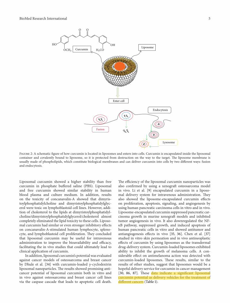
Liposomes are synthetic vesicles with globular character thatcan be produced from natural phospholipids [82]. &ey areself-assembling closed colloidal constructions composed oflipid bilayers, and they have a spherical shape in whichan outer lipid bilayer surrounds a central aqueous space[83]. &e liposome diameter varies from 25 nm to 2.5mm(Table 1). &ey are stated to act as immunological adjuvantsand drug carriers. Liposomes can encapsulate drugs withwidely varying solubility or lipophilicity, entrapped eitherin the aqueous core of the phospholipid bilayer or at thebilayer interface [84]. Moreover, they are able to deliverdrugs into cells by fusion or endocytosis, and practicallyany drug, irrespective of its solubility, can be entrapped intoliposomes (Figure 2). In this regard, to enhance the solubilityof curcumin, Rahman et al. [30] prepared !-cyclodextrin-curcumin inclusion complexes that entrapped both native
curcumin and the complexes separately into liposomes. Allcurcumin-containing formulations were e'ective in inhibit-ing cell proliferation in in vitro cell culture. In another study,Shi et al. [31] developed a water-soluble liposomal curcuminto examine curcumin’s preventive e'ects on lung (brosis viaintravenous administration in mice by using enzyme-linkedimmunosorbent assay method (ELISA). Results showed thatliposomal curcumin can e'ectively diminish radiation pneu-monitis and (brosis of lung and sensitize LL/2 cells toirradiation.&ese data suggest that the systemic administra-tion of liposomal curcumin with enhanced solubility is safeand deserves to be investigated for further clinical applica-tion.
Some studies showed that the drugs encapsulated inliposomes are expected to be transported without rapiddegradation and result in minimum side e'ects and showmore signs of stability in the recipients. In this regard,to assess curcumin tissue distribution, Matabudul et al.[32] questioned whether di'erent durations of intravenousinfusions of Lipocurc can alter curcumin metabolism and itstissue distribution and whether treating necropsied tissues ofBeagle dogs with phosphoric acid prior to measuring cur-cumin and its metabolite (tetrahydrocurcumin) can stabilizethe compounds allowing for accurate analytical measure-ments. Results demonstrated that the addition of liposomesmay inhibit or saturate a putative reductase enzyme thatconverts curcumin to tetrahydrocurcumin and stabilizes thelevels of curcumin. Tetrahydrocurcumin in some tissues(lung, spleen, and liver), but not all the examined tissues(lung, spleen, liver, pancreas, kidney, and urinary bladder),raised issues of tissue-speci(c curcumin and tetrahydrocur-cumin stability via a transporter-dependent mechanism thatelevated tissue concentrations of curcumin. Additionally,to obtain better understanding of curcumin interactionmechanisms with lipid membranes and improve the stabilityof curcumin, Karewicz et al. [33] banded curcumin toegg yolk phosphatidylcholine, dihexadecyl phosphate andcholesterol, then in order to determine curcumin bindingconstant to liposomes they used absorption and ,uorescencetechniques. &e egg yolk phosphatidylcholine/dihexadecylphosphate/cholesterol liposomal bilayer curcumin stabilizedthe system proportionally to its content, while the egg yolkphosphatidylcholine/dihexadecyl phosphate system destabi-lized upon drug loading.&e three-component lipid compo-sition of the liposome seems to be themost promising systemfor curcumin delivery. Furthermore, an interaction of freeand liposomal curcumin with egg yolk phosphatidylcholineand mixed monolayers was also studied by using Langmuirbalance measurements. Condensing e'ects of curcumin onegg yolk phosphatidylcholine and egg yolk phosphatidyl-choline/dihexadecyl phosphate monolayers and looseningin,uence on egg yolk phosphatidylcholine/dihexadecyl phos-phate/cholesterol ones were observed. It was also demon-strated that curcumin-loaded egg yolk phosphatidylcholineliposomes are more stable upon interaction with the modellipid membrane than the unloaded ones. In another study,Chen et al. [85] reported the e'ects of di'erent lipo-somal formulations on curcumin stability in phosphatebu'ered saline, human blood, plasma, and culture medium.

4 BioMed Research InternationalTa
ble1:Nanop
articles
-con
jugatedcurcum
incharacteriz
ationford
i'erentd
iseases
treatment.
Type
ofnano
particle
sFo
rmSize
(nm)
Used
mod
elsMetho
dsRe
sults
Reference
Lipo
some
Globu
lar25–205
(i)Breastcancer
(ii)M
elano
ma
(iii)Re
nalischemia
(iv)M
alaria
Invitro
Invivo
(dog
andmice
)
(i)Increasedsolubility,tissued
istrib
ution,andsta
bility
(ii)E
nhancedantitum
orandantia
ngiogenesis
e'ects
(iii)Show
edantim
elano
ma,anti-in,ammatory,and
antim
alaria
le'ects
[30–
33]
[34–
37]
[38,39]
Mice
lleSpheric
al10–100
(i)Lu
ngtumor
(ii)B
reastcancer
Invitro
Invivo
(mice
)
(i)Increasedsolubilitya
ndbioavailability
(ii)Improved
antio
xidativ
eand
antitum
ore'ects
(iii)Prolon
gedcir
culationtim
e(iv
)Enh
anced,u
orescencee
'ect
[40]
[41]
[42]
[43]
[44]
[45]
[46]
Noisome
Lamellar
190–
1140
(i)Albino
ratskin
(ii)C
ancerous
cells
Invitro
Invivo
(snake
andmice
)
(i)Increasedskin
penetra
tion
(ii)P
rolonged
deliverys
ystem
(iii)An
ti-infectionandantic
ancere
'ects
(iv)E
nhanced,u
orescenceintensity
[47]
[48]
[49]
OO
O O O
OO
O
OOO
O
n
OR 6
OR 6
OR 2O
R 2
OR 6
OR 6O
R 3
OR 3
OR 3
R 6O
R 2OR 3
OR 3
O
R 3OR 2
O
R 6O
R 2O
OR 2
Cyclo
dextrin
Cyclic
150–
500
(i)Bo
weld
isease
(ii)B
reast,lung
,pancreatic,
and
prostatecancer
Invitro
Invivo
(ratandmice
)
(i)Im
proved
solubility
(ii)E
nhancedantip
roliferatione'ects
(iii)Increasedantic
ancera
ndanti-in,ammatorye
'ects
(iv)D
evelo
pedbioavailability
[30]
[50–
56]
Dendrim
erGlobu
larpo
lymer
15–150
(i)Breastcancer
(ii)C
olon
cancer
Invitro
Invivo
(mice
)(i)
Improved
stability
(ii)Increased
antitum
orandantip
roliferativee
'ects
[57,58]
[59–
63]
Nanogel
Cross-lin
ked
polymer
network
10–200
(i)Mela
noma
(ii)B
reastand
pancreaticcancer
cells
Invitro
(i)Increasedsta
bility
(ii)E
nhanced,u
orescencee
'ects
(iii)Develo
pedbioavailability
(iv)Improved
antic
ancere
'ects
(v)G
etbette
rcon
trolledrelea
se(vi)Prolon
gedhalf-life
(vii)
Enhanced
treatmento
fmela
noma
[64]
[65]
Chito
san
Linear
polysaccha-
ride
compo
sed
100–
250
(i)Wou
nds
(ii)M
elano
matum
orsIn
vitro
Invivo
(ratandmice
)
(i)Im
proved
chem
icalstability
(ii)S
howe
dwo
undhealing
e'ects
(iii)Increasedantitum
ore'ects
(iv)Improved
antio
xidant
e'ects
(v)P
rolonged
bloo
dcir
culation
[66–
71]
Gold
Globu
lar200–
250
Cancerou
scells
Invitro
(i)Im
proved
solubility
(ii)E
nhancedantio
xidant
andantic
ancere
'ects
[72]
[73]
Silve
rFilm
layer
∼15(i)
Infections
(ii)S
kinwo
unds
Invitro
(i)Sh
owed
antim
icrob
iale'ects
(ii)Improved
woun
dhealing
(iii)Increasedantiv
iraland
antic
ancere
'ects
[74]
[75]
Lipi
d(s
olid
)Solid
lipid
Spheric
al50–100
0(i)
Cerebralisc
hemia
(ii)C
olitis
(iii)Aller
gy(iv
)Breastcancer
Invitro
Invivo
(ratandmice
)
(i)Prolon
gedcir
culationof
bloo
d(ii)Increased
anti-in,ammatorye
'ects
(iii)Im
proved
brain
delivery
[76–
78]
[79–
81]
BioMed Research International 5
Curcumin Liposome
Enter cell
Fusion
Endocytosis
Lysosome
OH
O O
H3COOCH3
HO
Figure 2: A schematic (gure of how curcumin is located in liposomes and enters into cells. Curcumin is encapsulated inside the liposomalcontainer and covalently bound to liposome, so it is protected from destruction on the way to the target. &e liposome membrane isusually made of phospholipids, which constitute biological membranes and can deliver curcumin into cells by two di'erent ways: fusionand endocytosis.
Liposomal curcumin showed a higher stability than freecurcumin in phosphate bu'ered saline (PBS). Liposomaland free curcumin showed similar stability in humanblood plasma and culture medium. In addition, resultson the toxicity of concanavalin-A showed that dimyris-toylphosphatidylcholine and dimyristoylphosphatidylglyc-erol were toxic on lymphoblastoid cell lines. However, addi-tion of cholesterol to the lipids at dimyristoylphosphatidyl-choline/dimyristoylphosphatidylglycerol/cholesterol almostcompletely eliminated the lipid toxicity to these cells. Liposo-mal curcumin had similar or even stronger inhibitory e'ectson concanavalin-A-stimulated human lymphocyte, spleno-cyte, and lymphoblastoid cell proliferation. &ey concludedthat liposomal curcumin may be useful for intravenousadministration to improve the bioavailability and e8cacy,facilitating the in vivo studies that could ultimately lead toclinical application of curcumin.
In addition, liposomal curcumin’s potential was evaluatedagainst cancer models of osteosarcoma and breast cancerby Dhule et al. [34] with curcumin-loaded #-cyclodextrinliposomal nanoparticles.&e results showed promising anti-cancer potential of liposomal curcumin both in vitro andin vivo against osteosarcoma and breast cancer cell linesvia the caspase cascade that leads to apoptotic cell death.
&e e8ciency of the liposomal curcumin nanoparticles wasalso con(rmed by using a xenogra4 osteosarcoma modelin vivo. Li et al. [9] encapsulated curcumin in a liposo-mal delivery system for intravenous administration. &eyalso showed the liposome-encapsulated curcumin e'ectson proliferation, apoptosis, signaling, and angiogenesis byusing human pancreatic carcinoma cells in vitro and in vivo.Liposome-encapsulated curcumin suppressed pancreatic car-cinoma growth in murine xenogra4 models and inhibitedtumor angiogenesis in vivo. It also downregulated the NF-$B pathway, suppressed growth, and induced apoptosis ofhuman pancreatic cells in vitro and showed antitumor andantiangiogenesis e'ects in vivo [35, 36]. Chen et al. [37]studied in vitro skin permeation and in vivo antineoplastice'ects of curcumin by using liposomes as the transdermaldrug-delivery system. Curcumin-loaded liposomes exhibitedability to inhibit the growth of melanoma cells. A con-siderable e'ect on antimelanoma action was detected withcurcumin-loaded liposomes. &ese results, similar to theresults of other studies, suggest that liposomes would be ahopeful delivery service for curcumin in cancer management[30, 86, 87]. &ese data indicate a signi(cant liposomalcurcumin potential as delivery vehicles for the treatment ofdi'erent cancers (Table 1).

6 BioMed Research International
Rogers et al. [38] also administered liposomes contain-ing curcumin to target delivery to renal tubular epithelialand antigen-presenting cells in mice renal ischemia model.Liposomal curcumin signi(cantly improved serum crea-tinine, reduced histological injury and cellular apoptosis,and lowered toll-like receptor-4, heat shock protein-70, andtumor necrosis factor alpha (TNF-%) mRNA expression, andit also decreased neutrophil in(ltration and in,ammatoryinterleukins expression. In this regard, Basnet et al. [39]developed vaginal administration of liposomal curcumin.Liposomal curcumin was found to be twofold to sixfold morepotent than corresponding free curcumin. Results showedthat liposomal delivery systems enhance anti-in,ammatoryproperties of curcumin. Also, evaluation of liposomal cur-cumin cytochrome P450 inhibition was conducted by Machet al. [88] in liver tissues. Results demonstrated that thereis low potential for CYP450 mediated drug interactions atphysiologic serum concentrations of liposomal curcumin.It will not interact with other chemotherapy agents thatare metabolized and/or eliminated via the primary drugmetabolizing cytochrome P450 pathways [88].
&e therapeutic e8cacies of novel liposomal deliverysystems based on artemisinin or artemisinin-based combi-nation therapy with curcumin have been investigated andreported by Isacchi et al. [89].&ey reported that artemisininalone began to decrease parasitaemia levels only 7 daysa4er the start of the treatment, and it appears to have a,uctuant trend in blood concentration which is re,ectedin the antimalarial e'ectiveness. By contrast, treatmentswith artemisinin loaded with liposomal delivery systemsappeared to have an immediate antimalarial e'ect whichcured all malaria-infected mice within the same postinocu-lation period of time. In particular, artemisinin loaded withliposomal curcumin seems to give the most pronouncedand statistically signi(cant therapeutic e'ect in this murinemodel of malaria. &e enhanced permanency in bloodof artemisinin loaded with liposomal curcumin suggestsapplication of these nanosystems as suitable passive targetedcarriers for parasitic infections [89]. &is strong e'ect offormulation is added up to the mechanism of action ofartemisinin which acts in the erythrocyte cycle stage ofhuman host as a blood schizonticide. Agarwal et al. [90] alsoassessed the acute e'ects of liposome-entrapped curcumin onincreasing current electroshock seizures, pentylenetetrazole-induced seizures, and status epilepticus in mice. Liposome-entrapped curcumin demonstrated signi(cant increase inseizure threshold current and latency to myoclonic andgeneralized seizures increasing current electroshock andpentylenetetrazole-induced seizures, respectively. It alsoincreased the latency to the onset and decreased the durationof seizures during status epilepticus. &erefore, liposomal-entrapped curcumin can possess anticonvulsant activityagainst status epilepticus in mice (Table 1).
To put it brie,y, the above data suggest that the admin-istration of liposomal curcumin has numerous bene(ciale'ects which could lead to required clinical applications.&ese better outcomes take place by means of enhancedsolubility, more safety and minimum side e'ects, moresigns of stability in the blood, increased bioavailability and
e8cacy, owning a potential role as delivery vehicles for thetreatment of di'erent cancers, potent anti-in,ammatory andantimalaria response, and, (nally, anticonvulsant activity.
4. Micelles
A typical micelle is a surfactant molecule aggregate dispersedin a liquid colloid. It is a nanosized vesicular membranewhich becomes soluble in water by gathering the hydrophilicheads outside in contact with the solvent and hydrophobictails inside, which is known as emulsi(cation. Micelles arelipid molecules that arrange themselves in a spherical formin aqueous solutions with a very narrow range from 10to 100 nm in size, which makes them more stable towarddilution in biological ,uids [84]. &e shape or morphologyof micelles is from amphiphilic block copolymers such asspherical, rodlike, and starlike, as well as vesicles (Table1). &e self-assembly of amphiphilic block copolymer is areversible process, and the shape varies with the copolymers’composition and length ratio [91].&e functional propertiesofmicelles are based on amphiphilic block copolymers, whichcome together to form a nanosized core/shell structure inaqueous media. &e hydrophobic core area hands out asa pool for hydrophobic drugs, while the hydrophilic shellarea stabilizes the hydrophobic core and makes the polymerswater soluble. Polymeric micelles can serve as transporters ofwater-insoluble drugs such as curcumin, which can augmentthe drug’s e8ciency by targeting de(nite cells or organs;therefore, fewer drugs accumulate in healthy tissues andtheir toxicity reduces, and occasionally higher doses can beadministered [92]. In this regard, to overcome the poor watersolubility of curcumin, Liu et al. [93] prepared curcumin-loaded biodegradable self-assembled polymeric micelles bysolid dispersion method, which was simple and easy toscale up. Release pro(le showed a signi(cant di'erencebetween rapid release of free curcumin and much slowerand sustained release of curcumin-loaded micelles. In addi-tion, the preparation of curcumin-loaded micelles basedon amphiphilic Pluronic/polycaprolactone block copolymerwas investigated by Raveendran et al. [40], which provedto be e8cient in enhancing curcumin’s aqueous solubility.Some other studies also deliberated on highly surface-activecompounds such as poloxamers or Pluronic that can self-assemble into spherical micelle. In vitro results showedthat spherical curcumin-loaded mixed micelles might serveas a potential nanocarrier to improve the solubility andbiological activity of curcumin [94–96]. In another study,the aqueous solubility of the curcumin was increased byencapsulation within the micelles [97]. Solubilization wasdirectly related to the compatibility between the solubilizateand polycaprolactone as determined by the Flory-Hugginsinteraction parameter. Molecular modeling study suggestedthat curcumin tended to interact with polycaprolactoneserving as a core embraced by polyethylene glycol as a shell.In addition, Yu et al. [41] showed the structure of modi(ed&-polylysine micelles and their application in improvingcellular antioxidant activity of curcuminoids. Results of theirinvestigation revealed that modi(ed &-polylysine micelleswere able to encapsulate curcuminoids and improve their

BioMed Research International 7
water solubility and cellular antioxidative activity comparedwith free curcuminoids. &ey suggested that these micellesmay be used as new biopolymermicelles for delivering poorlysoluble drugs such as curcumin. Another study synthesizedcurcumin in sodium dodecyl sulfate and cetyltrimethylam-monium bromide micelles to overcome the poor watersolubility of curcumin and demonstrated antioxidative e'ectsof curcumin analogues against the free-radical-induced per-oxidation of linoleic acid in these micelles [98, 99]. Kineticanalysis of the antioxidation processes demonstrated thatthese compounds exhibited extraordinarily higher antioxida-tive activity in micelles due to their solubility being higherthan free curcumin [98].
Drug release frommicelles is governed by di'erent issuesincluding micelle stability, the rate of copolymer biodegrada-tion, and drug di'usion. By the way, Sahu et al. [100] reportedthe potential of the two most common Pluronic triblockcopolymer micelles, Pluronic F127 and F68, for curcuminencapsulation e8ciency and stability. Pluronic F127 showedbetter encapsulation e8ciency and good stability for long-term storage than Pluronic F68. Atomic force microscopy(AFM) study revealed that the drug-encapsulatedmicelles arespherical in shape with diameters below 100 nm. Pluronic-encapsulated curcumin demonstrated slower and sustainedrelease of curcumin from the micelles and considerableanticancer activity in comparison with free curcumin in vitrocytotoxicity study. In addition, Podaralla et al. [42] reporteda natural protein core-based polymeric micelle and demon-strated its application for the delivery of hydrophobic anti-cancer drugs, speci(cally curcumin. &ey synthesized novelbiodegradable micelles by conjugatingmethoxy polyethyleneglycol and zein, a biodegradable hydrophobic plant proteinwhich can be found in Maize, and then encapsulating withcurcumin. Polyethylene glycol zein micelles sustained thecurcumin release up to 24 hrs in vitro and signi(cantlyenhanced its aqueous solubility and stability with the 3-fold reduction in IC50 value of curcumin. So, since thecurcumin is (nely protected from possible inactivation bytheir micellar surroundings, its retention and bioavailabilitycan be enhanced (Table 1).
Aiming to modify the pharmacokinetics of curcumin,Song et al. [43] synthesized a poly(D,L-lactide-co-glycolide)-b-poly(ethylene glycol)-b-poly(D,L-lactide-co-glycolide)(PLGA-PEG-PLGA) with micelles. PLGA-PEG-PLGAmicelles provided higher area under the concentrationcurve (AUC) and enhanced residence time, clearance, anddistribution half-life in comparison with curcumin solution.&e prolongation of half-life, enhanced residence time, anddecreased total clearance indicated that curcumin-loadedmicelles could prolong acting time of curcumin in vivo.&eseresults may be related to the curcumin location within themicelles and increased viscosity of copolymer solution at thebody temperature. &e variation of AUC indicated that thecurcumin-loaded micelles provided higher bioavailabilitythan curcumin solution, and the biodistribution studyshowed that the micelles had decreased drug uptake byliver and spleen and enhanced drug distribution in lungand brain. &ese results suggested that PLGA-PEG-PLGAmicelles would be a potential carrier for curcumin. In
addition, Ma et al. [94] demonstrated the pharmacokineticsof both solubilized curcumin and its polymeric micellarformulation in rats by using a simple, rapid, and reliableHPLC method. &ey concluded that encapsulation ofcurcumin in the polymeric micellar formulation led toincrease in curcumin’s half-life and distribution volume.
In addition, curcumin-micelles can be a'ected by physic-ochemical characteristics, concentration, and location withinthe micelles. &e polymeric micelles have a prolonged cir-culation time due to their small size and hydrophilic shellthat reduce the drug uptake by the mononuclear phagocytesystem [101]. Leung et al. [44] reported that encapsulatedcurcumin in cationic micelles suppresses alkaline hydrolysisthat was studied in three types of micelles composed ofthe cationic surfactants cetyltrimethylammonium bromide(CTAB) and dodecyltrimethylammonium bromide (DTAB)and the anionic surfactant sodium dodecyl sulfate (SDS).Curcumin underwent rapid degradation in the SDS micellarsolution by alkaline hydrolysis at pH of 13, while it wassigni(cantly suppressed with a yield of suppression closeto 90% in the presence of either CTAB or DTAB micelles.Results from ,uorescence spectroscopic studies revealed thatcurcumin is dissociated from the SDSmicelles to the aqueousphase at this pH while curcumin remains encapsulatedin CTAB and DTAB micelles at pH 13. &e absence ofencapsulation and stabilization in the SDS micellar solutionresulted in rapid hydrolysis of curcumin. Some other studiesshowed other curcumin-loaded micelles properties. Wanget al. [102] introduced the sensitive ,uorometric methodfor the determination of curcumin using the enhancementof mixed micelle. &is method had the advantages of highsensitivity, selectivity, and stability. &e ,uorescence of cur-cumin was greatly enhanced by mixed micelle of sodiumdodecylbenzenesulfonate and cetyltrimethylammoniumbro-mide (SDBS-CTAB). &is study indicated that ,uorescencequantum yield of curcumin in SDBS-CTAB micelle wasabout 55-fold larger than that of aqueous solution con-taining 1.0% ethanol, which was in agreement with their,uorescence intensity ratio. As a result, curcumin can beused as a ,uorophore in ,uorescence polarization anisotropymeasurement to determine the criticalmicellar concentrationof surfactant and to study the interaction between them.In addition, Adhikary et al. [45] performed femtosecond,uorescence upconversion experiments on the naturallyoccurringmedicinal pigment, curcumin, in anionic, cationic,and neutral micelles. &ese micelles were composed of SDS,dodecyltrimethylammonium bromide (DTAB), and TritonX-100.&ey revealed the curcumin’s excited-state kinetics inmicelles with fast (3–8 ps) and slow (50–80 ps) components.While deuteration of curcumin had a negligible e'ect onthe fast component, the slow component exhibited a pro-nounced isotope impact of approximately 1.6, which indi-cates thatmicelle-captured curcumin undergoes excited-stateintramolecular hydrogen atom transfer. Moreover, Beganet al. [46] had attached curcumin to phosphatidylcholinemicelles followed by ,uorescence measurements. Curcuminin aqueous solution did not inhibit dioxygenation of fattyacids by lipoxygenase 1, but it inhibited the oxidation offatty acids when bound to phosphatidylcholine micelles.

8 BioMed Research International
Results demonstrated that 8.6'M of curcumin bound to thephosphatidylcholine micelles is required for 50% inhibitionof linoleic acid peroxidation. Lineweaver-Burk plot analysishad indicated that curcumin is a competitive inhibitor oflipoxygenase 1 with Ki of 1.7 'M for linoleic acid and 4.3'Mfor arachidonic acid, respectively. By using spectroscopicmeasurement, they revealed that the inhibition of lipoxyge-nase 1 activity by curcumin can be due to binding to activecenter iron and curcumin a4er binding to the phosphatidyl-choline micelles acts as an inhibitor of lipoxygenase 1. In arecent investigation, the critical micelle concentration of theamphiphilic polymer was determined by using ,uorescentprobe. Outcomes indicated that Pluronic/polycaprolactonemicelles may be a promising candidate for curcumin deliveryto cancer cells of colorectal adenocarcinoma [40]. In anotherpharmacokinetic study, curcumin micelles demonstratedhigher concentration and longer retention time in plasmaand tumor sites, so they had stronger inhibitory e'ects onproliferation, migration, invasion, and tube formation ofcarcinoma cells than free curcumin; for example, curcuminmicelles were shown to be more e'ective, presumably dueto higher concentration in inhibiting tumor growth andprolonged survival in both subcutaneous and pulmonarymetastatic tumor models [103].
Investigating the in,uence of micelles on cytotoxicityof curcumin, speci(cally in cancer therapy, in vitro studyby Raveendran et al. [40] showed that Pluronic/polycapro-lactonemicelles could be a promising candidate for curcumindelivery to cancer cells regarding the cytotoxicity and cellularuptake of the curcumin-loaded micelles in colorectaladenocarcinoma cells. An investigation by Wang et al. [104]revealed that the encapsulated curcuminmaintains its potentantitumor e'ects; however, curcumin-loaded micelles weremore e'ective in inhibiting tumor growth and spontaneouspulmonary metastasis in subcutaneous 4T1 breast tumormodel and prolonged survival of tumor-bearingmice. Immu-no,uorescent and immunohistochemical studies alsoshowed that tumors of curcumin-loaded micelle-treatedmice had more apoptotic cells, fewer microvessels, and fewerproliferation-positive cells [104]. In addition, Yang et al.[19] had conjugated methoxypolyethylene glycol-polylacticacid (mPEG-PLA) micelle to multiple curcumin mole-cules; the cytotoxicity study results showed that the e'ect ofIC50 of mPEG-PLA-Tris-curcumin on human hepatocellularcarcinoma cells was similar to unmodi(ed curcumin.&e cel-lular uptake study demonstrated that these carriers could suc-cessfully transport the drug to the cytoplasm of hepatic cells.Micelles containing multiple drug molecules were an e8-cient means to increase loading and intracellular deliveryof low-potency curcumin [19]. Moreover, Mohanty et al.[105] reported that curcumin encapsulated in methoxypoly(ethylene glycol)/poly-epsilon-caprolactone diblockcopolymeric (MePEG/PCL) micelle, by varying the cop-olymer ratio (40 : 60MePEG/PCL ratio was selected due toits high encapsulation), had increased bioavailability due tointensi(ed uptake, 2.95 times more, with comparative cyto-toxic e'ects by induction of apoptosis in contrast withunmodi(ed curcumin at equimolar concentrations. Over-all, these data obviously showed the commitment of a
micellar system for e8cient solubilization, stabilization, andcontrolled delivery of the hydrophobic drug, such as cur-cumin, for cancer therapy.
Concisely, curcumin-loadedmicelles can boost the drug’se8ciency by targeting de(nite cells and result in less drugaccumulation in healthy tissues and reduction of toxicity.Curcumin’s aqueous solubility and much slower and sus-tained release of drug caused by curcumin-loaded micellesalso get in use in several conditions. &e retention andbioavailability of curcumin could be elevated since the cur-cumin is protected from possible inactivation by its micellarsurroundings. Locating the curcumin in the micelles can alsoenhance half-life and residence time and decrease total clear-ance leading to prolongation of acting time of curcumin.Curcumin micelles can be in,uenced by physicochemicalfeatures including their size and electrical charges, concentra-tion, and location within the micelles. &ese data obviouslyshowed the commitment of a micellar system for e8cientsolubilization, stabilization, and controlled delivery of thehydrophobic drug, such as curcumin, for cancer therapy(Table 1).
5. Niosomes
Niosomes aremicroscopic lamellar constructions of nonionicsurfactant of alkyl or dialkyl polyglycerol ether category withcholesterol that were (rst introduced in the 70s [106, 107].Niosomes can provide a container for drug molecules witha wide range of solubilities due to presence of hydrophilic,amphiphilic, and lipophilic moieties in the constitution(Table 1).&ey behave similar to liposomes in vivo and can beused as an e'ective alternative to liposomal drug carriers, andthose properties depend on the composition of the bilayer aswell as the method of their production [108]. Surfactant type,encapsulated drug nature, storage temperature, detergents,and use of membrane spanning lipids can a'ect niosomesstability [107]. Niosomes are also planned for use in a numberof potential therapeutic applications, such as anticancer andanti-infective drug targeting agents [84]. &ey can improvethe therapeutic indices of drugs by restricting their actionon the target cells. &ey also improve oral bioavailability ofpoorly absorbed drugs such as curcumin to design the noveldrug delivery system and increase the skin penetration ofdrugs [47]. In this regard, in an in vitro study which wasperformed using albino rat skin, proniosomes of curcuminwere prepared by encapsulation of the drug in a mixtureof Span 80, cholesterol, and diethyl ether to investigatetransdermal drug delivery system [109].&e planned systemsdistinguished between size, drug entrapment, repose angle,hydration rate, and vesicular stability under di'erent storagesettings. Results showed that proniosomes are very stable andpromising prolonged delivery systems for curcumin [109].Mandal et al. [48] also designed a comparative study withdi'erent microenvironments for photophysical propertiesof curcumin inside niosomes by means of steady state,time resolved ,uorescence spectroscopy, and dynamic lightscattering techniques. Outcomes showed that more rigidand con(ned microenvironments of niosomes improve thesteady state ,uorescence intensity alongwith the ,uorescence

BioMed Research International 9
lifetime of curcumin.&e data indicated that niosomes are agood tool for delivery system to suppress the level of degrada-tion of curcumin [48]. In another study, by Rungphanichkulet al., curcuminoid niosomes were developed with a seriesof nonionic surfactants to enhance skin permeation of cur-cuminoids. [49]. Results were evaluated based on entrapmente8ciency and in vitro penetration of curcuminoids via snakeskin. Niosomes drastically enhanced permeation of curcum-inoids compared with a vehicle solution of curcuminoids[49]. &e ,uxes of curcumin, desmethoxycurcumin, andbisdesmethoxycurcumin also were consistent with the quali-(ed hydrophobicity of curcumin, desmethoxycurcumin, andbisdesmethoxycurcumin, respectively. Data indicated thatcurcuminoids can be fruitfully prepared as niosomes, andsuch formulations have superior properties for transdermaldrug delivery system [49].
Brie,y, niosomes can be a potential delivery system forcurcumin in order to suppress the degradation of this agentand increase its life time. It has also been demonstrated thatniosomes boost the permeation of curcumin through skin(Table 1).
6. Cyclodextrins
Cyclodextrins (Cds) are a family of complexes prepared fromsugar molecules bound together in cyclic oligosaccharides[110]. &ey are created from starch by using enzymaticswitch. Cds are cyclic oligomers of glucose that can formwater-soluble inclusion complexes with small molecules andportions of large complexes [111]. &ey are exceptionalmolecules with pseudoamphiphilic construction, which areused industrially in pharmaceutical requirements [84]. Cdsare also used in agriculture and in environmental engineeringin food, drug delivery systems, and chemical industries [110].&ey have an interior hydrophobic surface which can providea place for residence of poorly water-soluble molecules, whilethe external hydrophilic area makes its solubility possible inthe aqueous setting with high stability (Table 1).
To improve the water solubility and the hydrolytic stabil-ity of curcumin, Tønnesen et al. [50] prepared cyclodextrin-curcumin complexes by using HPLC and UV/VIS scan-ning spectrophotometer techniques [50] (Figure 3). Resultsshowed that the hydrolytic stability of curcumin was sturdilyimproved by the complex, and also the photodecompositionrate was enhanced in organic solvents compared to the freecurcumin. As a result, the cavity size and charge of cyclodex-trin side-chains in,uenced the stability and degradation rateof curcumin [50]. In addition, other investigations on thesolubility, phase distribution, and hydrolytic and photochem-ical stability of curcumin showed that curcumin derivativesweremore stable towards hydrolytic degradation in cyclodex-trin solutions than free curcumin [51]. &e photochemicalstudies illustrated that curcumin is universally more stablethan its other derivatives. Solubility and phase-distributionstudies showed that curcuminoids with side groups on thephenyl moiety have higher a8nity for the hydroxypropyl-#-cyclodextrin (HP-#-CD) than the cyclodextrins. &e rad-ical scavenging investigations con(rmed that curcumin ismore active than its curcuminoids derivatives, and the
free phenolic hydroxyl group may possibly be necessaryfor the scavenging properties [51]. In another study, toincrease the solubility of curcumin, Darandale and Vavia [52]employed cyclodextrin-based nanosponges; they formulatedthe complex of curcumin with !-cyclodextrin nanospongeobtained with dimethyl carbonate as a cross-linker. &eloaded nanosponges have shown more solubilization e8-ciency compared to free curcumin and !-cyclodextrin com-plex.&e characterization of curcumin nanosponge complexcon(rmed the interactions of curcumin with nanosponges.Moreover, in vitro drug release of curcumin was controlledover a prolonged time period, and the complex was non-hemolytic [52]. &erefore, it seems that CDs are permittingvehicles that can be used for oral delivery to develop thebioavailability of insoluble drugs bymolecular dispersion anddegradation protection and for intravenous delivery to supplyas solubilizers for multifaceted hydrophobic drugs withoutaltering their pharmacokinetic properties [84].
Yadav et al. [53] developed a new cyclodextrin com-plex of curcumin to increase solubility of curcumin andstudied its anti-in,ammatory and antiproliferative e'ects.&ey showed that cyclodextrin-curcumin complex was moreactive than free curcumin in inhibiting the in,ammatorytranscription factor, such as nuclear factor kappa-b (NF-$B).In addition, it suppressed cyclin D1 as a cell proliferationmarker, matrix metallopeptidase 9 (MMP-9) as an invasionmarker in metastasis, and vascular endothelial growth factor(VEGF) as an angiogenesis marker. Cyclodextrin-curcumincomplex was alsomore active in inducing the death receptorsand apoptosis of leukemic cells as well as other cancer celllines.&ese suggest that cyclodextrin-curcumin complex hassuperior characteristics compared to free curcumin for celluptake and antiproliferative and anti-in,ammatory e'ects[53]. Yadav et al. [54] have also planned curcumin complexesby common methods to evaluate the anti-in,ammatorye'ects of cyclodextrin-curcumin complex for the treatmentof in,ammatory bowel disease (IBD) in an animal rat model.In vivo results showed that curcumin has higher a8nity forhydroxypropyl-!-cyclodextrin than other cyclodextrins. Inaddition, hydroxypropyl-!-cyclodextrin-curcumin complexproved to be a powerful antiangiogenesis complex. In vivodata also con(rmed that the scale of colitis was appreciablyattenuated by cyclodextrin-curcumin. In summary, cyclodex-trin complex was shown to be valuable in the therapeuticapproaches for IBD patients being a nontoxic natural dietaryyield [54].
Additionally, Cds can augment bioavailability of insolubledrugs such as curcumin by rising drug solubility and dissolu-tion [84].&ey also amplify the permeability of hydrophobicagents by making them accessible at the surface of the mem-brane’s biological barrier. A !-cyclodextrin-encapsulatedcurcumin drug delivery systemwas developed by Yallapu andcolleagues in order to get better curcumin hydrophilic anddrug delivery characteristics [55]. Encapsulated-curcumine8ciency was shown to be improved through increasingthe ratio of curcumin to cyclodextrin. &en, an optimizedcyclodextrin-curcumin complex was assessed for intracellu-lar uptake and anticancer e'ects. Cell proliferation and clono-genic examinations showed that !-cyclodextrin-curcumin

10 BioMed Research International
O–CH3 H3C–O
Curcumin
+
Cyclodextrin
OOH
OHHO
HOO
CC
CC
C CC
HO
OMeOMe
OH
12
3
45
6
78
910
2!3!
4!5!
6!
7!
8!9!
10!
HO
HO
HO
HO
HO
HO
OH
OH
OH
OH
OH
OHOH
OH
O
O
O
O
O
OOO
O
O
O
O
OO
HOCH3
HOCH3
CH3OH CH3OH
CH3OH
CH3OH
CH3OH
6
2
3
Figure 3: A schematic (gure of curcumin connection to the cyclodextrin nanoparticles.
self-assembly augmented curcumin delivery and improvedits therapeutic e8cacy in prostate cancer cells [55]. More-over, curcumin-loaded #-cyclodextrin liposomal nanoparti-cles as delivery vehicles were also explored by Dhule et al.[34] and evaluated against cancer models. &e resulting 2-hydroxypropyl-#-cyclodextrin/curcumin-liposome complexshowed promising anticancer potential both in vitro and invivo against osteosarcoma and breast cancer. Liposomal cur-cumin initiated the caspase cascade that led to apoptoticcell death in vitro. In addition, the e8ciency of the lipo-somal curcumin formulation was con(rmed in vivo byusing a xenogra4 osteosarcoma model. Data showed thatcurcumin-loaded #-cyclodextrin liposomes indicated con-siderable potential as delivery vehicles for cancer cure [34].Rahman et al. [30] prepared !-cyclodextrin-curcumin com-plexes, as a hydrophilic curcumin. &ey entrapped both
native curcumin as a hydrophobic agent and the complexesseparately into liposomes and then assessed them for theircytotoxicity in cancerous cell lines. &e aqueous solubilityof !-cyclodextrin-curcumin complexes enhanced noticeably,and successful entrapment of complexes into prepared lipo-somes was also achieved. &e median e'ective dose for allcurcumin formulations was found to be in a low range forboth lung and colon cancer cell lines [30]. Outcomes guar-anteed that !-cyclodextrin-curcumin complexes of weaklywater-soluble drugs such as curcumin can be tricked withinbiocompatible vesicles such as liposomes, and this does notprevent their anticancer e'ects [30]. In another study, anovel curcumin analogue (di,uorinated curcumin; CDF) andCDF-!-cyclodextrin-curcumin complex were synthesized toenhance anticancer e'ects against pancreatic cancer [56].Results showed that CDF-!-cyclodextrin was found to lower

BioMed Research International 11
IC50 value by half when tested against multiple cancercell lines. Following intravenous administration of CDF-!-cyclodextrin, it was specially accumulated in pancreatic tissue10 times higher than in serum. As a result, novel curcuminanalogue CDF outstanding gathering in pancreas tissue ledto its persuasive anticancer e'ects against pancreatic cancercells. So, synthesis of such CDF-!-cyclodextrin self-assemblyis a successful approach to improve its bioavailability andtissue distribution. Further evaluations on CDF delivery inclinical settings for treatment of human malignancies weresuggested by these authors [56]. Moreover, a novel poly(!-cyclodextrin)-curcumin self-assembly was approached toimprove curcumin’s delivery to prostate cancer cells byYallapu et al. [112]. Intracellular uptake of the self-assemblywas evaluated by means of ,ow cytometry and immuno,u-orescence microscopy. &e therapeutic values were estab-lished by cell proliferation and colony formation tests onprostate cancer cells. Results recommended that the poly(!-cyclodextrin)-curcumin formulation could be a valuablesystem for developing curcumin delivery and its therapeu-tic e'ectiveness in prostate cancer [112]. Additionally, inorder to improve solubility and drug delivery of curcumin,Lomedasht et al. [113] exploited a !-cyclodextrin-curcumininclusion complex and evaluated its cytotoxic e'ects byMTT assay in vitro. Breast cancer cells were treated withequal concentration of !-cyclodextrin-curcumin and freecurcumin. &en, telomerase gene expression was comparedby real-time PCR in two groups. In vitro results showedthat !-cyclodextrin-curcumin increased curcumin deliveryin breast cancer cells [113]. Telomerase gene expression waslower in !-cyclodextrin-curcumin-treated cells than freecurcumin-treated cells. As a result, !-cyclodextrin-curcumincomplex wasmore e'ectual than free curcumin in telomeraseexpression inhibition. Rocks et al. [114] have used cyclodex-trins as an excipient permitting a signi(cant enhancementof curcumin solubility and bioavailability. &en, complex’se'ects were evaluated in cell cultures as well as in vivo,in an orthotopic lung tumor mouse model. Cell prolifer-ation in the presence of curcumin-cyclodextrin complexwas decreased while apoptosis rates were increased in lungepithelial tumor cells in vitro. For in vivo experiments,cells were gra4ed into lungs of C57Bl/6 mice treated byan oral administration of a nonsoluble form of curcumin,Cds alone, or curcumin-CD complexes, combined with ornot combined with gemcitabine [114]. In addition, the sizeof orthotopically implanted lung tumors was noticeablyreduced by curcumin complex administration in compar-ison with nonsolubilized curcumin. Moreover, curcumin-cyclodextrin complex potentiated the gemcitabine-mediatedantitumor e'ects. Results underlined a prospective preser-vative e'ect of curcumin with gemcitabine, thus providinga pro(cient remedial alternative for anti-lung cancer treat-ment [114]. Moreover, for noninvasive imaging, encapsu-lated 4-[3,5-bis(2-chlorobenzylidene-4-oxo-piperidine-1-yl)-4-oxo-2-butenoic-acid] (CLEFMA) was developed by usinghydroxypropyl !-cyclodextrin [115]. CLEFMA possessedmore persuasive antiproliferative e'ects in lung adenocar-cinoma without any impact on normal lung (broblasts. Itseems that CLEFMA liposomes retained the antiproliferative
e'ectiveness of free CLEFMA while sustaining its nontoxiccharacter in normal lung (broblasts. In addition, tumorvolume extensively reduced a4er treatment with CLEFMA,to 94% in rat xenogra4 tumors. Outcomes revealed theusefulness of liposomes to supply as a carrier for CLEFMA,and this study was the (rst to exhibit the e8cacy of novelcurcuminoid CLEFMA in a preclinical model [115].
To sum up, these collected data show that Cds helpincrease the hydrolytic stability of curcumin, photodecompo-sition rate, protection against decomposition, bioavailability,and molecular dispersion compared to the free curcuminwithout altering their pharmacokinetic characteristics (Table1).&ese data also con(rm that cyclodextrin-curcumin com-plex has a priority against free curcumin in cell uptake,antiproliferative and anti-in,ammatory e'ects by suppres-sion of cyclin D1, MMP-9, and VEGF, and induction of deathreceptors and apoptosis.
7. Dendrimers
Dendrimers are a group of greatly branched globular poly-mers which are created with structural control rivalingtraditional biomolecules. &ey were introduced in the mid-1980s and are referred to as synthetic proteins. Dendrimersare a series of polymeric architectures with di'erent chem-ical and surface-related properties. &ey have much moreaccurately controlled structures, with a globular shape anda single molecular weight rather than a distribution ofmolecular weights in comparison with the traditional lin-ear polymers [116]. A number of properties put togetherdendrimers’ exceptional nanostructures with the interior-surface architecture or generations (Table 1). &e dendrimerstructure, consisting of a core, branched interiors, andnumerous surface functional groups, serves as a platform towhich additional substrates can be added to this sphericalmolecule in a highly controlled manner. &is nanospacerepresents an isolated environment, thus decreasing toxicityassociated with the payload. &e well-de(ned organization,dense spherical form, size, monodispersity, and controllable“surface” functionalities of dendrimers make them brilliantapplicants for assessment as drug delivery services [117].In addition, the biocompatibility silhouette of dendrimersdonates to their e'ectiveness in molecular imaging. &isbiocompatibility can be increased via functionalization withsmallmolecules. Increased biocompatibility is also associatedwith lower generation branch cells with anionic or neutralgroups compared to similar branch cells of higher generationswhich have cationic surface groups.
To test whether dendrimer curcumin displays both cyto-toxicity and water solubility, Debnath et al. [57] generateddendrimer curcumin conjugate, a water-soluble and e'ectivecytotoxic agent against breast cancer cell lines. In vitro resultsshowed that dendrimer curcumin conjugate dissolved inwaterwas signi(cantlymore e'ective in inducing cytotoxicityagainst SKBr3 and BT549 human breast cancer cells ande'ectively induced cellular apoptosis measured by caspase-3 activation. In another study, the interaction of curcumindendrimers with cancer cells, serum proteins, and human redblood cells was studied by Yallapu et al. [58]. &ey assessed

12 BioMed Research International
dendrimers’ potential application for in vivo preclinical andclinical studies. Protein interaction studies were conductedusing particle size analysis, zeta potential, and western blottechniques. To evaluate its acute toxicity and hemocompati-bility, curcumin-dendrimer was incubated with human redblood cells. In addition, the cellular uptake of curcumin-dendrimer was assessed by using curcumin levels in can-cer cells using ultraviolet-visible spectrophotometry. Resultsshowed a remarkable capacity of the dendrimer curcuminnanoformulation to bind to plasma protein. However, no sig-ni(cant changes were observed in the zeta potential and theextensive hemolysis of the dendrimer curcumin formulation.Results showed that the positively charged amino surfacegroups cause destabilize the cell membrane and cell lysis.&istype of lytic e'ect on erythrocytosis is extremely dangerouswhen administered in vivo. &erefore, polyethylene glycolconjugation of dendrimer formulations may be required todecrease this activity [118, 119].
Cao et al. [59] investigated the interactions betweenpolyamidoamine-C (a dendrimers) and curcumin by using,uorescence spectroscopy andmolecularmodelingmethods.Results showed that the polyamidoamine-C12 25% formationtogether with curcumin induced the ,uorescence quenchingof polyamidoamine-C12 25%. Curcumin entered the inter-face of polyamidoamine-C12 25% with mainly (ve classesof binding sites by hydrophobic bonds, hydrogen bonds,and van der Waals forces interactions. &e larger valuesof binding constants indicated that polyamidoamine-C1225% holds the curcumin strongly. Furthermore, in anotherstudy, polyamidoamine encapsulated curcumin inhibitedtelomerase activity in human breast cancer cell line [60].&ese researchers also used telomerase repeat ampli(cationprotocol (TRAP) assay and determined relative telomeraseactivity (%RTA). In vitro results demonstrated that den-drimers have no cytotoxicity in human breast cancer cellline. Also, polyamidoamine encapsulating curcumin con-centration increased while %RTA decreased. &ese resultssuggested that polyamidoamine encapsulating curcumin hada dose-dependent cytotoxicity e'ect on breast cancer cell linethrough downregulation and inactivation of telomerase andinducing apoptosis by enhancing curcumin uptake by cells(Table 1). So, polyamidoamine can be considered as a (necarrier especially for hydrophobic agents.
&e stability of curcumin and its antitumor propertieswere improved by using dendrosomal nanoparticles in vitroand in vivo by our team’s work [61–63, 120]. &e made den-drosomal nanoparticle-curcumin is a neutral, amphipathic,and biodegradable nanomaterial with variable monomerssuitable for inert cell drug porters. It is a new type of bio-compatible polymeric particle taken from plant fatty acidswhich keeps curcumin size at 80 nm (Table 1). Acute andchronic toxicity of dendrosomal nanoparticle-curcumin wasinvestigated in mice. Our results shed new light on den-drosomal nanoparticle-curcumin’s potential biocompatibilityfor in vitro and in vivo biological systems. In addition,the protective and the therapeutic e'ects of dendrosomalnanoparticle-curcumin were assessed on an animal modelof breast cancer through apoptosis, proliferation, andangiogenesis pathways. In our study, dendrosomal
nanoparticle-curcumin signi(cantly suppressed proliferationof human andmouse carcinoma cells. In vitro results showednot only that dendrosomes have signi(cantly increased theuptake of curcumin but also that dendrosomal nanoparticle-curcumin inhibited the growth of cancer cells rather thannormal ones by inducing apoptosis. In toxicity pro(le,based on hematological, blood chemical, and histologicalexaminations, minimal hepatic and renal toxicity wereseen with high dendrosomal nanoparticle-curcumin doses.In addition, in vivo results showed that tumor incidence,weight, and size were signi(cantly declined in dendrosomalnanoparticle-curcumin-treated group. Dendrosomal nano-particle-curcumin also induced the expression of proapop-totic Bax protein and reduced antiapoptotic Bcl-2 proteinexpression relative to the control group. Moreover, prolife-rative and angiogenic markers were lowered in dendrosomalnanoparticle-curcumin-treated animals.&ese (ndings pointto the features of the polymeric carrier as a promising drug-delivery system for cancer therapy. In another study, we alsoevaluated the antiproliferative and anticarcinogenic e'ectsof dendrosomal nanoparticle-curcumin in rat colon cancer.Our results demonstrated the potential anticancer e'ectsof dendrosomal nanoparticle-curcumin in a typical animalmodel of colon cancer. &e results provide evidence thatnanoparticle-curcumin exerts signi(cant chemoprotectiveand chemotherapeutic e'ects on colon cancer through inhi-bition of cell proliferation and apoptosis induction [61, 63].&ese tunable properties make dendrimers more attractiveagents for biomedical applications compared to other nano-vectors such as micelles, liposomes, or emulsion droplets(Table 1). &erefore, they are being preferred as carrierswhich are the foundation for new types of anticancer entities.Although the application of dendrimers as drug-deliveryinstruments has been advertised as a major area of theirpotential application, this part has really been little studied[121].
So, mentioned studies suggest that dendrimer curcuminconjugate in water was signi(cantly more e'ective in induc-ing cytotoxicity through downregulation and inactivation oftelomerase activity and in inducing apoptosis by induction ofthe expression of proapoptotic Bax protein and reduction ofantiapoptotic Bcl-2protein expression since curcuminuptakeenhances.
8. Nanogels
Nanogels are self-possessed of cross-linked three-dimen-sional polymer chain networks which are created throughcovalent linkages and can be customized to gel networkswith biocompatible and degradable properties. &e porosityamong these cross-linked networks not only provides aperfect reservoir for loading drugs but also keeps them fromenvironmental degradation [58].&e swelling of nanogels inan aqueous setting is controlled by using the polymer chem-ical structure, cross-linking degree, and the polyelectrolytegel’s charge density and/or by pH value, ionic strength, andchemical nature of low molecular mass (Table 1). Further-more, nanogels can be chemically modi(ed to incorporate
BioMed Research International 13
various ligands for targeted drug delivery, triggered drugrelease, or preparation of composite materials [122].
Nanogels are developed as carriers for drug delivery andcan be planned to spontaneously absorb biologically activemolecules via creation of salt bonds, hydrogen bonds, orhydrophobic interactions that can enhance oral and brainbioavailability of low-molecular-weight drugs and biomacro-molecules [122]. An important criterion for a nanogel carrierwith widespread biomedical abilities is to have good stabilityin biological ,uids, which would prohibit aggregation. In thisregard, Goncalves et al. (2012) applied a self-assembled dex-trin nanogel as curcumin delivery system by using dynamiclight scattering and,uorescencemeasurements.&ey showedthat the stability and loading e8ciency of curcumin-loadednanogel depend on the nanogel/curcumin ratio.&e in vitrorelease pro(le in HeLa cell cultures indicated that dextrinnanogel may act as a suitable carrier for the controlled releaseof curcumin [123]. Various nanogel properties can be attainedby altering the chemical functional groups, cross-linking den-sity, and surface-active and stimuli-responsive elements [58].Nanogels demonstrate excellent potential for systemic drugdelivery that should have a few common features includinga smaller particle size (10–200 nm), biodegradability and/orbiocompatibility, prolonged half-life, high stability, higheramount of drug loading and/or entrapment, and moleculesprotection from immune system [58]. Mangalathillam et al.(2011) loaded curcumin into chitin nanogels and analyzed itby dynamic light scattering (DLS), scanning electron micro-scope (SEM), and Fourier transform infrared spectroscopy(FTIR). &en, the nanogel’s cytotoxicity was analyzed onhuman dermal (broblast and human melanoma cells. &ecurcumin-chitin nanogels showed higher release at acidicpH compared to neutral pH. &e in vitro results showedthat curcumin-chitin nanogels have had a speci(c toxic-ity on melanoma cells in a concentration range of 0.1–1.0mg/mL, but less toxicity towards normal cells [64]. &econfocal analysis con(rmed the high uptake of curcumin-chitin nanogels by human melanoma cells. In addition, itwas indicated that curcumin-chitin nanogels at the higherconcentration of the cytotoxic range may show comparableapoptosis in comparison with free curcumin.&e curcumin-chitin nanogels also showed a 4-fold increase in steadystate transdermal ,ux of curcumin in comparison with freecurcumin. &e histopathology studies showed loosening ofthe horny layer of the epidermis, facilitating penetrationwith no observed signs of in,ammation in the group treatedwith curcumin-chitin nanogels [64]. &ese results suggestedthe formulated curcumin-chitin nanogels’ explicit advantagefor the treatment of melanoma by e'ective transdermalpenetration.
Drug release from nanogels’ networks depends on theinteraction of hydrophobic and hydrogen complicationand/or coordination of drug molecules with the polymerchain networks. Preclinical studies suggest that nanogels canbe used for the e8cient delivery of biopharmaceuticals in cellsas well as for increasing drug delivery across cellular barriers[124]. Wu et al. [125] designed a class of water-dispersiblehybrid nanogels for intracellular delivery of hydrophobiccurcumin. &ey synthesized hybrid nanogels by coating
the Ag/Au bimetallic nanoparticles with a hydrophobicpolystyrene gel layer as internal shell and a subsequent thinhydrophilic nonlinear poly(ethylene glycol-) based gel layeras external shell. &e Ag/Au core nanoparticles not onlyemitted well-built ,uorescence for imaging and monitoringat the cellular level but also exhibited burly absorption in thenear-infrared region for photothermal conversion and signif-icantly improved the therapeutic e8cacy. Furthermore, whilethe internal polystyrene gel layer was introduced to providestrong hydrophobic interactionswith curcumin for high drugloading yields, the external nontoxic and thermoresponsivepoly(ethylene glycol) analog gel layer was designed to triggerthe release of the preloaded curcumin by either variationof surrounding temperature or exogenous irradiation withnear-infrared light. &ese results suggest that such designedmultifunctional hybrid nanogels are properly suited for invivo and clinical trials by promising natural medicine ofcurcumin to the forefront of therapeutic agents for cancersand other diseases. In addition, hyaluronic acid- (HA-) basednanogel-drug conjugates with enhanced anticancer activitywere designed by Wei et al. for the targeting of CD44-positive and drug-resistant tumors [65]. &ese authors syn-thesized nanogel-drug conjugates based on membranotropiccholesteryl-HA for e8cient targeting and suppression ofdrug-resistant tumors. &is class of tumors expresses CD44receptors, cellular glycoproteins which bind to HA. &esenanogel conjugates have signi(cantly increased the bioavail-ability of poorly soluble drugs such as curcumin. In this study,the small nanogel particles with a hydrophobic core andhigh drug loads were formed a4er ultrasonication [65].&esenanogel particles demonstrated a sustained drug releasefollowing the hydrolysis of biodegradable ester linkage.Importantly, cholesteryl-HA-drug nanogels demonstrated a2–7 times higher cytotoxicity in CD44-expressing drug-resistant human breast and pancreatic adenocarcinoma cells[65]. &ese nanogels were e8ciently internalized via CD44receptor-mediated endocytosis and simultaneous interactionwith the cancer cellmembrane [65]. Anchoring by cholesterolmoieties in cellular membrane caused more e8cient drugaccumulation in cancer cells. &e cholesteryl-HA nanogelswere able to penetrate multicellular cancer spheroids andexhibited a higher cytotoxic e'ect in the system modelingtumor environment than both HA-drug conjugates and freedrugs [65].
Overall, the proposed design of nanogel-drug conjugatescan allow signi(cantly enhancing drug bioavailability, sta-bility, loading e8ciency, e'ective transdermal penetration,cancer cell targeting, and treatment e8cacy against drug-resistant cancer cells and multicellular spheroids (Table 1).
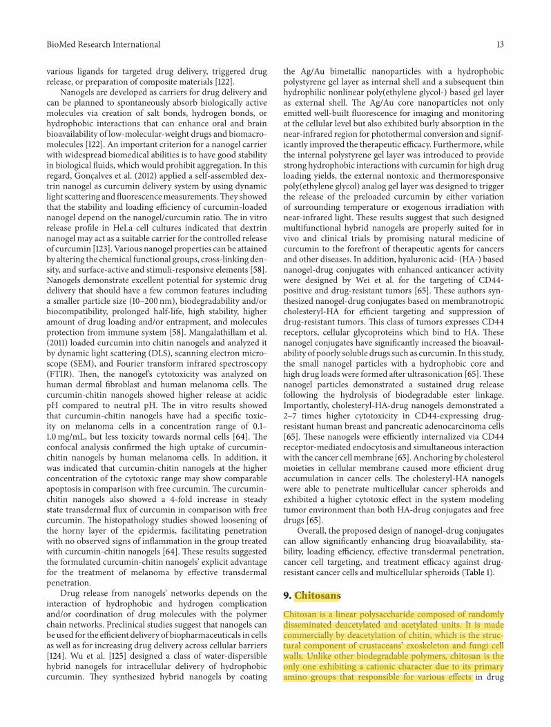
9. Chitosans
Chitosan is a linear polysaccharide composed of randomlydisseminated deacetylated and acetylated units. It is madecommercially by deacetylation of chitin, which is the struc-tural component of crustaceans’ exoskeleton and fungi cellwalls. Unlike other biodegradable polymers, chitosan is theonly one exhibiting a cationic character due to its primaryamino groups that responsible for various e'ects in drug

14 BioMed Research International
delivery systems [126]. It displays particular properties, forexample, solubility in various media, polyoxysalt creation,polyelectrolyte behavior, metal chelations, and structuraluniqueness (Table 1). One study showed that the ,uorescenceintensity of curcumin can be greatly improved in the presenceof chitosan by bovine and human serum albumin [104]. &emethod has been pro(tably used for the determination ofhuman serum albumin in real samples. Data analysis recom-mended that the highly enhanced ,uorescence of curcuminresulted from synergic e'ects of favorable hydrophobicmicroenvironment provided by bovine serum albumin andchitosan and e8cient intermolecular energy transfer betweenbovine serum albumin and curcumin. Bovine serum albuminmay bind to chitosan through hydrogen bonds, which causesthe protein conformation to switch from !-fold to %-helix.Curcumin can combine with bovine serum albumin from !-fold to %-helix and can also combine with the bovine serumalbumin-chitosan complex via its center carbonyl carbon.&erefore, chitosan plays a key role in promoting the energytransfer process by shortening the distance between bovineserum albumin and curcumin [104].
Polycaprolactone nanocarriers decorated with amucoad-hesive polysaccharide chitosan containing curcumin werealso developed [127]. In order to optimize the preparationconditions, these nanocarriers were prepared by the nano-precipitation method by using di'erent molar masses andconcentrations of chitosan and triblock surfactant polox-amer. Chitosan-coated nanocarriers revealed positive surfacecharge and a mean particle radius ranging between 114and 125 nm, con(rming the decoration of the nanocarrierswith the mucoadhesive polymer, through hydrogen bondsbetween ether and amino groups, from poloxamer andchitosan, respectively. Dynamic light scattering studies haveshown monodisperse nanocarriers. Furthermore, colloidalsystems showed mean drug content about 460 lg/mL andencapsulation e8ciency higher than 99%. In summary, thesenanocarriers showed a vast ability to interact with mucin,also indicating their suitability formucoadhesive applicationswhen coated with chitosan [127].
On the other hand, curcumin-phytosome-loaded chi-tosan microspheres were developed by combining polymer-and lipid-based delivery systems to improve the bioavailabil-ity and prolong the retention time of curcumin [66]. &esecomplexes were produced by encapsulating curcumin phy-tosomes in chitosan microspheres using ionotropic gelation.Di'erential scanning calorimetry and FUTI spectroscopyrevealed that the integrity of the phytosomes was pro-tected within the polymeric matrix of the microspheres.In vitro release rate of curcumin from the curcumin-phytosome-loaded chitosan microspheres was slower thancurcumin-loaded chitosan microspheres. Pharmacokineticstudies showed an increase in curcumin absorption incurcumin-phytosome-loaded chitosan microspheres com-pared with curcumin phytosomes and curcumin-loadedchitosan microspheres. Moreover, half-life of curcumin inoral administration of curcumin-phytosome-loaded chitosanmicrospheres was longer than the two other ones. &eseresults indicated that the novel curcumin-phytosome-loadedchitosan microspheres combined system has the advantages
of both the chitosanmicrospheres and the phytosomes, whichhad better e'ects of promoting oral absorption and prolong-ing retention time of curcumin than single curcumin phyto-somes or curcumin-loaded chitosanmicrospheres.&erefore,the phytosome chitosan microspheres may be used as asustained delivery system for lipophilic compounds withpoorwater solubility and loworal bioavailability [66]. A studyshowed that curcumin bound to chitosan nanoparticles wasnot rapidly degraded in comparison to free curcumin, andthe uptake of curcumin-loaded chitosan NPs by mouse’s redblood cells (RBC) was much better than free curcumin [67].Oral delivery of curcumin-loaded chitosan NPs improvedthe bioavailability of curcumin both in plasma and in RBC.Like chloroquine, conjugated curcumin inhibited parasitelysate induced heme polymerization in vitro in a dosedependentmanner, and it had a lower IC50 value than chloro-quine. Additionally, feeding of curcumin-loaded chitosanNPs caused a higher survival in mice infected with a lethalstrain of Plasmodium yoelii. &erefore, binding of curcuminto chitosan NPs improves its chemical stability and bioavail-ability. In vitro data also suggest that this complex can inhibithemozoin synthesis which is lethal for the parasite [67].
In another study, chitosan showed promising features asauxiliary agent in drug delivery (e.g., slimming, wound dress-ing, and tissue engineering). An in situ injectable nanocom-posite hydrogel curcumin was e'ectively developed for useas a treatment in the dermal wound repair process [68]. Invitro release studies disclosed that the encapsulated nanocur-cumin was slowly released from the N,O-carboxymethylchitosan/oxidized alginate hydrogel with the controllabledi'usion behavior. Additionally, in vivo wound healingstudies revealed that application of nanocurcumin/N,O-carboxymethyl chitosan/oxidized alginate hydrogel couldsigni(cantly improve the reepithelialization of epidermis andcollagen deposition on rat dorsal wounds. DNA, protein,and hydroxyproline content in wound tissue indicated thatmaking a combination by using nanocurcumin and N,O-carboxymethyl chitosan/oxidized alginate hydrogel couldsigni(cantly accelerate the process of wound healing. So,results suggested that the developed nanocurcumin/N,O-carboxymethyl chitosan/oxidized alginate hydrogel as apromising wound dressing might have potential applicationin the wound healing [68].
Water-soluble nanocarriers of curcumin were synthe-sized, characterized, and applied as a stable detoxifyingagent for arsenic poisoning [69]. &e therapeutic e8cacy ofencapsulated curcumin nanocarriers was investigated againstarsenic-induced toxicity in an animal model. In this regard,sodium arsenite and encapsulated curcumin were orallyadministered to male Wistar rats for 4 weeks. Arsenic dra-matically declined blood d-aminolevulinic acid dehydrataseactivity and glutathione and increased blood reactive oxygenspecies. &ese alterations were accompanied by increasesin hepatic total ROS, oxidized glutathione, and thiobar-bituric acid-reactive substance levels. By contrast, hepaticglutathione, superoxide dismutase, and catalase activitieswere considerably declined a4er arsenic exposure, indicativeof oxidative stress. Brain amines levels such as dopamine,norepinephrine, and 5-hydroxytryptamine also showed

BioMed Research International 15
considerable changes a4er arsenic exposure. Coadministra-tion of encapsulated curcumin nanocarriers providedobvious favorable e'ects on the adverse changes in oxidativestress parameters induced by arsenic.&e results revealed thatencapsulated curcumin nanocarriers have better antioxid-ant and chelating potential compared to free curcumin.&erefore, the signi(cant neurochemical and immunohisto-chemical protection a'orded by encapsulated curcumin nan-ocarriers shows their neuroprotective e'ectiveness [69].Chitosan also explains fungistatic, haemostatic, and anti-tumor e'ects [70]. In this regard stable vesicles for e8cientcurcumin encapsulation, delivery, and controlled releasehave been obtained by coating of liposomes with thin layerof newly synthesized chitosan derivatives [71]. Some spe-cial derivatives of chitosan were studied such as the cationic,hydrophobic, and cationic-hydrophobic derivatives. Zetapotential data proved e'ectual coating of liposomes withall these derivatives. In this regard, the liposomes coatedwith cationic-hydrophobic chitosan derivatives were themain promising curcumin carriers. &ey can easily entercell membrane and release curcumin in a controlledapproach, and the biological investigations showed that suchorganizations are nontoxic for normal murine (broblastswhile toxic for murine melanoma tumors [71].
In a recent study, Pluronic F127 was used to enhance thesolubility of curcumin in the alginate-chitosan NPs [128].Atomic force and scanning electron microscopic analysisdemonstrated that the particles were almost spherical inshape (100 ± 20 nm). Fourier transform infrared analysisshowed impending interactions among the components inthe composite NPs. Furthermore, encapsulated curcumine8ciency con(rmed considerable increase over alginate-chitosan NPs without Pluronic. Cytotoxicity assay explainedthat composite NPs at a concentration of 500 'g/mL werenontoxic for HeLa cells. Moreover, cellular internalizationof curcumin-loaded complex was con(rmed by green ,u-orescence inside the HeLa cells [128]. Curcumin-loadedbiodegradable thermoresponsive chitosan-g-poly copoly-mericNPswere prepared by using ionic cross-linkingmethod[129]. &e results showed that these NPs were nontoxic todi'erent cancerous cell lines, whereas the curcumin loadedwith NPs showed a speci(c toxicity for the abovementionedcell lines. Additionally, these results were further approvedby ,ow cytometry analysis which proved increased apoptosison these cell lines in a concentration-dependent manner.Furthermore, the blood compatibility assay showed the pos-sibility of an IV injection with this formulation. Preliminarystudy provided clear evidence for the thermal targetingof curcumin by being loaded with novel thermosensitivechitosan-g-PNIPAAm NPs, and e8cacies were achieved incancer therapy. &ese results indicated that thermorespon-sive chitosan-g-poly copolymeric NPs can be a potentialnanocarrier for curcumin drug delivery [129]. Novel cationicpoly(butyl) cyanoacrylate (PBCA) NPs coated with chitosanwere synthesized with curcumin. &e transmission electronmicroscopy showed the spherical shape of prepared NPsalong with the particle size. Curcumin NPs demonstratedmore therapeutic e8cacy than free curcumin against apanel of human hepatocellular cancer cell lines. Encapsulated
curcumin with PBCA NPs caused a profound change inthe pharmacokinetics of the drug. &e elimination half-life of curcumin was increased 52-fold in loaded form withPBCA NPs, and ultimately its clearance was also decreased2.5-fold. Additionally, the higher plasma concentration ofcurcumin for curcumin-PBCA NPs might be a result of theNPs size and chitosan coating to keep drug in the bloodcirculation for a more extended period. Besides, the meanresidence time of curcumin-PBCA NPs was longer thanfree curcumin. &ese results might be due to accumulationof NPs in endoplasmic reticulum system of organs andsustained release of the drug from them. Furthermore, thecarriers’ properties, for instance, shape, size, charge, andhydrophilicity, can prolong the retention of them in theblood circulation. &ere was also a substantial increase inthe distribution volume (51-fold) that was quite unexpected.Obviously, it was possible that the larger micellar carri-ers were sequestered by the reticuloendothelial system orother tissues and truly led to improved distribution volume[130]. Additionally, treatment with curcumin NPs resultedin reduced tumor size and visible blanching of tumors[131].
So far, curcumin-loaded chitosan NPs improve thebioavailability and prolong the retention time of curcumindue to accumulation of NPs in endoplasmic reticulum systemand the carriers’ features such as shape, size, charge, andhydrophilicity (Table 1). Gathered data also propose that thiscomplex can be lethal for the parasite because of hemozoinsynthesis inhibition. Some in vivo experiments also resultedin better wound healing a4er application of curcumin-loadedchitosan NP polymers by means of better reepithelializationof epidermis and collagen deposition. &is complex couldalso be administered in order to detoxify arsenic throughbetter antioxidant and chelating potential.&ese compoundsgained some achievements in cancer therapy as well.
10. Gold Nanoparticles
Metal nanoparticles have been known since very old times,and gold nanoparticles (AuNPs) with optical and electro-chemical uniqueness have proven to be a potent appara-tus in nanomedicinal requests [132]. &ey have also beenlargely used in immunochemistry, immunohistochemistry,and immunoblotting for electron microscopy.&ey are o4engenerated in various shapes [132], and their properties arestrongly dependent on the conditions in which they are pre-pared. Moreover, the stability of AuNPs and their capabilityto combine with biomolecules are their other outstandingproperties. AuNPs are studied broadly as imperative drugdelivery vectors due to some of their characteristic aspects,such as low cytotoxicity, tunable surface features, and stabilityin in vivo conditions, and can be easily synthesized andfunctionalized (Table 1). &ey can also act as drug pool forsmall drugmolecules, proteins, DNA, or RNAwith improvedlong life in the blood circulation. Rajesh et al. [133] usedpolyvinyl pyrrolidone (PVP) as a proven drug carrier tocurcumin conjugation with AuNPs to enhance solubility ofcurcumin. Results showed a superior assurance for suchconjugates as therapeutic-curcumin-imaging materials in

16 BioMed Research International
biomedical (eld [134]. Kumar et al. (2012) also preparedthe chitosan-curcumin nanocapsules with AuNPs via solventevaporation method. Scanning electron microscopy andtransmission electron microscopy were done to describethe drug entrapped nanocapsules. &e average diameter ofAuNPs was found to be in the range of 18–20 nm, andthe nanocapsules were found to be in the range of 200–250 nm. Furthermore, the Fourier transform infrared analysisrevealed no possible interactions among the constituentswith the chitosan nanoparticles. &e drug release studiesrevealed that curcumin encapsulated chitosan with AuNPswas controlled and steadied when compared with curcuminencapsulated chitosan nanoparticles. Use of in vitro drugrelease in various kinetic equations indicated a matrix modelwith uniform distribution of curcumin in the nanocapsules[135]. Additionally, the tunability of AuNPs allows for com-plete control of surface properties for targeting and sustainedrelease of the bioactive molecules [136].
In a study by Singh et al. [72] curcumin was bound on thesurface of AuNPs in order to increase the bioavailability ofit.&e AuNPs were synthesized by direct decline of HAuCl4by curcumin in aqueous part. Curcumin acted as both areducing and capping agent and a stabilizing gold sol formany months. Furthermore, these curcumin-capped AuNPsshowed an excellent antioxidant activity which was estab-lished by 2,2-diphenyl-l-picrylhydrazyl radical test. Conse-quently, the practical surface of AuNPs with curcumin maysuggest a new way of use of curcumin towards possible drugdelivery and therapeutics [72]. In another study, e'ect ofcurcumin-conjugated-AuNPs was investigated on peripheralblood lymphocytes [137]. &e treated lymphocytes showedtypical characteristics of apoptosis which included chromatincondensation and membrane blebbing and occurrence ofapoptotic bodies. Results revealed that these conjugatednanoparticles may be used as drugs in nontoxic range[137]. In order to target cancer at a single cell level, gold-citrate nanoparticles were also synthesized with diametersof 13 nm [73]. AuNPs were coated with sodium citrate.Outcomes revealed that cancerous cells were more proneto absorb nanomaterials coated with citrate than normalsomatic cells. Moreover, the damage was reversible withAuNPs and the normal dermal (broblast cells were able toregenerate stress (bers which were lost during exposure.However, cancer cells were unable to recover from the dam-age in,icted by Au/citrate nanoparticle exposure [73]. Manjuand Sreenivasan [136] also formulated a simple method forthe fabrication of water-soluble curcumin conjugated AuNPsto target various cancer cell lines. Curcumin conjugatedto hyaluronic acid to get a water-soluble compound. &eywere made AuNPs by diminishing chloroauric acid usinghyaluronic acid-curcumin, which played dual roles of areducing and a stabilizing agent and subsequently anchoredfolate conjugated PEG. &eir interaction with various can-cer cell lines was followed by ,ow cytometry and confo-cal microscopy. Blood-materials interactions studies provedthat the nanoparticles are extremely hemocompatible. Flowcytometry and confocal microscopy results demonstratedconsiderable cellular uptake and internalization of the par-ticles by various cancer cells [136].
In conclusion, curcumin conjugated AuNPs exhibitedmore cytotoxicity compared to free curcumin (Table 1).AuNPs also cause targeting and sustained release of curcuminand an excellent antioxidant activity.
11. Silvers
Silver has usually been utilized as an incredibly e8cientmate-rial for antimicrobial utility [138]. In small concentrations, itis safe for human cells but lethal for the majority of bacteriaand viruses [139]. With development of nanotechnology,it has become the metal of choice in restricting microbialgrowth and expansion in a variety of nanoparticle-relatedrequests [138]. Silver nanoparticles are identi(ed for theirbrilliant optoelectronic properties originated from surfaceplasmon resonance. &ey can be used in optoelectronics,biological labeling, and biological and chemical sensing(Table 1). &ey have shown excellent antimicrobial activitycompared to other available silver antimicrobial agents.
Sodium carboxylmethyl cellulose silver nanocomposite(lms were attempted for antibacterial applications, so, toimprove their applicability, novel (lm-silver nanoparticle-curcumin complexes have been developed [74]. &ese (lmswere described by FTIR,UV-visible, X-ray di'raction (XRD),thermogravimetric analysis (TGA), di'erential scanningcalorimetry (DSC), and TEM techniques. &e structuredsilver nanoparticles had a typical particle size of 15 nm. Cur-cumin loading into sodium carboxylmethyl cellulose silvernanocomposite (lms was achieved by di'usion mechanism.&e UV analysis showed superior encapsulation of curcuminin the (lms with higher sodium carboxylmethyl cellulosecontent. Additionally, it was surveyed that the presence ofsilver nanoparticles in the (lms improved the encapsulationof curcumin demonstrating an interaction between them.Moreover, results showed that the sodium carboxylmethylcellulose (lms produced with silver nanoparticles have asynergistic e'ect in the antimicrobial activity against E.coli. Furthermore, curcumin loaded with sodium carboxyl-methyl cellulose silver nanocomposite (lms extended consid-erable inhibition of E. coli growth compared with the silvernanoparticles and curcumin alone (lm. &erefore, the studyobviously supplied novel antimicrobial (lms which werepotentially helpful in preventing/treating infections [74]. Inanother study, novel hydrogel-silver nanoparticle-curcumincomposites have been built up to increase its applicability.&ese were (rst synthesized by polymerizing acrylamide inthe presence of polyvinyl sulfonic acid sodium salt and atrifunctional cross-linker (2,4,6-triallyloxy 1,3,5-triazine) byusing redox initiating system. Silver nanoparticles were thenproduced throughout the hydrogel networks by using in situmethod incorporating the silver ions and following dropwith sodium borohydride. Curcumin loading into hydrogel-silver nanoparticles complex was earned by di'usion mech-anism. An attractive arrangement of silver nanoparticles(shining sun ball in range 5 nm) with apparent smaller grownnanoparticles (1 nm) was detected. A comparative antimicro-bial study was performed for hydrogel-silver nanocompositesand hydrogel-silver nanoparticle-curcumin composites. &eresults indicated that hydrogel-AgNPs-curcumin composites
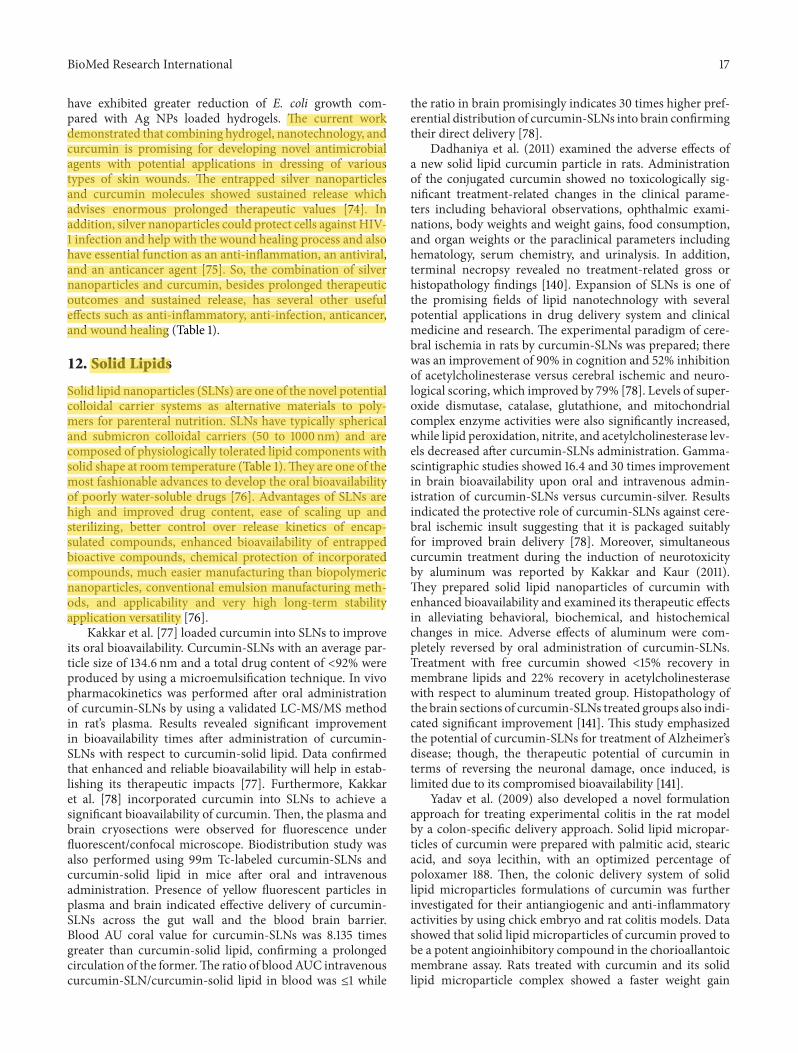
BioMed Research International 17
have exhibited greater reduction of E. coli growth com-pared with Ag NPs loaded hydrogels. &e current workdemonstrated that combining hydrogel, nanotechnology, andcurcumin is promising for developing novel antimicrobialagents with potential applications in dressing of varioustypes of skin wounds. &e entrapped silver nanoparticlesand curcumin molecules showed sustained release whichadvises enormous prolonged therapeutic values [74]. Inaddition, silver nanoparticles could protect cells against HIV-1 infection and help with the wound healing process and alsohave essential function as an anti-in,ammation, an antiviral,and an anticancer agent [75]. So, the combination of silvernanoparticles and curcumin, besides prolonged therapeuticoutcomes and sustained release, has several other usefule'ects such as anti-in,ammatory, anti-infection, anticancer,and wound healing (Table 1).
12. Solid Lipids
Solid lipid nanoparticles (SLNs) are one of the novel potentialcolloidal carrier systems as alternative materials to poly-mers for parenteral nutrition. SLNs have typically sphericaland submicron colloidal carriers (50 to 1000 nm) and arecomposed of physiologically tolerated lipid components withsolid shape at room temperature (Table 1).&ey are one of themost fashionable advances to develop the oral bioavailabilityof poorly water-soluble drugs [76]. Advantages of SLNs arehigh and improved drug content, ease of scaling up andsterilizing, better control over release kinetics of encap-sulated compounds, enhanced bioavailability of entrappedbioactive compounds, chemical protection of incorporatedcompounds, much easier manufacturing than biopolymericnanoparticles, conventional emulsion manufacturing meth-ods, and applicability and very high long-term stabilityapplication versatility [76].
Kakkar et al. [77] loaded curcumin into SLNs to improveits oral bioavailability. Curcumin-SLNs with an average par-ticle size of 134.6 nm and a total drug content of <92% wereproduced by using a microemulsi(cation technique. In vivopharmacokinetics was performed a4er oral administrationof curcumin-SLNs by using a validated LC-MS/MS methodin rat’s plasma. Results revealed signi(cant improvementin bioavailability times a4er administration of curcumin-SLNs with respect to curcumin-solid lipid. Data con(rmedthat enhanced and reliable bioavailability will help in estab-lishing its therapeutic impacts [77]. Furthermore, Kakkaret al. [78] incorporated curcumin into SLNs to achieve asigni(cant bioavailability of curcumin.&en, the plasma andbrain cryosections were observed for ,uorescence under,uorescent/confocal microscope. Biodistribution study wasalso performed using 99m Tc-labeled curcumin-SLNs andcurcumin-solid lipid in mice a4er oral and intravenousadministration. Presence of yellow ,uorescent particles inplasma and brain indicated e'ective delivery of curcumin-SLNs across the gut wall and the blood brain barrier.Blood AU coral value for curcumin-SLNs was 8.135 timesgreater than curcumin-solid lipid, con(rming a prolongedcirculation of the former.&e ratio of bloodAUC intravenouscurcumin-SLN/curcumin-solid lipid in blood was ≤1 while
the ratio in brain promisingly indicates 30 times higher pref-erential distribution of curcumin-SLNs into brain con(rmingtheir direct delivery [78].
Dadhaniya et al. (2011) examined the adverse e'ects ofa new solid lipid curcumin particle in rats. Administrationof the conjugated curcumin showed no toxicologically sig-ni(cant treatment-related changes in the clinical parame-ters including behavioral observations, ophthalmic exami-nations, body weights and weight gains, food consumption,and organ weights or the paraclinical parameters includinghematology, serum chemistry, and urinalysis. In addition,terminal necropsy revealed no treatment-related gross orhistopathology (ndings [140]. Expansion of SLNs is one ofthe promising (elds of lipid nanotechnology with severalpotential applications in drug delivery system and clinicalmedicine and research. &e experimental paradigm of cere-bral ischemia in rats by curcumin-SLNs was prepared; therewas an improvement of 90% in cognition and 52% inhibitionof acetylcholinesterase versus cerebral ischemic and neuro-logical scoring, which improved by 79% [78]. Levels of super-oxide dismutase, catalase, glutathione, and mitochondrialcomplex enzyme activities were also signi(cantly increased,while lipid peroxidation, nitrite, and acetylcholinesterase lev-els decreased a4er curcumin-SLNs administration. Gamma-scintigraphic studies showed 16.4 and 30 times improvementin brain bioavailability upon oral and intravenous admin-istration of curcumin-SLNs versus curcumin-silver. Resultsindicated the protective role of curcumin-SLNs against cere-bral ischemic insult suggesting that it is packaged suitablyfor improved brain delivery [78]. Moreover, simultaneouscurcumin treatment during the induction of neurotoxicityby aluminum was reported by Kakkar and Kaur (2011).&ey prepared solid lipid nanoparticles of curcumin withenhanced bioavailability and examined its therapeutic e'ectsin alleviating behavioral, biochemical, and histochemicalchanges in mice. Adverse e'ects of aluminum were com-pletely reversed by oral administration of curcumin-SLNs.Treatment with free curcumin showed <15% recovery inmembrane lipids and 22% recovery in acetylcholinesterasewith respect to aluminum treated group. Histopathology ofthe brain sections of curcumin-SLNs treated groups also indi-cated signi(cant improvement [141]. &is study emphasizedthe potential of curcumin-SLNs for treatment of Alzheimer’sdisease; though, the therapeutic potential of curcumin interms of reversing the neuronal damage, once induced, islimited due to its compromised bioavailability [141].
Yadav et al. (2009) also developed a novel formulationapproach for treating experimental colitis in the rat modelby a colon-speci(c delivery approach. Solid lipid micropar-ticles of curcumin were prepared with palmitic acid, stearicacid, and soya lecithin, with an optimized percentage ofpoloxamer 188. &en, the colonic delivery system of solidlipid microparticles formulations of curcumin was furtherinvestigated for their antiangiogenic and anti-in,ammatoryactivities by using chick embryo and rat colitis models. Datashowed that solid lipid microparticles of curcumin proved tobe a potent angioinhibitory compound in the chorioallantoicmembrane assay. Rats treated with curcumin and its solidlipid microparticle complex showed a faster weight gain

18 BioMed Research International
compared with dextran sulfate solution control rats. &eincrease in whole colon length appeared to be signi(-cantly greater in solid lipid microparticle-treated rats whencompared with free curcumin and control rats. Moreover,decreased mast cell numbers was observed in the colonmucosa of curcumin-solid lipid microparticle treated rats.&e degree of colitis caused by administration of dextran sul-fate solution was signi(cantly attenuated by colonic deliveryof curcumin-solid lipid microparticles [79]. Being a nontoxicnatural dietary product, it seems that curcumin can be usefulin the therapeutic strategy for in,ammatory bowel diseasepatients. Wang et al. (2012) aimed to formulate curcumin-SLNs to improve its therapeutic e8cacy in an ovalbumin-induced allergic rat model of asthma. in vitro tests wereperformed in order to check Physiochemical properties ofcurcumin-SLNs and its release experiments. &e pharma-cokinetics in tissue distribution and the therapeutic e'ectswere studied in mice. X-ray di'raction analysis revealedthe amorphous nature of the encapsulated curcumin. &ecurcumin concentrations in plasma suspension were consid-erably superior to free curcumin, and all the tissue concen-trations of curcumin increased a4er curcumin-SLNs admin-istration, especially in lung and liver. In addition, curcumin-SLNs e8ciently suppressed airway hyperresponsiveness andin,ammatory cell in(ltration. It also inhibited the expressionof T-helper-2-type cytokinesin bronchoalveolar lavage ,uidsigni(cantly compared to free curcumin.&ese observationsimply that curcumin-SLNs can be a promising candidate forasthma therapy [80]. In another study, transferrin-mediatedSLNs were prepared to increase photostability and anticanceractivity of curcumin against breast cancer cells in vitro [81].Microplate analysis and ,ow cytometry techniques were usedfor cytotoxicity and apoptosis studies.&e physical character-ization showed the suitability of preparation method. Trans-mission electron microscopy and X-ray di'raction studiesrevealed the spherical nature and entrapment of curcuminin amorphous form, respectively. Annexin V-FITC/PI doublestaining, DNA analysis, and reducedmitochondrial potentialcon(rmed the occurrence of apoptosis. &e ,ow cytometricstudies disclosed that the anticancer activity of curcuminis enhanced with transferrin-mediated SLNs compared tofree curcumin, and apoptosis is the mechanism underlyingthe cytotoxicity (Table 1). Results indicated the potential oftransferrin-mediated SLNs in enhancing the anticancer e'ectof curcumin in breast cancer cells in vitro [81].
13. Conclusion and Future Perspectives
&e use of nanotechnology in medicine and more purposelydrug delivery is set to spread quickly. Currently, manysubstances are under investigation for drug delivery andmorespeci(cally for cancer therapy. Fascinatingly, pharmaceuticalsciences are using nanoparticles to reduce toxicity and sidee'ects of drugs. Moreover nanoparticles augment solubilityand stability of some substances like curcumin. It is now clearthat further development of traditional natural compoundswith chemopreventive and chemotherapeutic potential suchas curcumin will be dictated by the advanced drug delivery
systems.Nanotechnology is assumed to be a fundamental set-ting in drug delivery system and human therapeutics. How-ever, considerable challenges remain in driving this (eld intoclinically practical therapies. Curcumin, an excellent repre-sentative derived from traditional natural compounds, hasbeen proven to be e'ectual in long-term application andpreclinical trials. &ere is no doubt that advance of noveldelivery systems of curcumin with better therapeutic e'ectswill be vital for future improvement of curcumin as a thera-peutic agent.&us, it is an enormous implication to overcomethe current limitations of curcumin. It seems that only bymultidisciplinary collaboration we can bring these promis-ing traditional natural compounds to the forefront of ther-apeutic agents for di'erent diseases. &erefore, the promiseof nanotechnology-based medicine may become a realitywith su8cient e'orts and further researches. Human trialsneed to be conducted to establish curcumin’s e'ectiveness inclinical applications as an improved therapeutic modality fortreatment of di'erent diseases.
Conflict of Interests
&e authors report no con,ict of interests.&e authors aloneare responsible for the content of the paper.
Acknowledgment
&is study was supported by Tehran University of MedicalSciences.
References
[1] E. Jaruga, S. Salvioli, J. Dobrucki et al., “Apoptosis-like, revers-ible changes in plasmamembrane asymmetry and permeability,and transientmodi(cations inmitochondrialmembrane poten-tial induced by curcumin in rat thymocytes,” FEBS Letters, vol.433, no. 3, pp. 287–293, 1998.
[2] S. Sreejayan and M. N. A. Rao, “Curcuminoids as potent inhi-bitors of lipid peroxidation,” Journal of Pharmacy and Pharma-cology, vol. 46, no. 12, pp. 1013–1016, 1994.
[3] R. S. Ramsewak, D. L. DeWitt, and M. G. Nair, “Cytotoxicity,antioxidant and anti-in,ammatory activities of curcumins I-IIIfrom Curcuma longa,” Phytomedicine, vol. 7, no. 4, pp. 303–308,2000.
[4] J. Milobedzka, S. V. Kostanecki, and V. Lampe, “Zur Kenntnisdes Curcumins,” Berichte der Deutschen Chemischen Gesells-cha#, vol. 43, no. 2, pp. 2163–2170, 1910.
[5] H. P. Ammon and M. A. Wahl, “Pharmacology of Curcumalonga,” Planta Medica, vol. 57, no. 1, pp. 1–7, 1991.
[6] N. K. Pandeya, “Old wives'tales: modern miracles—turmeric astraditionalmedicine in India,”Trees for Life Journal, vol. 1, article3, 2005.
[7] B. B. Aggarwal and B. Sung, “Pharmacological basis for the roleof curcumin in chronic diseases: an age-old spice with moderntargets,” Trends in Pharmacological Sciences, vol. 30, no. 2, pp.85–94, 2009.
[8] B. A. Bharat and K. B. Harikumar, “Potential therapeutic e'ectsof curcumin, the anti-in,ammatory agent, against neurode-generative, cardiovascular, pulmonary, metabolic, autoimmune

BioMed Research International 19
and neoplastic diseases,” International Journal of Biochemistryand Cell Biology, vol. 41, no. 1, pp. 40–59, 2009.
[9] L. Li, F. S. Braiteh, and R. Kurzrock, “Liposome-encapsulatedcurcumin: in vitro and in vivo e'ects on proliferation, apop-tosis, signaling, and angiogenesis,” Cancer, vol. 104, no. 6, pp.1322–1331, 2005.
[10] K. Maiti, K. Mukherjee, A. Gantait, B. P. Saha, and P. K.Mukherjee, “Curcumin-phospholipid complex: preparation,therapeutic evaluation and pharmacokinetic study in rats,”International Journal of Pharmaceutics, vol. 330, no. 1-2, pp. 155–163, 2007.
[11] L. Lin, Q. Shi, A. K. Nyarko et al., “Antitumor agents. 250.Design and synthesis of new curcumin analogues as potentialanti-prostate cancer agents,” Journal of Medicinal Chemistry,vol. 49, no. 13, pp. 3963–3972, 2006.
[12] H. Ohtsu, Z. Xiao, J. Ishida et al., “Antitumor agents. 217. Cur-cumin analogues as novel androgen receptor antagonists withpotential as anti-prostate cancer agents,” Journal of MedicinalChemistry, vol. 45, no. 23, pp. 5037–5042, 2002.
[13] B. K. Adams, E. M. Ferstl, M. C. Davis et al., “Synthesis andbiological evaluation of novel curcumin analogs as anti-cancerand anti-angiogenesis agents,” Bioorganic and Medicinal Chem-istry, vol. 12, no. 14, pp. 3871–3883, 2004.
[14] R. Benassi, E. Ferrari, R. Grandi, S. Lazzari, and M. Saladini,“Synthesis and characterization of new !-diketo derivativeswith iron chelating ability,” Journal of Inorganic Biochemistry,vol. 101, no. 2, pp. 203–213, 2007.
[15] T. N. Shankar, N. V. Shantha, H. P. Ramesh, I. A. Murthy, andV. S. Murthy, “Toxicity studies on turmeric (Curcuma longa):acute toxicity studies in rats, guinea pigs & monkeys,” IndianJournal of Experimental Biology, vol. 18, no. 1, pp. 73–75, 1980.
[16] K. B. Soni and R. Kuttan, “E'ect of oral curcumin administra-tion on serum peroxides and cholesterol levels in human vol-unteers,” Indian Journal of Physiology andPharmacology, vol. 36,no. 4, pp. 273–275, 1992.
[17] P.Anand,A. B.Kunnumakkara, R.A.Newman, andB. B.Aggar-wal, “Bioavailability of curcumin: problems and promises,”Molecular Pharmaceutics, vol. 4, no. 6, pp. 807–818, 2007.
[18] R. A. Sharma, W. P. Steward, and A. J. Gescher, “Pharma-cokinetics and pharmacodynamics of curcumin,” Advances inExperimental Medicine and Biology, vol. 595, pp. 453–470, 2007.
[19] R. Yang, S. Zhang, D. Kong, X. Gao, Y. Zhao, and Z. Wang,“Biodegradable polymer-curcumin conjugate micelles enhancethe loading and delivery of low-potency curcumin,” Pharma-ceutical Research, vol. 29, no. 12, pp. 3512–3525, 2012.
[20] B.Wahlstrom andG. Blennow, “A study on the fate of curcuminin the rat,”Acta Pharmacologica et Toxicologica, vol. 43, no. 2, pp.86–92, 1978.
[21] M. Lopez-Lazaro, “Anticancer and carcinogenic properties ofcurcumin: considerations for its clinical development as a can-cer chemopreventive and chemotherapeutic agent,” MolecularNutrition and Food Research, vol. 52, no. 1, pp. S103–S127, 2008.
[22] N. A. Kasim,M.Whitehouse, C. Ramachandran et al., “Molecu-lar properties ofWHO essential drugs and provisional biophar-maceutical classi(cation,”Molecular Pharmaceutics, vol. 1, no. 1,pp. 85–96, 2004.
[23] M.-H. Pan, T.-M. Huang, and J.-K. Lin, “Biotransformationof curcumin through reduction and glucuronidation in mice,”Drug Metabolism and Disposition, vol. 27, no. 4, pp. 486–494,1999.
[24] F. Payton, P. Sandusky, and W. L. Alworth, “NMR study of thesolution structure of curcumin,” Journal of Natural Products,vol. 70, no. 2, pp. 143–146, 2007.
[25] J. Ishida, H. Ohtsu, Y. Tachibana et al., “Antitumor agents—part 214: synthesis and evaluation of curcumin analogues ascytotoxic agents,” Bioorganic and Medicinal Chemistry, vol. 10,no. 11, pp. 3481–3487, 2002.
[26] C. Selvam, S. M. Jachak, R.&ilagavathi, and A. K. Chakraborti,“Design, synthesis, biological evaluation and molecular dock-ing of curcumin analogues as antioxidant, cyclooxygenase inhi-bitory and anti-in,ammatory agents,” Bioorganic andMedicinalChemistry Letters, vol. 15, no. 7, pp. 1793–1797, 2005.
[27] A. Sun,M. Shoji, Y. J. Lu, D. C. Liotta, and J. P. Snyder, “Synthesisof EF24-tripeptide chloromethyl ketone: a novel curcumin-related anticancer drug delivery system,” Journal of MedicinalChemistry, vol. 49, no. 11, pp. 3153–3158, 2006.
[28] H. Ohori, H. Yamakoshi, M. Tomizawa et al., “Synthesisand biolgical analysis of new curcumin analogues bearing anenhanced potential for the medicinal treatment of cancer,”Molecular Cancer $erapeutics, vol. 5, no. 10, pp. 2563–2571,2006.
[29] R. A. Freitas Jr., “What is nanomedicine?” Nanomedicine:Nanotechnology, Biology, and Medicine, vol. 1, no. 1, pp. 2–9,2005.
[30] S. Rahman, S. Cao, K. J. Steadman, M. Wei, and H. S. Parekh,“Native and !-cyclodextrin-enclosed curcumin: entrapmentwithin liposomes and their in vitro cytotoxicity in lung andcolon cancer,” Drug Delivery, vol. 19, no. 7, pp. 346–353, 2012.
[31] H. S. Shi, X. Gao, D. Li et al., “A systemic administrationof liposomal curcumin inhibits radiation pneumonitis andsensitizes lung carcinoma to radiation,” International Journal ofNanomedicine, vol. 7, pp. 2601–2611, 2012.
[32] D. Matabudul, K. Pucaj, G. Bolger, B. Vcelar, M. Majeed, andL. Helson, “Tissue distribution of (Lipocurc) liposomal cur-cumin and tetrahydrocurcumin following two- and eight-hourinfusions in Beagle dogs,” Anticancer Research, vol. 32, no. 10,pp. 4359–4364, 2012.
[33] A. Karewicz, D. Bielska, B. Gzyl-Malcher, M. Kepczynski, R.Lach, andM. Nowakowska, “Interaction of curcumin with lipidmonolayers and liposomal bilayers,” Colloids and Surfaces B:Biointerfaces, vol. 88, no. 1, pp. 231–239, 2011.
[34] S. S. Dhule, P. Penfornis, T. Frazier et al., “Curcumin-loaded#-cyclodextrin liposomal nanoparticles as delivery vehiclesfor osteosarcoma,”Nanomedicine: Nanotechnology, Biology, andMedicine, vol. 8, no. 4, pp. 440–451, 2012.
[35] W. S.Orr, J.W.Denbo,K. R. Saab et al., “Liposome-encapsulatedcurcumin suppresses neuroblastoma growth through nuclearfactor-kappa B inhibition,” Surgery, vol. 151, no. 5, pp. 736–744,2012.
[36] D. Wang, M. S. Veena, K. Stevenson et al., “Liposome-encap-sulated curcumin suppresses growth of head and neck squa-mous cell carcinoma in vitro and in xenogra4s through theinhibition of nuclear factor kappaB by an AKT-independentpathway,” Clinical Cancer Research, vol. 14, no. 19, pp. 6228–6236, 2008.
[37] Y. Chen, Q. Wu, Z. Zhang, L. Yuan, X. Liu, and L. Zhou, “Pre-paration of curcumin-loaded liposomes and evaluation of theirskin permeation and pharmacodynamics,”Molecules, vol. 17, no.5, pp. 5972–5987, 2012.
[38] N. M. Rogers, M. D. Stephenson, A. R. Kitching, J. D.Horowitz, andP. T.H.Coates, “Amelioration of renal ischaemia-reperfusion injury by liposomal delivery of curcumin to renal

20 BioMed Research International
tubular epithelial and antigen-presenting cells,”$e British Jour-nal of Pharmacology, vol. 166, no. 1, pp. 194–209, 2012.
[39] P. Basnet, H. Hussain, I.&o, andN. Skalko-Basnet, “Liposomaldelivery system enhances anti-in,ammatory properties of cur-cumin,” Journal of Pharmaceutical Sciences, vol. 101, no. 2, pp.598–609, 2012.
[40] R. Raveendran, G. Bhuvaneshwar, and C. P. Sharma, “In vitrocytotoxicity and cellular uptake of curcumin-loaded Pluro-nic/Polycaprolactone micelles in colorectal adenocarcinomacells,” Journal of Biomaterials Applications, vol. 27, no. 7, pp. 811–827, 2013.
[41] H. Yu, J. Li, K. Shi, andQ.Huang, “Structure ofmodi(ed &-poly-lysine micelles and their application in improving cellular anti-oxidant activity of curcuminoids,” Food and Function, vol. 2, no.7, pp. 373–380, 2011.
[42] S. Podaralla, R. Averineni, M. Alqahtani, and O. Perumal, “Syn-thesis of novel biodegradable methoxy poly(ethylene glycol)-zein micelles for e'ective delivery of curcumin,” MolecularPharmaceutics, vol. 9, no. 9, pp. 2778–2786, 2012.
[43] Z. Song, R. Feng, M. Sun et al., “Curcumin-loaded PLGA-PEG-PLGA triblock copolymericmicelles: preparation, pharmacoki-netics and distribution in vivo,” Journal of Colloid and InterfaceScience, vol. 354, no. 1, pp. 116–123, 2011.
[44] M. H. M. Leung, H. Colangelo, and T. W. Kee, “Encapsulationof curcumin in cationicmicelles suppresses alkaline hydrolysis,”Langmuir, vol. 24, no. 11, pp. 5672–5675, 2008.
[45] R. Adhikary, P. J. Carlson, T.W. Kee, and J.W. Petrich, “Excited-state intramolecular hydrogen atom transfer of curcumin insurfactantmicelles,” Journal of Physical Chemistry B, vol. 114, no.8, pp. 2997–3004, 2010.
[46] G. Began, E. Sudharshan, and A. G. Appu Rao, “Inhibitionof lipoxygenase 1 by phosphatidylcholine micelles-bound cur-cumin,” Lipids, vol. 33, no. 12, pp. 1223–1228, 1998.
[47] S. Jain, P. Singh, V. Mishra, and S. P. Vyas, “Mannosylated nio-somes as adjuvant-carrier system for oral genetic immunizationagainst hepatitis B,” Immunology Letters, vol. 101, no. 1, pp. 41–49, 2005.
[48] S. Mandal, C. Banerjee, S. Ghosh, J. Kuchlyan, and N. Sarkar,“Modulation of the photophysical properties of curcumin innonionic surfactant (Tween-20) forming micelles and nio-somes: a comparative study of di'erent microenvironments,”Journal of Physical Chemistry B, vol. 117, no. 23, pp. 6957–6968,2013.
[49] N. Rungphanichkul, U. Nimmannit, W. Muangsiri, and P.Rojsitthisak, “Preparation of curcuminoid niosomes forenhancement of skin permeation,” Pharmazie, vol. 66, no. 8,pp. 570–575, 2011.
[50] H. H. Tønnesen, M. Masson, and T. Lo4sson, “Studies of cur-cumin and curcuminoids. XXVII. Cyclodextrin complexation:solubility, chemical and photochemical stability,” InternationalJournal of Pharmaceutics, vol. 244, no. 1-2, pp. 127–135, 2002.
[51] M. A. Tomren, M. Masson, T. Lo4sson, and H. H. Tønnesen,“Studies on curcumin and curcuminoids. XXXI. Symmetric andasymmetric curcuminoids: stability, activity and complexationwith cyclodextrin,” International Journal of Pharmaceutics, vol.338, no. 1-2, pp. 27–34, 2007.
[52] S. S. Darandale and P. R. Vavia, “Cyclodextrin-based nano-sponges of curcumin: formulation and physicochemical char-acterization,” Journal of Inclusion Phenomena and MacrocyclicChemistry, vol. 75, no. 3-4, pp. 315–322, 2013.
[53] V. R. Yadav, S. Prasad, R. Kannappan et al., “Cyclodextrin-com-plexed curcumin exhibits anti-in,ammatory and antiprolifer-ative activities superior to those of curcumin through highercellular uptake,” Biochemical Pharmacology, vol. 80, no. 7, pp.1021–1032, 2010.
[54] V. R. Yadav, S. Suresh, K. Devi, and S. Yadav, “E'ect of cyclo-dextrin complexation of curcumin on its solubility and antian-giogenic and anti-in,ammatory activity in rat colitis model,”AAPS PharmSciTech, vol. 10, no. 3, pp. 752–762, 2009.
[55] M. M. Yallapu, M. Jaggi, and S. C. Chauhan, “!-cyclodextrin-curcumin self-assembly enhances curcumin delivery in prostatecancer cells,” Colloids and Surfaces B: Biointerfaces, vol. 79, no.1, pp. 113–125, 2010.
[56] P. R. Dandawate, A. Vyas, A. Ahmad et al., “Inclusion complexof novel curcumin analogue CDF and !-cyclodextrin (1:2)and its enhanced in vivo anticancer activity against pancreaticcancer,” Pharmaceutical Research, vol. 29, no. 7, pp. 1775–1786,2012.
[57] S. Debnath, D. Saloum, S. Dolai et al., “Dendrimer-curcuminconjugate: a water soluble and e'ective cytotoxic agent againstbreast cancer cell lines,”Anti-Cancer Agents inMedicinal Chem-istry, vol. 13, no. 10, pp. 1531–1539, 2013.
[58] M. M. Yallapu, M. C. Ebeling, N. Chauhan, M. Jaggi, and S.C. Chauhan, “Interaction of curcumin nanoformulations withhuman plasma proteins and erythrocytes,” International Journalof Nanomedicine, vol. 6, pp. 2779–2790, 2011.
[59] J. Cao, H. Zhang, Y. Wang, J. Yang, and F. Jiang, “Investigationon the interaction behavior between curcumin and PAMAMdendrimer by spectral and docking studies,” SpectrochimicaActa A: Molecular and Biomolecular Spectroscopy, vol. 108, pp.251–255, 2013.
[60] M. Mollazade, N. Zarghami, M. Nasiri, K. Nejati, M. Rahmati,and M. Pourhasan, “Polyamidoamine (PAMAM) encapsulatedcurcumin inhibits telomerase activity in breast cancer cell line,”Clinical Biochemistry, vol. 44, no. 13, supplement, p. S217, 2011.
[61] M. N. Sarbolouki, A. M. Alizadeh, M. Khaniki, S. Azizian,and M. A. Mohaghgheg, “Protective e'ect of dendrosomal cur-cumin combination on colon cancer in rat,” Tehran UniversityMedical Journal, vol. 69, no. 11, pp. 678–685, 2012.
[62] A. M. Alizadeh, M. Khaniki, S. Azizian, M. A. Mohaghgheghi,M. Sadeghizadeh, and F. Naja(, “Chemoprevention of azoxy-methane-initiated colon cancer in rat by using a novel poly-meric nanocarrier—curcumin,” European Journal of Pharma-cology, vol. 689, no. 1–3, pp. 226–232, 2012.
[63] E. Babaei, M. Sadeghizadeh, Z. M. Hassan, M. A. H. Feizi, F.Naja(, and S. M. Hashemi, “Dendrosomal curcumin signi(-cantly suppresses cancer cell proliferation in vitro and in vivo,”International Immunopharmacology, vol. 12, no. 1, pp. 226–234,2012.
[64] S. Mangalathillam, N. S. Rejinold, A. Nair, V.-K. Lakshmanan,S. V. Nair, and R. Jayakumar, “Curcumin loaded chitin nanogelsfor skin cancer treatment via the transdermal route,”Nanoscale,vol. 4, no. 1, pp. 239–250, 2012.
[65] X. Wei, T. H. Senanayake, G. Warren, and S. V. Vino-gradov, “Hyaluronic acid-based nanogel-drug conjugates withenhanced anticancer activity designed for the targeting ofCD44-positive and drug-resistant tumors,” Bioconjugate Chem-istry, vol. 24, no. 4, pp. 658–668, 2013.
[66] J. Zhang, Q. Tang, X. Xu, and N. Li, “Development and evalua-tion of a novel phytosome-loaded chitosan microsphere systemfor curcumin delivery,” International Journal of Pharmaceutics,vol. 448, no. 1, pp. 168–174, 2013.

BioMed Research International 21
[67] F. Akhtar, M. M. A. Rizvi, and S. K. Kar, “Oral delivery of cur-cumin bound to chitosan nanoparticles cured Plasmodiumyoelii infected mice,” Biotechnology Advances, vol. 30, no. 1, pp.310–320, 2012.
[68] X. Li, S. Chen, B. Zhang et al., “In situ injectable nano-compositehydrogel composed of curcumin, N,O-carboxymethyl chitosanand oxidized alginate for wound healing application,” Interna-tional Journal of Pharmaceutics, vol. 437, no. 1-2, pp. 110–119,2012.
[69] A. Yadav, V. Lomash, M. Samim, and S. J. Flora, “Curcuminencapsulated in chitosan nanoparticles: a novel strategy for thetreatment of arsenic toxicity,” Chemico-Biological Interactions,vol. 199, no. 1, pp. 49–61, 2012.
[70] S. K. Shukla, A. K. Mishra, O. A. Arotiba, and B. B.Mamba, “Chitosan-based nanomaterials: a state-of-the-artreview,” International Journal of Biological Macromolecules, vol.59, pp. 46–58, 2013.
[71] A.Karewicz,D. Bielska,A. Loboda et al., “Curcumin-containingliposomes stabilized by thin layers of chitosan derivatives,”Colloids and Surfaces B: Biointerfaces, vol. 109, pp. 307–316, 2013.
[72] D. K. Singh, R. Jagannathan, P. Khandelwal, P. M. Abraham,and P. Poddar, “In situ synthesis and surface functionalizationof gold nanoparticles with curcumin and their antioxidantproperties: an experimental and density functional theoryinvestigation,” Nanoscale, vol. 5, no. 5, pp. 1882–1893, 2013.
[73] A. Moten, “&e use of gold-citrate nanoparticles and curcuminnanomedicine to target cancer at a single cell level,” in Proceed-ings of the NSTI Nanotechnology Conference and Trade Show,June 2008.
[74] K. Varaprasad, Y. Murali Mohan, K. Vimala, and K. MohanaRaju, “Synthesis and characterization of hydrogel-silver nano-particle-curcumin composites for wound dressing and antibac-terial application,” Journal of Applied Polymer Science, vol. 121,no. 2, pp. 784–796, 2011.
[75] H. Zhou, X. Wu, W. Xu, J. Yang, and Q. Yang, “Fluorescenceenhancement of the silver nanoparticales—curcumin-cetyl-trimethylammonium bromide-nucleic acids system and itsanalytical application,” Journal of Fluorescence, vol. 20, no. 4, pp.843–850, 2010.
[76] P. Ekambaram andH. S. Abdul, “Formulation and evaluation ofsolid lipid nanoparticles of ramipril,” Journal of Young Pharm-acists, vol. 3, no. 3, pp. 216–220, 2011.
[77] V. Kakkar, S. Singh, D. Singla, and I. P. Kaur, “Exploring solidlipid nanoparticles to enhance the oral bioavailability of cur-cumin,” Molecular Nutrition and Food Research, vol. 55, no. 3,pp. 495–503, 2011.
[78] V. Kakkar, S. K. Muppu, K. Chopra, and I. P. Kaur, “Cur-cumin loaded solid lipid nanoparticles: an e8cient formulationapproach for cerebral ischemic reperfusion injury in rats,”European Journal of Pharmaceutics and Biopharmaceutics, vol.85, no. 3, pp. 339–345, 2013.
[79] V. R. Yadav, S. Suresh, K. Devi, and S. Yadav, “Novel formulationof solid lipid microparticles of curcumin for anti-angiogenicand anti-in,ammatory activity for optimization of therapy ofin,ammatory bowel disease,” Journal of Pharmacy and Pharm-acology, vol. 61, no. 3, pp. 311–321, 2009.
[80] W. Wang, R. Zhu, Q. Xie et al., “Enhanced bioavailabilityand e8ciency of curcumin for the treatment of asthma by itsformulation in solid lipid nanoparticles,” International Journalof Nanomedicine, vol. 7, pp. 3667–3677, 2012.
[81] R. S. Mulik, J. Monkkonen, R. O. Juvonen, K. R. Mahadik, andA. R. Paradkar, “Transferrin mediated solid lipid nanoparticles
containing curcumin: enhanced in vitro anticancer activity byinduction of apoptosis,” International Journal of Pharmaceutics,vol. 398, no. 1-2, pp. 190–203, 2010.
[82] A. H. Faraji and P. Wipf, “Nanoparticles in cellular drugdelivery,” Bioorganic and Medicinal Chemistry, vol. 17, no. 8, pp.2950–2962, 2009.
[83] K. Cho, X.Wang, S. Nie, Z. Chen, and D. M. Shin, “&erapeuticnanoparticles for drug delivery in cancer,” Clinical CancerResearch, vol. 14, no. 5, pp. 1310–1316, 2008.
[84] F. Aqil, R. Munagala, J. Jeyabalan, and M. V. Vadhanam, “Bio-availability of phytochemicals and its enhancement by drugdelivery systems,” Cancer Letters, vol. 334, no. 1, pp. 133–141,2013.
[85] C. Chen, T. D. Johnston, H. Jeon et al., “An in vitro study ofliposomal curcumin: stability, toxicity and biological activityin human lymphocytes and Epstein-Barr virus-transformedhumanB-cells,” International Journal of Pharmaceutics, vol. 366,no. 1-2, pp. 133–139, 2009.
[86] M. Pandelidou, K. Dimas, A. Georgopoulos, S. Hatziantoniou,and C. Demetzos, “Preparation and characterization of lyo-philised EGG PC liposomes incorporating curcumin and eval-uation of its activity against colorectal cancer cell lines,” Journalof Nanoscience andNanotechnology, vol. 11, no. 2, pp. 1259–1266,2011.
[87] C. N. Sreekanth, S. V. Bava, E. Sreekumar, and R. J. Anto,“Molecular evidences for the chemosensitizing e8cacy of lipo-somal curcumin in paclitaxel chemotherapy inmousemodels ofcervical cancer,” Oncogene, vol. 30, no. 28, pp. 3139–3152, 2011.
[88] C. M. Mach, J. H. Chen, S. A. Mosley, R. Kurzrock, and J. A.Smith, “Evaluation of liposomal curcumin cytochrome P450metabolism,” Anticancer Research, vol. 30, no. 3, pp. 811–814,2010.
[89] B. Isacchi, M. C. Bergonzi, M. Grazioso et al., “Artemisinin andartemisinin plus curcumin liposomal formulations: enhancedantimalarial e8cacy against Plasmodium berghei-infectedmice,” European Journal of Pharmaceutics and Biopharmaceu-tics, vol. 80, no. 3, pp. 528–534, 2012.
[90] N. B. Agarwal, S. Jain, D. Nagpal, N. K. Agarwal, P. K. Medi-ratta, and K. K. Sharma, “Liposomal formulation of curcuminattenuates seizures in di'erent experimental models of epilepsyin mice,” Fundamental & Clinical Pharmacology, vol. 27, no. 2,pp. 169–172, 2013.
[91] H. K. Cho, I. W. Cheong, J. M. Lee, and J. H. Kim, “Polymericnanoparticles, micelles and polymersomes from amphiphilicblock copolymer,” Korean Journal of Chemical Engineering, vol.27, no. 3, pp. 731–740, 2010.
[92] M.-C. Jones and J.-C. Leroux, “Polymeric micelles—a newgeneration of colloidal drug carriers,” European Journal ofPharmaceutics and Biopharmaceutics, vol. 48, no. 2, pp. 101–111,1999.
[93] L. Liu, L. Sun, Q. Wu et al., “Curcumin loaded polymericmicelles inhibit breast tumor growth and spontaneous pulmo-nary metastasis,” International Journal of Pharmaceutics, vol.443, no. 1-2, pp. 175–182, 2013.
[94] Z. Ma, A. Haddadi, O. Molavi, A. Lavasanifar, R. Lai, and J.Samuel, “Micelles of poly(ethylene oxide)-b-poly(&-caprolac-tone) as vehicles for the solubilization, stabilization, and con-trolled delivery of curcumin,” Journal of Biomedical MaterialsResearch A, vol. 86, no. 2, pp. 300–310, 2008.
[95] L. Zhao, J. Du, Y. Duan et al., “Curcumin loadedmixedmicellescomposed of Pluronic P123 and F68: preparation, optimization

22 BioMed Research International
and in vitro characterization,” Colloids and Surfaces B: Biointer-faces, vol. 97, pp. 101–108, 2012.
[96] M. Gou, K.Men, H. Shi et al., “Curcumin-loaded biodegradablepolymeric micelles for colon cancer therapy in vitro and invivo,” Nanoscale, vol. 3, no. 4, pp. 1558–1567, 2011.
[97] K. Letchford, R. Liggins, and H. Burt, “Solubilization of hydro-phobic drugs by methoxy poly(ethylene glycol)-block-poly-caprolactone diblock copolymer micelles: theoretical andexperimental data and correlations,” Journal of PharmaceuticalSciences, vol. 97, no. 3, pp. 1179–1190, 2008.
[98] F. Dai, W.-F. Chen, B. Zhou, L. Yang, and Z.-L. Liu, “Antiox-idative e'ects of curcumin and its analogues against the free-radical-induced peroxidation of linoleic acid in micelles,” Phy-totherapy Research, vol. 23, no. 9, pp. 1220–1228, 2009.
[99] S. Mondal and S. Ghosh, “Role of curcumin on the determina-tion of the critical micellar concentration by absorbance, ,uo-rescence and ,uorescence anisotropy techniques,” Journal ofPhotochemistry and Photobiology B, vol. 115, pp. 9–15, 2012.
[100] A. Sahu, N. Kasoju, P. Goswami, and U. Bora, “Encapsulationof curcumin in Pluronic block copolymer micelles for drugdelivery applications,” Journal of Biomaterials Applications, vol.25, no. 6, pp. 619–639, 2011.
[101] M. Yokoyama, “Clinical applications of polymeric micelle car-rier systems in chemotherapy and Image diagnosis of solidtumors,” Journal of Experimental and Clinical Medicine, vol. 3,no. 4, pp. 151–158, 2011.
[102] F.Wang, X.Wu, F.Wang, S. Liu, Z. Jia, and J. Yang, “&e sensitive,uorimetric method for the determination of curcumin usingthe enhancement of mixedmicelle,” Journal of Fluorescence, vol.16, no. 1, pp. 53–59, 2006.
[103] C.Gong, S.Deng,Q.Wuet al., “Improving antiangiogenesis andanti-tumor activity of curcumin by biodegradable polymericmicelles,” Biomaterials, vol. 34, no. 4, pp. 1413–1432, 2013.
[104] F. Wang, W. Huang, L. Jiang, and B. Tang, “Quantitative deter-mination of proteins based on strong ,uorescence enhance-ment in curcumin-chitosan-proteins system,” Journal of Fluo-rescence, vol. 22, no. 2, pp. 615–622, 2012.
[105] C. Mohanty, S. Acharya, A. K. Mohanty, F. Dilnawaz, and S. K.Sahoo, “Curcumin-encapsulated MePEG/PCL diblock copoly-meric micelles: a novel controlled delivery vehicle for cancertherapy,” Nanomedicine, vol. 5, no. 3, pp. 433–449, 2010.
[106] M.Malhotra andN. K. Jain, “Niosomes as drug carriers,” IndianDrugs, vol. 31, no. 3, pp. 81–86, 1994.
[107] M. Karim, A. Mandal, N. Biswas et al., “Niosome: a future oftargeted drug delivery systems,” Journal of Advanced Pharma-ceutical Technology and Research, vol. 1, no. 4, pp. 374–380, 2010.
[108] M. N. Azmin, A. T. Florence, R. M. Handjani-Vila, J. F. Stuart,G. Vanlerberghe, and J. S. Whittaker, “&e e'ect of non-ionicsurfactant vesicle (niosome) entrapment on the absorption anddistribution of methotrexate in mice,” Journal of Pharmacy andPharmacology, vol. 37, no. 4, pp. 237–242, 1985.
[109] K. Kumar and A. K. Rai, “Development and evaluation ofproniosome- encapsulated curcumin for transdermal adminis-tration,”Tropical Journal of Pharmaceutical Research, vol. 10, no.6, pp. 697–703, 2011.
[110] S. Menuel, J.-P. Joly, B. Courcot, J. Elysee, N.-E. Ghermani, andA. Marsura, “Synthesis and inclusion ability of a bis-!-cyclo-dextrin pseudo-cryptand towards Busulfan anticancer agent,”Tetrahedron, vol. 63, no. 7, pp. 1706–1714, 2007.
[111] M. E. Davis and M. E. Brewster, “Cyclodextrin-based phar-maceutics: past, present and future,” Nature Reviews DrugDiscovery, vol. 3, no. 12, pp. 1023–1035, 2004.
[112] M. M. Yallapu, M. Jaggi, and S. C. Chauhan, “Poly(!-cyclo-dextrin)/curcumin self-assembly: a novel approach to improvecurcumin delivery and its therapeutic e8cacy in prostate cancercells,” Macromolecular Bioscience, vol. 10, no. 10, pp. 1141–1151,2010.
[113] F. Lomedasht, A. Rami, and N. Zarghami, “Comparison ofinhibitory e'ect of curcumin nanoparticles and free curcuminin human telomerase reverse transcriptase gene expression inbreast cancer,” Advanced Pharmaceutical Bulletin, vol. 3, no. 1,pp. 127–130, 2013.
[114] N. Rocks, S. Bekaert, I. Coia et al., “Curcumin-cyclodextrincomplexes potentiate gemcitabine e'ects in an orthotopicmouse model of lung cancer,”$e British Journal of Cancer, vol.107, no. 7, pp. 1083–1092, 2012.
[115] H. Agashe, K. Sahoo, P. Lagisetty, and V. Awasthi, “Cyclodex-trin-mediated entrapment of curcuminoid 4-[3,5-bis(2-chloro-benzylidene-4-oxo-piperidine-1-yl)-4-oxo-2-butenoic acid] orCLEFMA in liposomes for treatment of xenogra4 lung tumorin rats,” Colloids and Surfaces B: Biointerfaces, vol. 84, no. 2, pp.329–337, 2011.
[116] H. Namazi and M. Adeli, “Dendrimers of citric acid and poly(ethylene glycol) as the new drug-delivery agents,”Biomaterials,vol. 26, no. 10, pp. 1175–1183, 2005.
[117] M. Longmire, P. L. Choyke, and H. Kobayashi, “Dendrimer-based contrast agents for molecular imaging,” Current Topics inMedicinal Chemistry, vol. 8, no. 14, pp. 1180–1186, 2008.
[118] W. Shi, S. Dolai, S. Rizk et al., “Synthesis of monofunctionalcurcumin derivatives, clicked curcumin dimer, and a PAMAMdendrimer curcumin conjugate for therapeutic applications,”Organic Letters, vol. 9, no. 26, pp. 5461–5464, 2007.
[119] E. Markatou, V. Gionis, G. D. Chryssikos, S. Hatziantoniou,A. Georgopoulos, and C. Demetzos, “Molecular interactionsbetween dimethoxycurcumin and Pamam dendrimer carriers,”International Journal of Pharmaceutics, vol. 339, no. 1-2, pp. 231–236, 2007.
[120] M. Khaniki, S. Azizian, A.M.Alizadeh,H.Hemmati, N. Emam-ipour, and M. A. Mohagheghi, “&e antiproliferative and anti-cancerogenic e'ects of nano-curcumin in rat colon cancer,”Tehran University Medical Journal, vol. 71, no. 5, pp. 277–284,2013.
[121] I. Haririan, M. S. Alavidjeh, M. R. Khorramizadeh, M. S.Ardestani, Z. Z. Ghane, and H. Namazi, “Anionic linear-glo-bular dendrimer-cis-platinum (II) conjugates promote cytotox-icity in vitro against di'erent cancer cell lines,” InternationalJournal of Nanomedicine, vol. 2, no. 5, pp. 63–75, 2010.
[122] A. V. Kabanov and S. V. Vinogradov, “Nanogels as pharmaceuti-cal carriers: (nite networks of in(nite capabilities,” AngewandteChemie—International Edition, vol. 48, no. 30, pp. 5418–5429,2009.
[123] C. Goncalves, P. Pereira, P. Schellenberg, P. Coutinho, and F.Gama, “Self-assembled dextrin nanogel as curcumin deliverysystem,” Journal of Biomaterials and Nanobiotechnology, vol. 3,no. 2, pp. 178–184, 2012.
[124] S. Maya, B. Sarmento, A. Nair, N. S. Rejnold, S. V. Nair, andR. Jayakumar, “Smart stimuli sensitive nanogels in cancer drugdelivery and imaging: a review,”Current Pharmaceutical Design,vol. 19, no. 41, pp. 7203–7218, 2013.
[125] W. Wu, J. Shen, P. Banerjee, and S. Zhou, “Water-dispersiblemultifunctional hybrid nanogels for combined curcumin andphotothermal therapy,” Biomaterials, vol. 32, no. 2, pp. 598–609,2011.

BioMed Research International 23
[126] A. Bernkop-Schnurch and S. Dunnhaupt, “Chitosan-baseddrug delivery systems,” European Journal of Pharmaceutics andBiopharmaceutics, vol. 81, no. 3, pp. 463–469, 2012.
[127] L. Mazzarino, C. Travelet, S. Ortega-Murillo et al., “Elaborationof chitosan-coated nanoparticles loaded with curcumin formucoadhesive applications,” Journal of Colloid and InterfaceScience, vol. 370, no. 1, pp. 58–66, 2012.
[128] R. K. Das, N. Kasoju, and U. Bora, “Encapsulation of cur-cumin in alginate-chitosan-pluronic composite nanoparticlesfor delivery to cancer cells,” Nanomedicine: Nanotechnology,Biology, and Medicine, vol. 6, no. 1, pp. 153–160, 2010.
[129] N. S. Rejinold, P. R. Sreerekha, K. P. Chennazhi, S. V. Nair,and R. Jayakumar, “Biocompatible, biodegradable and thermo-sensitive chitosan-g-poly (N-isopropylacrylamide) nanocarrierfor curcumin drug delivery,” International Journal of BiologicalMacromolecules, vol. 49, no. 2, pp. 161–172, 2011.
[130] S. Kommareddy, S. B. Tiwari, and M. M. Amiji, “Long-circulating polymeric nanovectors for tumor-selective genedelivery,” Technology in Cancer Research and Treatment, vol. 4,no. 6, pp. 615–625, 2005.
[131] J. Duan, Y. Zhang, S. Han et al., “Synthesis and in vitro/in vivoanti-cancer evaluation of curcumin-loaded chitosan/poly(butylcyanoacrylate) nanoparticles,” International Journal of Pharma-ceutics, vol. 400, no. 1-2, pp. 211–220, 2010.
[132] K. Omidfar, F. Khorsand, and M. Darziani Azizi, “New ana-lytical applications of gold nanoparticles as label in antibodybased sensors,” Biosensors and Bioelectronics, vol. 43, pp. 336–347, 2013.
[133] J. Rajesh, M. Rajasekaran, G. Rajagopal, and P. Athappan, “Ana-lytical methods to determine the comparative DNA bindingstudies of curcumin-Cu(II) complexes,” Spectrochimica Acta A:Molecular and Biomolecular Spectroscopy, vol. 97, pp. 223–230,2012.
[134] R. Gangwar, V. Dhumale, D. Kumari et al., “Conjugation ofcurcumin with PVP capped gold nanoparticles for improvingbioavailability,”Materials Science and Engineering C, vol. 32, no.8, pp. 2659–2663, 2012.
[135] K. Kumar, D. Gnanaprakash, K. Mayilvaganan, C. Arunraj,and S. Mohankumar, “Chitosan-gold nanoparticles as deliverysystems for curcumins,” International Journal of PharmaceuticalSciences & Research, vol. 3, no. 11, p. 4533, 2012.
[136] S. Manju and K. Sreenivasan, “Gold nanoparticles generatedand stabilized by water soluble curcumin-polymer conjugate:blood compatibility evaluation and targeted drug delivery ontocancer cells,” Journal of Colloid and Interface Science, vol. 368,no. 1, pp. 144–151, 2012.
[137] K. Sindhu, R. Indra, A. Rajaram, K. J. Sreeram, and R. Rajaram,“Investigations on the interaction of gold-curcumin nanopar-ticles with human peripheral blood lymphocytes,” Journal ofBiomedical Nanotechnology, vol. 7, no. 1, p. 56, 2011.
[138] M. J. Sweet and I. Singleton, “Silver nanoparticles: a microbialperspective,” Advances in Applied Microbiology, vol. 77, pp. 115–133, 2011.
[139] A. Ravindran, P. Chandran, and S. S. Khan, “Biofunctionalizedsilver nanoparticles: advances and prospects,” Colloids andSurfaces B: Biointerfaces, vol. 105, pp. 342–352, 2013.
[140] P. Dadhaniya, C. Patel, J.Muchhara et al., “Safety assessment of asolid lipid curcumin particle preparation: acute and subchronictoxicity studies,” Food and Chemical Toxicology, vol. 49, no. 8,pp. 1834–1842, 2011.
[141] V. Kakkar and I. P. Kaur, “Evaluating potential of cur-cumin loaded solid lipid nanoparticles in aluminium induced
behavioural, biochemical and histopathological alterations inmice brain,” Food and Chemical Toxicology, vol. 49, no. 11, pp.2906–2913, 2011.