nannochloropsis oculata under nitrogen repleted and...
TRANSCRIPT
Effect of various pretreatment for extracting intracellular lipid from 1
Nannochloropsis oculata under nitrogen repleted and depleted conditions 2
3
Duraiarasan Surendhiran and Mani Vijay* 4
5
*Corresponding Author, Bioelectrochemical Laboratory, Department of Chemical Engineering, 6
Faculty of Engineering and Technology, Annamalai University, Annamalai Nagar, Tamilnadu-7
608002. India 8
9
10
11
12
*Author to whom all correspondence should be addressed: 13
Dr.M.Vijay 14
Assistant Professor 15
Bioelectrochemical Laboratory 16
Annamalai University 17
Annamalai Nagar 18
Tamilnadu-608002, India 19
E-mail: [email protected] 20
Phone: +91-9443227891 21
22

Abstract 1
Microalga is one of the most compelling microbial biomass for biodiesel production. Various 2
pretreatment processes namely, enzyme treatment, lysis by acid, ultrasonicator, microwaves, 3
autoclave and 40% NaCl, for nitrogen repleted and depleted algal cultures of Nannochloropsis 4
oculata had been carried out to check the most feasible and effective technique to disrupt cells 5
for procuring lipids. From different cell lysis protocols, lipid concentrations were determined. 6
Fatty acid composition was analyzed by GC-MS and essential functional groups were identified 7
using FT-IR Spectroscopy. Cell disruption was visually checked by Nile red fluorescent 8
microscopy. The present investigation showed that lipid concentration was higher in nitrogen 9
depleted cells than that in normally nourished cells. GC-MS revealed the presence of major fatty 10
acids – palmitic, oleic, stearic, arachidic, lauric and linoleic acids. Highest efficiency was found 11
when cells were pretreated using acid for 3h. The lipid content was calculated as 33.18% and 12
54.26% for nitrogen rich cells and nitrogen starved cells respectively. This work thus aided in 13
identifying the most eligible pretreatment process for cell distortion to avail lipids which could to 14
be converted to third generation biofuel, biodiesel-an ecofriendly and non polluting fuel. 15
16
Keywords: Nannochloropsis oculata, pretreatment, lipid content, GC-MS, FT-IR Spectroscopy 17
18
19
20
21

1. Introduction 1
Increasing population and uncontrolled urbanization has created serious problems of energy 2
requirement. Due to a sudden hike in energy consumption, it is anticipated that there would be 3
deterioration in oil reserves by 2050. Continuous use of fossil fuels resulted in effect on 4
environment by increasing green house gas emission leads to climatic changes [1]. Therefore, 5
there is a current demand to find out the alternative eco friendly fuel against petrodiesel. 6
Biodiesel has been considered as a major alternative for fossil fuel, as it is a biodegradable, 7
renewable and non-toxic fuel [2]. Fatty acid methyl esters originating from vegetable oils and 8
animal fats are known as biodiesel. It does not contribute net carbon dioxide or sulfur to the 9
atmosphere and emits less gaseous pollutants than the petro diesel [3]. Plants and algae are good 10
candidates, as alternative energy sources, as they obtain their energy from the sunlight and build 11
up their biomass by removing carbon dioxide from atmosphere through photosynthesis [4]. 12
Recently, there is much interest in lipid production from microalgae because they have multiple 13
advantages over traditional energy crops [5]. Microalgae have a high photosynthetic efficiency, 14
rapid growth rate, shorter doubling time, higher biomass production rate, utilizes very less land 15
than conventional crops [6,7]. 16
Biodiesel from algae is widely considered as one of the most efficient method because 17
algae do not compete with food crops [8]. For example, using corn as a feedstock for making 18
ethanol creates a negative competition between human and animal consumption or fuel 19
production [7]. 20
Though algae contain high amount of lipid content, biodiesel production process is not 21
being commercially operated elsewhere, only some companies are involved in 22

commercialization. Since extraction of algal lipids is costly, it is one of the key challenges for the 1
commercial success of algae biofuel [9]. Biodiesel production from microalgae consists of 2
following steps including species selection, cultivation, harvest and cell disruption. Cell 3
disruption is particularly an important step as cell walls are generally thick and consist of 4
multiple layers [6,9]. Extracting oil from microalgae for biofuel production is one of the 5
principal steps of microalgae-based biodiesel production [10]. Since cell wall and membrane 6
present in algae are formidable barriers to permeation by extraction solvents, cells have to be 7
disrupted prior to extraction [11] which enhances oil recovery. Methods of cell wall disruption 8
and extracting solvents decide the efficiency of oil extraction from microalgae [11]. 9
To make it more economically attractive, a feasible cell disruption method should be 10
established to ensure a low operating cost, high product recovery, and high quality of the 11
recovered lipids. The purpose of this study was to compare and evaluate a range of different 12
physical and chemical treatments on the disruption of cells of marine Nannochloropsis oculata 13
for lipid recovery. Finding the most appropriate method of cell disruption for Nannochloropsis 14
oculata would maximize the lipid concentration and improve the quality of the extracted lipids. 15
Measurements of lipid concentrations were obtained to indicate cell disruption efficiency as a 16
correlating variable [6]. 17
From the literature survey, we have found only few researchers worked on enzymatic cell 18
disruptions, acid hydrolysis and osmotic shock using NaCl. In this work we had compared 19
different pretreatment process for optimistic extortion of oil from nitrogen availed culture and 20
nitrogen starved culture with the commonly available methods such as ultrasonication, 21
autoclaving and microwave method. The specific objective of this study was to investigate 22

several pretreatment for loosening the cell wall of microalgae, for intracellular oil extraction and 1
visualizing their cell wall after pretreatment using light microscopy. 2
2. Materials and methods 3
2.1. Culture condition 4
Nannochloropsis oculata was obtained from CMFRI, Tuticorin, Tamilnadu, India and cultivated 5
in 25 L photobioreactor using sterile Walne medium under 5000 lux illuminated with white 6
fluorescent bulb for 12:12 hr light and dark condition for 15 days. One reactor was filled with 7
nitrogen rich Walne’s medium and another medium was supplied with nitrogen for first 4 days 8
after which the nutrients were added to medium containing nitrogen and medium without 9
nitrogen for scaling up to 25 L. 10
2.2. Harvesting of cells 11
When the culture reached stationary phase, the biomass was harvested by centrifugation at 8500 12
rpm for 10 min to get thick algal paste. Then the microalgal paste was rinsed with distilled water 13
to remove residual salts and then dried in hot air oven at 600 C for 8 h. 14
2.3. Pretreatment of algal cells for oil extraction 15
2.3.1. Acid lysis of microalgae 16
A quantity of 3 g of dried microalgal biomass was added to sterile sea water, the pH was reduced 17
to 2.0 with HCl and the solution was shaken for 1h, 2h and 3h using orbital shaker at 180 rpm. 18
19
20

2.3.2. Enzymatic treatment 1
The microalgal suspension was disrupted with cellulase (Hi Media, Ltd, Mumbai, India). A 2
quantity of 2 g of dried microalgal biomass was taken in 250ml Erlenmeyer flask containing 3
cellulase enzyme solution prepared with 0.1M sodium citrate buffer and the enzymatic 4
hydrolysis was conducted at 370 C for 1 h, 2 h and 3 h. The concentration of cellulase enzyme 5
was 5mg L-1
. The pH was adjusted to 5.5 with diluted HCl before disruption. Then cellulase was 6
inactivated by heating at 1000
C for 10 min. 7
2.3.3. Thermal treatment 8
The heat treatment was performed for 2g of biomass using autoclave. In this experiment, the 9
autoclave was maintained at 1210 C, 15 lbs pressure for 10 min, 20 min and 30 min. 10
2.3.4. Microwave treatment 11
This experiment was conducted in the microwave oven (Model-National NN-S557WF) for 5 12
min, 10 min and 15 min at 1000C, 900W and 2455MHz. 13
2.3.5. Pretreatment with 40% NaCl solution 14
The algal dried biomass was treated with 40% NaCl solution in an Erlenmeyer flask and kept at 15
180 rpm in an orbital shaker for 24, 48 and 72 hrs. 16
2.3.6. Ultrasonic treatment: 17
The pretreatment process for microalgal cell wall destruction was also performed with 18
Ultrasonicator (VIBRCEL VX400, Sonic Limited, USA) at 24 kHz at a temperature at 500 C for 19
5 min. The algal biomass was mixed with 15ml of sterile distilled water and sonicated at 70 20

amptitude for 5 min, 10 min and 15 min. To avoid overheating of samples, they were kept in an 1
ice bath during the ultrasonic process. All the experiments were carried out for both microalgal 2
biomass harvested from nitrogen rich and depleted media. After pretreatment, the biomass slurry 3
was subjected to drying to remove excess moisture in hot air oven. 4
2.4. SEM analysis 5
The different pretreated microalgal cells were subjected to morphological analysis to examine 6
cell wall damage. Small amount of sample was taken from the suspension, dried and observed 7
with Scanning Electron Microscope (SEM). 8
2.5. Oil extraction 9
Cell slurries from acid treatment, enzymatic treatment, ultrasonication, autoclaving, plasmolysis 10
with 40% NaCl and microwave treatment were subjected to oil extraction by Bligh and Dyer 11
[12] with slight modification. In brief, the biomass suspension was mixed with chloroform: 12
methanol (1:2) ratio, vortexed it for few minutes and incubated on ice for 10 minutes. 13
Chloroform was then added, followed by addition of 1M HCl and was vortexed again few 14
minutes. Finally the whole suspension was centrifuged at maximum speed for 2 minutes. Bottom 15
layer containing lipid was transferred into fresh previously weighed beaker. Chloroform was 16
added to reextract the lipid from the aqueous sample. The solvent system was evaporated using 17
rotary evaporator at 300 C. Finally, the lipids from all the disruption methods were analyzed for 18
fatty acid composition analysis using GC-MS. 19
20
21

2.6. Calculation of oil yield 1
The liquid phase was transferred to pre-weighed flasks. Thereafter, the flasks were then placed in 2
a hot air oven for complete evaporation of the solvent, and were weighed again. The total lipid 3
fraction was calculated after obtaining the differences of final and initial flask weights. The lipid 4
concentration was defined as dry weight ratio of extracted lipids to biomass. According to 5
Suganya et al [8], the oil extraction yield (%w/w) was determined by following formula; 6
The extracted oil from untreated algal biomass (from nitrogen rich medium and nitrogen 7
depletion medium) was considered as control for comparing oil pretreated by different extraction 8
techniques. 9
2.7. FTIR Analysis 10
A quantity of 50mg of dried biomass was taken, mixed with 150mg of KBR powder and ground 11
well to fine mixture. The mixture was pressed to a disc using a hydraulic press. The disc was 12
subjected to FTIR spectral measurement in the frequency range of 4000-400cm-1
. The algal 13
powder was characterized using a Fourier Transfer Infrared Spectrophotometer (Bruker Optics, 14
GmBH, Germany). 15
2.8. Intracellular lipid identification by Nile Red staining 16
It is a specific stain to identify intracellular lipids present in biological samples. A stock solution 17
of Nile Red stain (9-diethlamino-5H-benzo (α) phenoxa-phenoxazine-5-one) was prepared 18
according to Mohamady et al [13]. A quantity of 2.5 mg of Nile Red was dissolved in brown 19

bottle containing 100 ml of acetone and this was stored at dark. Each 0.5 ml of microalgae 1
culture broth (both nitrogen rich and nitrogen depletion) was centrifuged at 1500 rpm for 10 2
minutes and the pellets were washed with sterile distilled water (equal volume) for several times. 3
The cell pellets were then mixed with 0.5 ml of Nile Red solution incubated for 10 min at room 4
temperature. After washing with distilled water, the stained cells observed under fluorescence 5
microscopy. 6
2.9. Chlorophyll content analysis 7
The chlorophyll a content (mg/L) was estimated according to Su et al. [14]. Two milliliter of 8
culture broth was taken in centrifuge tube, ultrasonic for 10 min in ice bath with two milliliter of 9
90% methanol over night. Then the homogenate was centrifuged at 3000 rpm for 5 min. The 10
supernatant was separated and absorbance was read at 665 and the amount of chlorophyll was 11
calculated using following formula; 12
Chlorophyll a (mg L-1
) = 13.43 x OD665 13
2.10. Total carbohydrate and protein estimation 14
The total carbohydrate content was determined with DNS method using glucose as reference and 15
the total protein content was estimated according to Lowry’s method [15] using bovine serum 16
albumin as standard. 17
2.11. Gas chromatography and Mass spectroscopic determination of fatty acid components 18
Fatty acid compositions of oil extracted from both nitrogen rich and nitrogen depleted cultures 19
were analyzed by Gas Chromatography-Mass Spectrometry (GC-MS-QP 2010, Shimadzu) 20
equipped with VF-5 MS capillary column (30mm length, 0.25mm diameter and 0.25µm film 21
thickness). The column temperature of each run was started at 700 C for 3 min, then raised to 1
3000 C and maintained at 300
0 C for 9 min. GC conditions were: column oven temperature-70
0 2
C, injector temperature-2400C, injection mode split, split ratio-10, flow control mode-linear 3
velocity, column flow-1.51ml/min, carrier gas- helium (99.9995% purity) and injection volume-4
1µl. MS conditions were: ion source temp-2000 C, interface temperature-240
0 C, scan range-40-5
1000m/z, solvent cut time-5 min, MS start time-5 min, end time-35 min and ionization-EI (-6
70eV) and scan speed-2000. 7
3. Results and Discussion 8
3.1. Growth aspects of N.oculata under normal and nitrogen depleted conditions 9
10
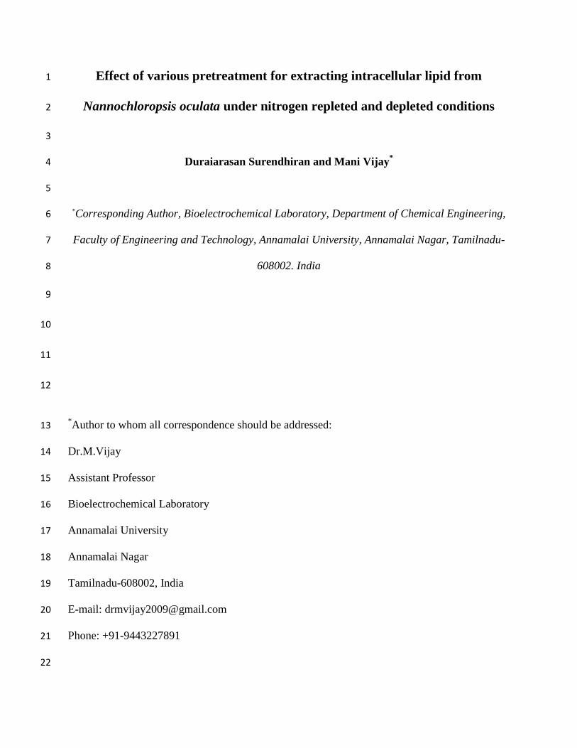
FIGURE 1: Growth curves of N.oculata grown on Walne’s medium with nitrogen repleted and 11
nitrogen depleted conditions 12
The effect of nitrogen on microalgal growth is shown in (Fig.1). The algal growth was 13
increased under nitrogen repleted condition, whereas some loss in growth was observed during 14
nitrogen limited or starvation condition [16]. A previous study by Alsull and Wan Omar [17] 15
0
0.2
0.4
0.6
0.8
1
1.2
0 1 2 3 4 5 6 7 8 9 10 11 12 13 14 15
Normal growth
Nitrogen depletedgrowth
Opti
cal
Den
sity
at
680nm
Incubation Time (Days)

also resulted in a decreasing yield of biomass when alga was grown without nitrogen. Because 1
without nitrogen, microalgal growth slows down and there is no requirement for the synthesis of 2
new membrane compounds. Nitrogen is an essential nutrient for algal growth; hence it was 3
added to the nitrogen depleted cells initially. At the 5th
day, the medium was supplied without 4
nitrogen to microalgal culture. The nitrogen rich cells was dark green pigmented, whereas 5
nitrogen depleted culture grew with same color till lag phase and turned yellow green at log and 6
stationary phases. This indicated that nitrogen was essential in synthesis of chlorophyll for 7
photosynthesis which also enhanced growth of microalgae. Similar result was also found in Beal 8
et al. [18] which showed that nitrogen starved algal paste was yellow-green in color and healthy 9
sample was dark green. 10
11
3.2. Chlorophyll, protein and total carbohydrate analysis 12
Chlorophyll and protein contents drastically decreased to half of its actual concentration (Table 13
1). Similar result was obtained by Alsul and Wan Omar et al. [17] for Tetraselmis sp. and 14
Nannochloropsis sp. The chlorophyll a content was decreased, while the total lipid content 15
increased under nitrogen limitation or starvation condition. In case of total carbohydrate, the 16
content decreased moderately than the protein and chlorophyll. In general, during nitrogen 17
limitation conditions the normal carbohydrate and protein metabolic pathways of cells are 18
reverted to lipid synthesis [19], which leads to high amount of intracellular lipid accumulation in 19
microalgae during nitrogen starvation. 20
21

TABLE 1: Chlorophyll, protein content and carbohydrate content of N.oculata under nitrogen 1
repleted and nitrogen depleted condition 2
Parameter N+ N
–
Chlorophyll content (µg/ml) 9.4 7.83
Protein content (µg/ml) 1.95 0.98
Total carbohydrate (µg/ml) 4.89 2.79
N+
cells grown in normal medium, N– cells grown in nitrogen depleted medium
3
3.3. Nile red staining for lipid identification 4
5
FIGURE 2: Nile Red stained cells of Nannochloropsis oculata under fluorescent microscope 6
(A). Normal cells (B). Nitrogen starved cells. 7
Intracellular lipid droplets of N.oculata nitrogen rich cells and nitrogen starved cells were 8
observed by Nile Red staining under fluorescent microscope with excitation at 450–490-nm and 9
emission at 515-nm. Neutral lipid or triglycerides appeared as yellow dots, whereas polar lipid 10
and chlorophyll were stained in red colour. Fig.2 shows that nitrogen starved cells contained 11
more lipid droplets with increased cell size than the normal cells. Ahlgren and Hyenstrand [20] 12
and Hoffman et al. [21] reported that under nitrogen-deficient conditions, algal cells often 13
accumulate a surplus of carbon metabolites as neutral lipids more than polar lipids. These neutral 14
lipids are located as lipid bodies in the cytoplasm of microalgal cells (Fig.2). It was also 15
A B

reported that microalgae respond to the nitrogen starvation condition by degrading nitrogen 1
containing macromolecules and accumulating carbon reserve compounds for the maintenance of 2
cells, such as polysaccharides and fats. Current findings support previous research by Elumalai et 3
al. [22] and Pick and Rachutin-Zalogin [23] mentioned that the Nile red staining technique was a 4
useful tool for rapid determination of lipids in microalgae. 5
3.4. FTIR analysis 6
Analysis of the microalgal biomass was performed by FTIR-Spectrophotometer. Figure 3 shows 7
the FT-IR spectra of dried biomass sample of normally nourished (Fig.3a) and nitrogen depleted 8
cells (Fig. 3b) of N.oculata. Table 2 reveals the various functional groups present in the samples. 9
Bands were attributed to -CH stretch, protein band, N-H and C=O stretches of peptide bond, -10
CH2 stretch of lipids, C=O stretch of ester, C-O-P, C-C and C-O stretches of polysaccharides. 11
Both the spectra were found to be almost matching with slight disparity in peaks. In figure 3b, 12
less intensified absorption bands were observed in the region of 1800-800 cm-1
, a region specific 13
for carbohydrate and protein, showing a decrease in their content. This could be a notification of 14
carbohydrate metabolism reverting to that of lipids when cells starved from nitrogen. Also, sharp 15
absorption bands in the region of 3100-2800 cm-1
were found in spectrum of nitrogen depleted 16
cells, showing increased lipid content and intensity of the particular band was lesser in nitrogen 17
repleted cells. From these spectrograms it was inferred that components present were mostly cis 18
isomers as the bands were between 700 and 3500 cm-1
(Konwar, 2011, Elumalai et al (2011) and 19
Rukminasari, 2013). 20

1
2
FIGURE 3: represents the FTIR spectrum of N.oculata grown under nitrogen depleted (a) and 3
repleted conditions (b) 4
5
6
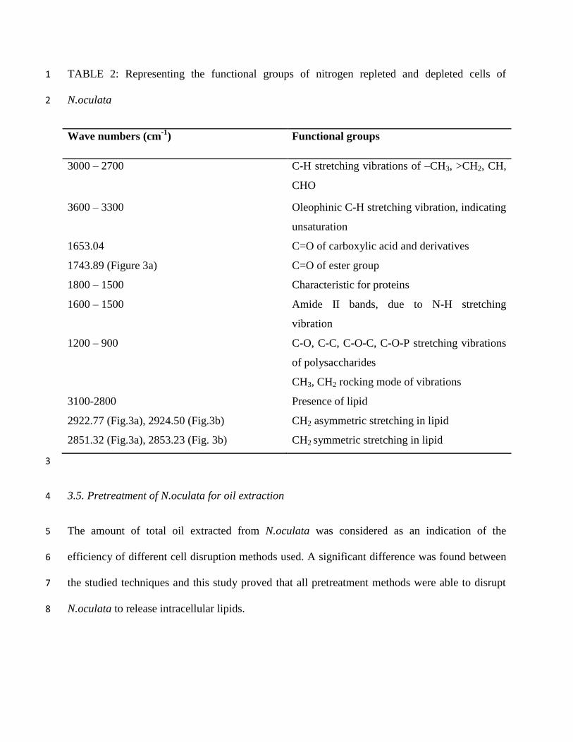
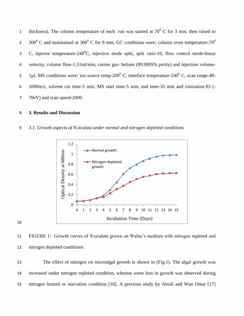
TABLE 2: Representing the functional groups of nitrogen repleted and depleted cells of 1
N.oculata 2
Wave numbers (cm-1
) Functional groups
3000 – 2700
C-H stretching vibrations of –CH3, >CH2, CH,
CHO
3600 – 3300 Oleophinic C-H stretching vibration, indicating
unsaturation
1653.04
C=O of carboxylic acid and derivatives
1743.89 (Figure 3a) C=O of ester group
1800 – 1500
Characteristic for proteins
1600 – 1500
Amide II bands, due to N-H stretching
vibration
1200 – 900
C-O, C-C, C-O-C, C-O-P stretching vibrations
of polysaccharides
CH3, CH2 rocking mode of vibrations
3100-2800
Presence of lipid
2922.77 (Fig.3a), 2924.50 (Fig.3b) CH2 asymmetric stretching in lipid
2851.32 (Fig.3a), 2853.23 (Fig. 3b) CH2 symmetric stretching in lipid
3
3.5. Pretreatment of N.oculata for oil extraction 4
The amount of total oil extracted from N.oculata was considered as an indication of the 5
efficiency of different cell disruption methods used. A significant difference was found between 6
the studied techniques and this study proved that all pretreatment methods were able to disrupt 7
N.oculata to release intracellular lipids. 8

Enzymatic pretreatment were carried out using cellulase enzyme (5mg L-1
) for 8 h, 10 h 1
and 12 h interval. A maximum efficiency by enzymatic pretreatment was found to be at 12 h as 2
32.74% from nitrogen rich cultures and 51.68% under nitrogen starvation condition. The 3
microalgal cell wall is made up of polysaccharide mainly comprising cellulose can be 4
hydrolyzed by the enzyme, cellulase [24]. From the data it was clear that the oil extraction was 5
found to be increased with increasing pretreatment time with cellulase. From the literature, the 6
benefit of using enzyme for hydrolyzing microalgal cell wall was evaluated by Sander and 7
Murthy [25]. Presently, enzymatic cell wall degradation is not widely practiced in industry 8
because cell lysing enzymes have traditionally been cost prohibitive. Though cost intensive, 9
enzyme degradation is important because the rigid cell wall of algal cell is resistance to 10
mechanical methods will require excess energy usage and multiple passes through disruption 11
equipment. The cost factor can be overcome by immobilization of enzyme. Moreover, an alga 12
does not have lignin in their cell wall, which is a major advantage to perform enzymatic 13
hydrolysis of its polysaccharide components [4]. Fu et al. [24] had successfully hydrolysed 14
Chlorella sp. using immobilized cellulase with electrospun polyacrylonitrile (PAN) producing 15
reducing sugar. Therefore use of enzyme to lyse algal cell wall might be advantageous as 16
compared to other methods. 17
Effect of acid treatment was studied at different time periods – 1h, 2h and 3h. During acid 18
lysis treatment of N.oculata, at pH 2.0, there was a high effect on oil released at an incubation of 19
2h. The marine microalgae have been used as feed for herbivorous fishes worldwide. The 20
researchers believed that many marine herbivorous fishes must possess acidic gastric condition 21
in their stomach to gain access to the intracellular nutrients from algae for their diet [26]. The 22
present study showed that the most efficient cell disruption occurred after 2h of incubation 23
giving 33.18% for nitrogen availed culture and 54.26% for nitrogen depleted condition. Similar 1
result was reported by Zemke-White et al. [25], in which they had analysed 4 macroalgae namely 2
Enteromorpho intestinalis, Ulva rigida, Porphyra sp. and Polysiphonia strictissima and found 3
that at pH 2.0 for 60 min the cell wall pore size of all four algal species increased up to at least 4
13.5nm than the normal cell wall size (8.8nm). In the present study, after 2h of treatment, the 5
amount of oil yield was found to be constant. Hence we concluded that the algal cell wall lysed 6
when treated for 2h at a low pH 2, the porosity of cell wall increased and intracellular lipid got 7
released. Also this finding was supported by Harun and Danquah [26]. They had reported that 8
acid pretreatment process was most suitable for hydrolyzing the cell wall of Chlorococcum 9
humicola to release and convert the polysaccharides, entrapped in microalgal cell wall, into 10
simple sugars for ethanol production. 11
Ultrasonication is a simple physical method for disrupting N.oculata. The cells grown in 12
normal nutrition condition were exposed to various time periods namely 5, 10 and 15 min and a 13
maximum of 30.12% oil was extracted at 15 min pretreatment. Similarly nitrogen depleted cells, 14
when ultrasonicated at above mentioned time intervals, showed higher extraction efficiency of 15
45.77 % at a pretreatment time of 15 min. Higher the prolongation of time for process, extraction 16
of oil increased. In contrast to the present study, Lee et al. [10] observed that ultrasonication 17
resulted in least efficiency for Botryococcus sp. Suganya and Renganathan [8] reported that 18
through ultrasonication, higher extraction efficiency was achieved 2.25 times higher than that of 19
direct extraction of oil from Ulva lactuca. Ultrasonication is one of the major pretreatments of 20
algal cells for extracting oil; hence higher physical stress due to vibration on prolonged exposure, 21
to extensive release of oil because of cell disruption, enhances the yield of oil for biodiesel 22
production. 23

Autoclaving of microalgal cells was carried out for extraction of oil at various time 1
intervals 10, 20 and 30 min at 1210 C. Higher yield of oil was obtained from nitrogen depleted 2
cells than the normally cultivated microalgal cells. At high thermal treatment, at 30min, 3
maximum amount of oil (28.81 %) was obtained for normal cultures. Likewise for the nitrogen 4
depleted cells the oil content was found to be at its maximum (43.90%) at 30min. Autoclaving 5
was found to be one of the most efficient methods yielding 7.88% of oil from Ulva lactuca by 6
disrupting the membranes of the cells [8]. Lee et al. [10] also reported that extraction of oil from 7
Chlorella vulgaris was at its maximum of 7.9% on autoclaving. Therefore higher the time of 8
autoclaving at a high temperature, higher was the oil yield. 9
The effect of 40% NaCl was studied by subjecting the normally nourished and nitrogen 10
starved cells at different time intervals. This higher osmotic shock resulted in cell lysis due to 11
susceptibility of cell membrane of microorganisms. For normal cells 24 h the highest oil content 12
was obtained for 48 h as 26.49 %. Similarly, under nitrogen depleted cells the maximum yield oil 13
was 40.56 % at 48h. For determination of optimistic time for oil extraction with NaCl, the cells 14
were treated above 48 h till 72h, but the amount of oil remained constant. Hence, 48 h was found 15
to be an optimum extraction time for 40% NaCl treatment. However, the osmotic shock is a 16
simple procedure for oil extraction but it did not show much disruption effect on microalgal cells 17
and required longer time duration. The current result was supported by the work of Lee et al. 18
[10] for treating microalgal species, C.vulgaris and Scenedesmus sp. for 48 h. 19
Dejoye et al. [27] reported that extraction of oil from microalgae with microwave 20
pretreated microalgae systematically presented higher yields. But in contrast, in this study, 21
microwave pretreated method did not significantly affect cell disruption. Microwave assisted 22
treatment was performed at 5, 10 and 15 min for oil extraction by cell distortion. The maximum 23

oil content using this method was found to be 26.51 % at 5 min for normal cells and for nitrogen 1
depleted cells it was 41.28 % at 5 min. A decline in oil content was experienced as the time of 2
exposure prolonged. A similar result was found in a previous study that microalgal lipid 3
extraction efficiency was not effective and the oil extracted from microalgae by microwave 4
method became volatile while disruption and extraction process [6]. 5
TABLE 3: Lipid content at various pretreatment process of N.oculata under nitrogen rich and 6
nitrogen starved conditions 7
Various
Pretreatment
Pretreatment Time
(min/h)
Normal Growth
Nitrogen depleted
Growth
Control 26.43 40.52
Acid 1 h 31.23 48.38
2 h 33.18 54.26
3 h 33.18 54.26
Enzyme 8 h 29.38 45.28
10 h 30.62 48.52
12 h 32.74 51.68
Ultrasonication 5 min 28.14 44.16
10 min 29.32 45.24
15 min 30.12 45.77
Autoclave 10 min 27.68 42.39
20 min 28.06 43.38
30 min 28.81 43.90
Microwave Oven 5 min 26.51 41.28
10 min 22.88 35.36
15 min 16.79 23.21
40 % NaCl 24 h 26.45 40.54
48 h 26.49 40.56
72 h 26.49 40.56
8
Higher lipid concentration represented the disruption efficiency in this study and nitrogen 9
depletion enhanced oil content on marine microalgae N.oculata, which was compared with the 10

control. Among several methods were for pretreating N.oculata for oil extraction, the enzymatic 1
method gave maximum efficiency for normal culture and nitrogen depleted culture and also 2
revealed that this method was the best for microalgal cell wall lysis. Acid pretreatment gave 3
second maximum yield. Poor efficiency was shown by ultrasonication, autoclaving, microwave 4
oven and 40% NaCl pretreatment. Similar results were also found in Zheng et al. [6], who had 5
studied various pretreatment on fresh water microalgae, C.vulgaris showing higher effects of 6
enzymes like lysozyme and cellulose on oil extraction by cell disruption. 7
In addition, from an extensive literature survey, it was noted that nowadays enzymatic 8
extraction has been widely used in extraction of bioactive compounds from plant based 9
materials. It is more attractive, with advantages like shorter extracting time, less pollution, higher 10
extraction yield and less decomposition of target compounds [28]. Moreover, enzymatic 11
hydrolysis is specific, gentle and has specific effect on cell wall of algae especially on 12
hemicelluloses and saccharides and accelerates the migration of bioactive compounds. Compared 13
with conventional methods, it utilizes less energy and gave higher extraction yield [28]. Zheng et 14
al. [6] stated that the enzymatic process could be worked at low temperature and could prevent 15
the oxidation of oil, thus improving the biodiesel quality. 16
17
18
19
20
21

3.6. SEM analysis of algal cells 1
2
3
FIGURE 4: Scanning Electron Microscopic images of morphological analysis of various 4
pretreatment processes A).Control, B).Acid pretreatment, C).Enzyme pretreatment, 5
D).Ultrasonication 6
7
For direct evidence on different pretreatment methods on cell wall damage, microalgal cells can 8
be observed by microscopic study (Figure 4). The undamaged cells of N.oculata (Figure 4A) 9
showed intact structures and had no indication of cell lysis. In addition, Acid treatment 1M HCl 10
totally disrupted the morphology of microalgal structure, appeared completely broken cells under 11
Scanning Electron Microscope (Figure 4B). Next to the acid lysis the effective cell disruption 12
was happened with enzymatic (cellulase) and ultrasonic disruption (Figures 4C, 4D). 13
3.7. Fatty acid composition analysis by GC-MS 14
B
C D
A
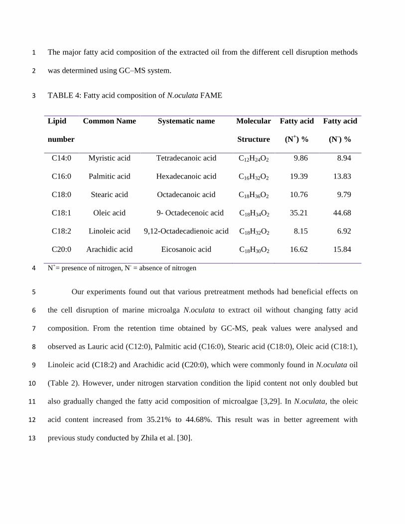
The major fatty acid composition of the extracted oil from the different cell disruption methods 1
was determined using GC–MS system. 2
TABLE 4: Fatty acid composition of N.oculata FAME 3
Lipid
number
Common Name Systematic name Molecular
Structure
Fatty acid
(N+) %
Fatty acid
(N-) %
C14:0 Myristic acid Tetradecanoic acid C12H24O2 9.86 8.94
C16:0 Palmitic acid Hexadecanoic acid C16H32O2 19.39 13.83
C18:0 Stearic acid Octadecanoic acid C18H36O2 10.76 9.79
C18:1 Oleic acid 9- Octadecenoic acid C18H34O2 35.21 44.68
C18:2 Linoleic acid 9,12-Octadecadienoic acid C18H32O2 8.15 6.92
C20:0 Arachidic acid Eicosanoic acid C18H30O2 16.62 15.84
N+= presence of nitrogen, N
- = absence of nitrogen 4
Our experiments found out that various pretreatment methods had beneficial effects on 5
the cell disruption of marine microalga N.oculata to extract oil without changing fatty acid 6
composition. From the retention time obtained by GC-MS, peak values were analysed and 7
observed as Lauric acid (C12:0), Palmitic acid (C16:0), Stearic acid (C18:0), Oleic acid (C18:1), 8
Linoleic acid (C18:2) and Arachidic acid (C20:0), which were commonly found in N.oculata oil 9
(Table 2). However, under nitrogen starvation condition the lipid content not only doubled but 10
also gradually changed the fatty acid composition of microalgae [3,29]. In N.oculata, the oleic 11
acid content increased from 35.21% to 44.68%. This result was in better agreement with 12
previous study conducted by Zhila et al. [30]. 13
Unsaturated fatty acids have been reported as reasonable balance of fuel properties [6]. 1
The chain length of fatty acids in N.oculata was observed between C12 and C20. In a previous 2
report, it was stated that the fatty acids with maximum of C16 and C18 series were recognized as 3
the most common components of biodiesel [31]. Therefore, fatty acids from N.oculata were 4
more applicable for producing high quality of biofuel, since it contained high content of C16 5
(palmitic acid) and C18 (oleic acid). 6
4. Conclusion 7
This paper demonstrated the utility of various pretreatment protocols to extract lipids by cell 8
disruption. Amongst all the procedures, acid hydrolysis proved to the appropriate method. 9
Additionally, it was found that cells when under stressed condition, i.e., nitrogen depleted state, 10
produced lipids at a higher rate due to the reversion of carbohydrate, protein metabolism to that 11
of lipid, which was clearly depicted by FT-IR spectral analysis. This technique could be used for 12
the large scale production and utilize lipids to generate biodiesel. 13
Conflict of interest 14
The authors of this research article claim no conflict of interest 15
References 16
[1] P. Schlagermann, G. Gottlicher, R. Dillschneider, R. Rosello-Sastre and C. Posten, 17
“Composition of Algal Oil and Its Potential as Biofuel”, Journal of Combustion, vol. 2012, 18
Article ID 285185, doi:10.1155/2012/285185. 19
[2] H. C. Chen, H. Y. Ju, T. T. Wu, Y. C. Liu, C.C. Lee, C. Chang, Y. L. Chung and C. J. 20
Shieh, “Continuous Production of Lipase-Catalyzed Biodiesel in a Packed-Bed Reactor: 21

Optimization and Enzyme Reuse Study”, Journal of Biomedicine and Biotechnology, vol. 1
2011, Article ID 950725, doi:10.1155/2011/950725. 2
[3] A. Widjaja, C. C. Chien and Y. H. Ju, “Study of increasing lipid production from fresh 3
water microalgae Chlorella vulgaris”, Journal of the Taiwan Institute of Chemical 4
Engineers, vol. 40, pp. 13–20, 2009. 5
[4] M. A. Rodrigues and E. P. S. Bon, “Evaluation of Chlorella (Chlorophyta) as Source of 6
Fermentable Sugars via Cell Wall Enzymatic Hydrolysis”, Enzyme Research, vol. 2011, 7
Article ID 405603, doi:10.4061/2011/405603. 8
[5] E. Ryckebosch, K. Muylaert and I. Foubert, “Optimization of an Analytical Procedure for 9
Extraction of Lipids from Microalgae”, Journal of American Chemical Society, DOI 10
10.1007/s11746-011-1903-z. 11
[6] H. Zheng, J. Yin, Z. Gao, H. Huang, X. Ji and C. Dou, “Disruption of Chlorella vulgaris 12
Cells for the Release of Biodiesel-Producing Lipids: A Comparison of Grinding, 13
Ultrasonication, Bead Milling, Enzymatic Lysis, and Microwaves”. Applied Biochemistry 14
and Biotechnology, vol. 164, pp. 1215–1224, 2011. 15
[7] Paula Mercer and Roberto E. Armenta, Developments in oil extraction from microalgae, 16
Eur. J. Lipid Sci. Technol. 2011, DOI: 10.1002/ejlt.201000455. 17
[8] Tamilarasan Suganya and Sahadevan Renganathan. Optimization and kinetic studies on 18
algal oil extraction from marine macroalgae Ulva lactuca. Bioresource Technology. 107, 19
319-326 (2012). 20
[9] Kyle Sander and Ganti S. Murthy. Enzymatic Degradation of Microalgal Cell Walls. An 21
ASABE Meeting Presentation, Paper Number: 1035636. June 21-June 24, (2009). 22
[10] Jae-Yon Lee, Chan Yoo, So-Young Jun, Chi-Yong Ahn, Hee-Mock Oh. Comparison of 1
several methods for effective lipid extraction from microalgae. Bioresource Technology. 2
101, S75-S77, (2010). 3
[11] Guojie Jin, Fan Yang, Cuimin Hu, Hongwei Shen, Zongbao K. Zhao. Enzyme-assisted 4
extraction of lipids directly from the culture of the oleaginous yeast Rhodosporidium 5
toruloides. Bioresource Technology. 111, 378-382 (2012). 6
[12] E. G. Bligh, W. J. Dyer, A rapid method of total lipid extraction and purification, Can J 7
Biochem. Physiol. 37 (1959) 911–917. 8
[13] Nagwa G.E. Mohammady, Christopher W. Ricken, Scott R. Lindell, Christopher M. 9
Reddy, Hala M. Taha, Connie Pui Ling Lau and Catherine A. Carmichael. Age of Nitrogen 10
Deficient Microalgal Cells is a key Factor for Maximizing Lipid content. Research Journal 11
of Phytochemistry. 6 (2): 42-53 (2012). 12
[14] C.H. Su, C.C. Fu, Y.C. Chang, G.R. Nair, J.L. Ye, M. Chu, W.T. Wu, Simultaneous 13
Estimation of Chlorophyll a and Lipid Contents in Microalgae by Three-Color Analysis, 14
Biotechnology and Bioengineering, 99, (4), 1034-1039. 15
[15] O. H. Lowry, N. J. Rosebrough, A. L. Farr, J. Randal, Protein measurement with the folin-16
phenol reagent. Journal of Biological Chemistry, 193 (1951) 265–275. 17
[16] Chittra Yeesang, Benjamas Cheirsilp, Effect of nitrogen, salt, and iron content in the 18
growth medium and light intensity on lipid production by microalgae isolated from 19
freshwater sources in Thailand, Bioresource Technology 102 (2011) 3034–3040. 20
[17] Mohamed Alsull and Wan Maznah Wan Omar. Responses of Tetraselmis sp. and 21
Nannochloropsis sp. Isolated from Penang National Park Coastal Waters, Malaysia, to the 22
Combined Influences of Salinity, Light and Nitrogen Limitation. International Conference 23
on Chemical, Ecology and Environmental Sciences (ICEES'2012) march 17-18, Pages 142-1
145 (2012), Bangkok. 2
[18] C.M. Beal, M.E. Webber, R.S. Ruoff, R.E. Hebner. Lipid Analysis of Neochloris 3
oleoabundans by Liquid State NMR. Biotechnology and Bioengineering. 106 (4), 573-583 4
(2010). 5
[19] Dina Feng, Zhaoan Chen, Song Xue, Wei Zhang. Increased lipid production of the marine 6
oleaginous microalgae Isochrysis zhangjiangensis (Chrysophyta) by nitrogen supplement. 7
Bioresource Technology. 102 (2011) 6710–6716. 8
[20] Ahlgren, G., Hyenstrand, P., 2003. Nitrogen limitation effects of different nitrogen sources 9
on the nutritional quality of two freshwater organisms, Scenedesmus quadricauda 10
(Chlorophyceae) and Synechococcus sp. (Cyanophyceae). J. Phycol. 39, 906–917. 11
[21] Maren Hoffmann, Kai Marxen, Rüdiger Schulz and Klaus Heinrich Vanselow, TFA and 12
EPA Productivities of Nannochloropsis salina Influenced by Temperature and Nitrate 13
Stimuli in Turbidostatic Controlled Experiments, Mar. Drugs 2010, 8, 2526-2545; 14
doi:10.3390/md8092526. 15
[22] Sanniyasi Elumalai, Velu Prakasam and Ramganesh Selvarajan. Optimization of abiotic 16
conditions suitable for the production of biodiesel from Chlorella vulgaris. Indian Journal 17
of Science and Technology. 4 (2), 91-97(2011). 18
[23] Uri Pick, Tatyana Rachutin-Zalogin, Kinetic anomalies in the interactions of Nile red with 19
microalgae, Journal of Microbiological Methods 88 (2012) 189–196. 20
[24] C.C. Fu, T.C. Huang, J. Y. Chen, C.H. Su, W. T. Wu, Hydrolysis of microalgae cell walls 21
for production of reducing sugar and lipid extraction, Bioresource Technology, 101 (2010) 22
8750-8754. 23

[25] W. L. Zemke-White, K.D. Clements, P.J. Harris. Acid lysis of macroalgae by marine 1
herbivorous fishes: effects of acid pH on cell wall porosity. Journal of Experimental 2
Marine Biology and Ecology. 245, 57-86 (2000). 3
[26] Razif Harun, Michael K. Danquah. Influence of acid pre-treatment on microalgal biomass 4
for bioethanol production. Process Biochemistry. 46 (2011) 304–309. 5
[27] Celine Dejoye, Maryline Abert Vian,Guy Lumia, Christian Bouscarle, Frederic,Charton, 6
and Farid Chemat. Combined Extraction Processes of Lipid from Chlorella vulgaris 7
Microalgae: Microwave Prior to Supercritical Carbon Dioxide Extraction. Int. J. Mol. Sci. 8
2011, 12, 9332-9341. 9
[28] Shengnan Li, Heng Zhang, Dandan Han, and Kyung Ho Row. Optimization of enzymatic 10
extraction of polysaccharides from some marine algae by response surface methodology. 11
Korean J. Chem. Eng., 29 (5), 650-656 (2012). 12
[29] Guan Hua Huang, Feng Chen, Dong Wei, Xue Wu Zhang, Gu Chen. Biodiesel production 13
by microalgal biotechnology. Applied Energy. 87 (2010) 38–46. 14
[30] Zhila NO, Kalacheva GS, Volova TG. Effect of nitrogen limitation on the growth and lipid 15
composition of the green alga Botryococcus braunii Kutz IPPAS H-252. Russ J Plant 16
Physiol 2005;52:357–65. 17
[31] Qiang Lin, Na Gu, Gang Li, Junda Lin, Liangmin Huang, LingLing Tan. Effects of 18
inorganic carbon concentration on carbon formation, nitrate utilization, biomass and oil 19
accumulation of Nannochloropsis oculata CS 179. Bioresource Technology. 111 (2012) 20
353–359. 21
22
23