nadph oxidase-4 mediates protection against …nadph oxidase-4 mediates protection against chronic...
TRANSCRIPT

NADPH oxidase-4 mediates protection againstchronic load-induced stress in mouse hearts byenhancing angiogenesisMin Zhanga,1,2, Alison C. Brewera,1, Katrin Schröderb,1, Celio X. C. Santosa, David J. Grievea, Minshu Wanga,Narayana Anilkumara, Bin Yua, Xuebin Donga, Simon J. Walkera, Ralf P. Brandesb, and Ajay M. Shaha,2
aCardiovascular Division, King’s College London British Heart Foundation Centre, London SE5 9PJ, United Kingdom; and bInstitut für KardiovaskulärePhysiologien, Goethe-Universität, 60590 Frankfurt am Main, Germany
Edited* by Salvador Moncada, University College London, London, United Kingdom, and approved September 14, 2010 (received for review July 4, 2010)
Cardiac failure occurs when the heart fails to adapt to chronicstresses. Reactive oxygen species (ROS)-dependent signaling isimplicated in cardiac stress responses, but the role of different ROSsources remains unclear. Here we report that NADPH oxidase-4(Nox4) facilitates cardiac adaptation to chronic stress. Unlike otherNox proteins, Nox4 activity is regulated mainly by its expressionlevel, which increases in cardiomyocytes under stresses such aspressure overload or hypoxia. To investigate the functional role ofNox4 during the cardiac response to stress, we generated mice witha genetic deletion of Nox4 or a cardiomyocyte-targeted overexpres-sion of Nox4. Basal cardiac function was normal in both models, butNox4-null animals developed exaggerated contractile dysfunction,hypertrophy, and cardiac dilatation during exposure to chronic over-load whereas Nox4-transgenic mice were protected. Investigationof mechanisms underlying this protective effect revealed a signifi-cant Nox4-dependent preservation of myocardial capillary densityafter pressure overload. Nox4 enhanced stress-induced activationof cardiomyocyte hypoxia inducible factor 1 and the release of vas-cular endothelial growth factor, resulting in increased paracrine an-giogenic activity. These data indicate that cardiomyocyte Nox4 is aunique inducible regulator ofmyocardial angiogenesis, a keydetermi-nant of cardiac adaptation to overload stress. Our results also havewider relevance to the use of nonspecific antioxidant approaches incardiac disease and may provide an explanation for the failure ofsuch strategies in many settings.
cardiac remodeling | hypoxia inducible factor | reactive oxygen species
Heart failure results from disease stresses that chronically in-crease cardiac workload (1). The cardiac response to such
insults involves remodeling of the cardiomyocytes, vasculature, andextracellular matrix that may initially be adaptive. Persistent stress,however, results in contractile dysfunction, fibrosis, ventriculardilatation, and capillary rarefaction. Specific pathways are thoughtto drive adaptive vs. maladaptive features of remodeling (1–3).Increased reactive oxygen species (ROS) production is impli-
cated in cardiac remodeling through several mechanisms, includingthe activation of signaling pathways that promote cardiomyocytehypertrophy, abnormal excitation-contraction coupling, mitochon-drial dysfunction, cell death, and extracellular matrix remodeling(4, 5). Clinical trials of antioxidants have yielded disappointing re-sults, however, and effective therapies based on targeting ROSremain elusive. ROS in a stressed heart may emanate from severalsources, including mitochondria, NADPH oxidases of the Noxfamily, uncoupled nitric oxide (NO) synthases, and xanthine oxi-dases, but the functional roles of individual sources remain unclear(5). It is recognized, however, that different sources may modulatedistinct signaling pathways through regulated, spatially restrictedROS production (6). Indeed, some ROS-mediated effects arebeneficial rather than detrimental, e.g., in cardioprotective signal-ing elicited by preconditioning (7).Nox family enzymes generate ROS by catalyzing electron
transfer from NADPH to molecular O2. Seven family membersexist (Nox1–5 and Duox1–2), each based on a distinct catalytic
subunit and with tissue-specific expression (8, 9). The prototypicNox enzyme, Nox2, mediates microbicidal activity in phagocytesthrough generation of large amounts of superoxide (O2
−). Innonphagocytic cells, however, Nox2 and other Nox proteins gen-erate low levels of ROS that are involved in intracellular signaling(9). Previous studies using Nox2-null mice and other modelsshowed that Nox2 in the heart is involved in the development ofcardiac hypertrophy and contractile dysfunction induced by an-giotensin II, pressure overload, or myocardial infarction (10–15).Nox4 differs from Nox2 and other Nox enzymes in that it is reg-ulated mainly by its expression level and does not require agoniststimulation or association with regulatory subunits for activation(16–18). Recent studies also suggest that it generates pre-dominantly H2O2 rather than O2
− (17–20). Previous work showedthat Nox4 is expressed at a low level in the adult mammalian heartand that its abundance increases during pressure overload (11),but its pathophysiological functions in vivo are unknown.In this study, we generated a Nox4-null mouse model and a
cardiomyocyte-targeted Nox4-transgenic model to elucidate the ef-fects of Nox4 during cardiac stress. In marked contrast to the effectsof Nox2 and other ROS sources, increases in cardiomyocyte Nox4resulted in protection against pressure overload-induced adversecardiac remodeling. Nox4 facilitated preservation of myocardialcapillary density during pressure overload by regulating stress-induced cardiomyocyte hypoxia inducible factor 1 (Hif1) activationand release of vascular endothelial growth factor (VEGF), resultingin increased paracrine angiogenic activity. These data indicate thatNox4 is a unique stress-inducible regulator of myocardial angio-genesis that facilitates adaptation to cardiac overload stress.
ResultsMyocardial Nox4 Expression Increases During Stress. We first ana-lyzed changes in myocardial Nox4 expression during developmentand in response to various stresses. Nox4 expression in-creased after in vivo pressure overload, myocardial infarction, orin vitro hypoxia (Fig. 1A). Nox4 expression was significantly lowerin themyocardium of healthy young or old animals compared withfetal hearts (Fig. 1B). The increase in Nox4 protein expressionafter pressure overload was largely in cardiomyocytes (Fig. S1A).This induction of Nox4 is similar to that of so-called fetal genesthat are reactivated in the adult heart during stress (3).
Author contributions: A.M.S. designed research; M.Z., A.C.B., K.S., C.X.C.S., D.J.G., M.W.,N.A., B.Y., X.D., and S.J.W. performed research; K.S. and R.P.B. contributed new reagents;M.Z., C.X.C.S., and A.M.S. analyzed data; and M.Z., R.P.B., and A.M.S. wrote the paper.
The authors declare no conflict of interest.
*This Direct Submission article had a prearranged editor.
Freely available online through the PNAS open access option.1M.Z., A.C.B., and K.S. contributed equally to this work.2To whom correspondence may be addressed. E-mail: [email protected] or [email protected].
This article contains supporting information online at www.pnas.org/lookup/suppl/doi:10.1073/pnas.1009700107/-/DCSupplemental.
www.pnas.org/cgi/doi/10.1073/pnas.1009700107 PNAS | October 19, 2010 | vol. 107 | no. 42 | 18121–18126
MED
ICALSC
IENCE
S
Dow
nloa
ded
by g
uest
on
Apr
il 17
, 202
0

Nox4-Null Mice Have Exaggerated Load-Induced Cardiac Dysfunction.To evaluate the role of increases in Nox4 during cardiac stress, wegenerated mice deficient in Nox4 (Fig. S1B). Deletion of en-dogenous Nox4 resulted in a total loss of Nox4 protein anda small reduction in H2O2 production as assessed by a homo-vanillic acid assay (Fig. 1 C and D). Myocardial levels of Nox2 andp22phox were unaffected by Nox4 deletion (Fig. 1C). Nox4-null
mice were born in the expected Mendelian ratio, bred normally,and showed no obvious abnormal baseline phenotype (Table S1).Basal cardiac size and function assessed by echocardiography
were unchanged in Nox4-null mice (Fig. 2A). To assess the effectsof the absence of Nox4 during cardiac stress, we performed su-prarenal aortic constriction to generate a chronic pressure over-load. The degree of pressure overload was similar in Nox4-nullmice and wild-type (WT) littermates, and there was no differencein mortality. As expected, 6 wk of pressure overload caused con-tractile impairment and ventricular dilatation in wild-type mice.Nox4-null animals, however, developed significantly greater car-diac dilatation and contractile impairment than wild type (Fig.2A). Nox4-null mice also developed exaggerated cardiac hyper-trophy after chronic pressure overload at both the whole-heartand the cardiomyocyte level, as well as increased interstitial fi-brosis (Fig. 2 B–D). These data therefore suggest that an increasein myocardial Nox4 expression is protective against the detri-mental consequences of chronic pressure overload.
Cardiomyocyte-Targeted Nox4-Transgenic Mice Show No BasalDysfunction. To further validate a protective role of Nox4 dur-ing cardiac overload, we generated transgenic mice with acardiomyocyte-targeted increase in Nox4. Nox4-transgenic micehad significantly increased Nox4 protein levels (Fig. 3A and Fig.S2A). Nox4 heterodimerizes with p22phox, with the two proteinsstabilizing each other (8, 16), and increased Nox4 expression wasaccompanied by a twofold increase in p22phox levels. Nox2 levelsby contrast were similar between strains. Nox4-transgenic micehad a modest elevation of myocardial H2O2 production (Fig. 3B)but did not show increased O2
− levels as assessed by electronparamagnetic resonance spectroscopy (EPR) (Fig. S2B), which isin line with data that suggest that Nox4 generates predominantlyH2O2 rather than O2
− (17–20). Consistent with this, myocardialnitrotyrosine levels—as a readout of nitrosative stress resultingfrom interaction of O2
− and NO—were unaltered in Nox4-transgenic mice, which also had unaltered endothelial NO syn-thase levels (Fig. 3A and Fig. S2A). Overexpressed Nox4 protein
Fetus 3m 12m0
1
2
Nox
4 m
RN
A (A
.U)
****
0
2
4
6
Ctl Band Ctl MI Ctl Hyp
**
**
**N
ox4
mR
NA
(A.U
)
A B
0
20
40
60
80
100
WT KO WT KO
Heart Kidney
*H
2O2
gene
ratio
n (n
mol
/mg)
DCHeart
KidneyActin
WT KO
Nox2
Actin
p22phox
WT KO
Fig. 1. Cardiac Nox4 induction during stress and generation of Nox4-nullmice. (A) Changes in Nox4 expression after pressure overload (Band), myo-cardial infarction (MI), or in vitro hypoxia (Hyp; 24 h) compared with re-spective controls (Ctl). **P < 0.01; n = 4–6/group. (B) Nox4 expression in 3-mo(3m)- and 12-mo (12m)-old mice compared with fetal heart. **P < 0.01; n = 5/group. (C) Western blots showing loss of Nox4 protein in heart and kidney ofhomozygous Nox4-KO mice (Top) and lack of change in cardiac Nox2 andp22phox levels (Bottom). (D) H2O2 production in heart and kidney of Nox4-KOcompared with WT. *P < 0.05; n = 6/group.
A
00.2
0.4
0.6
0.8
1.0
IVSD
(mm
)
##
***
Sham Band
LVES
V (μ
l)
0
20
40
60
80##
***
Sham Band0
20406080
100
LVED
V (μ
l)
##
**
Sham Band
EF (%
)
0
20
40
60 ##
****
Sham Band
012345
Fibr
osis
(%)
Sham Band
*
**##
B WT/Sham KO/Sham WT/Band KO/Band
WTKO
##
**
Cro
ss-s
ectio
nal a
rea
(μm
2 )
**
Sham Band
LV/B
W (m
g/g) **
##
**
0
2
4
6
Sham Band
C
D
Fig. 2. Nox4-null mice have exaggeratedload-induced dysfunction. (A) Echocardi-ography of WT and KO mice subjected to6 wk of chronic pressure overload. IVSD:interventricular septal diameter; LVESV:LV end-systolic volumes; LVEDV: LV end-diastolic volumes; EF: ejection fraction. (B)Representative H&E-stained longitudinalsections of WT and KO hearts. Scale bar: 2mm. (C and D) Representative sections forcardiomyocyte area (WGA-stained) andinterstitial fibrosis (Picrosirius red-stained),respectively. Scale bars: 20 μm. Mean dataare shown at the right. **P < 0.01; *P <0.05 for band vs. respective sham; ##P <0.01 for KO band vs. WT band; n = 12–15/group. LV/BW: LV/body weight.
18122 | www.pnas.org/cgi/doi/10.1073/pnas.1009700107 Zhang et al.
Dow
nloa
ded
by g
uest
on
Apr
il 17
, 202
0

in transgenic cardiomyocytes was found in a perinuclear location,similar to the location of endogenous Nox4 in normal car-diomyocytes or after myocyte transfection (Fig. S3), and asreported in other cell types (21–23). Nox4-transgenic mice weregrossly normal and showed no cardiac dysfunction up to 12 mo ofage, although the older animals had a slightly increased cardiacmass compared with wild-type littermates (Table S2). There wasno evidence of increased fibrosis or apoptosis in the hearts ofNox4-transgenic mice (Fig. S2 C and D). These results show thatan increase in myocardial Nox4 levels in the absence of stress hasno significant detrimental consequences.
Cardiomyocyte-Targeted Nox4-Transgenic Mice Are Protected AgainstLoad-Induced Stress. We next subjected Nox4-transgenic mice andwild-type littermates to chronic pressure overload. Quantificationof in vivo left ventricular (LV) pressure–volume relationsrevealed that both systolic and diastolic function were betterpreserved in Nox4-transgenic mice than in wild type after pressureoverload for 9 wk (Fig. 3C). The protective effect of Nox4 wasconfirmed by echocardiography (Fig. S4A) as well as in a second
independent transgenic line (Fig. S5). Nox4-transgenic mice de-veloped less cardiac hypertrophy after pressure overload thanwild-type littermates and also had significantly less interstitial fi-brosis (Fig. 3 D–F and Fig. S4 B–D). Taken together, the dataobtained so far using both loss-of-function and gain-of-functionapproaches indicate that an increase in myocardial Nox4 ex-pression is protective against chronic pressure overload-inducedcardiac dysfunction.
Nox4-Dependent Enhancement of Myocardial Capillary Density DuringPressure Overload. Signaling effectors of the cardiac response tochronic pressure overload include various protein kinases thatare potentially redox-sensitive (3, 5). We undertook an immu-noblotting-based profiling screen (Kinexus Bioinformatics) thatencompasses a broad range of signaling pathways in Nox4-transgenic and wild-type mice subjected to pressure overload.The only protein with a more than twofold difference in phos-phorylation between groups was Akt1. Quantitative immuno-blotting showed that phosphorylated Akt (S473) levels weremodestly elevated in Nox4-transgenic hearts, but there was nodifference between Nox4-null mice and wild-type mice subjectedto pressure overload (Fig. S5C). Levels of myocardial apoptosisafter pressure overload were also unaffected either by Nox4 de-letionor by cardiomyocyte-specific overexpression [WTband: 7.2±1.1; KO band: 6.7 ± 1.1; transgenic band: 6.0 ± 0.9/105 nuclei; n=6–9/group; P = NS]. A key determinant of functional cardiaccompensation during chronic pressure overload has recently beenrecognized as the extent of myocardial capillarization, with in-sufficient angiogenesis being a driver of heart failure (24–27).Quantification of myocardial capillary density in LV sections ofNox4-null mice, Nox4-transgenic mice, and respective wild-typelittermate controls showed that although there were no differ-ences between groups at baseline, after imposition of pressureoverload, capillary density was significantly lower in Nox4-nullmice compared with wild type (Fig. 4A). By contrast, Nox4-transgenic mice had significantly higher myocardial capillarydensity than wild-type littermates after pressure overload (Fig.4B). These results suggest that Nox4 up-regulation during cardiacstress is required to protect against load-induced cardiac dys-function by controlling the compensatory increase in myocardialcapillary density.
Nox4 Enhances Cardiomyocyte Hif1α and VEGF. Previous work showsthat a central mechanism underpinning myocardial stress-inducedangiogenesis is the release of angiogenic factors, notably VEGF(25–27). We found that cardiac VEGF-A protein levels weresignificantly increased in Nox4-transgenic mice whereas Nox4-nullmice had markedly lower levels than wild-type after pressureoverload (Fig. 5A). Immunostaining for VEGF in heart demon-strated a significant increase at cardiomyocyte membranes and invessels in Nox4-transgenic mice after aortic banding whereasNox4-null animals showed very little staining (Fig. S6A). An im-portant upstream transcriptional regulator of VEGF during load-induced stress is Hif1 (26), which is known to be redox-regulated(28). Hif1α protein levels were significantly higher in Nox4-transgenic hearts compared with wild type after pressure overload(Fig. 5B). By contrast, Hif1α levels in Nox4-null mice were sig-nificantly lower than in wild-type littermates after aortic banding.To more directly investigate the relationship between car-
diomyocyte Nox4, Hif1α, and VEGF, we studied cultured cardiaccells. Overexpression of Nox4 in cultured cardiomyocytes in-creased H2O2 levels and slightly increased Hif1α protein levelsduring normoxia but substantially enhanced them during hypoxia(Fig. 6A and Fig. S6 B and C). A similar Nox4-dependent aug-mentation of Hif1α levels was observed in H9c2 cardiomyoblastsafter treatment with cobalt chloride, a chemical hypoxia mimetic(Fig. 6B). Hypoxia-induced Hif1α accumulation in control cellswas significantly reduced by the potent but nonspecific Nox in-hibitor, diphenylene iodonium, or by catalase (Fig. 6C). Impor-tantly, similar effects were observed after specific down-regulationof endogenous Nox4 by siRNA-mediated knockdown (Fig. 6D).
p22phox
Nox2
Actin
Nox4
A WT TG
eNOS
Nitrotyrosine
B
0
20
40
60
H2O
2ge
nera
tion
(nm
ol/m
g)
WT TG
**
LV volume (μl)
LV volume (μl)0 10 20 30 40 50
0
40
80
120
LV p
ress
ure
(mm
Hg)
WT bandWT sham
0
40
80
120
0 10 20 30 40 50LV p
ress
ure
(mm
Hg)
TG bandTG sham
0
2
4
6
8
ESPV
R (m
mH
g/μl
)
**
##
PRSW
(mm
Hg/
g)
0
25
50
75
100
**
#
0
5
10
15
20
LVED
P (m
mH
g)
**#
dP/d
tmin
(-mm
Hg/
s)
-8000
-4000
0
*
C
Sham BandSham Band
Sham Band
Sham Band
LV/B
W (m
g/g)
0
2
4
6 ****
##
Sham Band0
100
200
Cro
ss-s
ectio
nal a
rea
(μm
2 )
Sham Band0
0.5
1.0
1.5
2.0
2.5
Fibr
osis
(%)
Sham Band
****
##
****
##D E F
WTTG
Fig. 3. Cardiomyocyte-targeted Nox4 overexpression protects against load-induced dysfunction. (A) Protein expression in hearts of Nox4-transgenic mice(TG) and wild-type littermates (WT). (B) H2O2 production in TG compared withWT myocardium. **P < 0.01; n = 6/group. (C) Contractile assessment ofpressure-overloaded Nox4-transgenic and WT mice by LV pressure–volumeanalysis. End-systolic pressure volume relation (ESPVR) and preload-recruitablestrokework (PRSW) aremeasures of systolic function. LV end-diastolic pressure(LVEDP) and dP/dtmin are measures of diastolic function. Representative pres-sure–volume curves are shown at the right. **P < 0.01; *P < 0.05 for band vs.respective sham; ##P < 0.01; #P < 0.05 for TG band vs. WT band; n = 15/group.(D–F) Mean data for heart hypertrophy (n > 20/group), cardiomyocyte cross-sectional area (n = 6/group), and interstitial fibrosis (n = 6/group), respectively.Statistics in D–F as in C.
Zhang et al. PNAS | October 19, 2010 | vol. 107 | no. 42 | 18123
MED
ICALSC
IENCE
S
Dow
nloa
ded
by g
uest
on
Apr
il 17
, 202
0
Both transcriptional and posttranslational mechanisms couldpotentially be involved in Hif1α up-regulation (28). We foundthat Hif1α mRNA expression in vivo was significantly higher inNox4-transgenic than in wild-type mice, but there was no sig-nificant difference in levels between Nox4-null mice and wild-type littermates (Fig. S6 D and E). Notably, Hif1α protein levelsin Nox4-transgenic mice were not elevated at baseline despiteincreased mRNA levels, suggesting that an effect at the proteinlevel was more important. Increasing Nox4 in cultured car-diomyocytes did not affect Hif1α mRNA levels during eithernormoxia or hypoxia (Fig. S6F). Because Hif1α protein stabilityis regulated by hydroxylation by specific prolyl hydroxylases,leading to targeting for proteosomal degradation (28), weassessed the effect of Nox4 on levels of hydroxylated Hif1α. Nox4overexpression in cardiomyocytes led to a substantial decrease inhydroxylated Hif1α levels as assessed by two different antibodies,an effect potentiated by hypoxia (Fig. 6 E and F). This wassimilar to the effect of a specific prolyl hydroxylase inhibitor,dimethyloxalylglycine, in reducing hydroxylated Hif1α and inincreasing total Hif1α levels (Fig. 6E). These results suggest thatthe inhibition of prolyl hydroxylase activity may be a majormechanism by which Nox4 increases cardiomyocyte Hif1α levels.
Paracrine Angiogenic Activity Mediated by Nox4. We found thatNox4-induced increases in cultured cardiomyocyte Hif1α duringhypoxia were accompanied by an increase in VEGF levels in thecells and even more so in their conditioned medium, indicative ofextracellular release (Fig. 7 A and B). To directly test the potentialangiogenic effects of Nox4-dependent changes in factors secretedby cardiomyocytes, we undertook in vitro endothelial cell tube for-mation assays. The conditioned medium of Nox4-overexpressing
cardiomyocytes subjected to hypoxia markedly enhanced endothe-lial tube formation as compared with that of hypoxic cardiomyo-cytes overexpressing a β-galactosidase (β-gal) control gene (Fig. 7CandFig. S6G). The angiogenic effect of cardiomyocyte-conditionedmedium was almost fully inhibited by a VEGF-blocking antibodybutwasunaltered by nonspecific IgG.These results indicate that theparacrine release ofVEGF is central to the proangiogenic effects ofcardiomyocyte Nox4.
DiscussionThis study provides definitive data on the in vivo function of Nox4in the heart with the use of complementary loss-of-function andgain-of-function models. We present a previously unrecognizedand unexpected protective role of an endogenous ROS-generatingenzyme in the cardiac response to load-induced stress, involving anenhancement ofmyocardial capillary density and functional cardiaccompensation. The proangiogenic role ofNox4 involves a paracrinemechanism in which Nox4 up-regulation in cardiomyocytes leadsto enhanced Hif1 activation and increased release of VEGF, whichin turn promotes capillarization. The beneficial effects of Nox4contrast markedly with those of other ROS sources in the remod-eling heart, such as mitochondria, which have been found to bedetrimental and have formed the basis for the testing of antioxidanttherapies in human heart failure (29, 30). Our findings, however,indicate that therapeutic strategies may need to be directed towardspecific ROS sources and pathways rather than the nonspecifictargeting of ROS.Nox enzymes differ from most other ROS sources in that
ROS generation is their primary function (8, 9). Among the Noxenzymes expressed in the cardiovascular system, Nox4 is uniquein that its activity is regulated mainly by its protein level whereasNox1/Nox2 activation are under control of posttranslationalmechanisms such as agonist-dependent phosphorylation of reg-ulatory subunits (8, 16–18). Nox4 appears to be largely stress-inducible with low expression in the healthy adult heart andmost other tissues apart from kidney (31), but with significantly
VEGF
Actin
AWT TG WT TG
Sham BandWT KO WT KO
Sham Band
B
HIF
-1α
prot
ein
(A.U
)
HIF-1α
Actin
WT TG WT TG
Sham Band Sham Band
HIF-1α
Actin
WT KO WT KO
00.51.01.52.02.5 #
HIF
-1α
prot
ein
(A.U
)
#
0
0.5
1.0
1.5
0
0.5
1.0
1.5
2.0
VEG
F pr
otei
n (A
.U)
###
Sham Band
WTTG
VEG
F pr
otei
n (A
.U)
0
0.4
0.8
1.2
#
Sham Band
VEGF
Actin
WTKO
WTTG
WTKO
Sham Band Sham Band
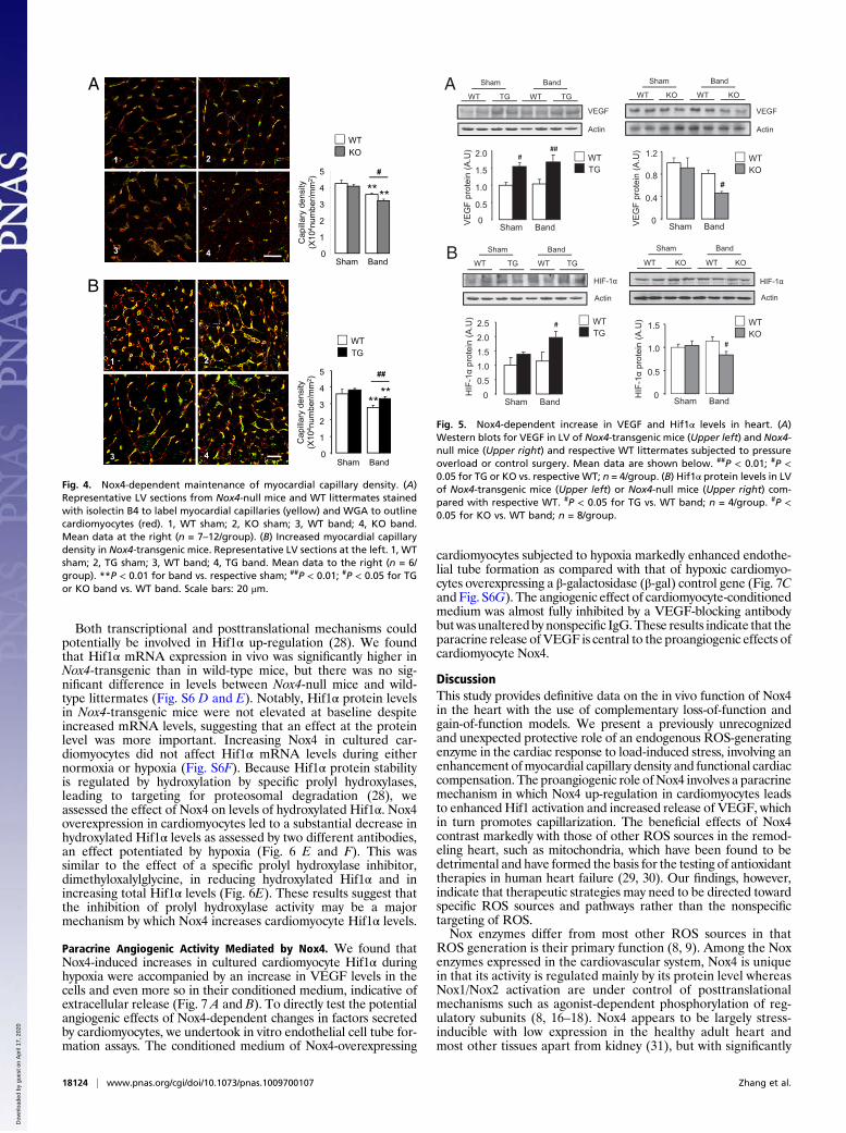
Fig. 5. Nox4-dependent increase in VEGF and Hif1α levels in heart. (A)Western blots for VEGF in LV of Nox4-transgenic mice (Upper left) and Nox4-null mice (Upper right) and respective WT littermates subjected to pressureoverload or control surgery. Mean data are shown below. ##P < 0.01; #P <0.05 for TG or KO vs. respective WT; n = 4/group. (B) Hif1α protein levels in LVof Nox4-transgenic mice (Upper left) or Nox4-null mice (Upper right) com-pared with respective WT. #P < 0.05 for TG vs. WT band; n = 4/group. #P <0.05 for KO vs. WT band; n = 8/group.
B
1 2
3 4
Cap
illary
den
sity
(Х
104 n
umbe
r/mm
2 )
**
#
**
0
1
2
3
4
5
Sham Band
A
WTKO
Cap
illary
den
sity
(Х
104 n
umbe
r/mm
2 )
****
##
1 2
3 4Sham Band
WTTG
0
1
2
3
4
5
Fig. 4. Nox4-dependent maintenance of myocardial capillary density. (A)Representative LV sections from Nox4-null mice and WT littermates stainedwith isolectin B4 to label myocardial capillaries (yellow) and WGA to outlinecardiomyocytes (red). 1, WT sham; 2, KO sham; 3, WT band; 4, KO band.Mean data at the right (n = 7–12/group). (B) Increased myocardial capillarydensity in Nox4-transgenic mice. Representative LV sections at the left. 1, WTsham; 2, TG sham; 3, WT band; 4, TG band. Mean data to the right (n = 6/group). **P < 0.01 for band vs. respective sham; ##P < 0.01; #P < 0.05 for TGor KO band vs. WT band. Scale bars: 20 μm.
18124 | www.pnas.org/cgi/doi/10.1073/pnas.1009700107 Zhang et al.
Dow
nloa
ded
by g
uest
on
Apr
il 17
, 202
0

increased levels after stresses such as pressure overload orhypoxia. Our findings that Nox4-null mice have no obvious ab-normalities in the absence of stress are consistent with the no-tion of stress inducibility. Nox4 differs from Nox2 in two otherimportant respects: First, its subcellular location in car-diomyocytes is in or associated with the perinuclear endoplas-mic reticulum whereas activated Nox2 is found predominantly atthe cell membrane (32). Second, several recent independentreports indicate that Nox4 generates predominantly H2O2 (asfurther confirmed in the current study) whereas Nox2 primarilygenerates O2
− (refs. 17–20). These differences in regulation,activation mechanism, subcellular location, and ROS generationtranslate into isoform-specific actions in isolated cellular models(9, 19, 22, 33). In the heart, previous studies in mouse models ofdefective Nox2 activation or its deletion showed that Nox2 isdetrimental during remodeling, causing increased hypertrophy,apoptosis, and contractile dysfunction (10–15). The currentresults on Nox4, taken together with previous data on Nox2,indicate that the two isoforms contrast markedly in their effectson cardiac remodeling, with Nox2 being detrimental and Nox4beneficial.The major mechanism underlying the beneficial effects of Nox4
during load-induced stress is an increase in myocardial capillaries,with capillary density being impaired in Nox4-null animals butbetter preserved in Nox4-transgenic mice compared with wildtype. Myocardial angiogenesis is tightly coupled to cardiomyocytegrowth during heart development (34) and is also an importantdeterminant of the response to disease-causing stresses such aspressure overload (24–27). Stress-induced angiogenesis has beenshown to be underpinned by the release of VEGF from car-diomyocytes to exert paracrine effects on adjacent vessels (25).
Myocardial VEGF production is under the control of transcrip-tion factors such as Hif1 and GATA4 (26, 27), but the upstreamsignals regulating activation of these factors during cardiac stressremain unclear. The results reported here suggest that stress-induced increase in Nox4 levels is a key mechanism that enhancescardiomyocyte Hif1α levels during overload, in turn leading toVEGF release and an increase in angiogenic capacity. Thisfacilitates functional compensation of the heart, which is man-ifested as better-preserved contractile function and a reducedextent of cardiac hypertrophy, fibrosis, and dilatation. Hif1α levelsare the primary regulator of Hif1 transcriptional activity throughdimerization with Hif1β and the recruitment of coactivators (28).Although Hif1α levels may be influenced by changes in mRNAexpression, the major regulatory mechanism is via oxygen-dependent hydroxylation of Hif1α protein by prolyl hydroxylases,which results in its targeting for proteosomal degradation. Hy-droxylase activity is inhibited during hypoxia, leading to increasedHif1α levels. Although we found an increase in Hif1α mRNAlevels inNox4-transgenic mice, the dominant mechanism by whichNox4 increased Hif1α appeared to be at the protein level. As-sessment of Hif1α hydroxylation indicated that Nox4 may act byinhibition of prolyl hydroxylase activity, thereby stabilizing Hif1αand increasing protein levels. Interestingly, previous studies havereported that ROS may increase Hif1α mRNA expression invascular cells (35) as well as inhibit Hif1 prolyl hydroxylases intumor cells (36).In contrast to load-induced cardiac stress or ischemia, angio-
genesis is detrimental in cancer by promoting tumor growth, andanti-angiogenic therapies are considered a promising strategy.Additionally, Nox4 reportedly has prosurvival effects in certaintumors and is thought to be a suitable therapeutic target (37, 38).Our results suggest, however, that caution should be exercised inusing Nox4-targeted cancer therapy in patients with cardiacoverload (e.g., hypertension) to avoid cardiotoxicity. In this study,we used a mouse model with global Nox4 deficiency so that partof the detrimental effect observed in pressure overloaded Nox4-null mice could potentially be due to loss of Nox4 in non-cardiomyocytes—in, for example, other cardiac cell types or organssuch as the kidneys—although the data in cardiomyocyte-specificNox4-transgenics argue against this. Regardless of this point, thecurrent results with global Nox4-null mice may be predictive ofpotential side effects with systemic Nox4-inhibitor therapy.Despite treatments such as beta blockers and angiotensin-con-
verting enzyme inhibitors that decrease mortality in heart failurepatients, prognosis remains poor. New therapeutic strategies thatcan impact on disease mechanisms are therefore needed. ROSimbalance has long been recognized as potentially important in theremodeling and failing heart, but its therapeutic targeting hasproven elusive. The present study indicates that ROS have not only
AHIF-1α
Actin
β-gal Nox4 β-gal Nox4
Normoxia Hypoxia
0 20 40Nox4
Actin
(MOI)
HIF-1α
Histone
0 2 4 0 2 4 h CoCl2
β-gal Nox4
Nox4
Nox4
Actin
β-gal
HIF-1α
Histone
0 2 4 16 0 2 4 16 h CoCl2
Scramble Nox4 siRNA
Nox4
Actin
Scb Nox4 siRNAHIF-1α
Histone
B
C D
EOH-HIF1α(P402)
OH-HIF1α(P564)
HIF-1α
Actin
DPICatalaseCoCl2+ + + +
++
β-gal Nox4 β-gal Nox4 DMOG
+ Hypoxia+02468
101214
P402 P564
OH
-Hif1
α/ H
IF1α
(A.U
)F
1 2 3 4 5 1 2 3 4 5
**
#
Fig. 6. Enhancement of cardiomyocyte Hif1α and VEGF by Nox4. Nox4-de-pendent regulation of Hif1α protein levels in (A) cardiomyocytes and (B–D)nuclear extracts of H9c2 cells. Doses were CoCl2 (200 μmol/L), diphenyleneiodonium (DPI; 20 μmol/L), and PEG-catalase (catalase; 125 U/mL). Scb: scram-bled siRNA. (E) Effect of β-gal control, Nox4, and a Hif1 prolyl hydroxylaseinhibitor, dimethyloxalylglycine (DMOG; 1 mmol/L) on Hif1α hydroxylationusing anti-hydroxylated-Pro402 and -Pro564 antibodies. Representative blots atthe left and mean data at the right. 1, β-gal; 2, Nox4; 3, β-gal hypoxia; 4, Nox4hypoxia; 5, DMOG. *P < 0.05 for all groups vs. β-gal control; #P < 0.05 vs. β-galhypoxia. Hydroxylated Hif1α was expressed relative to total HIF1α. All blotsare representative of three or more independent experiments.
0
100
200
300
400**
Tube
leng
th (%
of C
tl)
B**
CBVEGFActin
Ctl Nox4 Ctl Nox4Normoxia Hypoxia
VEGF
A
VE
GF
leve
l (A
.U)
0
1
2
3 *
VE
GF
leve
l (A
.U)
**
0
2
4
6
8 ##
β-galNox4VEGF ABIgG
+
++
+ + +
Normoxia Hypoxia Normoxia Hypoxia
CtlNox4
CtlNox4
CCtl Nox4 Ctl Nox4Normoxia Hypoxia
Fig. 7. Nox4-dependent enhancement of angiogenic response. Immunoblot-ting for VEGF in (A) cardiomyocytes and (B) culture supernatants of car-diomyocytes overexpressing Nox4 or a β-gal control (Ctl). Actin and Coomassieblue (CB) staining were used to confirm equal protein loading. Mean data areshownbelow. **P< 0.01; *P< 0.05 forNox4hypoxia vs. normoxia; ##P< 0.01 forNox4 vs. β-gal; n = 3/group. (C) Mean data showing effects of conditionedmedia on tube formation. **P < 0.01 vs. β-gal control (Ctl) or vs. Nox4 over-expression + VEGF antibody; n = 4/group.
Zhang et al. PNAS | October 19, 2010 | vol. 107 | no. 42 | 18125
MED
ICALSC
IENCE
S
Dow
nloa
ded
by g
uest
on
Apr
il 17
, 202
0

detrimental but also beneficial effects in the remodeling heartdepending upon the source and suggests that specific targeting ofan individual ROS source linked to an adaptive and potentiallydisease-preventing pathway (i.e., myocardial angiogenesis) may bea useful approach. Our results also have wider relevance to theuse of nonspecific antioxidant approaches in human diseases andmay provide an explanation for the failure of such strategies inmany settings.
MethodsDetailed methods are provided in SI Methods.
Gene-Modified Mice. Nox4-nullmiceweregeneratedby targeteddeletionof thetranslation initiation siteandexons1and2of thegene (Fig. S1B). Cardiomyocyte-targeted Nox4-transgenic mice were generated using the mouse Nox4 cDNAdownstream of the mouse α-myosin heavy chain promoter. All lines were back-crossed >10 generations onto a C57BL/6 background.
Animal Studies. Procedures were performed in accordance with the Guidanceon the Operation of the Animals (Scientific Procedures) Act, 1986 (UnitedKingdom). Aortic constriction was induced by suprarenal banding (11).
Histology. FITC-conjugated wheat germ agglutinin (WGA) was used to outlinecardiomyocytes and Picrosirius red staining to assess fibrosis (15). Capillarieswere immunostained with isolectin B4 (39).
Cell Studies. Neonatal rat cardiomyocytes were subjected to hypoxia usinga 95% N2/4% CO2/1% O2 mixture. Human umbilical vein endothelial cellsseeded on Matrigel-coated slides were used for tube formation assays.
Detection of ROS. H2O2 levels were detected with a homovanillic acid assay(22). EPR was used to measure O2
− generation by heart particulate fractions,using a 5,5-dimethylpyrroline-N-oxide (DMPO, 50 mmol/L) spin trap (40).
Statistics. All data are presented as mean ± SEM. Comparisons were un-dertaken by Student’s t test or one-way ANOVA, as appropriate, followed bya post hoc Tukey’s test. P < 0.05 was considered significant.
ACKNOWLEDGMENTS. We thank M. Mayr for advice on tube formationassays. This study was supported by the British Heart Foundation (Grants RG/08/011/25922, CH/99001, and RE/08/003), the Leducq Foundation, by EUGe-neHeart (EU FP6 Grant LSHM-CT-2005-018833), by the German Search Foun-dation (Grants SFB815 TPA1, SFB834 TPA2), and by the Excellence ClusterCardio-Pulmonary System and Goethe University.
1. Mudd JO, Kass DA (2008) Tackling heart failure in the twenty-first century. Nature451:919–928.
2. Catalucci D, Latronico MV, Ellingsen O, Condorelli G (2008) Physiological myocardialhypertrophy: How and why? Front Biosci 13:312–324.
3. Heineke J, Molkentin JD (2006) Regulation of cardiac hypertrophy by intracellularsignalling pathways. Nat Rev Mol Cell Biol 7:589–600.
4. Giordano FJ (2005) Oxygen, oxidative stress, hypoxia, and heart failure. J Clin Invest115:500–508.
5. Anilkumar N, Sirker A, Shah AM (2009) Redox sensitive signaling pathways in cardiacremodeling, hypertrophy and failure. Front Biosci 14:3168–3187.
6. Rhee SG, et al. (2005) Intracellular messenger function of hydrogen peroxide and itsregulation by peroxiredoxins. Curr Opin Cell Biol 17:183–189.
7. Saini HK, Machackova J, Dhalla NS (2004) Role of reactive oxygen species in ischemicpreconditioning of subcellular organelles in the heart. Antioxid Redox Signal 6:393–404.
8. Lambeth JD (2004) NOX enzymes and the biology of reactive oxygen. Nat RevImmunol 4:181–189.
9. Brown DI, Griendling KK (2009) Nox proteins in signal transduction. Free Radic BiolMed 47:1239–1253.
10. Bendall JK, Cave AC, Heymes C, Gall N, Shah AM (2002) Pivotal role of a gp91(phox)-containing NADPH oxidase in angiotensin II-induced cardiac hypertrophy in mice.Circulation 105:293–296.
11. Byrne JA, et al. (2003) Contrasting roles of NADPH oxidase isoforms in pressure-overload versus angiotensin II-induced cardiac hypertrophy. Circ Res 93:802–805.
12. Nakagami H, Takemoto M, Liao JK (2003) NADPH oxidase-derived superoxide anionmediates angiotensin II-induced cardiac hypertrophy. J Mol Cell Cardiol 35:851–859.
13. Satoh M, et al. (2006) Requirement of Rac1 in the development of cardiachypertrophy. Proc Natl Acad Sci USA 103:7432–7437.
14. Doerries C, et al. (2007) Critical role of the NAD(P)H oxidase subunit p47phox for leftventricular remodeling/dysfunction and survival after myocardial infarction. Circ Res100:894–903.
15. Looi YH, et al. (2008) Involvement of Nox2 NADPH oxidase in adverse cardiacremodeling after myocardial infarction. Hypertension 51:319–325.
16. Ambasta RK, et al. (2004) Direct interaction of the novel Nox proteins with p22phox isrequired for the formation of a functionally active NADPH oxidase. J Biol Chem 279:45935–45941.
17. Martyn KD, Frederick LM, von Loehneysen K, Dinauer MC, Knaus UG (2006)Functional analysis of Nox4 reveals unique characteristics compared to other NADPHoxidases. Cell Signal 18:69–82.
18. Serrander L, et al. (2007) NOX4 activity is determined by mRNA levels and revealsa unique pattern of ROS generation. Biochem J 406:105–114.
19. Dikalov SI, et al. (2008) Distinct roles of Nox1 and Nox4 in basal and angiotensin II-stimulated superoxide and hydrogen peroxide production. Free Radic Biol Med 45:1340–1351.
20. Nisimoto Y, Jackson HM, Ogawa H, Kawahara T, Lambeth JD (2010) ConstitutiveNADPH-dependent electron transferase activity of the Nox4 dehydrogenase domain.Biochemistry 49:2433–2442.
21. Chen K, Kirber MT, Xiao H, Yang Y, Keaney JF, Jr (2008) Regulation of ROS signaltransduction by NADPH oxidase 4 localization. J Cell Biol 181:1129–1139.
22. Anilkumar N, Weber R, Zhang M, Brewer A, Shah AM (2008) Nox4 and nox2 NADPH
oxidases mediate distinct cellular redox signaling responses to agonist stimulation.
Arterioscler Thromb Vasc Biol 28:1347–1354.23. Helmcke I, Heumüller S, Tikkanen R, Schröder K, Brandes RP (2009) Identification of
structural elements in Nox1 and Nox4 controlling localization and activity. Antioxid
Redox Signal 11:1279–1287.24. Hilfiker-Kleiner D, et al. (2004) Signal transducer and activator of transcription 3 is
required for myocardial capillary growth, control of interstitial matrix deposition, and
heart protection from ischemic injury. Circ Res 95:187–195.25. Shiojima I, et al. (2005) Disruption of coordinated cardiac hypertrophy and
angiogenesis contributes to the transition to heart failure. J Clin Invest 115:
2108–2118.26. Sano M, et al. (2007) p53-induced inhibition of Hif-1 causes cardiac dysfunction during
pressure overload. Nature 446:444–448.27. Heineke J, et al. (2007) Cardiomyocyte GATA4 functions as a stress-responsive
regulator of angiogenesis in the murine heart. J Clin Invest 117:3198–3210.28. Semenza GL, Prabhakar NR (2007) HIF-1-dependent respiratory, cardiovascular, and
redox responses to chronic intermittent hypoxia. Antioxid Redox Signal 9:1391–1396.29. Ide T, et al. (1999) Mitochondrial electron transport complex I is a potential source of
oxygen free radicals in the failing myocardium. Circ Res 85:357–363.30. Schriner SE, et al. (2005) Extension of murine life span by overexpression of catalase
targeted to mitochondria. Science 308:1909–1911.31. Geiszt M, Kopp JB, Várnai P, Leto TL (2000) Identification of renox, an NAD(P)H
oxidase in kidney. Proc Natl Acad Sci USA 97:8010–8014.32. Heymes C, et al. (2003) Increased myocardial NADPH oxidase activity in human heart
failure. J Am Coll Cardiol 41:2164–2171.33. Wu RF, Ma Z, Myers DP, Terada LS (2007) HIV-1 Tat activates dual Nox pathways
leading to independent activation of ERK and JNK MAP kinases. J Biol Chem 282:
37412–37419.34. Giordano FJ, et al. (2001) A cardiac myocyte vascular endothelial growth factor
paracrine pathway is required to maintain cardiac function. Proc Natl Acad Sci USA
98:5780–5785.35. Bonello S, et al. (2007) Reactive oxygen species activate the HIF-1α promoter via
a functional NFkappaB site. Arterioscler Thromb Vasc Biol 27:755–761.36. Gerald D, et al. (2004) JunD reduces tumor angiogenesis by protecting cells from
oxidative stress. Cell 118:781–794.37. Vaquero EC, Edderkaoui M, Pandol SJ, Gukovsky I, Gukovskaya AS (2004) Reactive
oxygen species produced by NAD(P)H oxidase inhibit apoptosis in pancreatic cancer
cells. J Biol Chem 279:34643–34654.38. Mochizuki T, et al. (2006) Inhibition of NADPH oxidase 4 activates apoptosis via the
AKT/apoptosis signal-regulating kinase 1 pathway in pancreatic cancer PANC-1 cells.
Oncogene 25:3699–3707.39. Hilfiker-Kleiner D, et al. (2007) A cathepsin D-cleaved 16 kDa form of prolactin
mediates postpartum cardiomyopathy. Cell 128:589–600.40. Janiszewski M, et al. (2005) Regulation of NAD(P)H oxidase by associated protein
disulfide isomerase in vascular smooth muscle cells. J Biol Chem 280:40813–40819.
18126 | www.pnas.org/cgi/doi/10.1073/pnas.1009700107 Zhang et al.
Dow
nloa
ded
by g
uest
on
Apr
il 17
, 202
0