myxofibrosarcoma of the mandible: a case report and review
TRANSCRIPT

CASE REPORT Open Access
Myxofibrosarcoma of the mandible: a casereport and review of the literatureZhengqiang Li, Xianwen Liu, Quanyin Zhang, Jie Zhang, Mingyi Huang and Shuguang Liu*
Abstract
Background: Myxofibrosarcoma (MFS) is a soft tissue sarcoma that commonly occurs in late adult life. It is mainlylocated in the subcutaneous soft tissues of extremities characterized by a high recurrence rate at the original site.MFS of the head and neck is rare, while it occurs in the maxilla and mandible is extremely rare.
Case presentation: We report a case of MFS of the mandible in a 51-year-old female who presented with apainless gingival swelling and mobile, super-erupted right mandibular second and third molars. Panoramic x-rayand maxillofacial CT revealed an ill-defined radiolucent lesion surrounding the mandibular molars giving a teeth-floating-in-air appearance. Histopathological examination showed scattered spindle and stellate cells with mildatypia distributed in the myxoid stroma. Only a few mitotic figures were identified and no area of tissue necrosiswas found. The characteristic thin-walled and curvilinear vasculature were prominent. Immunohistochemistryanalysis revealed the tumor cells being positive for vimentin and vascular CD31. CK, S-100, P63, HHF-35 stains werenegative. The labeling index of Ki-67 was about 30%. Based on the histopathological and immunohistochemicalexaminations, the diagnosis of a low-grade MFS was established. This patient underwent a radical segmentalexcision with a 2-cm margin, supraomohyoid neck dissection and immediate reconstruction of the mandibularcontinuity defect with a fibular osteocutaneous free flap. This patient has been followed for 20 months to date andhas remained disease free.
Conclusions: This report describes a rare case of MFS of the mandible. Recognizing the histopathological featuresof MFS and applying the appropriate immunohistochemical examinations are crucial in establishing the correctdiagnosis. Our case may provide diagnosis and treatment experiences of MFS occurs in the mandible.
Keywords: Head and neck, Mandible, Maxilla, Soft tissue tumor, Myxofibrosarcoma
BackgroundMyxofibrosarcoma (MFS) is a fibroblast-derived sarcoma,which accounts for approximately 5–10% of all soft tissuemalignant tumors [1]. The World Health Organization(WHO) defines MFS as the malignant fibroblastic neo-plasm characterized by cellular pleomorphism, variablyprominent myxoid stroma, and prominent elongated,thin-walled stromal blood vessels [2]. The mean age in pa-tients with MFS is between the fifth and seventh decades
[3]. Although some studies show a slight male predomin-ance [4], the current evidence suggests no significant gen-der predilection [5]. About 77% of MFS cases occur in theextremities with a predilection for the upper extremities.Other areas of the body including the trunk (12%), retro-peritoneum or mediastinum (8%) [4, 6], abdominal wall[7], heart [8] have also been reported. As a soft tissuetumor that mainly occurs in the subcutaneous tissue, MFSof the head and neck is quite rare. This case report de-scribes the clinical features, histopathological and immu-nohistochemical examinations and treatment experiencein managing this rare sarcoma.
© The Author(s). 2020 Open Access This article is licensed under a Creative Commons Attribution 4.0 International License,which permits use, sharing, adaptation, distribution and reproduction in any medium or format, as long as you giveappropriate credit to the original author(s) and the source, provide a link to the Creative Commons licence, and indicate ifchanges were made. The images or other third party material in this article are included in the article's Creative Commonslicence, unless indicated otherwise in a credit line to the material. If material is not included in the article's Creative Commonslicence and your intended use is not permitted by statutory regulation or exceeds the permitted use, you will need to obtainpermission directly from the copyright holder. To view a copy of this licence, visit http://creativecommons.org/licenses/by/4.0/.The Creative Commons Public Domain Dedication waiver (http://creativecommons.org/publicdomain/zero/1.0/) applies to thedata made available in this article, unless otherwise stated in a credit line to the data.
* Correspondence: [email protected] of Oral and Maxillofacial Surgery, Stomatological Hospital ofSouthern Medical University, 366 south of Jiangnan Road, Guangzhou510280, China
Li et al. BMC Oral Health (2020) 20:113 https://doi.org/10.1186/s12903-020-01094-7

Case presentationThis is a 51-year-old female who presented with a three-month history of an enlarging gingival mass of the rightmandible. Head and neck evaluation revealed no facialasymmetry, cervical lymphadenopathy nor trismus. She de-nied pain, sensory alterations or bleeding associated withthe lesion. The right mandibular second and third molarswere super-erupted, grossly mobile, but not painful to pal-pation. The exophytic gingival lesion was covered with ayellowish pseudomembrane (Fig. 1). A slight cortical expan-sion was noted at the buccal vestibule. An outside dentalprovider performed an incisional biopsy of the gingival le-sion prior to referring her to our institution for a higherlevel of care. The incisional biopsy performed suggestedpyogenic granuloma as the diagnosis (Fig. 2a, b).On admission, imaging studies including panoramic
radiograph and maxillofacial computed tomography (CT)without contrast were obtained. An ill-defined radiolucentlesion involving the right mandibular angle was noted.The lesion involved the entire bucco-lingual width of theright mandibular angle with extension through the lingualcortex. The right mandibular second and third molarswere both super-erupted with a teeth-floating-in-air ap-pearance (Fig. 3a, b, c, d). The outlines of the mandibularcanal were obscured by the intraosseous lesion.Incisional biopsies of the gingival mass and the right
mandibular lesion were performed under local anesthesia.Histopathology findings of the gingival lesion are shownin Fig. 4a. The gingival squamous epithelium was infil-trated with a large amount of plasma cells, neutrophilsand other inflammatory cells. Abundant inflammatory ex-udation and fibrous tissue proliferation were also identi-fied. Vascular cellulosic necrosis or granuloma was notobvious. PAS (Periodic Acid-Schiff stain) and methena-mine silver stain showed no fungal organisms (figures not
shown). These findings suggested chronic suppurative in-flammation of the gingival mass. The cut surface of themandibular lesion revealed an intraosseous nodular lesion.Histopathological exam of the mandibular biopsy showedscattered spindle and stellate cells with hyperchromatic nu-clei distributed in a mucous matrix. Mildly atypical and mi-totic figures were seen, while necrosis was not present (Fig. 4b, c). Immunohistochemistry showed that the tumor cellswere positive for vimentin (Fig. 4 d). Vascular CD31 stainshowed the characteristic thin-walled and curvilinear vascu-lature (Fig. 4 e). The labeling index of Ki-67 was about 30%(Fig. 4 f). The tumor cells were negative for CK, S-100, P63,HHF-35 (figures not shown). Based on the clinical presenta-tion, radiographic findings, histopathological and immuno-histochemical examinations, the diagnosis of a low-grademyxofibrosarcoma of the mandible was made.This patient was taken to the operating room for radical
segmental resection of the right mandibular MFS with 2cm margins, right supraomohyoid neck dissection, andimmediate reconstruction with a left fibular ostocutaneousfree flap. Due to the size of the lesion, a right mandibularhemimandibulectomy with disarticulation of the man-dibular condyle was performed. The resected tissues weresubmitted for histopathologic and immunohistochemicalexaminations. None of the resected cervical lymph nodeswere positive for malignancy. This patient was followedclosely in the past twenty months and no local recurrencenor distant metastasis was detected to date (Fig. 5).
Discussion and conclusionsThe term myxofibrosarcoma (MFS) was first reported inthe literature in 1951 [9]. It wasn’t until 1977, whenAngervall et al. reported 30 cases of MFS that MFS be-came a clinically distinct diagnostic entity. According tothe degree of cellularity, nuclear pleomorphism, and mi-totic activity, MFS was categorized into four grades: I-IV[10]. In 1996, Mentzel et al. classified the tumor intolow, intermediate, and high-grade MFS [11]. The 2002third edition of the WHO Classification of Soft TissueTumors classified MFS as malignant fibroblastic/myofi-broblastic tumors. Myxoid Malignant Fibrous Histiocy-tomas, for the first time, were considered a form of MFS[12]. The fourth edition WHO Classification of Soft Tis-sue Tumors regarding MFS was published in 2013 andthe classification remained unchanged to date [13].As a soft tissue sarcoma, MFS mainly occurs in the der-
mis or subcutaneous tissue, and occasionally occurs in thesubfascial or intramuscular tissue [14]. The incidence ofMFS in the head and neck region is quite rare [15]. Thereported cases of MFS in the head and neck region weremostly in the soft tissues, such as the Schneiderian mem-brane of the maxillary sinus [16], lining of the sphenoidsinus [17], parotid gland [18], thyroid gland [19], andpharynx [20]. MFS occurring intra-osseously in the head
Fig. 1 preoperative examinations. The figure showing apparentswelling of the gingiva lesion (the white box)
Li et al. BMC Oral Health (2020) 20:113 Page 2 of 8

and neck region is exceedingly rare. To the best of ourknowledge, only two cases of intraosseous MFS were re-ported in the maxilla [1, 21] and four cases reported in themandible [22–25] (Table 1). Most MFS tumor cells havethe ultrastructural characteristics of a tumor of fibroblastdifferentiation and secretory activity. They commonly ori-ginate from fibrous connective tissue in the soft tissue, butthe origin of MFS in bone tissue remains unclear. It isspeculated that MFS may originate from the fibrous con-nective tissue in the endosteum or the mesenchymal tissueduring tooth germ development.
Like most soft tissue sarcomas, the pathogenesis ofMFS is still unclear. It is suspected that multiple factorscontribute to the development of MFS. Current researchmainly focuses on the genetic characteristics of soft tis-sue sarcomas. It is found that MFS has a highly complexkaryotype and shows gains and losses of numerous chro-mosomes or chromosome regions. Therefore, it is specu-lated that the pathogenesis of MFS may be related tochromosome abnormalities [26].The clinical courses of MFS vary significantly and no
specific clinical features can be identified. It often
Fig. 2 Hematoxylin-Eosin Staining of the gingiva lesion in pre-admission. The two images of a (×100) and b (×200) were suspicious for adiagnose of granulomatous lesion
Fig. 3 Preoperative images of radiological examinations. Panoramic radiograph (a) and computed tomography (b, c, d) showing a large area ofright mandibular destruction (the red box)
Li et al. BMC Oral Health (2020) 20:113 Page 3 of 8

presents as a painless, slow-growing tumor. Low-gradeMFS often demonstrates expansive growth, while high-grade MFS often shows local invasion or compression ofthe surrounding anatomical structures. High-grade MFSinvolving the airway or the major vasculature of the headand neck can lead to life-threatening complications.The tumor in this case caused aggressive destruction of
the right mandible, but without significant cortical expan-sion leading to facial asymmetry. The patient did not ex-perience sensory alteration with the tumor involving themandibular canal. The predominant symptom was thelocal swelling of the overlying gingiva. The referring den-tist initially suspected inflammatory lesion of the oral mu-cosa, so radiographic examinations were not performed.The pathology report of the initial gingival biopsy sug-gested pyogenic granuloma. It wasn’t until the patient pre-sented to our department (Department of Oral andMaxillofacial Surgery, Stomatological Hospital of SouthernMedical University) for treatment, the necessary diagnos-tic radiographic examinations were performed. The
radiographic findings prompted us to perform incisionalbiopsies of the gingival mass and mandibular lesion. Thesurgical pathology report supported the diagnosis of low-grade MFS of the mandible and chronic suppurative in-flammation of the overlying gingiva.MFS is associated with a relatively high rate of local
recurrence or distant metastasis. Accurate preoperativediagnosis and proper treatment strategies are critical inmanaging patients with MFS. Preoperative imaging ex-aminations including ultrasound, CT, and MRI can beused to determine the anatomical extent of the lesion in-volvement. Histopathologic examinations are consideredthe gold standard in establishing the diagnosis of MFS.MFS is more likely to arise superficially in the subcuta-
neous tissue or dermis, than in deep soft tissues, such asthe sub-fascial or intramuscular tissues. On gross exam-ination, the superficial MFS tend to present as multinod-ular lesions along the epidermis or dermis anddemonstrate variability in the gelatinous cut surfaces.The deep tissue MFS, on the other hand, often presents
Fig. 4 Hematoxylin-Eosin Staining and immunohistochemical staining after admission. Hematoxylin-Eosin Staining showing chronic inflammationof the gingiva lesion (a, ×200) and low-grade myxofibrosarcoma of the right mandible (b, ×100, c, ×200), immunohistochemical staining showingvimentin positive (d, ×200), CD31 positive (e, ×200), and Ki-67 positive (f, ×200)
Li et al. BMC Oral Health (2020) 20:113 Page 4 of 8
as a solitary mass with a grayish, fish-meat appearancewith infiltrative borders.Although the microscopic morphologies of MFS vary,
there are some common histopathological features ofMFS. These commonalities include varying degrees ofmyxoid (hyaluronic acid) matrixes composed of spindleto stellate cells that demonstrate nuclear atypia andpleomorphism, and curvilinear vasculature. According tothe spectrum of cellularity, cytologic atypia, and mitoticactivity, MFS is categorized into low, intermediate andhigh-grade.In low-grade MFS, the tumor shows characteristics such
as hypocellularity and prominent myxoid stroma on hist-ology. Tumor cells are scattered and non-cohesive. Theyoften present as plump spindle or stellate shaped cells withill-defined and slightly eosinophilic cytoplasm and hyper-chromatic nuclei arranged disorderly or in bundles. Mitoticfigures are uncommon and tumor necrosis cannot be seen.In some cases, hyperchromatic multi-nuclear tumor cellscan be seen. Occasionally, vacuolar and acidic mucous-filled pseudolipoblasts can also be seen, which are positiveon AB-PAS stain. The essence of pseudoadipocyte is fibro-blast, and there are no fat droplets in the cytoplasm. Thecharacteristic thin-walled and curvilinear vasculature are
prominent. In a few cases, the vasculature is clustered orbranched. A variable number of inflammatory cells andtumor cells can be seen around the blood vessels [27, 28].High-grade MFS is either partially or predominantly
composed of solid sheets and cellular fascicles of spindleand pleomorphic tumor cells with marked nuclear andcytologic pleomorphism. Bizarre and multinucleatedtumor giant cells with abundant eosinophilic cytoplasmand irregularly shaped nuclei are common. Mitotic fig-ures, atypical cells, hemorrhage and coagulative necrosisare also common. Features such as prominent myxoidmatrix and numerous curvilinear vessels can be foundfocally. Mucous degeneration and rich, slender capillarynetworks can be seen in the tumor stroma [27, 28].The histopathological features of intermediate-grade
MFS are between the low and high-grade MFS.Intermediate-grade MFS is more cellular and pleomorphicthan the low-graded tumors. It lacks the extensive solid/sheet-like areas, pronounced cellular pleomorphism, andnecrosis found in high-grade MFS. Mitoses are more com-monly found than in low-grade tumors. Curvilinear vascu-lature is often prominent [27, 28].In immunohistochemistry, the tumor cells of MFS are
positive for vimentin. MSA and α-SMA can be focally
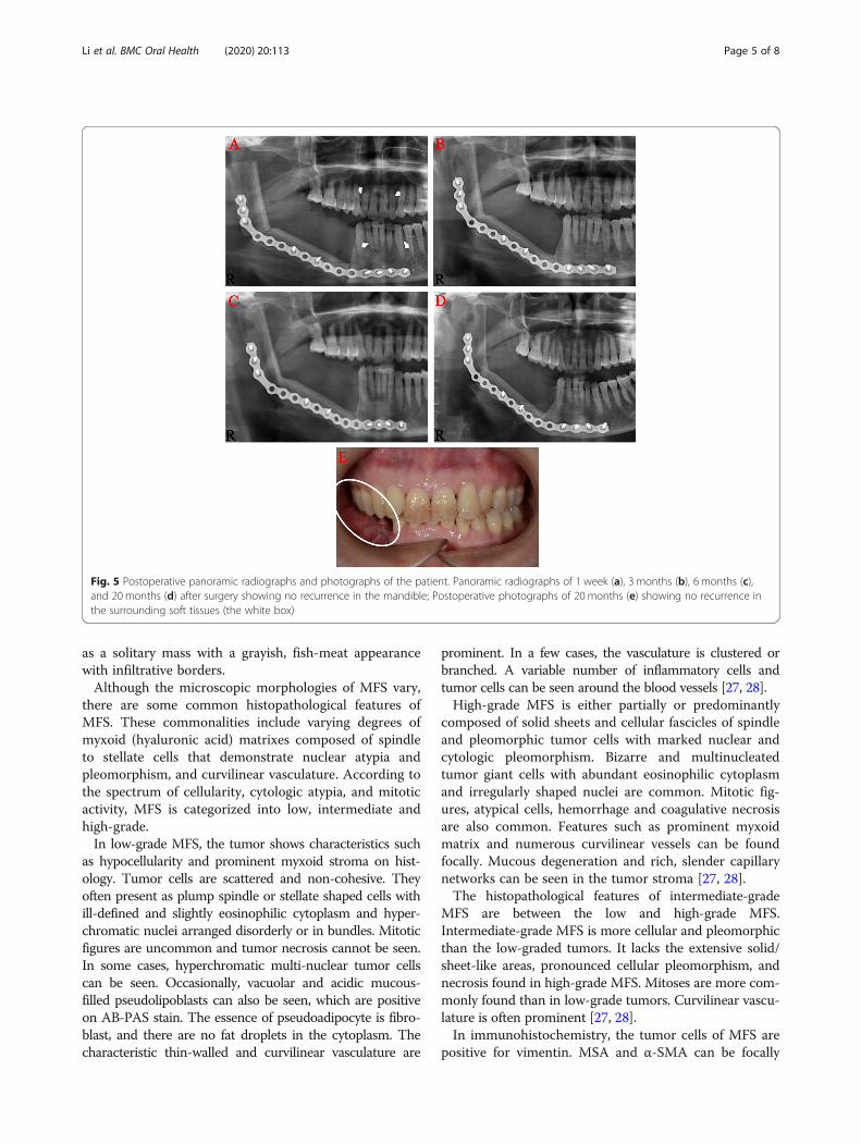
Fig. 5 Postoperative panoramic radiographs and photographs of the patient. Panoramic radiographs of 1 week (a), 3 months (b), 6 months (c),and 20 months (d) after surgery showing no recurrence in the mandible; Postoperative photographs of 20 months (e) showing no recurrence inthe surrounding soft tissues (the white box)
Li et al. BMC Oral Health (2020) 20:113 Page 5 of 8

Table
1Cases
ofmyxofibrosarcom
ain
themaxillaandmandible
Caserepo
rtAge
(years)/Sex
Locatio
nChief
complaint
Immun
ohistochem
icalfeatures
Diagn
osis
Treatm
ent
Follow-up
Nakaharaet
al.[1]
52/M
Maxilla
Che
ekdiscom
fortand
expansionof
theup
per
right
gumsfor3mon
ths
Pre-op
eration
myelin
(±),synaptop
hysin(−),
desm
in(−),s-100(−)
Inflammation
reactio
nSurgery,po
st-
operative
radiothe
rapy
Aliveafter
17mon
ths
Pre-op
eration
vimen
tin(+),Alcianblue
(±),
cytokeratin
(−),s-100(−)
MFS
Post-ope
ratio
nvimen
tin(+),CKAE1/3
(−),
CK7(−),CK20
(−),S-100(−),
CK34βE12
(−),de
smin
(−),a-SM
A(−),
ALK-1
(−)
MFS
Quimby
etal.[22]
72/F
Maxilla
Arapidlygrow
ingsofttissue
massin
thepo
steriorleft
maxilla
Pre-op
eration
NO
Perip
heralg
iant
cellgranulom
aPre-op
erative
radiothe
rapy,
surgery
Unkno
wn
Pre-op
eration
NO
Spindlecell
sarcom
a
Post-ope
ratio
nNO
MFS
Kummoo
naet
al.[23]
35/M
Mandible
Num
bnessof
theleftlower
lipof
2mon
ths,mandilbe
swelling
Pre-op
eration
NO
MFS
Surgery
Aliveafter
24mon
ths
Post-ope
ratio
nNO
MFS
Kargahietal.[24]
61/M
Mandible
Thefourth
timeof
local
recurren
cePre-op
eration
NO
Unkno
wn
Incomplete
resection
Aliveafter
10mon
ths
Post-ope
ratio
nvimen
tin(+),S100
(−),CK(−),Ki67
(1%)
MFS
Zoulou
miset
al.[25]
23/M
Mandible
2-mon
thshistoryof
asw
ellingon
theleftside
ofthelower
face
Pre-op
eration
NO
Unkno
wn
Surgery
Aliveafter
39mon
ths
Post-ope
ratio
nNO
MFS
Park
etal.[26]
59/M
Mandible
Pain
ofpo
steriorleft
mandible
Pre-op
eratio
NO
Myxofibroma
Surgery,chem
o-radiothe
rapy
Aliveafter
12mon
ths
Post-ope
ratio
nNO
MFS
Li et al. BMC Oral Health (2020) 20:113 Page 6 of 8

positive, which indicate myofibroblast differentiation.CD31 is positive in tumor vasculature. S-100, desmin,caldesmon, keratin and histocyte markers such as CD68,Mac387, XIIIa are negative. Ki-67 is partially positive.Ki-67 has a positive correlation with tumor recurrenceand can be used as an indicator of recurrence [27, 28].Based on the clinical and radiographic findings de-
scribed in this case report, the differential diagnoses in-clude low-grade malignant fibromyxoid sarcoma, myxoidliposarcoma, myxoma, Nodular fasciitis, Spindle cell lip-oma, and nerve sheath myxoma [27, 28]. Histopatho-logical and immunohistochemical examinations areessential in establishing the correct diagnosis.Although there are some variations in management of
MFS, the primary treatment approach is radical surgicalresection including a wide margin of adjacent disease-free tissue. Preoperative maxillofacial CT is necessary todetermine the anatomical extent of the tumor and in-volvement of the surrounding tissue. It is generally be-lieved that resection of at least 2 cm around the tumor isneeded to ensure a negative surgical margin and reducethe chance of local recurrence and distant metastasis[29]. For the surgical site defect, pedicled or free tissueflaps can be used to reconstruct the forms and functionsof the resected structures. Whether cervical lymph nodedissection is necessary for head and neck MFS patientswith clinically negative cervical lymph node remains un-clear. In order to decrease the risk of local recurrenceand distant metastasis, postoperative adjuvant treatmentsuch as radiotherapy and/or chemotherapy can be addedto the treatment regimen. In a randomized controlledtrial (RCT) involving multiple subtypes of soft tissue sar-comas, it was found that radiotherapy helped to reducethe recurrence rate of the sarcomas [30, 31]. However,there are no RCTs specifically evaluating the effect ofradiotherapy on MFS. Currently, there is no consensuson whether and when radiotherapy should be used inthe management of MFS. In cases where complete resec-tion was not possible or in patients with recurrent le-sions, radiotherapy is recommended. Chemotherapy iscommonly used in MFS patients with distant metastasis.Colia et al. found that chemotherapy agents such as thecombination of anthracycline and isocyclophosphamideincrease the survival rate in MFS patients. Other cohortstudies did not support the same conclusion [32]. Con-sidering the small sample sizes of the retrospective stud-ies and the absence of RCTs, further studies on the risksand benefits of chemotherapy are still needed.In this case report, this patient underwent radical hard
and soft tissue resection with right hemimandibulect-omy, disarticulation of the right mandibular condyle.The large mandibular defect was reconstructed with afibular osteocutaneous free flap. Due to the consider-ation that MFS can metastasize to the adjacent cervical
lymph nodes, supraomohyoid neck dissection was per-formed on the right side. The final surgical pathology re-port showed no cervical lymph node involvement,postoperative radiotherapy and chemotherapy were notprescribed.Based on the current literature, the local recurrence
rate for MFS does not correlate well with its histopatho-logical grade. Most cases of MFS are low-grade lesions,and the short-term prognosis of this subtype is generallygood. The local recurrence rate for a low-grade MFS issimilar to that of a high-grade MFS. Low-grade MFS,however, can transform into a high-grade MFS after re-currence [33]. The local recurrence rate of MFS is about16–57%. Multiple recurrences have also been reportedand the rate for multiple recurrence is estimated be-tween 25 and 52% [29]. Local recurrence has been re-ported as early as 2 months after resection and as far outas eight years postoperatively. The median recurrencetime is about 27 months [34]. A negative surgical marginis the most important prognostic factor in estimatingthe local recurrence rate [34, 35]. The prognosis is gen-erally poor for MFS that recurs within one year.Distant metastasis is rarely seen in low-grade MFS.
The rate of metastasis can be as high as 20 to 35% inintermediate and high-grade MFS. Distant metastasesusually occur in the lungs and bones. The 5-year overallsurvival rate for MFS is between 61 and 77% [34]. Me-tastasis and mortality are closely associated with thehistopathological grade of the MFS. For instance, metas-tasis is positively correlate with the depth of the tumorlocation, the size of necrosis areas, the size of tumor lar-ger than 5 cm, and mucoid area less than 75% [21]. Pa-tients with MFS need long-term follow-up to monitorfor tumor recurrence. In this patient, if local recurrenceand/or distant metastasis were detected in the future, re-operation, radiotherapy, chemotherapy or combinedtherapy will be considered.In conclusion, besides odontogenic tumors, soft tissue
malignancies such as MFS can occur in the mandibleand cause the radiographic findings described in thiscase report. Recognizing the histopathological featuresof MFS and applying the appropriate immunohisto-chemical examinations are crucial in establishing thecorrect diagnosis. Wide Surgical excision with at least 2-cm margin of adjacent normal tissue and adjuvant post-operative radiochemotherapy are the primary treatmentrecommendations in managing patients with MFS.Long-term follow up for tumor surveillance is advisablein all cases. Many clinical questions such as the patho-genesis of the disease, tumor origin, efficacy of the adju-vant chemoradiotherapy remain unanswered. Furtherstudies are necessary in improving our understanding ofthis malignancy which may help in providing better out-comes for our patients.
Li et al. BMC Oral Health (2020) 20:113 Page 7 of 8

AbbreviationsMFS: myxofibrosarcoma; WHO: The current World Health Organization;CT: computed tomography; RCT: randomized controlled trial
AcknowledgementsNot applicable.
Authors’ contributionsSL performed the clinical diagnosis, treatment and drafted the manuscript asthe corresponding author. ZL collected clinical data, conducted thehistopathological examination and was involved in writing the manuscript.XL and QZ were involved in drafting the manuscript. JZ and MH participatedin the surgery carried out in this case and performed the literature search. Allauthors read and approved the final manuscript prior to submission.
FundingNot applicable.
Availability of data and materialsNot applicable.
Ethics approval and consent to participateEthics approval was not required for this article.Consent to participate: Not applicable.
Consent for publicationWritten informed consent was obtained from the patient for publication ofher clinical details and clinical images. A copy of the consent form isavailable for review by the Editor of this journal.
Competing interestsThe authors declare that they have no competing interests.
Received: 28 November 2019 Accepted: 27 March 2020
References1. Nakahara S, Uemura H, Kurita T, et al. A case of myxofibrosarcoma of the
maxilla with difficulty in preoperative diagnosis. Int J Clin Oncol. 2012;17:390–4.
2. Gambarotti M. Myxofibrosarcoma: atlas of musculoskeletal tumors andTumorlike lesions. Cham, Heidelberg, Dordrecht, London, New York:Springer International Publishing; 2014.
3. EI Bahraouy A, Khmamouche R, Oukabli M, et al. Successful Management ofHigh Grade Myxofibrosarcoma of the forearm. J Case Reports. 2016;6:259–62.
4. Gopalratnam K, Rodriguez JA, Woodson KA, et al. A case of myxofibrosarcomain an unusual thoracic location. Case Rep Oncol. 2016;9:39–44.
5. Rongioletti F. Myxofibrosarcoma: rare malignant skin tumors. Heidelberg,Dordrecht, London, New York: Springer; 2014.
6. Hambleton C, Noureldine S, Gill F, et al. Myxofibrosarcoma with metastasisto the lungs, pleura, and mediastinum: a case report and review ofliterature. Int J Clin Exp Med. 2012;5:92–5.
7. Antbring R, Parker SG, Lordan JT, et al. High-grade myxofibrosarcoma of theabdominal wall. BMJ Case Rep. 2017;2017:1–5.
8. Lazaros GA, Matsakas EP, Madas JS, et al. Primary myxofibrosarcoma of the leftatrium: case report and review of the literature. Angiology. 2008;59:632–5.
9. HL VON. Myxofibrosarcoma of the external auditory canal. Ann Otol RhinolLaryngol. 1951;60:258–9.
10. Angervall L, Kindblom LG, Merck C. Myxofibrosarcoma: a study of 30 cases.Acta Pathol Microbiol Scand A. 1977;85:127–40.
11. Mentzel T, Calonje E, Wadden C, et al. Myxofibrosarcoma: clinicopathologicanalysis of 75 cases with emphasis on the low-grade variant. Am J SurgPathol. 1996;20:391–405.
12. Fletcher CDM. Myxofibrosarcoma: pathology and genetics of tumors of softtissue and bone, World Health Organization classification of tumors. Lyon:IARC Press; 2002.
13. Doyle LA. Sarcoma classification: an update based on the 2013 WorldHealth Organization classification of tumors of soft tissue and bone. Cancer.2014;120:1763–74.
14. Kaya M, Wada T, Nagoya S, et al. MRI and histological evaluation of theinfiltrative growth pattern of myxofibrosarcoma. Skelet Radiol. 2008;37:1085–90.
15. Odei B, Rwigema JC, Eilber FR, et al. Predictors of local recurrence inpatients with myxofibrosarcoma. Am J Clin Oncol. 2018;41:827–31.
16. Wong A, Park RCW, Mirani NM, et al. Myxofibrosarcoma of the maxillarysinus. Allergy Rhinol. 2017;8:e95–9.
17. Enomoto K, Inohara H, Hamada K, et al. FDG PET imaging ofmyxofibrosarcoma on the sphenoid sinus. Clin Nucl Med. 2008;33:421–2.
18. Srinivasan B, Ethunandan M, Hussain K, et al. Epitheloid myxofibrosarcomaof the parotid gland. Case Rep Pathol. 2011;2011:1–3.
19. Darouassi Y, Attifi H, Zalagh M, et al. Myxofibrosarcoma of the thyroid gland.Eur Ann Otorhinolaryngol Head Neck Dis. 2014;131:385–7.
20. Nishimura G, Sano D, Hanashi M, et al. Myxofibrosarcoma of thehypopharynx. Auris Nasus Larynx. 2006;33:93–6.
21. Quimby A, Estelle A, Gopinath A, et al. Myxofibrosarcoma in head and neck:case report of unusually aggressive presentation. J Oral Maxillofac Surg.2017;75:e1–e12.
22. Kummoona R. Central myxofibrosarcoma of the mandible treated by radicalresection. Oral Surg Oral Med Oral Pathol. 1975;39:713–7.
23. Kargahi N, Arjang E, Shahnaseri S, et al. Low-grade Myxofibrosarcoma in themandible: A rare case report. Middle East J Cancer. 2017;8:113–6.
24. Zouloumis L, Ntomouchtsis A, Lazaridis N. Giant myxofibrosarcoma of themandible. Balkan J Stomatol. 2010;14:41–4.
25. Park JH, Choi SY, Kwon TG, et al. Low-grade myxofibrosarcoma in themandible: a case report. J Korean Assoc Oral Maxillofac Surg. 2011;37:67–71.
26. Willems SM, Debiec-Rychter M, Szuhai K, et al. Local recurrence ofmyxofibrosarcoma is associated with increase in tumour grade andcytogenetic aberrations, suggesting a multistep tumour progression model.Mod Pathol. 2006;19:407–16.
27. Lindberg Matthew R. Myxofibrosarcoma: diagnostic pathology: soft tissuetumors (2nd edition). Philadelphia, PA: Elsevier Health Sciences; 2016.
28. Horvai Andrew E. Myxofibrosarcoma: bone and soft tissue pathology.Philadelphia, PA: Elsevier Saunders; 2012.
29. Roland CL, Wang WL, Lazar AJ, et al. Myxofibrosarcoma. Surg Oncol Clin NAm. 2016;25:775–88.
30. Yang JC, Chang AE, Baker AR, et al. Randomized prospective study of thebenefit of adjuvant radiation therapy in the treatment of soft tissuesarcomas of the extremity. J Clin Oncol. 1998;16:197–203.
31. Beane JD, Yang JC, White D, et al. Efficacy of adjuvant radiation therapy inthe treatment of soft tissue sarcoma of the extremity: 20-year follow-up of arandomized prospective trial. Ann Surg Oncol. 2014;21:2484–9.
32. Colia V, Fiore M, Provenzano S, et al. Activity of anthracycline-andifosfamide-based chemotherapy in a series of patients affected by advancedmyxofibrosarcoma. Clin Sarcoma Res. 2017;7:1–16.
33. Huang HY, Lal P, Qin J, et al. Low-grade myxofibrosarcoma: aclinicopathologic analysis of 49 cases treated at a single institution withsimultaneous assessment of the efficacy of 3-tier and 4-tier grading systems.Hum Pathol. 2004;35:612–21.
34. Sanfilippo R, Miceli R, Grosso F, et al. Myxofibrosarcoma: prognostic factorsand survival in a series of patients treated at a single institution. Ann SurgOncol. 2011;18:720–5.
35. Huang HY, Li CF, Fang FM, et al. Prognostic implication of ezrinoverexpression in myxofibrosarcomas. Ann Surg Oncol. 2010;17:3212–9.
Publisher’s NoteSpringer Nature remains neutral with regard to jurisdictional claims inpublished maps and institutional affiliations.
Li et al. BMC Oral Health (2020) 20:113 Page 8 of 8