myocarditis in pregnancy -...
TRANSCRIPT

Myocarditis in Pregnancy
Avraham Shotan
Heart Institute, Hillel Yaffe Medical Center, Hadera, Israel

Myocarditis – Definition
An inflammatory disease of the myocardium
diagnosed by established histological, immunological
and immunohistochemical criteria
Richardson P, McKenna W, Bristow M, Maisch B, Mautner B, O’Connell J, Olsen E, Thiene G, Goodwin J, Gyarfas I, Martin
I, Nordet P. Report of the 1995 World Health Organization/International Society and Federation of Cardiology Task
Force on the Definition and Classification of cardiomyopathies. Circulation 1996;93:841–842.

Myocarditis – Etiology
Causes of Myocarditis
Infectious
•Viral
• Bacterial
• Spirochetal
• Fungal
• Protozoal
• Parasitic
• Rickettsial
Immune-Mediated
•Allergens Tetanus toxoid, vaccines
serum sickness, drugs
•Autoallergens Heart transplant rejection
•Autoantigens Infection-negative lymphocytic,
Infection negative giant cell
Associated with autoimmune or
immune oriented disorders
Toxic
• Drugs
• Heavy Metals
• Hormones, e.g.
catecholamines
(pheochromocytoma)
• Physical agents
Keren A, Caforio A. Acute myocarditis. Acute Cardiovascular Care Association. Clinical Decision-Making TOOLKIT - European Society of
Cardiology. Bueno H editor. 2016, pp1001-1006

Myocarditis – Etiology
• Earliest evidence of virus infection during outbreaks of influenza, poliomyelitis,
measles, rubella, mumps, and pleurodynia associated with enterovirus infection
• Currently parvovirus (PV) B19 and human herpes virus type 6 (HHV-6) are the most
frequent viruses, while enteroviruses, like coxsackie B were in the past
• Viruses commonly tested are PVB19, adenovirus, cytomegalovirus, enterovirus,
Epstein- Barr, hepatitis C, herpes simplex 1, 2, and 6 and influenza viruses A and B
• In Latin America Trypanosoma cruzi infection – Chagas disease

Myocarditis – Etiology cont’
• Lymphocytic and giant cell myocarditis are presumed idiopathic or
autoimmune
• Autoimmune myocarditis may occur with exclusive cardiac involvement
or in the context of autoimmune disorders with extra-cardiac
manifestations, most frequently in sarcoidosis, hypereosinophilic
syndrome, scleroderma, and systemic lupus erythematosus

Myocarditis – Pathogenesis
The causative organism, usually viral, or the non-infectious insulting
process evokes immune responses. Two major pathways are responsible
for myocardial damage:
1. Direct cytopathic effects of the cardiotropic viruses
2. Virus-induced anticardiac immune response
Myocytolysis liberates cardiac antigens, evoking anticardiac
autoimmunity, which may persist even after complete viral elimination
Viral persistence perpetuates the anti-cardiac immune response.
Genetic predisposition – an important contributing factor for cardiac
pathogenicity of these viruses
Myocarditis – Pathogenesis cont’
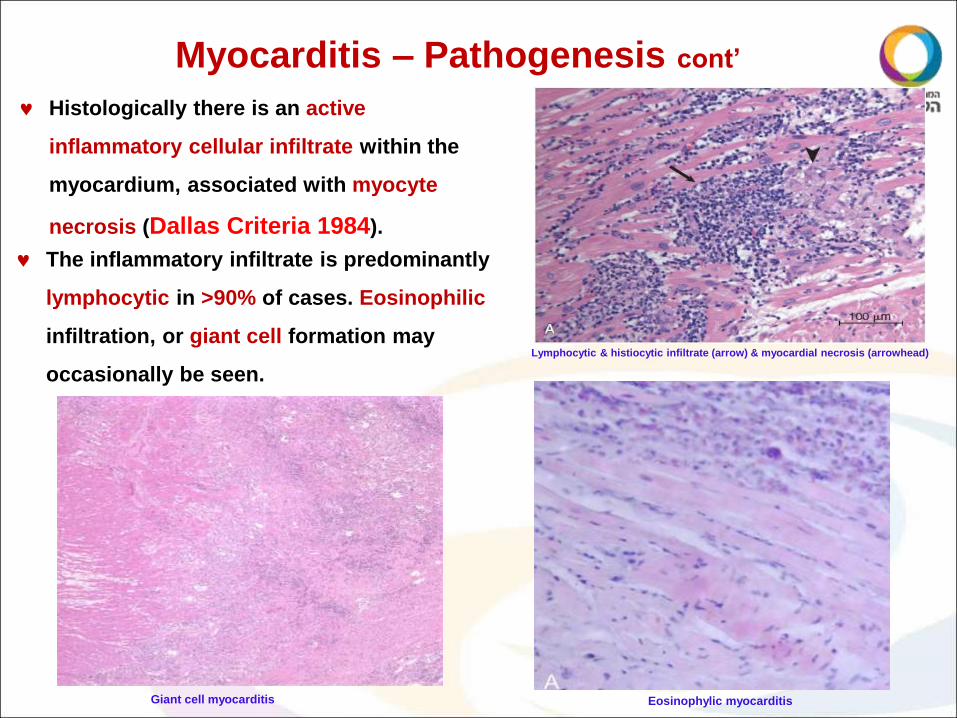
Lymphocytic & histiocytic infiltrate (arrow) & myocardial necrosis (arrowhead)
Histologically there is an active
inflammatory cellular infiltrate within the
myocardium, associated with myocyte
necrosis (Dallas Criteria 1984).
The inflammatory infiltrate is predominantly
lymphocytic in >90% of cases. Eosinophilic
infiltration, or giant cell formation may
occasionally be seen.
Eosinophylic myocarditis Giant cell myocarditis

Myocarditis – Pathogenesis cont’
Fulminant myocarditis
In a majority of cases the inflammatory process resolves with minimal or no damage or
remodeling
Ongoing myocardial inflammation may result in dilated cardiomyopathy or restrictive
cardiomyopathy
If the host immune response is overwhelming or inappropriate, the inflammation may
acutely destroy the heart tissue leading to LV failure frequently even without dilatation
(fulminant myocarditis) and death

Myocarditis – Clinical Features
A recent systemic illness with viral symptomatology: fever, sore throat,
cough, arthralgia, myalgia, abdominal pain, nausea, vomiting, diarrhea,
and skin rash.
Cardiac involvement usually becomes apparent only a few days to a
few weeks later. Usually manifested as fatigue, decreased exercise
tolerance, dyspnea, palpitations, and precordial discomfort.
Plueropericardial chest pain is not infrequent, especially when there is
associated pericarditis.
However, chest pain may occur without inflammatory involvement of
the pericardium as was proven by MRI.

Myocarditis – Clinical Features
New physical findings depends on the severity of the disease. They
include: persistent fever, excessive tachycardia, hypotension, and narrow
pulse pressure. Clinical findings of HF with mitral and tricuspid
regurgitation may occur in more severe cases.
Neck veins distention in HF, pericardial effusion, or both.
In the acute phase there is no cardiac dilatation, which after occurs days to
weeks, and is associated with a diffuse and displaced PMI and RV heave.
Auscultatory sounds may include a muffled first heart sound, a third heart
sound, a friction rub, and murmurs due to mitral and tricuspid
regurgitation.

Myocarditis – Laboratory Data
ESR and CRP levels are often raised, but they do not confirm the diagnosis.
WBC – slightly to moderately elevated, with 50% neutrophilia
Eosinophilia may indicate a parasitic etiology.
Biomarkers are usually elevated, especially high sensitive troponins, which are
more sensitive than CK, including CK-MB levels.
They are non-specific and when normal do not exclude myocarditis.
In comparison to acute MI CRP values are relatively much more elevated than
high sensitive troponin values.
Other biomarkers such as BNP/Pro-BNP, circulating cytokines, markers of
extracellular matrix degradation, pentraxin 3, galectin 3, and growth
differentiation factor 15 that are frequently elevated.

C-reactive protein to troponin ratio for the differentiation of
perimyocarditis from myocardial infarction
Meisel SR, Kleiner-Shochat M, Abu Fanne R, Blondheim DS, Frimerman A, Asif A,
Levy Y, Muhassen J, Kazatsker M, Shotan A
Perimyocarditis

Myocarditis – Laboratory Data
Antibodies are usually not found until about 1 week after the illness onset.
IgM antibody levels peak in 2-3 weeks and are later undetectable
IgG antibody levels peak later and may remain elevated for months or years
Positive viral serology does not imply myocardial infection, but rather
indicates the interaction of the peripheral immune system with an
infectious agent.
Viral serology is not diagnostic, as the prevalence of circulatory IgG ABs
to cardiotropic viruses in the general population is high without heart
disease.
Viral serology has, therefore, a low clinical value and is currently not
routinely recommended, as it did not correlate with endomyocardial
biopsy viral findings.

Myocarditis – Cardiovascular magnetic resonance (CMR) imaging
The diagnostic cardiac magnetic resonance criteria for myocarditis in the setting of clinically
suspected myocarditis (Lake Louise criteria) are:
1. Regional or global myocardial signal intensity increase in T2-weighted edema images.
2. Increased global myocardial early gadolinium enhancement ratio between myocardium
and skeletal muscle in gadolinium-enhanced T1-weighted images.
3. There is at least one focal lesion with non-ischemic regional distribution in inversion
recovery-prepared gadolinium-enhanced T1-weighted images (LGE).
A CMR study is consistent with:
Myocardial inflammation, if ≥2 criteria are present
Myocyte injury and/or scar caused by myocardial inflammation if criterion 3 is present.

Myocarditis – Cardiovascular magnetic resonance (CMR) imaging
CMR does not replace endomyocardial biopsy.
It is unable to differentiate between infectious and immune-mediated forms
Does not provide information on the type of inflammation including special types of
myocarditis, which may require special therapies (like giant cell, eosinophilic myocarditis or
sarcoidosis).
Does not provide information on the type of virus and non-diagnostic in milder cases.
CMR supports the clinical suspicion of myocarditis and for noninvasive follow-up.
Particularly important in patients with minor symptoms, e.g., young patients with
unexplained arrhythmia, or troponin positive patients with normal coronary arteries.

Myocarditis – Endomyocardial Biopsy
Since the Dallas criteria (1984), , endomyocardial biopsy has become the
"gold standard" for the diagnosis of myocarditis.
Endomyocardial biopsy confirms the diagnosis of myocarditis and
identifies the underlying etiology, especially specific type of inflammation
(giant cell, eosinophilic myocarditis, sarcoidosis) which imply different
treatments and prognosis.
However, the negative results of the
multicenter Myocarditis Treatment Trial
that used the Dallas criteria to recruit
myocarditis patients to 6 mths
immunosuppression had a negative
effect in the next decade on the use of
endomyocardial biopsy. Sarcoidosis

Myocarditis – Endomyocardial Biopsy
Cell-specific immunehistological staining for surface antigens: such as anti-
CD3 (T-cells), anti-CD4 (T-helper cells), anti-CD20 (B-cells), anti-CD68
(macrophages), and anti–human leukocyte antigen (HLA).
This technique is associated with less sampling error, therefore is more
sensitive than histopathology and has better prognostic value.
The diagnostic contribution of EMB is enhanced by molecular analysis with
DNA–RNA extraction and RT-PCR amplification of viral genome.
In order to exclude systemic infection, peripheral blood should be
investigated in parallel with EMB.
Viral isolation from biopsy culture is complementary to histopathology and
mandatory for identification & characterization of the inflammatory infiltrate.

Myocarditis – Endomyocardial Biopsy
The ability to perform endomyocardial biopsy is somewhat limited during
pregnancy, because the use of fluoroscopy is undesirable.
The procedure should, therefore, be performed under echocardiographic guidance.
Viral persistence in the myocardium has been associated with ventricular dysfunction,
and viral genome clearance with improvement of ventricular function and a better 10-
year prognosis.
In contrast, immunohistological evidence of inflammation, but not the presence of viral
genome alone, was an independent predictor of survival.

Myocarditis – Treatment
Acute myocarditis resolves within 2-4 weeks in 50% of cases, but about 25% will
develop persistent cardiac dysfunction and 12–25% may acutely deteriorate and either
die or progress to end-stage DCM.
The treatment of many milder forms of myocarditis is symptomatic, care of arrhythmia
and of heart failure and, where supported by evidence, etiology-targeted therapy.
All pregnant women with suspected myocarditis should be hospitalized for clinical
monitoring, until a definite diagnosis is established, since cardiopulmonary emergency,
like severe heart block or life threatening arrhythmia, may occur even if systolic
function is initially preserved.
Patients with hemodynamic instability, HF, significant pericardial effusion, at risk of
tamponade, and serious arrhythmias should be adequately monitored in ICCU.
Exercise testing is contraindicated in the acute stage, as it can precipitate arrhythmia.

Myocarditis – Treatment
HF should be treated with diuretics and beta blockers. The use of ACE-I, ARBs, MRAs and
Sacubitril / Valsartan is contraindicated during pregnancy, due to their teratogenicity
and/or lack of evidence.
Digoxin, commonly used, increases proinflammatory cytokines and mortality in a murine
model of viral myocarditis.
As therapeutic levels of digoxin may be associated with toxicity in myocarditis, and
serum digoxin levels cannot be accurately measured during pregnancy, it should be used
with caution and only at low doses.
In acute/fulminant cases with cardiogenic shock and severe ventricular dysfunction,
besides inotropic agents and intraaortic counterpulsation, ventricular assist devices
(VAD) or extracorporeal membrane oxygenation (ECMO) are needed early (sometimes
within 12-24 hours) to provide a bridge to cardiac transplantation or to recovery.

Myocarditis – Treatment
Important arrhythmias should be treated with beta blockers, lidocaine, quinidine, or
procainamide, which are relatively safe in gestation, and if persistent, the implantation
of a defibrillator should be considered. However, whenever clinically feasible, ICD
implantation should be deferred until resolution of the acute episode.
Temporary pacing should be inserted for high degree AV block. As conduction
disturbances are transient in the majority of patients with myocarditis, a permanent
pacemaker is usually not indicated.
Cardiac electronic implantable device, either pacemaker or ICD/CRTD can be implanted
during pregnancy using echocardiography with relatively minimal X-ray irradiation.
In the last years there is an increased use of an wearable defibrillator (LifeVest) during
pregnancy, awaiting cardiac recovery or internal ICD/CRTD implantation after delivery.

Wearable Defibrillator (LifeVest)

Myocarditis – Treatment
NSAIDs, in particular acetylsalicylic acid, are a cornerstone of treatment for acute
pericarditis, but have been associated with increased mortality in experimental models
of myocarditis. Clinical data for their administration in myocarditis are inconclusive,
and controlled trials are needed.
Anticoagulation may be added, especially if patients have severe LV dysfunction, with
or without evidence of a LV thrombus, to reduce the risk of emboli.
Anti-viral therapies – Interferon beta treatment can eliminate enteroviral and
adenoviral genomes in patients with LV dysfunction, is associated with improvement in
NYHA class, and in enteroviral infection is associated with a better 10-year prognosis.
However, Interferon beta was not effective against parvoviral B19 infection in the
recently published BICC trial

Myocarditis – Treatment
High dose intravenous immunoglobulin (IVIG) has been associated with
improved LVEF in chronic symptomatic HF of various causes.
However, IVIG was ineffective in the IMAC trial of recent-onset DCM.
As IVIG has no major side effects it may be used in myocarditis refractory
to conventional HF therapy, both viral and autoimmune forms, particularly if
autoantibody-mediated.
Immunosuppression should be started only after ruling out active infection
on EMB by PCR. Most data have been obtained using steroids alone,
azathioprine and steroids, or cyclosporine A.
Current recommendation of immunosuppression: proven autoimmune
forms of myocarditis, with no contraindications, including giant cell
myocarditis, cardiac sarcoidosis, eosinophilic myocarditis and myocarditis
associated with known extra-cardiac autoimmune disease.

Myocarditis – Treatment
Steroid therapy is indicated in cardiac sarcoidosis in the presence of ventricular
dysfunction and/or arrhythmia, and in some forms of infection-negative eosinophilic
or toxic myocarditis with HF and/or arrhythmia.
Immunosuppression may be considered by a recent position statement in infection-
negative lymphocytic myocarditis refractory to standard therapy in patients with no
contraindications to immunosuppression.
This approach is based on the positive results of the TIMIC randomized trial, and a
recently published observational retrospective study. These studies included patients
with inflammatory cardiomyopathy of at least 6 months duration. The use of
immunosuppression in infection negative acute myocarditis unresponsive to
supportive treatment had been documented only in sporadic cases.
Strenuous activity may be deleterious and should be prohibited during the acute
phase of myocarditis for at least 6 months both in athletes and nonathletes.

Myocarditis in Pregnancy
Only a few cases of myocarditis have been reported in pregnancy
In an early review (1968) 4 of 22 pts with viral myocarditis – in the postpartum period
Grimes (1980) reported 4 cases with fatal outcome following an abortion in early stage of
gestation – autopsy evidence of myocarditis
Gehrke (1994) reported a 28-year-old asthmatic female who developed postpartum acute
HF accompanied by diarrhea, fever, and hypereosinophilia. During steroid treatment,
cytomegalovirus-associated myocarditis developed
Chen (1994) described a patient with repeated episodes of acute myocarditis who
developed heart failure in the 36th week of gestation, with rapid deterioration and death

Myocarditis in Pregnancy
Ciccone (2016) reported a 40-year-old woman who developed, after childbirth,
hyperthermia with neck and left arm pain, who died suddenly few days later. Autopsy
disclosed normal sized heart with fulminant myocarditis, congested organs and negative
microbiological tests.
Massengill A (2016) described a pregnant woman who developed infectious myocarditis
presenting as acute respiratory distress.
Malhorta (2016) described a 38-year-old postpartum female who had a cesarean section
due to preeclampsia, who developed acute pericarditis and myocarditis related to SLE,
complicated by acute respiratory failure and cardiogenic shock with dramatic
improvement within days under steroid therapy

Myocarditis in Pregnancy
Several reports have demonstrated a relatively high incidence histologically proven
myocarditis in patients with PPCM, suggesting that myocarditis may be an important
etiologic factor in PPCM.
The incidence of active myocardial inflammation in PPCM varied:
Rizeq (1994) reported a low incidence 3(9%) of myocarditis in 34 patients with PPCM,
comparable to age- and sex-matched control population with idiopathic-dilated CMP.
Bültmann (2005) studied 26 patients with PPCM in whom endomyocardial biopsy
specimens revealed viral genomes: parvovirus B19, human herpes virus 6, Epstein-
Barr virus, and human cytomegalovirus in 8 patients (31%)

Comparison of Myocarditis during pregnancy to PPCM/PAC
PPCM / PAC Myocarditis
All, more frequent >30 All Age (years)
More frequent in multipara Unknown Number of pregnancies
Unknown Mostly viral Etiology
>16% Unknown Genetic background
16% Unknown Twin pregnancy
Yes Unknown Preeclampsia / HTN /
Tocolytic therapy

Comparison of Myocarditis during pregnancy to PPCM/PAC cont’
PPCM / PAC Myocarditis
Sometimes Frequently Flu-like preceding symptoms
delayed (on average 2 weeks
unless severe)
Delayed (usually within few
days unless severe)
Diagnosis
Sometimes Very frequently Fever
Rarely Quite frequently Pericardial pain
Sometimes Always Inflammatory markers
Currently not recommended In clinical severe cases Endomyocardial biopsy

Comparison of Myocarditis during pregnancy to PPCM/PAC cont’
PPCM / PAC Myocarditis
Treatment
Yes Yes HFrEF Guidelines
recommended
No When viral persistence
(not effective in parvovirus)
Interferon beta
No Yes - mostly biopsy guided Immunosuppression
Yes (still debatable) No Bromocriptine
20-40% recurrence Recurrence rate not reported.
Probably low. When full recovery
it may be safe
Subsequent pregnancy

Myocarditis in Pregnancy – Summary
Myocarditis during pregnancy is rare.
Its clinical presentation varies from asymptomatic, mild non-specific symptoms to cardiogenic shock
and/or life threatening arrhythmias.
Its diagnosis is based on combination of clinical features, electrocardiographic, laboratory,
echocardiographic and CMR findings.
Endomyocardial biopsy confirms the diagnosis of myocarditis, identifies the underlying etiology and
may reveal particular types of inflammation. (e.g. giant cell, eosinophilic myocarditis, sarcoidosis), which
require specific immunosuppressive treatment.
Immunosuppressive therapy may be considered in selected patients unresponsive to standard
therapy, in whom inflammation was demonstrated and viral persistence was excluded by
endomyocardial biopsy.
Myocarditis resolves within few weeks. However, patients may develop persistent cardiac dysfunction
and 12–25% of them may deteriorate to end-stage cardiomyopathy and even death.

Thank You Thank You
Thank You

Myocarditis – Prevalence
• Affects individuals of all ages, but most frequent in the young
• Global prevalence – 22 / 100,000 pts per year
• Age-standardized death rate due to myocarditis & cardiomyopathies –
6.1 / 100,000 pts
Global burden of disease task force. Global and regional mortality from 235 causes of death for 20 age groups in 1990
and 2010: A systematic analysis for the global burden of disease study 2010. Lancet. 2012;380:2095-2128
Cooper Lt Jr, Keren A, Sliwa K, Matsumori A, Menash GA. The global burden of myocarditis: part 1: a systematic
literature review for the Global Burden of Diseases, Injuries, and Risk Factors 2010 study. Global Heart 2014;9:121–129

Myocarditis – Clinical Features
Myocarditis may cause ventricular arrhythmias and heart block or mimic acute MI, especially
when presents with localized electrocardiographic changes and wall motion abnormalities.
Hemodynamic instability, and circulatory collapse, may develop with severe LV and/or RV
dysfunction, a high degree AV block, ventricular arrhythmias, or cardiac tamponade.
Myocarditis may be the cause of 20% of cases of sudden, unexpected death in young
adults, <40 years of age and in young athletes, secondary to tachyarrhythmias or complete
AV block.
In autopsies of young adults, myocarditis is responsible for 4-12% of sudden deaths, ranking
as the 3rd cause after hypertrophic cardiomyopathy and congenital and atherosclerotic
coronary artery disease.
Systemic and pulmonary emboli have been reported and may be the presenting feature.
Myocarditis resolves spontaneously in approximately 80% of patients, but in those who did
not recover, prospective studies revealed a 10-year survival rate of only 45%, mostly due to
manifestation of DCM and sudden cardiac death.

Myocarditis – Electrocardiogram
In the acute stage, the electrocardiogram is usually abnormal, demonstrating ST segment elevation with
inversion or flattening of the T wave and possible prolongation of the QT interval.
ST-T segment elevation in myocarditis is typically concave (rather than convex as seen in myocardial
infarction) and diffuse without reciprocal changes or limitation to a specific coronary territory.
The ST segment changes usually return to baseline within a few days, whereas T-wave changes may
persist for several weeks or months.
Abnormal Q waves may sometimes develop and mimic acute myocardial infarction.
Ventricular premature beats are common, and atrial and ventricular tachyarrhythmias are present in many
patients.
QRS prolongation may be an independent negative predictor for survival.
Atrioventricular conduction disturbances of varying degrees associated with ventricular tachyarrhythmias
should raise the suspicion of giant cell myocarditis, considered the most malignant form of myocarditis,
which requires histopathologic confirmation and aggressive immunosuppressive therapy.
A-V block in the presence of mild left ventricular dilatation may be also suggestive of Lyme disease or
cardiac sarcoidosis.