myocardial strain analysis in patients with heart failure with preserved ejection fraction using...
TRANSCRIPT
POSTER PRESENTATION Open Access
Myocardial strain analysis in patients with HeartFailure with preserved Ejection Fraction usingbright blood cine MR images: A comparison withspeckle-tracking echocardiographyPeter M Smith1*, Vistasp Daruwalla1, Benjamin H Freed3, Bruce S Spottiswoode2, Kevin Kalisz1, James C Carr1,Jeremy D Collins1
From 17th Annual SCMR Scientific SessionsNew Orleans, LA, USA. 16-19 January 2014
BackgroundChanges in myocardial strain parameters is of interest inpatients with heart failure as an objective measure ofdisease severity. Speckle-tracking echocardiography (ST-echo) is the accepted standard of reference for myocar-dial strain analysis given superior temporal resolution;however, difficult acoustic windows and limited contrastto noise resolution can limit strain analysis. Preliminarywork using deformation field analysis at steady state freeprecession (SSFP) cine MR imaging has shown thatstrain analysis at CMR is similar between conventionaland highly accelerated GRAPPA cine acquisitions. Thepurpose of this study is to compare the strain values inpatients with heart failure and preserved ejection frac-tion (HFpEF, left ventricular ejection fraction >50%) atSSFP cine MRI with ST-echo.
MethodsRetrospective analysis of Cardiac MR and echocardio-graphic images from 15 patients (5 men, avg age 61.2 yrs)with HFpEF. Cardiac MR images were obtained at 1.5 T(MAGNETOM Avanto, Siemens Medical Systems, Erlan-gen, AG) using GRAPPA factor 2 acceleration (temp res =39.2 msec, spatial res = 1.5 × 1.5 mm, thickness = 6 mm).Myocardial strain analysis at Cardiac MR was performedusing prototype software calculating Lagrangian strainfrom deformation field analysis (Siemens Corp, CorporateTechnology, Princeton, NJ). Transthoracic echocardiogra-phy exams included apical 4-chamber and mid-ventricular
short axis views. Left ventricular (LV) mid ventricularaverage and peak systolic radial and circumferential strainsas well as longitudinal strain data was calculated. Peak andaverage right ventricular (RV) longitudinal strain was alsoobtained. CMR and ST-Echo derived strain indices werecompared using the Pearson correlation. Inter andintraobserver variance was assessed for CMR-derived RVand LV longitudinal strain analysis using the intraclasscorrelation coefficient (ICC).
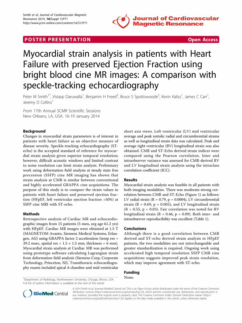
ResultsMyocardial strain analysis was feasible in all patients withboth imaging modalities. There was moderate-strong cor-relation between CMR and ST-Echo (Figure 1) as follows:LV radial strain (R = 0.79, p = 0.0004), LV circumferentialstrain (R = 0.69, p = 0.005), and LV longitudinal strain(R = 0.55, p = 0.03). Fair correlation was noted for RVlongitudinal strain (R = 0.46, p = 0.09). Both inter- andintraobserver reproducibility was excellent (Table 1).
ConclusionsAlthough there is a good correlation between CMRderived and ST-echo derived strain analysis in HFpEFpatients, the two modalities are not interchangeable andgreater standardization is required. Ongoing work usingaccelerated high temporal resolution SSFP CMR cineacquisitions suggests improved peak strain resolution,which may improve agreement with ST-echo.
FundingNone.1Department of Radiology, Northwestern University, Chicago, Illinois, USA
Full list of author information is available at the end of the article
Smith et al. Journal of Cardiovascular MagneticResonance 2014, 16(Suppl 1):P71http://www.jcmr-online.com/content/16/S1/P71
© 2014 Smith et al.; licensee BioMed Central Ltd. This is an Open Access article distributed under the terms of the Creative CommonsAttribution License (http://creativecommons.org/licenses/by/2.0), which permits unrestricted use, distribution, and reproduction inany medium, provided the original work is properly cited. The Creative Commons Public Domain Dedication waiver (http://creativecommons.org/publicdomain/zero/1.0/) applies to the data made available in this article, unless otherwise stated.
Authors’ details1Department of Radiology, Northwestern University, Chicago, Illinois, USA.2Cardiovascular MR R&D, Siemens Healthcare, Chicago, Illinois, USA.3Department of Cardiology, Northwestern University, Chicago, Illinois, USA.
Published: 16 January 2014
doi:10.1186/1532-429X-16-S1-P71Cite this article as: Smith et al.: Myocardial strain analysis in patientswith Heart Failure with preserved Ejection Fraction using bright bloodcine MR images: A comparison with speckle-tracking echocardiography.Journal of Cardiovascular Magnetic Resonance 2014 16(Suppl 1):P71.
Submit your next manuscript to BioMed Centraland take full advantage of:
• Convenient online submission
• Thorough peer review
• No space constraints or color figure charges
• Immediate publication on acceptance
• Inclusion in PubMed, CAS, Scopus and Google Scholar
• Research which is freely available for redistribution
Submit your manuscript at www.biomedcentral.com/submit
Figure 1 Correlation plots comparing CMR determined Lagrangian strains with those at ST-echo. ST-echo: speckle-trackingechocardiography, CMR: Cardiac MR.
Table 1 ICC values demonstrating reproducibility of LVand RV longitudinal CMR strain analysis.
ICC 95% Confidence Interval
LV Longitudinal 0.997 0.999-0.990
RV Longitudinal 0.981 0.993-0.944
Smith et al. Journal of Cardiovascular MagneticResonance 2014, 16(Suppl 1):P71http://www.jcmr-online.com/content/16/S1/P71
Page 2 of 2