mycology proficiency testing program
TRANSCRIPT

Test Event Critique May 2015
Mycology Proficiency Testing Program

1
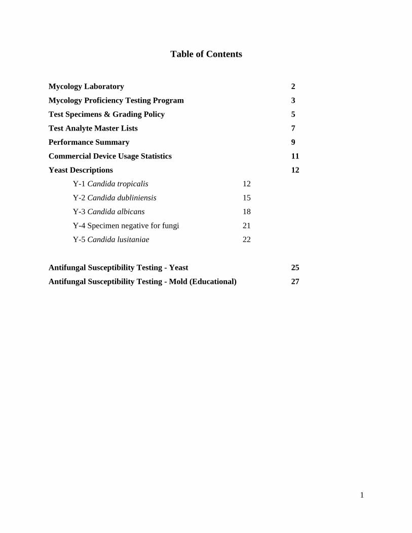
Table of Contents
Mycology Laboratory 2
Mycology Proficiency Testing Program 3
Test Specimens & Grading Policy 5
Test Analyte Master Lists 7
Performance Summary 9
Commercial Device Usage Statistics 11
Yeast Descriptions 12
Y-1 Candida tropicalis 12
Y-2 Candida dubliniensis 15
Y-3 Candida albicans 18
Y-4 Specimen negative for fungi 21
Y-5 Candida lusitaniae 22
Antifungal Susceptibility Testing - Yeast 25
Antifungal Susceptibility Testing - Mold (Educational) 27
2
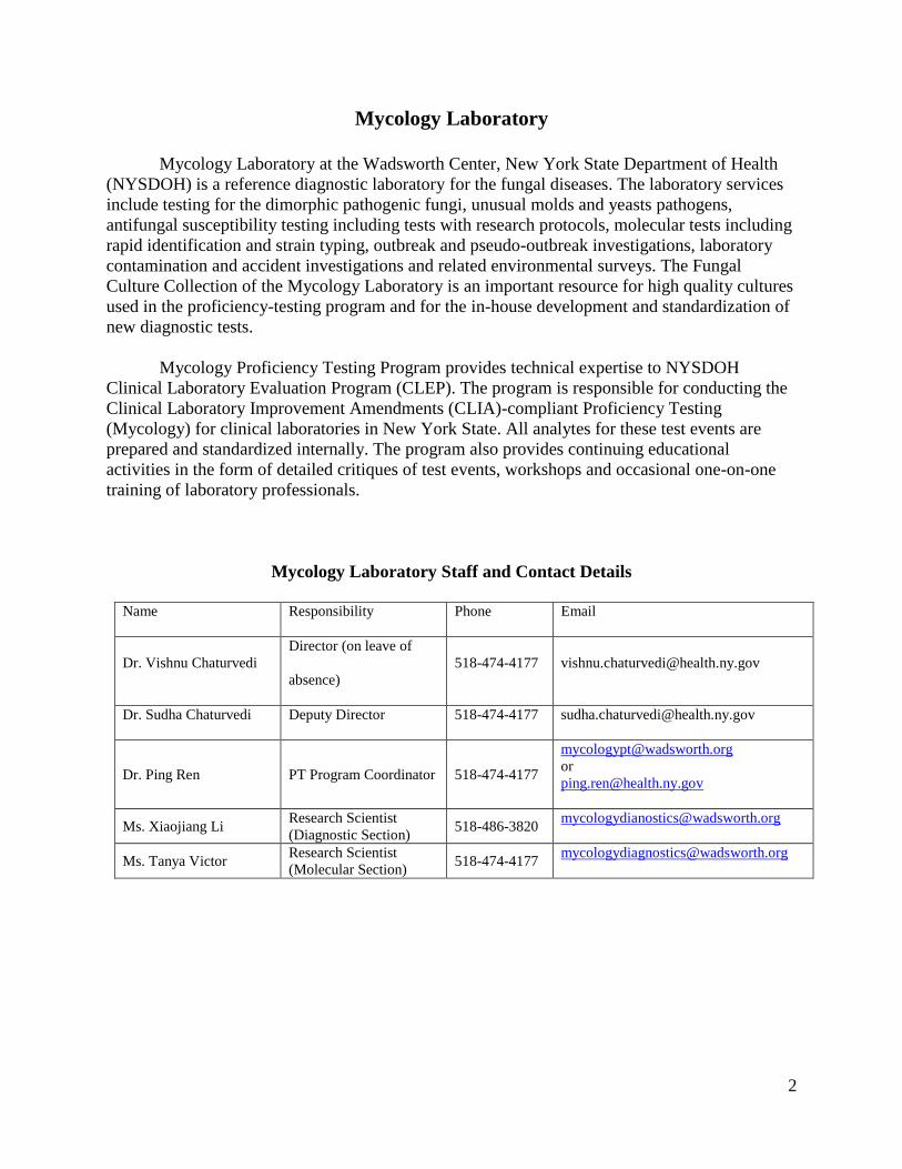
Mycology Laboratory
Mycology Laboratory at the Wadsworth Center, New York State Department of Health
(NYSDOH) is a reference diagnostic laboratory for the fungal diseases. The laboratory services
include testing for the dimorphic pathogenic fungi, unusual molds and yeasts pathogens,
antifungal susceptibility testing including tests with research protocols, molecular tests including
rapid identification and strain typing, outbreak and pseudo-outbreak investigations, laboratory
contamination and accident investigations and related environmental surveys. The Fungal
Culture Collection of the Mycology Laboratory is an important resource for high quality cultures
used in the proficiency-testing program and for the in-house development and standardization of
new diagnostic tests.
Mycology Proficiency Testing Program provides technical expertise to NYSDOH
Clinical Laboratory Evaluation Program (CLEP). The program is responsible for conducting the
Clinical Laboratory Improvement Amendments (CLIA)-compliant Proficiency Testing
(Mycology) for clinical laboratories in New York State. All analytes for these test events are
prepared and standardized internally. The program also provides continuing educational
activities in the form of detailed critiques of test events, workshops and occasional one-on-one
training of laboratory professionals.
Mycology Laboratory Staff and Contact Details
Name Responsibility Phone Email
Dr. Vishnu Chaturvedi
Director (on leave of
absence)
518-474-4177 [email protected]
Dr. Sudha Chaturvedi Deputy Director 518-474-4177 [email protected]
Dr. Ping Ren PT Program Coordinator 518-474-4177
or
Ms. Xiaojiang Li Research Scientist
(Diagnostic Section) 518-486-3820
Ms. Tanya Victor Research Scientist
(Molecular Section) 518-474-4177
3
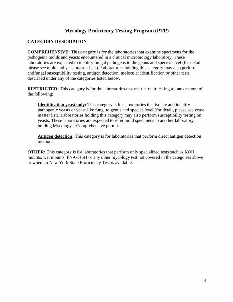
Mycology Proficiency Testing Program (PTP)
CATEGORY DESCRIPTION
COMPREHENSIVE: This category is for the laboratories that examine specimens for the
pathogenic molds and yeasts encountered in a clinical microbiology laboratory. These
laboratories are expected to identify fungal pathogens to the genus and species level (for detail,
please see mold and yeast master lists). Laboratories holding this category may also perform
antifungal susceptibility testing, antigen detection, molecular identification or other tests
described under any of the categories listed below.
RESTRICTED: This category is for the laboratories that restrict their testing to one or more of
the following:
Identification yeast only: This category is for laboratories that isolate and identify
pathogenic yeasts or yeast-like fungi to genus and species level (for detail, please see yeast
master list). Laboratories holding this category may also perform susceptibility testing on
yeasts. These laboratories are expected to refer mold specimens to another laboratory
holding Mycology – Comprehensive permit.
Antigen detection: This category is for laboratories that perform direct antigen detection
methods.
OTHER: This category is for laboratories that perform only specialized tests such as KOH
mounts, wet mounts, PNA-FISH or any other mycology test not covered in the categories above
or when no New York State Proficiency Test is available.

4
PROFICIENCY TESTING ANALYTES OFFERED
(CMS regulated analytes or tests are indicated with an asterisk)
Comprehensive
Culture and Identification*
Susceptibility testing
Cryptococcus neoformans Antigen Detection
Restricted
Identification Yeast Only
Culture and Identification of yeasts*
Susceptibility testing of yeasts
Antigen Detection
Antigen detection of Cryptococcus neoformans*
5
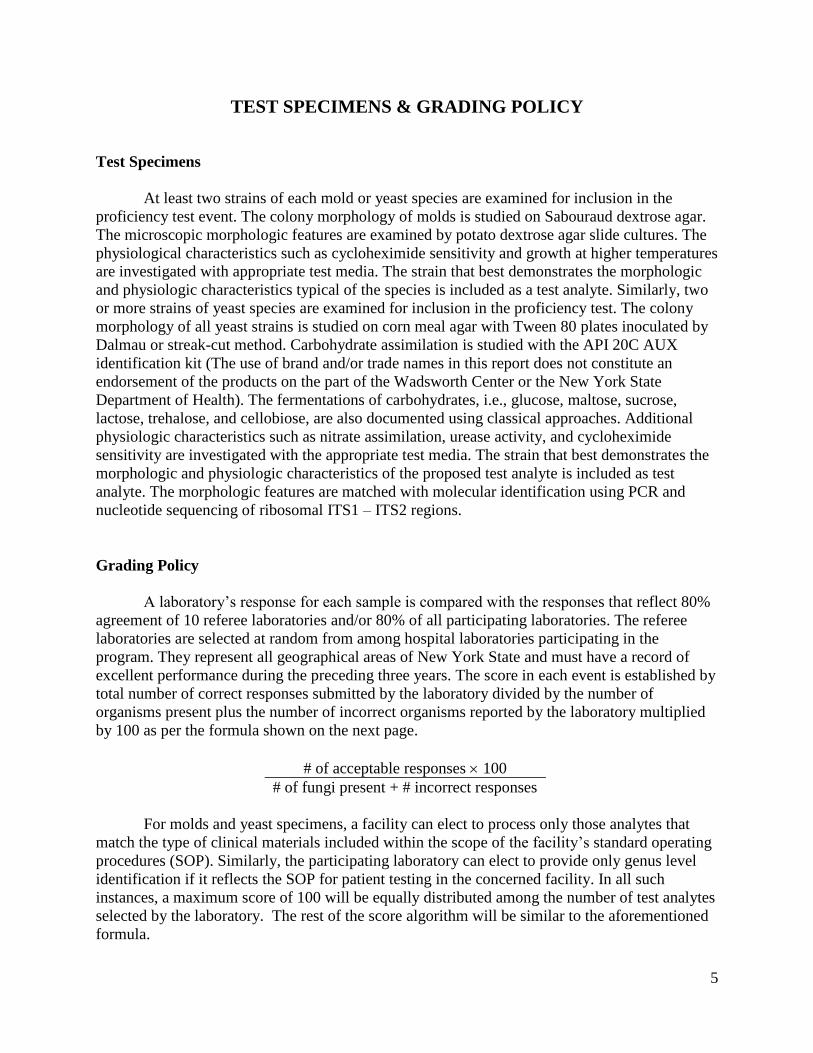
TEST SPECIMENS & GRADING POLICY
Test Specimens
At least two strains of each mold or yeast species are examined for inclusion in the
proficiency test event. The colony morphology of molds is studied on Sabouraud dextrose agar.
The microscopic morphologic features are examined by potato dextrose agar slide cultures. The
physiological characteristics such as cycloheximide sensitivity and growth at higher temperatures
are investigated with appropriate test media. The strain that best demonstrates the morphologic
and physiologic characteristics typical of the species is included as a test analyte. Similarly, two
or more strains of yeast species are examined for inclusion in the proficiency test. The colony
morphology of all yeast strains is studied on corn meal agar with Tween 80 plates inoculated by
Dalmau or streak-cut method. Carbohydrate assimilation is studied with the API 20C AUX
identification kit (The use of brand and/or trade names in this report does not constitute an
endorsement of the products on the part of the Wadsworth Center or the New York State
Department of Health). The fermentations of carbohydrates, i.e., glucose, maltose, sucrose,
lactose, trehalose, and cellobiose, are also documented using classical approaches. Additional
physiologic characteristics such as nitrate assimilation, urease activity, and cycloheximide
sensitivity are investigated with the appropriate test media. The strain that best demonstrates the
morphologic and physiologic characteristics of the proposed test analyte is included as test
analyte. The morphologic features are matched with molecular identification using PCR and
nucleotide sequencing of ribosomal ITS1 – ITS2 regions.
Grading Policy
A laboratory’s response for each sample is compared with the responses that reflect 80%
agreement of 10 referee laboratories and/or 80% of all participating laboratories. The referee
laboratories are selected at random from among hospital laboratories participating in the
program. They represent all geographical areas of New York State and must have a record of
excellent performance during the preceding three years. The score in each event is established by
total number of correct responses submitted by the laboratory divided by the number of
organisms present plus the number of incorrect organisms reported by the laboratory multiplied
by 100 as per the formula shown on the next page.
# of acceptable responses 100
# of fungi present + # incorrect responses
For molds and yeast specimens, a facility can elect to process only those analytes that
match the type of clinical materials included within the scope of the facility’s standard operating
procedures (SOP). Similarly, the participating laboratory can elect to provide only genus level
identification if it reflects the SOP for patient testing in the concerned facility. In all such
instances, a maximum score of 100 will be equally distributed among the number of test analytes
selected by the laboratory. The rest of the score algorithm will be similar to the aforementioned
formula.
6
Acceptable results for antifungal susceptibility testing are based on the
consensus/reference laboratories’ MIC values within +/- 2 dilutions and the interpretation per
CLSI (NCCLS) guidelines or related, peer-reviewed publications. One yeast species is to be
tested against following drugs: amphotericin B, anidulafungin, caspofungin, flucytosine,
fluconazole, itraconazole, ketoconazole, micafungin, posaconazole, and voriconazole. The
participating laboratories are free to select any number of antifungal drugs from the test panel
based upon test practices in their facilities. A maximum score of 100 is equally distributed to
account for the drugs selected by an individual laboratory. If the result for any drug is incorrect
then laboratory gets a score of zero for that particular test component or set.
For Cryptococcus neoformans antigen test, laboratories are evaluated on the basis of their
responses and on overall performance for all the analytes tested in the Direct Detection category.
The maximum score for this event is 100. Appropriate responses are determined by 80%
agreement among participant responses. Target values and acceptable ranges are mean value +/-
2 dilutions; positive or negative answers will be acceptable from laboratories that do not report
antigen titers. When both qualitative and quantitative results are reported for an analyte, ten
points are deducted for each incorrect result. When only qualitative OR quantitative results are
reported, twenty points are deducted from each incorrect result.
A failure to attain an overall score of 80% is considered unsatisfactory performance.
Laboratories receiving unsatisfactory scores in two out of three consecutive proficiency test
events may be subject to ‘cease testing’.
7
TEST ANALYTE MASTER LISTS
Yeast Master List
The yeast master list is intended to provide guidance to the participating laboratories
about the scope of the Mycology - Restricted to Yeasts Only Proficiency Testing Program. This
list includes most common pathogenic and non-pathogenic yeasts likely to be encountered in the
clinical laboratory. The list is compiled from published peer-reviewed reports as well as current
practices in other proficiency testing programs. The list is meant to illustrate acceptable
identifications used in grading of responses received after each test event. This list neither
includes all yeasts that might be encountered in a clinical laboratory nor is intended to be used
for the competency assessment of the laboratory personnel in diagnostic mycology.
The nomenclature used in this list is based upon currently recognized species in
published literature, monographs, and catalogues of recognized culture collections. No attempt
has been made to include teleomorphic states of fungi if they are not routinely encountered in the
clinical specimens. Where appropriate, current nomenclature has been included under
parentheses to indicate that commonly accepted genus and/or species name is no longer valid,
e.g. Blastoschizomyces capitatus (Geotrichum capitatum). These guidelines supersede any
previous instructions for identification of yeasts. The list is subject to change in response to
significant changes in nomenclature, human disease incidence or other factors.
It is expected that major pathogenic yeasts listed in the Master List will be completely
identified to genus and species levels while those yeasts not listed in the master list will be
identified to genus only (i.e. Candida inconspicua as Candida species). However, the laboratory
can elect to provide only genus level identification if it reflects the standard operating procedures
(SOP) for patient testing. Please use “species complex” where appropriate, e.g. Candida
parapsilosis species complex if it is consistent with current reporting format used by the
laboratory.

8
Blastoschizomyces capitatus (Geotrichum capitatum) Cryptococcus terreus
Blastoschizomyces species Cryptococcus uniguttulatus
Candida albicans Geotrichum candidum
Candida dubliniensis Geotrichum species
Candida famata Hansenula anomala (Candida pelliculosa)
Candida glabrata Malassezia furfur
Candida guilliermondii species complex Malassezia pachydermatis
Candida kefyr Malassezia species
Candida krusei Pichia ohmeri (Kodamaea ohmeri)
Candida lipolytica (Yarrowia lipolytica) Prototheca species
Candida lusitaniae Prototheca wickerhamii
Candida norvegensis Prototheca zopfii
Candida parapsilosis species complex Rhodotorula glutinis
Candida rugosa Rhodotorula minuta
Candida species Rhodotorula mucilaginosa (rubra)
Candida tropicalis Rhodotorula species
Candida viswanathii Saccharomyces cerevisiae
Candida zeylanoides Saccharomyces species
Cryptococcus albidus Sporobolomyces salmonicolor
Cryptococcus gattii Sporobolomyces species
Cryptococcus laurentii Trichosporon asahii
Cryptococcus neoformans Trichosporon inkin
Cryptococcus neoformans- Trichosporon mucoides
Cryptococcus gattii species complex Trichosporon species
Cryptococcus species

9
Summary of Laboratory Performance:
Mycology – Yeast Only
Specimen key Validated specimen Other acceptable
answers
Laboratories with correct responses /
Total laboratories (% correct responses)
Y-1 Candida tropicalis Candida tropicalis 108/110 (98%)
Y-2 Candida
dubliniensis
Candida
dubliniensis
102/108 (94%)
Y-3 Candida albicans Candida albicans 109/110 (99%)
Y-4 Specimen negative
for fungi
Specimen negative
for fungi
No fungal growth 104/108 (96%)
Y-5 Candida lusitaniae Candida lusitaniae 106/108(98%)

10
Antifungal Susceptibility Testing for Yeast (S-1: Candida parapsilosis M957)
*Please use interpretations for Candida spp. provided in CLSI M27-S4 document. If there is no
antifungal agent listed in CLSI M27-S4 document, CLSI M27-S3 document can be used as an
alternate guideline.
Drugs Acceptable MIC
(g/ml) Range
Acceptable
interpretation*
Laboratories with acceptable
responses/ Total laboratories
(% correct responses)
Amphotericin B 0.25 – 1 Susceptible /
No interpretation
21/21 (100%)
Anidulafungin 1 – 2 Susceptible 18/18 (100%)
Caspofungin 0.25 – 2 Susceptible 25/25 (100%)
Flucytosine (5-FC) 0.03 – 0.25 Susceptible / No
interpretation
22/22 (100%)
Fluconazole 0.25 - 4 Susceptible /
Susceptible-dose
dependent
32/32 (100%)
Itraconazole 0.015 – 0.125 Susceptible / No
interpretation
25/25 (100%)
Ketoconazole 0.015-0.06 Susceptible /
No interpretation
3/3 (100%)
Micafungin 1 – 2 Susceptible 18/18 (100%)
Posaconazole 0.015 – 0.06 Susceptible /
No interpretation
17/17 (100%)
Voriconazole 0.008 – 0.125 Susceptible 29/29 (100%)
11
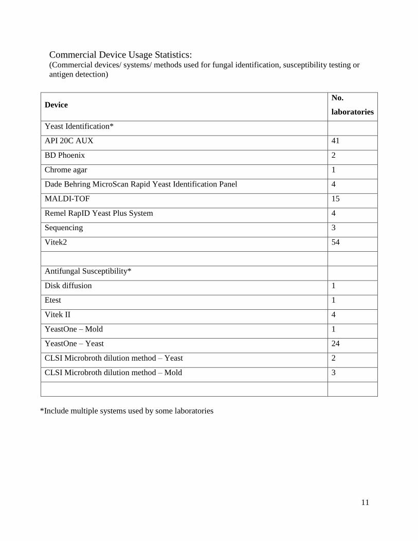
Commercial Device Usage Statistics: (Commercial devices/ systems/ methods used for fungal identification, susceptibility testing or
antigen detection)
*Include multiple systems used by some laboratories
Device No.
laboratories
Yeast Identification*
API 20C AUX 41
BD Phoenix 2
Chrome agar 1
Dade Behring MicroScan Rapid Yeast Identification Panel 4
MALDI-TOF 15
Remel RapID Yeast Plus System 4
Sequencing 3
Vitek2 54
Antifungal Susceptibility*
Disk diffusion 1
Etest 1
Vitek II 4
YeastOne – Mold 1
YeastOne – Yeast 24
CLSI Microbroth dilution method – Yeast 2
CLSI Microbroth dilution method – Mold 3
12
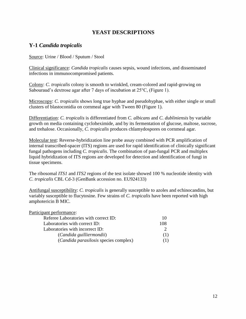
YEAST DESCRIPTIONS
Y-1 Candida tropicalis
Source: Urine / Blood / Sputum / Stool
Clinical significance: Candida tropicalis causes sepsis, wound infections, and disseminated
infections in immunocompromised patients.
Colony: C. tropicalis colony is smooth to wrinkled, cream-colored and rapid-growing on
Sabouraud’s dextrose agar after 7 days of incubation at 25°C, (Figure 1).
Microscopy: C. tropicalis shows long true hyphae and pseudohyphae, with either single or small
clusters of blastoconidia on cornmeal agar with Tween 80 (Figure 1).
Differentiation: C. tropicalis is differentiated from C. albicans and C. dubliniensis by variable
growth on media containing cycloheximide, and by its fermentation of glucose, maltose, sucrose,
and trehalose. Occasionally, C. tropicalis produces chlamydospores on cornmeal agar.
Molecular test: Reverse-hybridization line probe assay combined with PCR amplification of
internal transcribed-spacer (ITS) regions are used for rapid identification of clinically significant
fungal pathogens including C. tropicalis. The combination of pan-fungal PCR and multiplex
liquid hybridization of ITS regions are developed for detection and identification of fungi in
tissue specimens.
The ribosomal ITS1 and ITS2 regions of the test isolate showed 100 % nucleotide identity with
C. tropicalis CBL Cd-3 (GenBank accession no. EU924133)
Antifungal susceptibility: C. tropicalis is generally susceptible to azoles and echinocandins, but
variably susceptible to flucytosine. Few strains of C. tropicalis have been reported with high
amphotericin B MIC.
Participant performance:
Referee Laboratories with correct ID: 10
Laboratories with correct ID: 108
Laboratories with incorrect ID: 2
(Candida guilliermondii) (1)
(Candida parasilosis species complex) (1)

13
Illustrations:
Figure 1. Candida tropicalis, smooth-to-wrinkled, creamish colony, Sabouraud’s dextrose agar
7-days, 25°C. Microscopic morphology on cornmeal agar with Tween 80, showing long true
hyphae and pseudohyphae with clusters of blastoconidia (bar = 50 m). Scanning electron
micrograph illustrates true and pseudohyphae (with constrictions) and blastoconidia (bar = 2
m).
14
Further reading:
Chai LY, Denning DW, Warn P. 2010. Candida tropicalis in human disease. Crit Rev Microbiol. 36: 282-98.
de Carvalho Parahym AM, da Silva CM, Leão MP, Macario MC, Filho GA, de Oliveira NT, Neves RP. 2011.
Invasive infection in an acute myeloblastic leukemia patient due to triazole-resistant Candida tropicalis. Diagn
Microbiol Infect Dis. 71: 291-293.
Fesharaki SH, Haghani I, Mousavi B, Kargar ML, Boroumand M, Anvari MS, Abbasi K, Meis JF, Badali H. 2013.
Endocarditis due to a co-infection of Candida albicans and Candida tropicalis in a drug abuser. J Med Microbiol.
62(Pt 11): 1763-1767.
Hilmioglu S, Ilkit M, Badak Z. 2007. Comparison of 12 liquid media for germ tube production of Candida albicans
and C. tropicalis. Mycoses. 50: 282-285.
Lidder S, Tasleem A, Masterson S, Carrington RW. 2013. Candida tropicalis: diagnostic dilemmas for an unusual
prosthetic hip infection. J R Army Med Corps. 159: 123-125.
Magri MM, Gomes-Gouvêa MS, de Freitas VL, Motta AL, Moretti ML, Shikanai-Yasuda MA. 2012. Multilocus
sequence typing of Candida tropicalis shows the presence of different clonal clusters and fluconazole susceptibility
profiles in sequential isolates from candidemia patients in Sao Paulo, Brazil. J Clin Microbiol. 51: 268-277.
Muñoz P, Giannella M, Fanciulli C, Guinea J, Valerio M, Rojas L, Rodríguez-Créixems M, Bouza E. 2011. Candida
tropicalis fungemia: incidence, risk factors, and mortality in a general hospital. Clin Microbiol Infect. 17: 1538-
1545.
Negri M, Silva S, Henriques M, Oliveira R. 2011. Insights into Candida tropicalis nosocomial infections and
virulence factors. Eur J Clin Microbiol Infect Dis. DOI. 10.1007/s10096-011-1455-z.
Nucci M, Colombo AL. 2007. Candidemia due to Candida tropicalis: clinical, epidemiologic, and microbiologic
characteristics of 188 episodes occurring in tertiary care hospitals. Diagn Microbiol Infect Dis. 58: 77-82.
Pfaller MA, Castanheira M, Messer SA, Moet GJ, Jones RN. 2010. Variation in Candida spp. distribution and
antifungal resistance rates among bloodstream infection isolates by patient age: report from the SENTRY
Antimicrobial Surveillance Program (2008-2009). Diagn Microbiol Infect Dis. 68: 278-283.

15
Y-2 Candida dubliniensis
Source: Wound / Urine / Oral
Clinical significance: Candida dubliniensis was initially recovered from the oral cavities of HIV
infected individuals and AIDS patients causing erythematous and/or pseudomembranous oral
candidiasis or angular cheilitis. C. dubliniensis has also been isolated from other body sites
including lungs, vagina, blood, and feces.
Colony: C. dubliniensis colony is white to cream, smooth, and soft on Sabouraud’s dextrose agar
after 7 days of incubation at 25C (Figure 2). C. dubliniensis does not grow at 45C.
Microscopy: C. dubliniensis shows abundant, branched pseudohyphae and true hyphae with
blastoconidia. Chlamydospores are single, or in pairs, or in chains, or clusters on cornmeal agar
with Tween 80 (Figure 2).
Differentiation: C. dubliniensis is practically indistinguishable from C. albicans on the basis of
many common phenotypic tests. One physiologic feature that does appear to be fairly stable is
that C. dubliniensis grows poorly at 42C or does not at all at 45C while C. albicans grows well
at both of these temperatures. In addition, C. dubliniensis is able to assimilate glycerol, but not
xylose or trehalose as opposed to observations in C. albicans. Some commercial yeast
identification kits such as the API 20C AUX, VITEK2, or the ID 32C have biocodes for C.
dubliniensis included in the databases. These two closely related yeasts can also be distinguished
by molecular methods.
Molecular test: Genetically, C. dubliniensis has been found to be distinct from C. albicans in
DNA fingerprinting studies even though the two species are closely related phylogenetically.
Several C. dubliniensis molecular probes are available in reference laboratories.
The ribosomal ITS1 and ITS2 regions of the test isolate showed 100 % nucleotide identity with
Candida dubliniensis isolate CD36 (GenBank accession no. FM992695.1).
Antifungal susceptibility: Several isolates of C. dubliniensis have been found to have higher
resistance to fluconazole than other pathogenic species of Candida, and the resistance to
fluconazole may be induced in some originally sensitive strains. This fact may have serious
implications for immunocompromised individuals prescribed fluconazole for prolonged periods.
Participant performance:
Referee Laboratories with correct ID: 10
Laboratories with correct ID: 102
Laboratories with incorrect ID: 4
(Candida albicans) (2)
(Candida zeylanoides) (1)
(Rhodotorula sp.) (1)
16
Illustrations:
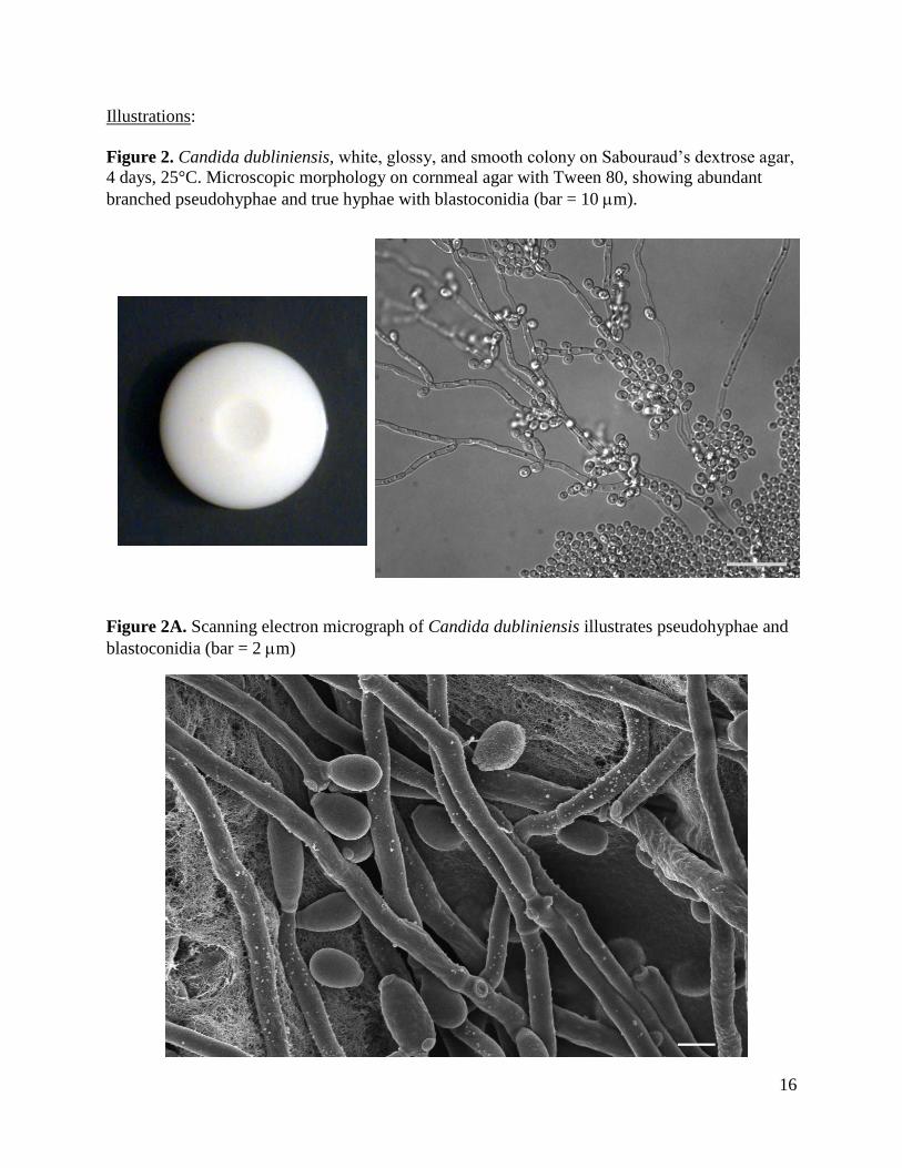
Figure 2. Candida dubliniensis, white, glossy, and smooth colony on Sabouraud’s dextrose agar,
4 days, 25°C. Microscopic morphology on cornmeal agar with Tween 80, showing abundant
branched pseudohyphae and true hyphae with blastoconidia (bar = 10 m).
Figure 2A. Scanning electron micrograph of Candida dubliniensis illustrates pseudohyphae and
blastoconidia (bar = 2 m)
17
Further reading:
Bosco-Borgeat ME, Taverna CG, Cordoba S, Isla MG, Murisengo OA, Szusz W, Vivot W, Davel G. 2011.
Prevalence of Candida dubliniensis fungemia in Argentina: identification by a novel multiplex PCR and comparison
of different phenotypic methods. Mycopathologia. 172(5):407-414.
Cardenes-Perera CD, Torres- Lana A, Alonso-Vargas R, Moragues-Tosantas MD, Emeterio JP, Quindos-Andres G,
Arevalo-Morales MP. 2004. Evaluation of API ID 32C® and Vitek-2® to identify Candida dubliniensis. Diagn
Microbiol & Infect Dis. 50: 219 – 221.
Ellepola AN, Khan ZU. 2012. Rapid Differentiation of Candida dubliniensis from Candida albicans by Early D-
Xylose Assimilation. Med Princ Pract. 21: 375-378.
Espinosa-Heidmann DG, McMillan BD, Lasala PR, Stanley J, Larzo CR. 2012. Candida dubliniensis
endophthalmitis: first case in North America. Int Ophthalmol. 32: 41-45.
Khan Z, Ahmad S, Chandy R, Joseph L. 2012. A simple xylose-based agar medium for the differentiation of
Candida dubliniensis and Candida albicans. Diagn Microbiol Infect Dis. 72: 285-287.
Khan Z, Ahmad S, Joseph L, Chandy R. 2012. Candida dubliniensis: an appraisal of its clinical significance as a
bloodstream pathogen. PLoS One. 7:e32952.
Mirhendi H, makimura K, Zomorodian K, Maeda N, Ohshima T, Yamaguchi H. 2005. Differentiation of Candida
albicans and Candida dubliniensis using a single enzyme PCR-RFLP method. Jpn J Infect Dis. 58: 235 – 237.
Romeo O, Criseo G. 2009. Molecular epidemiology of Candida albicans and its closely related yeasts Candida
dubliniensis and Candida africana. J Clin Microbiol. 47: 212-214.
Salgado-Parreno FJ, Alcoba-Florez J, Arias A, Moragues MD, Quindos G, Ponton J, Arevalo MP. 2006. In vitro
activities of voriconazole and five licensed antifungal agents against Candida dubliniensis: comparison of CLSI
M27-A2, Sensititre YeastOne, disk diffusion, and Etest methods. Microb Drug Resist. 12: 246-51.
Scheid LA, Mario DA, Kubiça TF, Santurio JM, Alves SH. 2012. In vitro activities of antifungal agents alone and in
combination against fluconazole-susceptible and -resistant strains of Candida dubliniensis. Braz J Infect Dis. 16:
78-81.
Sullivan DJ, Moran GP, Pinjon E, Al-Mosaid A, Stokes C, Vaughan C, Coleman DC. 2004. Comparison of the
epidemiology, drug resistance mechanisms and virulence of Candida dubliniensis and Candida albicans. FEMS
Yeast Research. 4: 369 – 376.
Tsuruta R, Oda Y, Mizuno H, Hamada H, Nakahara T, Kasaoka S, Maekawa T. 2007. Candida dubliniensis isolated
from the sputum of a patient with end-stage liver cirrhosis. Intern Med. 46: 597-600.
Us E, Cengiz SA. 2007. Prevalence and phenotypic evaluation of Candida dubliniensis in pregnant women with
vulvovaginal candidosis in a university hospital in Ankara. Mycoses. 50: 13-20.
Yu N, Kim HR, Lee MK. 2012. The First Korean Case of Candidemia due to Candida dubliniensis. Ann Lab Med.
32: 225-228.
18
Y-3 Candida albicans
Source: Eye / Vaginal Swab / Urine
Clinical significance: Candida albicans is the most common cause of candidiasis. It is ubiquitous
in humans who probably encounter it initially during passage through the birth canal. The serious
infections are generally seen in immunocompromised patients.
Colony: C. albicans colony is white to creamy, glossy, smooth and soft on Sabouraud’s dextrose
agar at 25°C for 3 to 5 days (Figure 3).
Microscopy: C. albicans yeasts are round blastoconidia bunched together with pseudohyphae on
cornmeal agar with Tween 80. Thick walled, mostly terminal chlamydospores are prominent
(Figure 3).
Differentiation: By morphological criterion, C. albicans is difficult to distinguish from C.
dubliniensis. However, C. albicans grows well at 42°C and 45C, but C. dubliniensis grows
poorly or not at all at 42°C or 45C. C. dubliniensis generally produces more abundant
chlamydospores than C. albicans. If the CHEOMagar is used for diagnosis, bluish green color
distinguishes C. albicans from dark-green color of C. dubliniensis. The positive germ tube test
for C. albicans distinguishes it from C. tropicalis.
Molecular test: Molecular tests are available for identification of C. albicans. A large number of
DNA typing and nucleotide sequencing methods are available for molecular epidemiology of C.
albicans strains.
The ribosomal ITS1 and ITS2 regions of the test isolate showed 100 % nucleotide identity with
Candida albicans strain CS-KW8723 (GenBank accession no. KC176533.1).
Antifungal susceptibility: C. albicans is sensitive to amphotericin B, anidulafungin, caspofungin,
micafungin, fluconazole, and posaconazole. Fluconazole-resistant isolates of C. albicans are also
reported.
Participant performance:
Referee Laboratories with correct ID: 10
Laboratories with correct ID: 109
Laboratories with incorrect ID: 1
(Sacchromycetes cerevisiae) (1)

19
Illustrations:
Figure 3. Candida albicans, glossy and smooth colony on Sabouraud’s dextrose agar, 25°C.
Candida albicans on corn meal agar with Tween 80 showing pseudohyphae with blastoconidia
(bar = 25 m).
Figure 3A. Scanning electron micrograph illustrating pseudohyphae with blastoconidia of
Candida albicans.

20
Further reading:
Bartie KL, Williams DW, Wilson MJ, Potts AJ, Lewis MA. 2001. PCR fingerprinting of Candida albicans
associated with chronic hyperplastic candidosis and other oral conditions. J Clin Microbiol. 39: 4066-4075.
Chi HW, Yang YS, Shang ST, Chen KH, Yeh KM, Chang FY, Lin JC. 2011. Candida albicans versus non-albicans
bloodstream infections: The comparison of risk factors and outcome. J Microbiol Immunol Infect. 44: 369-375.
Donelli G. 2006. Vascular catheter-related infection and sepsis. Surg Infect (Larchmt). 7 Suppl 2:S25-7.
Eraso E, Moragues MD, Villar-Vidal M, Sahand IH, Gonzalez-Gomez N, Ponton J, Quindos G. 2006. Evaluation of
the new chromogenic medium Candida ID 2 for isolation and identification of Candida albicans and other
medically important Candida species. J Clin Microbiol. 44: 3340-3345.
Kim D, Shin W-S, Lee K-H, Kim K, Park JY. 2002. Rapid differentiation of Candida albicans from other Candida
species using its unique germ tube formation at 39C. Yeast 19: 957-962.
Krcmery V, Huttova M, Mateicka F, Laho L, Jurga L, Ondrusova A, Tarekova Z, Kralinsky K, Hanzen J, Liskova
A, Mrazova M, Sabo A, Pisarcikova M, Kovacicova G, Chovancova D, Szovenyiova Z. 2001. Breakthrough
fungaemia in neonates and infants caused by Candida albicans and Candida parapsilosis susceptible to fluconazole
in vitro. J Antimicrob Chemother. 8: 521-525.
Liguori G, Di Onofrio V, Gallé F, Lucariello A, Albano L, Catania MR, Guida M. 2010. Candida albicans
identification: comparison among nine phenotypic systems and a multiplex PCR. J Prev Med Hyg. 51: 121-124.
Manfredi R, Sabbatani S. 2006. Severe Candida albicans panophthalmitis treated with all available and potentially
effective antifungal drugs: Fluconazole, liposomal amphotericin B, caspofungin, and voriconazole. Scand J Infect
Dis. 38: 950-951.
Mean M, Marchetti O, Calandra T. 2008. Bench-to-bedside review: Candida infections in the intensive care unit.
Crit Care. 12: 204.
Mirhendi H, Makimura K, Khoramizadeh M, Yamaguchi H. 2006. A one-enzyme PCR-RFLP assay for
identification of six medically important Candida species. Nippon Ishinkin Gakkai Zasshi. 47: 225-229.
Moudgal V, Sobel J. 2010. Antifungals to treat Candida albicans. Expert Opin Pharmacother. 2010 11: 2037-2048.
Odds FC. 2010. Molecular phylogenetics and epidemiology of Candida albicans. Future Microbiol. 5: 67-79.
Patel M, Shackleton JT, Coogan MM. 2006. Effect of antifungal treatment on the prevalence of yeasts in HIV-
infected subjects. J Med Microbiol. 55: 1279-1284.
Rautemaa R, Richardson M, Pfaller MA, Perheentupa J, Saxén H. 2008. Activity of amphotericin B, anidulafungin,
caspofungin, micafungin, posaconazole, and voriconazole against Candida albicans with decreased susceptibility to
fluconazole from APECED patients on long-term azole treatment of chronic mucocutaneous candidiasis. Diagn
Microbiol Infect Dis. 62:182-185.
Spiess B, Seifarth W, Hummel M, Frank O, Fabarius A, Zheng C, Mörz H, Hehlmann R, Buchheidt D. 2007. DNA
microarray-based detection and identification of fungal pathogens in clinical samples from neutropenic patients. J
Clin Microbiol. 45: 3743-3753.

21
Y-4 Specimen negative for fungal
Source: Lung / Vaginal / Throat
Only Actinomyces neuii was included in this specimen. So no fungus (neither yeast nor mold)
should be recovered. Identification of Actinomyces species is not required.
Participant performance:
Referee Laboratories with correct ID: 10
Laboratories with correct ID: 105
Laboratories with incorrect ID: 3
(Candida albicans) (1)
(Candida guilliermondii) (1)
(Malassezia furfur) (1)

22
Y-5 Candidda lusitaniae
Source: Body fluid / Bronchial lavage / Skin
Clinical significance: Candida lusitaniae causes fungemia and sepsis in immunocompromised
and debilitated patients with cancer, diabetes, or asthma, and also neonates in intensive care
units. The common clinical samples are blood, urine, and respiratory tract secretions.
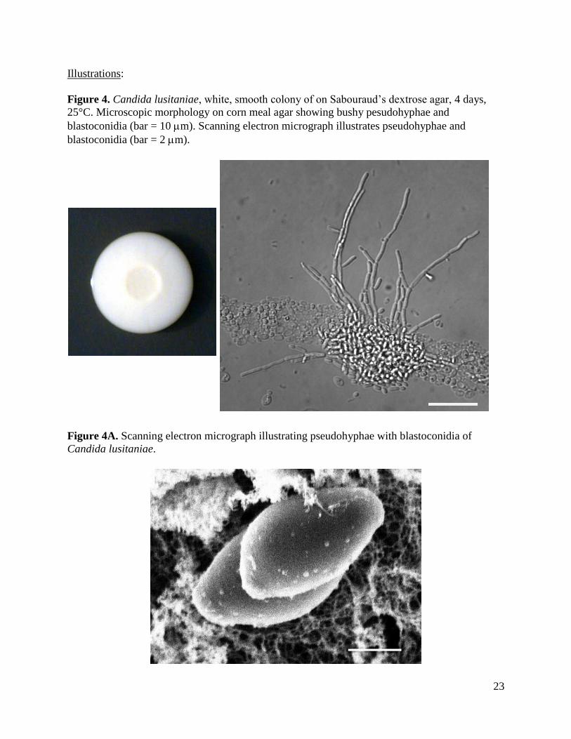
Colony: C. lusitaniae colony is white to creamish, shiny, and slightly raised in the center on
Sabouraud’s dextrose agar, after 7 days of incubation at 25°C (Figure 4).
Microscopy: C. lusitaniae produced many short, branched (“bushy”) pseudohyphae. Along the
length of the pseudohyphae, elongated blastoconidia formed in short chains on cornmeal agar
with Tween 80 (Figure 4).
Differentiation: C. lusitaniae is able to ferment and assimilate cellobiose, which differentiates it
from C. parapsilosis.
Molecular test: Specific nucleic acid probes targeting the large subunit rRNA genes have been
developed for identification of C. lusitaniae. Three pulsed-field electrophoretic methods and a
random amplified polymorphic DNA (RAPD) method were also reported to delineate strains of
C. lusitaniae.
The ribosomal ITS1 and ITS2 regions of the test isolate showed 100 % nucleotide identity with
Candida lusitaniae (Clavispora lusitaniae) isolate F47819-04 (GenBank accession no.
HQ693785.1).
Antifungal susceptibility: Some C. lusitaniae strains are reported to be inherently resistant to
amphotericin B. Amphotericin B susceptible strains are also known to develop resistance during
the course of treatment with this drug. C. lusitaniae is reported as more susceptible to
voriconazole than fluconazole.
Participant performance:
Referee Laboratories with correct ID: 10
Laboratories with correct ID: 106
Laboratories with incorrect ID: 1
(Trichosporon sp.) (1)
23
Illustrations:
Figure 4. Candida lusitaniae, white, smooth colony of on Sabouraud’s dextrose agar, 4 days,
25°C. Microscopic morphology on corn meal agar showing bushy pesudohyphae and
blastoconidia (bar = 10 m). Scanning electron micrograph illustrates pseudohyphae and
blastoconidia (bar = 2 m).
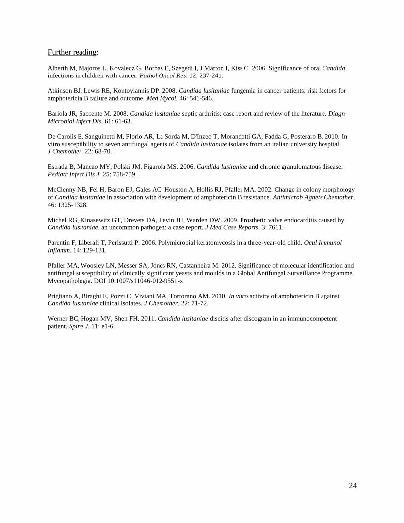
Figure 4A. Scanning electron micrograph illustrating pseudohyphae with blastoconidia of
Candida lusitaniae.
24
Further reading:
Alberth M, Majoros L, Kovalecz G, Borbas E, Szegedi I, J Marton I, Kiss C. 2006. Significance of oral Candida
infections in children with cancer. Pathol Oncol Res. 12: 237-241.
Atkinson BJ, Lewis RE, Kontoyiannis DP. 2008. Candida lusitaniae fungemia in cancer patients: risk factors for
amphotericin B failure and outcome. Med Mycol. 46: 541-546.
Bariola JR, Saccente M. 2008. Candida lusitaniae septic arthritis: case report and review of the literature. Diagn
Microbiol Infect Dis. 61: 61-63.
De Carolis E, Sanguinetti M, Florio AR, La Sorda M, D'Inzeo T, Morandotti GA, Fadda G, Posteraro B. 2010. In
vitro susceptibility to seven antifungal agents of Candida lusitaniae isolates from an italian university hospital.
J Chemother. 22: 68-70.
Estrada B, Mancao MY, Polski JM, Figarola MS. 2006. Candida lusitaniae and chronic granulomatous disease.
Pediatr Infect Dis J. 25: 758-759.
McClenny NB, Fei H, Baron EJ, Gales AC, Houston A, Hollis RJ, Pfaller MA. 2002. Change in colony morphology
of Candida lusitaniae in association with development of amphotericin B resistance. Antimicrob Agnets Chemother.
46: 1325-1328.
Michel RG, Kinasewitz GT, Drevets DA, Levin JH, Warden DW. 2009. Prosthetic valve endocarditis caused by
Candida lusitaniae, an uncommon pathogen: a case report. J Med Case Reports. 3: 7611.
Parentin F, Liberali T, Perissutti P. 2006. Polymicrobial keratomycosis in a three-year-old child. Ocul Immunol
Inflamm. 14: 129-131.
Pfaller MA, Woosley LN, Messer SA, Jones RN, Castanheira M. 2012. Significance of molecular identification and
antifungal susceptibility of clinically significant yeasts and moulds in a Global Antifungal Surveillance Programme.
Mycopathologia. DOI 10.1007/s11046-012-9551-x
Prigitano A, Biraghi E, Pozzi C, Viviani MA, Tortorano AM. 2010. In vitro activity of amphotericin B against
Candida lusitaniae clinical isolates. J Chemother. 22: 71-72.
Werner BC, Hogan MV, Shen FH. 2011. Candida lusitaniae discitis after discogram in an immunocompetent
patient. Spine J. 11: e1-6.
25
ANTIFUNGAL SUSCEPTIBILITY TESTING FOR YEASTS
Introduction: Clinical laboratories perform susceptibility testing of pathogenic yeasts to
determine their in vitro resistance to antifungal drugs. This test is also useful in conducting
surveillance for evolving patterns of antifungal drug resistance in a healthcare facility. The
results are likely to facilitate the selection of appropriate drugs for treatment. Clinical
Laboratory Standards Institute (CLSI) documents of M27-A3, M27-S3, M27-S4, and M44-A,
describe the current standard methods for antifungal susceptibility testing of pathogenic yeasts.
Another resource for standardized method is the EUCAST Definitive Document EDef 7.1:
method for the determination of broth dilution MICs of antifungal agents for fermentative yeasts.
The FDA approved devices for antifungal susceptibility testing of yeasts include Sensititre
YeastOne Colorimetric Panel (Trek Diagnostic Systems Inc. Cleveland, OH) and Etest
(bioMérieux, Inc., Durham, NC). The following ten drugs are included in the Mycology
Proficiency Test Program - amphotericin B, anidulafungin, caspofungin, flucytosine (5-FC),
fluconazole, itraconazole, ketoconazole, micafungin, posaconazole, and voriconazole. The
participating laboratories are allowed to select any number of antifungal drug(s) from this test
panel based upon practices in their facilities.
Materials: Candida parapsilosis (S-1) was the analyte in the May 27, 2015 antifungal
proficiency testing event. The interpretation of MIC values for antifungal susceptibility testing of
yeasts and molds is in a state of constant change. These changes are necessitated by new
information emerging from clinical trials and laboratory susceptibility testing. NYSDOH
Mycology Laboratory uses latest CLSI and EUCAST documents to score proficiency testing
results. However, the participating laboratories are advised to regularly consult these
organizations for the latest version of their standard documents.
Comments: Acceptable results were MICs +/-2 dilutions of the reference laboratory results for
any single drug. Only 2 of the 32 laboratories participating in this test event tested all 10
antifungal drugs. The reported results were as follows: voriconazole (29 laboratories),
caspofungin and itraconazole (25 laboratories respectively), flucytosine (22 laboratories),
amphotericin B (21 laboratories), anidulafungin and micafungin (18 laboratories respectively),
posacoanazole (17 laboratories), and ketoconazole (3 laboratories). Fluconazole was the only
drug tested by all 32 laboratories.
26
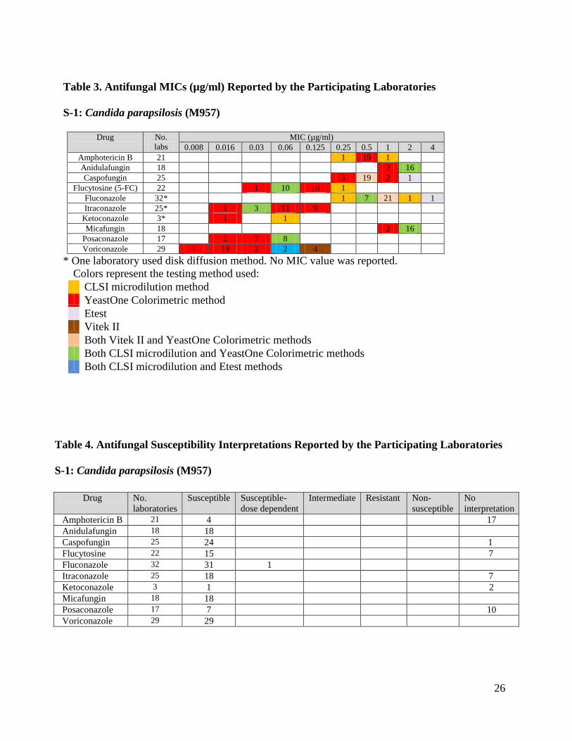
Table 3. Antifungal MICs (µg/ml) Reported by the Participating Laboratories
S-1: Candida parapsilosis (M957)
Drug
No.
labs
MIC (µg/ml)
0.008 0.016 0.03 0.06 0.125 0.25 0.5 1 2 4
Amphotericin B 21 1 19 1
Anidulafungin 18 2 16
Caspofungin 25 3 19 2 1
Flucytosine (5-FC) 22 1 10 10 1
Fluconazole 32* 1 7 21 1 1
Itraconazole 25* 1 3 11 9
Ketoconazole 3* 1 1
Micafungin 18 2 16
Posaconazole 17 2 7 8
Voriconazole 29 1 19 2 2 4
* One laboratory used disk diffusion method. No MIC value was reported.
Colors represent the testing method used: CLSI microdilution method YeastOne Colorimetric method Etest Vitek II Both Vitek II and YeastOne Colorimetric methods Both CLSI microdilution and YeastOne Colorimetric methods Both CLSI microdilution and Etest methods
Table 4. Antifungal Susceptibility Interpretations Reported by the Participating Laboratories
S-1: Candida parapsilosis (M957)
Drug No.
laboratories
Susceptible
Susceptible-
dose dependent
Intermediate Resistant Non-
susceptible
No
interpretation
Amphotericin B 21 4 17
Anidulafungin 18 18
Caspofungin 25 24 1
Flucytosine 22 15 7
Fluconazole 32 31 1
Itraconazole 25 18 7
Ketoconazole 3 1 2
Micafungin 18 18
Posaconazole 17 7 10
Voriconazole 29 29
27
ANTIFUNGAL SUSCEPTIBILITY TESTING FOR MOLDS
(EDUCATIONAL)
Introduction: Clinical laboratories perform susceptibility testing of pathogenic molds to
determine their in vitro resistance to antifungal drugs. This test is also useful in conducting
surveillance for evolving patterns of antifungal drug resistance in a healthcare facility. It is not
clear at this juncture if the results of mold susceptibility testing have direct relevance in the
selection of appropriate drugs for treatment. Clinical Laboratory Standards Institute (CLSI)
document of M38-A2 describes the current standard methods for antifungal susceptibility testing
of pathogenic molds. Another resource for standardized method is the EUCAST Technical Note
on the method for the determination of broth dilution minimum inhibitory concentrations of
antifungal agents for conidia-forming moulds. The following nine drugs are included in the
antifungal susceptibility panel - amphotericin B, anidulafungin, caspofungin, fluconazole,
itraconazole, ketoconazole, micafungin, posaconazole, and voriconazole.
Materials: Aspergillus fumigatus M2040 was used as a test analyte; it was obtained from a
reference laboratory. Participating laboratories volunteered to perform the test and they were free
to choose any number of drugs and a test method. Three laboratories used CLSI broth
microdilution method while the remaining one laboratories used TREK YeastOne Colorimetric
method.
Comments: Four out of thirty-two laboratories, which hold antifungal susceptibility testing for
yeasts permit, voluntarily participated in this test event for molds. Please refer to Table 5 for
summary of performances. Since too few laboratories have participated in this test, no consensus
data could be generated.

28
Table 5. MIC (g/ml) Values of Mold Antifungal Susceptibility: Aspergillus fumigatus M2040
Drugs (µg/ml) Total # of labs 0.008 0.015 0.03 0.06 0.12 0.25 0.5 1.0 2.0 4.0 8.0 16 32 64 256
Amphotericin B 4 2 1 1
Anidulafungin 4 2 1 1
Caspofungin 4 2 1 1
Fluconazole 3 2 1
Itraconazole 5 1 3
Ketoconazole 1 1
Micafungin 4 1 2 1
Posaconazole 4 3 1
Voriconazole 4 1 2 1
Colors represent the testing method used:
CLSI microdilution method
YeastOne Colorimetric method
Both CLSI microdilution and YeastOne Colorimetric methods
29
Further Reading:
Canton E, Peman J, Gobernado M, Alvarez E, Baquero F, Cisterna R, Gil J, Martin-Mazuelos E, Rubio C, Sanchez-
Sousa A, Settano C. 2005. Sensititre YeastOne caspofungin susceptibility testing of Candida clinical isolates:
correlation with results of NCCLS M27-A2 multicenter study. Antimicrobiol Agents Chemother. 49: 1604-1607.
Clinical and Laboratory Standards Institute. 2008. Reference Method for Broth Dilution Antifungal Susceptibility
Testing of Yeasts; Approved Standard - Third Edition. CLSI document M27-A3 (ISBN 1-56238-666-2).
Clinical and Laboratory Standards Institute. 2008. Quality Control Minimal Inhibitory Concentration (MIC) Limits
for Broth Microdilution and MIC Interpretive Breakpoints; Informational Supplement - Third Edition. CLSI
document M27-S3 (ISBN 1-56238-667-0).
Clinical and Laboratory Standards Institute. 2008. Reference Method for Broth Dilution Antifungal Susceptibility
Testing of Filamentous Fungi; Approved Standard – Second Edition. CLSI document M38-A2 (1-56238-668-9).
Clinical and Laboratory Standards Institute. 2009. Method for Antifungal Disk Diffusion Susceptibility Testing of
Yeasts; Approved Guideline – Second Edition. CLSI document M44-A2 (ISBN 1-56238-703-0).
Clinical and Laboratory Standards Institute. 2009. Zone Diameter Interpretive Standards, Corresponding Minimal
Inhibitory Concentration (MIC) Interpretive Breakpoints, and Quality Control Limits for Antifungal Disk Diffusion
Susceptibility Testing of Yeasts; Informational Supplement. CLSI document M44-S3.
Clinical and Laboratory Standards Institute. 2010. Method for Antifungal Disk Diffusion Susceptibility Testing of
Nondermatophyte Filamentous Fungi; Approved Guideline. CLSI document M51-A (ISBN 1-56238-725-1).
Clinical and Laboratory Standards Institute. 2010. Performance Standards for Antifungal Disk Diffusion
Susceptibility Testing of Filamentous Fungi; Informational Supplement. CLSI document M51-S1 (ISBN 1-56238-
725-1).
Clinical and Laboratory Standards Institute. 2012. Reference Method for Broth Dilution Antifungal Susceptibility
Testing of Yeasts; Fourth Informational Supplement. CLSI document M27-S4 (ISBN 1-56238-863-0).
Subcommittee on Antifungal Susceptibility Testing (AFST) of the ESCMID European Committee for Antimicrobial
Susceptibility Testing (EUCAST). 2008. EUCAST technical note on fluconazole. Clin Microbiol Infect. 14: 193-
195.
Subcommittee on Antifungal Susceptibility Testing (AFST) of the ESCMID European Committee for Antimicrobial
Susceptibility Testing (EUCAST). 2008. EUCAST definitive document Edef 7.1: method for the determination of
broth dilution MICs of antifungal agents for fermentative yeasts. Clin Microbiol Infect. 14: 398-405.
Subcommittee on Antifungal Susceptibility Testing (AFST) of the ESCMID European Committee for Antimicrobial
Susceptibility Testing (EUCAST). 2008. EUCAST technical note on the method for the determination of broth
dilution minimum inhibitory concentrations of antifungal agents for conidia–forming moulds. Clin Microbiol Infect.
14: 982-984.
Subcommittee on Antifungal Susceptibility Testing (AFST) of the ESCMID European Committee for Antimicrobial
Susceptibility Testing (EUCAST). 2008. EUCAST technical note on voriconazole. Clin Microbiol Infect. 14: 985-
987.
Copyright 2015 Wadsworth Center
New York State Department of Health