mycobacterium tuberculosis utilizes a unique heterotetrameric structure for...
TRANSCRIPT

Mycobacterium tuberculosis Utilizes a Unique HeterotetramericStructure for Dehydrogenation of the Cholesterol Side ChainSuzanne T. Thomas and Nicole S. Sampson*
Department of Chemistry, Stony Brook University, Stony Brook, New York 11794, United States
*S Supporting Information
ABSTRACT: Compounding evidence supports the importantrole in pathogenesis that the metabolism of cholesterol byMycobacterium tuberculosis plays. Elucidating the pathway bywhich cholesterol is catabolized is necessary to understand themolecular mechanism by which this pathway contributes toinfection. On the basis of early metabolite identification studiesin multiple actinomycetes, it has been proposed thatcholesterol side chain metabolism requires one or more acyl-CoA dehydrogenases (ACADs). There are 35 genes annotatedas encoding ACADs in the M. tuberculosis genome. Here wecharacterize a heteromeric ACAD encoded by Rv3544c and Rv3543c, formerly named fadE28 and fadE29, respectively. We nowrefer to genes Rv3544c and Rv3543c as chsE1 and chsE2, respectively, in recognition of their validated activity in cholesterol sidechain dehydrogenation. Analytical ultracentrifugation and liquid chromatography−ultraviolet experiments establish that ChsE1−ChsE2 forms an α2β2 heterotetramer, a new architecture for an ACAD. Our bioinformatic analysis and mutagenesis studies revealthat heterotetrameric ChsE1−ChsE2 has only two active sites. E241 in ChsE2 is required for catalysis of dehydrogenation byChsE1−ChsE2. Steady state kinetic analysis establishes the enzyme is specific for an intact steroid ring system versushexahydroindanone substrates with specificity constants (kcat/KM) of (2.5 ± 0.5) × 105 s−1 M−1 versus 9.8 × 10² s−1 M−1,respectively, at pH 8.5. The characterization of a unique ACAD quaternary structure involved in sterol metabolism that isencoded by two distinct cistronic ACAD genes opens the way to identification of additional sterol-metabolizing ACADs in M.tuberculosis and other actinomycetes through bioinformatic analysis.
Mycobacterium tuberculosis, the causative agent of tuberculosis,is the second most deadly infection in the adult populationworldwide; 8.8 million cases were reported in 2010 and 1.4million deaths attributed to the disease.1 During infection, M.tuberculosis bacilli are phagocytosed by macrophages, and thehost inflammatory response results in the formation of agranuloma.2 Bacteria can persist within the granuloma fordecades.M. tuberculosis metabolism of cholesterol, an abundant host
lipid, is important for the persistence of infection in vivo.3,4
Disruption of an Mce4 ABC transporter mutant results in thedefective transport of cholesterol into the mycobacterium.4,5 Ininfected mice, the mce4 mutant’s growth is slowed in thechronic phase of infection. fadA5, a cholesterol-regulated genethat encodes a thiolase, is required for the growth of M.tuberculosis on cholesterol as a carbon source.6 The number ofbacteria in the lungs of mice infected with the fadA5 mutantdecreased after induction of the cellular immune response,demonstrating a persistence phenotype identical to that seenupon mutation of the Mce4 transporter.4 Thiolases catalyze thereverse Claisen condensation step in lipid β-oxidation.Similarly, the M. tuberculosis igr operon, which encodes sixproteins, is important for growth on cholesterol in vitro and formycobacterial survival in vivo and encodes homologues of lipid-modifying enzymes.7,8
The M. tuberculosis igr operon (Rv3545c−Rv3540c) isrequired for complete degradation of the cholesterol sidechain. Upon disruption of this operon, steroid-derivedmetabolite methyl 1β-(2′-propanoate)-3aα-H-4α-(3′-propanoicacid)-7aβ-methylhexahydro-5-indanone accumulates in M.tuberculosis Δigr cultures grown in the presence of cholesterol(Scheme 1).9 Rv3544c and Rv3543c, annotated as potentialacyl-CoA dehydrogenases, are encoded by two adjacent
Received: March 7, 2013Revised: April 4, 2013Published: April 5, 2013
Scheme 1. Methyl 1β-(2′-Propanoate)-3aα-H-4α-(3′-propanoic acid)-7aβ-methylhexahydro-5-indanoneAccumulates in an igr-Disrupted Strain of M. tuberculosiswhen It Is Cultured with Cholesterol
Article
pubs.acs.org/biochemistry
© 2013 American Chemical Society 2895 dx.doi.org/10.1021/bi4002979 | Biochemistry 2013, 52, 2895−2904

cistronic genes. Heterologously co-expressed N-His6-taggedRv3544c and tagless Rv3543c are copurified upon immobilizedmetal affinity chromatography, indicating they form aquaternary complex. We demonstrated that the complex is afunctional acyl-CoA dehydrogenase that dehydrogenatessteroid-derived substrates analogous to the metabolite isolatedfrom an igr knockout strain.9
To the best of our knowledge, this was the first report of aheteromeric acyl-CoA dehydrogenase (ACAD). All ACADscharacterized to date form homotetrameric assembliescomprised of four ∼43 kDa monomers, with one exception.The ACAD subfamilies specific for very long chain fatty acids(VLCAD and ACAD9) form homodimers from two ∼73 kDamonomers.10,11
Herein, we present a full enzymatic and biophysicalcharacterization of Rv3543c−Rv3544c and demonstrate thatRv3543c−Rv3544c is an α2β2 heterotetramer. Neither Rv3543cnor Rv3544c is functionally competent by itself. Extensivesequence alignments of Rv3543c and Rv3544c with acyl-CoAdehydrogenases with known three-dimensional structuresreveal that a canonical active site glutamate required forgeneral base catalysis is conserved in Rv3543c, but not inRv3544c. Mutagenesis in combination with kinetic assaysconfirmed that Glu241 of Rv3543c is the base required forcatalysis by this enzyme. Furthermore, Rv3543c−Rv3544c hasonly two conserved FAD binding sites per tetramer. Formationof the Rv3543c−Rv3544c complex is required to constitutefunctional binding sites and consequently catalytic activity. Wepropose that this unique heteromeric structure is required foreffecting dehydrogenation of steroyl-CoA substrates and maybe a motif that is utilized for polycyclic-CoA esters in general.Therefore, we now refer to the Rv3544c and Rv3543c acyl-CoAdehydrogenase genes as chsE1 and chsE2, respectively, forcholesterol side chain metabolism E, to distinguish them frommembers of the bacterial fadE, fatty acid degradation E acyl-CoA dehydrogenase, gene family.
■ MATERIALS AND METHODS
Materials, Strains, Media, and General Methods.Ferricenium hexafluorophosphate and ergocalciferol werepurchased from Sigma-Aldrich (St. Louis, MO). Coenzyme Awas purchased from MP Biomedicals (Solon, OH). Isopropylβ-D-1-thiogalactopyranoside was from Denville Scientific(Metuchen, NJ). Tryptone, HEPES, Tris, and ampicillin werepurchased from Fisher Scientific (Pittsburgh, PA). L-Arabinose,chloramphenicol, and sodium chlorite were purchased fromAcros Organics. Tetracycline was from U.S. Biochemical Corp.(Cleveland, OH), and kanamycin was from IBI Scientific(Peosta, IA). Yeast extract was purchased from Research
Products International Co. (Mount Prospect, IL). iProof DNApolymerase was from Bio-Rad (Hercules, CA). Restrictionendonucleases, T4 DNA ligase, T4 polynucleotide kinase, andthe protein ladder were from New England Biolabs (Beverly,MA). HisTrap FF columns, MonoQ, and Superdex 75 HiLoad16/60 and 10/300 GL columns were from GE HealthcareBiosciences Corp. (Piscataway, NJ). Oligonucleotides werefrom IDT Inc. (Coralville, IA). Total genomic DNA of M.tuberculosis H37Rv was obtained from the TB ResearchMaterials Facility at Colorado State University (Fort Collins,CO) (NIAD NO1-AI40091). MALDI mass spectra wereacquired on a Bruker Autoflex II TOF/TOF instrument. BigDye DNA sequencing (Applied Biosystems, Foster City, CA;performed by the Stony Brook University Sequencing Facility)was used to verify the coding sequence of the expressionplasmids. BL21(DE3) Escherichia coli was obtained from Bio-Rad, and chaperone plasmid pG-KJE8 was obtained fromTakara. Mycobacterium smegmatis strain mc2155 was obtainedfrom E. Dubnau (PHRI). 2× YT is composed of 16 g oftryptone, 10 g of yeast extract, and 5 g of NaCl per liter. LC−UV−MS analysis was conducted on a Waters UPLC/MSinstrument with diode array and SQD detectors. Buffer Aconsisted of 20 mM Tris-HCl buffer (pH 8.0), supplementedwith 300 mM NaCl, 1 mM TCEP, and 10 mM imidazole.Buffer B consisted of 50 mM Tris-HCl buffer (pH 8.0). BufferC consisted of 50 mM Tris-HCl buffer (pH 8.0), supplementedwith 200 mM NaCl and 1 mM TCEP. Buffer D consisted of 20mM sodium phosphate buffer (pH 7.5), supplemented with200 mM NaCl and 1 mM TCEP.
Expression Plasmid Construction. The desired geneswere amplified from M. tuberculosis H37Rv total genomic DNAby polymerase chain reaction (PCR) using forward and reverseprimers. The PCR product was digested with the appropriaterestriction endonuclease and ligated into a similarly digestedvector (Table 1). DNA sequencing of the plasmids confirmedthat the sequence was correct and that no mutations wereintroduced during the cloning procedures. ChsE2 glutamate241 was mutated to a glutamine in pigr5 using the method ofMoore.12 The mutation was confirmed by DNA sequencing.
Individual Gene Expression. pChsE1N or pChsE2Nconstructs were transformed into BL21(DE3) E. coli, andsingle colonies were selected on LB plates containing 30 μg/mL kanamycin and cultured in 2× YT medium at 37 °C.Expression was induced at an A600 of 0.6−0.8 by the addition of50 μM to 1 mM isopropyl 1-thio-β-D-galactopyranoside(IPTG), and cells were grown for 20 h at 16 or 25 °C.
Gene Expression in M. smegmatis. pChsE2Ms waselectroporated into M. smegmatis mc2155, and single colonieswere selected on 7H10 plates supplemented with 100 μg/mL
Table 1. Plasmids Constructed
construct name plasmid gene fusion tag restriction sites used antibiotic marker source or ref
pET20b amp NovagenpET28b kan NovagenpSD31 hygro 13pG-KJE8 cam Takara
pChsE1N pET28b Rv3544c N-terminal His6 NdeI/NotI kan this workpChsE2N pET28b Rv3543c N-terminal His6 NdeI/XhoI kan this workpChsE1 pET20b Rv3544c − NdeI/NotI amp this workpChsE2Ms pSD31 Rv3543c N-terminal His6 EcoRv hygro this workpigr5 pET28b Rv3544c−Rv3540c N-terminal His6 NdeI/HindIII kan 9pigr5E241Q pET28b Rv3544c−Rv3540c N-terminal His6 NdeI/HindIII kan this work
Biochemistry Article
dx.doi.org/10.1021/bi4002979 | Biochemistry 2013, 52, 2895−29042896

ampicillin, 10 μg/mL cycloheximide, and 50 μg/mL hygrom-ycin and grown in 7H9 medium supplemented with 0.2%glycerol. After 2 days, expression was induced with 0.2%acetamide and cultures were grown for an additional 24 h.Gene Co-Expression. Constructs pChsE2N and pChsE1
were cotransformed into BL21(DE3) E. coli::pG-KJE8 (chap-erone plasmid, Takara). Single colonies were selected on LBplates containing the appropriate antibiotics (100 μg/mLampicillin for pET20b, 30 μg/mL kanamycin for pET28b, and20 μg/mL chloramphenicol for pG-KJE8) and cultured in 2×YT medium at 37 °C. Chaperone expression was induced uponinoculation with 2 mg/mL L-arabinose and 5 ng/mLtetracycline. ChsE expression was induced at an A600 of 0.6−0.8 with the addition of 1 mM IPTG, and cells were grown for20 h at 25 °C. Gene co-expression in cis with construct pigr5was performed as reported previously.9 Similarly, theChsE2E241Q mutant protein was prepared with constructpigr5E241Q using the expression conditions that were used forpigr5.Protein Purification. ChsE1- or ChsE2-expressing cells
were lysed with a French press in buffer A, and cellular debriswas removed by centrifugation at 125000g for 1 h. Proteinswere purified by immobilized metal affinity chromatographyusing Hisbind resin (Novagen) following the manufacturer’sprotocol. Soluble ChsE2 was buffer exchanged with buffer Band further purified by anion exchange chromatography on aMonoQ column (1 mL) equilibrated in buffer B. Protein waseluted at a flow rate of 0.5 mL/min with a linear gradient from100 to 100% buffer B supplemented with 1 M NaCl. Afterinjection, the column was washed with 5 column volumes (CV)of buffer B, which was then changed to 80% buffer B over 20CV. Next, the gradient was changed from 80 to 25% buffer Bover 10 CV and then to 0% buffer B over 5 CV. Eluted proteinwas further purified by size exclusion chromatography on aSuperdex 200 HiLoad 16/60 column (GE Healthcare)equilibrated with buffer C. ChsE1−ChsE2 and ChsE1−ChsE2E241Q were purified as detailed for ChsE2, excluding theanion exchange chromatography step. All protein samples wereanalyzed by reducing sodium dodecyl sulfate−polyacrylamidegel electrophoresis (SDS−PAGE), and protein band identitieswere confirmed by in-gel tryptic digestion as reportedpreviously.14 ChsE proteins were stored in 50 mM HEPESbuffer (pH 8.0) at −80 °C.Identification and Quantification of a Flavin Cofactor.
A 15 μM (1.2 mg/mL) solution of ChsE1−ChsE2 in buffer Cwas denatured by being boiled for 2 min. The sample waschilled on ice and centrifuged to pellet precipitated protein.The supernatant was analyzed by reverse phase C18 liquidchromatography−mass spectrometry in ESI positive mode andcompared to the flavin adenine dinucleotide standard. Thequantity of FAD obtained was determined using the absorbanceand extinction coefficient of FAD at 260 nm (ε260 = 37000 M−1
cm−1). The protein pellet was dissolved in 6 M guanidinehydrochloride and the concentration determined using thecalculated extinction coefficients at 280 nm: ε280(ChsE1) =35410 M−1 cm−1, ε280(ChsE2) = 58900 M−1 cm−1, andε280(ChsE1−ChsE2) = 188620 M−1 cm−1.Biophysical Analysis of ChsE Proteins. ChsE2, ChsE1−
ChsE2, and ChsE1−ChsE2E241Q samples were analyzed byanalytical gel filtration on a Superdex 75 10/300 GL column(GE Healthcare) equilibrated with buffer D. Samples wereeluted isocratically in the same buffer, monitoring at 220 and280 nm.
Molecular masses of ChsE2 and ChsE1−ChsE2 weredetermined using analytical ultracentrifugation sedimentationequilibrium with a Beckman Optima XL-A centrifuge. ChsE2(8.5, 3.4, and 1.7 μM) in 5 mM sodium phosphate buffer (pH7.5) and ChsE1−ChsE2 (10.6, 5.3, and 2.6 μM) in buffer Bwere centrifuged at 20K, 25K, and 30K rpm and 20 °C. Scanswere acquired after centrifugation for 18 and 20 h at each speedby monitoring at 280 nm. The protein partial-specific volumesof 0.7336 for ChsE2 and 0.7350 for ChsE1−ChsE2 and thesolvent densities of 0.9994 for ChsE2 and 1.0079 for ChsE1−ChsE2 were calculated using SEDNTERP (University of NewHampshire, Durham, NH). Data were fit globally to the ideal,single-species model using Heteroanalysis (University ofConnecticut, Storrs, CT) to determine the molecular mass.The protein complex stoichiometry of ChsE1−ChsE2 was
confirmed by LC−UV−MS (Waters UPLC/diode array/SQD). ChsE1 and ChsE2 were separated on a Waters XBridgeBEH300 C4 3.5 μm column (2.1 mm × 100 mm) at 40 °C witha linear gradient from 95% A to 95% B over 15 min, where Aconsists of 5% 2-propanol and 0.1% trifluoroacetic acid and Bconsists of 99.9% 2-propanol and 0.1% trifluoroacetic acid. MSspectra were collected in ESI positive ion mode with a conevoltage of 40 V, a capillary voltage of 4.5 kV, and a sourcetemperature of 150 °C. MS spectra were deconvolved usingESIprot version 1.0,15 and peaks in the UV 280 nmchromatograms were integrated using R. The integrated peakareas of each protein were divided by the corresponding molarextinction coefficient for the protein to yield the molarconcentrations. The protein stoichiometry was determinedfrom the ratio of the molar concentrations.
Bioinformatic Analysis. ACAD protein sequences wereobtained from UniProt, and sequence alignments wereconducted with ClustalW2 (EMBL-EBI, European Bioinfor-matics Institute, Cambridge, U.K.) using the default parame-ters.16 FAD binding and CoA binding in published ACADProtein Data Bank (PPDB) structures were investigated withPoseView (Center of Bioinformatics, University of Hamburg,Hamburg, Germany).17 A ChsE1−ChsE2 ligand binding modelwas created using the MCAD homodimer (PDB entry 1EGC)as a template. The model is based on a ChsE1−ChsE2heterodimer assembly and highlights conserved residuesdetermined from the ACAD sequence alignment with ChsE1and ChsE2.
Dehydrogenase Assay. CoA thioesters, 3-oxo-4-pregnene-20-carboxyl-CoA (1) and 1β-(2′-propanoyl-CoA)-3aα-H-7aβ-methylhexahydro-4-indanone (2), were prepared and purifiedas reported previously.9,18 The identities and purity of CoAthioesters were assessed by LC−UV−MS.Dehydrogenase activity was tested with artificial electron
acceptor ferricenium hexafluorophosphate as reported pre-viously.19 Product formation was monitored spectroscopicallyat 300 nm and 25 °C. Initial velocities were determined for thefirst 100 s of the reaction. Enzyme activity was optimized forpH using the three component buffer system reported by Ellisand Morrison with 50 μM acyl-CoA 1.20 Subsequent assayswere conducted in 100 mM TAPS buffer (pH 8.5). Theoptimal ionic strength was investigated at NaCl concentrationsfrom 0 to 1.0 M. Protein stability and substrate 1 solubility weretested at concentrations of 100 nM and 50 μM, respectively,with and without 0.2 M NaCl by dynamic light scattering on a90 Plus particle size analyzer from Brookhaven InstrumentsCorp. Data were acquired in triplicate at 25 °C.
Biochemistry Article
dx.doi.org/10.1021/bi4002979 | Biochemistry 2013, 52, 2895−29042897

Steady state kinetic analysis was conducted in 100 mM TAPSbuffer (pH 8.5) with CoA thioesters 1 and 2. The rates ofproduct formation were fit to the Michaelis−Menten equation(eq 1) to determine the KM values for substrates 1 and 2 andthe kcat for enoyl-CoA product formation.
= +v V K[S]/( [S])max M (1)
where v is the initial velocity, Vmax is the maximal velocity, S isthe varied substrate, and KM is the Michaelis−Menten constant.NMR Analysis of the Assay Product. The product of the
ChsE1−ChsE2 assay with substrate 3-oxo-4-pregnene-20-carboxyl-CoA was purified on a 3 cc sep-pak C18 cartridge(Waters) equilibrated in a 5% MeOH/95% 10 mM ammoniumacetate mixture (pH 8.0). After the sample had been loaded, thecartridge was washed with 6 mL of equilibration buffer followedby 6 mL of a 25% MeOH/75% 10 mM ammonium acetatemixture (pH 8.0). The product was eluted in a 45% MeOH/55% 10 mM ammonium acetate mixture (pH 8.0). The driedcompound was dissolved in D2O for NMR analysis on a Bruker700 MHz NMR instrument. 1H NMR spectra of substrate 1and the purified product were acquired using excitationsculpting for water suppression.21
■ RESULTSChsE2 Is Isolated as a Monomeric Apoenzyme When
Heterologously Expressed in either E. coli or M.smegmatis. Initially, chsE1 and chsE2 were expressedindividually in E. coli as N-terminally His6-tagged proteins.ChsE1 was found to be insoluble and was localized in inclusionbodies. ChsE2 was isolated and purified by immobilized metalaffinity, anion exchange, and size exclusion chromatographies(Figure S1 of the Supporting Information). UV−visiblespectroscopy revealed the protein was purified as theapoprotein, without the flavin cofactor necessary for catalysis(Figure 1). Attempts to reconstitute the protein with FAD werenot successful.
ChsE2 was analyzed by analytical size exclusion chromatog-raphy (Figure 2) and analytical ultracentrifugation (AUC)sedimentation equilibrium experiments (Figure 3A). Thepredicted monomer molecular mass with a His6 tag is 44.9kDa. The AUC data fit an ideal model with a molecular mass of42.0 ± 0.5 kDa, where the reported error is the measure of thefit and indicates the chosen model is satisfactory. Thus, under
the conditions tested, ChsE2 is a monomer. In crystalstructures of ACADs, one molecule of FAD binds non-covalently per monomer and the adenosine lies at the interfaceof the two monomers. Therefore, two monomer chainscomprise the FAD binding pocket, and ChsE2 may not bindFAD because of its monomeric state.Next, chsE2 was expressed in M. smegmatis, a nonpathogenic,
faster-growing relative of M. tuberculosis, as an N-terminallyHis6-tagged protein. The protein was isolated by immobilizedmetal affinity chromatography (IMAC) and further purified byanion exchange and size exclusion chromatographies. Analysisby analytical size exclusion chromatography indicated that M.
Figure 1. UV−visible spectra of purified ChsE2 (50 μM), ChsE1−ChsE2 (15 μM), and ChsE1−ChsE2E241Q (15 μM). Relative spectralmaxima at 370 and 446 nm for ChsE1−ChsE2 and ChsE1−ChsE2E241Q are characteristic of oxidized flavin. No flavin absorbancewas observed for ChsE2 alone.
Figure 2. Analytical gel filtration of ChsE2 and ChsE1−ChsE2complexes. ChsE2 expressed in E. coli and M. smegmatis and ChsE1−ChsE2 and ChsE1−ChsE2E241Q expressed in E. coli were analyzed byanalytical gel filtration on a Superdex 75 column under identicalconditions. The signal at 220 nm was normalized to 1.0. The ChsE1−ChsE2 protein formed a stable complex with a molecular mass higherthan that of ChsE2 expressed alone in E. coli or M. smegmatis.
Figure 3. Analytical ultracentrifugation sedimentation equilibrium datafor ChsE proteins. (A) ChsE2 and (B) ChsE1−ChsE2 were analyzedat three concentrations ranging from 1 to 11 μM at centrifugationspeeds of 20K, 25K, and 30K rpm at 20 °C. A representative fit foreach sample is shown. The solid line shows the fit of the data to theideal species model, and the residuals of the fit are graphed below. Theglobal fit for each protein provided molecular masses of 42.0 ± 0.5 and156 ± 1 kDa for ChsE2 and ChsE1−ChsE2, respectively.
Biochemistry Article
dx.doi.org/10.1021/bi4002979 | Biochemistry 2013, 52, 2895−29042898

smegmatis expressed ChsE2 was also monomeric (Figure 2).Analysis of ChsE2 expressed in E. coli and in M. smegmatisdemonstrates that the protein is not in the predicted quaternarystructure of ACADs, lacks FAD, and therefore is inactive.The ChsE1−ChsE2 Complex Forms an Obligate α2β2
Tetramer with Noncovalently Bound FAD. Isolation ofmonomeric ChsE2 apoprotein and insoluble ChsE1 indicatedthey do not form native structures as individual proteins.Previously, we had reasoned that these proteins might form acomplex because they are encoded in a single operon. Co-expression would allow assembly of ChsE1 and ChsE2 thatwould otherwise be insoluble or inactive when expressedindividually. Indeed, when chsE1 and chsE2 were expressed in acistronic construct on a single plasmid, pigr5, a complex ofChsE1 and ChsE2 was isolated in a pull-down assay.9
Here, we further investigated expression conditions andconstructs. We found that the ChsE1−ChsE2 complex wasobtained upon expressing chsE1 as a tagless protein and chsE2as an N-terminally His6-tagged protein from separate plasmids(in trans) in E. coli with co-expression of chaperones. We usedE. coli harboring plasmid pG-KJE8, which encodes five foldingchaperones, DnaK, DnaJ, GrpE, GroES, and GroEL. TheChsE1−ChsE2 protein complex was isolated regardless ofwhich protein, ChsE1 or ChsE2, contained an N-terminal His6tag. The identity of ChsE1 and ChsE2 in the complex wasconfirmed by SDS−PAGE (Figure S1 of the SupportingInformation) and in-gel tryptic digest followed by MALDI-TOF MS analysis.Under both in cis and in trans expression conditions, the
isolated complex was yellow in appearance and had acharacteristic UV−visible flavin spectrum, with absorbancemaxima at 370 and 446 nm (Figure 1). Expression in cis,however, yielded higher A446/A280 ratios, indicative of higherFAD occupancy. Kinetic studies were conducted with ChsE1−ChsE2 expressed with construct pigr5.The flavin cofactor was released from ChsE1−ChsE2 by heat
denaturation of the protein and was analyzed by LC−UV−MS.A protonated molecular ion at m/z 786 was observed (FigureS2 of the Supporting Information), confirming the identity ofthe cofactor as FAD. In addition, the retention time andabsorbance spectrum were identical to those of a FADstandard. Therefore, ChsE1−ChsE2 is isolated as a holoproteinwith the FAD cofactor.Analysis of the complex by analytical size exclusion
chromatography confirmed that a stable quaternary complexis formed (Figure 2). Further analysis by AUC sedimentationequilibrium experiments established that ChsE1−ChsE2 existsas a tetramer with a molecular mass of 156 ± 1 kDa (Figure3B). The predicted molecular masses of N-tagged ChsE1 andChsE2 are 37.6 and 42.7 kDa, respectively. To determineprecisely the stoichiometry of the complex, the assembly wasanalyzed by LC−UV−MS (Figure 4). The two observed peakswere identified as ChsE1 and ChsE2 by ESI+ MS. Theabsorbance peaks at 280 nm for ChsE1 and ChsE2 wereintegrated, and using the extinction coefficient for each protein,the relative molar stoichiometry of ChsE1 to ChsE2 wascalculated to be 1:1. This ratio indicates that the ChsE1−ChsE2species forms an α2β2 complex.The Tetrameric ChsE1−ChsE2 Complex Has Two
Cofactor Binding Sites. The FAD stoichiometry in theprotein complex was determined spectrophotometrically afterprotein denaturation. The protein concentration was deter-mined for the unfolded protein using the calculated extinction
coefficient of ChsE1 and ChsE2 at 280 nm. The concentrationof FAD was calculated from the extinction coefficient at 260nm after removal of precipitated ChsE1−ChsE2. TheFAD:protein molar ratio was 1.4 ± 0.1 FAD molecules perα2β2 ChsE1−ChsE2 tetramer. This result suggests that ChsE1−ChsE2 has two FAD bindings sites in contrast to typical ACADtetramers that have four FAD binding sites.To corroborate the FAD stoichiometry in heterotetrameric
ChsE1−ChsE2, we analyzed the protein sequences of ChsE1and ChsE2 for FAD binding residues that are conserved inother ACADs. First, we examined ligand−protein interactionsbetween FAD and several ACADs in reported crystal structures(PDB entry in parentheses), including human SCAD (2VIG),MCAD (1EGC), SBCAD (2JIF), IBD (1RX0), IVD (1IVH),GD (1SIQ), VLCAD (3B96), and ACAD11 (2WBI) andbacterial ACADS from Megsphaera elsdenii (1BUC) andMycobacterium thermoresistibile (3NF4). In each structure, asingle FAD cofactor binding site is comprised of two chainsfrom the ACAD tetramer or dimer, in the case of VLCAD andACAD9. Each chain interacts with adenosine in one bindingpocket and with riboflavin in a second binding pocket. Thepolar side chains of Thr161A, Ser167A, Thr193A, and Thr403Aform hydrogen bonds with the isoalloxazine and ribityldiphosphate moieties (numbering from MCAD). These Serand Thr residues are conserved within the ACAD family(Figure 5A).Analysis of the ChsE2 and ChsE1 sequence alignments with
these ACADs revealed that Thr161A, Ser167A, and Thr193Aare conserved in ChsE2 as residues Thr126, Thr132, andThr158, respectively. Thr403A is not conserved in ChsE2(Figure 5A). In contrast, Thr161A, Ser167A, Thr193A, andThr403A are not conserved in ChsE1.Residues involved in forming hydrogen bonds with
adenosine of FAD, Arg306A′, Gln317A′, Gln374A′, andGly378A′, are from the second chain that contributes to theFAD cofactor binding sites in ACADs and are highly conserved.Sequence analysis of ChsE1 revealed that Arg306A′, Gln317A′,Gln374A′, and Gly378A′ align with residues Arg227, Gln238,His295, and Gly299, respectively (Figure 5A). However,Arg306A′, Gln317A′, Gln374A′, and Gly378A′ are notconserved in ChsE2.
Figure 4. Reverse phase LC−UV chromatogram of ChsE1−ChsE2.Peak a and peak b were identified as ChsE2 and ChsE1, respectively,by deconvolution of multiple charged states in the corresponding ESI+MS spectra. The absorbance peaks were integrated and relativeconcentrations determined from the calculated extinction coefficientsof ChsE1 and ChsE2 [ε280(ChsE1) = 35410 M−1 cm−1, andε280(ChsE2) = 58900 M−1 cm−1].
Biochemistry Article
dx.doi.org/10.1021/bi4002979 | Biochemistry 2013, 52, 2895−29042899

In all ACAD crystal structures determined to date, two FADcofactors bind at each dimer interface. In contrast, only one setof FAD binding site residues is conserved in the homologymodel of the ChsE1−ChsE2 interface. The lack of conservedriboflavin binding residues in ChsE1 and adenosine bindingresidues in ChsE2 indicates that one FAD cofactor binds at theChsE1−ChsE2 heterodimer interface (Figure 5B), and that twoheterodimeric units comprise the heterotetramer. Thishomology model is consistent with the experimentallydetermined FAD stoichiometry for ChsE1−ChsE2. Isolationof monomeric apo-ChsE2 suggests it does not self-associate toform a homodimer and further supports our model.Tetrameric ChsE1−ChsE2 Has Two Active Sites. The
FAD binding stoichiometry and binding residue conservation intetrameric ChsE1−ChsE2 suggest that the complex has two
active sites. ACADs require a catalytic base for activity, and thisbase is highly conserved in the ACAD protein family (Figure5A). The catalytic base is most often a glutamate and is locatedat one of two positions in helix G or helices J and K.22
Glutamate at either of these two positions orients in the activesite in a catalytically competent configuration in the three-dimensional protein structures of ACADs.In the ChsE2 sequence alignment with representative ACAD
family members, Glu241 aligns with the glutamate of IVD andLCAD in helix G (Figure 5A). There is not a conservedglutamate in the alignment of helices J and K with ChsE2.Notably, ChsE1 does not have a glutamate or aspartate thataligns with either helix G or helices J and K. The conservedglutamates in helix G and helices J and K align with glycineresidues in ChsE1. The lack of a potential active site base, in
Figure 5. Bioinformatic analysis of ChsE1 and ChsE2. (A) Sequence alignment of ChsE1 and ChsE2 against human and bacterial ACADS: MCAD(P11310), SCAD (P16219), LCAD (P28330), SBCAD (P45954), iBD (Q9UKU7), IVD (P26440), GD (Q92947), VLCAD (P49748), ACAD9(Q9H845), ACAD10 (Q6JQN1), ACAD11 (Q709F0), 1BUC (SCAD Mg. elsdenii, Q06319), and 3NF4 (M. thermoresistibile, G7CDN2). Residueshighlighted in green bind the isoalloxazine and ribityl diphosphate moieties of FAD. Residues highlighted in blue bind adenosine of FAD. Residueshighlighted in purple bind CoA. Residues highlighted in yellow are the catalytic bases. (B) Homology model of FAD binding residues and (C)octanoyl-CoA binding residues in ChsE1−ChsE2 based on the human MCAD homodimer (PDB entry 1EGC). FAD is colored yellow and octanoyl-CoA green. FAD and CoA cofactors are shown adjacent to conserved binding residues.
Biochemistry Article
dx.doi.org/10.1021/bi4002979 | Biochemistry 2013, 52, 2895−29042900

addition to the lack of riboflavin binding residues in ChsE1,suggests that ChsE1−ChsE2 has only two active sites, incontrast to traditional homotetrameric ACADs, which have fouractive sites. On the basis of these alignments, we hypothesizedthat Glu241 of ChsE2 is the catalytic base responsible fordeprotonation during catalysis.Analysis of the CoA binding site in the three-dimensional
structures of ACADs identified conserved CoA hydrogen-bonding residues Ser142A, Arg256A, and Asp253A (numberingfor human MCAD). The side chain of Arg256 forms ahydrogen bond network with the pantetheine group of CoAand is highly conserved in the ACAD family. Arg256 isconserved in ChsE2 as Arg242. In ChsE1, Leu202 aligns withArg256. Asp253 in MCAD forms a hydrogen bond with theadenine moiety of CoA. In GD and IBD, however, this residueis an Asn, and analysis of the crystallographic structure showsthe backbone carbonyl of Asn forms a hydrogen bond withadenine. Asn is conserved in ChsE2 as residue Asn239, and thisresidue is Val204 in ChsE1. The lack of CoA binding residuesin ChsE1 supports our hypothesis that ChsE1 is not an activehomtetrameric ACAD.The ChsE1−ChsE2 Complex Forms 3-Oxo-pregna-
4,17-dien-20-carboxyl-CoA (3). 1H NMR spectra of
substrate 1 and product 3 were acquired in D2O (Figure 6).Comparison of substrate and product 1H NMR spectrahighlights the disappearance of the coupling of the C21 methylhydrogen to H21 (δ 1.09, d). The C21 methyl becomes asinglet at 1.84 ppm (Figure 6B). If C20−C21 bonddehydrogenation had occurred, two new alkene protonswould have replaced the C21 methyl in the product spectrum.However, a new proton resonance in the alkene region was notobserved (Figure 6C). We conclude that the ChsE1−ChsE2complex forms the thermodynamically favored tetrasubstitutedalkene product 3.
Steady State Kinetics of ChsE1−ChsE2. Previously, wehad demonstrated that the pregnene-carboxyl-CoA ester (1)and indanone CoA ester (2) are substrates of ChsE1−ChsE2(Figure 7A). Here, we determine the pH and ionic strengthdependence of ChsE1−ChsE2. Initial velocity data werecollected for ChsE1−ChsE2 with substrate 1 from pH 5.5 to9.5 in 0.5 pH unit steps. No activity was observed at pH ≤6.0,and the highest activity was observed at pH >8.0 (Figure 7B).Further experiments were conducted at pH 8.5 in TAPS buffer.The ChsE1−ChsE2 complex was then assayed as a function
of NaCl concentration to determine the optimal ionic strength(Figure 6C). Initial velocities were determined at NaCl
Figure 6. Characterization of the dehydrogenated product by ChsE1−ChsE2. 1H NMR spectra (700 MHz) of substrate 1 and ChsE1−ChsE2 assayproduct 3 illustrating the changes in the methyl (B) and alkene (C) regions. The alkene stereochemistry is represented as E but was not determined.
Figure 7. Optimization of dehydrogenase activity of ChsE1−ChsE2. (A) 3-Oxo-4-pregnene-20-carboxyl-CoA 1 and 1β-(2′-propanoyl-CoA)-3aα-H-7aβ-methylhexahydro-4-indanone 2, substrates of ChsE1−ChsE2. (B) pH and (C) ionic strength optimization of dehydrogenase activity. Assayswere conducted with 50 μM substrate 1 and 250 μM ferricenium hexafluorophosphate at 25 °C. Assays used to produce the data in panel C wereconducted in 100 mM TAPS buffer (pH 8.5).
Biochemistry Article
dx.doi.org/10.1021/bi4002979 | Biochemistry 2013, 52, 2895−29042901
concentrations from 0 to 1.0 M with 50 μM substrate 1.Increasing the ionic strength by addition of NaCl diminishedactivity, and no rate enhancement was observed even at lowconcentrations of NaCl. We tested the protein stability andsubstrate solubility at working concentrations using dynamiclight scattering at 0 and 0.2 M NaCl. The activity of ChsE1−ChsE2 was reduced by 50% in 0.2 M NaCl. However, neitherprotein aggregation nor substrate precipitation was observed.The decrease in enzyme activity is not due to proteindenaturation or substrate aggregation.Next, the ionic strength was increased with halide salts KCl,
MgCl2, CaCl2, NaI, and NaBr, as well as sodium acetate (FigureS3 of the Supporting Information). The specific activity wasreduced in the presence of all the tested salts. Further analysiswould be required to determine the cause of rate reduction.Steady state kinetic constants KM and kcat were determined
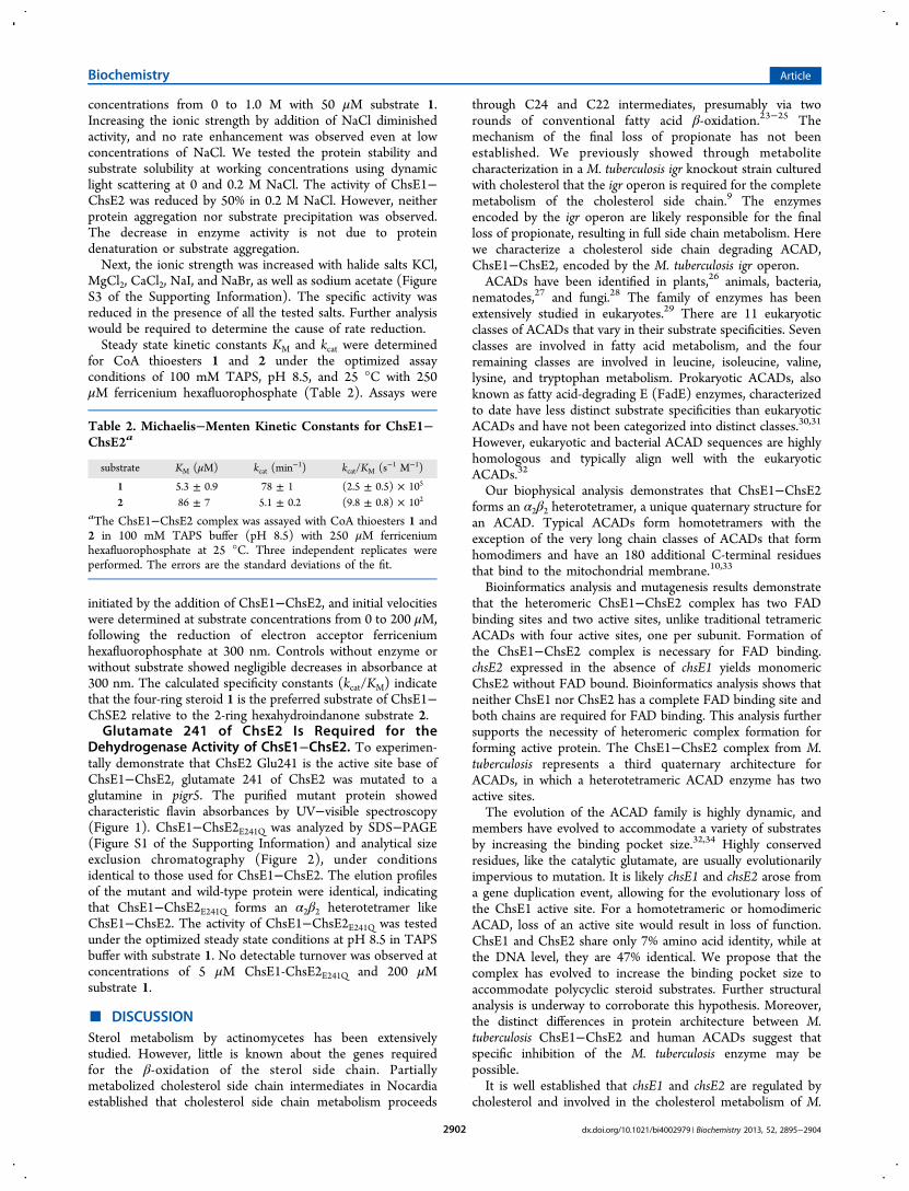
for CoA thioesters 1 and 2 under the optimized assayconditions of 100 mM TAPS, pH 8.5, and 25 °C with 250μM ferricenium hexafluorophosphate (Table 2). Assays were
initiated by the addition of ChsE1−ChsE2, and initial velocitieswere determined at substrate concentrations from 0 to 200 μM,following the reduction of electron acceptor ferriceniumhexafluorophosphate at 300 nm. Controls without enzyme orwithout substrate showed negligible decreases in absorbance at300 nm. The calculated specificity constants (kcat/KM) indicatethat the four-ring steroid 1 is the preferred substrate of ChsE1−ChSE2 relative to the 2-ring hexahydroindanone substrate 2.Glutamate 241 of ChsE2 Is Required for the
Dehydrogenase Activity of ChsE1−ChsE2. To experimen-tally demonstrate that ChsE2 Glu241 is the active site base ofChsE1−ChsE2, glutamate 241 of ChsE2 was mutated to aglutamine in pigr5. The purified mutant protein showedcharacteristic flavin absorbances by UV−visible spectroscopy(Figure 1). ChsE1−ChsE2E241Q was analyzed by SDS−PAGE(Figure S1 of the Supporting Information) and analytical sizeexclusion chromatography (Figure 2), under conditionsidentical to those used for ChsE1−ChsE2. The elution profilesof the mutant and wild-type protein were identical, indicatingthat ChsE1−ChsE2E241Q forms an α2β2 heterotetramer likeChsE1−ChsE2. The activity of ChsE1−ChsE2E241Q was testedunder the optimized steady state conditions at pH 8.5 in TAPSbuffer with substrate 1. No detectable turnover was observed atconcentrations of 5 μM ChsE1-ChsE2E241Q and 200 μMsubstrate 1.
■ DISCUSSIONSterol metabolism by actinomycetes has been extensivelystudied. However, little is known about the genes requiredfor the β-oxidation of the sterol side chain. Partiallymetabolized cholesterol side chain intermediates in Nocardiaestablished that cholesterol side chain metabolism proceeds
through C24 and C22 intermediates, presumably via tworounds of conventional fatty acid β-oxidation.23−25 Themechanism of the final loss of propionate has not beenestablished. We previously showed through metabolitecharacterization in a M. tuberculosis igr knockout strain culturedwith cholesterol that the igr operon is required for the completemetabolism of the cholesterol side chain.9 The enzymesencoded by the igr operon are likely responsible for the finalloss of propionate, resulting in full side chain metabolism. Herewe characterize a cholesterol side chain degrading ACAD,ChsE1−ChsE2, encoded by the M. tuberculosis igr operon.ACADs have been identified in plants,26 animals, bacteria,
nematodes,27 and fungi.28 The family of enzymes has beenextensively studied in eukaryotes.29 There are 11 eukaryoticclasses of ACADs that vary in their substrate specificities. Sevenclasses are involved in fatty acid metabolism, and the fourremaining classes are involved in leucine, isoleucine, valine,lysine, and tryptophan metabolism. Prokaryotic ACADs, alsoknown as fatty acid-degrading E (FadE) enzymes, characterizedto date have less distinct substrate specificities than eukaryoticACADs and have not been categorized into distinct classes.30,31
However, eukaryotic and bacterial ACAD sequences are highlyhomologous and typically align well with the eukaryoticACADs.32
Our biophysical analysis demonstrates that ChsE1−ChsE2forms an α2β2 heterotetramer, a unique quaternary structure foran ACAD. Typical ACADs form homotetramers with theexception of the very long chain classes of ACADs that formhomodimers and have an 180 additional C-terminal residuesthat bind to the mitochondrial membrane.10,33
Bioinformatics analysis and mutagenesis results demonstratethat the heteromeric ChsE1−ChsE2 complex has two FADbinding sites and two active sites, unlike traditional tetramericACADs with four active sites, one per subunit. Formation ofthe ChsE1−ChsE2 complex is necessary for FAD binding.chsE2 expressed in the absence of chsE1 yields monomericChsE2 without FAD bound. Bioinformatics analysis shows thatneither ChsE1 nor ChsE2 has a complete FAD binding site andboth chains are required for FAD binding. This analysis furthersupports the necessity of heteromeric complex formation forforming active protein. The ChsE1−ChsE2 complex from M.tuberculosis represents a third quaternary architecture forACADs, in which a heterotetrameric ACAD enzyme has twoactive sites.The evolution of the ACAD family is highly dynamic, and
members have evolved to accommodate a variety of substratesby increasing the binding pocket size.32,34 Highly conservedresidues, like the catalytic glutamate, are usually evolutionarilyimpervious to mutation. It is likely chsE1 and chsE2 arose froma gene duplication event, allowing for the evolutionary loss ofthe ChsE1 active site. For a homotetrameric or homodimericACAD, loss of an active site would result in loss of function.ChsE1 and ChsE2 share only 7% amino acid identity, while atthe DNA level, they are 47% identical. We propose that thecomplex has evolved to increase the binding pocket size toaccommodate polycyclic steroid substrates. Further structuralanalysis is underway to corroborate this hypothesis. Moreover,the distinct differences in protein architecture between M.tuberculosis ChsE1−ChsE2 and human ACADs suggest thatspecific inhibition of the M. tuberculosis enzyme may bepossible.It is well established that chsE1 and chsE2 are regulated by
cholesterol and involved in the cholesterol metabolism of M.
Table 2. Michaelis−Menten Kinetic Constants for ChsE1−ChsE2a
substrate KM (μM) kcat (min−1) kcat/KM (s−1 M−1)
1 5.3 ± 0.9 78 ± 1 (2.5 ± 0.5) × 105
2 86 ± 7 5.1 ± 0.2 (9.8 ± 0.8) × 102
aThe ChsE1−ChsE2 complex was assayed with CoA thioesters 1 and2 in 100 mM TAPS buffer (pH 8.5) with 250 μM ferriceniumhexafluorophosphate at 25 °C. Three independent replicates wereperformed. The errors are the standard deviations of the fit.
Biochemistry Article
dx.doi.org/10.1021/bi4002979 | Biochemistry 2013, 52, 2895−29042902

tuberculosis. We have previously shown that this enzymecomplex is able to bind and turn over steroid substrates. Inthis work, our experimentally determined specificity constantsfor 3-oxo-4-pregnene-20-carboxyl-CoA (1) and 1β-(2′-prop-anoyl-CoA)-3aα-H-7aβ-methylhexahydro-4-indanone (2) dem-onstrate that the ChsE1−ChsE2 complex strongly prefers thering nucleus intact species to the hexahydroindinone species, invitro. This suggests the side chain of cholesterol is fullymetabolized prior to aromatization and degradation of the ringsystem.Assignment of biochemical function to genes is complicated
by the large number of annotated fatty acid oxidation genes inM. tuberculosis.35 It is impossible to assign enzymes to specificbiochemical steps in sterol metabolism from sequence dataalone. We propose that the heterotetrameric architecture isimportant for binding sterols and possibly other polycyclicsubstrates. Our biochemical characterization and bioinformaticanalysis of a heteromeric sterol oxidizing ACAD provideinformation that will be applied to further proper annotation ofthe M. tuberculosis genome.
■ ASSOCIATED CONTENT*S Supporting InformationSDS−PAGE of purified proteins (Figure S1), LC−UV−MSanalysis of isolated flavin from ChsE1−ChsE2 (Figure S2), andthe salt dependence of dehydrogenase activity (Figure S3). Thismaterial is available free of charge via the Internet at http://pubs.acs.org.
■ AUTHOR INFORMATIONCorresponding Author*Department of Chemistry, Stony Brook University, StonyBrook, NY 11794-3400. E-mail: [email protected]. Phone: (631) 632-7952.FundingThis work was supported by the National Institutes of Health(R21AI092455, R01HL53306, and S10RR021008, N.S.S.),National Science Foundation Grant BIO1039771 (NMR),and a DOE-GAANN fellowship (S.T.T.).NotesThe authors declare no competing financial interest.
■ ACKNOWLEDGMENTSWe thank Francis Picart (NMR Coordinator, Stony BrookUniversity) for his assistance in NMR acquisition.
■ ABBREVIATIONSACAD, acyl-CoA dehydrogenase; igr, intracellular growth;IPTG, isopropyl β-D-1-thiogalactopyranoside; HEPES, 4-(2-hydroxyethyl)-1-piperazineethanesulfonic acid; Tris, tris-(hydroxymethyl)aminomethane; IMAC, immobilized metalion affinity chromatography; TAPS, N-tris(hydroxymethyl)-methyl-3-aminopropanesulfonic acid; TCEP, tris(2-carboxyethyl)phosphine; FAD, flavin adenine dinucleotide;AUC, analytical ultracentrifugation; CoA, coenzyme A.
■ REFERENCES(1) World Health Organization (2011) Global Tuberculosis Control2011.(2) Russell, D. G., Cardona, P. J., Kim, M. J., Allain, S., and Altare, F.(2009) Foamy macrophages and the progression of the humantuberculosis granuloma. Nat. Immunol. 10, 943−948.
(3) Peyron, P., Vaubourgeix, J., Poquet, Y., Levillain, F., Botanch, C.,Bardou, F., Daffe, M., Emile, J. F., Marchou, B., Cardona, P. J., deChastellier, C., and Altare, F. (2008) Foamy macrophages fromtuberculous patients’ granulomas constitute a nutrient-rich reservoirfor M. tuberculosis persistence. PLoS Pathog. 4, e1000204.(4) Pandey, A. K., and Sassetti, C. M. (2008) Mycobacterialpersistence requires the utilization of host cholesterol. Proc. Natl. Acad.Sci. U.S.A. 105, 4376−4380.(5) Mohn, W. W., van der Geize, R., Stewart, G. R., Okamoto, S., Liu,J., Dijkhuizen, L., and Eltis, L. D. (2008) The actinobacterial mce4locus encodes a steroid transporter. J. Biol. Chem. 283, 35368−35374.(6) Nesbitt, N., Yang, X., Fontan, P., Kolesnikova, I., Smith, I.,Sampson, N. S., and Dubnau, E. (2010) A thiolase of M. tuberculosis isrequired for virulence and for production of androstenedione andandrostadienedione from cholesterol. Infect. Immun. 78, 275−282.(7) Chang, J. C., Miner, M. D., Pandey, A. K., Gill, W. P., Harik, N. S.,Sassetti, C. M., and Sherman, D. R. (2009) igr Genes andMycobacterium tuberculosis cholesterol metabolism. J. Bacteriol. 191,5232−5239.(8) Chang, J. C., Harik, N. S., Liao, R. P., and Sherman, D. R. (2007)Identification of Mycobacterial genes that alter growth and pathologyin macrophages and in mice. J. Infect. Dis. 196, 788−795.(9) Thomas, S. T., VanderVen, B. C., Sherman, D. R., Russell, D. G.,and Sampson, N. S. (2011) Pathway profiling in Mycobacteriumtuberculosis: Elucidation of cholesterol-derived catabolite and enzymesthat catalyze its metabolism. J. Biol. Chem. 286, 43668−43678.(10) Zhang, J., Zhang, W., Zou, D., Chen, G., Wan, T., Zhang, M.,and Cao, X. (2002) Cloning and functional characterization of ACAD-9, a novel member of human acyl-CoA dehydrogenase family. Biochem.Biophys. Res. Commun. 297, 1033−1042.(11) Souri, M., Aoyama, T., Hoganson, G., and Hashimoto, T.(1998) Very-long-chain acyl-CoA dehydrogenase subunit assembles tothe dimer form on mitochondrial inner membrane. FEBS Lett. 426,187−190.(12) Moore, S. (2012) ’Round-the-horn site-directed mutagenesis.Wikiomics.(13) Daugelat, S., Kowall, J., Mattow, J., Bumann, D., Winter, R.,Hurwitz, R., and Kaufmann, S. H. (2003) The RD1 proteins ofMycobacterium tuberculosis: Expression inMycobacterium smegmatis andbiochemical characterization. Microbes Infect. 5, 1082−1095.(14) Shevchenko, A., Tomas, H., Havlis, J., Olsen, J. V., and Mann,M. (2006) In-gel digestion for mass spectrometric characterization ofproteins and proteomes. Nat. Protoc. 1, 2856−2860.(15) Winkler, R. (2010) ESIprot: A universal tool for charge statedetermination and molecular weight calculation of proteins fromelectrospray ionization mass spectrometry data. Rapid Commun. MassSpectrom. 24, 285−294.(16) Thompson, J. D., Higgins, D. G., and Gibson, T. J. (1994)CLUSTAL W: Improving the sensitivity of progressive multiplesequence alignment through sequence weighting, position specific gappenalties and weight matrix choice. Nucleic Acids Res. 22, 4673−4680.(17) Stierand, K., Maass, P. C., and Rarey, M. (2006) Molecularcomplexes at a glance: Automated generation of two-dimensionalcomplex diagrams. Bioinformatics 22, 1710−1716.(18) Capyk, J. K., Casabon, I., Gruninger, R., Strynadka, N. C., andEltis, L. D. (2011) Activity of 3-ketosteroid 9α-hydroxylase (KshAB)indicates cholesterol side chain and ring degradation occursimultaneously in Mycobacterium tuberculosis. J. Biol. Chem. 286,40717−40724.(19) Lehman, T. C., and Thorpe, C. (1990) Alternate electronacceptors for medium-chain acyl-CoA dehydrogenase: Use offerricenium salts. Biochemistry 29, 10594−10602.(20) Ellis, K. J., and Morrison, J. F. (1982) Buffers of constant ionicstrength for studying pH-dependent processes. Methods Enzymol. 87,405−426.(21) Hwang, T. L., and Shaka, A. J. (1995) Water suppression thatworks: Excitation sculpting using arbitrary wave-forms and pulsed-fieldgradients. J. Magn. Reson., Ser. A 112, 275−279.
Biochemistry Article
dx.doi.org/10.1021/bi4002979 | Biochemistry 2013, 52, 2895−29042903

(22) Djordjevic, S., Dong, Y., Paschke, R., Frerman, F. E., Strauss, A.W., and Kim, J. J. (1994) Identification of the catalytic base in longchain acyl-CoA dehydrogenase. Biochemistry 33, 4258−4264.(23) Sih, C. J., Tai, H. H., and Tsong, Y. Y. (1967) The mechanismof microbial conversion of cholesterol into 17-keto steroids. J. Am.Chem. Soc. 89, 1957−1958.(24) Sih, C. J., Tai, H. H., Tsong, Y. Y., Lee, S. S., and Coombe, R. G.(1968) Mechanisms of steroid oxidation by microorganisms. XIV.Pathway of cholesterol side-chain degradation. Biochemistry 7, 808−818.(25) Sih, C. J., Wang, K. C., and Tai, H. H. (1968) Mechanisms ofsteroid oxidation by microorganisms. XIII. C22 acid intermediates indegradation of cholesterol side chain. Biochemistry 7, 796−807.(26) Bode, K., Hooks, M. A., and Couee, I. I. (1999) Identification,separation, and characterization of acyl-coenzyme A dehydrogenasesinvolved in mitochondrial β-oxidation in higher plants. Plant Physiol.119, 1305−1314.(27) Komuniecki, R., Fekete, S., and Thissen-Parra, J. (1985)Purification and characterization of the 2-methyl branched-chain Acyl-CoA dehydrogenase, an enzyme involved in NADH-dependent enoyl-CoA reduction in anaerobic mitochondria of the nematode, Ascarissuum. J. Biol. Chem. 260, 4770−4777.(28) Kionka, C., and Kunau, W. H. (1985) Inducible β-oxidationpathway in Neurospora crassa. J. Bacteriol. 161, 153−157.(29) Kunau, W. H., Dommes, V., and Schulz, H. (1995) β-Oxidationof fatty acids in mitochondria, peroxisomes, and bacteria: A century ofcontinued progress. Prog. Lipid Res. 34, 267−342.(30) Campbell, J. W., and Cronan, J. E., Jr. (2002) The enigmaticEscherichia coli fadE gene is yafH. J. Bacteriol. 184, 3759−3764.(31) Shen, Y. Q., Lang, B. F., and Burger, G. (2009) Diversity anddispersal of a ubiquitous protein family: Acyl-CoA dehydrogenases.Nucleic Acids Res. 37, 5619−5631.(32) Swigonova, Z., Mohsen, A. W., and Vockley, J. (2009) Acyl-CoAdehydrogenases: Dynamic history of protein family evolution. J. Mol.Evol. 69, 176−193.(33) McAndrew, R. P., Wang, Y., Mohsen, A. W., He, M., Vockley, J.,and Kim, J. J. (2008) Structural basis for substrate fatty acyl chainspecificity: Crystal structure of human very-long-chain acyl-CoAdehydrogenase. J. Biol. Chem. 283, 9435−9443.(34) Hiltunen, J. K., and Qin, Y. (2000) β-Oxidation: Strategies forthe metabolism of a wide variety of acyl-CoA esters. Biochim. Biophys.Acta 1484, 117−128.(35) Cole, S. T., Brosch, R., Parkhil, J., Garnier, T., Churcher, C.,Harris, D., Gordon, S. V., Eiglmeier, K., Gas, S., Barry, C. E., III,Tekaia, F., Badcock, K., Basham, D., Brown, D., Chillingworth, T.,Connor, R., Davies, R., Devlin, K., Feltwell, T., Gentles, S., Hamlin, N.,Holroyd, S., Hornsby, T., Jagels, K., Krogh, A., McLean, J., Moule, S.,Murphy, L., Oliver, K., Osborne, J., Quail, M. A., Rajandream, M. A.,Rogers, J., Rutter, S., Seeger, K., Skelton, J., Squares, R., Squares, S.,Sulston, J. E., Taylor, K., Whitehead, S., and Barrell, B. G. (1998)Deciphering the biology of Mycobacterium tuberculosis from thecomplete genome sequence. Nature 393, 537−544.
Biochemistry Article
dx.doi.org/10.1021/bi4002979 | Biochemistry 2013, 52, 2895−29042904