mycobacterial siga and sigb cotranscribe essential … · mycobacterial siga and sigb cotranscribe...
TRANSCRIPT

Mycobacterial SigA and SigB Cotranscribe EssentialHousekeeping Genes during Exponential Growth
Kelley Hurst-Hess,a Rajesh Biswas,a* Yong Yang,a* Paulami Rudra,a Erica Lasek-Nesselquist,a Pallavi Ghosha,b
aDivision of Genetics, Wadsworth Center, New York State Department of Health, Albany, New York, USAbSchool of Public Health, University at Albany, Albany, New York, USA
ABSTRACT Mycobacterial �B belongs to the group II family of sigma factors, whichare widely considered to transcribe genes required for stationary-phase survival andthe response to stress. Here we explored the mechanism underlying the observedhypersensitivity of ΔsigB deletion mutants of Mycobacterium smegmatis, M. abscessus,and M. tuberculosis to rifampin (RIF) and uncovered an additional constitutive role of�B during exponential growth of mycobacteria that complements the function ofthe primary sigma factor, �A. Using chromatin immunoprecipitation sequencing(ChIP-Seq), we show that during exponential phase, �B binds to over 200 promoterregions, including those driving expression of essential housekeeping genes, like therRNA gene. ChIP-Seq of ectopically expressed �A-FLAG demonstrated that at least 61promoter sites are recognized by both �A and �B. These results together suggestthat RNA polymerase holoenzymes containing either �A or �B transcribe housekeep-ing genes in exponentially growing mycobacteria. The RIF sensitivity of the ΔsigBmutant possibly reflects a decrease in the effective housekeeping holoenzyme pool,which results in susceptibility of the mutant to lower doses of RIF. Consistent withthis model, overexpression of �A restores the RIF tolerance of the ΔsigB mutant tothat of the wild type, concomitantly ruling out a specialized role of �B in RIF toler-ance. Although the properties of mycobacterial �B parallel those of Escherichia coli�38 in its ability to transcribe a subset of housekeeping genes, �B presents a cleardeparture from the E. coli paradigm, wherein the cellular levels of �38 are tightlycontrolled during exponential growth, such that the transcription of housekeepinggenes is initiated exclusively by a holoenzyme containing �70 (E.�70).
IMPORTANCE All mycobacteria encode a group II sigma factor, �B, closely related tothe group I principal housekeeping sigma factor, �A. Group II sigma factors arewidely believed to play specialized roles in the general stress response andstationary-phase transition in the bacteria that encode them. Contrary to this widelyaccepted view, we show an additional housekeeping function of �B that comple-ments the function of �A in logarithmically growing cells. These findings implicate anovel and dynamic partnership between �A and �B in maintaining the expression ofhousekeeping genes in mycobacteria and can perhaps be extended to other bacte-rial species that possess multiple group II sigma factors.
KEYWORDS ChIP-Seq, Mycobacterium, rifampin, sigB, sigma factor
Transcription in bacteria is carried out by a multisubunit RNA polymerase (RNAP) thatassociates with an interchangeable sigma subunit and directs the transcription
machinery to specific promoter regions (1–4). All bacteria encode an essential principalsigma factor and a variable number of alternative sigma factors. Sigma factors areclassified into four groups based on the presence of conserved domains 1 to 4. GroupI sigma factors are required for transcription of housekeeping genes and are essential(5–7). Group II sigma factors are closely related to those in group I, lack domain 1.1, but
Citation Hurst-Hess K, Biswas R, Yang Y, RudraP, Lasek-Nesselquist E, Ghosh P. 2019.Mycobacterial SigA and SigB cotranscribeessential housekeeping genes duringexponential growth. mBio 10:e00273-19.https://doi.org/10.1128/mBio.00273-19.
Editor Christina L. Stallings, WashingtonUniversity in St. Louis School of Medicine
Copyright © 2019 Hurst-Hess et al. This is anopen-access article distributed under the termsof the Creative Commons Attribution 4.0International license.
Address correspondence to Pallavi Ghosh,[email protected].
* Present address: Rajesh Biswas, Sackler Schoolof Graduate Sciences, Tufts University, Boston,Massachusetts, USA; Yong Yang, School ofPublic Health, Jiaotong University, Shanghai,China.
K.H.-H. and R.B. contributed equally to thisarticle.
Received 31 January 2019Accepted 11 April 2019Published 21 May 2019
RESEARCH ARTICLEMolecular Biology and Physiology
crossm
May/June 2019 Volume 10 Issue 3 e00273-19 ® mbio.asm.org 1
on March 18, 2020 by guest
http://mbio.asm
.org/D
ownloaded from
are nonessential. Group III sigma factors contain domains 2 to 4, whereas the group IVsigma factors contain only domains 2 and 4. Mycobacteria encode one sigma factorbelonging to each of groups I to III and a variable number of group IV sigma factors:10 in Mycobacterium tuberculosis, 16 in M. abscessus, and 25 in M. smegmatis (8). GroupIV sigma factors have been studied extensively and are involved in heat shock, coldshock, hypoxia, carbon starvation, surface and oxidative stresses, and virulence (8–10).The mycobacterial �A, a group I sigma factor, is essential and highly similar to theprimary sigma factors from other bacteria, suggesting that it is the principal sigmafactor in mycobacteria (6, 11). �A mRNA levels are constant under different growthconditions, though the levels of the �A protein have been seen to decrease duringstationary phase (6, 7). The mycobacterial �B, a group II sigma factor, lacks domain 1.1and shows an �64% sequence identity with �A; in fact, residues important for recog-nition of �10 and �35 promoter elements are identical between mycobacterial �A and�B (12, 13). Although it is not essential for survival, �B is �90% conserved acrossmycobacterial species. A deletion in sigB results in sensitivity to heat, oxidative, andsurface stress in vitro and an increased sensitivity to p-aminosalicylic acid, sulfame-thoxazole, and ethambutol but does not impact the survival of M. tuberculosis inmacrophages or mouse lungs (10, 12, 14–19). Two attempts to characterize the �B
regulon yielded contradictory results. The global transcription profile of a strain over-expressing �B identified 72 �B-dependent genes, while the global transcription profileof a ΔsigB strain compared to that of wild-type (WT) bacteria identified only 8 �B-dependent genes during exponential growth (12, 14). This disparity can be resolved bydetermining the binding sites of �B; although a comprehensive map of transcriptionregulators, including sigma factors, has been determined in M. tuberculosis usingchromatin immunoprecipitation sequencing (ChIP-Seq), this does not include SigBbinding sites (20, 21). Exposure to diamide and SDS stress resulted in the downregu-lation of 40 and 72 genes, respectively, in the ΔsigB strain compared to their expressionin wild-type bacteria (14). Furthermore, the transcription of �B was shown to occur fromtwo promoters: one recognized by the stress-inducible sigma factors �E, �H, and �L andthe other recognized by �F (22–25). These observations together led to the generalnotion that �B has little role in exponential growth; rather, it is required solely for themycobacterial response to stress.
RNA polymerase is a target for the broad-spectrum antibiotic rifampin (RIF), whichcomprises a frontline therapy against M. tuberculosis infection. RIF exerts its effect bybinding to the � subunit of RNA polymerase in a region comprising the DNA/RNAchannel and sterically blocks the extrusion of elongating RNA when the transcriptexceeds 2 to 3 nucleotides (nt) in length (26). High levels of clinically acquired RIFresistance involve rpoB mutations in four distinct sequence clusters (clusters N, I, II, andIII), the majority of which map to cluster I (27–32). In contrast to acquired resistance, thefast-growing mycobacteria, such as M. smegmatis and M. abscessus, are naturally RIFresistant, albeit to various extents. This intrinsic rifampin resistance has been attributedto the presence of a rifampin ADP-ribosyltransferase (Arr), which inactivates the drug byribosylation (33–35). The association of RNAP with accessory proteins, such as certainsigma factors and RbpA, has also been shown to influence its susceptibility to RIF.Wegrzyn et al. showed that the Escherichia coli �70-RNAP is considerably more sensitivethan �32-RNAP in vitro and in vivo and that a deletion of Bacillus subtilis sigB, theorthologue of mycobacterial sigF, renders the bacteria more sensitive to RIF (43). RbpA,an RNAP binding protein conserved in actinomycetes, has been shown to prevent RIFinhibition in vitro (37, 38). While RbpA is essential in mycobacteria, a deletion of RbpAin Streptomyces coelicolor results in RIF sensitivity and a slow-growth phenotype (37,38). It is unlikely that RbpA is involved in the degradation or efflux of RIF but, rather,modifies RNAP. RbpA interacts exclusively with group I and II sigma factors in Strepto-myces and mycobacteria and stabilizes the formation of open promoter complexes,thereby enhancing the transcription efficiency of holoenzymes containing �A and �B.The mechanism by which RbpA confers RIF tolerance is unknown but has been shown
Hurst-Hess et al. ®
May/June 2019 Volume 10 Issue 3 e00273-19 mbio.asm.org 2
on March 18, 2020 by guest
http://mbio.asm
.org/D
ownloaded from

to not involve an occlusion of the RIF binding site in RNAP, and its effect is presumablyindirect (39, 40).
In the current work, we explore the underlying mechanism of RIF sensitivity of ΔsigBmutants of M. smegmatis, M. abscessus, and M. tuberculosis and demonstrate that theRIF sensitivity of ΔsigB strains is likely not attributable to the lack of transcription of�B-dependent RIF resistance genes. The study has uncovered that, contrary to previousmodels, �B is transcriptionally active during the exponential phase of growth of M.smegmatis and actively transcribes several �A-dependent housekeeping genes. Ourresults therefore demonstrate an active role for �B in the exponential phase ofmycobacterial growth, in addition to its role as a stress response sigma factor.
RESULTSDeletion of sigB results in RIF hypersensitivity in M. smegmatis, M. tuberculosis,
and M. abscessus. To understand the role of sigma factors in mycobacterial drugtolerance, we constructed isogenic deletions in 14 out of 28 randomly selected sigmafactor genes in M. smegmatis using recombineering and assayed the sensitivity of thedeletion strains to a variety of antibiotics (41). Deletion of the primary-like sigma factorsigB resulted in hypersensitivity to RIF (Fig. 1a). We then explored if the phenotype ofthe ΔsigB mutant could be recapitulated in the pathogenic mycobacteria M. tuberculosisand M. abscessus. sigB deletion mutations were constructed in the attenuated M.tuberculosis strain mc27000 and the M. abscessus ATCC 19977 strain using recombineer-ing. The ΔsigB strains of M. tuberculosis and M. abscessus were found to be hypersen-sitive to RIF compared to their corresponding wild types (Fig. 1b and c), suggesting thatthe �B-mediated basal RIF tolerance may be conferred by a conserved mechanism.Growth of the ΔsigB mutant of M. smegmatis in Middlebrook 7H10 agar lacking
FIG 1 Deletion of �B confers RIF sensitivity in M. smegmatis (Msm), M. abscessus (Mab), and M.tuberculosis (Mtb). (a to c) Tenfold serial dilutions of M. smegmatis mc2155, M. abscessus ATCC 19977, M.tuberculosis mc27000, and their respective ΔsigB and complemented strains were grown to an A600 of 0.7and spotted on Middlebrook 7H10 ADC or OADC containing the indicated concentrations of RIF. Deletionof sigB results in RIF sensitivity in all three strains. The mutant phenotype can be complemented byconstitutive expression of the respective sigB gene.
SigB-Dependent Transcription during Exponential Growth ®
May/June 2019 Volume 10 Issue 3 e00273-19 mbio.asm.org 3
on March 18, 2020 by guest
http://mbio.asm
.org/D
ownloaded from

antibiotics was unaffected but was reduced in 7H9 broth compared to that of WTbacteria (see Fig. S1a and b in the supplemental material). The M. tuberculosis ΔsigBmutant displayed a slow-growth phenotype on Middlebrook 7H10 agar lacking anti-biotics and has previously been shown to exhibit slow growth in liquid media (Fig. S1d)(18).
SigB-mediated resistance to RIF is independent of Arr. Intrinsic tolerance toRIF in mycobacteria has been attributed to the ribosylation of RIF by ADP-ribosyltransferases (Arr), encoded by the fast-growing mycobacteria (42). We firstinvestigated the most likely scenario that �B is required for the transcription of arr,either directly or indirectly, such that a deletion in sigB abrogates arr expression,resulting in RIF sensitivity. We therefore determined the relative abundance of the arrtranscript in WT M. smegmatis and the ΔsigB mutant of M. smegmatis (the MsΔsigBmutant) upon exposure to RIF by quantitative PCR (qPCR) analysis. Figure 2a shows thatthe level of arr induction upon RIF exposure did not decrease in the ΔsigB mutantstrain, as would be expected if its transcription were solely dependent on SigB. Instead,arr transcript levels increased �6-fold in a ΔsigB strain and may reflect a compensatoryresponse. Although this does not rule out the possibility that �B is required for thetranscription of arr, it suggests the presence of redundant pathways for arr expression.Nevertheless, this demonstrates that the RIF sensitivity of the ΔsigB strain cannot beattributed to a compromised transcription of arr. However, it is possible that the cellularlevel of the Arr protein is indirectly influenced by the absence of �B. If this were thecase, we would anticipate that the RIF sensitivities of the Δarr mutant and the ΔarrΔsigB double mutant would be indistinguishable. However, we observed that the RIFsensitivity of the double mutant was significantly higher than that of each of the singlemutants, suggesting that their effect is additive and mediated through independentpathways (Fig. 2b; Table 1). Moreover, a ΔsigB strain of M. tuberculosis, which naturallylacks arr, is also hypersensitive to RIF and provides additional support for the sugges-tion that the �B-mediated resistance to RIF is independent of ADP-ribosyltransferases.
FIG 2 �B-mediated resistance to RIF is independent of ADP-ribosyltransferase (Arr) and putative effectorgenes. (a) Wild-type M. smegmatis (MsWT) and the MsΔsigB strain were grown to an A600 of 0.7 andexposed to 4 �g/ml RIF for either 20 min or 2 h, and the amount of M. smegmatis arr transcripts wasdetermined by qPCR and plotted as the fold induction over the level of expression for an unexposedcontrol. Data represent the mean � SD (n � 3). sigA was used as an endogenous control. (b) Tenfoldserial dilutions of WT strain M. smegmatis mc2155 and the MsΔarr and MsΔsigB MsΔarr strains weregrown to an A600 of 0.7 and spotted on Middlebrook 7H10 ADC containing the indicated concentrationof RIF.
Hurst-Hess et al. ®
May/June 2019 Volume 10 Issue 3 e00273-19 mbio.asm.org 4
on March 18, 2020 by guest
http://mbio.asm
.org/D
ownloaded from

The RIF sensitivity of the �sigB mutant is independent of the transcription ofknown and putative RIF resistance effectors. We next tested if �B regulates theexpression of genes besides arr that mitigate the effect of RIF. We analyzed thetranscription profile of wild-type mc2155 and the ΔsigB mutant upon exposure tosublethal doses of RIF (4 �g/ml) using RNA sequencing (RNA-seq). �B-dependent genesthat confer RIF resistance would be detectable as those that are RIF inducible in thewild type but not in the ΔsigB mutant. An exposure time of 20 min was found to bemost appropriate to enable detection of the gene expression changes that immediatelyfollow RIF exposure. Exposure of wild-type bacteria to RIF caused a �4-fold inductionof 101 genes with a q value of �0.001 (Data Set S1), of which the top 50 are representedin Fig. 3a, left. The most highly induced genes were MSMEG_2252 (homologue ofrifampin monooxygenase [Rox]), MSMEG_2539 (thiopurine methyltransferase), MS-MEG_2174 (helicase), MSMEG_2254 (oxalate decarboxylase), MSMEG_1221 (ADP-ribosyltransferases [Arr]), and MSMEG_1224 (Arr). Surprisingly, however, genes thatwere RIF inducible in wild-type bacteria showed comparable levels of induction in theΔsigB mutant strain (Fig. 3a, right; Fig. 3b; Fig. S2; Data Set S1). Consistent with thisobservation, the RIF tolerance of the ΔsigB strain could not be restored to wild-typelevels by overexpression of MSMEG_2252, MSMEG_2254, MSMEG_2539, or MS-MEG_2174 (Fig. 3c). Interestingly, although a deletion in MSMEG_2174 increased RIFsusceptibility, its expression was unchanged in the ΔsigB mutant, indicating that thephenotype is sigB independent (Fig. S3).
We also considered the possibility that �B-dependent RIF resistance effectors areconstitutively expressed. We therefore compared the transcription profiles of thewild-type and ΔsigB strains of M. smegmatis mc2155 grown to mid-log phase. RNA-seqanalysis showed that 13 genes were significantly (q value � 0.01) underexpressed by�3-fold in the mutant (Table S1 and Data Set S2), which is consistent with previouslypublished results (14). We evaluated the role of two of the most highly affected genesthat were underexpressed in the ΔsigB mutant strain: MSMEG_4708, which encodes amethyltransferase, and MSMEG_6241, which encodes an AAATPase. Overexpression ofeither of these genes did not complement the RIF sensitivity of the ΔsigB mutant strain(Fig. 3c).
Lastly, we evaluated the role of RbpA, an RNA polymerase-associated protein thathas been shown to affect the RIF sensitivity of S. coelicolor and is RIF inducible inmycobacteria (37, 38). Figure 3d shows that RbpA transcript levels increased �2-fold inwild-type bacteria and �6-fold in the ΔsigB mutant upon RIF exposure, consistent withthe results of the RNA-seq experiments. Moreover, overexpression of RbpA failed torestore the RIF tolerance of the ΔsigB mutant to that of the wild type (Fig. 3e). Together,these observations suggest that the RIF sensitivity of the ΔsigB mutant cannot beattributed to the lack of RbpA, a known effector of RIF resistance.
�A- and �B-containing holoenzymes are indistinguishable in their RIF suscep-tibility. Taken together, the data likely rule out the possibility that the RIF-sensitivephenotype of ΔsigB is due to the lack of expression of either novel or previouslydescribed effectors of RIF resistance. We speculated that the observed RIF sensitivitycould be a reflection of the interaction of �B with RNAP, the target of RIF. The sensitivityof RNAP to RIF has previously been demonstrated to depend on its association withparticular species of sigma factors; holoenzymes associated with primary sigma factors
TABLE 1 MIC of RIF for the M. smegmatis WT, ΔsigB, Δarr, and ΔsigB Δarr strainsa
Strain MIC of RIF (�g/ml)
WT mc2155 10mc2155 ΔsigB 2.5mc2155 Δarr 0.25mc2155 ΔsigB Δarr 0.0625aThe survival of the M. smegmatis mc2155 wild-type, ΔsigB, Δarr, and ΔsigB Δarr strains was determined in a2-fold dilution series of RIF in Middlebrook 7H9 medium. The minimum concentration of antibiotic requiredto inhibit 99% of the growth is shown.
SigB-Dependent Transcription during Exponential Growth ®
May/June 2019 Volume 10 Issue 3 e00273-19 mbio.asm.org 5
on March 18, 2020 by guest
http://mbio.asm
.org/D
ownloaded from
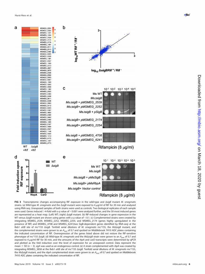
FIG 3 Transcriptomic changes accompanying RIF exposure in the wild-type and ΔsigB mutant M. smegmatisstrains. (a) Wild-type M. smegmatis and the ΔsigB mutant were exposed to 4 �g/ml of RIF for 20 min and analyzedusing RNA-seq. Unexposed samples of both strains were used as controls. Two biological replicates of each samplewere used. Genes induced �4-fold with a q value of �0.001 were analyzed further, and the 50 most induced genesare represented as a heat map. (Left) WT; (right) ΔsigB mutant. (b) RIF-induced changes in gene expression in theWT versus ΔsigB mutant are shown using genes with a q value of �0.1. (c) Complemented strains were created byintegrating MSMEG_2539, MSMEG_2252, MSMEG_2254, and MSMEG_2174 (genes highly upregulated in thepresence of RIF) and MSMEG_4708 and MSMEG_6241(two SigB-dependent genes identified by RNA-seq) at theBxb1 attB site of mc2155 ΔsigB. Tenfold serial dilutions of M. smegmatis mc2155, the MsΔsigB mutant, andthe complemented strains were grown to an A600 of 0.7 and spotted on Middlebrook 7H10 ADC plates containingthe indicated concentration of RIF. Overexpression of the genes listed above did not restore the RIF-sensitivephenotype of mc2155 ΔsigB. (d) Wild-type M. smegmatis and the MsΔsigB strain were grown to an A600 of 0.7 andexposed to 4 �g/ml RIF for 30 min, and the amounts of the rbpA and carD transcripts were determined by qPCRand plotted as the fold induction over the level of expression for an unexposed control. Data represent themean � SD (n � 3). sigA was used as an endogenous control. (e) A strain complemented with rbpA was created byintegrating MSMEG_3858 at the Bxb1 attB site of mc2155 ΔsigB. Tenfold serial dilutions of M. smegmatis mc2155,the MsΔsigB mutant, and the rbpA complemented strain were grown to an A600 of 0.7 and spotted on Middlebrook7H10 ADC plates containing the indicated concentration of RIF.
Hurst-Hess et al. ®
May/June 2019 Volume 10 Issue 3 e00273-19 mbio.asm.org 6
on March 18, 2020 by guest
http://mbio.asm
.org/D
ownloaded from

are more sensitive than those associated with alternate sigma factors (38, 43). Inaddition, �B has been shown to recognize several �A-dependent promoters in vitro (13).Based on these two lines of evidence, we propose that the RIF sensitivity of the ΔsigBmutant can be explained by one of two scenarios: (i) a holoenzyme containing �B (E.�B)is more resistant to RIF than a holoenzyme containing �A (E.�A) and is recruited tohousekeeping promoters in the presence of RIF when transcription by E.�A is compro-mised, or (ii) E.�A and E.�B are equally sensitive to RIF but are both involved in thetranscription of housekeeping genes in exponentially growing bacteria. The toxicity ofRIF would become pronounced when one of the sigma factors is missing, especially ifthe expression of neither sigA nor sigB is inducible. Since �A is essential, this phenotypeis apparent only in a ΔsigB mutant.
We determined the RIF sensitivity of �A-RNAP and �B-RNAP by assaying their activityat the sigA promoter (sigAP) in multiple-round in vitro transcription assays. Assays wereperformed both in the presence and in the absence of RbpA, since RbpA has beenshown to assist with open complex formation by �A and �B, as well as offer protectionagainst RIF inhibition (13, 38–40, 44, 45). Figure 4a and b show that RbpA greatly(�100-fold) enhanced transcription by E.�B, but its effect on E.�A was modest (�2-fold)and is consistent with previously published results (13, 40). In the presence of RbpA,
FIG 4 �A and �B containing holoenzymes are equally RIF susceptible. (a and b) Multiple-round in vitrotranscription assays were performed on the sigA promoter using 200 nM �A-RNAP/�B-RNAP. RbpA(600 nM) was added where indicated. RIF was added to the indicated concentrations for 30 min at 37°C.Transcription was initiated by addition of 2 �l of an NTP mix (1.5 mM ATP, GTP, and CTP and 0.5 mM UTP)plus 2 �Ci of [�-32P]UTP. The reaction mixtures were incubated at 37°C for 30 min, and the reactions wereterminated by the addition of 5 mM EDTA and 100 �g/ml tRNA. Samples were ethanol precipitated andseparated using denaturing PAGE (6% urea polyacrylamide gel). (c) The products were visualized usinga Typhoon imager (GE Healthcare) and quantitated using ImageQuant software. Inhibition of RNAPactivity at 50 nM RIF is expressed as a ratio of the activity in the presence and absence of RIF.
SigB-Dependent Transcription during Exponential Growth ®
May/June 2019 Volume 10 Issue 3 e00273-19 mbio.asm.org 7
on March 18, 2020 by guest
http://mbio.asm
.org/D
ownloaded from

although the overall yield of the transcript was �100-fold higher when using E.�B, theinhibition of transcription at each RIF concentration was comparable when using eitherE.�A or E.�B (�70% inhibition was seen with both holoenzymes at 50 nM RIF) (Fig. 4c).This suggests that E.�A and E.�B are equally RIF sensitive and that the association of �B
with RNAP does not offer any additional protection against RIF.�B actively transcribes housekeeping genes in exponentially growing M. smeg-
matis. We next explored the alternate scenario that E.�A and E.�B are both involved inthe transcription of housekeeping genes in exponentially growing bacteria. This hy-pothesis is contrary to the currently accepted notion that �B is required only duringtransition to stationary phase and in response to environmental stress (7, 8, 12, 14).However, RNA-seq of exponentially growing M. smegmatis showed comparable levelsof sigA and sigB transcripts (Data Set S2) (10). We therefore determined the relativelevels of the �A and �B proteins at various stages of M. smegmatis growth by Westernblot analysis using an anti-�70 antibody that recognizes an epitope in domain 3.1common to mycobacterial �A and �B and E. coli �70 (46) (Fig. S4a). Figure 5a shows that
FIG 5 �B is transcriptionally active in exponentially growing M. smegmatis. (a) (Top) Growth kinetics of wild-type M.smegmatis indicating the growth phase and samples used for Western blotting. (Bottom) Relative levels of the �A and �B
proteins at the indicated optical densities determined by Western blotting using an anti-�70 monoclonal antibody. Sampleswere normalized by wet weight and protein concentration to ensure equivalent loading at each OD. Purified �A and �B
proteins were used as controls. The ratio of �A/�B was quantitated using ImageJ software and is shown below. Equivalentamounts of protein were loaded in each lane of the Coomassie-stained gel (see Fig. S4b in the supplemental material). (b)Sequence logo of enriched motif in �B-FLAG-bound sites identified using the MEME Suite of tools (MEME Evalue � 7.0e�003). (c) The ChIP-Seq peaks of �B bound to promoters of key housekeeping genes visualized withSignalMap software are shown. The transcript levels of the corresponding genes (RPKM values) in the wild type (red) andthe ΔsigB strain (blue) are plotted.
Hurst-Hess et al. ®
May/June 2019 Volume 10 Issue 3 e00273-19 mbio.asm.org 8
on March 18, 2020 by guest
http://mbio.asm
.org/D
ownloaded from
�B is consistently present in exponentially growing M. smegmatis; in fact, �B proteinlevels were comparable to those of �A during early logarithmic phase of growth.
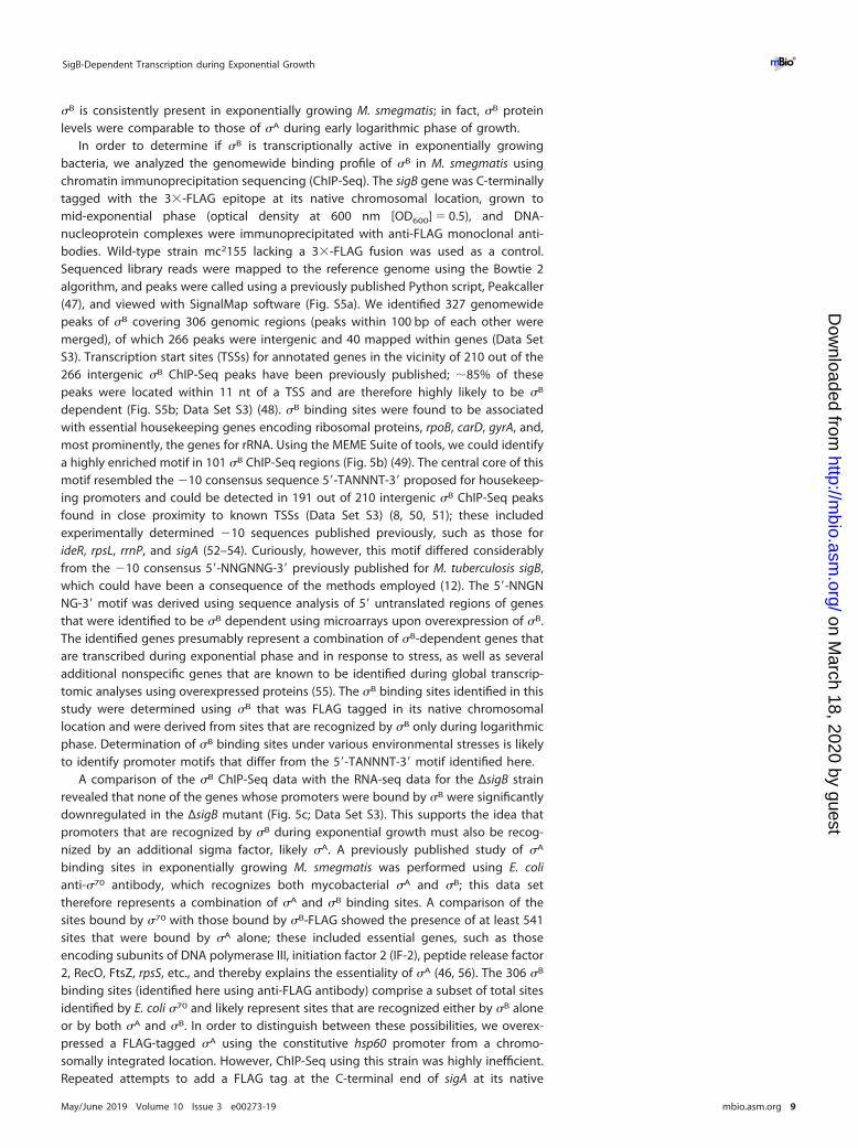
In order to determine if �B is transcriptionally active in exponentially growingbacteria, we analyzed the genomewide binding profile of �B in M. smegmatis usingchromatin immunoprecipitation sequencing (ChIP-Seq). The sigB gene was C-terminallytagged with the 3�-FLAG epitope at its native chromosomal location, grown tomid-exponential phase (optical density at 600 nm [OD600] � 0.5), and DNA-nucleoprotein complexes were immunoprecipitated with anti-FLAG monoclonal anti-bodies. Wild-type strain mc2155 lacking a 3�-FLAG fusion was used as a control.Sequenced library reads were mapped to the reference genome using the Bowtie 2algorithm, and peaks were called using a previously published Python script, Peakcaller(47), and viewed with SignalMap software (Fig. S5a). We identified 327 genomewidepeaks of �B covering 306 genomic regions (peaks within 100 bp of each other weremerged), of which 266 peaks were intergenic and 40 mapped within genes (Data SetS3). Transcription start sites (TSSs) for annotated genes in the vicinity of 210 out of the266 intergenic �B ChIP-Seq peaks have been previously published; �85% of thesepeaks were located within 11 nt of a TSS and are therefore highly likely to be �B
dependent (Fig. S5b; Data Set S3) (48). �B binding sites were found to be associatedwith essential housekeeping genes encoding ribosomal proteins, rpoB, carD, gyrA, and,most prominently, the genes for rRNA. Using the MEME Suite of tools, we could identifya highly enriched motif in 101 �B ChIP-Seq regions (Fig. 5b) (49). The central core of thismotif resembled the �10 consensus sequence 5=-TANNNT-3= proposed for housekeep-ing promoters and could be detected in 191 out of 210 intergenic �B ChIP-Seq peaksfound in close proximity to known TSSs (Data Set S3) (8, 50, 51); these includedexperimentally determined �10 sequences published previously, such as those forideR, rpsL, rrnP, and sigA (52–54). Curiously, however, this motif differed considerablyfrom the �10 consensus 5=-NNGNNG-3= previously published for M. tuberculosis sigB,which could have been a consequence of the methods employed (12). The 5=-NNGNNG-3= motif was derived using sequence analysis of 5= untranslated regions of genesthat were identified to be �B dependent using microarrays upon overexpression of �B.The identified genes presumably represent a combination of �B-dependent genes thatare transcribed during exponential phase and in response to stress, as well as severaladditional nonspecific genes that are known to be identified during global transcrip-tomic analyses using overexpressed proteins (55). The �B binding sites identified in thisstudy were determined using �B that was FLAG tagged in its native chromosomallocation and were derived from sites that are recognized by �B only during logarithmicphase. Determination of �B binding sites under various environmental stresses is likelyto identify promoter motifs that differ from the 5=-TANNNT-3= motif identified here.
A comparison of the �B ChIP-Seq data with the RNA-seq data for the ΔsigB strainrevealed that none of the genes whose promoters were bound by �B were significantlydownregulated in the ΔsigB mutant (Fig. 5c; Data Set S3). This supports the idea thatpromoters that are recognized by �B during exponential growth must also be recog-nized by an additional sigma factor, likely �A. A previously published study of �A
binding sites in exponentially growing M. smegmatis was performed using E. colianti-�70 antibody, which recognizes both mycobacterial �A and �B; this data settherefore represents a combination of �A and �B binding sites. A comparison of thesites bound by �70 with those bound by �B-FLAG showed the presence of at least 541sites that were bound by �A alone; these included essential genes, such as thoseencoding subunits of DNA polymerase III, initiation factor 2 (IF-2), peptide release factor2, RecO, FtsZ, rpsS, etc., and thereby explains the essentiality of �A (46, 56). The 306 �B
binding sites (identified here using anti-FLAG antibody) comprise a subset of total sitesidentified by E. coli �70 and likely represent sites that are recognized either by �B aloneor by both �A and �B. In order to distinguish between these possibilities, we overex-pressed a FLAG-tagged �A using the constitutive hsp60 promoter from a chromo-somally integrated location. However, ChIP-Seq using this strain was highly inefficient.Repeated attempts to add a FLAG tag at the C-terminal end of sigA at its native
SigB-Dependent Transcription during Exponential Growth ®
May/June 2019 Volume 10 Issue 3 e00273-19 mbio.asm.org 9
on March 18, 2020 by guest
http://mbio.asm
.org/D
ownloaded from

chromosomal location were also unsuccessful, suggesting that the presence of a FLAGtag may compromise the function of �A. Despite the inefficiency of ChIP-Seq using�A-FLAG, we could identify 94 �A binding sites that were common with �B binding sites(and that were also recognized by E. coli anti-�70 antibody) and likely representhigh-affinity binding sites of �A. Out of the 94 �A binding sites, 61 were associated withknown TSSs, and these therefore constitute the minimum number of promoters thatare recognized by both �A and �B, including promoters for essential genes such asthose for rRNA, tRNAs, ribosomal proteins, sigA, and rpoB (Data Set S3).
Overexpression of �A restores the RIF tolerance of Ms�sigB to that of the wildtype. We reasoned that if transcription of housekeeping genes is initiated by both E.�A
and E.�B, the absence of �B could be compensated for by increasing the copy numberof �A in a way that mitigates the deleterious effect of RIF. Figure 6a and Table 2 showthat the constitutive overexpression of M. smegmatis sigA from a chromosomallyintegrated location restored the RIF sensitivity of the MsΔsigB mutant. Further, weobserved that overexpression of either sigA or sigB from M. abscessus and M. tubercu-losis could complement the phenotype of the MsΔsigB mutant (Fig. 6a). However, thiseffect was restricted to the group I and II sigma factors; sigF (group III) and theextracytoplasmic function (ECF) sigma factors were unable to restore the RIF sensitivityof the MsΔsigB mutant (Fig. 6b), presumably because of their inability to initiatetranscription at housekeeping promoters. An alternate explanation is that a deletion ofsigB reduces the levels of �A, which can be complemented by overexpression of �A. Wetherefore followed the expression of the sigA transcript as well as protein levels in theMsΔsigB mutant in the presence and absence of exposure to RIF. Figure 6c and d showthat sigA transcript and protein levels did not decrease in the MsΔsigB bacteriacompared to wild-type bacteria; furthermore, sigA expression was also not RIF induciblein either the wild type or the MsΔsigB mutant (Fig. 6e).
DISCUSSION
All mycobacterial species contain a highly conserved group II sigma factor, theproduct of the sigB gene. Global transcriptomic analyses have failed to identify sizablenumbers of �B-dependent genes during exponential growth of mycobacteria. More-over, sigB mRNA levels increase upon entry into stationary phase and in response toheat shock and surface and oxidative stresses (5, 7, 10, 14). These observations have ledto the inference that �B is specialized for transcription during transition to stationaryphase and in the global stress response but does not play an active role in transcriptionduring the logarithmic phase of growth. Herein we present a series of observationswhich together illuminate a role for �B during exponential growth, in addition to itbeing a stress response sigma factor.
Using ChIP-Seq analysis under exponential growth conditions, we demonstratedthat �B binds to over 200 promoter regions, several of which control the transcriptionof essential housekeeping genes, such as the rRNA gene, carD, rpoB, etc. This finding isconsistent with that of RNA-seq analysis, which showed that sigB mRNA is as abundantas sigA mRNA during exponential phase of M. smegmatis growth (see Data Set S2 in thesupplemental material) and that the �B protein is consistently present in all stages ofgrowth (Fig. 5a). A limited ChIP-Seq data set for ectopically overexpressed FLAG-tagged�A confirmed at least 61 promoter sites that were recognized by both �A and �B,including those that control the vital cellular functions of ribosome biogenesis andtranscription. The most plausible explanation for �B occupancy at such crucial sites isthat it is engaged in active transcription of these genes, or else its occupancy wouldinterfere with the �A-dependent transcription initiation from these sites. These resultsimply that E.�A and E.�B together transcribe a subset of housekeeping genes duringexponential growth of M. smegmatis. While further data are required to determine therelative occupancy of E.�A and E.�B at a given promoter, we predict that this varies withthe promoter site, its association with accessory proteins, as well as with changinggrowth and environmental conditions. An example of such a scenario has previouslybeen demonstrated in vitro by Hu et al., in which E.�A and E.�B are transcriptionally
Hurst-Hess et al. ®
May/June 2019 Volume 10 Issue 3 e00273-19 mbio.asm.org 10
on March 18, 2020 by guest
http://mbio.asm
.org/D
ownloaded from

active at the sigAP but open complex formation on this promoter is seen only with E.�B
in the presence of RbpA, suggesting a higher affinity of E.�B at the sigA promoter (13).It is noteworthy that the transcript levels of sigA as well as �A protein levels are not
responsive to the lack of �B in a ΔsigB mutant strain (Fig. 6c and e); sigA is also not RIF
FIG 6 Overexpression of �A restores the RIF sensitivity of the MsΔsigB mutant. (a to c) Complemented strainswere created by integrating Ms_SigA, Mtb_SigA, Mab_SigA, Ms_SigB, Mtb_SigB, Mab_SigB, Ms_Ms1804,Ms_Ms3296, Ms_Ms5444, and MSMEG_1418 at the Bxb1 attB site of mc2155 ΔsigB. Tenfold serial dilutions ofM. smegmatis mc2155, the mc2155 ΔsigB mutant, and the complemented strains were grown to an A600 of 0.7and spotted on Middlebrook 7H10 ADC plates containing the indicated concentrations of RIF. The RIFsensitivity of mc2155 ΔsigB could be complemented by the constitutive expression of sigA and sigB from allmycobacterial strains but not by ECF sigma factors. (b) Wild-type M. smegmatis and the MsΔsigB strain weregrown to an A600 of 0.7 and exposed to 4 �g/ml RIF for 30 min, and the amount of the M. smegmatis sigAtranscript was determined by qPCR and plotted as the fold induction of sigA levels in the MsΔsigB strain overthe level of expression in the wild-type strain. The data represent the mean � SD (n � 3). MSMEG_4936 wasused as an endogenous control, as its levels were unchanged under various conditions in RNA-seq experi-ments. (d) Wild-type M. smegmatis and the MsΔsigB strain were grown to an A600 of 0.7 and exposed to4 �g/ml RIF for 30 min, and the amounts of the M. smegmatis sigA and sigB transcripts were determined byqPCR and plotted as the fold induction upon RIF exposure over the level of expression for an unexposedcontrol. The data represent the mean � SD (n � 3). MSMEG_4936 was used as an endogenous control. (e)Wild-type M. smegmatis and the MsΔsigB strain were grown to an A600 of 0.7 and exposed to 4 �g/ml RIF for30 min. The levels of �A protein were determined by Western blotting using an anti-�70 monoclonal antibody.Samples were normalized by wet weight and protein concentration to ensure equivalent loading of eachsample. Purified �A was used as a control.
SigB-Dependent Transcription during Exponential Growth ®
May/June 2019 Volume 10 Issue 3 e00273-19 mbio.asm.org 11
on March 18, 2020 by guest
http://mbio.asm
.org/D
ownloaded from

inducible in either the wild type or the ΔsigB mutant (Fig. 6d). The absence of �B in aΔsigB mutant strain must therefore result in a decrease in the concentration ofholoenzyme that is available for transcription of a subset of housekeeping genes. Wewould therefore predict a decrease in transcription of some housekeeping genes in theΔsigB mutant strain; however, this was not reflected in our RNA-seq experiments. Sincethe most prominent binding of �B is observed at rRNA gene promoters (rrnP), it isplausible that the deletion of sigB largely impacts transcription from rRNA promoters,changes in which cannot be captured by the RNA-seq approach. Moreover, the rRNAgene promoter has been shown to be particularly susceptible to inhibition by RIF (40).It is therefore tempting to suggest that �B-RNAP plays a crucial role in maintaining thelevels of rRNA and that the slow-growth phenotype of ΔsigB in liquid media could bea reflection of the decrease in rRNA levels. Addition of RIF to a ΔsigB strain couldpotentially reduce the transcription at rrnP further, resulting in growth arrest and theobserved sensitivity to RIF. Although we cannot completely rule out the possibility thata �B-dependent gene affects the translation of a RIF effector transcript, we favor anexplanation that the addition of RIF targeting a decreased pool of holoenzyme capableof transcribing housekeeping genes contributes to its increased lethality. Restoration ofRIF tolerance in the ΔsigB mutant by overexpression of �A further rules out thepossibility of a specialized role of �B in RIF tolerance. The RIF-sensitive phenotype ofΔsigB mutants of M. abscessus, M. tuberculosis, and M. smegmatis and the cross-speciesfunctional complementarity between �A and �B among these species suggest that therole of �B in the transcription of housekeeping genes during exponential growth islikely to be conserved in all mycobacterial species.
The RNAP-associated protein RbpA has previously been shown to protect againstRIF inhibition in S. coelicolor. Although RbpA is incapable of protecting against RIFinhibition at mycobacterial rrnP as well as sigAP, we and others have noted that theoverall levels of transcription by E.�A at these promoters are higher in the presence ofRbpA; moreover, the transcription efficiency of E.�B at these promoters greatly exceedsthat of E.�A but is strictly RbpA dependent (Fig. 4) (40). Consistent with this observation,a CHIP-Seq analysis of RbpA showed that �75% of �B-bound sites are also bound byRbpA (K. Hurst-Hess, R. Biswas, and P. Ghosh, unpublished results). Curiously, expressionof RbpA increases �2-fold in wild-type bacteria and �6-fold in ΔsigB mutant bacteriaupon RIF exposure (Fig. 3d). We speculate that in the presence of RIF, the growth ofbacteria can be stimulated by RbpA by increasing the transcription efficiency of E.�A
and E.�B and the increase in RbpA transcription in ΔsigB reflects an attempt tocompensate for the lack of transcription by E.�B.
The behavior of mycobacterial �B is reminiscent of that of rpoS (�38), a group IIsigma factor of E. coli induced during stress and stationary phase with a high degree ofsimilarity to the primary sigma factor of E. coli, �70. �70 and �38 display extensiveoverlap between their target promoters, and �38 has been shown to take over severalhousekeeping duties of �70 during stationary phase (57–59). Despite the overlap inpromoter recognition, E. coli �70 and �38 have distinct but complementary roles in vivo,and �38 transcribes its regulon only under relevant physiological conditions (59, 60).This is achieved in part by tightly regulating the cellular concentration of �38 at the
TABLE 2 MIC of RIF for M. smegmatis wild-type, ΔsigB, and ΔsigB strains overexpressingeither M. smegmatis sigB or sigAa
Strain MIC of RIF (�g/ml)
WT mc2155 10mc2155 ΔsigB 2.5mc2155 ΔsigB sigBOE �10mc2155 ΔsigB sigAOE �10aThe survival of M. smegmatis wild-type strain mc2155, mc2155 ΔsigB, and mc2155 ΔsigB overexpressing M.
smegmatis sigB and sigA (mc2155 ΔsigB sigBOE and mc2155 ΔsigB sigAOE, respectively) was determined in a2-fold dilution series of RIF in Middlebrook 7H9 medium. The minimum concentration of antibiotic requiredto inhibit 99% of the growth is shown.
Hurst-Hess et al. ®
May/June 2019 Volume 10 Issue 3 e00273-19 mbio.asm.org 12
on March 18, 2020 by guest
http://mbio.asm
.org/D
ownloaded from
levels of transcription, translation, and proteolysis such that the �38 protein is nearlyundetectable during exponential growth but increases during entry into stationaryphase (58, 61–63). Additionally, the promoter specificity of E.�38 is modulated to allowtranscription of housekeeping genes under appropriate conditions; the precise mech-anism by which this is achieved is unclear but may be mediated in part by cis-actingpromoter features as well as trans-acting proteins, such as Crl, an activator thatstimulates E.�38 activity at certain promoters, and global regulators like H-NS and IHF(64–68). The association of mycobacterial �B with RbpA has similarly been shown toallow the recognition of �A-specific promoters by �B (13). Although the roles of Crl andRbpA appear to be similar, they have been shown to act via distinct mechanisms: Crlincreases the affinity of �38 to core RNAP, whereas RbpA stimulates open complexformation without stabilizing the holoenzyme. Mycobacterial �B, nevertheless, presentsa clear departure from the E. coli paradigm: the cellular levels of �B are not controlledduring exponential growth, and �B instead actively participates in the cotranscriptionof housekeeping genes. Variation in the relative levels of these sigma factors may playa key role in the global regulation of gene expression. We speculate that the presenceof �B may offer an advantage in the survival of mycobacteria under conditions whereeither the function of �A is compromised or the bacteria could benefit from theincreased transcription of housekeeping genes. Since the expression of �A itself isnoninducible, any demand for increasing the housekeeping gene expression could beachieved by inducing �B expression. Moreover, we speculate that this mechanism ofregulation of gene expression could be more widely utilized, especially in the closelyrelated Streptomyces coelicolor and cyanobacteria that encode multiple group II sigmafactors (69).
MATERIALS AND METHODSMedia and strains. Mycobacterium smegmatis was grown at 37°C in Middlebrook 7H9 (Difco)
supplemented with 10% albumin-dextrose-catalase (ADC) and 0.05% Tween 20. Mycobacterium abscessusATCC 19977 was grown at 37°C in Middlebrook 7H9 (Difco) supplemented with 10% oleic acid-albumin-dextrose-catalase (OADC) and 0.05% Tween 20. Mycobacterium tuberculosis mc27000, an attenuatedstrain of Mycobacterium tuberculosis H37Rv which carries deletions in the RD1 and panCD loci, both ofwhich are critical for the virulence of M. tuberculosis, was grown at 37°C in Middlebrook 7H9 (Difco)supplemented with 10% OADC and 0.05% Tween 20 (70). Antibiotics were added as required to theamounts indicated below. Gene replacement mutants were constructed using recombineering asdescribed previously (41). The recombineering construct was generated by cloning in the multiple-cloning sites flanking the apramycin cassette of pYUB854. Mutant clones were checked using the Fcheck
and Rcheck primers flanking the deletion site. The sigB-FLAG-tagged strain was confirmed using sequenc-ing as well as by Western blot analysis with anti-FLAG antibody.
Antibiotic sensitivity assays. Wild-type and mutant strains of M. smegmatis, M. abscessus, and M.tuberculosis were grown to an A600 of 0.6 to 0.7. Cells were tested for their susceptibility to RIF by spottinga 10-fold serial dilution on Middlebrook 7H10 (Difco) plates containing the concentration of RIF indicatedabove and below. Antibiotic susceptibility in liquid media was assayed by inoculating the desired strainin a 2-fold dilution series of each antibiotic at an initial A600 of 0.0004. The cultures were incubated at37°C, and the A600 was measured after 48 h for M. smegmatis.
RNA preparation, qPCR, and RNA-seq analysis. Wild-type M. smegmatis mc2155 as well as theΔsigB deletion strain were grown to exponential phase (OD600 � 0.4) in Middlebrook 7H9-ADC, exposedto 4 �g/ml of RIF for various periods of time (0 to 90 min), and evaluated for the lethality of RIF. TotalRNA was prepared from wild-type and mutant strains exposed to RIF (4 �g/ml) for 20 min using a QiagenRNA preparation kit, followed by DNase I treatment. Unexposed samples were used as controls.Approximately 5-�g total RNA samples were treated by the Ribo-Zero rRNA removal procedure (Illumina)to enrich for mRNA. Approximately 500 ng of RNA was used for library preparation using a ScriptSeq (v2)RNA-seq kit and high-throughput sequencing on an Illumina NextSeq platform. The sequence data wereanalyzed using the reference-based analysis and default parameters in the Rockhopper (v2.03) program,in which the data are normalized by upper quartile normalization and transcript abundance is reportedas the number of reads per kilobase per million (RPKM). Differential gene expression was tested for eachtranscript, and q values, which control the false-discovery rate, were then reported (71, 72). RNA-seqexperiments were performed three independent times, using two biological replicates each time.
M. smegmatis wild-type and ΔsigB deletion strains were exposed to RIF (4 �g/ml) for the requiredtimes. Total RNA was prepared using a Qiagen RNA preparation kit, followed by DNase I treatment.Primers for quantitative reverse transcription-PCR (qRT-PCR) were generated using Primer Quest software(Integrated DNA Technologies). cDNA was generated using random hexamers and Maxima reversetranscriptase (Fisher Scientific), and qRT-PCR was performed using the Maxima SYBR green qPCR mastermix (Fisher Scientific) and the following primer pair for MSMEG_1221: 5=-CCTGTGGTTCGCGGAAA-3=/5=-
SigB-Dependent Transcription during Exponential Growth ®
May/June 2019 Volume 10 Issue 3 e00273-19 mbio.asm.org 13
on March 18, 2020 by guest
http://mbio.asm
.org/D
ownloaded from

CCCTGCTCAAGAATCTCACC-3=. An Applied Biosystems 7300 real-time PCR system was used with cyclingconditions of 50°C for 2 min, 95°C for 10 min, and 40 cycles of 95°C for 15 s and 60°C for 1 min.
Chromatin immunoprecipitation sequencing and data analysis. ChIP-Seq was performed aspreviously described with minor modifications (73). mc2155 sigB-FLAG was grown at 37°C in Middlebrook7H9 broth (Becton, Dickinson) supplemented with ADS (albumin [50 g liter�1], dextrose [20 g liter�1],NaCl [8.1 g liter�1]), 0.2% glycerol, and 0.05% Tween 80 to an OD600 of 0.4. This was followed bycross-linking with 1% formaldehyde for 30 min with constant agitation and quenching with 250 mMglycine. The cells were pelleted, washed with Tris-buffered saline buffer, and resuspended in buffer 1containing a protease inhibitor cocktail (73). Cells were lysed on a Covaris S220 Focused ultrasonicatorfor 30 min (amplitude � 20%, intensity � 5, number of cycles/burst � 200), immunoprecipitated withanti-FLAG monoclonal antibody M2 (Sigma) for 12 h at 4°C, and further processed as described previously(73). Each CHIP-Seq experiment was performed three independent times using two replicates of cultureeach time.
Genomic DNA libraries enriched for �B binding were sequenced on the Illumina platform (WadsworthCenter, Sequencing Core Facility). The reads were aligned to the reference genome using the Bowtie2and SAMtools algorithms (74). Regions of enrichment were identified using a custom Python script asdescribed previously (47). Briefly, for each replicate data set in the pair, an appropriate threshold, T1 orT2, was determined for the plus and minus strands. Values for T1 and T2 were considered to be between1 and 1,000. For each combination of values for T1 and T2, the number of genome positions with valuesgreater than or equal to the value for T1 in the first replicate and with values greater than or equal to thevalue for T2 in the second replicate was determined. The false-discovery rate was estimated using the nullhypothesis that no regions are enriched. The combination of thresholds yielding the highest number oftrue-positive positions with an estimated false-discovery rate of less than 0.01 was selected. Once T1 andT2 were chosen, a region was identified as a peak if both replicates showed enrichment above thecorresponding thresholds for each strand. For a peak to be called, there must be a peak on the plusstrand within a threshold distance of a peak on the minus strand. Peaks obtained with the Peakcallerprogram were verified using the MACS2 algorithm and viewed with SignalMap (v2.0.05) software (RocheNimbleGen). Relative enrichment is reported as the fold-over-threshold (FAT) score. The enriched regionswere analyzed using MEME Suite (v5.0.3) tools and the default parameters (49).
Protein overexpression and purification. M. tuberculosis �A, �B, and RbpA were cloned in pET21awith a C-terminal His tag, transformed into E. coli BL21(DE3)pLysS, grown to an A600 of 0.4, and inducedwith 1 mM IPTG (isopropyl-�-D-thiogalactopyranoside) at 30°C. The cells were lysed in a buffer containing50 mM Tris-HCl (pH 8.0), 300 mM NaCl, and 5% glycerol, and the clarified lysate was loaded on anNi-nitrilotriacetic acid (NTA) column (Qiagen). Nonspecifically bound proteins were removed by washingwith lysis buffer containing 40 mM, 35 mM, and 20 mM imidazole, and the protein was eluted with150 mM imidazole. For purification of M. tuberculosis RNA polymerase, BL21(DE3)pLysS was cotrans-formed with pETDuet-Mtb��= and pRsfDuet-Mtb��, grown at 30°C to an A600 of 0.4, and induced with0.4 mM IPTG at 16°C for a period of 18 h. The cells were lysed by sonication and passed through anNi-NTA column (Qiagen) that had been equilibrated with 50 mM Tris, 300 mM NaCl, and 5% glycerol (lysisbuffer). The column was washed with lysis buffer and 40 mM imidazole and eluted with lysis buffer and150 mM imidazole. Fractions containing RNAP were loaded on a heparin-Sepharose matrix (GE Health-care) that had been equilibrated with 50 mM Tris, 300 mM NaCl, and 5% glycerol and eluted with a buffercontaining 1 M NaCl.
In vitro transcription assays. Multiple-round in vitro transcription was performed as previouslydescribed (40). In short, 200 nM M. tuberculosis RNAP was assembled with 600 nM the desired sigmafactor in a volume of 10 �l for 10 min at 37°C. RbpA (600 nM) was added during assembly to the indicatedsamples, followed by a further incubation for 5 min. sigAP DNA (20 nM) was added to the mixtures, andthe mixtures were incubated for 10 min at 37°C. RIF was added to the concentrations indicated belowfor 30 min at 37°C. Transcription was initiated by addition of 2 �l of a nucleoside triphosphate (NTP) mix(1.5 mM ATP, GTP, and CTP and 0.5 mM UTP) plus 2 �Ci of [�-32P]UTP. The reaction mixtures wereincubated at 37°C for 30 min, and the reactions were terminated by the addition of 5 mM EDTA plus100 �g/ml tRNA. Samples were ethanol precipitated, resuspended in stop buffer (80% [vol/vol] forma-mide, 10 mM EDTA, 0.01% xylene cyanol, 0.01% bromophenol blue), and separated using denaturingPAGE (18% urea polyacrylamide gel). The products were visualized using a Typhoon imager (GEHealthcare) and quantitated using ImageQuant software.
Western blot analysis. M. smegmatis was grown in Middlebrook 7H9 supplemented with ADS andTween 20. Aliquots were removed at different stages of growth (A600 � 0.2, 0.4, 0.7, 1.0, 2.0, 2.8, and 4.0),pelleted, and washed with phosphate-buffered saline (PBS) buffer. Pellets were normalized by weight,resuspended in the required volumes of PBS, and lysed by sonication. The lysate was clarified bycentrifugation, the protein concentration was determined at the A260, and equal quantities of proteinfrom different growth stages were separated using 10% SDS-PAGE, transferred to a polyvinylidenedifluoride membrane, and probed with anti-�70 monoclonal antibody 2G10. Purified �A and �B were usedas controls.
SUPPLEMENTAL MATERIALSupplemental material for this article may be found at https://doi.org/10.1128/mBio
.00273-19.FIG S1, PDF file, 1.4 MB.FIG S2, PDF file, 0.5 MB.
Hurst-Hess et al. ®
May/June 2019 Volume 10 Issue 3 e00273-19 mbio.asm.org 14
on March 18, 2020 by guest
http://mbio.asm
.org/D
ownloaded from

FIG S3, PDF file, 1.6 MB.FIG S4, PDF file, 1.6 MB.FIG S5, PDF file, 1.6 MB.TABLE S1, DOCX file, 0.04 MB.DATA SET S1, XLSX file, 0.02 MB.DATA SET S2, XLSX file, 0.02 MB.DATA SET S3, XLSX file, 0.1 MB.
ACKNOWLEDGMENTSWe thank The Wadsworth Center’s Applied Genomics Technology Core for sequenc-
ing of RNA-seq libraries, the Media Core for the preparation of media and buffers, andthe Wadsworth Bioinformatics Core. pETDuet-Mtb��= was a kind gift from JayantMukhopadhyay, Bose Institute, Kolkata, India. We thank Joe Wade and Anil Ojha fordiscussions and critical reading of the manuscript.
P.G. is supported by a Cystic Fibrosis Foundation grant and the Wadsworth Center.
REFERENCES1. Gruber TM, Gross CA. 2003. Multiple sigma subunits and the partitioning
of bacterial transcription space. Annu Rev Microbiol 57:441– 466. https://doi.org/10.1146/annurev.micro.57.030502.090913.
2. Saecker RM, Record MT, Jr, Dehaseth PL. 2011. Mechanism of bacterialtranscription initiation: RNA polymerase-promoter binding, isomeriza-tion to initiation-competent open complexes, and initiation of RNAsynthesis. J Mol Biol 412:754 –771. https://doi.org/10.1016/j.jmb.2011.01.018.
3. Paget MS, Helmann JD. 2003. The sigma70 family of sigma factors.Genome Biol 4:203. https://doi.org/10.1186/gb-2003-4-1-203.
4. Murakami KS, Masuda S, Campbell EA, Muzzin O, Darst SA. 2002. Struc-tural basis of transcription initiation: an RNA polymerase holoenzyme-DNA complex. Science 296:1285–1290. https://doi.org/10.1126/science.1069595.
5. Manganelli R, Provvedi R, Rodrigue S, Beaucher J, Gaudreau L, Smith I,Proveddi R. 2004. Sigma factors and global gene regulation in Myco-bacterium tuberculosis. J Bacteriol 186:895–902. https://doi.org/10.1128/JB.186.4.895-902.2004.
6. Gomez M, Doukhan L, Nair G, Smith I. 1998. sigA is an essential gene inMycobacterium smegmatis. Mol Microbiol 29:617– 628. https://doi.org/10.1046/j.1365-2958.1998.00960.x.
7. Hu Y, Coates AR. 1999. Transcription of two sigma 70 homologue genes,sigA and sigB, in stationary-phase Mycobacterium tuberculosis. J Bacte-riol 181:469 – 476.
8. Sachdeva P, Misra R, Tyagi AK, Singh Y. 2010. The sigma factors ofMycobacterium tuberculosis: regulation of the regulators. FEBS J 277:605– 626. https://doi.org/10.1111/j.1742-4658.2009.07479.x.
9. Rodrigue S, Provvedi R, Jacques PE, Gaudreau L, Manganelli R. 2006. Thesigma factors of Mycobacterium tuberculosis. FEMS Microbiol Rev 30:926 –941. https://doi.org/10.1111/j.1574-6976.2006.00040.x.
10. Manganelli R, Dubnau E, Tyagi S, Kramer FR, Smith I. 1999. Differentialexpression of 10 sigma factor genes in Mycobacterium tuberculosis. MolMicrobiol 31:715–724. https://doi.org/10.1046/j.1365-2958.1999.01212.x.
11. Predich M, Doukhan L, Nair G, Smith I. 1995. Characterization of RNApolymerase and two sigma-factor genes from Mycobacterium smegma-tis. Mol Microbiol 15:355–366. https://doi.org/10.1111/j.1365-2958.1995.tb02249.x.
12. Lee JH, Karakousis PC, Bishai WR. 2008. Roles of SigB and SigF in theMycobacterium tuberculosis sigma factor network. J Bacteriol 190:699 –707. https://doi.org/10.1128/JB.01273-07.
13. Hu Y, Morichaud Z, Perumal AS, Roquet-Baneres F, Brodolin K. 2014.Mycobacterium RbpA cooperates with the stress-response sigmaB sub-unit of RNA polymerase in promoter DNA unwinding. Nucleic Acids Res42:10399 –10408. https://doi.org/10.1093/nar/gku742.
14. Fontan PA, Voskuil MI, Gomez M, Tan D, Pardini M, Manganelli R,Fattorini L, Schoolnik GK, Smith I. 2009. The Mycobacterium tuberculosissigma factor sigmaB is required for full response to cell envelope stressand hypoxia in vitro, but it is dispensable for in vivo growth. J Bacteriol191:5628 –5633. https://doi.org/10.1128/JB.00510-09.
15. Doukhan L, Predich M, Nair G, Dussurget O, Mandic-Mulec I, Cole ST,
Smith DR, Smith I. 1995. Genomic organization of the mycobacterialsigma gene cluster. Gene 165:67–70. https://doi.org/10.1016/0378-1119(95)00427-8.
16. Hu Y, Coates AR. 1999. Transcription of the stationary-phase-associatedhspX gene of Mycobacterium tuberculosis is inversely related to synthe-sis of the 16-kilodalton protein. J Bacteriol 181:1380 –1387.
17. Mukherjee R, Chatterji D. 2005. Evaluation of the role of sigma B inMycobacterium smegmatis. Biochem Biophys Res Commun 338:964 –972. https://doi.org/10.1016/j.bbrc.2005.10.038.
18. Yang SS, Hu YB, Wang XD, Gao YR, Li K, Zhang XE, Chen SY, Zhang TY,Gu J, Deng JY. 2017. Deletion of sigB causes increased sensitivity topara-aminosalicylic acid and sulfamethoxazole in Mycobacterium tuber-culosis. Antimicrob Agents Chemother 61:e00551-17. https://doi.org/10.1128/AAC.00551-17.
19. Pisu D, Provvedi R, Mata Espinosa D, Barrios Payan J, Boldrin F, Palu G,Hernandez-Pando R, Manganelli R. 2017. The alternative sigma factorsSigE and SigB are involved in tolerance and persistence to antituberculardrugs. Antimicrob Agents Chemother 61:e01596-17. https://doi.org/10.1128/AAC.01596-17.
20. Minch KJ, Rustad TR, Peterson EJ, Winkler J, Reiss DJ, Ma S, Hickey M,Brabant W, Morrison B, Turkarslan S, Mawhinney C, Galagan JE, Price ND,Baliga NS, Sherman DR. 2015. The DNA-binding network of Mycobac-terium tuberculosis. Nat Commun 6:5829. https://doi.org/10.1038/ncomms6829.
21. Galagan JE, Minch K, Peterson M, Lyubetskaya A, Azizi E, Sweet L, GomesA, Rustad T, Dolganov G, Glotova I, Abeel T, Mahwinney C, Kennedy AD,Allard R, Brabant W, Krueger A, Jaini S, Honda B, Yu WH, Hickey MJ,Zucker J, Garay C, Weiner B, Sisk P, Stolte C, Winkler JK, Van de Peer Y,Iazzetti P, Camacho D, Dreyfuss J, Liu Y, Dorhoi A, Mollenkopf HJ,Drogaris P, Lamontagne J, Zhou Y, Piquenot J, Park ST, Raman S,Kaufmann SH, Mohney RP, Chelsky D, Moody DB, Sherman DR, SchoolnikGK. 2013. The Mycobacterium tuberculosis regulatory network and hyp-oxia. Nature 499:178 –183. https://doi.org/10.1038/nature12337.
22. Dainese E, Rodrigue S, Delogu G, Provvedi R, Laflamme L, Brzezinski R,Fadda G, Smith I, Gaudreau L, Palu G, Manganelli R. 2006. Posttransla-tional regulation of Mycobacterium tuberculosis extracytoplasmic-function sigma factor sigma L and roles in virulence and in globalregulation of gene expression. Infect Immun 74:2457–2461. https://doi.org/10.1128/IAI.74.4.2457-2461.2006.
23. Manganelli R, Voskuil MI, Schoolnik GK, Smith I. 2001. The Mycobacte-rium tuberculosis ECF sigma factor sigmaE: role in global gene expres-sion and survival in macrophages. Mol Microbiol 41:423– 437. https://doi.org/10.1046/j.1365-2958.2001.02525.x.
24. Raman S, Song T, Puyang X, Bardarov S, Jacobs WR, Jr, Husson RN. 2001. Thealternative sigma factor SigH regulates major components of oxidative andheat stress responses in Mycobacterium tuberculosis. J Bacteriol 183:6119–6125. https://doi.org/10.1128/JB.183.20.6119-6125.2001.
25. Chauhan R, Ravi J, Datta P, Chen T, Schnappinger D, Bassler KE, BalazsiG, Gennaro ML. 2016. Reconstruction and topological characterization of
SigB-Dependent Transcription during Exponential Growth ®
May/June 2019 Volume 10 Issue 3 e00273-19 mbio.asm.org 15
on March 18, 2020 by guest
http://mbio.asm
.org/D
ownloaded from

the sigma factor regulatory network of Mycobacterium tuberculosis. NatCommun 7:11062. https://doi.org/10.1038/ncomms11062.
26. Campbell EA, Korzheva N, Mustaev A, Murakami K, Nair S, Goldfarb A,Darst SA. 2001. Structural mechanism for rifampicin inhibition of bacte-rial RNA polymerase. Cell 104:901–912. https://doi.org/10.1016/S0092-8674(01)00286-0.
27. Jin DJ, Gross CA. 1988. Mapping and sequencing of mutations in theEscherichia coli rpoB gene that lead to rifampicin resistance. J Mol Biol202:45–58. https://doi.org/10.1016/0022-2836(88)90517-7.
28. Lisitsyn NA, Gur’ev SO, Sverdlov ED, Moiseeva EP, Nikiforov VG. 1984.Nucleotide substitutions in the rpoB gene leading to rifampicin resis-tance of E. coli RNA polymerase. Bioorg Khim 10:127–128. (In Russian.)
29. Lisitsyn NA, Sverdlov ED, Moiseyeva EP, Danilevskaya ON, Nikiforov VG.1984. Mutation to rifampicin resistance at the beginning of the RNApolymerase beta subunit gene in Escherichia coli. Mol Gen Genet 196:173–174. https://doi.org/10.1007/BF00334112.
30. Ovchinnikov YA, Monastyrskaya GS, Guriev SO, Kalinina NF, Sverdlov ED,Gragerov AI, Bass IA, Kiver IF, Moiseyeva EP, Igumnov VN, Mindlin SZ,Nikiforov VG, Khesin RB. 1983. RNA polymerase rifampicin resistancemutations in Escherichia coli: sequence changes and dominance. MolGen Genet 190:344 –348. https://doi.org/10.1007/BF00330662.
31. Ramaswamy S, Musser JM. 1998. Molecular genetic basis of antimicrobialagent resistance in Mycobacterium tuberculosis: 1998 update. TuberLung Dis 79:3–29. https://doi.org/10.1054/tuld.1998.0002.
32. Severinov K, Soushko M, Goldfarb A, Nikiforov V. 1994. RifR mutations inthe beginning of the Escherichia coli rpoB gene. Mol Gen Genet 244:120 –126.
33. Quan S, Imai T, Mikami Y, Yazawa K, Dabbs ER, Morisaki N, Iwasaki S,Hashimoto Y, Furihata K. 1999. ADP-ribosylation as an intermediate stepin inactivation of rifampin by a mycobacterial gene. Antimicrob AgentsChemother 43:181–184. https://doi.org/10.1128/AAC.43.1.181.
34. Quan S, Venter H, Dabbs ER. 1997. Ribosylative inactivation of rifampinby Mycobacterium smegmatis is a principal contributor to its low sus-ceptibility to this antibiotic. Antimicrob Agents Chemother 41:2456 –2460. https://doi.org/10.1128/AAC.41.11.2456.
35. Rominski A, Roditscheff A, Selchow P, Bottger EC, Sander P. 2017.Intrinsic rifamycin resistance of Mycobacterium abscessus is mediatedby ADP-ribosyltransferase MAB_0591. J Antimicrob Chemother 72:376 –384. https://doi.org/10.1093/jac/dkw466.
36. Reference deleted.37. Dey A, Verma AK, Chatterji D. 2010. Role of an RNA polymerase inter-
acting protein, MsRbpA, from Mycobacterium smegmatis in phenotypictolerance to rifampicin. Microbiology 156:873– 883. https://doi.org/10.1099/mic.0.033670-0.
38. Newell KV, Thomas DP, Brekasis D, Paget MS. 2006. The RNA polymerase-binding protein RbpA confers basal levels of rifampicin resistance onStreptomyces coelicolor. Mol Microbiol 60:687– 696. https://doi.org/10.1111/j.1365-2958.2006.05116.x.
39. Hubin EA, Tabib-Salazar A, Humphrey LJ, Flack JE, Olinares PD, Darst SA,Campbell EA, Paget MS. 2015. Structural, functional, and genetic analy-ses of the actinobacterial transcription factor RbpA. Proc Natl Acad SciU S A 112:7171–7176. https://doi.org/10.1073/pnas.1504942112.
40. Hu Y, Morichaud Z, Chen S, Leonetti JP, Brodolin K. 2012. Mycobacteriumtuberculosis RbpA protein is a new type of transcriptional activator thatstabilizes the sigma A-containing RNA polymerase holoenzyme. NucleicAcids Res 40:6547– 6557. https://doi.org/10.1093/nar/gks346.
41. van Kessel JC, Hatfull GF. 2007. Recombineering in Mycobacteriumtuberculosis. Nat Methods 4:147–152. https://doi.org/10.1038/nmeth996.
42. Alexander DC, Jones JR, Liu J. 2003. A rifampin-hypersensitive mutantreveals differences between strains of Mycobacterium smegmatis andpresence of a novel transposon, IS1623. Antimicrob Agents Chemother47:3208 –3213. https://doi.org/10.1128/AAC.47.10.3208-3213.2003.
43. Wegrzyn A, Szalewska-Pałasz A, Błaszczak A, Liberek K, Wegrzyn G. 1998.Differential inhibition of transcription from sigma70- and sigma32-dependent promoters by rifampicin. FEBS Lett 440:172–174. https://doi.org/10.1016/S0014-5793(98)01449-5.
44. Tabib-Salazar A, Liu B, Doughty P, Lewis RA, Ghosh S, Parsy M-L, SimpsonPJ, O’Dwyer K, Matthews SJ, Paget MS. 2013. The actinobacterial tran-scription factor RbpA binds to the principal sigma subunit of RNApolymerase. Nucleic Acids Res 41:5679 –5691. https://doi.org/10.1093/nar/gkt277.
45. Bortoluzzi A, Muskett FW, Waters LC, Addis PW, Rieck B, Munder T,Schleier S, Forti F, Ghisotti D, Carr MD, O’Hare HM. 2013. Mycobacterium
tuberculosis RNA polymerase-binding protein A (RbpA) and its interac-tions with sigma factors. J Biol Chem 288:14438 –14450. https://doi.org/10.1074/jbc.M113.459883.
46. Breyer MJ, Thompson NE, Burgess RR. 1997. Identification of the epitopefor a highly cross-reactive monoclonal antibody on the major sigmafactor of bacterial RNA polymerase. J Bacteriol 179:1404 –1408. https://doi.org/10.1128/jb.179.4.1404-1408.1997.
47. Fitzgerald DM, Bonocora RP, Wade JT. 2014. Comprehensive mapping ofthe Escherichia coli flagellar regulatory network. PLoS Genet 10:e1004649. https://doi.org/10.1371/journal.pgen.1004649.
48. Li X, Mei H, Chen F, Tang Q, Yu Z, Cao X, Andongma BT, Chou SH, He J.2017. Transcriptome landscape of Mycobacterium smegmatis. FrontMicrobiol 8:2505. https://doi.org/10.3389/fmicb.2017.02505.
49. Bailey TL, Elkan C. 1994. Fitting a mixture model by expectation maxi-mization to discover motifs in biopolymers. Proc Int Conf Intell Syst MolBiol 2:28 –36.
50. Agarwal N, Tyagi AK. 2006. Mycobacterial transcriptional signals: require-ments for recognition by RNA polymerase and optimal transcriptionalactivity. Nucleic Acids Res 34:4245– 4257. https://doi.org/10.1093/nar/gkl521.
51. Gomez M, Smith I. 2000. Determinants of mycobacterial gene expres-sion, p 111–129. In Hatfull GF, Jacobs WR, Jr (ed), Molecular genetics ofmycobacteria. ASM Press, Washington, DC.
52. Kenney TJ, Churchward G. 1996. Genetic analysis of the Mycobacteriumsmegmatis rpsL promoter. J Bacteriol 178:3564 –3571. https://doi.org/10.1128/jb.178.12.3564-3571.1996.
53. Bashyam MD, Kaushal D, Dasgupta SK, Tyagi AK. 1996. A study ofmycobacterial transcriptional apparatus: identification of novel featuresin promoter elements. J Bacteriol 178:4847– 4853. https://doi.org/10.1128/jb.178.16.4847-4853.1996.
54. Gonzalez-y-Merchand JA, Colston MJ, Cox RA. 1996. The rRNA operonsof Mycobacterium smegmatis and Mycobacterium tuberculosis: compar-ison of promoter elements and of neighbouring upstream genes. Mi-crobiology 142:667– 674. https://doi.org/10.1099/13500872-142-3-667.
55. Chiner-Oms Á, González-Candelas F, Comas I. 2018. Gene expressionmodels based on a reference laboratory strain are poor predictors ofMycobacterium tuberculosis complex transcriptional diversity. Sci Rep8:3813. https://doi.org/10.1038/s41598-018-22237-5.
56. Landick R, Krek A, Glickman MS, Socci ND, Stallings CL. 2014. Genome-wide mapping of the distribution of CarD, RNAP sigma(A), and RNAPbeta on the Mycobacterium smegmatis chromosome using chromatinimmunoprecipitation sequencing. Genom Data 2:110 –113. https://doi.org/10.1016/j.gdata.2014.05.012.
57. Raffaelle M, Kanin EI, Vogt J, Burgess RR, Ansari AZ. 2005. Holoenzymeswitching and stochastic release of sigma factors from RNA polymerasein vivo. Mol Cell 20:357–366. https://doi.org/10.1016/j.molcel.2005.10.011.
58. Tanaka K, Takayanagi Y, Fujita N, Ishihama A, Takahashi H. 1993. Heter-ogeneity of the principal sigma factor in Escherichia coli: the rpoS geneproduct, sigma 38, is a second principal sigma factor of RNA polymerasein stationary-phase Escherichia coli. Proc Natl Acad Sci U S A 90:8303.
59. Gaal T, Ross W, Estrem ST, Nguyen LH, Burgess RR, Gourse RL. 2001.Promoter recognition and discrimination by EsigmaS RNA polymer-ase. Mol Microbiol 42:939 –954. https://doi.org/10.1046/j.1365-2958.2001.02703.x.
60. Tanaka K, Takayanagi Y, Fujita N, Ishihama A, Takahashi H. 1993. Heter-ogeneity of the principal sigma factor in Escherichia coli: the rpoS geneproduct, sigma 38, is a second principal sigma factor of RNA polymerasein stationary-phase Escherichia coli. Proc Natl Acad Sci U S A 90:3511–3515. https://doi.org/10.1073/pnas.90.8.3511.
61. Battesti A, Majdalani N, Gottesman S. 2011. The RpoS-mediated generalstress response in Escherichia coli. Annu Rev Microbiol 65:189 –213.https://doi.org/10.1146/annurev-micro-090110-102946.
62. Hengge R. 2009. Proteolysis of sigmaS (RpoS) and the general stressresponse in Escherichia coli. Res Microbiol 160:667– 676. https://doi.org/10.1016/j.resmic.2009.08.014.
63. Lange R, Hengge-Aronis R. 1994. The cellular concentration of the sigmaS subunit of RNA polymerase in Escherichia coli is controlled at the levelsof transcription, translation, and protein stability. Genes Dev 8:1600 –1612. https://doi.org/10.1101/gad.8.13.1600.
64. Typas A, Becker G, Hengge R. 2007. The molecular basis of selectivepromoter activation by the sigmaS subunit of RNA polymerase. MolMicrobiol 63:1296 –1306. https://doi.org/10.1111/j.1365-2958.2007.05601.x.
Hurst-Hess et al. ®
May/June 2019 Volume 10 Issue 3 e00273-19 mbio.asm.org 16
on March 18, 2020 by guest
http://mbio.asm
.org/D
ownloaded from

65. Arnqvist A, Olsen A, Normark S. 1994. Sigma S-dependent growth-phaseinduction of the csgBA promoter in Escherichia coli can be achieved invivo by sigma 70 in the absence of the nucleoid-associated proteinH-NS. Mol Microbiol 13:1021–1032. https://doi.org/10.1111/j.1365-2958.1994.tb00493.x.
66. Olsen A, Arnqvist A, Hammar M, Sukupolvi S, Normark S. 1993. TheRpoS sigma factor relieves H-NS-mediated transcriptional repressionof csgA, the subunit gene of fibronectin-binding curli in Escherichiacoli. Mol Microbiol 7:523–536. https://doi.org/10.1111/j.1365-2958.1993.tb01143.x.
67. Arnqvist A, Olsen A, Pfeifer J, Russell DG, Normark S. 1992. The Crlprotein activates cryptic genes for curli formation and fibronectin bind-ing in Escherichia coli HB101. Mol Microbiol 6:2443–2452.
68. Colland F, Barth M, Hengge-Aronis R, Kolb A. 2000. Sigma factor selec-tivity of Escherichia coli RNA polymerase: role for CRP, IHF and lrptranscription factors. EMBO J 19:3028 –3037. https://doi.org/10.1093/emboj/19.12.3028.
69. Buttner MJ, Lewis CG. 1992. Construction and characterization of Strep-tomyces coelicolor A3(2) mutants that are multiply deficient in thenonessential hrd-encoded RNA polymerase sigma factors. J Bacteriol174:5165–5167. https://doi.org/10.1128/jb.174.15.5165-5167.1992.
70. Ojha AK, Baughn AD, Sambandan D, Hsu T, Trivelli X, Guerardel Y, AlahariA, Kremer L, Jacobs WR, Jr, Hatfull GF. 2008. Growth of Mycobacteriumtuberculosis biofilms containing free mycolic acids and harbouring drug-tolerant bacteria. Mol Microbiol 69:164 –174. https://doi.org/10.1111/j.1365-2958.2008.06274.x.
71. Tjaden B. 2015. De novo assembly of bacterial transcriptomes fromRNA-seq data. Genome Biol 16:1. https://doi.org/10.1186/s13059-014-0572-2.
72. McClure R, Balasubramanian D, Sun Y, Bobrovskyy M, Sumby P, GencoCA, Vanderpool CK, Tjaden B. 2013. Computational analysis of bacterialRNA-Seq data. Nucleic Acids Res 41:e140. https://doi.org/10.1093/nar/gkt444.
73. Jaini S, Lyubetskaya A, Gomes A, Peterson M, Tae Park S, Raman S,Schoolnik G, Galagan J. 2014. Transcription factor binding site mappingusing ChIP-Seq. Microbiol Spectr 2:MGM2-0035-2013. https://doi.org/10.1128/microbiolspec.MGM2-0035-2013.
74. Langmead B, Trapnell C, Pop M, Salzberg SL. 2009. Ultrafast andmemory-efficient alignment of short DNA sequences to the humangenome. Genome Biol 10:R25. https://doi.org/10.1186/gb-2009-10-3-r25.
SigB-Dependent Transcription during Exponential Growth ®
May/June 2019 Volume 10 Issue 3 e00273-19 mbio.asm.org 17
on March 18, 2020 by guest
http://mbio.asm
.org/D
ownloaded from